Article

Cytotoxic Effect of Soluble TRAIL and Its Combination with Irinotecan on the Chemoresistance of Colorectal Cancer


by
Adriana G. Quiroz-Reyes
,
Gladys Selene Pérez-Contreras
,
Maria Elena Vazquez-Chavez
,
Paulina Delgado González
,
Jose F. Islas
and
Elsa Nancy Garza-Treviño
*
Laboratorio de Terapia Celular, Departamento de Bioquímica y Medicina Molecular, Facultad de Medicina, Universidad Autónoma de Nuevo León, Av. Dr. José Eleuterio González 235, Monterrey 64460, Nuevo León, Mexico
*
Author to whom correspondence should be addressed.
J. Oman Med. Assoc. 2026, 3(1), 9; https://doi.org/10.3390/joma3010009
Submission received: 6 October 2025 / Revised: 6 April 2026 / Accepted: 28 May 2026 / Published: 3 June 2026

Abstract

Colorectal cancer (CRC) presents high incidence and mortality, largely due to late diagnosis and the persistence of cancer stem cells (CSCs), which contribute to chemoresistance and poor patient outcomes. TRAIL (TNF-related apoptosis-inducing ligand) is considered a promising therapeutic agent because of its ability to selectively induce apoptosis through DR4/DR5 receptors. Mesenchymal stem cells (MSCs) have been explored as TRAIL delivery vehicles, taking advantage of their tumor-homing capacity and sustained protein expression. However, TRAIL monotherapy has shown limited efficacy, prompting research into strategies to enhance its pro-apoptotic effect, including its combination with chemotherapeutics that upregulate TRAIL receptors. Methods: We evaluated the effect of irinotecan (IRINO) in Caco-2 cells and primary CRC cultures. In addition, we analyzed the cytotoxic activity of sTRAIL-MSCs and its impact on CSC markers, both alone and in combination with IRINO in Caco-2 cells. Results: Caco-2 cells and 87.5% of primary cultures were resistant to IRINO. sTRAIL-MSCs induced higher cell death (50–80%) at ratios of 1:3 and 1:6, while its combination with IRINO achieved 50–60%. Additionally, sTRAIL-MSCs reduced CSC marker expression at 24 and 48 h. Conclusions: IRINO did not enhance the cytotoxicity of sTRAIL-MSC, but it did enhance downregulated markers such as KRT-18, CD44v6, and EpCAM, and it represents a promising therapeutic strategy against CRC.

1. Introduction

Colorectal cancer (CRC) ranks second in incidence and first in cancer-associated deaths worldwide [1]. Current therapy approaches include surgery, radiotherapy, and chemotherapy, commonly using Oxaliplatin, 5-Fluorouracil, and Leucovorin (FOLFOX) regimens [2]. However, therapy resistance is still a challenge, often linked with cancer stem cells (CSC), which have been identified through a wide range of molecular markers [3]. The overexpression of CD44, particularly the CD44v6 isoform, has been associated with chemoresistance, epithelial-to-mesenchymal transition (EMT) development, and activation of survivor pathways such as PI3K-Akt and MAPK-Ras-Erk [4]. The high expression of KRT-18 correlates with advanced clinical stage, increased tumor invasiveness and metastasis, and poorer patient prognosis [5]. Similarly, EpCAM expression is associated with enhanced proliferation and poor differentiation, and it serves as a potential prognostic marker [6,7].
TNF-related apoptosis-inducing ligand (TRAIL) is an apoptosis-inducing agent that selectively targets tumoral cells overexpressing the death receptors DR4 and DR5 [8,9]. Nevertheless, the clinical application of TRAIL has been limited due to its short half-life, sequestration by decoy receptors (DcR1 or DcR2, and also TRAIL-R3 and TRAIL-R4), and the development of resistance [10]. To enhance TRAIL delivery, mesenchymal stem cells (MSCs) can be employed as expression systems for sustained administration. Moreover, MSCs possess immunomodulatory and migratory capabilities, regulating cell growth through paracrine signals [11]
The development of TRAIL resistance in CSC can be reversed through drug sensitization using agents such as oxaliplatin, valproic acid, desipramine, resveratrol, and others. This effect is often achieved through the upregulation of TRAIL receptors [12,13,14,15]. In this context, the synergy of Irinotecan (IRINO) and TRAIL has not yet been explored, representing an alternative therapeutic strategy. IRINO, a camptothecin derivative and prodrug of SN-38, acts as a Topoisomerase I inhibitor by stabilizing the Topo I–DNA complex, thereby inducing replication fork stalling and cell death [16]. Although irinotecan is not widely recognized for directly upregulating DR4/DR5 or modulating apoptotic signaling, prior studies have suggested that CPT-11 in combination with TRAIL can alter apoptotic regulators such as Bax, Bak, and Bcl-XL in prostate cancer models [17]. These findings have provided a rationale for exploring whether irinotecan, similar to oxaliplatin or INFβ, could sensitize colorectal cancer cells to TRAIL [16]. In this study, we evaluated the combination treatment of soluble TRAIL delivered by MSCs and IRINO against a chemo-resistant colorectal cancer cell line expressing CSC markers.

2. Materials and Methods

2.1. MSC Isolation and Characterization

We isolated and propagated a primary culture of bone marrow-mesenchymal stem cells (BM-MSCs) from mice. The BM-MSC isolation and characterization were developed as described in our previous work [18]. Briefly, the BM-MSCS were obtained from 6 to 8-week-old Balb/c mice sacrificed in a CO2 chamber. The cells were deposited into a 25 cm2 culture flask (Corning Inc., One Riverfront Plaza, Corning, New York, NY, USA) with DMEM/F12-GlutaMAX (Gibco, Life Technologies, Grand Island, NY, USA) supplemented with 10% fetal bovine serum (FBS) (Gibco, Life Technologies, Grand Island, NY, USA), gentamicin (100 µg/mL) (Gibco, Life Technologies, Grand Island, NY, USA), amphotericin B (2.5 µg/mL) (Gibco, Life Technologies, Grand Island, NY, USA), and glutamine (2 nM) (Gibco, Life Technologies, Grand Island, NY, USA). An immunohistochemical analysis was used to identify the MSCs based on the expression of CD105 (C2446-55), CD90 (C2441-60), and CD34 (ab81289) using primary the monoclonal antibodies anti-CD90, anti-CD105, and anti-CD34 (United States Biologicals, Salem, MA, USA), as the International Society of Cell Therapy specifies [19] In addition, a Mouse Mesenchymal Stem Cell Functional Identification Kit (R&D Systems, Inc., Minneapolis, MN, USA) was used for a multipotency evaluation of the BM-MSCs to differentiate the osteoblasts and adipocytes.

2.2. Lentiviral Transduction of the BM-MSCS

The lentiviral construct used in the study was previously designed by our group [16] using the vector-building platform created by Cyagen (Santa Clara, CA, USA) to express soluble TRAIL (sTRAIL) with the configuration pLV[Exp]-EGFP/Neo-EF1A > {sMurTRAIL}. Lentiviral transduction was performed following the protocol described by Yuan et al. [8]. Culture flasks were seeded with 5.0 × 10⁵ BM-MSCS and incubated for 16 h with 5% CO2 at 37 °C. Protamine sulfate (5 µg/mL) (Sigma-Aldrich, Merck, St. Louis, MO, USA) was added after 16 h. All cultures were infected with lentivirus at 2 MOI (multiplicity of infection). The cells were incubated for an additional 48 h under the same conditions. Geneticin (400 µg/mL) (Gibco, Life Technologies, Grand Island, NY, USA) was added to select transduced cells. Transduction efficiency was evaluated by fluorescent count using epifluorescence microscopy (Nikon, Eclipse 50i, Tokyo, Japan). The cells were seeded onto glass slides at a density of 1.0 × 10⁵ cells and cultured for 24 h in DMEM/F12-GlutaMAX (Gibco, Life Technologies, Grand Island, NY, USA) supplemented with 10% FBS and antibiotics at the previously mentioned concentrations.
The slides were washed with phosphate-buffered saline (PBS; Gibco, Life Technologies, Grand Island, NY, USA), and the cells were fixed with cold methanol-acetone (4:1) for 10 min at 4 °C. Finally, the slides were covered with 7 µL of VECTASHIELD mounting medium containing DAPI (Vector laboratories, Burlingame, CA, USA) and covered with a coverslip. An epifluorescence microscopy (Nikon, Eclipse 50i) was employed.

2.3. Cell Line and Half-Maximal Inhibitory Concentration Determination

The Caco-2 human adenocarcinoma cell line (ATCC®HTB-37) was used for the assays. The cells were cultured in DMEM 1X-GlutaMAX medium (Gibco, Life Technologies, Grand Island, NY, USA) supplemented with 10% FBS (Gibco, Life Technologies, Grand Island, NY, USA), gentamicin (100 µg/mL) (Gibco, Life Technologies, Grand Island, NY, USA), amphotericin B (2.5 µg/mL) (Gibco, Life Technologies, Grand Island, NY, USA), and glutamine (2 nm) (Gibco, Life Technologies, Grand Island, NY, USA).
The half-maximal inhibitory concentration (IC50) of IRINO was determined using the Caco-2 cell line. The cells were seeded in 96-well plates at a density of 2.0 × 10³ cells per well and incubated for 24 h at 37 °C with 5% CO2. Following incubation, IRINO (Colizactive, Glenmark Pharmaceuticals, Bombay, India) was added at concentrations ranging from 0 to 80 µg/mL, and the cells were incubated for 48 h.

2.4. Chemosensitivity of the Primary Colorectal Cancer Cultures

We evaluated the response to the maximal plasma concentrations of each drug—IRINO (1.97 µg/mL), 5-fluorouracil (10 µg/mL; Teva Pharmaceutical Industries, Ltd., Singapore), and Oxaliplatin (2.9 µg/mL; Asofarma de Mexico, Mexico City, México)—in primary colorectal cancer cultures obtained from patients with colorectal cancer after receiving signed informed consent (approved project BI11-004) and isolated following the previously described protocol [18]. The patient cohort consisted of 8 individuals, aged 42–79 years, with a predominance of males (5 men and 3 women). Tumors were classified as intermediate to advanced stage (T3–T4) and were located in the rectum (5 cases, including 2 with hepatic metastases) and colon (3 cases). The cells were isolated and seeded in 96-well plates at a density of 2.0 × 10³ cells per well, and the combination of 5FU and oxaliplatin or IRINO was immediately added, after which the cells were incubated for 48 h. All experiments were performed in triplicate.

2.5. Viability Assay

Cell viability was assessed using CellTiter-Glo (Promega, Madison, WI, USA), which quantifies intracellular ATP as an indicator of metabolically active cells. A volume of 100 µL of CellTiter-Glo reagent was added to each well of the 96-well plate, followed by agitation for 2 min at 300–500 rpm. The plates were then incubated for 10 min at room temperature. Luminescence was measured in a Cytation 3 plate reader (BioTek, Winooski, VT, USA). Cell-death percentage was calculated with the following formula: [1 − (treatment mean luminescence/control mean luminescence)] × 100. IC50 was calculated with a linear regression analysis. All experiments were performed in triplicate using three independent biological replicates.

2.6. Cytotoxicity and Apoptotic Effect of the MSCs Expressing TRAIL by a Co-Culture Assay

The colorectal cancer cell line (Caco-2) was seeded in 96-well plates at a density of 2.0 × 10³ cells per well and incubated with or without IRINO treatment at a maximum peak plasma concentration (1.97 µg/mL) as reported by Garza-Treviño et al. [18]. For the combination treatment, MSCs expressing sTRAIL were co-cultured with cancer cells at ratios of 1:3 or 1:6 (6.0 × 10³ and 12.0 × 10³ sTRAIL MSCs per 2.0 × 10³ cancer cells, respectively). The cells were incubated for 48 h to evaluate the optimal cytotoxic MSC ratio. For the viability analysis, 100 µL of CellTiter-Glo reagent (Promega, Madison, WI, USA) was added to each well, followed by agitation at 300–500 rpm for 2 min. The plates were incubated at room temperature for 10 min. Luminescence was measured using a Cytation 3 plate reader (BioTek). The cell-death percentage was calculated with the following formula: [1 − (mean luminescence–effector mean luminescence/control mean luminescence)] × 100. All experiments were performed in triplicate using three independent biological replicates.

Evaluation of the CSC Molecular Markers

Specific primers were selected for the amplification of genes associated with the following CSC markers: CD44v6, KRT-18, and EpCAM. GAPDH was used as the reference housekeeping gene. Primer sequences designed to amplify human genes, shown in Table 1, were validated using the Primer BLAST tool (NCBI) version 1.0.3 and Oligo Analyzer version 1.0.3 (IDT, Integrated Technologies, Wilton, IA, USA) for secondary structures and thermodynamic properties. All primers were synthesized by T4 oligo (Irapuato, GTO, Mexico).
The relative expression of the CSC molecular markers was assessed by qPCR. Caco-2 cells were seeded in 24-well plates at a density of 5.0 × 10⁴ per well for 24 h at 37 °C. Subsequently, sTRAIL-expressing MSCs were added in a co-culture at a 1:6 ratio (1.2 × 10⁴ cells) and incubated for 24 and 48 h. Total RNA was extracted using the RNeasy Mini Kit (Qiagen, Hilden, Germany) according to the manufacturer’s instructions. Briefly, the cells were lysed in 350 µL of Buffer RLT, and the lysate was homogenized by pipetting and vortexing for 1 min. Then, 350 µL of 70% ethanol was added, and the mixture was transferred (700 µL) to an RNeasy spin column placed in a 2 mL collection tube. The column was centrifuged for 15 s at 10,000 rpm, and the flow-through was discarded. The column was washed with 700 µL of Buffer RW1 and centrifuged again for 15 s at 10,000 rpm, and the flow-through discarded. A volume of 500 µL of Buffer RPE was added to an RNeasy spin column, followed by centrifugation at 10,000 rpm for 15 s. This wash step was repeated with another 500 µL of Buffer RPE and centrifugation at 10,000 rpm for 2 min. To elute RNA, 20 µL of RNAase-free water was added directly to the column, followed by centrifugation at 10,000 rpm for 1 min. RNA concentration and purity were measured using a NanoDrop 2000 spectrophotometer (Thermo Fisher, Scientific, Waltham, MA, USA).
Complementary DNA (cDNA) was synthetized using the SuperScripTM IV VILOTM Master Mix kit (Invitrogen, Waltham, MA, USA). Briefly, the reaction mixture consisted of 4 µL of 5× VILO reaction mix, 2 µL of 10× Super Script Enzyme mix, 2.5 µg of total RNA, and nuclease-free DEPC-treated water to a final volume of 20 µL. The cDNA synthesis was performed under the following thermal conditions: primer annealing at 25 °C for 10 min, extension at 42 °C for 60 min, and reverse transcriptase inactivation at 85 °C for 5 min. The resulting cDNA was stored at −20 °C until use. Quantitative PCR (qPCR) was carried out using SYBRTM Green Master Mix 2× (Thermo Fisher, Scientific, MA, USA). Each 10 µL reaction included 5 µL of 2× SYBR Green Master Mix, 1 µL of forward primer (200 nM), 1 µL of reverse primer (200 nM), 200 ng of cDNA, and nuclease-free DEPC water until reaching the final volume. The qPCR was performed under the following cycling conditions: Taq activation at 50 °C for 2 min, initial denaturation at 95 °C for 10 min followed by 40 cycles of denaturation at 95 °C for 15 s, and extension at 60 °C for 60 s. Relative gene expression was quantified using GAPDH as an endogenous control. The 2−ΔΔCt method and the Pfaffl equation were applied for a comparative analysis of the gene expression levels. All experiments were performed in triplicate using three independent biological replicates.

2.7. Statistical Analysis

Statistical analysis was performed using GraphPad Prism (Ver. 9.0.0, GraphPad Software, 225 Franklin Street. Fl. 26 Boston, MA, USA). A p value < 0.05 was considered statistically significant. All experiments were performed in triplicate using three independent biological replicates. The Shapiro–Wilk normality test was used. For normally distributed data, comparisons were made using the Student’s t-test. For non-parametric data, we applied the Kruskal–Wallis test followed by the Dunn’s multiple comparison post hoc test.

3. Results

3.1. Sensitivity Response to Chemotherapy on the Colorectal Cancer Cells

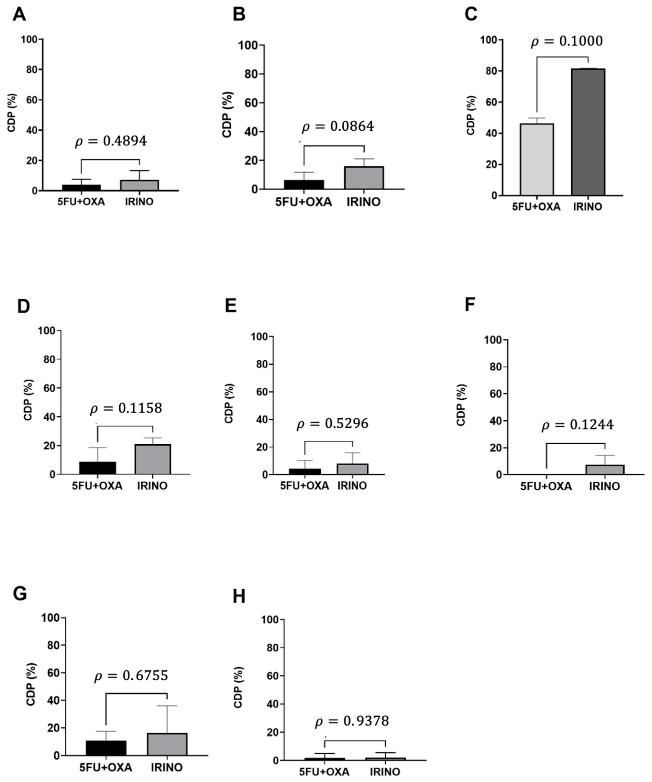
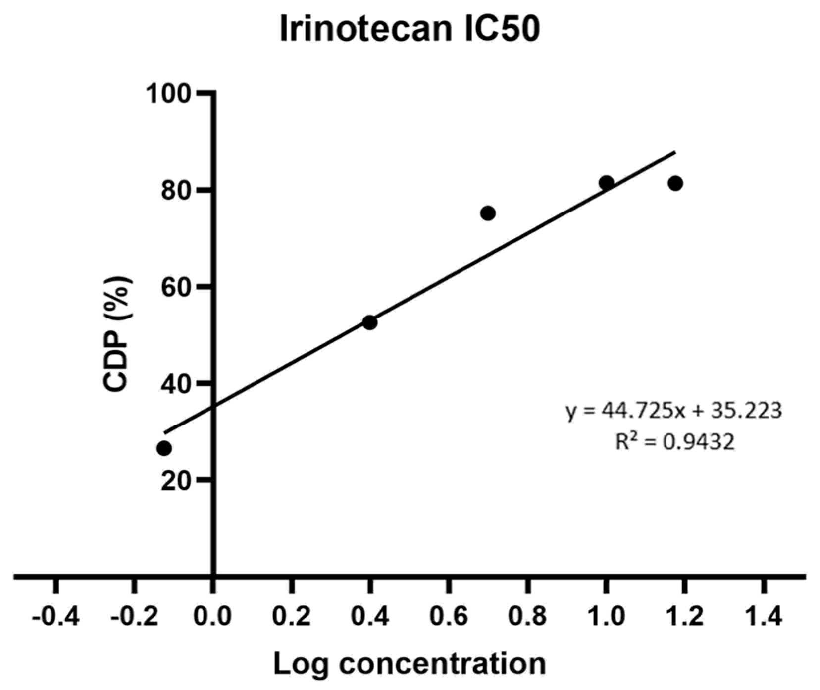
Primary cultures from eight CRC biopsies were treated with chemotherapy agent 5-FU (10 µg/mL) combined with Oxaliplatin (2.9 µg/mL) (5FU + OXA) or IRINO (1.97 µg/mL) (IRINO) and incubated for 48 h. We classified as responders those cultures that showed cell-death percentages (CDP) of ≥30% and as non-responders or resistant those cultures with a CDP of ≤29%. From these primary cultures, only Patient 81 was a responder for both treatments of 5FU + OXA and IRINO (Figure 1C) and 87.5% of cultures were non-responders for chemotherapy, and there was no statistical difference between the treatments (Student’s t-test and Mann–Whitney U-Test, p < 0.05) (Figure 1A–H). Furthermore, we selected the Caco-2 cell line to determine the half-maximal inhibitory concentration (IC50) for IRINO. The Caco-2 cells were exposed to increasing IRINO concentrations. Using linear regression analysis, we determined a value of IC50 of 2.14 µg/mL (Figure 2). This concentration exceeded the maximum peak plasma concentration previously reported by our group (1.97 µg/mL) [20]. Thus, similar to what was observed in primary cell cultures, the Caco-2 cell line was classified as IRINO-resistant. From this point, all experiments were developed with the Caco-2 cell line.

3.2. Genetically Modified Mesenchymal Stem Cells Expressing the GFP-sTRAIL Transgene

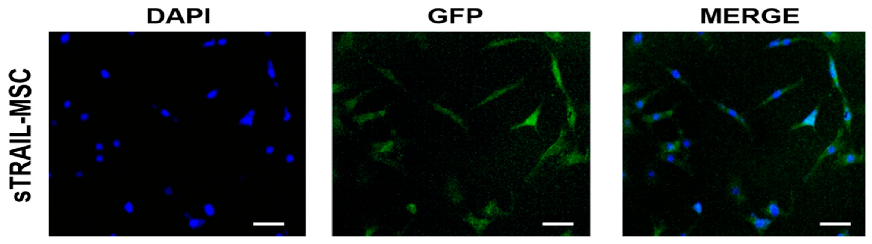
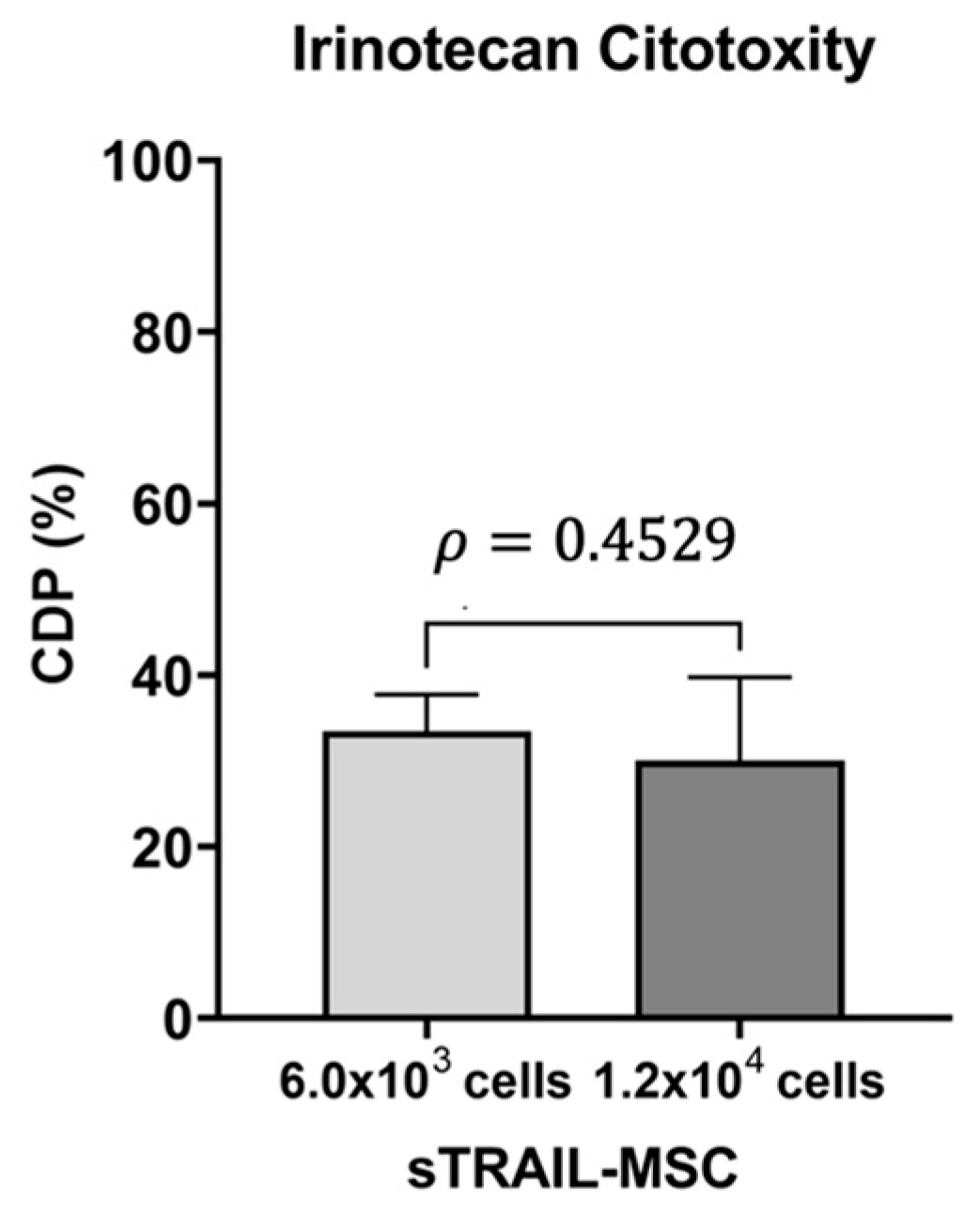
We used the MSC cell line previously transduced with lentiviral vectors to overexpress soluble TRAIL (sTRAIL) and validated by Western blot by our group [18]. The lentiviral construct included green fluorescent protein (GFP) as a reporter gene. Following a one-week geneticin selection, the cells were fixed and analyzed by fluorescence microscopy. GFP expression was visualized as green fluorescence, while cell nuclei were blue-stained with DAPI. The merge images revealed a transduction efficiency from 75–90% (Figure 3). IRINO was applied at the maximum plasma concentration (1.97 µg/mL) to the sTRAIL-MSCs at two different cell ratios (6.0 × 10³ and 1.2 × 10⁴ cells/well), showing CDPs of 25 to 35% (Figure 4). This indicated that the MSCs showed low sensitivity to IRINO cytotoxicity.
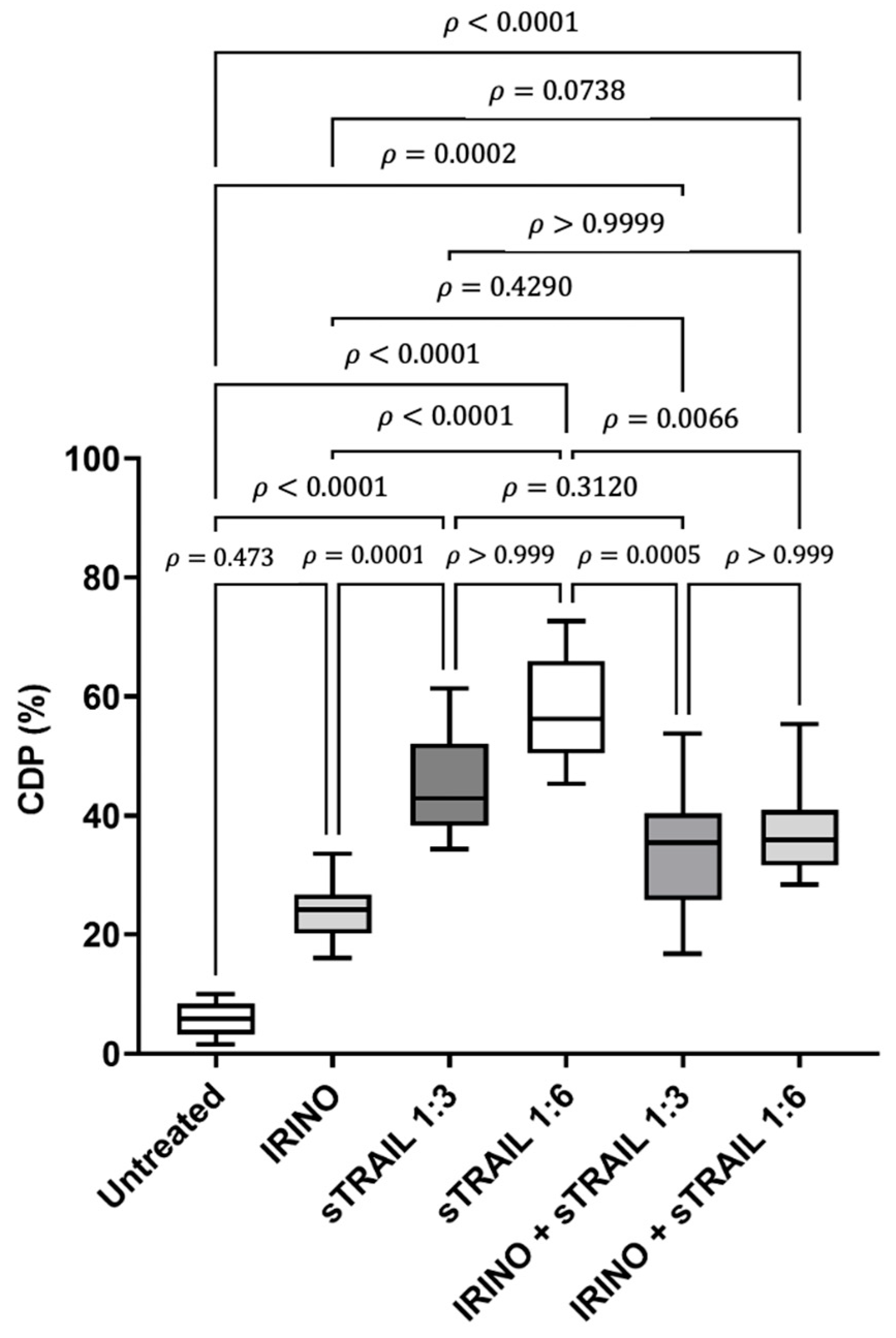
The cytotoxic activity of sTRAIL was demonstrated through co-culture assays, either in monotherapy or in combination with IRINO (Figure 5). As observed before, IRINO did not produce a statistically significant increase in CDP (p = 0.4737) compared to the untreated control. In contrast, the sTRAIL-MSCs induced a marked increase in CDP at 1:3 (6.0 × 10³) and 1:6 (1.2 × 10⁴) ratios, reaching CDPs of 50 and 80%, respectively. These increases were statistically significant (p < 0.001, Kruskal–Wallis and Dunn’s multiple comparison tests). In addition, the sTRAIL treatments induced significantly greater CDPs than IRINO alone compared to the untreated group (p < 0.001, Kruskal–Wallis and Dunn’s multiple comparison tests). The combination of IRINO with the sTRAIL-MSCs also resulted in a significant increase in CDP of 50 to 60% (p < 0.0001, Kruskal–Wallis and Dunn’s multiple comparison tests) compared with the untreated group. However, this combinatory effect was lower than that observed with the sTRAIL-MSCs at a 1:6 ratio as a monotherapy (80% CDP) (p < 0.0001, Kruskal–Wallis and Dunn’s multiple comparison tests). These results indicate that IRINO does not potentiate the cytotoxic effect of sTRAIL by sensitization. The treatment that achieved the highest CDP was the sTRAIL-MSCs at a 1:6 ratio, classified as the most effective treatment condition in this assay.

3.3. Effect of the sTRAIL-MSCs on the Expression of CSC Markers

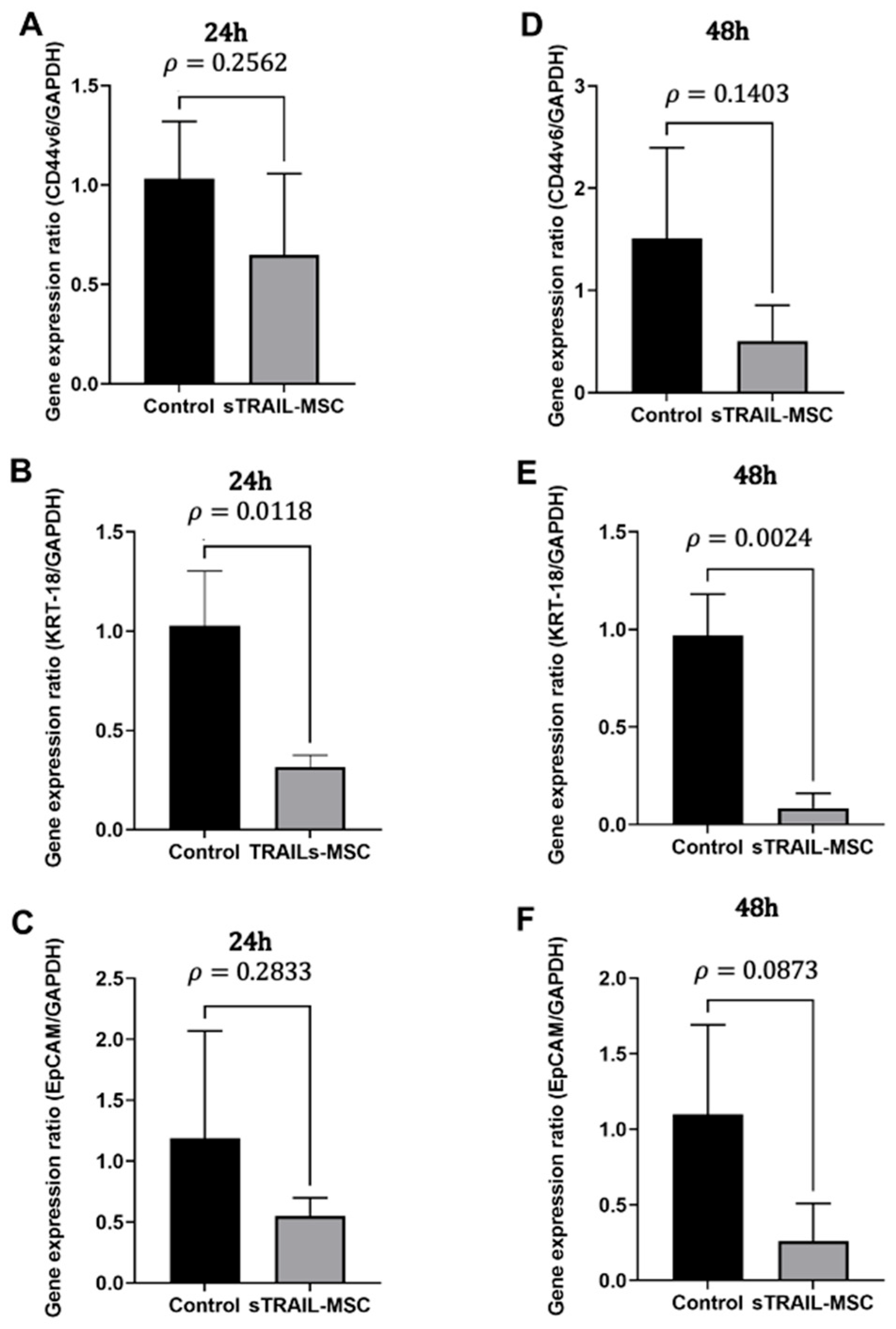
As sTRAIL treatment induced higher CDPs on the Caco-2 cell line, we investigated the specific effect on CSC population by evaluating the expression of CD44v6, KRT-18, and EpCAM levels. Using a co-culture assay, we seeded sTRAIL-expressing MSCs at a 1:6 ratio (5 × 10⁴ Caco-2 cells and 3 × 10⁵ sTRAIL-MSCs), with incubation periods of 24 and 48 h. A relative expression analysis revealed a statistically significant downregulation of KRT-18 as early as 24 h post-treatment (p < 0.0118, Student’s t-test) (Figure 6). Meanwhile, CD44v6 and EpCAM exhibited a downward trend at 24 h (p > 0.05, Student’s t-test) (Figure 6A–C). Moreover, at 48 h, KRT-18 expression was further reduced (p < 0.0024, Student’s t-test) (Figure 6D–F), and a continued downward trend in CD44v6 and EpCAM expression was observed. These results suggest that, in addition to enhancing overall cytotoxicity, sTRAIL delivered via MSCs contributes to the suppression of CSC molecular markers expression as early as 24 h, and this effect was significantly higher at 48 h.

4. Discussion

The high rate of resistance to first-line therapies in colorectal cancer has prompted the search for strategies capable of enhancing or overcoming current treatment limitations. The Caco-2 colorectal adenocarcinoma cell line has been previously classified as moderately resistant to TRAIL at 0.16 nM (500 ng/dL) [14] and fully resistant at 0.1 nM (In addition, Caco-2 cells exhibit a low expression of DR5 TRAIL death receptor [21]. However, the sTRAIL-MSCs expression system at a 1:6 ratio induces a significant increase in cytotoxicity (50–80% CDP). These results suggest that sTRAIL delivered by MSCs over 24 h achieves concentrations equivalent to or exceeding the IC50.
In a previous study, our group described Caco-2 resistance to Oxaliplatin at the maximum peak plasma concentration (2.9 µg/mL). In addition, Oxaliplatin sensitized both murine and human CRC cells to TRAIL-induced cytotoxicity [14]. In the present study, we evaluated whether IRINO could enhance TRAIL sensitivity. However, the cytotoxic effect of sTRAIL as a monotherapy surpassed the effect induced by the combination with IRINO, showing that co-administration does not potentiate sTRAIL activity using the conditions of this model. According to previous reports, irinotecan lacks the ability to modulate TRAIL receptor (DR4/DR5) expression, in contrast to what has been described for pre-treatment with 5-fluorouracil and oxaliplatin prior to TRAIL exposure. Although IRINO was expected to enhance TRAIL sensitivity by regulating pro-apoptotic (Bax) and anti-apoptotic (Bcl-xL) proteins, these levels were not directly assessed, restricting the mechanistic interpretation of our findings. A primary limitation was the administration of a single IRINO dose without evaluating its active metabolite SN-38, which is a significantly more potent inducer of apoptosis [22,23].
To confirm the specificity of sTRAIL treatment, the relative gene expression of KRT-18, CD44v6, and EpCAM, was evaluated. KRT-18 expression was significantly downregulated at 24 h (p < 0.05), and this reduction persisted at 48 h (p < 0.01). KRT-18 downregulation has also been linked to reduced patient survival in colorectal cancer [5,18]. However, KRT-18 is not considered a canonical CSC marker, and its decrease may reflect general cytotoxicity rather than CSC-specific targeting.
Moreover, CD44v6 and EpCAM showed a consistent trend toward decreased expression. These markers are known to improve colonization, invasion, and metastasis in colorectal cancer CSCs, and their overexpression correlates with poor prognoses and advanced disease stages [24]. Furthermore, the reduced expression of CD44v6 and EpCAM could also result from the selective elimination of marker-positive cells rather than the direct modulation of CSC biology. Furthermore, CD44 and KRT-18 are frequently overexpressed in chemo-resistant tumors [18]. Thus, the observed downregulation of these genes, together with the high cytotoxicity percentage of cancer cells, supports the notion that sTRAIL may target the CSC subpopulation. Nevertheless, functional assays such as flow cytometry or sphere formation were essential to directly confirm the efficacy of TRAIL on Caco-2 and CRC.
Although MSCs are widely explored as therapeutic vehicles, concerns remain regarding their long-term engraftment, possible immunogenicity, and tumor-promoting effects in certain contexts. Our delivery system for sTRAIL-MSCs was used under controlled in vitro conditions, and no adverse effects were observed in a previously studied murine model [16]. Nevertheless, we recognize that further preclinical and clinical evaluations are required to ensure the safety and translational applicability of MSC-based delivery systems.

5. Conclusions

sTRAIL delivered by MSCs exerts a robust cytotoxic effect against the chemo-resistant colorectal cancer cell line Caco-2. Our results demonstrated that the co-administration of sTRAIL by MSCs with IRINO does not yield a synergistic cytotoxic effect. However, it downregulates markers such as KRT-18, CD44v6, and EpCAM, indicating a promising therapeutic strategy against CRC.

Author Contributions

Conceptualization: E.N.G.-T., P.D.G., and J.F.I.; methodology, A.G.Q.-R., G.S.P.-C., and M.E.V.-C.; software, A.G.Q.-R., and G.S.P.-C.; validation, E.N.G.-T., P.D.G., and J.F.I.; formal analysis, A.G.Q.-R., G.S.P.-C., and M.E.V.-C.; investigation, A.G.Q.-R., G.S.P.-C., and M.E.V.-C.; resources, E.N.G.-T., P.D.G., and J.F.I.; data curation, A.G.Q.-R., G.S.P.-C., and M.E.V.-C.; writing—original draft preparation, A.G.Q.-R., G.S.P.-C., and M.E.V.-C.; writing—review and editing, E.N.G.-T., P.D.G., and J.F.I.; supervision, project administration and funding acquisition, E.N.G.-T., P.D.G., and J.F.I. All authors have read and agreed to the published version of the manuscript.

Funding

This research was partially funded by Programa de Apoyo a la Investigacion Cientifica y Tecnologica (PROACTI-UANL).

Institutional Review Board Statement

The study was conducted in accordance with the Declaration of Helsinki and approved by the Institutional Committee for the Care and Use of Laboratory Animals (protocol code number PI19-00183) as well as by Institutional Research Ethics Committee of the School of Medicine, UANL (protocol code number BI11-004 and BI21-00005) for studies involving humans.

Informed Consent Statement

Informed consent was obtained from all subjects involved in the study. Written informed consent was obtained from all patients for participation in the study and for publication of the results.

Data Availability Statement

Data is contained within the article. The original contributions presented in this study are included in the article. Further inquiries can be directed to the corresponding author.

Acknowledgments

We are grateful to the students PROVERICYT of Universidad Autonoma de Nuevo Leon for their assistance in the literature analysis that contributed to this manuscript.

Conflicts of Interest

The authors declare no conflicts of interest.

Abbreviations

The following abbreviations are used in this manuscript:
CDPCell-death percentage
CRCColorectal cancer
CSCCancer stem cell
TRAILTNF-related apoptosis-inducing ligand
MSCMesenchymal stem cells
FOLFOXOxaliplatin, 5-Fluorouracil, and Leucovorin
EMTEpithelial-to-mesenchymal transition
BM-MSCSBone marrow mesenchymal stem cells
sTRAILSoluble TRAIL
MOIMultiplicity of infection
DAPI4′,6-diamino-2-phenylindole
5FU5-Fluorouracil
OXAOxaliplatin
IRINOIrinotecan

References

  1. Sung, H.; Ferlay, J.; Siegel, R.L.; Laversanne, M.; Soerjomataram, I.; Jemal, A.; Bray, F. Global Cancer Statistics 2020: GLOBOCAN Estimates of Incidence and Mortality Worldwide for 36 Cancers in 185 Countries. CA Cancer J. Clin. 2021, 71, 209–249. [Google Scholar] [CrossRef]
  2. Neugut, A.I.; Lin, A.; Raab, G.T.; Hillyer, G.C.; Keller, D.; O’Neil, D.S.; Accordino, M.K.; Kiran, R.P.; Wright, J.; Hershman, D.L. FOLFOX and FOLFIRI Use in Stage IV Colon Cancer: Analysis of SEER-Medicare Data. Clin. Color. Cancer 2019, 18, 133–140. [Google Scholar] [CrossRef]
  3. Ashique, S.; Bhowmick, M.; Pal, R.; Khatoon, H.; Kumar, P.; Sharma, H.; Garg, A.; Kumar, S.; Das, U. Multi drug resistance in Colorectal Cancer- approaches to overcome, advancements and future success. Adv. Cancer Biol.-Metastasis 2024, 10, 100114. [Google Scholar] [CrossRef]
  4. Lv, L.; Liu, H.G.; Dong, S.Y.; Yang, F.; Wang, Q.X.; Guo, G.L.; Pan, Y.-F.; Zhang, X.-H. Upregulation of CD44v6 contributes to acquired chemoresistance via the modulation of autophagy in colon cancer SW480 cells. Tumor Biol. 2016, 37, 8811–8824. [Google Scholar] [CrossRef] [PubMed]
  5. Zhang, J.; Hu, S.; Li, Y. KRT18 is correlated with the malignant status and acts as an oncogene in colorectal cancer. Biosci. Rep. 2019, 39, BSR20190884. [Google Scholar] [CrossRef]
  6. Lee, C.C.; Yu, C.J.; Panda, S.S.; Chen, K.C.; Liang, K.H.; Huang, W.C.; Wang, Y.-S.; Ho, P.-C.; Wu, H.-C. Epithelial cell adhesion molecule (EpCAM) regulates HGFR signaling to promote colon cancer progression and metastasis. J. Transl. Med. 2023, 5, 530. [Google Scholar] [CrossRef]
  7. Gaghana, L.O.; Miskad, U.A.; Cangara, M.H.; Zainuddin, A.A.; Mardiati, M.; Arsyadi, G.; Wahid, S. The Relationship Between Expression of EpCAM Cancer Stem Cell Marker with Histopathological Grading, Lymphovascular Invasion, and Metastases in Colorectal Adenocarcinoma. Asian Pac. J. Cancer Prev. 2023, 24, 929–934. [Google Scholar] [CrossRef] [PubMed]
  8. Yuan, X.; Gajan, A.; Chu, Q.; Xiong, H.; Wu, K.; Wu, G.S. Developing TRAIL/TRAIL death receptor-based cancer therapies. Cancer Metastasis Rev. 2018, 37, 733–748. [Google Scholar] [CrossRef]
  9. Trivedi, R.; Mishra, D.P. Trailing TRAIL Resistance: Novel Targets for TRAIL Sensitization in Cancer Cells. Front. Oncol. 2015, 5, 69. [Google Scholar] [CrossRef] [PubMed]
  10. Ashkenazi, A.; Holland, P.; Eckhardt, S.G. Ligand-Based Targeting of Apoptosis in Cancer: The Potential of Recombinant Human Apoptosis Ligand 2/Tumor Necrosis Factor–Related Apoptosis-Inducing Ligand (rhApo2L/TRAIL). J. Clin. Oncol. 2008, 26, 3621–3630. [Google Scholar] [CrossRef]
  11. Lan, T.; Luo, M.; Wei, X. Mesenchymal stem/stromal cells in cancer therapy. J. Hematol. Oncol. 2021, 14, 195. [Google Scholar] [CrossRef]
  12. Zinnah, K.M.A.; Munna, A.N.; Seol, J.W.; Park, B.Y.; Park, S.Y. An Antidepressant Drug Increased TRAIL Receptor-2 Expression and Sensitized Lung Cancer Cells to TRAIL-induced Apoptosis. Anti-Cancer Agents Med. Chem. 2023, 23, 2225–2236. [Google Scholar] [CrossRef]
  13. Park, S.A.; Han, H.R.; Ahn, S.; Ryu, C.H.; Jeun, S.S. Combination treatment with VPA and MSCs-TRAIL could increase anti-tumor effects against intracranial glioma. Oncol. Rep. 2021, 45, 869–878. [Google Scholar] [CrossRef] [PubMed]
  14. Quiroz-Reyes, A.G.; Delgado-González, P.; Islas, J.F.; Soto-Domínguez, A.; González-Villarreal, C.A.; Padilla-Rivas, G.R.; Garza-Trevino, E.N. Oxaliplatin Enhances the Apoptotic Effect of Mesenchymal Stem Cells, Delivering Soluble TRAIL in Chemoresistant Colorectal Cancer. Pharmaceuticals 2023, 16, 1448. [Google Scholar] [CrossRef]
  15. Fulda, S.; Debatin, K.M. Resveratrol-mediated sensitisation to TRAIL-induced apoptosis depends on death receptor and mitochondrial signalling. Eur. J. Cancer 2005, 41, 786–798. [Google Scholar] [CrossRef]
  16. Quiroz-Reyes, A.G.; González-Villarreal, C.A.; Martínez-Rodriguez, H.; Said-Fernández, S.; Salinas-Carmona, M.C.; Limón-Flores, A.Y.; Soto-Dominguez, A.; Padilla-Rivas, G.; Montes De Oca-Luna, R.; Islas, J.F.; et al. A combined antitumor strategy of separately transduced mesenchymal stem cells with soluble TRAIL and IFNβ produces a synergistic activity in the reduction of lymphoma and mice survival enlargement. Mol. Med. Rep. 2022, 25, 206. [Google Scholar] [CrossRef]
  17. Dominici, M.; Le Blanc, K.; Mueller, I.; Slaper-Cortenbach, I.; Marini, F.C.; Krause, D.S.; Deans, R.J.; Keating, A.; Prockop, D.J.; Horwitz, E.M. Minimal criteria for defining multipotent mesenchymal stromal cells. The International Society for Cellular Therapy position statement. Cytotherapy 2006, 8, 315–317. [Google Scholar] [CrossRef]
  18. Garza-Treviño, E.; Martínez-Rodríguez, H.; Delgado-González, P.; Solís-Coronado, O.; Ortíz-Lopez, R.; Soto-Domínguez, A.; Trevino, V.M.; Padilla-Rivas, G.R.; Islas, J.F.; Quiroz-Reyes, A.G.; et al. Chemosensitivity analysis and study of gene resistance on tumors and cancer stem cell isolates from patients with colorectal cancer. Mol. Med. Rep. 2021, 24, 721. [Google Scholar] [CrossRef]
  19. Kopczynski, M.; Statkiewicz, M.; Cybulska, M.; Kuklinska, U.; Unrug-Bielawska, K.; Sandowska-Markiewicz, Z.; Grochowska, A.; Gajewska, M.; Kulecka, M.; Ostrowski, J.; et al. Cytotoxic efficacy and resistance mechanism of a TRAIL and VEGFA-peptide fusion protein in colorectal cancer models. Int. J. Mol. Sci. 2021, 22, 3160. [Google Scholar] [CrossRef] [PubMed]
  20. Kim, D.O.; Jang, E.H.; Kwon, Y.D.; Yoo, J.H.; Ma, X.; Kim, K.H.; Hong, D.G.; Kim, C.K.; Nam, H.; Choi, J.W.; et al. Mesenchymal Stem Cells Expressing CES1 and Soluble TRAIL Activate CPT-11 and Induce Apoptosis in Lung Cancer Brain Metastatic Lesions. Cancer Res. Commun. 2025, 5, 1552–1565. [Google Scholar] [CrossRef] [PubMed]
  21. Ma, X.L.; Sun, Y.F.; Wang, B.L.; Shen, M.N.; Zhou, Y.; Chen, J.W.; Hu, B.; Gong, Z.J.; Zhang, X.; Cao, Y.; et al. Sphere-forming culture enriches liver cancer stem cells and reveals Stearoyl-CoA desaturase 1 as a potential therapeutic target. BMC Cancer 2019, 19, 760. [Google Scholar] [CrossRef] [PubMed]
  22. Ray, S.; Almasan, A. Apoptosis induction in prostate cancer cells and xenografts by combined treatment with Apo2 ligand/tumor necrosis factor-related apoptosis-inducing ligand and CPT-11. Cancer Res. 2003, 63, 4713–4723. [Google Scholar] [PubMed]
  23. McKenzie, J.A.; Mbofung, R.M.; Malu, S.; Zhang, M.; Ashkin, E.; Devi, S.; Williams, L.; Tieu, T.; Peng, W.; Pradeep, S.; et al. The Effect of Topoisomerase I Inhibitors on the Efficacy of T-Cell-Based Cancer Immunotherapy. JNCI J. Natl. Cancer Inst. 2018, 110, 777–786. [Google Scholar] [CrossRef]
  24. Ma, L.; Dong, L.; Chang, P. CD44v6 engages in colorectal cancer progression. Cell Death Dis. 2019, 10, 30. [Google Scholar] [CrossRef] [PubMed]
Figure 1. Cytotoxicity effect of chemotherapy on the primary cultures of colorectal cancer. (A) Patient 16; (B) Patient 15; (C) Patient 91; (D) Patient 12; (E) Patient 13; (F) Patient 14; (G) Patient 8; (H) Patient 11. After 48 h of Irinotecan (IRINO), the samples presented higher cell-death percentages (CDPs) than the combinatory 5-Fluorouracil plus oxaliplatin (5FU + OXA) samples (Student t-test and Mann–Whitney U-test).
Joma 03 00009 g001
Figure 2. Irinotecan IC50 standard curve at 48 h. The cell-death percentages were plotted against the logarithm of concentration. The IC50 value was determined using a linear regression analysis, resulting in 2.14 µg/mL. Cell-death percentages (CDP); logarithm (Log).
Joma 03 00009 g002
Figure 3. Genetically modified mesenchymal stem cells sTRAIL expression. Cell nucleus stained by DAPI (200×). The green-stained cytoplasm shows the sTRAIL-GFP transgene insertion. The merge image superposition shows the correlation of the nucleus and cytoplasm blue- and green-stained. sTRAIL: soluble TRAIL; GFP: green fluorescent protein. The shown cell images are after one-week of antibiotic selection.
Joma 03 00009 g003
Figure 4. sTRAIL-MSC Irinotecan sensitivity. Cell-death percentage after 48 h of sTRAIL-MSC treated with Irinotecan at the maximum plasma concentration (1.97 µg/mL). sTRAIL: soluble TRAIL; MSC: mesenchymal stem cells; CDP: Cell-death percentage. Student’s t-test.
Joma 03 00009 g004
Figure 5. Cell-death percentages of the Caco-2 cell line after 48 h with individual and combined treatments of Irinotecan and sTRAIL MSCs. sTRAIL: soluble TRAIL; MSC: mesenchymal stem cells; IRINO: Irinotecan. Kruskal–Wallis and Dunn’s multiple comparison tests.
Joma 03 00009 g005
Figure 6. Gene relative expression of the CSC markers. (A) The relative expression of CD44v6 at 24 h. (B) The relative expression ofKRT-18 at 24 h. (C) The relative expression of EpCAM at 24 h. (D) The relative expression of CD44v6 at 48 h. (E) The relative expression of KRT-18 at 48 h. (F) The relative expression of EpCAM at 48 h. Housekeeping gene GAPDH. Student’s t-test.
Joma 03 00009 g006
Table 1. Primer Sequences of the CSC genes.
MarkerForwardReverse
CD44V65′GACAGAATCCCTGCTACCAATAG 3′5′TCCTTCGTGTGTGGGTAATG 3′
KRT-185′TCGCAAATACTGTGGACAATGC 3′5′GCAGTCGTGTGATATTGGTGT 3′
EpCAM5′GCTGGAATTGTTGTGCTGGTTA 3′5′AGATGTCTTCGTCCCACGC 3′
GAPDH5′TCGCCAGCCGAGCCA 3′5′CCTTGACGGTGCCATGGAAT 3′
Disclaimer/Publisher’s Note: The statements, opinions and data contained in all publications are solely those of the individual author(s) and contributor(s) and not of MDPI and/or the editor(s). MDPI and/or the editor(s) disclaim responsibility for any injury to people or property resulting from any ideas, methods, instructions or products referred to in the content.