1. Introduction
Uvular necrosis is a rare and under-reported post-operative complication, reported to have an estimated incidence of 0.03% post-general anesthesia with a reported higher prevalence in young male patients undergoing endotracheal tube (ETT) intubation in the supine position [
1].
The true incidence remains uncertain due to limited studies and the condition may be under-recognized due to overlap with routine post-operative oropharyngeal pain. The ischemic uvular injury results in pain 2–3 days post-insult. Cases reported in the literature attribute injuries to the uvula to various aspects of oropharyngeal manipulation, including ETT intubation, laryngeal mask placement, oral–pharyngeal suction, or the insertion of other medical devices (e.g., bronchoscopy, transesophageal echocardiography probe, scopes) [
2]. Nevertheless, a direct causal relationship or risk factors has not been elucidated. Uvular necrosis presents diagnostic and management challenges.
This case presents the unique dual perspective of both patient and medical trainee (first author), which provides a detailed description of the clinical course, management challenges, and recovery following uvular necrosis after an elective septoturbinoplasty. Informed written patient consent was obtained for the purposes of this publication, and Institutional Research Ethics Board approval was waived.
2. Case Description
A 24-year-old male (75 Kg, BMI: 23.4) underwent septoplasty and bilateral inferior turbinoplasty for allergic chronic rhinosinusitis. The patient had a history of dust mite allergy and had been managed pre-operatively with daily cetirizine (Reactine®, Kenvue Canada Inc., Guelph, ON, Canada), intranasal corticosteroid ciclesonide (Omnaris®, Covis Pharma, Zug, Switzerland), and intranasal saline irrigation. Additional medical history was notable for the use of inhaled corticosteroids (budesonide/formoterol fumarate dihydrate (Symbicort®, Astrazeneca, Dunkerque, France)) and gastroesophageal reflux disease. Pre-operative airway factors include grade 1 tonsils, class II Mallampati score, and a uvula of 2.5 cm (estimated from post-operative measurements). The patient was otherwise healthy. The patient received general anesthesia and a single-attempt orotracheal intubation using direct laryngoscopy with a size 7.5 ETT and a size 3 MacIntosh blade. The integrated suction in the General Electric® Aisys CS2 anesthesia machine (Datex-Ohmeda Inc., Madison, WI, USA) was utilized in the case. No abnormalities or complications were reported upon extubation.
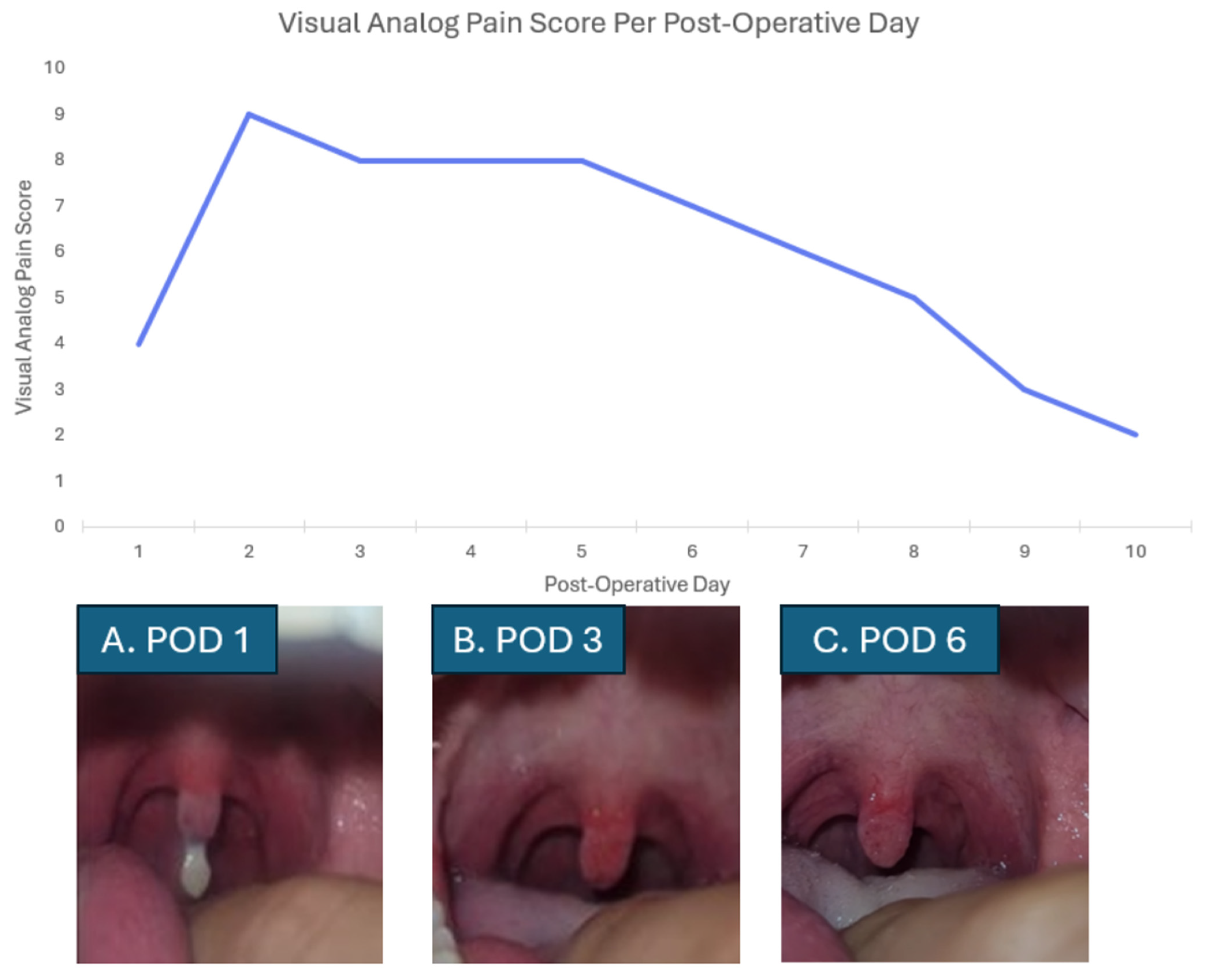
Immediately post-operatively (POD 0), the patient reported mild throat discomfort (Visual Analog Pain Scale (VAS) 2/10), managed conservatively with poor response with oral acetaminophen, tramadol, saltwater rinses, and use of a face mask to reduce oropharyngeal dryness (
Table 1). Self-examination revealed a swollen, discolored uvula with necrotic changes involving the distal third (
Figure 1). On POD 2, pain escalated to VAS 8/10, described as constant, dull, and burning pain with referred otalgia. Pain was aggravated by swallowing, eating, and oral dryness, with minimal relief from acetaminophen q6 h (
Table 1). By POD 3, necrotic uvular tissue had spontaneously sloughed, and the patient was assessed for urgent follow-up at an otolaryngology clinic, where uvular necrosis was confirmed (
Table 1). Oral 1% viscous lidocaine was prescribed for gargling as needed, providing significant but short-lived relief (60–90 min), necessitating frequent use. On POD5, the patient found that dipping a cotton swab into the solution and manually covering the uvula (2 mis) provided just as much analgesia as gargling the liquid (10–20 mls). From POD 6 onward, there was a gradual improvement in pain control and symptom trajectory (
Table 1). The frequency of lidocaine use decreased progressively. No further complications were reported during this recovery phase.
Despite being a medical trainee with direct access to the surgical team, the patient remained uncertain regarding whether additional medical evaluation was warranted, highlighting the potential for even greater anxiety among the general patient population. From the patient perspective, symptoms did not correspond with expected post-operative pain as described by the anesthesiologist and surgeon and thus the patient requested urgent follow-up.
Pain management was also challenging. The institutional post-operative septotubrinoplasty analgesic regimen consisted of tramadol, six tablets of 50 mg, and acetaminophen. The severity and persistence of symptoms was not adequately controlled on this regimen alone, prompting the patient to seek care. Pain significantly impacted daily activities, including interrupted sleep due to the need for reapplication of topical lidocaine for symptom control every hour. Given that the patient was already involved in the healthcare system and had email access directly to the surgeon, more uncertainty may be encountered by patients.
3. Discussion
This case highlights uvular necrosis as an uncommon but clinically significant anesthetic-related complication often characterized by delayed onset (1 day in this case, 2 h to 2 days in the literature), oropharyngeal pain, and a prolonged recovery course [
2]. Pain escalation occurred one day after surgery, peaked over several post-operative days, and was disproportionate to typical post-operative throat discomfort. Post-operative throat pain with referred otalgia following septo-turbinoplasty is atypical and should prompt consideration of anesthesia and/or surgical-related oropharyngeal injury, particularly uvular ischemia or necrosis and glossopharyngeal nerve irritation. Additional differentials include pharyngeal mucosal trauma, eustachian tube dysfunction, and, less commonly, otologic or deep neck space complications [
2]. Diagnosis of uvular necrosis can be made based on visual inspection of the uvula and oropharynx.
Among the analgesic strategies trialed, topical viscous lidocaine 1% was consistently the most effective intervention, providing meaningful symptomatic relief when systemic analgesics were perceived ineffective and difficult to swallow due to oropharyngeal inflammation. Although the pain relief duration was short, necessitating frequent reapplication, lidocaine remained central to pain control throughout the course and allowed gradual functional recovery as mucosal healing progressed. This important finding and patient experience sheds some light on the analgesic treatment for uvular necrosis, as treatment still requires study. A review found that uvular necrosis patients received varying treatments including antibiotics, oral prednisone (50 mg), oral analgesia, antiseptic mouth wash and conservative treatment [
2]. Nevertheless, the authors of this study have called into question the use of antibiotics in this condition, due to its localized and non-infectious nature [
2]. The authors also noted that single-dose corticosteroids have been shown to be effective in undifferentiated throat pain and uvular necrosis in patients without contraindications [
2]. Our group did not use oral steroids due to an ongoing topical analgesia regimen and poor evidence (case studies) of its use in uvular necrosis [
2].
Viscous lidocaine toxicity has been reported in the literature including an FDA box warning for 2% viscous lidocaine given cases of seizures, cardiopulmonary arrest and death in children under the age of 3 years old [
3,
4]. The maximum safe dosing for adults is 4.5 mg/kg or 300 mg total per dose, with at least 3 h intervals between applications and a maximum of eight doses per 24 h [
3]. The risk of toxicity increases substantially with traumatized or inflamed mucosa, as this enhances systemic absorption [
3].
The need for counseling patients for safe dosing and route of administration (cotton swab onto affected area and avoiding swallowing to decrease absorption) is required, especially since viscous lidocaine can be purchased without a prescription in many countries, and due to pharmacokinetics of topical lidocaine resulting in limited analgesia duration and frequent reapplication [
4]. Longer-acting and higher-potency topical analgesics such as liquid bupivacaine have further risk of toxicity and side effects. Future studies and clinical practice should explore the role of topical liquid lidocaine solutions, adjuncts such as oral corticosteroids, and the use of other treatments [
4].
The etiology of uvular necrosis in this case is iatrogenic, with plausible contributing factors including ETT pressure and overly vigorous oropharyngeal suctioning in the peri-extubation period [
2,
5]. The literature has described this complication after the insertion ETT, LMA, throat packs, oro/nasalpharyngeal airways, transesophageal echocardiography and various scopes [
2,
6,
7]. There are limited intensive care unit (ICU)-related and prolonged surgery (>4 h) cases reported in the literature, with most cases involving post-operative otolaryngology cases which require short-stay ICU admission [
2,
6]. The authors believe that the true incidence is likely under-reported, as uvular necrosis is self-limiting and may resolve prior to extubation or recognition in ICU, particularly in sedated patients receiving significant analgesia.
Preventative measures described in the literature include improved non-blinded suctioning techniques and the placement of the ETT laterally to avoid uvula compression [
2,
8]. Our case had other risk factors in terms of airway inflammation; allergic rhinitis, asthma, topical steroid treatment, and gastroesophageal reflux disease. Male sex was reported as being more common, although its relationship with oral pharyngeal injury during general anesthesia is unclear [
5]. Male sex predominance in the literature is speculated to be a possible surrogate marker for other airway risk factors by the authors such as large size, elevated BMI, obstructive sleep apnea, smoking history, larger airway instrumentation, and peri-extubation agitation. There is limited evidence exploring post-operative uvular necrosis risk factors.
Our institution oral–pharyngeal suctioning is conducted via the Aisys CS2 integrated suction which is attached to a suction canister and a hard plastic Yankauer suction instrument with a ball-style tip and no vent. According to the Aisys CS2 user manual, the maximum power of the integrated suction is 540 mmHg and 40 L/min free flow. Although the suction switch can be adjusted, we have found that staff often set the suction to the maximum power. Artificial airway suctioning pressures of 200 mmHg are recommended for adults and 100–80 mmHg for children, although evidence is limited [
9]. Literature and recommendations regarding post-surgical and post-extubation surgical oropharyngeal suctioning are similarly limited [
9].
Institutional quality improvement members speculated that the hard non-flexible plastic of the Yankauer suction may have also contributed to physical trauma to the uvula [
2,
6]. Post-extubation suctioning trauma, both physical and negative pressure trauma, may explain why it is seen in both laryngeal mask airways and endotracheal tube intubation cases in the literature.
In response to this case and other similar cases, our institution’s anesthesia department has identified this complication as a quality improvement opportunity and is planning initiatives focused on suction technique optimization, including the introduction of suction devices with graded pressure settings to minimize mucosal trauma. Greater awareness among anesthesia providers, improved documentation, and preventive strategies targeting airway instrumentation may reduce the risk of uvular necrosis [
2,
8]. Additionally, early recognition and prompt initiation of topical analgesic therapy may substantially improve patient comfort and reduce morbidity during recovery.
Importantly, although uvular necrosis is fundamentally an anesthetic complication, it is frequently under-recognized, under-documented, and often not directly observed by anesthesia providers due to its delayed presentation [
1,
2]. Patients with post-op complications are assessed by the operating surgeon, family physician, or via the emergency room rather than anesthesia [
7]. This disconnect likely contributes to under-reporting and limited awareness of the true incidence and patient burden associated with this complication.
The take-home messages in this case are:
What is Uvular Necrosis: Uvular necrosis is a rare but meaningful iatrogenic complication post-airway instrumentation characterized by delayed onset, disproportionate oropharyngeal pain, and pale or petechiae discoloration of the uvula which may progress to gangrenous necrosis and auto-amputation.
Management: Recommended treatments are based upon limited case reports in the literature. Treatments include oral analgesics, oral steroids (50 mg, prednisone), and topical viscous lidocaine (1%).
Topical viscous lidocaine (1–2%) was an effective option in this case. Application with cotton swab reduced dose and toxicity risk (20–10 mls to 2 mls) with no loss in analgesic efficacy.
Etiology is not fully elucidated, but potential causes in this case may include oral secretion suctioning, midline placement of endotracheal tube, and oropharyngeal manipulation (endotracheal tube placement, oral airway placement or surgical manipulation). Safe suctioning technique was identified as an area of improvement at our institution. Departmental quality improvement leaders recommended non-blind suctioning, and to limit suction power to better align within artificial airway suction standards (200 mmHg for adults and 100–80 mmHg for children) [
9].
Uvular necrosis is under-recognized and under-reported. The first contact in this case was the surgeon. There is also an element of patient uncertainty, as patients wonder whether their symptoms are consistent with an urgent post-surgical complication or expected post-operative pain.