Abstract
Metal–organic frameworks (MOFs) have been increasingly recognized as promising platforms for enzyme modulation, owing to their tunable porosity, high surface area, and versatile chemical functionality. In this review, the potential of MOFs for the inhibition and modulation of protein kinases and phosphatases—key regulators of cellular signaling and disease progression—is examined. The structural fundamentals of MOFs are outlined, followed by a discussion of common synthesis strategies, including solvothermal, microwave-assisted, sonochemical, and mechanochemical methods. Emphasis is placed on how synthesis conditions influence critical features such as particle size, crystallinity, surface chemistry, and functional group accessibility, all of which impact biological performance. Four primary mechanisms of MOF–enzyme interaction are discussed: surface adsorption, active site coordination, catalytic mimicry, and allosteric modulation. Each mechanism is linked to distinct physicochemical parameters, including pore size, surface charge, and metal node identity. Special focus is given to biologically relevant metal centers such as Zr4+, Ce4+, Cu2+, Fe3+, and Ti4+, which have been shown to contribute to both MOF stability and enzymatic inhibition through Lewis acid or redox-mediated mechanisms. Recent in vitro studies are reviewed, in which MOFs demonstrated selective inhibition of disease-relevant enzymes with minimal cytotoxicity. Despite these advancements, several limitations have been identified, including scalability challenges, limited physiological stability, and potential off-target effects. Strategies such as post-synthetic modification, green synthesis, and biomimetic surface functionalization are being explored to overcome these barriers. Through an integration of materials science, coordination chemistry, and molecular biology, this review aims to provide a comprehensive perspective on the rational design of MOFs for targeted enzyme inhibition in therapeutic contexts.
1. Introduction
Abnormal protein phosphorylation plays a central role in the onset and progression of various human diseases, making it a pressing issue in biomedical research. Protein phosphorylation, as a reversible post-translational modification [1], regulates a wide array of cellular processes including cell proliferation, differentiation, metabolism, and apoptosis [2]. This dynamic equilibrium is maintained through the opposing actions of two major classes of enzymes: protein kinases and protein phosphatases [3]. Kinases catalyze the transfer of phosphate groups from high-energy molecules such as ATP to target proteins, while phosphatases remove these phosphate groups. Dysregulation in either process contributes to pathologies such as cancer, diabetes, inflammatory diseases, and neurodegenerative disorders [4,5]. For example, hyperactivation of specific kinases has been implicated in uncontrolled cell proliferation in cancer, while reduced phosphatase activity can lead to aberrant signal transduction and cellular dysfunction in diseases like Alzheimer’s and Parkinson’s [6].
Figure 1 illustrates the dynamic roles and localization of protein kinases and phosphatases within the cellular environment. It depicts how protein kinases catalyze the phosphorylation of substrate proteins by transferring phosphate groups (Figure 1A), thereby modulating critical cellular processes such as metabolism, proliferation, and apoptosis [7]. In contrast, protein phosphatases reverse this modification by dephosphorylating target proteins (Figure 1B), ensuring the reversibility and balance of signaling events. The diagram emphasizes the spatial organization of these enzymes by showing anchoring proteins, such as A-Kinase Anchoring Proteins (AKAPs), which localize phosphatases and kinases to specific cellular compartments like the plasma membrane or nucleus (Figure 1C) [8,9]. This compartmentalization is essential for achieving signaling specificity and efficiency. Additionally, the figure incorporates a simplified representation of key signaling pathways, including the PI3K/AKT pathway (Figure 1D), highlighting how kinase and phosphatase activities integrate into broader cellular regulatory networks [10]. That reflects the complex interplay between enzyme activity, subcellular localization, and pathway regulation in maintaining cellular homeostasis.
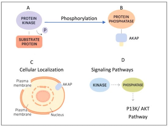
Figure 1.
Dynamic roles and localization of protein kinases and phosphatases within the cellular environment, (A) Protein kinase activity, (B) Protein phosphatase activity, (C) Subcellular localization and anchoring, and (D) Integration into signaling pathways.
Current pharmacological interventions often rely on small-molecule inhibitors targeting kinase activity, with many kinase inhibitors already approved for clinical use [5]. However, challenges such as poor selectivity, off-target toxicity, rapid degradation, and drug resistance remain critical barriers [11]. Similarly, therapeutic strategies involving phosphatase modulation have been largely underdeveloped due to the structural complexity and diversity of phosphatase enzymes, as well as the lack of selective modulators [12]. Furthermore, in biochemical and in vitro studies, there is a growing need for materials that can act as enzyme inhibitors or regulators while offering reusability, stability under physiological conditions, and tunable interaction properties.
In recent years, metal–organic frameworks (MOFs) have emerged as a promising class of hybrid materials in biomedical applications [13]. MOFs are crystalline porous structures composed of metal ions or clusters coordinated with organic linkers, forming highly ordered and customizable frameworks [14]. Originally developed for gas storage and catalysis [15], MOFs have since expanded into areas such as drug delivery, biosensing, tissue engineering, and biocatalysis [16]. More recently, their potential in protein interaction and enzyme modulation has been recognized, positioning MOFs as innovative tools for kinase and phosphatase inhibition studies [17].
The key advantage of MOFs in enzyme modulation lies in their high surface area, tunable pore sizes, and functionalizable surface chemistry. Specific metal ions incorporated within MOFs can directly interact with phosphate groups, mimic active sites, or even produce reactive species influencing enzymatic activity. Among these, MOFs containing metals such as zirconium (Zr) [18], cerium (Ce) [19], copper (Cu) [20], iron (Fe) [21], and lanthanides (e.g., Eu [22], Tb [23]) have demonstrated notable bioactivity. For instance, Zr-based MOFs (Zr-MOFs) are renowned for their chemical and thermal stability, large pore size, and strong affinity for phosphate-containing substrates. Cerium-based MOFs (Ce-MOFs), on the other hand, exhibit intrinsic catalytic activity resembling phosphatase enzymes, often referred to as “nanozymes”. Copper and iron-based MOFs contribute redox-active properties, allowing additional control over oxidative stress and biochemical pathways.
These distinctive properties make MOFs an attractive alternative to conventional small-molecule inhibitors for kinase and phosphatase modulation in vitro. Nevertheless, several challenges persist. One primary concern is biocompatibility and cytotoxicity. While some MOFs show low toxicity profiles, others may release harmful metal ions under physiological conditions [24,25]. Additionally, the stability of MOFs under biological pH ranges is a significant factor influencing their performance, as some frameworks may degrade or lose functionality in acidic or basic environments [26]. Another concern is the scalability and reproducibility of MOF synthesis. While laboratory-scale production often uses solvothermal, microwave-assisted, or sonochemical techniques, transitioning to larger-scale synthesis for biomedical applications requires addressing issues related to cost, environmental impact, and reproducibility [27,28].
The ability to functionalize MOF surfaces with biomolecules, targeting ligands, or specific functional groups is also critical for tailoring their interaction with kinase and phosphatase enzymes. Techniques such as post-synthetic modification and linker exchange have been explored to fine-tune MOF properties for biological applications [29]. Furthermore, ensuring reusability and regeneration efficiency of MOF-based enzyme modulators is essential for practical applications in biochemical assays, where cost-effectiveness and operational simplicity are prioritized.
In addition to MOFs, several emerging platforms (Table 1) have been developed for the modulation of protein kinases and phosphatases, each offering unique advantages and limitations. Polymeric nanocarriers, such as PEGylated micelles, dendrimers, and block copolymers, provide tunable architectures with high drug-loading capacity and biocompatibility, and they have been widely explored for targeted delivery of kinase inhibitors; however, they often face challenges with premature drug release and stability under physiological conditions. Nanoclusters, particularly gold or metal oxide clusters, possess intrinsic catalytic or redox properties that enable enzyme modulation or signal amplification in biosensing contexts, though concerns remain regarding their long-term toxicity and biodistribution. Peptidomimetics represent a molecular strategy that uses synthetic peptide-like molecules to mimic natural substrates or regulatory motifs, thereby enabling selective inhibition of kinase or phosphatase pathways; however, their susceptibility to enzymatic degradation and limited bioavailability can hinder translation. Placing MOFs in direct contrast with these systems highlights both the distinctive structural advantages of MOFs (e.g., permanent porosity, modular design) and the current limitations that remain to be addressed [30,31,32,33].

Table 1.
Comparative features of MOF-based modulators versus other emerging platforms for enzyme inhibition.
In this review, we provide a comprehensive overview of metal–organic frameworks (MOFs) as inhibitors or modulators of protein kinase and phosphatase enzymes in vitro. We specifically focus on MOFs constructed with biologically relevant metals such as zirconium, cerium, copper, iron, and lanthanides, highlighting their synthesis methods, structural properties, and mechanisms of enzyme interaction. Special attention is given to the advantages and limitations of various synthesis techniques including solvothermal, microwave-assisted, and sonochemical methods, and their impact on MOF performance in biological settings. Additionally, we discuss the key challenges such as biocompatibility, pH sensitivity, scalability, and cytotoxicity that must be addressed to translate MOF-based enzyme modulators from laboratory research to biomedical applications. Finally, we present insights and future perspectives for optimizing MOF design to expand their utility as selective, stable, and efficient enzyme inhibitors in vitro.
2. Metal–Organic Frameworks (MOFs) as Platforms for Protein Kinase and Phosphatase Inhibition
2.1. Fundamentals of MOFs Relevant to Enzyme Modulation
Conventional small-molecule inhibitors targeting protein kinases and phosphatases have played a pivotal role in both clinical therapy and biochemical research; however, these molecules exhibit inherent limitations that restrict their broader application. For example, Imatinib (Gleevec Novartis, United States of America), a well-known inhibitor targeting BCR-ABL tyrosine kinase, is widely used for chronic myeloid leukemia (CML) [34]. Similarly, Sorafenib targets RAF kinases, VEGFR, and PDGFR, and is clinically employed for hepatocellular carcinoma and renal cell carcinoma [35]. Gefitinib, another kinase inhibitor, is specific to the epidermal growth factor receptor (EGFR) and is utilized in treating non-small cell lung cancer [36]. Despite their clinical success, these inhibitors often face issues such as acquired resistance, limited specificity—leading to off-target effects—and rapid metabolic degradation. This is particularly challenging in kinase inhibition where high selectivity is crucial to avoid disrupting normal cellular functions.
For protein phosphatases, small-molecule inhibitors are primarily used in research due to their toxicity and lack of therapeutic viability. Compounds like Okadaic Acid and Calyculin A target protein phosphatase 1 (PP1) and protein phosphatase 2A (PP2A) but are highly toxic, preventing their clinical application [37]. Cantharidin, while historically used as a topical agent in dermatology, also targets PP2A but with limited therapeutic scope [38]. The common limitations across both kinase and phosphatase inhibitors include poor solubility, lack of sustained release profiles, and instability under physiological conditions.
While these molecules are typically employed in vivo, in vitro modulation of kinase and phosphatase activity remains essential for applications such as biosensing, inhibitor screening, and mechanistic enzymology. In biosensing, precise in vitro control of enzyme activity enables the quantitative detection of phosphorylation events and ATP consumption, which are key readouts for disease diagnostics and drug discovery assays. MOF-based systems are therefore designed not to replace clinical kinase inhibitors, but to serve as analytical and modulatory tools in vitro—where the enzyme’s catalytic response can be finely monitored and quantified under controlled conditions.
Unlike conventional small-molecule inhibitors, MOFs offer a highly tunable platform capable of interacting with enzymes through multiple mechanisms [39,40,41]. In this context, comparison with small molecules is valuable, not to equate their therapeutic use, but to highlight how MOFs overcome intrinsic limitations such as non-specific binding, instability, and lack of reusability in enzyme assays. By mimicking inhibitory behavior through metal coordination or spatial confinement, MOFs enable reproducible and selective modulation of kinase and phosphatase activity, providing an advanced in vitro alternative for biosensing, catalytic mimicry, and preclinical drug screening applications.
Structurally, MOFs are built from two primary components: metal nodes, which act as coordination centers and may mimic enzyme active sites; and organic linkers, often benzene rings functionalized with chemical groups like –COOH, –OH, or –NH2 (Figure 2) [42]. These functional groups enable non-covalent interactions with amino acid residues on the enzyme surface through hydrogen bonding, electrostatic forces, or π–π stacking [43].
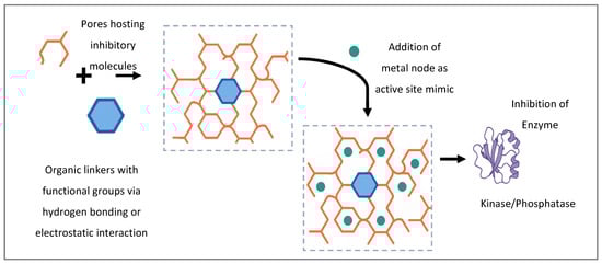
Figure 2.
Schematic illustration of metal nodes function as coordination centers within MOF and can mimic enzyme active sites, contributing directly to inhibitory mechanisms.
The combination of metal nodes and organic linkers forms a three-dimensional framework characterized by open channels or pores [44]. These pores allow enzymes or substrates to enter, with their size and shape directly influencing whether a protein can be hosted within the MOF or blocked at its surface. Additionally, in some MOF designs, exposed metal sites can chelate catalytic residues, phosphate groups, or cofactors associated with kinase or phosphatase active sites, providing a direct mode of inhibition [45].
In the context of protein kinases and phosphatases, MOFs can serve as scaffolds that anchor or block enzyme active sites, mimic biological inhibitors, or deliver inhibitory agents in a controlled manner [46]. Their hybrid inorganic–organic nature allows for the design of MOFs with specific surface charge, hydrophobicity, pore size, and functionalization tailored to interact with phosphorylation-related enzymes [43]. Functionalized linkers interact directly with enzyme surfaces, while pore systems can physically host portions of enzyme molecules, modulating catalytic behavior through steric and spatial effects.
Improving the selective capture of phosphorylated peptides is central to developing accurate kinase biosensors. To this end, Jia et al. [47] utilized a titanium-based MOF (Ti-MIL125-NH2) that provided a highly specific interaction between phosphate groups and titanium centers. Their system enriched phosphopeptides through Ti–O–P coordination, generating a strong fluorescence signal for detecting protein kinase A (PKA) with exceptional sensitivity down to 0.00003 U/μL [47]. Similarly, Bai et al. [48] leveraged the high surface area and abundant Zr defect sites in UiO-66 to develop a fluorescence-based sensor capable of efficiently capturing phosphorylated peptides. This platform achieved a detection limit of 0.00005 U/μL, demonstrating excellent affinity for kinase activity at low concentrations [48].
Building upon these findings, Zr-based MOFs have emerged as particularly robust candidates due to their strong phosphate affinity and chemical stability. Radfar et al. [49] and Alizadeh et al. [50] both developed nearly identical solvothermal synthesis routes for Zr-based MOF nanoplates, specifically the two-dimensional dca-Zr12 framework, with comparable crystallinity and particle dimensions (~100–150 nm). Both systems exploited the same carboxylate–Zr4+–phosphate coordination for selective phosphopeptide recognition. However, their analytical modes diverged: Radfar et al. used ferrocene-labeled peptides within a signal-off electrochemiluminescence (ECL) platform, achieving high selectivity and sensitivity toward phosphorylation events, whereas Alizadeh et al. employed a signal-on/signal-off dual ECL strategy to enhance phosphate detection, reaching a detection limit of 0.35 mU/mL [49,50]. Discussing these studies jointly highlights both the reproducibility of Zr-based frameworks and how subtle methodological adjustments—such as ligand substitution and detection mode—can fine-tune sensitivity and application range.
Another key area of focus has been signal amplification for enhanced biosensing sensitivity. Yan et al. [51] integrated gold and platinum nanoparticles into a UiO-66 scaffold (Au&Pt@UiO-66), leveraging bimetallic catalytic enhancement of the luminol–H2O2 ECL system. This dual mechanism of catalytic amplification and phosphate recognition resulted in a detection limit of 0.009 U/mL for PKA activity, with excellent performance in complex biological matrices [51]. In a related approach, Chang et al. [52] encapsulated methylene blue (MB) molecules within UiO-66-NH2, using the Zr–O clusters for phosphate group binding and MB as an electrochemical signal generator. This design enabled real-time electrochemical detection of kinase activity in serum, achieving a detection limit of 0.02 U/mL [52].
Recognizing the need for programmable systems to monitor ATP-to-ADP conversion during kinase reactions, Yu et al. [53] designed a family of Eu-based lanthanide MOFs (Ln-MOFs), identifying Eu-TPTC as a ratiometric fluorescence probe capable of molecular-level discrimination between ATP and ADP. Density functional theory (DFT) was used to verify the binding mechanism. The system was integrated into a paper-based chip that enabled rapid and precise creatine kinase detection in under 20 min, achieving a detection limit of 0.34 U/L [53].
Taken together, these studies converge on the critical role of metal centers (Ti and Zr) in mediating phosphate recognition, while illustrating how synthesis design, ligand chemistry, and detection modality dictate biosensor performance. The combination of fluorescence, ECL, and electrochemical modes across different MOF platforms reflects a broader evolution of MOFs—from simple recognition frameworks to modular scaffolds for enzyme activity mapping and inhibitor screening.
Looking forward, artificial intelligence and computational tools can accelerate progress in this field by addressing the complexity of MOF design [46]. Machine learning algorithms trained on experimental datasets could predict optimal linker–metal combinations for phosphate recognition, while molecular docking and molecular dynamics simulations may reveal how MOFs interact with kinase active sites or allosteric regions at the atomic level. Density functional theory (DFT), already applied in Eu-based MOF phosphosensors, can be extended to systematically probe catalytic mimicry and transition-state stabilization [54]. Furthermore, AI-guided synthesis planning and high-throughput virtual screening could shorten the design-to-application pipeline, guiding experimentalists toward the most promising candidates for selective, stable, and biocompatible MOF-based enzyme modulators. Together, these computational approaches position MOFs not merely as experimental curiosities, but as rationally designed therapeutic and diagnostic platforms at the intersection of materials science, biochemistry, and data-driven innovation.
2.2. Synthesis Methods of MOFs for Enzyme Inhibition Applications
The synthesis of metal–organic frameworks (MOFs) is a pivotal step that defines their crystal structure, pore size, surface functionality, and stability—factors that are essential for effective interaction with protein kinases and phosphatases. The choice of synthesis method directly influences the MOF’s physicochemical properties, such as crystallinity, surface chemistry, and biocompatibility, which are critical for applications in enzyme modulation. As summarized in Table 2, several synthesis strategies are commonly used in biomedical contexts, with solvothermal synthesis being the most widely employed due to its versatility and ability to produce highly crystalline, thermally stable frameworks.
Table 2.
Features and limitation of synthesis methods of MOFs-based on biological applications.
In solvothermal synthesis, metal salts and organic linkers are typically dissolved in solvents like dimethylformamide (DMF) and heated in sealed vessels under high temperatures for extended periods, often ranging from several hours to days. This technique reliably yields MOFs with high surface areas and well-defined porous structures, making them suitable for drug delivery, biosensing, and enzyme inhibition. However, the method also presents drawbacks: it often involves toxic solvents and produces relatively large particle sizes, which can limit biocompatibility and cellular uptake. Pourmadadi et al. (2023) highlights solvothermal synthesis as one of the most established techniques in MOF fabrication for biomedical use, alongside mechanochemical, microwave-assisted, and ultrasonic methods [55]. The study also underscores how this method supports the development of widely used frameworks such as zeolitic imidazolate frameworks (ZIFs) and Materials of Institute Lavoisier (MILs), although challenges like long-term toxicity and biological stability remain open issues for further research [23,45,50,53,55,56].
Building on this foundational insight, Qian et al. [57] focuses on copper-based MOFs (Cu-MOFs) synthesized primarily through solvothermal techniques, highlighting their multi-functionality in antitumor applications [57]. These frameworks exhibit excellent photothermal conversion, catalytic activity, and metabolic regulation—features particularly enabled by the structural integrity and porosity achieved via solvothermal synthesis. The author stresses that the solvothermal approach facilitates incorporation of specific organic ligands and topological features essential for optimizing tumor targeting, imaging, and drug delivery performance. This synthesis strategy also enables fine-tuning of Cu-MOFs’ bioresponsive behavior, a key factor in their increasing use in theranostics [57].
In a more targeted proteomic application, Xiao et al. [58] employed 2D solvothermally synthesized MOF nanosheets composed of Hf-BTB to enrich and selectively capture monophosphopeptides from complex biological samples [58]. The large surface area, ultrathin morphology, and abundant active sites—attributed to controlled solvothermal conditions—resulted in materials with exceptional specificity and sensitivity (detection limit of 0.4 fmol μL−1) for monophosphopeptide isolation. The nanosheets were successfully applied in proteomic studies of Alzheimer’s disease, demonstrating their utility in distinguishing phosphoprotein profiles from biological tissues. This highlights how solvothermal synthesis can be harnessed to develop advanced MOF architectures for highly selective biomedical assays [58].
Microwave-assisted synthesis offers a promising alternative to traditional solvothermal methods by addressing key limitations such as long reaction times and inconsistent particle sizes [59]. Through rapid and uniform microwave heating, this technique accelerates crystal formation, significantly reducing synthesis durations from hours to minutes. The result is the production of smaller, more uniform MOF nanoparticles with improved surface-area-to-volume ratios, which are particularly beneficial for enzyme inhibition due to enhanced interaction potential and cellular uptake. As a result, researchers have increasingly adopted microwave-assisted methods to improve the efficiency, scalability, and functional uniformity of MOFs for biomedical applications. This approach provides not only time and energy efficiency but also enhances structural precision—making it ideal for producing bioactive MOFs with tailored therapeutic properties.
To develop MOFs with both anticancer and antibacterial properties, Altharawi et al. [60] synthesized a novel titanium-based MOF (Ti/BTB-MOF) using a microwave-assisted route. This method involved combining titanium nitrate and 1,3,5-tris(4-carboxyphenyl)benzene (BTB) under controlled microwave radiation to generate a crystalline, porous structure. The authors demonstrated that this rapid synthetic approach yielded a bioactive MOF with significant cytotoxicity against MG-63 bone cancer and A-431 skin cancer cell lines, with IC50 values of 152 μg/mL and 201 μg/mL, respectively. Additionally, the material exhibited strong antibacterial activity across multiple pathogenic strains, surpassing the efficacy of standard antibiotics such as penicillin and gentamicin. These findings suggest that microwave-assisted synthesis not only facilitates time-efficient production but also preserves the structural integrity and functionality required for therapeutic applications [60].
Microwave irradiation also plays a key role in the synthesis of bioactive small molecules, including kinase inhibitors. Iorkula et al. [61] reviewed recent developments in the synthesis of pyrazolo[1,5-a]pyrimidine derivatives, a class of potent protein kinase inhibitors widely used in cancer treatment. Among the various synthetic strategies, microwave-assisted cyclization and multi-component reactions were highlighted as effective techniques for rapidly generating pharmacologically active derivatives with high yield and purity. These compounds target several kinases—including CK2, EGFR, MEK, and Pim-1—through both ATP-competitive and allosteric mechanisms. Although not focused on MOFs specifically, this study underscores how microwave-based synthetic methodologies can streamline the production of kinase-modulating agents and enhance the precision of biological targeting in cancer therapy [61].
Sonochemical synthesis on the other hand, uses high-frequency ultrasonic waves to create localized high-temperature and high-pressure zones (acoustic cavitation), promoting rapid nucleation of MOF particles. This technique often results in nanometer-scale particles with functionalized surfaces, beneficial for tailoring enzyme interaction sites. However, its lower yield and challenges in scaling up production limit its application to laboratory-scale research [62,63,64]
Abazari et al. [65] conducted an experimental study on the synthesis of a magnetic bio-MOF (Fe3O4@bio-MOF) using a sonochemical route and evaluated its anti-leishmanial activity under both in vitro and in vivo conditions. The research optimized synthesis parameters such as sonication time and power, and characterized the resulting nanocomposites using various techniques (PXRD, TEM, FTIR, etc.). The MBMOF nanostructures demonstrated potent cytotoxicity against Leishmania promastigotes and amastigotes, with reduced lesion size and parasitic load in treated BALB/c mice. This work represents a significant contribution by applying in situ sonosynthesis for fabricating biologically active MOFs with demonstrated therapeutic efficacy.
Moreover, mechanochemical synthesis methodology, which involve grinding solid reactants without solvents. This method is a greener and safer alternative. Although it produces smaller batches with potentially lower crystallinity, it eliminates the need for toxic solvents and is more environmentally sustainable. This method is being increasingly investigated for biomedical MOFs, particularly where biocompatibility and environmental considerations are critical [64,66,67,68].
Reported MOF synthesis methods for biomedical applications exhibit considerable variability in reaction time, yields, and particle characteristics. For example, highly crystalline Zr-BDC MOFs have been synthesized via microwave heating in as little as 1 min, while analogous solvothermal routes require multiple hours to days to achieve similar crystallinity and surface area [69]. Sonochemical methods have produced Cu-MOFs with average particle sizes around 60 nm, which is a favorable range for cellular uptake [70]. Yields differ substantially across methods: solvothermal approaches often achieve yields in the range of 40–60%, while mechanochemical (ball milling) methods can approach or exceed ~80% when solvent use is minimized [71]. Rapid synthesis methods (e.g., microwave-assisted) usually yield highly crystalline products, but when reaction times are very short or modulators/conditions are sub-optimal, semi-crystalline or smaller nanocrystalline materials result, sometimes requiring post-synthetic annealing or other treatments. These benchmarks underscore the challenge of balancing synthesis speed and efficiency with structural quality in MOF design for biological applications.
To optimize MOFs for enzyme inhibition, a rational design framework is required that integrates both structural and biological considerations. The first principle involves metal selection, where Zr(IV) and Ti(IV) nodes provide stability and catalytic activity, while Fe(III) and Ce(IV) introduce redox properties relevant to phosphatase and kinase modulation. Linker choice represents the second pillar, as biofunctional groups (e.g., phosphate, carboxylate, sulfonate) enhance affinity for enzyme residues. The third element is pore engineering, ensuring appropriate dimensions for substrate access or enzyme domain accommodation. Fourth, surface functionalization with targeting ligands, polymers, or biomimetic coatings can improve selectivity, reduce immune recognition, and modulate cellular uptake. Finally, the synthesis route—whether solvothermal, microwave-assisted, or mechanochemical—dictates scalability, reproducibility, and biocompatibility. Together, these principles provide a roadmap for tailoring MOFs with precision toward specific enzymatic targets.
2.3. Synthesis Influences MOF Properties (Porosity, Stability, Functional Groups)
The characteristics of metal–organic frameworks are not solely defined by their constituent components; rather, the method of synthesis plays a determining role in shaping critical material attributes. Variations in synthetic approaches directly influence essential properties such as porosity, thermal and chemical stability, and the presence of surface functional groups—all of which are central to their effectiveness in biomedical applications, particularly enzyme modulation and drug delivery.
Among these features, porosity stands out as one of the most sensitive to synthetic conditions. Techniques like solvothermal and microwave-assisted synthesis typically result in well-ordered frameworks with high surface areas and tunable pore sizes due to their controlled thermal and pressure environments [72,73]. Conversely, sonochemical and mechanochemical methods, which involve more mechanical or high-frequency agitation, often yield smaller particle sizes and sometimes reduced crystallinity [74,75,76]. These differences can either enhance or hinder pore accessibility, depending on the biological application in question.
In addition to porosity, the stability of MOFs—both thermal and chemical—is heavily shaped by synthesis parameters. Solvothermal methods tend to offer superior crystallinity and robust framework integrity, making them suitable for biological environments that demand structural resilience. On the other hand, the rapid and energy-efficient nature of microwave and ultrasonic methods may produce frameworks that require further treatment or functionalization to achieve comparable stability, especially under physiological conditions [73].
Functional versatility is another aspect significantly influenced by the synthetic route. Surface chemistry and the ability to introduce biologically active or reactive groups are dictated by both the precursor chemistry and the reaction environment. For instance, mechanochemical synthesis, which avoids harsh solvents and high temperatures, often preserves labile functional groups better than other methods [76]. Meanwhile, solvothermal processes offer greater flexibility for incorporating sophisticated organic ligands designed to enable specific interactions with biomolecules or serve as anchors for post-synthetic modifications.
Beyond synthetic control over crystallinity and morphology, the biological performance of MOFs is ultimately governed by how their structure enables or limits interaction with target enzymes like protein kinases and phosphatases. Porosity and pore size are central to this function: larger pores can facilitate deeper enzyme penetration and promote selective binding, especially for phosphatases like PTP1B with recessed active sites. Similarly, surface functional groups—such as carboxylates, sulfonates, or phosphate analogs—can be introduced or modified through tailored synthesis, enhancing electrostatic and molecular recognition. Table 3 shows the metal nodes within MOFs are chemically active contributors; Zr(IV) centers, for example, provide Lewis acidic sites capable of interfering with kinase ATP-binding pockets, while Ce(IV) nodes impart oxidative properties useful for modulating phosphatase activity. Importantly, the structural stability of MOFs under physiological conditions—such as resistance to hydrolysis in aqueous buffers and pH resilience—is closely tied to both synthetic methodology and metal-ligand coordination strength. Therefore, selecting an appropriate synthesis route not only dictates the physical structure of the MOF but also its suitability for biological inhibition, making structural engineering through synthesis a strategic tool in the design of bioactive MOFs [77].
Table 3.
Summary of specific MOFs studied for Kinase and Phosphatase Modulation.
3. MOFs as Enzyme Inhibitors: Mechanisms, Efficacy, and Translational Challenges
The design of metal–organic frameworks (MOFs) for biomedical applications hinges critically on the choice of metal ions. These metal centers not only contribute to the structural robustness of the MOFs but also impart functional bioactivity essential for enzyme inhibition. In particular, metal ions can participate directly in active-site interactions, playing a central role in modulating biological targets. One area of growing interest is the modulation of kinases and phosphatases by MOFs, which occurs through multiple distinct mechanistic pathways. Understanding these mechanisms is crucial for rational MOF design and therapeutic application. Figure 3 shows the four distinct mechanisms by which metal–organic frameworks (MOFs) interact with enzymes to modulate their activity. In the surface adsorption mechanism, MOFs bind non-covalently to enzyme surfaces through functional groups such as carboxyl, amine, or sulfonate. These interactions occur outside the active site and can influence enzyme stability or block substrate access. Adsorption onto the MOF surface can result in sequestration of enzymes or their substrates, disrupting enzymatic cascades. Active site coordination, in contrast, involves direct interaction between the MOF’s metal centers (e.g., Zr4+) and key residues within the enzyme’s active site, forming coordination bonds that inhibit or compete with natural substrate binding. Coordination mechanisms involve the metal nodes or functional groups directly interacting with active site residues (e.g., cysteines or histidines), altering enzyme conformation or function. Catalytic mimicry represents a unique mechanism where the MOF acts independently of the enzyme, mimicking enzymatic cofactors to catalyze substrate conversion (e.g., hydrolysis or oxidation) and thereby bypassing the natural biological pathway. Redox-active metals (like Ce4+ or Fe3+) simulate the role of enzyme cofactors, leading to oxidative or hydrolytic inactivation [42,80]. In allosteric modulation, the MOF binds to a site distinct from the active site, inducing conformational changes that upregulate or suppress enzymatic function [81,82]. Together, these mechanisms underline the multifaceted potential of MOFs in enzyme modulation and their expanding role in therapeutic and diagnostic applications. These mechanisms are highly dependent on MOF surface chemistry, particle size, pore accessibility, and the presence of targeting functional groups such as carboxylates or sulfonates [83]. As such, tuning these parameters is key to optimizing selective enzyme inhibition.
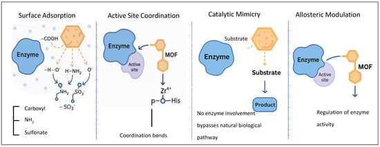
Figure 3.
Schematic representation of four primary mechanisms by which metal–organic frameworks (MOFs) modulate enzymatic activity.
Recent in vitro studies have validated the effectiveness of MOFs as enzyme inhibitors with high selectivity and manageable cytotoxicity profiles. For example, Ti-based MOFs synthesized via microwave-assisted methods demonstrated significant inhibition of cancer-related kinases and cytotoxicity against A431 skin carcinoma cells at low micromolar IC50 levels [60]. Fe3O4@bio-MOFs produced by sonochemical routes inhibited Leishmania phosphatases and showed therapeutic efficacy in both in vitro and in vivo models, with minimal off-target toxicity [65]. Additionally, Ce-MOFs functionalized with phosphate-mimicking ligands exhibited preferential binding to PTP1B and SHP2 phosphatases, reducing signaling activity in insulin-resistance models [84]. These studies confirm the potential of MOFs as bioactive materials in targeted enzyme inhibition, although long-term cytotoxicity, cellular uptake variability, and off-target effects remain key considerations. Despite their promise, several challenges hinder the clinical translation of MOFs in enzymatic modulation. Scalability and reproducibility remain critical bottlenecks, particularly for complex MOFs requiring precise ligand-metal ratios and energy-intensive synthesis methods [85,86]. The translational application of MOFs is critically shaped by biological barriers that extend beyond their demonstrated in vitro efficacy. Biodistribution is strongly size- and surface-dependent, with nanoparticles under 10 nm typically cleared by renal filtration, whereas larger frameworks tend to accumulate in the liver and spleen via the reticuloendothelial system (RES). Clearance pathways, whether renal or hepatic, must therefore be carefully controlled through rational size engineering, surface coatings, and functionalization strategies. Stability in biological environments represents another major challenge; MOFs are prone to degradation through linker hydrolysis or metal ion leaching under acidic or high-ionic-strength conditions typical of cellular and extracellular fluids [87]. Such degradation not only compromises functional activity but also risks cytotoxicity through the release of toxic ions. Immune recognition further complicates systemic delivery, as protein corona formation and complement activation can trigger opsonization and rapid clearance, while off-target binding to non-specific proteins may exacerbate toxicity [88]. These challenges underscore the importance of comprehensive pharmacokinetic studies and rational design approaches—such as biomimetic coatings, post-synthetic functionalization, and surface chemistry optimization—to improve stability, minimize immune response, and ensure effective therapeutic translation.
To overcome these limitations, recent efforts focus on post-synthetic functionalization, biomimetic coatings, and green synthesis approaches using water or ethanol as solvents instead of toxic solvents such as DMF [89,90,91] with mechanochemical and sonochemical approaches eliminating solvent use altogether. Energy-efficient microwave-assisted synthesis further reduces the environmental and economic cost of production. Despite these advances, clinical translation barriers persist. The reproducibility of green synthesis routes remains challenging at scale, while potential contamination from precursors or solvents can compromise biocompatibility. Additionally, regulatory approval requires comprehensive toxicity, pharmacokinetics, and degradation studies, which are often underexplored in green-synthesized MOFs. Thus, the integration of eco-friendly synthesis must be accompanied by rigorous evaluation of safety and stability.
4. Conclusions
In this review, we examined the potential of metal–organic frameworks (MOFs) as enzyme inhibitors, with a particular focus on their application in modulating protein kinases and phosphatases. We began by outlining the structural fundamentals of MOFs, highlighting the roles of metal nodes and organic linkers. Various synthesis strategies—solvothermal, microwave-assisted, and sonochemical—were discussed in the context of how they influence the resulting physicochemical and biological properties of MOFs. Particular emphasis was placed on biologically relevant metals such as Zr, Ce, Fe, Ti, Cu, and lanthanides, which contribute not only to structural integrity but also to catalytic and inhibitory activity. Four principal mechanisms of MOF–enzyme interaction were detailed: surface adsorption, active site coordination, catalytic mimicry, and allosteric modulation. These mechanistic pathways were supported by recent in vitro and in vivo studies demonstrating promising bioactivity, target selectivity, and manageable toxicity profiles. Challenges such as poor stability under physiological conditions, synthesis reproducibility, and limited clinical data were also critically discussed.
Taken together, the data suggest that MOFs represent a versatile and tunable class of materials for therapeutic enzyme inhibition. Their structural modularity, surface functionalizability, and catalytic potential enable them to address enzyme targets previously considered challenging for small molecules or biologics. To further guide their development, a rational design framework is essential, encompassing strategic metal selection (e.g., Zr(IV), Ti(IV), Fe(III), Ce(IV)), biofunctional linker incorporation (e.g., phosphate, carboxylate, sulfonate), and pore engineering to ensure optimal enzyme accessibility. Surface functionalization through polymers or biomimetic ligands can enhance selectivity and biocompatibility, while synthesis route optimization can improve scalability and reproducibility.
Looking ahead, the synthesis of MOFs for biomedical enzyme modulation will likely evolve along two complementary tracks. The first focuses on scalable and sustainable production, where continuous-flow and mechanochemical synthesis could reduce batch variability and align with green chemistry principles. The second involves precision-guided functionalization, leveraging post-synthetic modification and modular linker design to fine-tune enzyme specificity and pharmacokinetic profiles. Integration of computational tools—such as machine learning for linker selection, molecular docking for enzyme binding prediction, and density functional theory (DFT) for catalytic mechanism modeling—can accelerate discovery and improve structure–activity relationships.
Ultimately, the translation of MOFs into therapeutic platforms will depend on interdisciplinary collaboration—bridging materials science, enzymology, pharmacology, and computational chemistry. With continued innovation, sustainable synthesis, and rigorous biological evaluation, MOFs are poised to complement and potentially redefine enzyme inhibition strategies, offering selective, adaptable, and mechanistically distinct pathways for future disease intervention.
Author Contributions
Contributions: writing—review and editing, T.B.; writing—original draft preparation, A.A. All authors have read and agreed to the published version of the manuscript.
Funding
This research received no external funding.
Institutional Review Board Statement
Not applicable.
Informed Consent Statement
Not applicable.
Data Availability Statement
No new data were created or analyzed in this study. Data sharing is not applicable to this article.
Conflicts of Interest
The authors declare no conflicts of interest.
References
- Balbaied, T.; Moore, E. Overview of Capillary Electrophoresis Analysis of Alkaline Phosphatase (ALP) with Emphasis on Post-Translational Modifications (PTMs). Kinases Phosphatases 2023, 1, 206–219. [Google Scholar] [CrossRef]
- Hunter, T. Signaling—2000 and beyond. Cell 2000, 100, 113–127. [Google Scholar] [CrossRef]
- Bononi, A.; Agnoletto, C.; De Marchi, E.; Marchi, S.; Patergnani, S.; Bonora, M.; Giorgi, C.; Missiroli, S.; Poletti, F.; Rimessi, A.; et al. Protein kinases and phosphatases in the control of cell fate. Enzym. Res. 2011, 2011, 329098. [Google Scholar] [CrossRef] [PubMed] [PubMed Central]
- Cohen, P. The role of protein phosphorylation in human health and disease. Eur. J. Biochem. 2001, 268, 5001–5010. [Google Scholar] [CrossRef]
- Copeland, R.A. Evaluation of Enzyme Inhibitors in Drug Discovery: A Guide for Medicinal Chemists and Pharmacologists; John Wiley & Sons: Hoboken, NJ, USA, 2023. [Google Scholar]
- Baldi, S.; Long, N.; Ma, S.; Liu, L.; Al-Danakh, A.; Yang, Q.; Deng, X.; Xie, J.; Tang, H. Advancements in Protein Kinase Inhibitors: From Discovery to Clinical Applications. Research 2025, 8, 747. [Google Scholar] [CrossRef] [PubMed] [PubMed Central]
- Pawson, T.; Scott, J.D. Protein phosphorylation in signaling–50 years and counting. Trends Biochem. Sci. 2005, 30, 286–290. [Google Scholar] [CrossRef] [PubMed]
- Burton, J.C.; Royer, F.; Grimsey, N.J. Spatiotemporal control of kinases and the biomolecular tools to trace activity. J. Biol. Chem. 2024, 300, 107846. [Google Scholar] [CrossRef] [PubMed] [PubMed Central]
- Wong, W.; Scott, J.D. AKAP signalling complexes: Focal points in space and time. Nat. Rev. Mol. Cell Biol. 2004, 5, 959–970. [Google Scholar] [CrossRef]
- Hoxhaj, G.; Manning, B.D. The PI3K-AKT network at the interface of oncogenic signalling and cancer metabolism. Nat. Rev. Cancer 2020, 20, 74–88. [Google Scholar] [CrossRef] [PubMed] [PubMed Central]
- Wang, B.; Wu, H.; Hu, C.; Wang, H.; Liu, J.; Wang, W.; Liu, O. An overview of kinase downregulators and recent advances in discovery approaches. Signal Transduct. Target. Ther. 2021, 6, 423. [Google Scholar] [CrossRef]
- Klebe, G. Transferase Inhibitors. In Drug Design; Springer: Berlin/Heidelberg, Germany, 2024. [Google Scholar] [CrossRef]
- Hefayathullah, M.; Singh, S.; Ganesan, V.; Maduraiveeran, G. Metal-organic frameworks for biomedical applications: A review. Adv. Colloid Interface Sci. 2024, 331, 103210. [Google Scholar] [CrossRef] [PubMed]
- Sezgin, P.; Gulcay-Ozcan, E.; Vučkovski, M.; Bondžić, A.M.; Erucar, I.; Keskin, S. Biomedical Applications of Metal-Organic Frameworks Revisited. Ind. Eng. Chem. Res. 2025, 64, 1907–1932. [Google Scholar] [CrossRef] [PubMed] [PubMed Central]
- Alamro, A.; Balbaied, T. Boron Nitride Nanostructures (BNNs) Within Metal–Organic Frameworks (MOFs): Electrochemical Platform for Hydrogen Sensing and Storage. Analytica 2024, 5, 599–618. [Google Scholar] [CrossRef]
- Khafaga, D.S.; El-Morsy, M.T.; Faried, H.; Diab, A.H.; Shehab, S.; Saleh, A.M.; Ali, G.A. Metal–organic frameworks in drug delivery: Engineering versatile platforms for therapeutic applications. RSC Adv. 2024, 14, 30201–30229. [Google Scholar] [CrossRef]
- van Veldhuisen, T.W.; Dijkstra, R.M.J.; Koops, A.A.; Cossar, P.J.; van Hest, J.C.M.; Brunsveld, B. Modulation of Protein–Protein Interactions with Molecular Glues in a Synthetic Condensate Platform. J. Am. Chem. Soc. 2025, 147, 5386–5397. [Google Scholar] [CrossRef]
- Chattopadhyay, K.; Mandal, M.; Maiti, D.K. A review on zirconium-based metal–organic frameworks: Synthetic approaches and biomedical applications. Mater. Adv. 2024, 5, 51–67. [Google Scholar] [CrossRef]
- Bhattacharjee, S.; Chakraborty, T.; Bhaumik, A. A Ce-MOF as an alkaline phosphatase mimic: Ce-OH2 sites in catalytic dephosphorylation. Inorg. Chem. Front. 2022, 9, 5735–5744. [Google Scholar] [CrossRef]
- Cun, J.E.; Fan, X.; Pan, Q.; Gao, W.; Luo, K.; He, B.; Pu, Y. Copper-based metal–organic frameworks for biomedical applications. Adv. Colloid Interface Sci. 2022, 305, 102686. [Google Scholar] [CrossRef]
- Zhu, R.; Cai, M.; Fu, T.; Yin, D.; Peng, H.; Liao, S.; Du, Y.; Kong, J.; Ni, J.; Yin, X. Fe-Based Metal Organic Frameworks (Fe-MOFs) for Bio-Related Applications. Pharmaceutics 2023, 15, 1599. [Google Scholar] [CrossRef]
- Hu, S.; Liu, J.; Wang, Y.; Liang, Z.; Hu, B.; Xie, J.; Wong, W.-L.; Wong, K.-Y.; Qiu, B.; Peng, W. A new fluorescent biosensor based on inner filter effect and competitive coordination with the europium ion of non-luminescent Eu-MOF nanosheets for the determination of alkaline phosphatase activity in human serum. Sens. Actuators B Chem. 2023, 380, 133379. [Google Scholar] [CrossRef]
- Yu, L.; Feng, L.; Li, X.; Li, S.; Xu, Q.; Pan, X.; Xiao, Y. Rational Design of Dual-Emission Lanthanide Metal–Organic Framework for Visual Alkaline Phosphatase Activity Assay. ACS Appl. Mater. Interfaces 2021, 13, 11646–11656. [Google Scholar] [CrossRef]
- Wiśniewska, P.; Haponiuk, J.T.; Saeb, M.R.; Rabiee, N.; Bencherif, S.A. Mitigating metal-organic framework (MOF) toxicity for biomedical applications. Chem. Eng. J. 2023, 471, 144400. [Google Scholar] [CrossRef]
- Singh, N.; Qutub, S.; Khashab, N.M. Biocompatibility and biodegradability of metal organic frameworks for biomedical applications. J. Mater. Chem. B 2021, 9, 5925–5934. [Google Scholar] [CrossRef]
- Pramanik, B.; Sahoo, R.; Das, M.C. pH-stable MOFs: Design principles and applications. Coord. Chem. Rev. 2023, 493, 215301. [Google Scholar] [CrossRef]
- Han, W.; Shi, M.; Jiang, H.-L. Scalable and Low-energy Synthesis of Metal-organic Frameworks by a Seed-mediated Approach. Angew. Chem. Int. Ed. 2025, 64, e202421942. [Google Scholar] [CrossRef]
- Nazari, M.; Zadehahmadi, F.; Sadiq, M.M.; Sutton, A.L.; Mahdavi, H.; Hill, M.R. Challenges and solutions to the scale-up of porous materials. Commun. Mater. 2024, 5, 170. [Google Scholar] [CrossRef]
- Karami, Z.; Khodaei, M.M. Post-synthetic modification of IR-MOF-3 as acidic-basic heterogeneous catalyst for one-pot synthesis of pyrimido [4,5-b]quinolones. Res. Chem. Intermed. 2022, 48, 1773–1792. [Google Scholar] [CrossRef]
- Xu, D.; Zhang, W.; Pan, Y.; Wang, W.; Wang, D.; Ding, J. Progress in nanocarriers-based approaches for the delivery of tyrosine kinase inhibitors in bone cancer: Trends and prospects. IUBMB Life 2025, 77, e70052. [Google Scholar] [CrossRef]
- Akhuli, A.; Chakraborty, D.; Preeyanka, N.; Simanchal, A.; Sarkar, D.M. Copper nanoclusters as an effective enzyme inhibitor on the activity modulation of α-chymotrypsin. ACS Appl. Nano Mater. 2023, 6, 4910–4924. [Google Scholar] [CrossRef]
- Pan, X.; Yao, Y.; Zhang, M.; Yuan, X.; Yao, Q.; Hua, W. Enzyme-mimic catalytic activities and biomedical applications of noble metal nanoclusters. Nanoscale 2024, 16, 8196–8215. [Google Scholar] [CrossRef] [PubMed]
- Gomari, M.M.; Abkhiz, S.; Pour, T.G.; Lotfi, E.; Rostami, N.; Monfared, F.N.; Ghobari, B.; Mosavi, M.; Alipour, B.; Dokholyan, N.V. Peptidomimetics in cancer targeting. Mol. Med. 2022, 28, 146. [Google Scholar] [CrossRef]
- Zhang, Y.; Yang, C.-J.; Melrose, A.R.; Pang, J.; Schofield, K.; Song, S.D.; Parra-Izquierdo, I.; Zheng, T.J.; Lyssikatos, J.P.; Gross, S.D.; et al. Pharmacological effects of small molecule BCR-ABL tyrosine kinase inhibitors on platelet function. J. Pharmacol. Exp. Ther. 2025, 392, 100020. [Google Scholar] [CrossRef]
- Yao, S.-X.; Huang, Y.-J.; Zhang, Y.-X.; Cui, Z.-X.; Lu, H.-Y.; Wang, R.; Shi, L. Revisiting VEGF/VEGFR-2 signaling as an anticancer target and its inhibitor discovery: Where are we and where should we go? J. Drug Target. 2025, 33, 1471–1494. [Google Scholar] [CrossRef]
- Kim, M.S.; Kim, M.S. Deubiquitination of epidermal growth factor receptor by ubiquitin-specific peptidase 54 enhances drug sensitivity to gefitinib in gefitinib-resistant non-small cell lung cancer cells. PLoS ONE 2025, 20, e0320668. [Google Scholar] [CrossRef]
- Ramos-Alvarez, I.; Jensen, R.T. Elucidation of roles of serine/threonine phosphatases PP1 and PP2A in mediating CCK-stimulated growth and enzyme secretion in pancreatic acinar cells. AJP Gastrointest. Liver Physiol. 2025, 329, G102–G121. [Google Scholar] [CrossRef]
- Guo, H.; Ren, W.; Guo, M.; Wu, X.; Guo, Q. A Comprehensive Review on Ethnopharmacology, Phytochemistry of Mylabris, and Pharmacology of Cantharidin. Chem. Biodivers. 2025, 22, e202500266. [Google Scholar] [CrossRef]
- Nath, I.; Chakraborty, J.; Verpoort, F. Metal organic frameworks mimicking natural enzymes: A structural and functional analogy. Chem. Soc. Rev. 2016, 45, 4127–4170. [Google Scholar] [CrossRef]
- Cai, H.; Huang, Y.-L.; Li, D. Biological metal–organic frameworks: Structures, host–guest chemistry and bio-applications. Coord. Chem. Rev. 2019, 378, 207–221. [Google Scholar] [CrossRef]
- Xu, H.; Liu, M.; Huang, X.; Min, Q.; Zhu, J.-J. Multiplexed Quantitative MALDI MS Approach for Assessing Activity and Inhibition of Protein Kinases Based on Postenrichment Dephosphorylation of Phosphopeptides by Metal–Organic Framework-Templated Porous CeO2. Anal. Chem. 2018, 90, 9859–9867. [Google Scholar] [CrossRef] [PubMed]
- Wang, K.; Zhang, J.; Hsu, Y.-C.; Lin, H.; Han, Z.; Pang, J.; Yang, Z.; Liang, R.-R.; Shi, W.; Zhou, H.-C. Bioinspired Framework Catalysts: From Enzyme Immobilization to Biomimetic Catalysis. Chem. Rev. 2023, 123, 5347–5420. [Google Scholar] [CrossRef] [PubMed]
- Du, C.; Xu, Y.; Wei, G. Recent Advances in Biomolecule-Engineered Metal-Organic Frameworks (Bio-MOFs): From Design, Bioengineering, and Structural/functional Regulation to Biocatalytic Applications. Chem. Rec. 2025, 25, e202500001. [Google Scholar] [CrossRef]
- Chen, F.; Zheng, H.; Yusran, Y.; Li, H.; Qiu, S.; Fang, Q. Exploring high-connectivity three-dimensional covalent organic frameworks: Topologies, structures, and emerging applications. Chem. Soc. Rev. 2024, 54, 484–514. [Google Scholar] [CrossRef]
- Tang, H.; Fan, D.; Chen, Y.; Han, S.-Y. Exploring Enzyme-MOF (Metal-Organic Framework) Catalytic Systems: Trade-offs Between Enzyme Activity and MOF Stability. Green Chem. 2024, 27, 2605–2628. [Google Scholar] [CrossRef]
- Ellis, G.A.; Klein, W.P.; Lasarte-Aragonés, G.; Thakur, M.; Walper, S.A.; Medintz, I.L. Artificial Multienzyme Scaffolds: Pursuing in Vitro Substrate Channeling with an Overview of Current Progress. ACS Catal. 2019, 9, 10812–10869. [Google Scholar] [CrossRef]
- Jia, C.; Bai, J.; Liu, Z.; Gao, S.; Han, Y.; Yan, H. Application of a titanium-based metal-organic framework to protein kinase activity detection and inhibitor screening. Anal. Chim. Acta 2020, 1128, 99–106. [Google Scholar] [CrossRef]
- Bai, J.; Liu, L.; Jia, C.; Liu, Z.; Gao, S.; Han, Y.; Yan, H. Fluorescence Method for the Detection of Protein Kinase Activity by Using a Zirconium-Based Metal–Organic Framework as an Affinity Probe. ACS Appl. Bio Mater. 2019, 2, 6021–6028. [Google Scholar] [CrossRef] [PubMed]
- Radfar, S.; Sheikh, M.; Akhavantabib, A.; Heidari, A.; Ghasemi, M.; Naghavi, M.; Ghanbari, R.; Zibadi, F.; Jamshidi, B.; Alizadeh, A. Application of a porous zirconium-based MOF nanoplate as an affinity ECL platform for the detection of protein kinase activity and inhibitor screening. Talanta 2025, 287, 127675. [Google Scholar] [CrossRef]
- Alizadeh, A.; Sheikh, M.; Akhavantabib, A.; Heidari, A.; Ghasemi, M.; Naghavi, M.; Ghanbari, R.; Radfar, S. A Porous Zirconium-Based MOF Nanoplate as an Affinity ECL Platform for the Detection of Protein Kinase Activity and Inhibitor Screening. Preprint 2024. [Google Scholar] [CrossRef]
- Yan, Z.; Wang, F.; Deng, P.; Wang, Y.; Cai, K.; Chen, Y.; Wang, Z.; Liu, Y. Sensitive electrogenerated chemiluminescence biosensors for protein kinase activity analysis based on bimetallic catalysis signal amplification and recognition of Au and Pt loaded metal-organic frameworks nanocomposites. Biosens. Bioelectron. 2018, 109, 132–138. [Google Scholar] [CrossRef]
- Chang, Y.; Gao, F.; Wu, T.; Pan, Q.; Liu, L.; Song, Q. Electrochemical detection of protein kinases with methylene blue-functionalized Zr-based metal-organic frameworks as signal labels. Int. J. Electrochem. Sci. 2023, 18, 100338. [Google Scholar] [CrossRef]
- Yu, L.; Shen, Y.; Xu, Q.; Gan, Z.; Feng, Y.; Yang, C.; Xiao, Y. Enhancing Kinase Activity Detection with a Programmable Lanthanide Metal–Organic Framework via ATP-to-ADP Conversion. Anal. Chem. 2024, 96, 12139–12146. [Google Scholar] [CrossRef]
- Jodaeeasl, N.; Wang, S.; Hu, A.; Peslherbe, G.H. Comprehensive DFT investigation of small-molecule adsorption on the paradigm M-mof-74 family of metal–organic frameworks. Phys. Chem. Chem. Phys. 2025, 27, 3068–3082. [Google Scholar] [CrossRef]
- Pourmadadi, M.; Ostovar, S.; Eshaghi, M.M.; Rajabzadeh-Khosroshahi, M.; Safakhah, S.; Ghotekar, S.; Rahdar, A.; Díez-Pascual, A.M. Nanoscale metallic-organic frameworks as an advanced tool for medical applications: Challenges and recent progress. Appl. Organomet. Chem. 2023, 37, e6982. [Google Scholar] [CrossRef]
- Dalapati, S.; Jana, R.; Banerjee, R. Solvothermal synthesis of metal-organic frameworks for catalytic applications. Coord. Chem. Rev. 2022, 471, 214729. [Google Scholar] [CrossRef]
- Qian, Y.; Wang, C.; Xu, R.; Wang, J.; Chen, Q.; Zhu, Z.; Hu, Q.; Shen, Q.; Shen, J.-W. Copper-based metal–organic frameworks for antitumor application. J. Nanobiotechnol. 2025, 23, 135. [Google Scholar] [CrossRef]
- Xiao, J.; Yang, S.-S.; Wu, J.-X.; Wang, H.; Yu, X.; Shang, W.; Chen, G.-Q.; Gu, Z.-Y. Highly Selective Capture of Monophosphopeptides by Two-Dimensional Metal–Organic Framework Nanosheets. Anal. Chem. 2019, 91, 9093–9101. [Google Scholar] [CrossRef]
- Wang, H.; Chen, X.; Zhao, X. Microwave-assisted synthesis of MOFs: Methods and applications. Microporous Mesoporous Mater. 2021, 318, 111036. [Google Scholar] [CrossRef]
- Altharawi, A.; Alqahtani, S.M.; Aldakhil, T.; Ahmad, I. Microwave-assisted synthesis of novel Ti/BTB-MOFs as porous anticancer and antibacterial agents. Front. Chem. 2024, 12, 1386311. [Google Scholar] [CrossRef] [PubMed]
- Iorkula, T.H.; Osayawe, O.J.K.; Odogwu, D.A.; Ganiyu, L.O.; Faderin, E.; Awoyemi, R.F.; Akodu, B.O.; Ifijen, I.H.; Aworinde, O.R.; Agyemang, P.; et al. Advances in pyrazolo [1,5-a]pyrimidines: Synthesis and their role as protein kinase inhibitors in cancer treatment. RSC Adv. 2025, 15, 3756–3828. [Google Scholar] [CrossRef] [PubMed]
- Wang, S.; Li, Y.; Zhang, G. Recent advances in sonochemical synthesis of MOFs. Ultrason. Sonochem. 2023, 94, 106352. [Google Scholar] [CrossRef]
- Wang, J.; Hu, T.; Han, Q.; Luo, W.; Zhong, J.; Ding, M. The synthesis and functionalization of metal organic frameworks and their applications for the selective separation of proteins/peptides. Anal. Bioanal. Chem. 2023, 415, 5859–5874. [Google Scholar] [CrossRef]
- Scattolin, T.; Tonon, G.; Botter, E.; Canale, V.C.; Hasanzadeh, M.; Cuscela, D.M.; Buschini, A.; Zarepour, A.; Khosravi, A.; Cordani, M.; et al. Synergistic applications of cyclodextrin-based systems and metal–organic frameworks in transdermal drug delivery for skin cancer therapy. J. Mater. Chem. B 2024, 12, 3807–3839. [Google Scholar] [CrossRef] [PubMed]
- Abazari, R.; Mahjoub, A.R.; Molaie, S.; Ghaffarifar, F.; Ghasemi, E.; Slawin, A.M.; Carpenter-Warren, C.L. The effect of different parameters under ultrasound irradiation for synthesis of new nanostructured Fe3O4@bio-MOF as an efficient anti-leishmanial in vitro and in vivo conditions. Ultrason. Sonochem. 2018, 43, 248–261. [Google Scholar] [CrossRef]
- Chen, L.; Xu, X.; Hu, Y. Mechanochemical synthesis of bio-compatible MOFs: Towards green and scalable production. Adv. Mater. Interfaces 2024, 11, 2400190. [Google Scholar] [CrossRef]
- Mao, H.; Yu, L.; Tu, M.; Wang, S.; Zhao, J.; Zhang, H.; Cao, Y. Recent Advances on the Metal-Organic Frameworks-Based Biosensing Methods for Cancer Biomarkers Detection. Crit. Rev. Anal. Chem. 2024, 54, 1273–1289. [Google Scholar] [CrossRef]
- Mannias, G. Development of New Synthesis Methods of Proteins/Enzymes-Metal Organic Frameworks (MOFs) Hybrid Composite Materials for Biomedical Applications. Depositolegale.it; Università degli Studi di Sassari. Available online: https://tesidottorato.depositolegale.it/handle/20.500.14242/117581 (accessed on 21 February 2025).
- Griffin, S.L.; Briuglia, M.L.; ter Horst, J.H.; Forgan, R.S. Assessing crystallisation kinetics of zr metal–organic frameworks through turbidity measurements to inform rapid microwave-assisted synthesis. Chem.-A Eur. J. 2020, 26, 6910–6918. [Google Scholar] [CrossRef]
- Abaszadeh, N.; Afzali, D.; Sargazi, G.; Golpayegani, A. Sonochemical-assisted method for efficient synthesis of Cu-MOF and evaluating its antibacterial properties. Heliyon 2024, 10, e31024. [Google Scholar] [CrossRef] [PubMed]
- Głowniak, S.; Szczęśniak, B.; Choma, J.; Jaroniec, M. Mechanochemical synthesis of MOF-303 and its CO2 adsorption at ambient conditions. Molecules 2024, 29, 2698. [Google Scholar] [CrossRef]
- Phan, P.T.; Hong, J.; Tran, N.; Le, T.H. The Properties of Microwave-Assisted Synthesis of Metal–Organic Frameworks and Their Applications. Nanomaterials 2023, 13, 352. [Google Scholar] [CrossRef]
- Zheng, Z.; Jiang, X.; Yang, X.; Ma, M.; Ji, S.; Jiang, F. Microwave- and ultrasonic-assisted synthesis of 2D La-based MOF nanosheets by coordinative unsaturation degree to boost phosphate adsorption. RSC Adv. 2022, 12, 35517–35530. [Google Scholar] [CrossRef] [PubMed]
- Głowniak, S.; Szczęśniak, B.; Choma, J.; Jaroniec, M. Recent Developments in Sonochemical Synthesis of Nanoporous Materials. Molecules 2023, 28, 2639. [Google Scholar] [CrossRef] [PubMed]
- Ismail, K.M.; Hassan, S.S.; Medany, S.S.; Hefnawy, M.A. A facile sonochemical synthesis of the Zn-based metal–organic framework for electrochemical sensing of paracetamol. Mater. Adv. 2024, 5, 5870–5884. [Google Scholar] [CrossRef]
- Steenhaut, T.; Grégoire, N.; Barozzino-Consiglio, G.; Filinchuk, Y.; Hermans, S. Mechanochemical defect engineering of HKUST-1 and impact of the resulting defects on carbon dioxide sorption and catalytic cyclopropanation. RSC Adv. 2020, 10, 19822–19831. [Google Scholar] [CrossRef] [PubMed]
- Yang, J.; Yang, Y. Metal–Organic Frameworks for Biomedical Applications. Small 2020, 16, 1906846. [Google Scholar] [CrossRef]
- Liu, R.; Chi, L.; Wang, X.; Wang, Y.; Sui, Y.; Xie, T.; Arandiyan, H. Effective and selective adsorption of phosphate from aqueous solution via trivalent-metals-based amino-MIL-101 MOFs. Chem. Eng. J. 2019, 357, 159–168. [Google Scholar] [CrossRef]
- Alexander, C.; Guo, Z.; Glover, P.B.; Faulkner, S.; Pikramenou, Z. Luminescent Lanthanides in Biorelated Applications: From Molecules to Nanoparticles and Diagnostic Probes to Therapeutics. Chem. Rev. 2025, 125, 2269–2370. [Google Scholar] [CrossRef]
- Shteinman, A.A. Metallocavitins as Advanced Enzyme Mimics and Promising Chemical Catalysts. Catalysts 2023, 13, 415. [Google Scholar] [CrossRef]
- Changeux, J.-P.; Christopoulos, A. Allosteric Modulation as a Unifying Mechanism for Receptor Function and Regulation. Cell 2016, 166, 1084–1102. [Google Scholar] [CrossRef]
- Bhattacharya, A.; Pandit, S.; Lee, S.; Ebrahimi, S.B.; Samanta, D. Modulating Enzyme Activity using Engineered Nanomaterials. ChemBioChem 2024, 26, e202400520. [Google Scholar] [CrossRef] [PubMed]
- Li, T.; Liu, Y.; Wang, T.; Wu, Y.; He, Y.; Yang, R.; Zheng, S.-R. Regulation of the surface area and surface charge property of MOFs by multivariate strategy: Synthesis, characterization, selective dye adsorption and separation. Microporous Mesoporous Mater. 2018, 272, 101–108. [Google Scholar] [CrossRef]
- Saint-Laurent, C.; Mazeyrie, L.; Tajan, M.; Paccoud, R.; Castan-Laurell, I.; Valet, P.; Edouard, T.; Pradère, J.P.; Dray, C.; Yart, A. The Tyrosine Phosphatase SHP2: A New Target for Insulin Resistance? Biomedicines 2022, 10, 2139. [Google Scholar] [CrossRef] [PubMed]
- Xie, F.; Li, J. Toward Scalable and Sustainable Synthesis of Metal–Organic Frameworks. ACS Mater. Lett. 2024, 6, 2400–2408. [Google Scholar] [CrossRef]
- Shrivastav, V.; Mansi; Gupta, B.; Dubey, P.; Deep, A.; Nogala, W.; Shrivastav, V.; Sundriyal, S. Recent advances on surface mounted metal-organic frameworks for energy storage and conversion applications: Trends, challenges, and opportunities. Adv. Colloid Interface Sci. 2023, 318, 102967. [Google Scholar] [CrossRef] [PubMed]
- Mosca, L.P.L.; Gapan, A.B.; Angeles, R.A.; Lopez, E.C.R. Stability of Metal–Organic Frameworks: Recent Advances and Future Trends. Eng. Proc. 2023, 56, 146. [Google Scholar] [CrossRef]
- Chen, G.; Dai, B.; Hao, J.-N.; Li, Y. A dual-excitation-driven full-component-responsive lanthanide-based metal-organic framework for switchable profiling of multi-disease markers. Sci. China Mater. 2025, 68, 666–676. [Google Scholar] [CrossRef]
- Baa, E.; Watkins, G.M.; Krause, R.W.; Tantoh, D.N. Current Trend in Synthesis, Post-Synthetic Modifications and Biological Applications of Nanometal-Organic Frameworks (NMOFs). Chin. J. Chem. 2019, 37, 378–404. [Google Scholar] [CrossRef]
- Bagheri, A.; Hoseinzadeh, H.; Hayati, B.; Mahmoodi, N.M.; Mehraeen, E. Post-synthetic functionalization of the metal-organic framework: Clean synthesis, pollutant removal, and antibacterial activity. J. Environ. Chem. Eng. 2020, 9, 104590. [Google Scholar] [CrossRef]
- Gupta, A.; Singh, S.; Sharma, A.L.; Deep, A. Green Synthesis and Applications of Metal-Organic Frameworks; Springer: Cham, Switzerland, 2022; pp. 1–20. [Google Scholar] [CrossRef]


Disclaimer/Publisher’s Note: The statements, opinions and data contained in all publications are solely those of the individual author(s) and contributor(s) and not of MDPI and/or the editor(s). MDPI and/or the editor(s) disclaim responsibility for any injury to people or property resulting from any ideas, methods, instructions or products referred to in the content. |
© 2025 by the authors. Licensee MDPI, Basel, Switzerland. This article is an open access article distributed under the terms and conditions of the Creative Commons Attribution (CC BY) license (https://creativecommons.org/licenses/by/4.0/).