Article

Mass Spectrometry and 3D Modeling Indicate the SBK2 Kinase Phosphorylates Splicing Factor SRSF7 to Regulate Cardiac Development

Division of Biomedical and Translational Sciences, University of South Dakota Sanford School of Medicine, Vermillion, SD 57069, USA
*
Authors to whom correspondence should be addressed.
Kinases Phosphatases 2025, 3(4), 20; https://doi.org/10.3390/kinasesphosphatases3040020
Submission received: 6 August 2025 / Revised: 18 September 2025 / Accepted: 18 September 2025 / Published: 23 September 2025

Abstract

SH3 Domain Binding Kinase Family Member 2 (SBK2) is a critical kinase in atrial cardiomyocyte differentiation. However, its phospho-targets, its role in ventricle function, and its role in cardiac disease progression are unknown. Notably, SBK2 has been shown to be downregulated in the ventricular myocardium of several mouse models that recapitulate human desmin-related cardiomyopathies. To restore SBK2 expression, adenoviruses were constructed to promote cardiomyocyte-restricted SBK2 expression and injected at postnatal day 0. This significantly increased ejection fraction at 1 month of age relative to control hearts. However, in 3-month nontransgenic (NTG) and desmin-related cardiomyopathy hearts, the overexpression of SBK2 opposed increases in ejection fraction and left ventricular posterior wall thickness. These findings provide the first in vivo evidence that SBK2 plays a vital role in left ventricular function. To elucidate the molecular mechanism behind the physiological effects of SBK2 on the heart, we performed mass spectrometry combined with phospho-enrichment on ventricular tissue with and without SBK2 overexpression. We identified multiple phosphorylation sites on SBK2 and used AlphaFold3 to model how this phosphorylation likely affects SBK2’s role in phosphorylating the splicing factor SRSF7. We propose a novel mechanism by which SBK2 regulates splicing to promote cardiomyocyte development.

1. Introduction

SBK2 is required for the differentiation of immortalized neonatal rat atrial myocytes [1]. An atrial cardiomyocyte cell model has revealed that downregulation of SBK2 results in the disruption of the sarcomere, but the molecular mechanisms behind the disruption were unclear [1]. Further mystery surrounding SBK2 emerged when our group found that mouse models of desmin-related cardiomyopathy have a persistent downregulation of SBK2.
Desmin is an intermediate filament protein. Desmin and its accessory proteins organize the cardiac microfibrils to maintain muscle architecture [2,3,4]. Specific mutations in desmin or its chaperone protein crystallin alpha B result in a class of disorders called desmin-related cardiomyopathies (DRC) [4,5]. Additionally, desmin is one of only 12 genes for which there is definitive evidence of causing dilated cardiomyopathy [6]. DRC has a severe impact on a patient’s cardiac health [7]. Impaired proteostasis is a hallmark of the disease as well as reduced cellular respiration, cardiomyocyte death, heart failure, and hypertrophy [3,4,7,8,9,10]. In 2001, the human R120G mutation in the heat shock protein CRYAB (which caused DRC in patients) was successfully recapitulated in a mouse model [5]. This model exhibits heart-specific overexpression of the R120G mutant protein, and the R120G-CryAB mouse has been the subject of intensive study since its creation. The hearts of these mice progress through restrictive cardiomyopathy, dilated cardiomyopathy, and hypertrophy, and the disease ultimately results in premature death, often at only 6–7 months of age [5,11,12]. The R120G mouse is a stable transgenic mouse model, and in the past 25 years, these cardiac phenotypes have been repeatedly verified. R120G cardiomyocyte hypertrophy was originally characterized at multiple levels, including gravimetric assessment at the organ level, histopathology at the tissue level, isolated cardiomyocyte morphometry at the cell level, and fetal gene expression at the molecular level when the R120G model was created [5] and subsequently validated [13]. Fibrosis has been confirmed histologically in the R120G model vs. NTG controls through Masson’s trichrome stain and picrosirius red staining [11]. Cardiomyocyte death has been validated using TUNEL staining [9], and we recently confirmed the cytotoxic and morphological effects of the R120G mutation on cardiomyocytes using immunostaining and gene expression analysis [14]. Furthermore, R120G mice display overt hypertrophy that is readily identifiable via echocardiography at 3 months of age, even without quantitative analysis—though such analysis confirms a state of cardiac hypertrophy [15]. Cardiac hypertrophy can be determined through measurements of elevated ejection fraction and increased left ventricular posterior wall thickness, as outlined in the guidelines for assessing cardiac physiology in mice [16].
We have seen in 2 separate mouse models of DRC that they have a persistent downregulation of SBK2. Furthermore, SBK2’s role in development and differentiation implies that it may play a role in cardiac regeneration or repair. We therefore hypothesized that restoring SBK2 levels would improve cardiac function in DRC mouse models. Finally, we utilized phospho-mass spectrometry and AlphFold3 to begin to elucidate the mechanisms by which SBK2 promotes cardiac development and function.

2. Results

2.1. SBK2 Overexpression Impacts Ventricular Structure and Function

To determine the effects of restoring SBK2 levels in DRC mice, we generated SBK2 overexpression adeno-associated viruses (AAV) specifically targeting cardiomyocytes: AAV9.cTNT.SBK2.P2A.mcherry (Figure 1A). The AAV9 viral serotype has a higher transduction efficiency in cardiac tissue than other AAV vectors, while the cardiac troponin T (cTnT) promoter drives cardiomyocyte-specific gene expression [17,18]. The P2A sequence induces ribosome skipping so that the mcherry is translated separately from SBK2, thereby allowing mcherry to be used as a marker without affecting SBK2’s function [19].
The heart tissue-specific targeting and viral concentration were tested and validated (Figure S1). Neonatal day zero NTG and R120G pups were injected intraperitoneally with AAV9.cTNT.SBK2.P2A.mcherry (AAV9.SBK2) or AAV9.cTNT.P2A.mcherry (AAV9.mch) control viruses. Echocardiography was then performed at one and three months. Heart rate, stroke volume, and cardiac output were unaffected after one month of viral expression (Figure 1B–D). However, SBK2 overexpression resulted in a significantly increased ejection fraction (Figure 1E) in most SBK2-injected groups when compared to AAV9.mch injected controls, which indicated that SBK2 overexpression had affected ventricular function. Although mean left ventricular posterior wall thickness slightly increased in AAV9.SBK2 vs. AAV9.mch-injected groups, overall increases were not significant (Figure 1F).
At three months, stroke volume was unaffected, but as expected, the R120G mice had reduced heart rate and cardiac output compared to NTG mice (Figure 1G–I). Conversely, both ejection fraction and left ventricular posterior wall thickness increased in R120G mice (Figure 1J,K). These measurements validate our recent report in which we demonstrated the cardiac structure of the R120G mouse enters a hypertrophic compensatory phase (in a sex-independent manner) by 3 months of age [15]. In that earlier study, we also used immunostaining to show histologically how the accumulation of misfolded protein aggregates and ubiquitinated proteins contributed to structural alterations in R120G cardiomyocytes [13]. In the present study, we showed that ejection fraction was significantly reduced in NTG males treated with AAV9.SBK2 compared to NTG mice receiving the AAV9.mCherry control virus. However, this reduction did not reach statistical significance in females (although the overall physiological responses to SBK2 overexpression did exhibit similar trends in both male and female mice). In both sexes, ejection fraction and left ventricular posterior wall thickness in R120G mice injected with AAV9.SBK2 were not significantly different from NTG mice treated with the control virus. This suggests that while SBK2 overexpression may be detrimental to cardiac function in healthy NTG mice, it could potentially mitigate some of the hypertrophic response in 3-month-old R120G mice. Collectively, these results provide the first in vivo evidence that SBK2 overexpression alters ventricular structure and functional output.

2.2. Mass Spectrometry Reveals Proteome Changes Resulting from SBK2 Overexpression in 2-Week-Old Ventricles
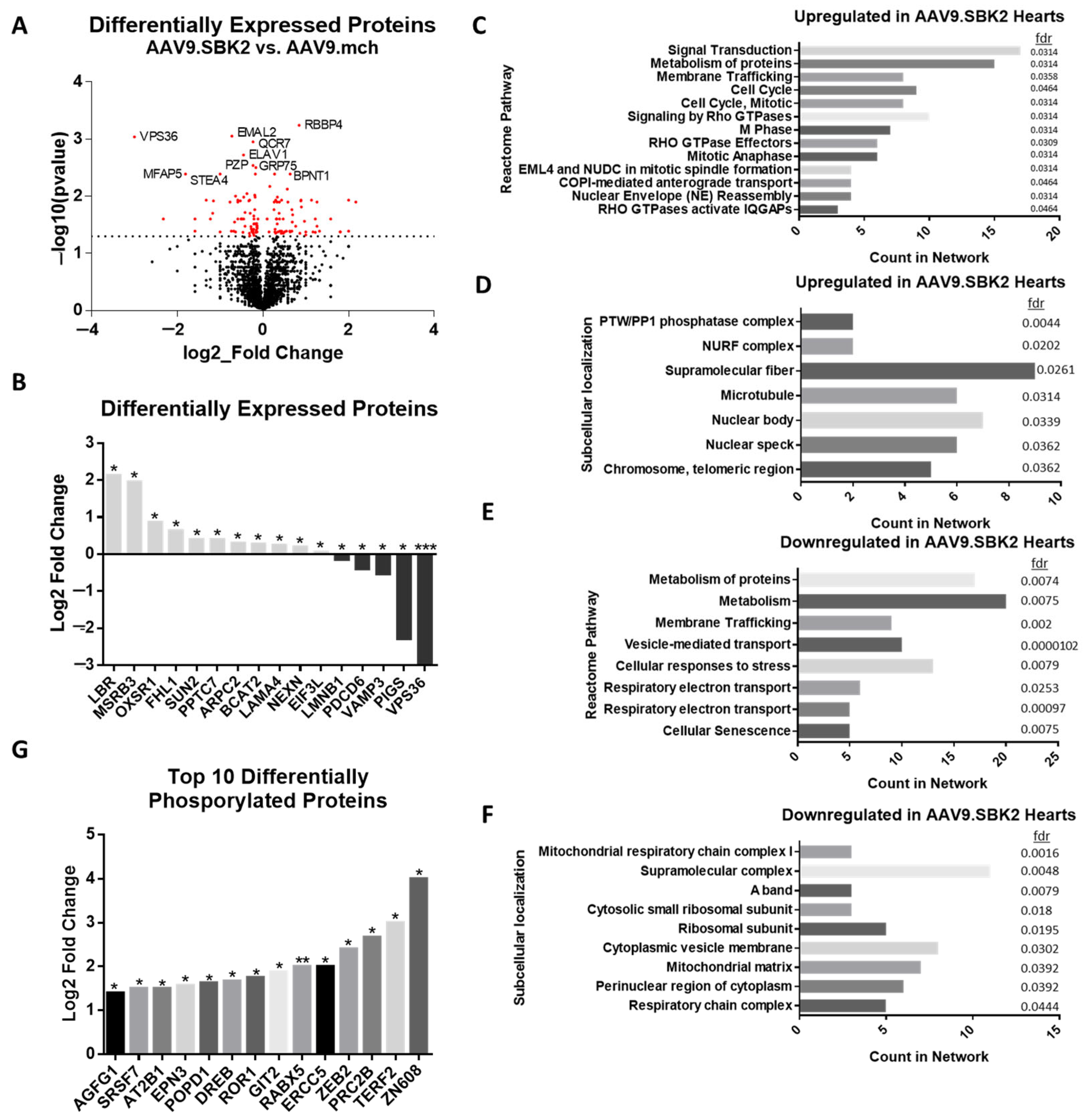
To begin to elucidate the molecular mechanisms behind the physiological effects of SBK2 overexpression, day zero pups were injected with either AAV9.SBK2 or AAV9.mch control virus, and ventricles were collected at 2 weeks of age for mass spectrometry-based proteomics. Of the 2993 proteins detected, 68 proteins were significantly upregulated and 76 downregulated in AAV9.SBK2-treated hearts relative to AAV9.mch-treated hearts (Figure 2A, Table S1). It is noteworthy that CRYAB was downregulated in AAV9.SBK2 hearts, indicating a possible regulatory relationship in DRC hearts. Of the differentially expressed proteins, several proteins were inversely regulated relative to their corresponding mRNA levels under SBK2-RNAi in cell lines from previous studies (Figure 2B) [1].
From the top Reactome Pathways for the upregulated proteins in AAV9.SBK2 injected hearts, notable categories include those related to the cell cycle and Rho GTPase signaling (Figure 2C). Included in the Rho GTPase signaling category are myosin regulatory light chain 12B (Myl12b) and a component of the actin-related protein 2/3 complex (Arpc2), further implicating SBK2 as a player in actin/myosin cytoskeletal organization. Notable subcellular localization (compartments) terms included microtubules and the PTW/PP1 phosphatase complex (Figure 2D). The PTW/PP1 phosphatase complex is an important regulator of cardiac development and function [20,21]. This complex therefore deserves further investigation, as it could imply a method of dephosphorylation related to SBK2 pathways.
Top Reactome Pathways for the downregulated proteins in AAV9.SBK2 hearts included categories related to metabolism, electron transport, and vesicle-mediated transport (Figure 2E). While the downregulated subcellular localization (compartments) terms included the mitochondrial respiratory chain and the A-band, which both also are severely affected by the R120G mutation in CRYAB [9].
To identify possible phospho-targets of SBK2, we combined two phosphopeptide pre-enrichment steps before running mass spectrometry. The top enriched phosphopeptides from the AAV9.SBK2-treated hearts included RABX5 and SRSF7 (Figure 2G). Phosphorylation of Rab5 GDP/GTP exchange factor (RABX5/RABGEF1) was the most significant phospho-target identified by mass spec. This is particularly interesting as it could explain why GTPase activity is a top upregulated Reactome pathway when overexpressing SBK2. RABX5 homolog RABGEF1 is a ubiquitin-binding protein that targets the mitochondria [22]. A cascade of other RAB proteins are then recruited to regulate Parkin-mediated mitophagy. If SBK2 directly phosphorylates RABX5, it could be an important player in mitophagy and further tie SBK2 dysregulation to R120G mice, as R120G mice have severe mitochondrial dysfunction [9].
Figure 2. Mass spectrometry reveals proteome changes resulting from SBK2 overexpression in 2-week-old ventricles. (A) Differentially expressed proteins in AAV9.cTNT.SBK2.P2A.mcherry or AAV9.cTNT.P2A.mcherry-treated hearts. Red p ≤ 0.05, black = ns (see the Methods section for calculations). (B) Proteins that are differentially expressed in AAV9.SBK2 vs. AAV9.mch injected groups but also have significant inverse expression of mRNA in SBK2-RNAi atrial cardiomyocyte cell lines from van Gorp et al. 2022 [1]. (CF) Highest strength Reactome Pathways and subcellular localizations up- or down-regulated, with corresponding false discovery rates (fdr) of pathway enrichment, from AAV.SBK2 vs. AAV9.mch mass spectrometry. Strength and fdr’s were calculated using STRING-db [23]. (G) The top 10 proteins with increased phosphorylation in AAV.SBK2 hearts (n = 6) compared to AAV9.mch hearts (n = 6) as identified by phospho-mass spectrometry. Student’s paired t-test. * p ≤ 0.05, ** p ≤ 0.01, *** p ≤ 0.001. AAV9.SBK2 males n = 3, AAV9.SBK2 females n = 3, vs. AAV9.mch males n = 3, vs. AAV9.mch females n = 3.
Kinasesphosphatases 03 00020 g002

2.3. Phosphorylation of SBK2

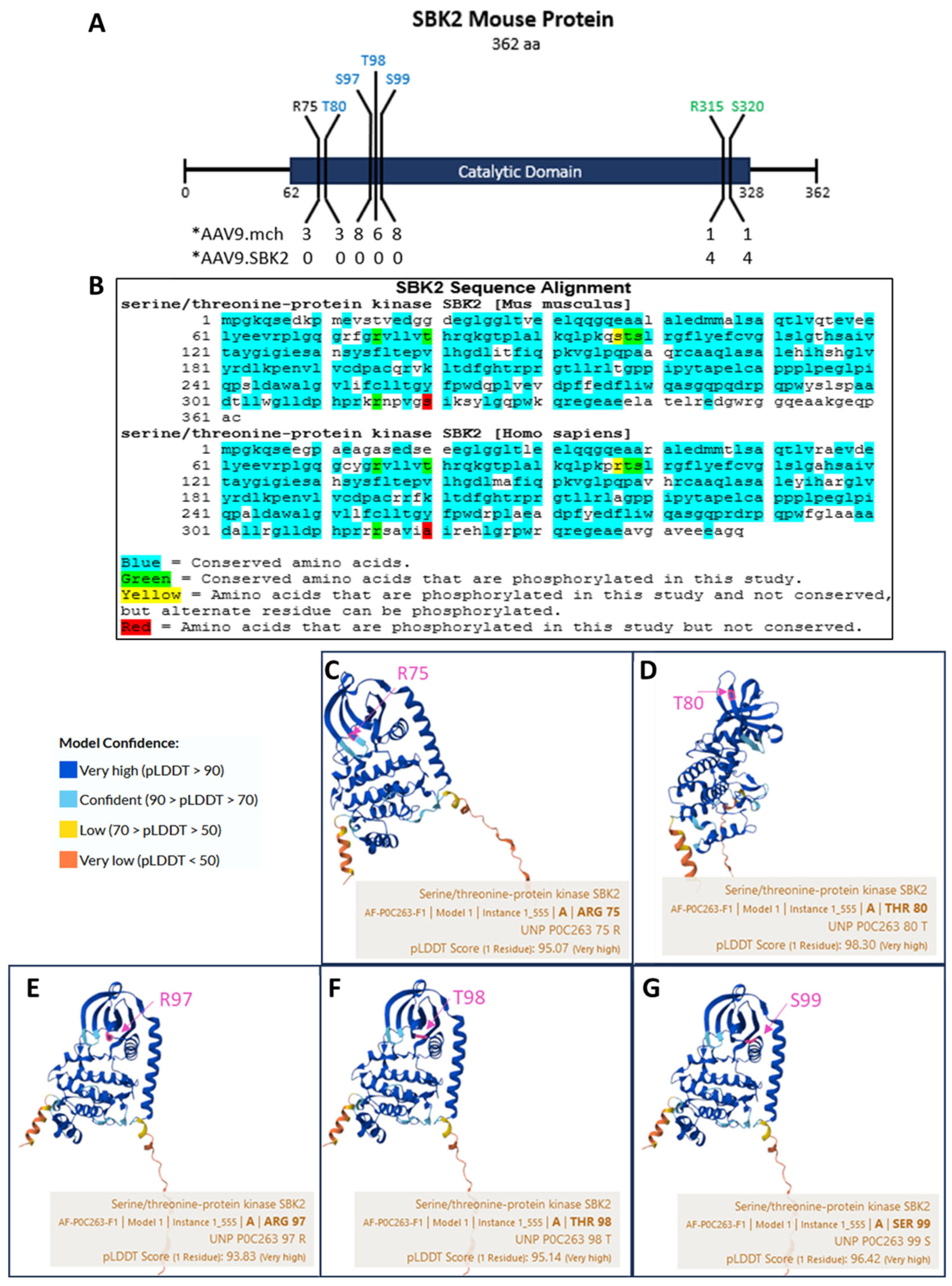
To identify SBK2 phosphorylation levels, we used the MASCOT program (see methods). The software returned peptides that are present in SBK2 and the amino acids in the respective peptides that were phosphorylated. We then aligned each peptide to the full SBK2 sequence and tallied each sample’s phosphorylation levels at each site (Table S1). SBK2 itself was phosphorylated at several sites: R75, T80, S97, T98, S99, R315, and S320, all within the catalytic domain (Figure 3A). The catalytic domain was defined by Kinexus Bioinformatics Corporation (RRID: SCR_012553). Interestingly, in the mice given AAV9.SBK2, only R315 and R320 at the C-terminal end of the catalytic site were phosphorylated. Both of these amino acids also had higher levels of phosphorylation than in AAV9.mch hearts. The differential phosphorylation at the 2 ends of the catalytic domain strongly suggests that phosphorylation plays a role in regulating SBK2.
Following this discovery, we used PROSITE to characterize the phosphorylation sites and found that the N-terminal sites are predicted to be protein kinase C (PKC) target sites, while the C-terminal sites are predicted to be cAMP- and cGMP-dependent kinase (PKA and PKG) phosphorylation sites (Figure 3A) [24]. To determine if these phosphorylation sites are conserved, we aligned the phosphorylated sites in the mouse sequence to the SBK2 sequence in humans (Figure 3B). Only one site, S320, was not conserved, and one (S97) had an alternative amino acid, arginine, which conserves the potential to be phosphorylated.
To further investigate whether the phosphorylation sites may have functional roles, we examined the human sequence in AlphaFold [25]. AlphaFold indicated 93.8–98.3% confidence in the structural position of the R75, T80, S97, T98, and S99 amino acids (Figure 3C–G). The close geometric location of these phospho-sites to the predicted binding sites on SBK2 (LGQGCYGRV68–76 and K91) indicates that phosphorylation of these sites likely plays a role in SBK2 interactions.
Although the majority of kinases are phosphorylated in the conserved activation segment, it does not appear to be a property of SBK2, as the activation segment starts with the DFG motif (amino acids 203–205) and ends with the APE motif (amino acids 225–227), which does not coincide with any of the phosphorylation sites we detected [26,27].

2.4. SBK2 R315 Phosphorylation Occurs on a Conserved Amino Acid of a Turn Motif

AlphaFold indicated that the R315 amino acid was located at a structurally significant site (Figure 4A). According to the bonding alignment, R315 was predicted to be an important structural component of the turn between two α-helices (Figure 4B). Two of the N-terminal amino groups of R315 were predicted to form four hydrogen bonds with Ala 243 and Glu 227 of the distal α-helix (Figure 4C). Phosphorylation of R315 would alter arginine’s positive charge, thereby disrupting these hydrogen bonds. As predicted, modeling the neutralization of arginine via the citrulline post-translational modification generated large changes. It resulted in folding the two disordered regions of SBK2 onto the main body of the protein (Figure 4D) (note: AlphaFold could not model arginine phosphorylation at R315 nor negatively charged aspartic acid at this site, so Citrullination was used to model neutralization of the positive charge) [28].
We next employed AlphaMissense, which can estimate pathogenicity of amino acid substitutions for a particular amino acid position by using related sequences that have been confirmed as pathogenic [29]. The R315 site returned 70–100% pathogenicity for any single amino acid change at this site (Figure 4E). These structural annotations and pathogenicity predictions indicate that the phosphorylation of R315 likely plays a critical role in SBK2’s function.

2.5. Modeling of SBK2 Binding to Phospho-Target Proteins

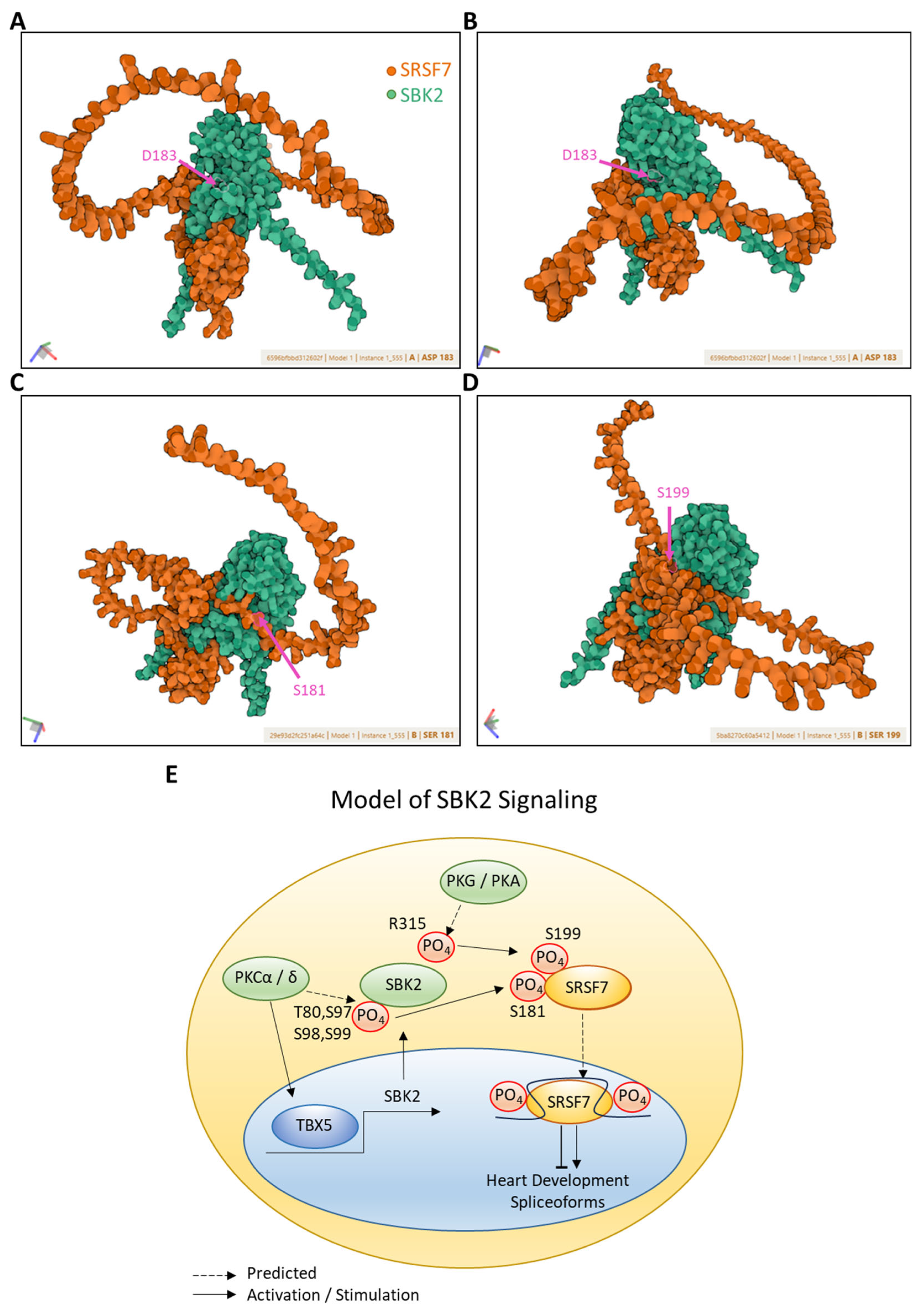
To model SBK2 and its possible partner proteins from our phospho-proteomics data we used AlphFold3 [28]. We modeled serine/arginine-rich splicing factor 7 (SRSF7) because it had increased phosphorylation under SBK2 overexpression and was previously shown to be significantly enriched in co-immunoprecipitation of SBK2 [1].
AlphaFold3 indicated that SRSF7 was predicted to bind SBK2 but not at the active site of SBK2 (Figure 5A,B). Critically, when we modeled phosphorylation via the neutralization of the positive charge at the SBK2 phospho-site R315 to 315CIR, SRSF7 remained at its primary SBK2 binding site, but secondary binding sites on the tail of SRSF7 became bound to the SBK2 active site; D183 (Figure 5C). In particular, two motifs on SRSF7’s tail (RSRSASPR and SGSIIGSR) were phosphorylated at several serines in our phospho-proteomics data and modeling R315CIR in SBK2 causes the SGSIIGSR motif of SRSF7 (which includes pS181) to bind near SBK2’s active site (Figure 5C).
Currently, there are no published structural analyses, such as CryoEM or X-ray diffraction, of SBK2. Therefore, R315’s predicted effect on SBK2 binding is only computational. However, AlphaFold3 has been shown to accurately predict protein complex structures and has been trained with structural models of phosphorylation from the Protein Data Bank [28].
When we modeled SBK2 phosphorylation at the C-terminal sites pT80, pS97, pT98, and pS99, a different SRSF7 tail motif, SISRPR, became oriented into SBK2’s active site (Figure 5D). This is important because S199 of the SISRPR motif in SRSF7 was phosphorylated in our phospho-proteomics data.
The SRSF7 phosphorylation sites that are elevated by SBK2 overexpression are all located in the tail region of SRSF7. This provides insight into the SBK2 regulatory mechanism of SRSF7 (previously known as 9G8), as the tail contains a carboxy-terminal arginine/serine-rich (RS) domain that can be differentially phosphorylated to regulate its shuttling between the cytoplasm and the nucleus or to regulate translation of SRSF7’s target mRNA [30].
To conclude, SBK2 regulates sarcomereogenesis during cardiomyocyte differentiation, while SRSF7 regulates the transition from a juvenile to adult-type transcriptome in certain cell types [1,31]. This, combined with our data, provides a likely mechanism by which SBK2 binds and phosphorylates SRSF7 to regulate splicing and promote differentiation/maturation of cardiomyocyte sarcomeres. In summary, SBK2 is predicted to phosphorylate different regions of SRSF7 depending on which region of SBK2 itself is phosphorylated.

3. Discussion

We have identified novel in vivo functions and possible mechanisms by which SBK2 functions and have identified likely factors both upstream and downstream of SBK2 (modeled in Figure 5E and clarified further below). First, SBK2 overexpression alters ventricular function and may attenuate hypertrophy in DRC mice. Second, SBK2 likely phosphorylates SRSF7 to regulate its RNA splicing function. Third, differential phosphorylation of SBK2 may control its substrate specificity.
SBK2 itself is critical for cardiomyocyte differentiation, which it likely achieves in part through its downstream phosphorylation of SRSF7. SRSF7 regulates the transition from a juvenile to adult-type transcriptome [31]. It regulates this transition by acting on RNA via multiple mechanisms. First, SRSF7 increases DROSHA binding of pri-miRNAs and, in turn, process them into miRNAs [32]. Second, SRSF7 interacts with FIP1 to promote splicing of 3′UTRs [33].
Upstream of SBK2, the pT80, pS97, pT98, and pS99 phosphorylation sites that we identified on the N-terminal end of SBK2’s kinase domain are predicted to be PKC targets. PKC targeting is significant because it has been shown that overexpression of PKCα and δ isoforms leads to cardiomyocyte differentiation and increased expression of TBX5 [34]. The transcription factor Tbx5, in turn, is a transcriptional activator of genes involved with cardiomyocyte maturation, and Tbx5 cardiac precursor cells in the heart are being investigated for regeneration of the heart following injury [35,36]. This is particularly relevant because a recent report emphasized that ATAC-seq revealed one of the genes most strongly regulated by Tbx5 is Sbk2 [37]. Further investigation of the various PKC isoform’s effect on SBK2 will help to clarify the precise regulatory mechanisms.
Alternatively, the R315 phosphorylation site on the N-terminal end of SBK2’s kinase domain is predicted to be a PKA- or PKG-dependent kinase target. Currently, there are no published structural analyses, such as CryoEM or X-ray diffraction, of SBK2. Therefore, R315’s predicted effect on SBK2 binding is only computational. However, AlphaFold3 has been shown to accurately predict protein complex structures and has been trained with structural models of phosphorylation from the Protein Data Bank [28]. We show that this phosphorylation is the likely signal for SBK2 to phosphorylate alternative domains on the tail of SRSF7. The differential tail phosphorylation of SRSF7 could provide activation signals for SRSF7 nuclear shuttling or mRNA splicing, which may be how SBK2 regulates cardiomyocyte differentiation/maturation.
The Missense data indicates that R315 is almost certain to result in disease, and due to SBK2 being required for cardiac development, it is likely that generating an R315 mutant would be lethal in the fetus [1]. This, combined with the observation that R315 was phosphorylated in both AAV9.mch and AAV9.SBK2, means that identifying the upstream signaling that promotes R315 phosphorylation of SBK2 will be a critical step in the study of SRSF7 phosphorylation. The manipulation of upstream signaling will allow the generation of a control group in future SRSF7 studies.
The specific phosphorylation sites we identified now enable targeted investigation of how upstream PKC, PKG, and PKA pathways influence SBK2 activity. Furthermore, determining SRSF7’s splicing targets in the heart when differentially phosphorylated by SBK2 will contribute to a more complete understanding of regeneration of the heart following injury and open possible regenerative therapeutic avenues. Combined, these findings suggest a new regulatory axis (PKC → TBX5→ SBK2 → SRSF7) critical to cardiomyocyte maturation.

4. Materials and Methods

4.1. Animals

FVB/NJ (FVB or NTG) (Jackson Laboratories Strain #001800) females were crossed with heterozygous males containing R120G (line 134) [5]. Resultant pups were injected on postnatal day zero. Experimental animals were maintained and housed in the same cages with control siblings. The protocols for animal care and experimentation used in this study were approved by the University of South Dakota Institutional Animal Use and Care Committee. A pilot study of one cohort of R120G mice (28 mice) was examined after one month to generate a final power analysis for the primary study. No criteria were set for excluding animals, and no exclusions were needed.

4.2. RT-qPCR

RNA was extracted using tri-reagent (Invitrogen AM9738, Carlsbad, CA, USA), chloroform, and isopropanol and washed in RNase-free ethanol. DNase treatment (Promega M6101, Madison, WI, USA) was performed following manufacturer protocol. RNA was quantified using a Nanodrop 2000 Spectrophotometer (Thermo Fisher Scientific, Waltham, MA, USA). cDNA was synthesized using the High Capacity cDNA Reverse Transcription Kit (Applied Biosystems #4308228, Foster City, CA, USA) (Thermo Fisher Scientific, Waltham, USA) on the Bio-Rad T100 thermal cycler (Bio-Rad, Hercules, CA, USA). qPCR was performed using PowerUp SYBR Green Master Mix (Thermo Fisher# A25742) (Thermo Fisher Scientific, Waltham, USA) on Applied Biosystems QuantStudio 5 (Thermo Fisher Scientific, Waltham, USA) following manufacturer protocols. Primer sequences for SBK2 and GAPDH can be found in Table S2. SBK2 relative expression was calculated using the ΔΔCT method.

4.3. Western Blotting

Tissues were bead blended (Next Advance Bullet Blender Storm Pro) (Next Advance Inc., Troy, MI, USA) on high for 5 min in 1× lysis buffer (0.5 mL 3 M Tris HCl pH 6.8, 4.5 mL 10% SDS, 3.0 mL glycerol, and 28.5 mL H2O) containing protease inhibitor cocktail (Boster Bio AR1182-1) (Boster BIO, Pleasanton, CA, USA). Samples were boiled for 5 min and then centrifuged at 14,000 rpm for 20 min. Proteins were quantified using BCA. Gels were run at 110 V and then wet-transferred at 250 mAmps for 140 min at 4 degrees. Membranes were blocked in 5% BSA in 1× TBST for 1 h, then incubated with primary antibodies, α-SBK2 1:500 (Atlas Antibodies HPA030631) (Atlas Antibodies, Stockholm, Sweden), α-CRYAB 1:1000 (ENZO ADI-SPA-223-F) (Enzo Life Sciences, Farmingdale, NJ, USA), and α-DES 1:2000 (Abcam ab15200) (Abcam, Cambridge, UK) overnight. Membranes were then washed 3 times in TBS, incubated in chemiluminescent substrate, and imaged on Bio-Rad Chemidoc MP (Bio-Rad, Hercules, USA). Membranes were normalized to whole protein, and relative abundance was calculated using Bio-Rad Image Lab Version 6.1 Software.

4.4. AAV Generation and Injection

The SBK2 gene (Genscript Clone ID OMu07315) was cloned into the pP2A-mCherry-N1 (Addgene Plasmid #84329) (Addgene, Watertown, NY, USA), and the sequence was verified by Genscript. The AAV9.cTNT.SBK2.P2A.mcherry or AAV9.cTNT.P2A.mcherry plasmids were packaged, purified, and resulting viruses were tittered by Charles River Labs. All injections were performed in Day 0 pups intraperitoneally with AAV9.cTNT.SBK2.P2A.mcherry or AAV9.cTNT.P2A.mcherry viruses at a concentration of 1 × 10¹⁰ genome copies/pup in 0.9% saline solution. Viral expression was validated via SBK2 Western blotting (Figure S1). Because their genotype was unknown at the time of injection, this ensured randomization, and further blinding was not used.

4.5. Echocardiography

Mice were anesthetized via inhalation of isoflurane at 4% for ~60 s and adjusted to 1.0% for measurements. Transthoracic echocardiography was performed using the VisualSonics Vevo 3100 system (FUJIFILM VisualSonics, Toronto, ON, Canada) and a 40 MHz probe (FUJIFILM VisualSonics, Toronto, Canada). Short axis, M-mode, two-dimensional echocardiograms were acquired through the left ventricular (LV) anterior and posterior walls. Graphpad Prism version 6 software was used for statistical analysis.

4.6. Mass Spectrometry

4.6.1. Protein Isolation for Mass Spectrometry Analysis

Twelve hearts, 3 males and 3 females per group, were injected with AAV9.cTNT.SBK2.P2A.mcherry or AAV9.cTNT.P2A.mcherry control virus at day 0. After 14 days, hearts were removed into TBS containing 1× protease and Phosphatase inhibitor cocktail solution (EDTA free) (Hello Bio HB9105) (Hello Bio, Princeton, NJ, USA) on ice and then snap frozen within ~20 s. Lysis buffer (4% SDS, 0.1 M Tris-HCL, pH 8.0 with 1× protease and phosphatase inhibitor cocktail) was added, then samples underwent bead blender (Next Advance Bullet Blender Storm Pro) (Next Advance Inc., Troy, USA) homogenization 2 times for 5 min on high speed. Samples were centrifuged at 14,000× g for 15 min, supernatant transferred to new low-binding tube. Further sonication on ice was performed for 10 s 3 times. Samples were boiled for 5 min. Centrifuged at 14,000× g for 15 min and supernatant transferred to new low-binding tube. Protein was quantified using the bicinchoninic acid assay (BCA). Aliquots for Western blotting were used to validate successful AAV transfection.

4.6.2. Protein Identification by Mass Spectrometry Analysis

The proteins were in-solution digested following this protocol: (a) the buffer was adjusted to meet the salt concentration as well the pH required for the protease (Trypsin) (Promega, Madison, WI, USA) used for in-solution digestion; (b) the reduction and alkylation were performed using 1/10 v/v 50 mM DTT (Sigma-Aldrich, Saint Louis, MO, USA) at 65 °C for 5 min, followed by 1/10 v/v 100 mM Iodoacetamide (Sigma-Aldrich, Saint Louis, MO) at room temperature in dark condition for 30 min; (c) Trypsin sequencing grade (Promega, Madison, WI) was used for the in solution digestion at 37 °C overnight; (d) the reaction was stopped by changing the pH to 4 using TFA (Sigma-Aldrich, Saint Louis, MO) at a final concentration of 0.5%; (e) finally, the tryptic digested peptides were concentrated at a centrifuge concentrator (SpeedVac, Thermo Scientific) (Thermo Fisher Scientific, Waltham, USA). Peptides were desalted using Peptide Desalting Spin Columns (Thermo Scientific 89852) (Thermo Fisher Scientific, Waltham, USA) and quantified using the Quantitative Fluorometric Peptide Assay (Pierce 23290) (Thermo Fisher Scientific, Waltham, USA). Phosphopeptide enrichment was performed sequentially using the High-Select TiO2 Phosphopeptide Enrichment Kit (A32993, ThermoFisher) (Thermo Fisher Scientific, Waltham, USA) and the High-Select Fe-NTA Phosphopeptide Enrichment Kit (A32992, ThermoFisher) (Thermo Fisher Scientific, Waltham, USA). Five percent of total peptides before pre-enrichment was reserved for proteome analysis. The peptides were in-line desalted using an Easy nLC 1200 nanoUHPLC (Thermo Fisher Scientific, Waltham, USA) through a trapping and desalting online trap column (300 μm × 20 mm Acclaim PepMap C18 100 Å (Thermo Fisher Scientific, Waltham, USA) and separated by an Easy-Spray PepMap RSLC 2 μm, 75 μm × 15 cm, nanoViper column (Thermo Fisher Scientific, Waltham, USA) coupled to nanoESI orbitrap Exploris 240 HR/MA mass (Thermo Fisher Scientific, Waltham, USA). The chromatography conditions were as follows: 0–1 min, 1% B isocratic; 2–60 min, 1–25%; 61–90 min 25–50%; and 92–101 min 50–100% B linear. Mobile Phase A (water/formic acid, 99.9:0.1% v/v) and Phase B (water/acetonitrile/formic acid, 20/80/0.1% v/v). The solvent flow rate was 300 nL per min. The orbitrap Exploris 240 instrument (Thermo Fisher Scientific, Waltham, USA) was operated under data-dependent mode acquisition (DDA) top 12 modes to automatically switch between full scan MS and MS/MS acquisition. The ions were analyzed in positive MS ion mode (m/z 375–1500) with 120,000 resolution (m/z 200) after accumulation with target ions to 1 × 10⁶ value based on predictive AGC. The MS/MS ion selection was set at m/z 65–1800 to 1 × 10⁵ counts with 30,000 resolutions. The spectrum was deconvoluted and analyzed using the Mascot Distiller v2.6 (Matrix Science, London, UK) and Proteome Discoverer v2.5 (Thermo Fisher Scientific, Waltham, USA). A mascot generic format list (MGF format) was generated to identify +1 or multiple charged precursor ions from the MS data file. Mascot server v2.8.1 6 (Matrix Science, London, UK) in MS/MS ion search mode (local licenses) was applied to conduct peptide matches (peptide masses and sequence tags) and protein searches against SwissProt Mus musculus 092022 (17,562 sequences; 9,985,721 residues) (Proteome ID UP000000589) database (refence sequence 2009 version, UniProt consortium, Geneva, Switzerland). The following parameters were set for the search: carbamidomethyl (C) on cysteine was fixed, and variable modifications included Phosphor Ser (S), Phospho Arg (R), Phospho Thr (T), asparagine and glutamine deamidation, and methionine oxidation. Two missed cleavages were allowed; monoisotopic masses were counted; the precursor peptide and fragment mass tolerance were set at 15 ppm and 0.02 Da, respectively; and the ion score or expected cut-off was set at 5. The MS/MS spectra were searched with MASCOT using a 95% confidence interval (% C.I.) threshold (p < 0.05), with which a minimum score of 37 was used for peptide identification, and an FDR < 5% was considered. The protein redundancy at the database under different accession numbers was limited to Mus musculus. All the proteins identified in the current study were found in these domains. The comparative analysis was performed using ProteoIQ v2.8 (local license).

Supplementary Materials

The following supporting information can be downloaded at: https://www.mdpi.com/article/10.3390/kinasesphosphatases3040020/s1. Figure S1. AAV verification; Table S1. Differentially Expressed Proteins and pathways from Mass Spectrometry; Table S2. qPCR primers.

Author Contributions

Conceptualization, formal analysis, investigation, and writing—original draft preparation, M.B.; methodology and software, M.B. and E.C.; resources and data curation, D.P. and E.C.; resources, supervision, and writing—review and editing, X.W.; funding acquisition, M.B. and X.W. All authors have read and agreed to the published version of the manuscript.

Funding

This research was funded by THE AMERICAN HEART ASSOCIATION grant numbers AHA 20TPA35490091 and AHA 23POST1018909 and US NATIONAL INSTITUTES OF HEALTH grants R01HL072166, R01HL153614, and P20GM103443.

Institutional Review Board Statement

The protocols for animal care and experimentation used in this study were approved by University of South Dakota Institutional Animal Use and Care Committee on 1/12/23, protocol# 13-11-22-25C.

Data Availability Statement

The proteomics data sets presented in this study are openly available at MassIVE UCSD (MassIVE MSV000098733) (https://massive.ucsd.edu) (accessed on 5 August 2025).

Acknowledgments

The authors would like to thank the University of South Dakota Proteomics Core, South Dakota Biomedical Research Infrastructure Network (SD BRIN), as well as Messrs. Ryan Johnson and Bill Conn from USD-IT Research Computing for their help in the databases installation and server operation and maintenance as well as the people from University of South Dakota IT Research Computing.

Conflicts of Interest

The authors declare no conflicts of interest.

Abbreviations

The following abbreviations are used in this manuscript:
AAV9.SBK2AAV9.cTNT.SBK2.P2A.mcherry
AAV9.mchAAV9.cTNT.P2A.mcherry
CRYABCrystallin Alpha B
DRCDesmin-Related Cardiomyopathy
R120GCrystallin Alpha B R120G mutation
RABX5/RABGEF1Rab5 GDP/GTP exchange factor
SBK2SH3 Domain Binding Kinase Family Member 2
SRSF7Serine/arginine-rich splicing factor 7

References

  1. van Gorp, P.R.; Zhang, J.; Liu, J.; Tsonaka, R.; Mei, H.; Dekker, S.O.; Bart, C.I.; De Coster, T.; Post, H.; Heck, A.J.; et al. Sbk2, a newly discovered atrium-enriched regulator of sarcomere integrity. Circ. Res. 2022, 131, 24–41. [Google Scholar] [CrossRef]
  2. McLendon, P.M.; Robbins, J. Desmin-related cardiomyopathy: An unfolding story. Am. J. Physiol.-Heart Circ. Physiol. 2011, 301, H1220–H1228. [Google Scholar] [CrossRef] [PubMed]
  3. Wang, X.; Osinska, H.; Dorn, G.W.; Nieman, M.; Lorenz, J.N.; Gerdes, A.M.; Witt, S.; Kimball, T.; Gulick, J.; Robbins, J. Mouse model of desmin-related cardiomyopathy. Circulation 2001, 103, 2402–2407. [Google Scholar] [CrossRef] [PubMed]
  4. Goldfarb, L.G.; Dalakas, M.C. Tragedy in a heartbeat: Malfunctioning desmin causes skeletal and cardiac muscle disease. J. Clin. Investig. 2009, 119, 1806–1813. [Google Scholar] [CrossRef] [PubMed]
  5. Wang, X.; Osinska, H.; Klevitsky, R.; Gerdes, A.M.; Nieman, M.; Lorenz, J.; Hewett, T.; Robbins, J. Expression of R120G–αB-crystallin causes aberrant desmin and αB-crystallin aggregation and cardiomyopathy in mice. Circ. Res. 2001, 89, 84–91. [Google Scholar] [CrossRef]
  6. Jordan, E.; Peterson, L.; Ai, T.; Asatryan, B.; Bronicki, L.; Brown, E.; Celeghin, R.; Edwards, M.; Fan, J.; Ingles, J. Evidence-based assessment of genes in dilated cardiomyopathy. Circulation 2021, 144, 7–19. [Google Scholar] [CrossRef]
  7. Bermudez-Jimenez, F.J.; Protonotarios, A.; García-Hernández, S.; Pérez Asensio, A.; Rampazzo, A.; Zorio, E.; Brodehl, A.; Arias, M.A.; Macías-Ruiz, R.; Fernández-Armenta, J.; et al. Phenotype and clinical outcomes in desmin-related arrhythmogenic cardiomyopathy. Clin. Electrophysiol. 2024, 10, 1178–1190. [Google Scholar] [CrossRef]
  8. Vicart, P. On noxious desmin: Functional effects of a novel heterozygous desmin insertion mutation on the extrasarcomeric desmin cytoskeleton and mitochondria. Hum. Mol. Genet. 2003, 12, 657–669. [Google Scholar] [CrossRef]
  9. Maloyan, A.; Sanbe, A.; Osinska, H.; Westfall, M.; Robinson, D.; Imahashi, K.I.; Murphy, E.; Robbins, J. Mitochondrial dysfunction and apoptosis underlie the pathogenic process in α-B-crystallin desmin-related cardiomyopathy. Circulation 2005, 112, 3451–3461. [Google Scholar] [CrossRef]
  10. Oka, H.; Nakau, K.; Imanishi, R.; Furukawa, T.; Tanabe, Y.; Hirono, K.; Hata, Y.; Nishida, N.; Azuma, H. A case report of a rare heterozygous variant in the desmin gene associated with hypertrophic cardiomyopathy and complete atrioventricular block. CJC Open 2021, 3, 1195–1198. [Google Scholar] [CrossRef]
  11. Tannous, P.; Zhu, H.; Johnstone, J.L.; Shelton, J.M.; Rajasekaran, N.S.; Benjamin, I.J.; Nguyen, L.; Gerard, R.D.; Levine, B.; Rothermel, B.A.; et al. Autophagy is an adaptive response in desmin-related cardiomyopathy. Proc. Natl. Acad. Sci. USA 2008, 105, 9745–9750. [Google Scholar] [CrossRef]
  12. Li, J.; Horak, K.M.; Su, H.; Sanbe, A.; Robbins, J.; Wang, X. Enhancement of proteasomal function protects against cardiac proteinopathy and ischemia/reperfusion injury in mice. J. Clin. Investig. 2011, 121, 3689–3700. [Google Scholar] [CrossRef]
  13. Zheng, H.; Tang, M.; Zheng, Q.; Kumarapeli, A.R.K.; Horak, K.M.; Tian, Z.; Wang, X. Doxycycline attenuates protein aggregation in cardiomyocytes and improves survival of a mouse model of cardiac proteinopathy. J. Am. Coll. Cardiol. 2010, 56, 1418–1426. [Google Scholar] [CrossRef] [PubMed]
  14. Cai, M.; Pan, B.; Xiao, P.; Bouska, M.; Lewno, M.T.; Xing, Y.; Zeng, E.; Liang, H.; Li, F.; Gao, X. Sustained but Decoyed Activation of the JAK1-STAT Pathway by Aberrant Protein Aggregation Exacerbates Proteotoxicity. Circulation 2024, 150, 1302–1305, Erratum in Circulation 2025, 152, e47. [Google Scholar] [CrossRef]
  15. Yang, L.; Parajuli, N.; Wu, P.; Liu, J.; Wang, X. S14-phosphorylated RPN6 mediates proteasome activation by PKA and alleviates proteinopathy. Circ. Res. 2023, 133, 572–587. [Google Scholar] [CrossRef]
  16. Lindsey, M.L.; Kassiri, Z.; Virag, J.A.; de Castro Brás, L.E.; Scherrer-Crosbie, M. Guidelines for measuring cardiac physiology in mice. Am. J. Physiol. Heart Circ. Physiol. 2018, 314, H733–H752. [Google Scholar] [CrossRef]
  17. Bish, L.T.; Morine, K.; Sleeper, M.M.; Sanmiguel, J.; Wu, D.; Gao, G.; Wilson, J.M.; Sweeney, H.L. Adeno-associated virus (AAV) serotype 9 provides global cardiac gene transfer superior to AAV1, AAV6, AAV7, and AAV8 in the mouse and rat. Hum. Gene Ther. 2008, 19, 1359–1368. [Google Scholar] [CrossRef]
  18. Prasad, K.M.; Xu, Y.; Yang, Z.; Acton, S.T.; French, B.A. Robust cardiomyocyte-specific gene expression following systemic injection of AAV: In vivo gene delivery follows a Poisson distribution. Gene Ther. 2011, 18, 43–52. [Google Scholar] [CrossRef]
  19. Donnelly, M.L.; Luke, G.; Mehrotra, A.; Li, X.; Hughes, L.E.; Gani, D.; Ryan, M.D. Analysis of the aphthovirus 2A/2B polyprotein ‘cleavage’mechanism indicates not a proteolytic reaction, but a novel translational effect: A putative ribosomal ‘skip’. J. Gen. Virol. 2001, 82, 1013–1025. [Google Scholar] [CrossRef]
  20. Liu, R. Complex functionality of protein phosphatase 1 isoforms in the heart. Cell. Signal. 2021, 85, 110059. [Google Scholar] [CrossRef]
  21. Sun, X.; Tang, X.; Qiu, H. Cardiac-Specific Suppression of Valosin-Containing Protein Induces Progressive Heart Failure and Premature Mortality Correlating with Temporal Dysregulations in mTOR Complex 2 and Protein Phosphatase 1. Int. J. Mol. Sci. 2024, 25, 6445. [Google Scholar] [CrossRef]
  22. Yamano, K.; Wang, C.; Sarraf, S.A.; Münch, C.; Kikuchi, R.; Noda, N.N.; Hizukuri, Y.; Kanemaki, M.T.; Harper, W.; Tanaka, K.; et al. Endosomal Rab cycles regulate Parkin-mediated mitophagy. Elife 2018, 7, e31326. [Google Scholar] [CrossRef]
  23. Szklarczyk, D.; Kirsch, R.; Koutrouli, M.; Nastou, K.; Mehryary, F.; Hachilif, R.; Gable, A.L.; Fang, T.; Doncheva, N.T.; Pyysalo, S. The STRING database in 2023: Protein–protein association networks and functional enrichment analyses for any sequenced genome of interest. Nucleic Acids Res. 2023, 51, D638–D646. [Google Scholar] [CrossRef]
  24. Sigrist, C.J.; Cerutti, L.; De Castro, E.; Langendijk-Genevaux, P.S.; Bulliard, V.; Bairoch, A.; Hulo, N. PROSITE, a protein domain database for functional characterization and annotation. Nucleic Acids Res. 2010, 38 (Suppl. S1), D161–D166. [Google Scholar] [CrossRef] [PubMed]
  25. Jumper, J.; Evans, R.; Pritzel, A.; Green, T.; Figurnov, M.; Ronneberger, O.; Tunyasuvunakool, K.; Bates, R.; Žídek, A.; Potapenko, A.; et al. Highly accurate protein structure prediction with AlphaFold. Nature 2021, 596, 583–589. [Google Scholar] [CrossRef] [PubMed]
  26. Johnson, L.N.; Noble, M.E.; Owen, D.J. Active and inactive protein kinases: Structural basis for regulation. Cell 1996, 85, 149–158. [Google Scholar] [CrossRef] [PubMed]
  27. Gógl, G.; Kornev, A.P.; Reményi, A.; Taylor, S.S. Disordered protein kinase regions in regulation of kinase domain cores. Trends Biochem. Sci. 2019, 44, 300–311. [Google Scholar] [CrossRef]
  28. Abramson, J.; Adler, J.; Dunger, J.; Evans, R.; Green, T.; Pritzel, A.; Ronneberger, O.; Willmore, L.; Ballard, A.J.; Bambrick, J.; et al. Accurate structure prediction of biomolecular interactions with AlphaFold 3. Nature 2024, 630, 493–500. [Google Scholar] [CrossRef]
  29. Cheng, J.; Novati, G.; Pan, J.; Bycroft, C.; Žemgulytė, A.; Applebaum, T.; Pritzel, A.; Wong, L.H.; Zielinski, M.; Sargeant, T.; et al. Accurate proteome-wide missense variant effect prediction with AlphaMissense. Science 2023, 381, eadg7492. [Google Scholar] [CrossRef]
  30. Cáceres, J.F.; Screaton, G.R.; Krainer, A.R. A specific subset of SR proteins shuttles continuously between the nucleus and the cytoplasm. Genes Dev. 1998, 12, 55–66. [Google Scholar] [CrossRef]
  31. Kadota, Y.; Jam, F.A.; Yukiue, H.; Terakado, I.; Morimune, T.; Tano, A.; Tanaka, Y.; Akahane, S.; Fukumura, M.; Tooyama, I. Srsf7 establishes the juvenile transcriptome through age-dependent alternative splicing in mice. iScience 2020, 23, 100929. [Google Scholar] [CrossRef]
  32. Le, M.N.; Nguyen, T.D.; Nguyen, T.A. SRSF7 and SRSF3 depend on RNA sequencing motifs and secondary structures to regulate Microprocessor. Life Sci. Alliance 2023, 6, e202201779. [Google Scholar] [CrossRef] [PubMed]
  33. Schwich, O.D.; Blümel, N.; Keller, M.; Wegener, M.; Setty, S.T.; Brunstein, M.E.; Poser, I.; Mozos, I.R.D.L.; Suess, B.; Münch, C.; et al. SRSF3 and SRSF7 modulate 3′ UTR length through suppression or activation of proximal polyadenylation sites and regulation of CFIm levels. Genome Biol. 2021, 22, 82. [Google Scholar] [CrossRef] [PubMed]
  34. Lee, K.E.; Choi, D.H.; Kim, W.H.; Park, Y.S. Up-regulation of PKCα and δ during beating cardiomyocyte differentiation of P19CL6 cells with suppressed apoptotic cell populations. Mol. Cell. Toxicol. 2024, 20, 167–176. [Google Scholar] [CrossRef]
  35. Steimle, J.D.; Moskowitz, I.P. TBX5: A key regulator of heart development. Curr. Top. Dev. Biol. 2017, 122, 195–221. [Google Scholar] [PubMed]
  36. Siatra, P.; Vatsellas, G.; Chatzianastasiou, A.; Balafas, E.; Manolakou, T.; Papapetropoulos, A.; Agapaki, A.; Mouchtouri, E.T.; Ruchaya, P.J.; Korovesi, A.G.; et al. Return of the Tbx5; lineage-tracing reveals ventricular cardiomyocyte-like precursors in the injured adult mammalian heart. NPJ Regen. Med. 2023, 8, 13. [Google Scholar] [CrossRef]
  37. Sweat, M.E.; Cao, Y.; Zhang, X.; Burnicka-Turek, O.; Perez-Cervantes, C.; Kulandaisamy, A.; Lu, F.; Keating, E.M.; Akerberg, B.N.; Ma, Q.; et al. Tbx5 maintains atrial identity in postnatal cardiomyocytes by regulating an atrial-specific enhancer network. Nat. Cardiovasc. Res. 2023, 2, 881–898, Correction in Nat. Cardiovasc. Res. 2023, 2, 1095. [Google Scholar] [CrossRef]
Figure 1. SBK2 overexpression impacts ventricular structure and function. (A) SBK2 construct used for generating AAV9.cTNT.SBK2.P2A.mcherry viruses; AAV9.cTNT.P2A.mcherry control viruses contained the same sequence except for the SBK2 sequence. (BK) Heart rate, stroke volume, cardiac output, ejection fraction, and diastolic left ventricular posterior wall thickness from 1- and 3-month-old mice hearts injected with either AAV9.cTNT.SBK2.P2A.mcherry or AAV9.cTNT.P2A.mcherry control virus. (BF) One-month echocardiography measurements. (GK) Three-month echocardiography measurements. Abbreviations and formulas: Beats Per Minute (BPM); d = diastolic; s = systolic; LV = left ventricular; Vol = volume; PW = posterior wall. Stroke Volume = (Diastolic − systolic volume); Ejection fraction = (LV Vol;d − LV Vol;s/LV Vol;d); Fractional Shortening = (Avg. Diastolic diameter − Avg. Systolic diameter)/Avg. Diastolic diameter. Two-way Anova Tukey’s multiple comparisons test * p ≤ 0.05; ** p ≤ 0.01; *** p ≤ 0.001 **** p ≤ 0.0001; (n ≥ 9).
Kinasesphosphatases 03 00020 g001
Figure 3. SBK2 phosphosites. (A) Seven amino acid (aa) phospho-sites detected by phospho-ms mapped to the catalytic domain of SBK2. Blue amino acids are predicted to be protein kinase C phospho-targets, while green are predicted to be cAMP- and cGMP-dependent kinase targets. Numbers below represent the number of mass-spec hits for SBK2-OE vs. mcherry-OE control. * = The number of samples phosphorylated at site in AAV9.mch vs AAV9.SBK2 hearts. (B) Sequences of SBK2 in mouse vs. human. Green-highlighted amino acids are conserved amino acids that were phosphorylated in mice. (CG) Backbone and Ribbon Diagrams of N-terminal phospho-site locations on SBK2.
Kinasesphosphatases 03 00020 g003
Figure 4. Conserved R315 phospho-site in SBK2 predicted to affect SBK2 function. (A) Location of the R315 phospho-site in SBK2. (B) R315 is a component of a loop between two alpha helices. (C) Hydrogen bonding (blue dotted lines) that form between R315 and residues in the two helices. (D) Neutralization of R315 via Citrullination is predicted to alter the orientation of the two tail regions of SBK2. (E) Alpha Missense Pathogenicity Heatmap indicates that any mutation of the R315 amino acid in SBK2 has a high likelihood of resulting in disease [29]. Confidence measures by AlphaFold’s predicted Local Distance Difference Test (pLDDT).
Kinasesphosphatases 03 00020 g004
Figure 5. Phosphorylation at SBK2’s conserved sites is predicted to influence which secondary binding sites on SRSF7’s tail are directed into SBK2’s active site. (A,B) Space-filling diagrams of SRSF7 (Orange) binding to unphosphorylated SBK2 (turquoise). Two views are shown to demonstrate that the SBK2 active site D183 (pink) remains open. (C) Mutation of the SBK2 phosphorylation site R315 to 315CIR (PKA/PKG targeted phosphorylation site) resulted in the tail of SRSF7 binding to the SBK2 active site. Critically, Ser181 (pink) of SRSF7 was near the active site of SBK2 when R315 was neutralized. Ser181 was phosphorylated in our phospho-MS data. (D) When the PKC phosphorylation sites pT80, pS97, pT98, and pS99 are phosphorylated on SBK2, Ser199 of SRSF7 (pink) is near the active site of SBK2 (D183). Ser199 was phosphorylated in our phospho-MS data. All models had a predicted template modeling (pTM) score above 0.5 indicating overall the complex structure might be similar to the true structure. (E) Hypothetical model of SBK2 pathway.
Kinasesphosphatases 03 00020 g005
Disclaimer/Publisher’s Note: The statements, opinions and data contained in all publications are solely those of the individual author(s) and contributor(s) and not of MDPI and/or the editor(s). MDPI and/or the editor(s) disclaim responsibility for any injury to people or property resulting from any ideas, methods, instructions or products referred to in the content.