Review

Digital Twin-Based Multiscale Models for Biomarker Discovery in Kinase and Phosphatase Tumorigenic Processes


by
Sara Sadat Aghamiri
1 and
Rada Amin
2,*
1
Decision Neuroscience Laboratory, Center for Brain, Biology, and Behavior, University of Nebraska, Lincoln, NE 68503, USA
2
Department of Biochemistry, University of Nebraska, Lincoln, NE 68503, USA
*
Author to whom correspondence should be addressed.
Kinases Phosphatases 2025, 3(3), 18; https://doi.org/10.3390/kinasesphosphatases3030018
Submission received: 30 July 2025 / Revised: 28 August 2025 / Accepted: 29 August 2025 / Published: 31 August 2025

Abstract

Digital twin is a mathematical model that virtually represents a physical object or process and predicts its behavior at future time points. These simulation models enable a deeper understanding of tumorigenic processes and improve biomarker discovery in cancer research. Tumor microenvironment is marked by dysregulated signaling pathways, where kinases and phosphatases serve as critical regulators and promising sources for biomarker discovery. These enzymes operate within multiscale and context-dependent processes where spatial and temporal coordination determine cellular outcomes. Digital Twin technology provides a platform for multimodal and multiscale modeling of kinase and phosphatase processes at the patient-specific level. These models have the potential to transform biomarker validation processes, enhance the prediction of therapeutic responses, and support precision decision-making. In this review, we present the major alterations affecting kinases and phosphatase functions within the tumor microenvironment and their clinical relevance as biomarkers, and we address how digital twins in oncology can augment and refine each stage of the biomarker discovery pipeline. Introducing this emerging technology for cancer biomarker discovery will assist in accelerating its adoption and translation into precision diagnostics and targeted therapies.

1. Introduction

Cancer biomarker discovery plays a critical role in precision oncology, supporting early detection, prognosis, therapeutic monitoring, and patient stratification. Yet, this process is often challenged by the inherent complexity of tumor microenvironment (TME), including intratumoral heterogeneity, resistant clones, dynamic molecular states, and context-dependent signaling [1]. Protein phosphorylation is a fundamental signaling mechanism in tumor biology, regulating key processes such as proliferation, differentiation, and metastasis, driving cancer initiation, and progression. This reversible modification is governed by two opposing enzyme families: kinases, which catalyze the addition of phosphate groups, and phosphatases, which remove them. Together, they maintain cellular homeostasis through intricate, tightly controlled signaling networks [2]. However, in cancer, these molecules are often deregulated, often due to genetic, epigenetic, post-translational modifications (PTMs), or autocrine loops, thereby driving uncontrolled growth, treatment resistance, and tumor escape [3,4].
Kinases, which number over 500 in the human genome, have been extensively studied as oncogenic drivers and drug targets, leading to the development of over 80 small-molecule kinase inhibitors approved for clinical use [5]. Phosphatases, in contrast, have historically received less attention, despite increasing evidence supporting their dual roles as tumor suppressors or oncogenes depending on cellular context [6]. The dual importance of kinases and phosphatases extends beyond therapy; they also serve as valuable biomarkers for disease diagnosis, prognosis, and response prediction. Importantly, both kinase and phosphatase activities are highly dynamic, non-linear, and regulated by additional cues such as subcellular localization, feedback regulation, PTMs and protein–protein interactions [7,8]. The complexity of these enzymes is further amplified by their activity across multiple biological scales, ranging from molecular interactions to cellular signaling pathways and tissue-level responses. Due to these characteristics, the identification and validation of clinically relevant kinase and phosphatase biomarkers remain challenging. In recent years, computational methods such as artificial intelligence (AI) algorithms including machine learning (ML), deep learning (DL), generative AI, and foundation models, have played increasingly significant roles in various stages of medical diagnosis, prognosis, and treatment planning [9]. While AI models and other data-driven algorithms have demonstrated promise in oncology for biomarker discovery, they face critical limitations when modeling the underlying tumorigenic processes driven by complex kinase-phosphatase signaling networks. These models typically function as black boxes that provide a fixed snapshot of signaling pathways, lacking mechanistic interpretability and failing to integrate pathway dynamics, feedback loops, or spatial-temporal heterogeneity, which are central to tumor progression and therapeutic resistance. Also, their reliance on large-scale, high-quality labeled datasets poses challenges in cancer research, where data is often sparse, noisy, and patient-specific [10,11]. In contrast, Digital Twins (DTs) for healthcare is an emerging field that offer a hybrid modeling paradigm that fuses data-driven AI with mechanistic, multiscale biological models such as those representing kinase and phosphatase regulatory pathways, thereby enabling biophysically grounded, patient-specific simulations [12].
Digital twin technology has emerged as a cutting-edge approach in biomedical research [13]. Originally developed for real-time monitoring and simulation in engineering and manufacturing, there is a growing traction in recent years to utilize this technology for dynamic, virtual representations of biological systems. These computational frameworks integrate multiscale data from molecular to physiological levels, creating dynamic, predictive models that can simulate, predict, and optimize biological processes in real-time [14,15]. In the context of kinase and phosphatase biology, DTs can model the non-linear, feedback-loop signaling dynamics that define tumor states, subtypes, clinical characteristics, advancing kinase/phosphatase biomarker discovery. This cutting-edge approach offers a novel platform, supporting dynamic simulation, monitoring, and prediction for biomarker discovery.
This review examines the potential of using DTs to model the dynamic behavior of kinases and phosphatases, with a particular focus on their applications in cancer biomarker discovery. First, we present the mutational landscape of kinases and phosphatases, highlighting recurrent alterations, pathway-level disruptions, and their functional implications. We also outline the existing biomarker discovery pipeline, including key steps such as candidate identification, analytical validation, and clinical qualification. Through this lens, we evaluate how DTs can enhance each stage of the biomarker discovery pipeline and provide several case studies. Ultimately, we propose that DTs represent a paradigm shift in how we model kinase-phosphatase networks and translate their complex biology into clinical insights.

2. Kinases and Phosphatases in Cancer: Molecular Drivers and Therapeutic Nodes

2.1. Categories of Alterations Affecting Kinases and Phosphatases in Cancer

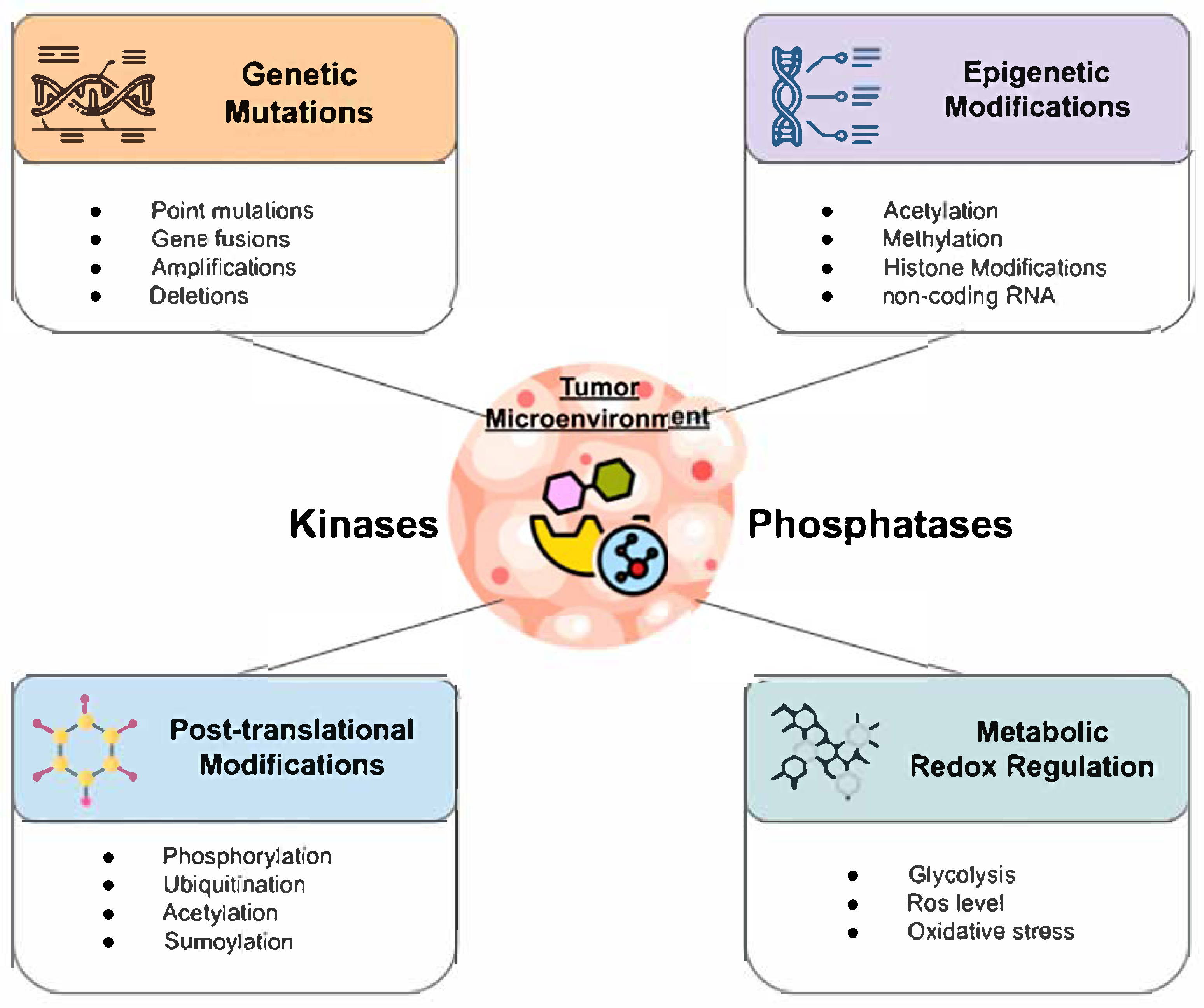
The functional dysregulation of kinases and phosphatases in cancer arises through a wide array of molecular alterations that affect their expression, localization, catalytic activity, and interaction with other signaling components. These alterations can be classified into general categories: genetic, epigenetic, PTMs, metabolic and redox modifications. All of these factors contribute to abnormal signaling dynamics that promote oncogenesis and progression. Importantly, these dysregulated pathways also contribute to tumor relapse and therapeutic failure by sustaining cancer stem cells (CSCs), which are key drivers of tumor recurrence even after apparent tumor clearance [4]. Here, we explored these mechanisms underlying the regulation of kinases and phosphatases in the context of cancer, illustrating them through selected examples that highlight their roles in tumorigenesis (Figure 1).

2.1.1. Genetic Alterations

Genetic mutations represent one of the most well-characterized mechanisms driving kinase dysregulation in cancer. These include activating point mutations, gene fusions, amplifications, and deletions. For example, a single mutations in B-Raf proto-oncogene, serine/threonine kinase (BRAF) (e.g., V600E) result in constitutive activation of the mitogen-Activated Protein Kinase (MAPK) signaling pathway, leading to uncontrolled cell growth and proliferation [16]. Similarly, epidermal growth factor receptor (EGFR) mutations in non-small cell lung cancer or anaplastic lymphoma kinase (ALK) fusions in anaplastic large cell lymphoma create hyperactive kinases that act as oncogenic drivers [17,18]. On the phosphatase side, tumor suppressors such as phosphatase and TENsin homolog (PTEN), ranked as the second most frequently mutated gene in cancer [19], and protein tyrosine phosphatase T (PTPRT) often exhibit inactivating mutations or deletions [20], resulting in the loss of negative regulation over key signaling pathways that control cell survival, migration, and proliferation [21].

2.1.2. Epigenetic Modifications

Epigenetic modifications, including DNA methylation, histone modifications, and non-coding RNAs, critically regulate the expression of kinases and phosphatases in cancer by altering chromatin accessibility and transcriptional activity [22,23]. Although these modifications are usually reversible, they can lead to persistent and heritable changes in gene expression that contribute to oncogenic signaling and tumor heterogeneity [24]. For example, in oral squamous cell carcinoma (OSCC), progressive loss of DNA methylation at the protein tyrosine kinase 6 (PTK6) promoter leads to its overexpression, which in turn enhances OSCC cell proliferation, migration, and invasion by activating oncogenic pathways, including increased signal transducer and activator of transcription 3 (STAT3) phosphorylation and elevated zinc finger and homeodomain transcription factor (ZEB1) expression [25]. Aberrant epigenetic silencing of tumor suppressor phosphatases or activation of oncogenic kinases can disrupt signaling homeostasis, while the compromised integrity of chromatin architecture, driven by mutations in epigenetic regulators, can further lock cells into pathological states, fueling cancer progression and resistance to kinase- or phosphatase-targeted therapies [26,27,28]. Interestingly, epigenetic silencing of phosphatases is a common mechanism by which tumor cells subvert negative regulatory inputs. Promoter hypermethylation of PTEN [29], PTPRK [30], or dual specificity phosphatase 6 (DUSP6) [31] has been reported in multiple tumor types, resulting in decreased transcription and loss of function.

2.1.3. Post-Translational Modifications

Post-translational modifications, such as phosphorylation, ubiquitination, acetylation, and sumoylation, are essential for the precise regulation of kinase and phosphatase activity, stability, localization, and substrate specificity [32,33]. These dynamic modifications function as molecular switches, fine-tuning signal transduction networks in response to cellular cues. Aberrant PTMs can disrupt these regulatory mechanisms, leading to the constitutive activation of oncogenic kinases or the inactivation of tumor-suppressive phosphatases [8]. For instance, a comprehensive proteogenomics analysis across 11 solid tumor types, conducted by Geffen et al., revealed that protein acetylation and phosphorylation are closely linked to key oncogenic processes. Their findings highlighted diverse mechanisms, such as impaired DNA repair linked to aberrant phosphorylation, immune and metabolic dysregulation driven by acetylation changes, and a functional interplay between acetylation and phosphorylation that shapes kinase activity and chromatin dynamics. Together, this study highlighted the complexity of PTMs in rewiring cancer signaling [34]. Genetic alteration can also confer advantages to the regulation of PTMs. For example, the R776H mutation in EGFR leads to constitutive activation through ligand-independent autophosphorylation, bypassing the normal requirement for EGF ligand binding [35]. This constitutive activation of EGFR signaling represents hallmark features of metastasis and contributes to therapeutic resistance [36]. Other studies have shown that tumor suppressors such as PP2A and PTEN can be functionally inactivated through various PTMs, which alter their expression levels, stability, subcellular localization, or enzymatic activity [37,38]. Interestingly, PTEN phosphorylation at specific tyrosine residues can preserve its phosphatase activity while impairing its nuclear localization, which is essential for maintaining chromatin condensation and decreasing aberrant transcription [39].

2.1.4. Metabolic and Redox Regulation

Regulation of cellular metabolism is crucial for cancer cell growth and survival, as it supports energy demands and biosynthetic needs. Cancer cells predominantly rely on aerobic glycolysis (the Warburg effect), converting glucose to lactate even in the presence of oxygen to fuel rapid growth [40]. Elevated glycolytic metabolism increases levels of intermediates like glucose-6-phosphate and alters redox balance through the accumulation of reactive oxygen species (ROS) such as H2O2, which can reversibly oxidize critical cysteine residues of several tyrosine phosphatases (e.g., PTEN, PP2A, Src homology-2 protein tyrosine phosphatase, and protein tyrosine phosphatase 1B), leading to their inactivation [41,42]. This redox-mediated inhibition of phosphatases results in increased activation of the oncogenic PI3K/AKT/mTOR axis, promoting cell survival, growth, and metabolic reprogramming, forming a feed-forward loop that reinforces oncogenic signaling [42]. It is important to note that ROS level can play a dual role in tumor cells. Low levels of ROS promote cancer stem cell maintenance, cell proliferation, differentiation, and migration, whereas high levels induce oxidative stress and DNA damage, leading to cancer cell death [43]. This dual role of ROS highlights the delicate balance cancer cells maintain in their metabolic activity to support growth and survival while avoiding cytotoxicity.
In addition, PTMs not only regulate signaling proteins but also directly modulate the metabolic activity of cancer cells by enhancing the function of key glycolytic enzymes. For example, the activity of hexokinase 1 and 2, key rate-limiting enzymes in glycolysis, is enhanced through phosphorylation mediated by the non-receptor tyrosine kinase c-SRC, which promotes pathways related to cell survival, migration, and metastasis [44]. Additional PTMs further regulate the enzymatic activity of hexokinase. Ubiquitination enhances the stability and mitochondrial localization of HK2, thereby supporting its glycolytic function, while de-SUMOylation modulates HK2 association with the mitochondrial membrane, influencing both its metabolic activity and apoptotic resistance [45,46].
Altogether, alterations in kinases and phosphatases occur across multiple biological scales, ranging from genetic to metabolic levels, may arise concurrently but through distinct mechanisms, highlighting the need to comprehend the multilevel complexity in biomarker discovery.

3. Biomarker Relevance and Clinical Utility

3.1. Kinases and Phosphatases as Biomarker

3.1.1. Kinases and Phosphatases in the Current Biomarker Landscape

Cancer biomarkers are increasingly recognized as multiscale entities that encompass a wide spectrum of biological, functional, and molecular features, collectively reflecting tumor presence, behavior, and therapeutic response. These biomarkers can be classified into several types such as genetic (e.g., point mutations, insertions, deletions, copy number variations), epigenetic (e.g., DNA methylation, histone modifications), transcriptomic (e.g., mRNA or non-coding RNA expression), proteomic (e.g., kinases, phosphatases, adaptor proteins, immune checkpoint molecules, tumor-associated antigens), or metabolic (e.g., altered metabolite levels in biofluids or tissues). Also, extracellular vesicles (EVs) and even whole-cell populations (e.g., circulating tumor cells, immune cells, and tumor-initiating cells) can function as biomarkers that predict therapeutic response and disease progression [47].
Antigens represent one of the most widely used categories of proteomic biomarkers. They are generally divided into two classes: tumor-associated antigens (TAAs) and tumor-specific antigens (TSAs) [48]. TAAs are proteins that are normally expressed at low levels in healthy adult tissues but become re-expressed or overexpressed in tumors. Examples include carcinoembryonic antigen (CEA) in colorectal cancer, alpha-fetoprotein (AFP) in hepatocellular carcinoma, and prostate-specific antigen (PSA) in prostate cancer, all of which are routinely applied in clinical practice for screening, monitoring, and prognosis. By contrast, TSAs are antigens that are expressed exclusively, or nearly exclusively, by tumor cells but not by normal tissues, making them highly specific indicators of malignancy. A notable example is human epidermal growth factor receptor-2 (HER2) kinase in breast cancer, which functions not only as a biomarker but also as a direct therapeutic target for monoclonal antibodies such as trastuzumab [49]. Many clinically relevant antigens are glycoproteins, where the post-translational glycosylation patterns provide more informative biomarkers than the protein backbone alone. Some examples include CA125/MUC16 mucin, a diagnosis biomarker in ovarian cancer and CA19-9 in pancreatic cancer [50]. However, antigens as biomarkers face important limitations, including the lack of specificity of TAAs, instability of expression driven by tumor immune evasion and genetic instability, inherently low immunogenicity, and significant intra-tumoral heterogeneity [51]. These challenges highlight the need to expand the biomarker landscape to enable more accurate and comprehensive tumor monitoring.
Within this broad biomarker landscape, kinases and phosphatases occupy a central role due to their dual relevance as both biomarkers and actionable therapeutic targets. Kinases, which number over 500 in the human genome, are the second most targeted drug class after GPCRs, owing to their conserved ATP-binding pockets and their central role in oncogenic signaling [52]. With the central role of tumor suppressor, phosphatases, long considered “undruggable”, are now emerging as therapeutic targets. Notable advances include inhibitors of SHP2 in RAS/MAPK-driven cancers and efforts to restore tumor-suppressive function of PP2A [6]. While expression level has traditionally been the focus of biomarker studies, it does not always reflect the functional state of a molecule. Numerous studies have demonstrated that expression levels alone are insufficient as prognostic or predictive biomarkers. This limitation arises from the lack of correlation between level expression and protein activity, which is control through its phosphorylated form. For example, the phosphorylated form of EGFR, AKT, and STAT3 provides a more accurate indication of receptor activation and downstream signaling potential than total expression, highlighting why activity-based measures can serve as more informative biomarkers, particularly in the context of targeted therapies [53,54,55]. PTMs are receiving increasing attention as cancer biomarkers due to their high specificity and the unique molecular signatures they provide. Several studies have been investigated PTMs, not only in cancer, but in other disorders, using a variety of biological fluids. Depending on the fluid analyzed, the platforms and detection methods required for PTM measurement can vary considerably, which can influence the quality of biomarker detection. Plasma and serum remain the most analyzed samples due to their accessibility and abundance of circulating proteins. Urine and saliva are also attractive for non-invasive biomarker discovery. More specialized fluids, such as cerebrospinal fluid, are particularly valuable for studies related to brain cancer [56].

3.1.2. Complexity of Kinases and Phosphatases as Biomarkers

The selection of an ideal biomarker requires balancing biological relevance with clinical applicability. However, a major challenge in using kinases and phosphatases as biomarkers lies in their intra- and inter-tumoral heterogeneity, where expression and activity can vary across different tumor subclones, spatial regions, or disease stages [57]. This heterogeneity is further shaped by therapeutic pressure, often leading to changes in biomarker stability over time. For example, breast cancer patients undergoing chemotherapy often exhibit dynamic changes in the expression of estrogen receptor (ER), progesterone receptor (PR), and HER2, which have been shown to correlate with both therapeutic response and overall survival [58].
Moreover, the context-dependent roles of kinases and phosphatases add another layer of complexity [59]. Certain enzymes can behave as oncogenes in one context while acting as tumor suppressors in another. For example, the nuclear localization of Shp2 has been correlated with poor survival in NSCLC, while in the liver, Shp2 acts as tumor suppressor by inhibiting cholangiocarcinoma progression [60,61]. Such context-specificity complicates the interpretation of these proteins as universal biomarkers.
The functional state of kinases and phosphatases is often regulated not by expression alone but by PTMs, subcellular localization, protein–protein interactions and the temporal persistence of these modifications [62]. Expression levels may not reliably predict activity, as illustrated by EGFR, where phosphorylated forms provide a more accurate measure of signaling activity and therapeutic response than total receptor expression. Upon ligand stimulation, EGFR undergoes rapid tyrosine phosphorylation, peaking within 5–10 min to trigger downstream signaling cascades. By 15 min, the receptor is internalized, and approximately 50% of the receptors recycle back to the cell surface within 60 min [63]. Similarly, some proteins undergo multiple PTMs that finely regulate their diverse functions. For instance, PTEN has multiple phosphorylation sites (e.g., Ser380, Thr382, Thr383, and Ser385), which regulates differently its stability, activity, and several cellular functions, affecting its role as a tumor suppressor [64]. Thus, the selection of the optimal PTM or activity readout is critical for biomarker development, as it must capture the relevant tumor-suppressive or oncogenic function without overlooking other essential roles of the protein.
When selecting effective biomarkers, practical factors beyond biological relevance, such as abundance, and accessibility, must be considered for clinical relevance. Low-abundance biomarkers, like phosphorylated kinases p-AKT or p-EGFR in the rare circulating breast tumor cells, require highly sensitive assays for reliable detection [65]. Accessibility favors minimally invasive samples, such as blood or urine; for example, ctDNA with EGFR mutations enables non-invasive monitoring of tumor evolution, while surface proteins like HER2 can be tracked via extracellular vesicles or circulating tumor cells [66,67]. Careful consideration of these factors ensures biomarkers are both informative and clinically actionable.
Taken together, these factors demonstrate that kinases and phosphatases are multifaceted but challenging biomarkers, requiring careful consideration of sample type, molecular context, PTM status, and tumor heterogeneity. Integrating these parameters with complementary molecular and clinical data will be essential to fully leverage their potential in precision oncology.

3.2. Clinical Classification of Kinase and Phosphatase Biomarkers

Tumor biomarkers are essential components across the entire cancer care continuum, supporting applications from early detection and diagnosis to therapeutic decision-making, treatment monitoring, and relapse surveillance [47]. Their classification into distinct clinical categories provides a structured framework for integrating these molecules into precision oncology workflows and optimizing therapeutic strategies. Clinically, tumor biomarkers are typically categorized into different groups: diagnostic, prognostic, and predictive biomarkers. Due to their significant role in tumorigenesis, kinases and phosphatases have emerged as clinically relevant biomarkers, contributing to multiple phases of cancer management, including diagnosis, prognosis, prediction of treatment response, and disease monitoring [68,69] (Table 1).

3.2.1. Diagnostic Biomarkers

These measurable indicators help detect or confirm disease, subtypes, and guide accurate diagnosis and personalized treatment decisions. For instance, prostate-specific antigen (PSA) is an approved biomarker for prostate cancer, as elevated serum levels of PSA suggest the presence of prostate cancer and are commonly used to support early detection and diagnosis. However, PSA testing is not cancer-specific (around 20–40% specificity) and may also rise in benign conditions such as prostatitis or benign prostatic hyperplasia. This can lead to false positives and unnecessary biopsies [70]. Another example is the presence of phosphatidylinositol-4,5-bisphosphate 3-kinase catalytic subunit alpha (PIK3CA) mutations in breast cancer, a gene that encodes for the p110α catalytic subunit of PI3K. Mutations in PIK3CA are among the most common genetic alterations in hormone receptor–positive (HR+), HER2-negative breast cancers. Detecting these mutations through molecular testing helps confirm the molecular subtype of the tumor, contributing to a more precise diagnosis. Additionally, identification of PIK3CA mutations not only informs diagnosis but also opens the door to targeted therapy with PI3K inhibitors such as alpelisib, further highlighting the clinical utility of biomarker discovery [76].

3.2.2. Prognostic Biomarkers

These types provide information about the likely course or outcome of a disease, independent of treatment. They are useful for assessing disease aggressiveness and stratifying patients into different risk groups. For example, the BRAF V600E mutation in colorectal cancer is associated with poor prognosis regardless of the therapy administered. This mutation helps identify patients with more aggressive disease who may benefit from closer monitoring or enrollment in clinical trials [72,77]. In non-small cell lung cancer (NSCLC), elevated EGFR expression, particularly in the presence of activating mutations such as exon 19 deletions or L858R, can serve as a prognostic biomarker, often associated with a more favorable prognosis and improved response to targeted therapies [78,79]. Interestingly, PSA levels in prostate cancer are also considered as prognostic markers because higher baseline PSA levels often correlate with more advanced disease and poorer clinical outcomes [70]. As prognostic biomarkers, phosphatases have shown limited clinical validation and present a more complex and heterogeneous biological profile compared to other biomarker classes [6]. Given its central role in regulating oncogenic signaling pathways, the loss or inactivation of tumor suppressor phosphatase PP2A has been discussed as a potential prognostic biomarker due to its association with aggressive histological features in endometrial cancer [80]. However, these alterations do not independently predict patient survival. Moreover, the interaction of PP2A with other pathways, such as the p53 signaling pathway, suggests that its clinical relevance may be best captured in combination with other molecular biomarkers [81].

3.2.3. Predictive Biomarkers

They determine whether a patient is likely to respond to a specific treatment or experience a particular outcome. They are crucial in personalized medicine, allowing clinicians to select the most effective therapies for individual patients based on their unique biological profile. Predictive biomarkers are typically measured before treatment begins to inform treatment decisions. For example, the EGFR L858R mutation is a well-established predictive biomarker for responsiveness to tyrosine kinase inhibitors (TKIs) in NSCLC. Patients harboring this activating mutation typically experience significant clinical benefit from targeted EGFR inhibition [82]. However, resistance mutations such as EGFR T790M or alternative pathway activators like BRAF V600E often emerge during treatment, leading to acquired resistance to first- and second-generation TKIs [83]. As predictive biomarkers, these secondary mutations guide interventions toward alternative targeted therapies to overcome resistance and improve patient outcomes. This evolving landscape highlights the critical role of predictive biomarkers in dynamic treatment adaptation and precision oncology. Phosphatase biomarkers are gaining attention in precision oncology, particularly SHP2, where high expression levels may predict responsiveness to SHP2 inhibitors associated with other TKIs to overcome therapeutic resistance [75,84].
Biomarker discovery in oncology has evolved significantly over the past two decades, driven by advances in high-throughput technologies, multi-omics platforms, large-scale data portals, and precision medicine frameworks [1,85,86]. While thousands of candidate biomarkers have been identified, only a small fraction have achieved clinical implementation, largely due to rigorous demands for analytical validity, clinical utility, and regulatory approval [87,88]. The current landscape is increasingly shaped by the integration of systems biology, AI, and spatial omics, which together aim to refine kinase and phosphatase biomarker identification and address persistent challenges such as tumor heterogeneity, CSC persistence, temporal disease evolution, and therapeutic resistance [13,14,89]. As a result, biomarker discovery with a focus on kinases and phosphatases continues to serve as a foundational pillar of cancer research and a critical gateway to unlocking the full promise of personalized oncology.

3.3. The Process of Biomarker Discovery: From Data to Clinical Utility

The biomarker discovery pipeline is a multi-step, iterative process that combines basic research, high-throughput technologies, computational modeling, and clinical validation. For kinases and phosphatases, this pipeline must accommodate the dynamic and context-dependent nature of phosphorylation networks, which cannot be fully captured by static genomic profiling alone [90,91,92].

3.3.1. Biomarker Identification

The discovery phase serves as the foundation of the biomarker pipeline, aiming to identify molecular candidates associated with a biological state. First, it is essential to define the clinical utility (e.g., diagnosis, prognosis, and predictive) to determine the appropriate samples for investigation. Various biological materials can be suitable for discovery, including tumor tissues, blood, cerebrospinal fluid (CSF), urine, saliva, or in vitro models like cell lines and organoids [92,93]. Each biological material offers unique advantages for candidate discovery. For instance, tumor tissues provide direct, site-specific molecular insights; while blood samples are minimally invasive and suitable for systemic monitoring, CSF reflects the central nervous system for conditions like brain cancer; urine and saliva offer non-invasive options for certain cancers; and in vitro models allow controlled testing and mechanistic validation [94,95]. Ensuring sample integrity and standardized collection protocols is critical to minimize variability and bias in downstream analyses.
Because approved biomarkers can be from different types (e.g., genetic, epigenetic, transcriptomics, proteomic, and metabolic biomarkers), comprehensive molecular profiling can be conducted using advanced high-throughput omics platforms, including genomics (e.g., whole genome or targeted sequencing), transcriptomics (e.g., single or bulk RNA sequencing), proteomics (e.g., mass spectrometry-based protein quantification), and metabolomics (e.g., mass spectrometry or NMR-based metabolic profiling). These platforms allow the simultaneous measurement of thousands of molecular features, capturing the complexity and heterogeneity of cancer at multiple biological levels [96]. However, due to the variable data types, such as differences in scale, dimensionality, measurement noise, and biological context, integrating and interpreting these datasets remains a major challenge. Advanced computational approaches, including ML, network-based modeling, and multi-omics integration frameworks, are therefore essential to extract meaningful patterns, prioritize candidate biomarkers, and understand the functional relationships between biological layers [14,97]. To minimize the cost and time associated with data generation and to address the limitations in biological sample availability, researchers often leverage publicly available datasets such as The Cancer Genome Atlas (TCGA), Gene Expression Omnibus (GEO), and the Clinical Proteomic Tumor Analysis Consortium (CPTAC), cBioportal, Xena and Gepia [98,99]. In addition, specialized repositories provide access to PTM data, including PhosphoSitePlus, dbPTM, and Phospho.ELM, among others, which catalog experimentally validated kinases, phosphatases and PTM events [100]. For functional and preclinical studies, resources such as the Cancer Cell Line Encyclopedia, COSMIC Cell Lines Project, and DepMap offer detailed molecular and pharmacogenomic profiles of cancer cell lines [101]. Other valuable repositories include the Human Protein Atlas for protein expression across tissues, Reactome and Kyoto Encyclopedia of Genes and Genomes (KEGG) for pathway annotations, and proteomic identification database (PRIDE) for mass spectrometry-based proteomics data. For metabolic profiling and pathway integration, databases such as the Human Metabolome Database, METLIN, and the KEGG Metabolism module provide comprehensive metabolite and reaction-level data. These resources provide access to high-quality, large-scale multi-omics data across diverse cancer types, supporting hypothesis generation without the need for immediate experimental data collection [85,86,89]. At the end of the discovery phase, a panel of candidates, not yet biomarkers, is identified for further analytical and clinical validation. However, due to the high dimensionality of omics data and biological variability, this stage often generates a large number of candidates, many of which may not ultimately prove clinically relevant, highlighting the importance of rigorous downstream filtering and validation. Furthermore, many studies do not progress beyond the initial discovery phase, primarily due to the substantial costs and the prolonged timeline required for biomarker validation and development [102].

3.3.2. Preclinical Validation

The goal of preclinical validation is to establish the biological significance and reproducibility of candidate biomarkers, ensuring that initial discovery-phase findings are consistent and disease-specific [103]. For kinase and phosphatase biomarkers, this step involves validating differential expressions, activity, or alteration patterns across independent cohorts of diseased and normal samples. This is essential for ensuring that observed changes are not artifacts of the discovery platform or specific to a single dataset.
A range of molecular and biochemical techniques is employed at this stage to validate candidate biomarkers firs in vitro [1]. Quantitative PCR and enzyme-linked immunosorbent assay (ELISA) are among the most straightforward and widely used platforms for validating mRNA and protein levels, respectively, due to their simplicity, cost-effectiveness, and scalability [104,105]. Western blotting is also commonly used for protein expression validation, while immunohistochemistry provides spatial localization of biomarkers within tissue samples [106,107]. More complex validation, involving PTMs, pathway analysis, metabolic or functional interactions, may require advanced methods like mass spectrometry-based phosphoproteomics and metabolomics [108,109].
Additional preclinical models are employed to screen and evaluate potential biomarkers, including 2D and 3D cell cultures, patient-derived xenografts (PDX), genetically engineered mouse models (GEMMs), and organoids [110,111,112]. These models are primarily utilized for their ability to mimic key aspects of TME, enabling the evaluation of biomarker expression, activity, and function under controlled and physiologically relevant conditions. For kinase and phosphatase biomarkers, such systems allow researchers to study phosphorylation dynamics, and pathway activation under physiologically relevant conditions [113,114]. Organoids and PDX models, particularly derived from individual patients, offer the advantage of preserving patient-specific tumor architecture and heterogeneity, making them as valuable systems for screening molecular activity profiles for personalized applications [115,116].
Functional assays are often included to examine the biological impact of candidate kinases or phosphatases on cellular processes. Inhibition or overexpression studies using cell lines or patient-derived models, combined with CRISPR-Cas genome editing, RNA interference and inducible expression systems, can provide mechanistic insights into how a candidate contributes to proliferation, apoptosis, tumorigenesis or stemness or resistance pathways [117,118,119].
Overall, this step not only confirms the consistency and specificity of biomarkers in preclinical models but also begins to establish its biological significance to patients, subtypes, and functionality.

3.3.3. Analytical Validation

The next step in the biomarker pipeline is the development and analytical validation of assays capable of accurately measuring the biomarker in biological samples. The main objective is to confirm the sensitivity, specificity, reproducibility, and robustness of the assay. It ensures that the findings are robust and reproducible, providing a solid foundation before transitioning to clinical testing in humans [92]. Analytical validation is typically performed using a well-characterized cohort of biospecimens, such as tumor and matched normal tissues, cancer stage, subtypes, demographic or clinical characteristics, to evaluate assay performance under clinically relevant conditions. In contrast to preclinical validation, analytical validation focuses on assessing the performance characteristics of the assay itself rather than the function of the biomarker [120,121].
Given the enzymatic nature and post-translational regulation of kinases and phosphatases, assays must demonstrate high sensitivity and specificity to distinguish cancer-associated alterations, such as aberrant expression, phosphorylation states, or activity levels, from background noise in complex biological samples [47,122]. The choice of assays depends on the scale of interest, the clinical application, and the expression compartment. Various assays can be developed depending on the category of biomarker, including antibody-based platforms (e.g., ELISA, Western blotting, immunohistochemistry) or more advanced technologies such as advanced mass spectrometry-based proteomics [123,124]. Additionally, automated high-throughput platforms are typically reserved for large-scale screening efforts or multiplex biomarker panels, especially in later stages of translational or clinical research settings [125,126]. These assays must undergo rigorous optimization to ensure consistent performance across sample types, batches, and laboratory settings to allow widespread clinical applicability. Of note, analytical validation of proteomic and metabolomic biomarkers poses distinct challenges. Proteins exhibit complex PTMs and wide dynamic ranges, requiring precise assay optimization and reliable internal standards [127]. Similarly, metabolites are chemically diverse and prone to degradation, with validation affected by matrix effects and batch variability [128]. These factors complicate reproducibility and standardization, especially in high-throughput settings.
Several key parameters, including accuracy, precision, sensitivity, specificity, reportable range, and stability, are commonly used to evaluate assay performance, with specific metrics prioritized based on the goals and context of the study [121]. Within these criteria, quantitative metrics are determined: the limit of blank, which describe the highest value that is likely to be found in a blank sample, the limit of detection, which refers to the lowest concentration of the biomarker that can be reliably distinguished from background noise, and the limit of quantification, which represents the lowest concentration that can be quantitatively measured with acceptable accuracy and precision [129,130]. A biomarker used for early detection may require a lower limit of detection and a higher level of sensitivity than one used for monitoring treatment response. This is because, at early stages of cancer, often before clinical symptoms emerge, the biomarker may be present at extremely low concentrations in biological fluids [1,131]. In contrast, biomarkers used for monitoring therapeutic response generally appear in higher concentrations once the disease is established, and the goal shifts toward tracking fluctuations over time, often prioritizing assay reproducibility and dynamic range over ultra-high sensitivity [132]. Therefore, the analytical performance requirements of a biomarker assay must be aligned with its intended clinical use, whether for early detection, diagnosis, prognosis, or monitoring.
Together, assay development and analytical validation form the technical backbone for translating kinase and phosphatase biomarkers into clinical applications, ensuring the measurements are both biologically meaningful and technically robust before proceeding to clinical validation and regulatory review.

3.3.4. Clinical Validation

Following analytical validation, clinical validation represents the last important step in the biomarker development pipeline. Its primary objective is to establish that the biomarker is clinically reliable in distinguishing between patient groups (e.g., diseased vs. non-diseased, responders vs. non-responders) and correlating with outcomes such as disease progression, survival, or therapeutic response [91,92]. Unlike analytical validation, which focuses on the technical robustness of the assay, clinical validation is concerned with how the biomarker performs in a real-world or clinical trial setting. Specifically, it involves evaluating the biomarker’s diagnostic, prognostic, or predictive performance in well-characterized clinical cohorts under real-world or trial-like conditions. Clinical validation typically requires prospective or retrospective studies using appropriately powered patient samples. More importantly, clinical validation requires a significantly larger cohort compared to analytical validation, to ensure statistical power and generalizability across varied patient populations [102].
Clinical validation is also a costly and time-consuming process, requiring large cohorts and often multi-institutional collaboration under strict ethical and regulatory frameworks [133]. Designing trials to detect meaningful differences in biomarker levels or signaling activity is particularly complex given the dynamic and context-dependent nature of kinase and phosphatase signaling [134,135]. For instance, a significant translational gap remains; kinase or phosphatase biomarkers that show promise in controlled validation studies may underperform in real-world clinical settings due to variability in sample processing, patient populations, and integration into existing diagnostic workflows [136]. To interrogate clinical specificity, patient heterogeneity, including differences in tumor genetics, signaling activity, spatial localization, and treatments, should be considered during study design. These factors can affect phosphorylation states and biomarker behavior, thereby limiting the generalizability of kinase and phosphatase biomarkers across patients [137,138]. Therefore, the availability of high-quality, significant sample sizes is essential for conducting meaningful clinical validation at the appropriate scale and specificity [139].
Ultimately, successful clinical validation provides the evidence base needed for regulatory approval and clinical implementation. It transforms a technically validated biomarker into a clinically actionable tool, paving the way for use in diagnostics, prognostics, or therapeutic design.

3.3.5. Regulatory Qualification and Approval

The biomarker at this stage is already supported by robust evidence generated through analytical and clinical validation, including clearly defined assay performance metrics (e.g., sensitivity, specificity, reproducibility), clinical utility, and relevance to patient cancer. The final stage of regulatory qualification and approval supports the transition of a biomarker from research into clinical practice. This process ensures that the biomarker meets the stringent standards required for use in diagnosis, prognosis, patient stratification, or as a companion diagnostic [140,141]. Regulatory qualification refers to the formal recognition by regulatory bodies, such as the U.S. Food and Drug Administration (FDA), European Medicines Agency (EMA), or PMDA in Japan, that a biomarker is valid for a specific context of use in drug development or clinical decision-making. The regulatory process also requires adherence to Good Laboratory Practice (GLP) and, in many cases, validation within CLIA-certified (Clinical Laboratory Improvement Amendments) laboratories to ensure assay consistency in clinical settings [142]. All regulatory submissions must include comprehensive documentation detailing the context of use, assay methodology, clinical study design, statistical analysis, and risk–benefit assessment [143]. For biomarkers intended for in vitro diagnostic use, the FDA may require approval via pathways such as 510(k) or Premarket Approval (PMA), depending on risk classification. In the case of companion diagnostics co-developed with targeted therapies, coordination with therapeutic regulatory submissions is essential [144]. The biomarker qualification program in the U.S. provides a formal framework for evaluating biomarkers outside the context of a specific drug, while the EMA’s qualification opinion offers similar guidance in the EU [145]. Navigating this regulatory pathway is essential for translating biomarker research into clinically actionable tools that can guide precision oncology and improve patient outcomes.
Targeting kinases is a significant area of research and development in cancer treatment, particularly through the development of small-molecule inhibitors that modulate their activity [122]. While targeting phosphatases has proven challenging due to several intrinsic biochemical and structural factors [6]. More than eighty kinase inhibitors have received FDA approval for cancer treatment, with EGFR inhibitors being the highly represented (ten FDA-approved inhibitors). These inhibitors are used in a wide spectrum of malignancies, including breast, glioma, lymphoma, leukemia, lung cancer, melanoma, and carcinoma [5]. Despite their clinical success, kinase inhibitors are often limited by the emergence of drug resistance, which can be de novo or acquired resistance [146].
Overall, the development of a biomarker from discovery to clinical approval is a lengthy and complex process that can span more than a decade. This is exemplified by the case of EGFR, first identified as a potential oncogenic driver in the 1980s. Although EGFR mutations (exon 21 L858R and exon 19) were later discovered in a subset of NSCLC in the early 2000s [147], it took years of rigorous validation and regulatory evaluation before the approval of first generation of targeted therapies such as erlotinib and afatinib were approved as first-line treatments for EGFR-mutant NSCLC in 2013 [148].
However, given the clinical needs and the pace of scientific discovery, there is a growing demand for new strategies and integrated platforms that can accelerate biomarker development, enhance validation efficiency, and reduce the overall time from candidate selection to implementation in clinical practice.

4. Integrating Digital Twins in Kinase and Phosphatase Biomarker Discovery

4.1. Definition of Digital Twins

DTs are advanced virtual models that replicate real-world objects, systems, or processes in a dynamic and interactive way, allowing them to evolve in parallel with their physical counterparts. DTs operate as bidirectional systems, continuously integrating real-world data to update the model, while simultaneously generating predictive simulations that guide future experimental or clinical decisions. DTs originally applied in engineering and manufacturing, however in recent years DTs have been adopted for medicine and healthcare sector [149,150].
DTs in healthcare have demonstrated various applications spanning from hospital operations, emergency response, medical device innovation, drug discovery, biomanufacturing, surgical preparation, virtual clinical trials, health monitoring, and individualized care [151,152]. This broad spectrum of uses highlights the increasing importance of DTs and their transformative potential to reshape modern healthcare. In oncology, they are paving the way to a new frontier for advancing precision medicine, with key roles in diagnosis, treatment planning, patient monitoring, clinical trial design, and biomedical research [153,154]. Several DTs have been developed for different solid cancers to optimize treatment strategies, predict therapeutic resistance and reassign treatment regimens [155,156,157]. By advancing complex modeling techniques that handle historical and real-time data, DT technology offers insights that go beyond static snapshots as they support proactive action.
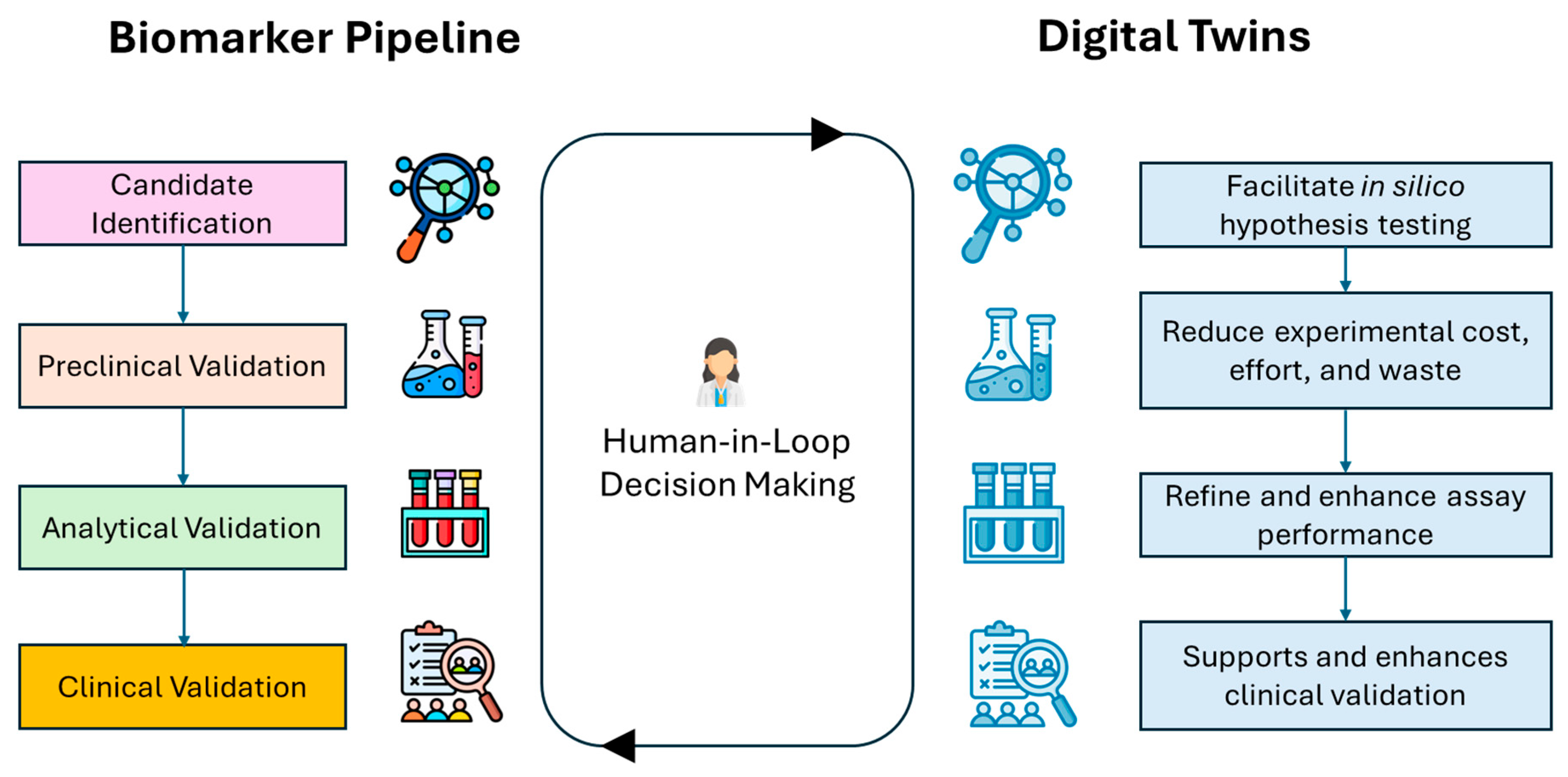
A functional DT consists of three main components: the real-world entity (e.g., a tumor, patient, or cellular system), the digital model (a computational representation of the patient biological landscape), and a data feedback loop that continuously updates the model with experimental or clinical data to keep it aligned with the physical system [158]. The development of DTs involves several key steps, including defining the biological or clinical scope, collecting relevant multimodal data, building a functional prototype, integrating patient-specific parameters for personalization, and ultimately validating the model for clinical application [12]. The validation and uncertainty quantification of emerging technologies such as DT and AI in healthcare require a collaborative effort among engineers, researchers, healthcare providers, and stakeholders [159,160]. Maintaining a human-in-the-loop paradigm is indispensable to ensure contextual interpretation and biological relevance, and although DT–based methodologies provide a robust computational framework for the systematic identification of kinase- and phosphatase-modified molecular entities, it is critical to underscore that such in silico approaches are designed to augment, rather than supplant, conventional biomarker discovery pipelines. Integrating DTs into clinical practice can reduce workload by simulating the repetitive and time-consuming steps of the biomarker discovery pipeline [161]. This synergy between human judgment and computational power accelerates the identification of actionable biomarkers while maintaining a focus on patient-specific and translational outcomes (Figure 2) [162].
Given the multiscale network that kinase and phosphatases operate within a tumor cell, DTs hold a great potential in advancing the biomarker discovery. A DT designed around these enzymes should replicate the dynamic behavior of key signaling pathways, capturing modifications between kinases, phosphatases, substrates, and various PTMs across molecular, cellular, and tissue scales, ultimately reflecting how these regulatory networks evolve in response to alterations, environmental cues, or therapeutic interventions. This approach not only could enhance our understanding of pathway dynamics but also accelerates the identification and validation of clinically actionable biomarkers. Moreover, DTs can also accelerate biomarker discovery by tackling every step of biomarker pipeline that for example, used retrospective data, and clinical trials [163]. Here, we focus on how DTs can enhance every aspect of biomarker discovery from candidate selection to the whole pipeline framework (Table 2).

4.2. Accelerating the Biomarker Discovery Pipeline with Digital Twins

4.2.1. Virtual High-Throughput Screening for Candidate Selection

One of the major bottlenecks in the biomarker discovery pipeline is the identification of candidate biomarkers from large-scale genomic, transcriptomic, and proteomic data. Candidate biomarkers must undergo a rigorous, multi-phase validation process that includes data filtering, experimental verification, and clinical correlation. This approach has limitation for dynamic systems like kinase and phosphatase function, where context, timing, and network compensation play essential roles [10]. Updating the patient’s DT with time-series single-cell transcriptomic and phosphoproteomic datasets can provide a virtual platform to capture all what-if scenarios related to the temporal and cell-type-specific regulation of kinase and phosphatase function during cancer progression or therapy response [164,165]. For example, as demonstrated by Li et al., their DT framework integrates a time series of single cell RNA-sequencing to model the dynamics changes in cell and genes, to prioritize potential biomarker and drug targets. The framework revealed multiple branching trajectories across cell types and time points, reflecting the inherent heterogeneity and plasticity of cellular responses. By analyzing these dynamic trajectories, the model identified and ranked upstream regulators responsible for context-specific alterations in signaling networks [166]. Furthermore, DTs can identify biomarker signatures associated with specific clinical trajectories by integrating multi-omics data from patient-derived samples. Scott et al. developed an adaptive DT model using proteomic data collected from a cohort of 1364 blood samples at the time of admission to emergency for sepsis. Their model could predicted protein panel associated with several outcome including organ dysfunction, and early-mortality-risk patients [167]. Based on this approach, patient DTs can evaluate the feasibility of assessing synergy between kinase and phosphatase associated with clinical outcome and could later guide the design of therapeutic interventions tailored to individual tumor signaling profiles.
While these two studies generated their own data to construct DT models, the broader applicability of this approach can be significantly enhanced by leveraging publicly available and retrospective datasets. For instance, Chang et al. proposed a DT pipeline that integrates existing multi-omics data and associated clinical features from TCGA, to support rapid prototyping and validation of DT models across diverse patient populations and mutational landscape [168]. Retrospective omic datasets, when paired with matched clinical data, can provide various biomedical data modalities that can update virtual models of patient cohorts. These models can then be used as virtual replica of TMEs to predict tumor trajectories in new patients, infer temporal dynamics in kinase and phosphatase activity, and identify candidate biomarkers all without the immediate need for new experimental data [158]. This approach significantly reduces both the time and cost associated with candidate identification and model development, while maximizing the utility of existing data resources. DTs provide an advanced platform for integrating multimodal data to enhance the selection of robust candidates. This integrative approach is particularly valuable as it takes into account the multiscale model of kinase and phosphatase enzymatic processes. For example, the combined overexpression of a kinase and concurrent loss of its regulatory phosphatase may serve as a more robust and predictive biomarker than a single alteration [169]. This multiscale strategy allows for the benchmarking of DT performance in modeling therapy response, resistance mechanisms, and tumor evolution across various therapeutic settings [15,155,170].
Overall, DTs enhance this step by modeling molecular flux in patient-specific contexts, helping to identify not just static biomarkers but those that shift in response to treatment or tumor evolution.

4.2.2. Virtual Preclinical System

While experimental validation remains essential for confirming the functional and clinical significance of candidate biomarkers, DTs can play a transformative role in streamlining and enhancing this process [171,172]. The incorporation of a closed-loop feedback mechanism between in vitro/in vivo experimental systems and in silico models enables continuous calibration of the DT. Initial simulations performed by the DT serve as hypotheses, which are empirically validated through biological assays. Quantitative data derived from these experiments, such as dynamic signaling profiles, phenotypic trajectories, and dose–response characteristics, are systematically reintegrated into the DT framework. This process facilitates model parameter refinement, state-space adjustment, and structural recalibration, thereby aligning the virtual model more closely with the underlying biological system. Through this iterative loop, the DT evolves into a self-updating system capable of accurately predicting the future behavior of its physical counterpart under varying conditions. Such adaptive learning mechanisms are critical for maintaining temporal coherence, physiological relevance, and mechanistic interpretability within human DT frameworks [173]. For example, Filipo et al. introduced a metabolic DT framework known as single-cell flux balance analysis, which integrates single-cell RNA-seq data into patient-based metabolic models. The authors were able to characterize metabolically distinct subpopulations to stratify heterogenous cancer subclones [174]. A similar framework, adapted to kinase and phosphatase involved in metabolism, could be used to complement wet-lab experiments by prioritizing high-confidence metabolic targets and generating mechanistic hypotheses at the individualized patient scale.
Importantly, wet-lab experiments are often constrained by practical limitations, including sample availability, restricted cell numbers, reagent costs, and time-consuming protocols. In contrast, DTs can simulate cellular responses at a much larger scale, encompassing thousands of virtual cells across multiple conditions, time points, and perturbations, far beyond what is feasible in a standard experimental setup [173]. For example, Behle et al. developed a 3D, large-scale cellular simulation capable of modeling cancerous tissue at the resolution of tens of millions of individual cells. The modular model includes several components that can be parameterized, such as cell–cell signaling, metabolic content, cell death, division, and mutations, enabling the prediction of behaviors ranging from single-cell morphology to macroscopic tumor tissue organization [175]. This model can then be used as a complementary tool to experimental validation, supporting the prediction of tissue-level organization and cellular dynamics in response to kinase and phosphatase targeting strategies. The computational scalability of DTs could help the exploration of a vast experimental space, including rare tumor cell populations, PTMs, and complex kinase/phosphatase interactions that might be missed in traditional assays [173]. By complementing wet-lab research, DTs help prioritize relevant candidates for next step of biomarker pipeline, reduce unnecessary experimental trials, and uncover mechanistic insights.
Three-dimensional culture systems are essential tools in the preclinical validation of cancer biomarkers, as they recapitulate the spatial organization, cell–cell interactions, and microenvironmental cues of in vivo tumors compared to traditional 2D cultures. These models include spheroids, organoids, and scaffold based system, providing high-content readouts for proliferation, apoptosis, invasion, and dynamic signaling responses [176,177,178]. Moreover, tumor-on-a-chip platforms further enhance the scalability by integrating microfluidics to monitor cell–cell interaction, tumor vascularity, mechanical stress, and multi-cellular interactions in a controllable setting [179,180]. When combined with DT frameworks, these platforms offer a personalized preclinical system, and iterative approach [181,182]. For example, Logun et al. demonstrated that patient-derived organoids from individuals undergoing immunotherapy could replicate the responses observed in corresponding clinical trial participants. This finding highlights the potential of organoids as monitoring systems in updating DT models with real-time data which can provide a virtual platform to predict individual treatment outcomes over time, and prioritize prognosis and predictive biomarkers for longitudinal monitoring [183]. Also, Bretti et al. introduced an in silico model as a foundational step toward developing future DTs that integrate data from breast cancer-on-chip experiments to investigate mechanisms of immunosurveillance. The model dynamically simulates immune cell migration in response to chemotactic signals released by tumor cells, providing insight into spatial immune-tumor interactions [184]. DTs can predict kinase and phosphatase crosstalk across different temporal and spatial conditions by virtually integrating different biological scales from patient-derived 3D system to omics [185,186]. DT technology enables large scale prediction of signaling pathways, immune response, and molecular landscape while reducing experimental burden by focusing on clinically actionable targets.
Altogether, the synergistic DTs–experimental workflow could support the virtual preclinical phase and provide a systems-level understanding that traditional pipelines may miss. Ultimately, this iterative process accelerates the identification of robust kinase and phosphatase biomarkers and supports their translation into assay development.

4.2.3. Virtual Analytical Validation

Analytical validation is fundamental to ensuring that biomarker assays are accurate, reliable, and reproducible across different platforms, sample types, and patient populations. However, this step can be resource-intensive, time-consuming, and often limited in their ability to comprehend complex patient and tumor biological variability [187,188]. Furthermore, sample source, quality, handling, and processing introduce additional variability, affecting enzyme stability, phosphorylation status, and assay signal fidelity. These variables often confound traditional analytical pipelines and hinder the development of scalable, reproducible biomarker platforms [189,190]. Through their predictive capabilities, DTs can simulate the performance of kinase- and phosphatase-based assays under a wide array of virtual experimental conditions and patient-specific profiles. For example, DTs can model how post-translationally modified proteins (e.g., phosphorylated substrates) behave under varying sample integrity, collection times, buffer conditions, or freeze–thaw cycles, without the need for extensive wet-lab experimentation [120]. These in silico testing can help anticipate assay failures, reduce false positives/negatives, and standardize protocols for challenging sample types such as formalin-fixed tissues or cerebrospinal fluid.
Analytical quality control (QC) is essential for ensuring data reliability across multiple runs, instruments, and laboratories. DTs can be employed to simulate technical variability in QC parameters such as stability over time, variability, reagent lot differences and signal saturation or background interference [120]. By modeling how these QC variables impact biomarker readouts, DTs can identify thresholds where assay performance deteriorates, enabling early detection of QC failures and suggesting optimal calibration or re-standardization steps. Additionally, DTs can serve as virtual benchmarking tools, simulating known biomarker responses under ideal and degraded conditions, providing synthetic reference standards when physical QC materials are limited [191,192].
Achieving sensitivity, and specificity can be challenging especially with variable expression levels, as low sensitivity may result in false negatives, while low specificity may lead to false readings [193]. Particularly, proteomic and metabolomic biomarkers poses distinct challenges, due to variability in protein abundance, stability, context-dependent PTMs, and the influence of complex biological matrices. These factors can compromise the sensitivity and specificity of assays, reducing performance [194]. To address these limitations, DT framework could integrate high-dimensional transcriptomic, proteomic, or metabolomic data into dynamic, cell-type-specific models [163]. By adjusting steady-state or time-resolved concentrations of enzymes, kinases, phosphatases, and metabolites to match experimentally observed values, DTs introduce biological realism into canonical signaling models [195]. This refinement enhances both the predictive power and analytical performance of biomarker assays, improving the ability to distinguish true biological signals from background noise. Also, DTs can provide virtual environment to estimate protein half-lives by computationally modeling the temporal stability, modification state transitions, and degradation kinetics of phosphorylated and non-phosphorylated targets. By simulating these molecular dynamics in silico, DTs provide insights into proteostatic regulation that are difficult to capture experimentally due to temporal constraints, limited resolution, and the inherent challenges of high-throughput biochemical assays. This virtual assessment framework enables the integration of time-resolved molecular behavior into predictive models, enhancing the DT’s utility in dynamic systems biology and mechanistic biomarker discovery [196].
These models can also perform in silico perturbation testing, modeling how biomarkers respond to dynamic changes across different analytical platforms. By simulating variations in detection sensitivity, signal calibration, and reagent performance, DTs assess assay consistency and reliability between technologies, such as mass spectrometry, Q-PCR, ELISA, or multiplex platforms. This enables early identification of discrepancies, standardization challenges, or platform-specific biases, ultimately supporting the optimal platform for a more robust and scalable biomarker assays [197].
Integrating DTs into the analytical validation phase offers a predictive, scalable, and cost-efficient complement to assay developments. As the field moves toward precision medicine, DTs provide a critical bridge between complex molecular biology and clinically actionable, analytically validated biomarkers.

4.2.4. Virtual Clinical Validation

Clinical validation is the final checkpoint before a biomarker is implemented in clinical decision-making. However, this stage remains one of the most resource-intensive and failure-prone steps in the biomarker development pipeline. They often require large, diverse, and well-annotated patient cohorts, which are logistically and ethically challenging to assemble. The recruitment into biomarker-driven clinical trials is often slow, costly, and logistically difficult due to several reasons such as strict inclusion/exclusion criteria, insufficient recruitment, trial fatigue and patient burden [198]. Furthermore, subgroups such as ethnic minorities, rare molecular subtypes, pediatric patients, or treatment-resistant cases are frequently underrepresented, limiting the generalizability of biomarker performance [199]. These gaps contribute to the high failure rate of biomarkers during late-stage validation. DTs could offer an interesting alternative by enabling in silico clinical validation across a wide spectrum of patient conditions. From an initial experimental or clinical cohort, DTs can generate synthetic, biologically realistic virtual populations that mirror real-world heterogeneity, including differences in age, sex, comorbidities, molecular subtypes, treatment history, and socioeconomic background [200]. These models could allow researchers to test biomarker performance in diverse simulated clinical scenarios, especially in underrepresented or underpowered subgroups. For example, DTs can model disease progression or treatment response in populations with limited real-world data, such as African American patients with triple-negative breast cancer, or pediatric rare cancers [201,202]. DTs help anticipate inter-patient variability and evaluate biomarker robustness across clinical and biological contexts, without requiring immediate access to large clinical datasets [199]. From this foundation, DTs can simulate biomarker performance across a spectrum of clinical variables that are difficult to capture in physical trials, including disease stages (early vs. advanced), treatment history (naïve vs. pretreated), comorbidities or immune status, rare subtypes, and therapy-resistant phenotypes. With this approach, it improves the generalizability and robustness of biomarker performance and addresses health equity gaps.
Another key advantage of DTs over traditional clinical trials is their ability to evolve dynamically over time, enabling real-time prediction of clinical behavior. This is particularly valuable where the extended duration often leads to participant attrition, introducing the risk of bias and compromising the generalizability of the findings [203]. Moreover, the analysis of longitudinal data collected throughout the course of a trial also faces additional challenges including missing data due to dropout, variations in follow-up intervals, and intra-individual variability over time. Addressing these issues typically requires advanced methods such as mixed-effects modeling, multiple imputation, or time-series analysis [204,205]. DTs offer a robust framework to overcome these limitations by simulating patient trajectories and adjusting for missing or inconsistent data in a virtual environment. They not only preserve data integrity but also provide a more accurate and resilient platform for testing hypotheses, predicting outcomes, and supporting decision-making in clinical research [155,206]. In the case of kinase or phosphatase biomarkers that are sensitive to treatment or cancer status, DTs can help identify when and in whom the biomarker is most predictive, improving the timing and context of its clinical application.
Through in silico modeling, DTs can identify optimal trial endpoints, enrich study populations with patients most likely to respond to kinase-targeted therapies, and estimate the probability of trial success even before patient enrollment begins [206]. This predictive capacity helps to de-risk costly late-phase trials, inform the design of adaptive or basket trial frameworks, and support narrow recruitment strategies through avatar matching [207,208]. Also, DTs can assist in trial feasibility assessments by simulating accrual rates, patient dropout risk, and geographic coverage based on synthetic populations, offering critical operational insights long before the first patient is enrolled [208,209].
DTs bridge the gap between laboratory discovery and clinical implementation by addressing patient disparities, enrollment challenges, and longitudinal modeling of patient responses. As precision medicine continues to evolve, the integration of DTs provides a scalable, ethical, and data-driven framework to validate biomarkers in a more inclusive, predictive, and cost-effective manner.

4.3. Case Studies

In the first study, Osipov et al. introduces the molecular twin, a data-driven DT designed to improve outcome prediction and clinical decision-making for patients with pancreatic ductal adenocarcinoma (PDAC) [169]. The platform integrates diverse multi-omic datasets, including clinical parameters, somatic mutations, gene expression profiles, tissue and plasma proteomics, lipidomics, and computational pathology features, encompassing over 6000 features. Using seven machine learning models, molecular twin synthesizes these heterogeneous data types to generate patient-specific predictions of disease survival. Validation of the models was performed on independent repositories from TCGA, Johns Hopkins University, and Massachusetts General Hospital, demonstrating robust predictive performance. Among the different data modalities, plasma proteomic signatures emerged as the most informative single-omics predictor, surpassing the traditional biomarker CA 19-9. Moreover, parsimonious models that incorporated a reduced subset of features (~589) achieved similar accuracy to the full model, highlighting the potential for cost-effective biomarker panels in clinical practice. This study helps narrow down both the type of actionable biomarker and the optimal sample source, identifying non-invasive plasma-based biomarkers while simultaneously supporting patient-specific therapeutic strategies in PDAC. By combining multi-omics information within a single predictive framework, molecular twin offers a predictive tool, patient-specific digital representation of tumor biology, effectively acting as a molecular-level DT.
In the second study, Kolokotroni et al., developed a mechanistic DT, based on multiscale hypermodeling framework that support clinical decision-making [210] [PMID: 38793058]. Their hypermodel integrates five distinct “hypomodels”: (i) cell kinetics to capture tumor growth in response to chemotherapy and radiotherapy; (ii) biomechanical to model the constraint of mechanical stress on the tumor growth and spreading; (iii) nutrient transport reflecting spatial delivery of essential resources; (iv) cancer metabolism, incorporating genomic alterations that rewire metabolic fluxes; and (v) molecular networks, which includes EGFR, KRAS, BRAF, and AML/ALK mutations, Erb receptor-mediated Ras-MAPK, PI3K/AKT pathway, and the p53-mediated DNA damage-response that determine death, proliferation and treatment resistance. Importantly, the proof-of-concept applications were not performed on large cohorts, but rather on unique patient cases: one with Wilms tumor and another with NSCLC. In each case, clinical imaging and treatment data were used to initialize the simulation, creating a personalized DT of that patient’s tumor. The personalized simulations were able to predict tumor shrinkage in response to different radiotherapy doses and neoadjuvant chemotherapy, thereby informing whether surgery might be necessary or potentially avoidable. By embedding molecular pathways into the hypermodel, the DT links molecular aberrations to tissue-level tumor dynamics, enhancing mechanistic interpretation and supporting future clinical decision-making. This highlights the clinical potential of integrating large scale genetic and signaling biomarkers as decision support tools for oncologists, where individual patient molecular landscape can be directly translated into predictive treatment scenarios.
In the third study, Lammert et al., propose a proof-of-concept based on large langue model (LLMs) enabled DT framework to support precision medicine for rare gynecological tumors [211]. The LLMs were used to integrate and structure both clinical and biomarker data from institutional cases (n = 21) and a broad literature dataset (n = 655 publications) to construct personalized DTs that synthesize patient trajectories and biomarker profiles. These DTs were applied to metastatic uterine carcinosarcoma cases with detailed biomarker profiling, such as proficient mismatch repair status, tumor mutational burden (TMB), high PD-L1 expression and specific molecular alterations including PIK3CA, PTEN (frameshift and deletion variants), BRAF, KRAS, CHEK2, ESR1 amplification, ERα, HER2-low/positive status, Trop2 protein expression, and FRα protein loss. The LLM-enabled system then proposed biomarker-informed treatment options, including therapies potentially overlooked by conventional molecular tumor board reviews. This study demonstrates how DTs powered by LLMs can enhance biomarker performance in guiding treatment in rare cancers by extracting, synthesizing, and applying complex molecular insights within patient-tailored virtual models.
Together, these examples illustrate the spectrum of DT strategies in oncology, ranging from LLMs, AI-driven predictive models to mechanistic, multiscale simulations, both aiming to support personalized clinical decision-making.

5. Conclusions

Kinases and phosphatases are central regulators of oncogenic signaling, but their context-specific dynamics, multi-layered regulation, and multiscale integration have limited their translation into clinically actionable biomarkers. In this context, DT technology represents a paradigm shift offering virtual models of enzymatic processes and patient-specific systems that can simulate dynamic signaling behavior across time, space, and scales. This review examined the molecular complexity, regulatory diversity, and clinical relevance of kinases and phosphatases in cancer, and evaluated how DTs can advance biomarker discovery through multimodal and multiscale modeling. We outlined the potential of DTs to support each stage of the biomarker development pipeline, from candidate identification to clinical validation, by enabling time-resolved pathway modeling, in silico–in vitro feedback loops, virtual assay simulations, and population-scale virtual clinical trials. By integrating various data modalities within a virtual TME, DTs offer a scalable, systems-level framework to predict emergent behaviors, resistance mechanisms, and combinatorial biomarker signatures that are often difficult to resolve with traditional experimental methods. There are still several challenges including standardizing DT architectures, validating predictive performance across diverse patient populations, and integrating these models into regulatory and clinical workflows. Addressing these limitations through ethical and robust frameworks is essential to unlocking the full potential of DTs in kinase and phosphatase biomarker discovery enabling more timely, precise, and context-specific decision-making in precision oncology [12].

Author Contributions

S.S.A. corrected, revised, provided critical feedback, contributed ideas, and finalized the manuscript. R.A. conceived the original idea, wrote, revised, and supplemented the manuscript. All authors have read and agreed to the published version of the manuscript.

Funding

This research received no external funding.

Conflicts of Interest

The authors declare no conflicts of interest.

References

  1. Passaro, A.; Al Bakir, M.; Hamilton, E.G.; Diehn, M.; André, F.; Roy-Chowdhuri, S.; Mountzios, G.; Wistuba, I.I.; Swanton, C.; Peters, S. Cancer biomarkers: Emerging trends and clinical implications for personalized treatment. Cell 2024, 187, 1617–1635. [Google Scholar] [CrossRef] [PubMed]
  2. Ardito, F.; Giuliani, M.; Perrone, D.; Troiano, G.; Lo Muzio, L. The crucial role of protein phosphorylation in cell signaling and its use as targeted therapy (Review). Int. J. Mol. Med. 2017, 40, 271–280. [Google Scholar] [CrossRef] [PubMed]
  3. Singh, V.; Ram, M.; Kumar, R.; Prasad, R.; Roy, B.K.; Singh, K.K. Phosphorylation: Implications in cancer. Protein J. 2017, 36, 1–6. [Google Scholar] [CrossRef]
  4. Turdo, A.; D’Accardo, C.; Glaviano, A.; Porcelli, G.; Colarossi, C.; Colarossi, L.; Mare, M.; Faldetta, N.; Modica, C.; Pistone, G.; et al. Targeting phosphatases and kinases: How to checkmate cancer. Front. Cell Dev. Biol. 2021, 9, 690306. [Google Scholar] [CrossRef]
  5. Roskoski, R. Properties of FDA-approved small molecule protein kinase inhibitors: A 2024 update. Pharmacol. Res. 2024, 200, 107059. [Google Scholar] [CrossRef]
  6. Guo, M.; Li, Z.; Gu, M.; Gu, J.; You, Q.; Wang, L. Targeting phosphatases: From molecule design to clinical trials. Eur. J. Med. Chem. 2024, 264, 116031. [Google Scholar] [CrossRef]
  7. Qi, X.-M.; Wang, F.; Mortensen, M.; Wertz, R.; Chen, G. Targeting an oncogenic kinase/phosphatase signaling network for cancer therapy. Acta Pharm. Sin. B 2018, 8, 511–517. [Google Scholar] [CrossRef]
  8. Dutta, H.; Jain, N. Post-translational modifications and their implications in cancer. Front. Oncol. 2023, 13, 1240115. [Google Scholar] [CrossRef]
  9. Mahesh, N.; Devishamani, C.S.; Raghu, K.; Mahalingam, M.; Bysani, P.; Chakravarthy, A.V.; Raman, R. Advancing healthcare: The role and impact of AI and foundation models. Am. J. Transl. Res. 2024, 16, 2166–2179. [Google Scholar] [CrossRef]
  10. Rangamani, P.; Iyengar, R. Modelling cellular signalling systems. Essays Biochem. 2008, 45, 83–94. [Google Scholar] [CrossRef]
  11. Choudhuri, S.; Yendluri, M.; Poddar, S.; Li, A.; Mallick, K.; Mallik, S.; Ghosh, B. Recent Advancements in Computational Drug Design Algorithms through Machine Learning and Optimization. Kinases Phosphatases 2023, 1, 117–140. [Google Scholar] [CrossRef]
  12. Aghamiri, S.S.; Amin, R. The Potential Use of Digital Twin Technology for Advancing CAR-T Cell Therapy. Curr. Issues Mol. Biol. 2025, 47, 321. [Google Scholar] [CrossRef] [PubMed]
  13. Puniya, B.L. Artificial-intelligence-driven Innovations in Mechanistic Computational Modeling and Digital Twins for Biomedical Applications. J. Mol. Biol. 2025, 437, 169181. [Google Scholar] [CrossRef] [PubMed]
  14. Isavand, P.; Aghamiri, S.S.; Amin, R. Applications of Multimodal Artificial Intelligence in Non-Hodgkin Lymphoma B Cells. Biomedicines 2024, 12, 1753. [Google Scholar] [CrossRef] [PubMed]
  15. Aghamiri, S.S.; Amin, R. From multi-omics to cancer digital twins: Novel paradigm in cancer research and treatment response. Clin. Transl. Discov. 2025, 5, e70035. [Google Scholar] [CrossRef]
  16. Castellani, G.; Buccarelli, M.; Arasi, M.B.; Rossi, S.; Pisanu, M.E.; Bellenghi, M.; Lintas, C.; Tabolacci, C. BRAF mutations in melanoma: Biological aspects, therapeutic implications, and circulating biomarkers. Cancers 2023, 15, 4026. [Google Scholar] [CrossRef]
  17. Iyer, R.S.; Needham, S.R.; Galdadas, I.; Davis, B.M.; Roberts, S.K.; Man, R.C.H.; Zanetti-Domingues, L.C.; Clarke, D.T.; Fruhwirth, G.O.; Parker, P.J.; et al. Drug-resistant EGFR mutations promote lung cancer by stabilizing interfaces in ligand-free kinase-active EGFR oligomers. Nat. Commun. 2024, 15, 2130. [Google Scholar] [CrossRef]
  18. Zeng, Y.; Feldman, A.L. Genetics of anaplastic large cell lymphoma. Leuk. Lymphoma 2016, 57, 21–27. [Google Scholar] [CrossRef]
  19. Fusco, N.; Sajjadi, E.; Venetis, K.; Gaudioso, G.; Lopez, G.; Corti, C.; Rocco, E.G.; Criscitiello, C.; Malapelle, U.; Invernizzi, M. PTEN alterations and their role in cancer management: Are we making headway on precision medicine? Genes 2020, 11, 719. [Google Scholar] [CrossRef]
  20. Zhao, S.; Sedwick, D.; Wang, Z. Genetic alterations of protein tyrosine phosphatases in human cancers. Oncogene 2015, 34, 3885–3894. [Google Scholar] [CrossRef]
  21. Du, Y.; Grandis, J.R. Receptor-type protein tyrosine phosphatases in cancer. Chin. J. Cancer 2015, 34, 61–69. [Google Scholar] [CrossRef]
  22. Gomaa, A.; Peng, D.; Chen, Z.; Soutto, M.; Abouelezz, K.; Corvalan, A.; El-Rifai, W. Epigenetic regulation of AURKA by miR-4715-3p in upper gastrointestinal cancers. Sci. Rep. 2019, 9, 16970. [Google Scholar] [CrossRef]
  23. Hu, D.; Zhao, T.; Xu, C.; Pan, X.; Zhou, Z.; Wang, S. Epigenetic modifiers in cancer metastasis. Biomolecules 2024, 14, 916. [Google Scholar] [CrossRef] [PubMed]
  24. Cheng, Y.; He, C.; Wang, M.; Ma, X.; Mo, F.; Yang, S.; Han, J.; Wei, X. Targeting epigenetic regulators for cancer therapy: Mechanisms and advances in clinical trials. Signal Transduct. Target. Ther. 2019, 4, 62. [Google Scholar] [CrossRef] [PubMed]
  25. Hsieh, Y.-P.; Chen, K.-C.; Chen, M.-Y.; Huang, L.-Y.; Su, A.-Y.; Chiang, W.-F.; Huang, W.-T.; Huang, T.-T. Epigenetic deregulation of protein tyrosine kinase 6 promotes carcinogenesis of oral squamous cell carcinoma. Int. J. Mol. Sci. 2022, 23, 4495. [Google Scholar] [CrossRef] [PubMed]
  26. Tinsley, S.L.; Allen-Petersen, B.L. PP2A and cancer epigenetics: A therapeutic opportunity waiting to happen. NAR Cancer 2022, 4, zcac002. [Google Scholar] [CrossRef]
  27. Wang, N.; Ma, T.; Yu, B. Targeting epigenetic regulators to overcome drug resistance in cancers. Signal Transduct. Target. Ther. 2023, 8, 69. [Google Scholar] [CrossRef]
  28. Cheng, M.W.; Mitra, M.; Coller, H.A. Pan-cancer landscape of epigenetic factor expression predicts tumor outcome. Commun. Biol. 2023, 6, 1138. [Google Scholar] [CrossRef]
  29. Luo, S.; Chen, J.; Mo, X. The association of PTEN hypermethylation and breast cancer: A meta-analysis. Onco Targets Ther. 2016, 9, 5643–5650. [Google Scholar] [CrossRef]
  30. Stevenson, W.S.; Best, O.G.; Przybylla, A.; Chen, Q.; Singh, N.; Koleth, M.; Pierce, S.; Kennedy, T.; Tong, W.; Kuang, S.Q.; et al. DNA methylation of membrane-bound tyrosine phosphatase genes in acute lymphoblastic leukaemia. Leukemia 2014, 28, 787–793. [Google Scholar] [CrossRef]
  31. Xu, S.; Furukawa, T.; Kanai, N.; Sunamura, M.; Horii, A. Abrogation of DUSP6 by hypermethylation in human pancreatic cancer. J. Hum. Genet. 2005, 50, 159–167. [Google Scholar] [CrossRef] [PubMed]
  32. Zhong, Q.; Xiao, X.; Qiu, Y.; Xu, Z.; Chen, C.; Chong, B.; Zhao, X.; Hai, S.; Li, S.; An, Z.; et al. Protein posttranslational modifications in health and diseases: Functions, regulatory mechanisms, and therapeutic implications. MedComm 2023, 4, e261. [Google Scholar] [CrossRef] [PubMed]
  33. Chen, L.; Liu, S.; Tao, Y. Regulating tumor suppressor genes: Post-translational modifications. Signal Transduct. Target. Ther. 2020, 5, 90. [Google Scholar] [CrossRef]
  34. Geffen, Y.; Anand, S.; Akiyama, Y.; Yaron, T.M.; Song, Y.; Johnson, J.L.; Govindan, A.; Babur, Ö.; Li, Y.; Huntsman, E.; et al. Pan-cancer analysis of post-translational modifications reveals shared patterns of protein regulation. Cell 2023, 186, 3945–3967.e26. [Google Scholar] [CrossRef] [PubMed]
  35. Ruan, Z.; Kannan, N. Mechanistic Insights into R776H Mediated Activation of Epidermal Growth Factor Receptor Kinase. Biochemistry 2015, 54, 4216–4225. [Google Scholar] [CrossRef]
  36. Sasaki, T.; Hiroki, K.; Yamashita, Y. The role of epidermal growth factor receptor in cancer metastasis and microenvironment. Biomed Res. Int. 2013, 2013, 546318. [Google Scholar] [CrossRef]
  37. Seshacharyulu, P.; Pandey, P.; Datta, K.; Batra, S.K. Phosphatase: PP2A structural importance, regulation and its aberrant expression in cancer. Cancer Lett. 2013, 335, 9–18. [Google Scholar] [CrossRef]
  38. González-García, A.; Garrido, A.; Carrera, A.C. Targeting PTEN regulation by post translational modifications. Cancers 2022, 14, 5613. [Google Scholar] [CrossRef]
  39. Amin, R.; Tripathi, K.; Sanderson, R.D. Nuclear heparanase regulates chromatin remodeling, gene expression and PTEN tumor suppressor function. Cells 2020, 9, 2038. [Google Scholar] [CrossRef]
  40. Yang, J.; Shay, C.; Saba, N.F.; Teng, Y. Cancer metabolism and carcinogenesis. Exp. Hematol. Oncol. 2024, 13, 10. [Google Scholar] [CrossRef]
  41. Kwon, J.; Lee, S.-R.; Yang, K.-S.; Ahn, Y.; Kim, Y.J.; Stadtman, E.R.; Rhee, S.G. Reversible oxidation and inactivation of the tumor suppressor PTEN in cells stimulated with peptide growth factors. Proc. Natl. Acad. Sci. USA 2004, 101, 16419–16424. [Google Scholar] [CrossRef]
  42. Puente-Cobacho, B.; Varela-López, A.; Quiles, J.L.; Vera-Ramirez, L. Involvement of redox signalling in tumour cell dormancy and metastasis. Cancer Metastasis Rev. 2023, 42, 49–85. [Google Scholar] [CrossRef]
  43. Zhao, Y.; Ye, X.; Xiong, Z.; Ihsan, A.; Ares, I.; Martínez, M.; Lopez-Torres, B.; Martínez-Larrañaga, M.-R.; Anadón, A.; Wang, X.; et al. Cancer metabolism: The role of ROS in DNA damage and induction of apoptosis in cancer cells. Metabolites 2023, 13, 796. [Google Scholar] [CrossRef]
  44. Zhang, J.; Wang, S.; Jiang, B.; Huang, L.; Ji, Z.; Li, X.; Zhou, H.; Han, A.; Chen, A.; Wu, Y.; et al. c-Src phosphorylation and activation of hexokinase promotes tumorigenesis and metastasis. Nat. Commun. 2017, 8, 13732. [Google Scholar] [CrossRef] [PubMed]
  45. Lee, H.-J.; Li, C.-F.; Ruan, D.; He, J.; Montal, E.D.; Lorenz, S.; Girnun, G.D.; Chan, C.-H. Non-proteolytic ubiquitination of Hexokinase 2 by HectH9 controls tumor metabolism and cancer stem cell expansion. Nat. Commun. 2019, 10, 2625. [Google Scholar] [CrossRef] [PubMed]
  46. Shangguan, X.; He, J.; Ma, Z.; Zhang, W.; Ji, Y.; Shen, K.; Yue, Z.; Li, W.; Xin, Z.; Zheng, Q.; et al. SUMOylation controls the binding of hexokinase 2 to mitochondria and protects against prostate cancer tumorigenesis. Nat. Commun. 2021, 12, 1812. [Google Scholar] [CrossRef] [PubMed]
  47. Das, S.; Dey, M.K.; Devireddy, R.; Gartia, M.R. Biomarkers in cancer detection, diagnosis, and prognosis. Sensors 2023, 24, 37. [Google Scholar] [CrossRef]
  48. Feola, S.; Chiaro, J.; Martins, B.; Cerullo, V. Uncovering the Tumor Antigen Landscape: What to Know about the Discovery Process. Cancers 2020, 12, 1660. [Google Scholar] [CrossRef]
  49. Swain, S.M.; Shastry, M.; Hamilton, E. Targeting HER2-positive breast cancer: Advances and future directions. Nat. Rev. Drug Discov. 2023, 22, 101–126. [Google Scholar] [CrossRef]
  50. Llop, E.; Guerrero, P.E.; Duran, A.; Barrabés, S.; Massaguer, A.; José Ferri, M.; Albiol-Quer, M.; de Llorens, R.; Peracaula, R. Glycoprotein biomarkers for the detection of pancreatic ductal adenocarcinoma. World J. Gastroenterol. 2018, 24, 2537–2554. [Google Scholar] [CrossRef]
  51. Xie, N.; Shen, G.; Gao, W.; Huang, Z.; Huang, C.; Fu, L. Neoantigens: Promising targets for cancer therapy. Signal Transduct. Target. Ther. 2023, 8, 9. [Google Scholar] [CrossRef] [PubMed]
  52. Arter, C.; Trask, L.; Ward, S.; Yeoh, S.; Bayliss, R. Structural features of the protein kinase domain and targeted binding by small-molecule inhibitors. J. Biol. Chem. 2022, 298, 102247. [Google Scholar] [CrossRef] [PubMed]
  53. Pernas, F.G.; Allen, C.T.; Winters, M.E.; Yan, B.; Friedman, J.; Dabir, B.; Saigal, K.; Mundinger, G.S.; Xu, X.; Morris, J.C.; et al. Proteomic signatures of epidermal growth factor receptor and survival signal pathways correspond to gefitinib sensitivity in head and neck cancer. Clin. Cancer Res. 2009, 15, 2361–2372. [Google Scholar] [CrossRef] [PubMed]
  54. Wang, F.; Wang, S.; Wang, Z.; Duan, J.; An, T.; Zhao, J.; Bai, H.; Wang, J.; Key Laboratory of Carcinogenesis and Translational Research (Ministry of Education). Phosphorylated EGFR expression may predict outcome of EGFR-TKIs therapy for the advanced NSCLC patients with wild-type EGFR. J. Exp. Clin. Cancer Res. 2012, 31, 65. [Google Scholar] [CrossRef]
  55. Kriegs, M.; Clauditz, T.S.; Hoffer, K.; Bartels, J.; Buhs, S.; Gerull, H.; Zech, H.B.; Bußmann, L.; Struve, N.; Rieckmann, T.; et al. Analyzing expression and phosphorylation of the EGF receptor in HNSCC. Sci. Rep. 2019, 9, 13564. [Google Scholar] [CrossRef]
  56. Pascovici, D.; Wu, J.X.; McKay, M.J.; Joseph, C.; Noor, Z.; Kamath, K.; Wu, Y.; Ranganathan, S.; Gupta, V.; Mirzaei, M. Clinically Relevant Post-Translational Modification Analyses-Maturing Workflows and Bioinformatics Tools. Int. J. Mol. Sci. 2018, 20, 16. [Google Scholar] [CrossRef]
  57. Sharifi, M.N.; Shi, Y.; Chrostek, M.R.; Callahan, S.C.; Shang, T.; Berg, T.J.; Helzer, K.T.; Bootsma, M.L.; Sjöström, M.; Josefsson, A.; et al. Clinical cell-surface targets in metastatic and primary solid cancers. JCI Insight 2024, 9, e183674. [Google Scholar] [CrossRef]
  58. Kong, L.; Ren, C. Biomarker alteration following chemotherapy-based systemic therapy in de novo metastatic breast cancer. Breast 2025, 81, 104471. [Google Scholar] [CrossRef]
  59. Shen, L.; Shi, Q.; Wang, W. Double agents: Genes with both oncogenic and tumor-suppressor functions. Oncogenesis 2018, 7, 25. [Google Scholar] [CrossRef]
  60. Chen, M.-J.; Wang, Y.-C.; Wu, D.-W.; Chen, C.-Y.; Lee, H. Association of nuclear localization of SHP2 and YAP1 with unfavorable prognosis in non-small cell lung cancer. Pathol. Res. Pract. 2019, 215, 801–806. [Google Scholar] [CrossRef]
  61. Buckarma, E.H.; Werneburg, N.W.; Conboy, C.B.; Kabashima, A.; O’Brien, D.R.; Wang, C.; Ilyas, S.I.; Smoot, R.L. The YAP-Interacting Phosphatase SHP2 Can Regulate Transcriptional Coactivity and Modulate Sensitivity to Chemotherapy in Cholangiocarcinoma. Mol. Cancer Res. 2020, 18, 1574–1588. [Google Scholar] [CrossRef]
  62. Kim, D.N.; Yin, T.; Zhang, T.; Im, A.K.; Cort, J.R.; Rozum, J.C.; Pollock, D.; Qian, W.-J.; Feng, S. Artificial Intelligence Transforming Post-Translational Modification Research. Bioengineering 2024, 12, 26. [Google Scholar] [CrossRef] [PubMed]
  63. Tanaka, T.; Zhou, Y.; Ozawa, T.; Okizono, R.; Banba, A.; Yamamura, T.; Oga, E.; Muraguchi, A.; Sakurai, H. Ligand-activated epidermal growth factor receptor (EGFR) signaling governs endocytic trafficking of unliganded receptor monomers by non-canonical phosphorylation. J. Biol. Chem. 2018, 293, 2288–2301. [Google Scholar] [CrossRef] [PubMed]
  64. Dempsey, D.R.; Viennet, T.; Iwase, R.; Park, E.; Henriquez, S.; Chen, Z.; Jeliazkov, J.R.; Palanski, B.A.; Phan, K.L.; Coote, P.; et al. The structural basis of PTEN regulation by multi-site phosphorylation. Nat. Struct. Mol. Biol. 2021, 28, 858–868. [Google Scholar] [CrossRef] [PubMed]
  65. Kallergi, G.; Agelaki, S.; Kalykaki, A.; Stournaras, C.; Mavroudis, D.; Georgoulias, V. Phosphorylated EGFR and PI3K/Akt signaling kinases are expressed in circulating tumor cells of breast cancer patients. Breast Cancer Res. 2008, 10, R80. [Google Scholar] [CrossRef]
  66. Bartolomucci, A.; Nobrega, M.; Ferrier, T.; Dickinson, K.; Kaorey, N.; Nadeau, A.; Castillo, A.; Burnier, J.V. Circulating tumor DNA to monitor treatment response in solid tumors and advance precision oncology. npj Precis. Oncol. 2025, 9, 84. [Google Scholar] [CrossRef]
  67. Nanou, A.; Zeune, L.L.; Bidard, F.-C.; Pierga, J.-Y.; Terstappen, L.W.M.M. HER2 expression on tumor-derived extracellular vesicles and circulating tumor cells in metastatic breast cancer. Breast Cancer Res. 2020, 22, 86. [Google Scholar] [CrossRef]
  68. Quan, C.; Xiao, J.; Liu, L.; Duan, Q.; Yuan, P.; Zhu, F. Protein kinases as tumor biomarkers and therapeutic targets. Curr. Pharm. Des. 2017, 23, 4209–4225. [Google Scholar] [CrossRef]
  69. Ruvolo, P.P. Role of protein phosphatases in the cancer microenvironment. Biochim. Biophys. Acta Mol. Cell Res. 2019, 1866, 144–152. [Google Scholar] [CrossRef]
  70. Sundaresan, V.M.; Smani, S.; Rajwa, P.; Renzulli, J.; Sprenkle, P.C.; Kim, I.Y.; Leapman, M.S. Prostate-specific antigen screening for prostate cancer: Diagnostic performance, clinical thresholds, and strategies for refinement. Urol. Oncol. 2025, 43, 41–48. [Google Scholar] [CrossRef]
  71. Nero, C.; Ciccarone, F.; Pietragalla, A.; Scambia, G. PTEN and gynecological cancers. Cancers 2019, 11, 1458. [Google Scholar] [CrossRef]
  72. Chu, J.E.; Johnson, B.; Kugathasan, L.; Morris, V.K.; Raghav, K.; Swanson, L.; Lim, H.J.; Renouf, D.J.; Gill, S.; Wolber, R.; et al. Population-based Screening for BRAFV600E in Metastatic Colorectal Cancer Reveals Increased Prevalence and Poor Prognosis. Clin. Cancer Res. 2020, 26, 4599–4605. [Google Scholar] [CrossRef]
  73. Jamaspishvili, T.; Berman, D.M.; Ross, A.E.; Scher, H.I.; De Marzo, A.M.; Squire, J.A.; Lotan, T.L. Clinical implications of PTEN loss in prostate cancer. Nat. Rev. Urol. 2018, 15, 222–234. [Google Scholar] [CrossRef]
  74. Le, L.T.; Nguyen, N.V.; Trinh, H.L.; Van Le, Q.; Dao, T.H.T.; Nguyen Ngoc, S.; Van, L.D.; Thi Nguyen, T. Differential Responses to Targeted Therapies in Non-Small Cell Lung Cancer: A Comparative Analysis of Outcomes in Patients with Single EGFR Mutation and Concurrent Gene Alterations. Appl. Clin. Genet. 2025, 18, 153–164. [Google Scholar] [CrossRef] [PubMed]
  75. Della Corte, C.M.; Gay, C.M.; Byers, L.A.; Morgillo, F. ILK and SHP2 expression identify a poor prognostic cohort of EGFR-mutant lung cancer. EBioMedicine 2019, 39, 5–6. [Google Scholar] [CrossRef] [PubMed]
  76. Kaklamani, V.G.; Richardson, A.L.; Arteaga, C.L. Exploring Biomarkers of Phosphoinositide 3-Kinase Pathway Activation in the Treatment of Hormone Receptor Positive, Human Epidermal Growth Receptor 2 Negative Advanced Breast Cancer. Oncologist 2019, 24, 305–312. [Google Scholar] [CrossRef] [PubMed]
  77. Guerrero, R.M.; Labajos, V.A.; Ballena, S.L.; Macha, C.A.; Lezama, M.S.; Roman, C.P.; Beltran, P.M.; Torrejon, A.F. Targeting BRAF V600E in metastatic colorectal cancer: Where are we today? Ecancermedicalscience 2022, 16, 1489. [Google Scholar] [CrossRef]
  78. Bethune, G.; Bethune, D.; Ridgway, N.; Xu, Z. Epidermal growth factor receptor (EGFR) in lung cancer: An overview and update. J. Thorac. Dis. 2010, 2, 48–51. [Google Scholar]
  79. Zhao, D.; Chen, X.; Qin, N.; Su, D.; Zhou, L.; Zhang, Q.; Li, X.; Zhang, X.; Jin, M.; Wang, J. The prognostic role of EGFR-TKIs for patients with advanced non-small cell lung cancer. Sci. Rep. 2017, 7, 40374. [Google Scholar] [CrossRef]
  80. Remmerie, M.; Janssens, V. PP2A: A promising biomarker and therapeutic target in endometrial cancer. Front. Oncol. 2019, 9, 462. [Google Scholar] [CrossRef]
  81. Khatun, M.; Pasanen, A.; Kanerva, A.; Koivisto-Korander, R.; Tuomi, T.; Bützow, R.; Loukovaara, M. PPP2R1A mutation status as a predictive and prognostic factor in molecularly characterized endometrial carcinoma: A cohort study. Int. J. Gynecol. Cancer 2025, 101934. [Google Scholar] [CrossRef]
  82. Shah, R.; Lester, J.F. Tyrosine Kinase Inhibitors for the Treatment of EGFR Mutation-Positive Non-Small-Cell Lung Cancer: A Clash of the Generations. Clin. Lung Cancer 2020, 21, e216–e228. [Google Scholar] [CrossRef]
  83. Lai-Kwon, J.; Tiu, C.; Pal, A.; Khurana, S.; Minchom, A. Moving beyond epidermal growth factor receptor resistance in metastatic non-small cell lung cancer—A drug development perspective. Crit. Rev. Oncol. Hematol. 2021, 159, 103225. [Google Scholar] [CrossRef]
  84. Ahmed, T.A.; Adamopoulos, C.; Karoulia, Z.; Wu, X.; Sachidanandam, R.; Aaronson, S.A.; Poulikakos, P.I. SHP2 Drives Adaptive Resistance to ERK Signaling Inhibition in Molecularly Defined Subsets of ERK-Dependent Tumors. Cell Rep. 2019, 26, 65–78.e5. [Google Scholar] [CrossRef] [PubMed]
  85. Aghamiri, S.S.; Amin, R. Glioma Stem Cells: GPRC5A as a Novel Predictive Biomarker and Therapeutic Target Associated with Mesenchymal and Stemness Features. Appl. Sci. 2024, 14, 8482. [Google Scholar] [CrossRef]
  86. Aghamiri, S.S.; Amin, R. COSMC-Regulated O-Glycosylation: A Bioinformatics-Driven Biomarker Identification for Stratifying Glioblastoma Stem Cell Subtypes. Kinases Phosphatases 2024, 2, 391–412. [Google Scholar] [CrossRef]
  87. Ioannidis, J.P.A.; Bossuyt, P.M.M. Waste, leaks, and failures in the biomarker pipeline. Clin. Chem. 2017, 63, 963–972. [Google Scholar] [CrossRef] [PubMed]
  88. Purkayastha, K.; Dhar, R.; Pethusamy, K.; Srivastava, T.; Shankar, A.; Rath, G.K.; Karmakar, S. The issues and challenges with cancer biomarkers. J. Cancer Res. Ther. 2023, 19, S20–S35. [Google Scholar] [CrossRef]
  89. Aghamiri, S.S.; Amin, R. Glioblastoma Stem Cells: MAP17 as a Novel Predictive Biomarker and Therapeutic Target Associated with Quiescence and Immune Evasion. Discov. Med. 2025, 37, 166–181. [Google Scholar] [CrossRef]
  90. Goossens, N.; Nakagawa, S.; Sun, X.; Hoshida, Y. Cancer biomarker discovery and validation. Transl. Cancer Res. 2015, 4, 256–269. [Google Scholar] [CrossRef]
  91. Drabovich, A.P.; Martínez-Morillo, E.; Diamandis, E.P. Toward an integrated pipeline for protein biomarker development. Biochim. Biophys. Acta 2015, 1854, 677–686. [Google Scholar] [CrossRef]
  92. Navanandan, N.; Searns, J.; Ambroggio, L. Method/ology of phases of biomarker discovery. Hosp. Pediatr. 2023, 13, e181–e185. [Google Scholar] [CrossRef]
  93. Martella, S.; Wekking, D.; Lai, E.; Lambertini, M.; Pettinato, A.; Parrino, A.; Semonella, F.; Sanna, G.; Maccioni, A.; Scartozzi, M.; et al. Liquid biopsy: An innovative tool in oncology. Where do we stand? Semin. Oncol. 2025, 52, 152343. [Google Scholar] [CrossRef]
  94. Crotty, E.E.; Paulson, V.A.; Ronsley, R.; Vitanza, N.A.; Lee, A.; Hauptman, J.; Goldstein, H.E.; Lockwood, C.M.; Leary, S.E.S.; Cole, B.L. Cerebrospinal fluid liquid biopsy by low-pass whole genome sequencing for clinical disease monitoring in pediatric embryonal tumors. Neurooncol. Adv. 2024, 6, vdae126. [Google Scholar] [CrossRef]
  95. Chacko, N.; Ankri, R. Non-invasive early-stage cancer detection: Current methods and future perspectives. Clin. Exp. Med. 2024, 25, 17. [Google Scholar] [CrossRef]
  96. Vitorino, R. Transforming Clinical Research: The Power of High-Throughput Omics Integration. Proteomes 2024, 12, 25. [Google Scholar] [CrossRef] [PubMed]
  97. Mi, H.; Sivagnanam, S.; Ho, W.J.; Zhang, S.; Bergman, D.; Deshpande, A.; Baras, A.S.; Jaffee, E.M.; Coussens, L.M.; Fertig, E.J.; et al. Computational methods and biomarker discovery strategies for spatial proteomics: A review in immuno-oncology. Brief. Bioinform. 2024, 25, bbae421. [Google Scholar] [CrossRef] [PubMed]
  98. Essegian, D.; Khurana, R.; Stathias, V.; Schürer, S.C. The clinical kinase index: A method to prioritize understudied kinases as drug targets for the treatment of cancer. Cell Rep. Med. 2020, 1, 100128. [Google Scholar] [CrossRef] [PubMed]
  99. Das, T.; Andrieux, G.; Ahmed, M.; Chakraborty, S. Integration of Online Omics-Data Resources for Cancer Research. Front. Genet. 2020, 11, 578345. [Google Scholar] [CrossRef]
  100. Savage, S.R.; Zhang, B. Using phosphoproteomics data to understand cellular signaling: A comprehensive guide to bioinformatics resources. Clin. Proteom. 2020, 17, 27. [Google Scholar] [CrossRef]
  101. Navarrete-Bencomo, E.; Campos Segura, A.V.; Sevillano, O.R.; Mayanga, A.; Buleje Sono, J.L.; Ortiz Rojas, C.A.; Murillo Carrasco, A.G. ShinyTHOR app: Shiny-built tumor high-throughput omics-based roadmap. Bioinform. Adv. 2025, 5, vbaf061. [Google Scholar] [CrossRef] [PubMed]
  102. Rifai, N.; Gillette, M.A.; Carr, S.A. Protein biomarker discovery and validation: The long and uncertain path to clinical utility. Nat. Biotechnol. 2006, 24, 971–983. [Google Scholar] [CrossRef] [PubMed]
  103. Sauer, J.-M.; Porter, A.C. Biomarker Programs, Predictive Safety Testing Consortium Preclinical biomarker qualification. Exp. Biol. Med. 2017, 243, 1535370217743949. [Google Scholar] [CrossRef]
  104. Hu, R.; Sou, K.; Takeoka, S. A rapid and highly sensitive biomarker detection platform based on a temperature-responsive liposome-linked immunosorbent assay. Sci. Rep. 2020, 10, 18086. [Google Scholar] [CrossRef]
  105. Grätz, C.; Bui, M.L.U.; Thaqi, G.; Kirchner, B.; Loewe, R.P.; Pfaffl, M.W. Obtaining Reliable RT-qPCR Results in Molecular Diagnostics-MIQE Goals and Pitfalls for Transcriptional Biomarker Discovery. Life 2022, 12, 386. [Google Scholar] [CrossRef]
  106. Mino-Kenudson, M. Immunohistochemistry for predictive biomarkers in non-small cell lung cancer. Transl. Lung Cancer Res. 2017, 6, 570–587. [Google Scholar] [CrossRef]
  107. Owen, C.; Fader, K.A.; Hassanein, M. Western blotting: Evolution of an old analytical method to a new quantitative tool for biomarker measurements. Bioanalysis 2024, 16, 319–328. [Google Scholar] [CrossRef]
  108. Schmidlin, T.; Debets, D.O.; van Gelder, C.A.G.H.; Stecker, K.E.; Rontogianni, S.; van den Eshof, B.L.; Kemper, K.; Lips, E.H.; van den Biggelaar, M.; Peeper, D.S.; et al. High-Throughput Assessment of Kinome-wide Activation States. Cell Syst. 2019, 9, 366–374.e5. [Google Scholar] [CrossRef]
  109. Lv, W.; Shi, W.; Zhang, Z.; Ru, L.; Feng, W.; Tang, H.; Wang, X. Identification of volatile biomarkers for lung cancer from different histological sources: A comprehensive study. Anal. Biochem. 2024, 690, 115527. [Google Scholar] [CrossRef]
  110. Bernsen, M.R.; Kooiman, K.; Segbers, M.; van Leeuwen, F.W.B.; de Jong, M. Biomarkers in preclinical cancer imaging. Eur. J. Nucl. Med. Mol. Imaging 2015, 42, 579–596. [Google Scholar] [CrossRef]
  111. Powley, I.R.; Patel, M.; Miles, G.; Pringle, H.; Howells, L.; Thomas, A.; Kettleborough, C.; Bryans, J.; Hammonds, T.; MacFarlane, M.; et al. Patient-derived explants (PDEs) as a powerful preclinical platform for anti-cancer drug and biomarker discovery. Br. J. Cancer 2020, 122, 735–744. [Google Scholar] [CrossRef]
  112. Franklin, M.R.; Platero, S.; Saini, K.S.; Curigliano, G.; Anderson, S. Immuno-oncology trends: Preclinical models, biomarkers, and clinical development. J. Immunother. Cancer 2022, 10, e003231. [Google Scholar] [CrossRef]
  113. Paweletz, C.P.; Andersen, J.N.; Pollock, R.; Nagashima, K.; Hayashi, M.L.; Yu, S.U.; Guo, H.; Bobkova, E.V.; Xu, Z.; Northrup, A.; et al. Identification of direct target engagement biomarkers for kinase-targeted therapeutics. PLoS ONE 2011, 6, e26459. [Google Scholar] [CrossRef]
  114. Yi, T.; Zhai, B.; Yu, Y.; Kiyotsugu, Y.; Raschle, T.; Etzkorn, M.; Seo, H.-C.; Nagiec, M.; Luna, R.E.; Reinherz, E.L.; et al. Quantitative phosphoproteomic analysis reveals system-wide signaling pathways downstream of SDF-1/CXCR4 in breast cancer stem cells. Proc. Natl. Acad. Sci. USA 2014, 111, E2182–E2190. [Google Scholar] [CrossRef] [PubMed]
  115. Ma, H.-C.; Zhu, Y.-J.; Zhou, R.; Yu, Y.-Y.; Xiao, Z.-Z.; Zhang, H.-B. Lung cancer organoids, a promising model still with long way to go. Crit. Rev. Oncol. Hematol. 2022, 171, 103610. [Google Scholar] [CrossRef] [PubMed]
  116. Liu, Y.; Wu, W.; Cai, C.; Zhang, H.; Shen, H.; Han, Y. Patient-derived xenograft models in cancer therapy: Technologies and applications. Signal Transduct. Target. Ther. 2023, 8, 160. [Google Scholar] [CrossRef] [PubMed]
  117. Uhlitz, F.; Bischoff, P.; Peidli, S.; Sieber, A.; Trinks, A.; Lüthen, M.; Obermayer, B.; Blanc, E.; Ruchiy, Y.; Sell, T.; et al. Mitogen-activated protein kinase activity drives cell trajectories in colorectal cancer. EMBO Mol. Med. 2021, 13, e14123. [Google Scholar] [CrossRef]
  118. Lieshout, R.; Faria, A.V.S.; Peppelenbosch, M.P.; van der Laan, L.J.W.; Verstegen, M.M.A.; Fuhler, G.M. Kinome profiling of cholangiocarcinoma organoids reveals potential druggable targets that hold promise for treatment stratification. Mol. Med. 2022, 28, 74. [Google Scholar] [CrossRef]
  119. Madorsky Rowdo, F.P.; Martini, R.; Ackermann, S.E.; Tang, C.P.; Tranquille, M.; Irizarry, A.; Us, I.; Alawa, O.; Moyer, J.E.; Sigouros, M.; et al. Kinome-Focused CRISPR-Cas9 Screens in African Ancestry Patient-Derived Breast Cancer Organoids Identify Essential Kinases and Synergy of EGFR and FGFR1 Inhibition. Cancer Res. 2025, 85, 551–566. [Google Scholar] [CrossRef]
  120. Chau, C.H.; Rixe, O.; McLeod, H.; Figg, W.D. Validation of analytic methods for biomarkers used in drug development. Clin. Cancer Res. 2008, 14, 5967–5976. [Google Scholar] [CrossRef]
  121. Ou, F.-S.; Michiels, S.; Shyr, Y.; Adjei, A.A.; Oberg, A.L. Biomarker discovery and validation: Statistical considerations. J. Thorac. Oncol. 2021, 16, 537–545. [Google Scholar] [CrossRef]
  122. Bhullar, K.S.; Lagarón, N.O.; McGowan, E.M.; Parmar, I.; Jha, A.; Hubbard, B.P.; Rupasinghe, H.P.V. Kinase-targeted cancer therapies: Progress, challenges and future directions. Mol. Cancer 2018, 17, 48. [Google Scholar] [CrossRef]
  123. Hristova, V.A.; Chan, D.W. Cancer biomarker discovery and translation: Proteomics and beyond. Expert Rev. Proteom. 2019, 16, 93–103. [Google Scholar] [CrossRef] [PubMed]
  124. Wenk, D.; Zuo, C.; Kislinger, T.; Sepiashvili, L. Recent developments in mass-spectrometry-based targeted proteomics of clinical cancer biomarkers. Clin. Proteom. 2024, 21, 6. [Google Scholar] [CrossRef] [PubMed]
  125. Williams, S.P.; McDermott, U. The Pursuit of Therapeutic Biomarkers with High-Throughput Cancer Cell Drug Screens. Cell Chem. Biol. 2017, 24, 1066–1074. [Google Scholar] [CrossRef] [PubMed]
  126. Cui, M.; Cheng, C.; Zhang, L. High-throughput proteomics: A methodological mini-review. Lab. Investig. 2022, 102, 1170–1181. [Google Scholar] [CrossRef]
  127. Sun, B.B.; Suhre, K.; Gibson, B.W. Promises and Challenges of populational Proteomics in Health and Disease. Mol. Cell. Proteom. 2024, 23, 100786. [Google Scholar] [CrossRef]
  128. Li, S.; Looby, N.; Chandran, V.; Kulasingam, V. Challenges in the Metabolomics-Based Biomarker Validation Pipeline. Metabolites 2024, 14, 200. [Google Scholar] [CrossRef]
  129. Armbruster, D.A.; Pry, T. Limit of blank, limit of detection and limit of quantitation. Clin. Biochem. Rev. 2008, 29 (Suppl. 1), S49–S52. [Google Scholar]
  130. Christenson, R.H.; Duh, S.-H. Methodological and analytic considerations for blood biomarkers. Prog. Cardiovasc. Dis. 2012, 55, 25–33. [Google Scholar] [CrossRef]
  131. Nagpal, M.; Singh, S.; Singh, P.; Chauhan, P.; Zaidi, M.A. Tumor markers: A diagnostic tool. Natl. J. Maxillofac. Surg. 2016, 7, 17–20. [Google Scholar] [CrossRef]
  132. Mahadevarao Premnath, S.; Zubair, M. Laboratory evaluation of tumor biomarkers. In StatPearls; StatPearls Publishing: Treasure Island, FL, USA, 2025. [Google Scholar]
  133. Fogel, D.B. Factors associated with clinical trials that fail and opportunities for improving the likelihood of success: A review. Contemp. Clin. Trials Commun. 2018, 11, 156–164. [Google Scholar] [CrossRef]
  134. Stephenson, E.H.; Higgins, J.M.G. Pharmacological approaches to understanding protein kinase signaling networks. Front. Pharmacol. 2023, 14, 1310135. [Google Scholar] [CrossRef] [PubMed]
  135. Burton, J.C.; Royer, F.; Grimsey, N.J. Spatiotemporal control of kinases and the biomolecular tools to trace activity. J. Biol. Chem. 2024, 300, 107846. [Google Scholar] [CrossRef] [PubMed]
  136. Antoniou, M.; Kolamunnage-Dona, R.; Wason, J.; Bathia, R.; Billingham, C.; Bliss, J.M.; Brown, L.C.; Gillman, A.; Paul, J.; Jorgensen, A.L. Biomarker-guided trials: Challenges in practice. Contemp. Clin. Trials Commun. 2019, 16, 100493. [Google Scholar] [CrossRef] [PubMed]
  137. Mund, A.; Coscia, F.; Kriston, A.; Hollandi, R.; Kovács, F.; Brunner, A.-D.; Migh, E.; Schweizer, L.; Santos, A.; Bzorek, M.; et al. Deep Visual Proteomics defines single-cell identity and heterogeneity. Nat. Biotechnol. 2022, 40, 1231–1240. [Google Scholar] [CrossRef]
  138. Lagache, L.; Zirem, Y.; Le Rhun, É.; Fournier, I.; Salzet, M. Predicting protein pathways associated to tumor heterogeneity by correlating spatial lipidomics and proteomics: The dry proteomic concept. Mol. Cell. Proteom. 2025, 24, 100891. [Google Scholar] [CrossRef]
  139. Riley, R.D.; Ensor, J.; Snell, K.I.; Archer, L.; Whittle, R.; Dhiman, P.; Alderman, J.; Liu, X.; Kirton, L.; Manson-Whitton, J.; et al. Importance of sample size on the quality and utility of AI-based prediction models for healthcare. Lancet Digit. Health 2025, 7, 100857. [Google Scholar] [CrossRef] [PubMed]
  140. Mattes, W.B.; Goodsaid, F. Regulatory landscapes for biomarkers and diagnostic tests: Qualification, approval, and role in clinical practice. Exp. Biol. Med. 2017, 243, 1535370217739629. [Google Scholar] [CrossRef]
  141. Aghamiri, S.S. Digital Twin Technology in Medicine: The Ethical and Regulatory Dilemma. Personal communication. 2025. Available online: https://digitaltwinstechnology.blogspot.com/ (accessed on 22 August 2025).
  142. Swanson, B.N. Delivery of high-quality biomarker assays. Dis. Markers 2002, 18, 47–56. [Google Scholar] [CrossRef]
  143. Hendrix, S.B.; Mogg, R.; Wang, S.J.; Chakravarty, A.; Romero, K.; Dickson, S.P.; Sauer, J.-M.; McShane, L.M. Perspectives on statistical strategies for the regulatory biomarker qualification process. Biomark. Med. 2021, 15, 669–684. [Google Scholar] [CrossRef]
  144. Marton, M.J.; Weiner, R. Practical guidance for implementing predictive biomarkers into early phase clinical studies. Biomed Res. Int. 2013, 2013, 891391. [Google Scholar] [CrossRef]
  145. Bakker, E.; Hendrikse, N.M.; Ehmann, F.; van der Meer, D.S.; Llinares Garcia, J.; Vetter, T.; Starokozhko, V.; Mol, P.G.M. Biomarker qualification at the european medicines agency: A review of biomarker qualification procedures from 2008 to 2020. Clin. Pharmacol. Ther. 2022, 112, 69–80. [Google Scholar] [CrossRef] [PubMed]
  146. Ou, X.; Gao, G.; Habaz, I.A.; Wang, Y. Mechanisms of resistance to tyrosine kinase inhibitor-targeted therapy and overcoming strategies. MedComm 2024, 5, e694. [Google Scholar] [CrossRef]
  147. Cooper, A.J.; Sequist, L.V.; Lin, J.J. Third-generation EGFR and ALK inhibitors: Mechanisms of resistance and management. Nat. Rev. Clin. Oncol. 2022, 19, 499–514. [Google Scholar] [CrossRef]
  148. Khozin, S.; Blumenthal, G.M.; Jiang, X.; He, K.; Boyd, K.; Murgo, A.; Justice, R.; Keegan, P.; Pazdur, R.U.S. Food and Drug Administration approval summary: Erlotinib for the first-line treatment of metastatic non-small cell lung cancer with epidermal growth factor receptor exon 19 deletions or exon 21 (L858R) substitution mutations. Oncologist 2014, 19, 774–779. [Google Scholar] [CrossRef]
  149. Ferrari, A.; Willcox, K. Digital twins in mechanical and aerospace engineering. Nat. Comput. Sci. 2024, 4, 178–183. [Google Scholar] [CrossRef]
  150. Bongomin, O.; Mwape, M.C.; Mpofu, N.S.; Bahunde, B.K.; Kidega, R.; Mpungu, I.L.; Tumusiime, G.; Owino, C.A.; Goussongtogue, Y.M.; Yemane, A.; et al. Digital twin technology advancing industry 4.0 and industry 5.0 across sectors. Results Eng. 2025, 26, 105583. [Google Scholar] [CrossRef]
  151. Katsoulakis, E.; Wang, Q.; Wu, H.; Shahriyari, L.; Fletcher, R.; Liu, J.; Achenie, L.; Liu, H.; Jackson, P.; Xiao, Y.; et al. Digital twins for health: A scoping review. npj Digit. Med. 2024, 7, 77. [Google Scholar] [CrossRef]
  152. Tortora, M.; Pacchiano, F.; Ferraciolli, S.F.; Criscuolo, S.; Gagliardo, C.; Jaber, K.; Angelicchio, M.; Briganti, F.; Caranci, F.; Tortora, F.; et al. Medical digital twin: A review on technical principles and clinical applications. J. Clin. Med. 2025, 14, 324. [Google Scholar] [CrossRef]
  153. Vidovszky, A.A.; Fisher, C.K.; Loukianov, A.D.; Smith, A.M.; Tramel, E.W.; Walsh, J.R.; Ross, J.L. Increasing acceptance of AI-generated digital twins through clinical trial applications. Clin. Transl. Sci. 2024, 17, e13897. [Google Scholar] [CrossRef]
  154. Ștefănigă, S.A.; Cordoș, A.A.; Ivascu, T.; Feier, C.V.I.; Muntean, C.; Stupinean, C.V.; Călinici, T.; Aluaș, M.; Bolboacă, S.D. Advancing Precision Oncology with Digital and Virtual Twins: A Scoping Review. Cancers 2024, 16, 3817. [Google Scholar] [CrossRef]
  155. Stahlberg, E.A.; Abdel-Rahman, M.; Aguilar, B.; Asadpoure, A.; Beckman, R.A.; Borkon, L.L.; Bryan, J.N.; Cebulla, C.M.; Chang, Y.H.; Chatterjee, A.; et al. Exploring approaches for predictive cancer patient digital twins: Opportunities for collaboration and innovation. Front. Digit. Health 2022, 4, 1007784. [Google Scholar] [CrossRef]
  156. Chaudhuri, A.; Pash, G.; Hormuth, D.A.; Lorenzo, G.; Kapteyn, M.; Wu, C.; Lima, E.A.B.F.; Yankeelov, T.E.; Willcox, K. Predictive digital twin for optimizing patient-specific radiotherapy regimens under uncertainty in high-grade gliomas. Front. Artif. Intell. 2023, 6, 1222612. [Google Scholar] [CrossRef] [PubMed]
  157. Giansanti, D.; Morelli, S. Exploring the potential of digital twins in cancer treatment: A narrative review of reviews. J. Clin. Med. 2025, 14, 3574. [Google Scholar] [CrossRef] [PubMed]
  158. Shen, S.; Qi, W.; Liu, X.; Zeng, J.; Li, S.; Zhu, X.; Dong, C.; Wang, B.; Shi, Y.; Yao, J.; et al. From virtual to reality: Innovative practices of digital twins in tumor therapy. J. Transl. Med. 2025, 23, 348. [Google Scholar] [CrossRef] [PubMed]
  159. Aghamiri, S.S. How Digital Twin Technology Can Revolutionize Healthcare. SIIM, 11 March 2025. Available online: https://siim.org/artificial-intelligence/how-digital-twin-technology-can-revolutionize-healthcare/ (accessed on 11 March 2025).
  160. Aghamiri, S.S.; Amin, R.; Isavand, P.; Vahdati, S.; Zeinoddini, A.; Kitamura, F.C.; Moy, L.; Kline, T. Digital twin technology in radiology. J. Digit Imaging. Inform. Med. 2025. [Google Scholar] [CrossRef]
  161. Xames, M.D.; Topcu, T.G. Toward Digital Twins for Human-in-the-loop Systems: A Framework for Workload Management and Burnout Prevention in Healthcare Systems. In Proceedings of the 2023 IEEE 3rd International Conference on Digital Twins and Parallel Intelligence (DTPI), Orlando, FL, USA, 7–9 November 2023; IEEE: Piscataway, NJ, USA, 2023; pp. 1–6. [Google Scholar]
  162. Agrawal, A.; Thiel, R.; Jain, P.; Singh, V.; Fischer, M. Digital Twin: Where do humans fit in? Autom. Constr. 2023, 148, 104749. [Google Scholar] [CrossRef]
  163. Kemkar, S.; Tao, M.; Ghosh, A.; Stamatakos, G.; Graf, N.; Poorey, K.; Balakrishnan, U.; Trask, N.; Radhakrishnan, R. Towards verifiable cancer digital twins: Tissue level modeling protocol for precision medicine. Front. Physiol. 2024, 15, 1473125. [Google Scholar] [CrossRef]
  164. Kirschenbaum, D.; Xie, K.; Ingelfinger, F.; Katzenelenbogen, Y.; Abadie, K.; Look, T.; Sheban, F.; Phan, T.S.; Li, B.; Zwicky, P.; et al. Time-resolved single-cell transcriptomics defines immune trajectories in glioblastoma. Cell 2024, 187, 149–165.e23. [Google Scholar] [CrossRef]
  165. Rosenberger, G.; Li, W.; Turunen, M.; He, J.; Subramaniam, P.S.; Pampou, S.; Griffin, A.T.; Karan, C.; Kerwin, P.; Murray, D.; et al. Network-based elucidation of colon cancer drug resistance mechanisms by phosphoproteomic time-series analysis. Nat. Commun. 2024, 15, 3909. [Google Scholar] [CrossRef]
  166. Li, X.; Lee, E.J.; Lilja, S.; Loscalzo, J.; Schäfer, S.; Smelik, M.; Strobl, M.R.; Sysoev, O.; Wang, H.; Zhang, H.; et al. A dynamic single cell-based framework for digital twins to prioritize disease genes and drug targets. Genome Med. 2022, 14, 48. [Google Scholar] [CrossRef]
  167. Scott, A.M.; Mellhammar, L.; Malmström, E.; Goch Gustafsson, A.; Bakochi, A.; Isaksson, M.; Mohanty, T.; Thelaus, L.; Kahn, F.; Malmström, L.; et al. Population scale proteomics enables adaptive digital twin modelling in sepsis. medRxiv 2024. [Google Scholar] [CrossRef]
  168. Chang, H.-C.; Gitau, A.M.; Kothapalli, S.; Welch, D.R.; Sardiu, M.E.; McCoy, M.D. Understanding the need for digital twins’ data in patient advocacy and forecasting oncology. Front. Artif. Intell. 2023, 6, 1260361. [Google Scholar] [CrossRef] [PubMed]
  169. Osipov, A.; Nikolic, O.; Gertych, A.; Parker, S.; Hendifar, A.; Singh, P.; Filippova, D.; Dagliyan, G.; Ferrone, C.R.; Zheng, L.; et al. The Molecular Twin artificial-intelligence platform integrates multi-omic data to predict outcomes for pancreatic adenocarcinoma patients. Nat. Cancer 2024, 5, 299–314. [Google Scholar] [CrossRef] [PubMed]
  170. Milne-Ives, M.; Fraser, L.K.; Khan, A.; Walker, D.; van Velthoven, M.H.; May, J.; Wolfe, I.; Harding, T.; Meinert, E. Life Course Digital Twins-Intelligent Monitoring for Early and Continuous Intervention and Prevention (LifeTIME): Proposal for a Retrospective Cohort Study. JMIR Res. Protoc. 2022, 11, e35738. [Google Scholar] [CrossRef]
  171. Kuchemüller, K.B.; Pörtner, R.; Möller, J. Digital Twins and Their Role in Model-Assisted Design of Experiments. Adv. Biochem. Eng. Biotechnol. 2021, 177, 29–61. [Google Scholar] [CrossRef]
  172. Park, S.-Y.; Kim, S.-J.; Park, C.-H.; Kim, J.; Lee, D.-Y. Data-driven prediction models for forecasting multistep ahead profiles of mammalian cell culture toward bioprocess digital twins. Biotechnol. Bioeng. 2023, 120, 2494–2508. [Google Scholar] [CrossRef]
  173. Alsalloum, G.A.; Al Sawaftah, N.M.; Percival, K.M.; Husseini, G.A. Digital twins of biological systems: A narrative review. IEEE Open J. Eng. Med. Biol. 2024, 5, 670–677. [Google Scholar] [CrossRef]
  174. Filippo, M.D.; Damiani, C.; Vanoni, M.; Maspero, D.; Mauri, G.; Alberghina, L.; Pescini, D. Single-cell Digital Twins for Cancer Preclinical Investigation. Methods Mol. Biol. 2020, 2088, 331–343. [Google Scholar] [CrossRef]
  175. Berghoff, M.; Rosenbauer, J.; Hoffmann, F.; Schug, A. Cells in Silico—Introducing a high-performance framework for large-scale tissue modeling. BMC Bioinform. 2020, 21, 436. [Google Scholar] [CrossRef] [PubMed]
  176. Abuwatfa, W.H.; Pitt, W.G.; Husseini, G.A. Scaffold-based 3D cell culture models in cancer research. J. Biomed. Sci. 2024, 31, 7. [Google Scholar] [CrossRef] [PubMed]
  177. Xu, H.; Wen, J.; Yang, J.; Zhou, S.; Li, Y.; Xu, K.; Li, W.; Li, S. Tumor-microenvironment-on-a-chip: The construction and application. Cell Commun. Signal. 2024, 22, 515. [Google Scholar] [CrossRef]
  178. Dobric, A.; Tape, C.J. High-dimensional signalling analysis of organoids. Curr. Opin. Cell Biol. 2025, 94, 102488. [Google Scholar] [CrossRef]
  179. Özyurt, C.; Uludağ, İ.; İnce, B.; Sezgintürk, M.K. Lab-on-a-chip systems for cancer biomarker diagnosis. J. Pharm. Biomed. Anal. 2023, 226, 115266. [Google Scholar] [CrossRef]
  180. Feng, Z.; Wu, Y.; Wang, Y.; Zhang, Y.; Zhang, L.; Li, D.; Liu, Q. Research progress of microfluidic chip for detecting tumor protein biomarkers in blood tests. Microchem. J. 2025, 214, 114113. [Google Scholar] [CrossRef]
  181. Abdollahi, H.; Saboury, B.; Soltani, M.; Shi, K.; Uribe, C.; Rahmim, A. Radiopharmaceutical therapy on-a-chip: A perspective on microfluidic-driven digital twins towards personalized cancer therapies. Sci. Bull. 2023, 68, 1983–1988. [Google Scholar] [CrossRef]
  182. Akbarialiabad, H.; Seyyedi, M.S.; Paydar, S.; Habibzadeh, A.; Haghighi, A.; Kvedar, J.C. Bridging silicon and carbon worlds with digital twins and on-chip systems in drug discovery. npj Syst. Biol. Appl. 2024, 10, 150. [Google Scholar] [CrossRef]
  183. Logun, M.; Wang, X.; Sun, Y.; Bagley, S.J.; Li, N.; Desai, A.; Zhang, D.Y.; Nasrallah, M.P.; Pai, E.L.-L.; Oner, B.S.; et al. Patient-derived glioblastoma organoids as real-time avatars for assessing responses to clinical CAR-T cell therapy. Cell Stem Cell 2025, 32, 181–190.e4. [Google Scholar] [CrossRef]
  184. Bretti, G.; De Ninno, A.; Natalini, R.; Peri, D.; Roselli, N. Estimation Algorithm for a Hybrid PDE–ODE Model Inspired by Immunocompetent Cancer-on-Chip Experiment. Axioms 2021, 10, 243. [Google Scholar] [CrossRef]
  185. Codrich, M.; Dalla, E.; Mio, C.; Antoniali, G.; Malfatti, M.C.; Marzinotto, S.; Pierobon, M.; Baldelli, E.; Di Loreto, C.; Damante, G.; et al. Integrated multi-omics analyses on patient-derived CRC organoids highlight altered molecular pathways in colorectal cancer progression involving PTEN. J. Exp. Clin. Cancer Res. 2021, 40, 198. [Google Scholar] [CrossRef] [PubMed]
  186. Jose, A.; Kulkarni, P.; Thilakan, J.; Munisamy, M.; Malhotra, A.G.; Singh, J.; Kumar, A.; Rangnekar, V.M.; Arya, N.; Rao, M. Integration of pan-omics technologies and three-dimensional in vitro tumor models: An approach toward drug discovery and precision medicine. Mol. Cancer 2024, 23, 50. [Google Scholar] [CrossRef] [PubMed]
  187. Badrick, T. Biological variation: Understanding why it is so important? Pract. Lab. Med. 2021, 23, e00199. [Google Scholar] [CrossRef]
  188. Vincent, F.; Loria, P.; Pregel, M.; Stanton, R.; Kitching, L.; Nocka, K.; Doyonnas, R.; Steppan, C.; Gilbert, A.; Schroeter, T.; et al. Developing predictive assays: The phenotypic screening “rule of 3”. Sci. Transl. Med. 2015, 7, 293ps15. [Google Scholar] [CrossRef] [PubMed]
  189. Espina, V.; Mueller, C.; Liotta, L.A. Phosphoprotein stability in clinical tissue and its relevance for reverse phase protein microarray technology. Methods Mol. Biol. 2011, 785, 23–43. [Google Scholar] [CrossRef]
  190. Nanni, U.; Ferroni, P.; Riondino, S.; Spila, A.; Valente, M.G.; Del Monte, G.; Roselli, M.; Guadagni, F. Biospecimen Digital Twins: Moving from a “High Quality” to a “Fit-for-Purpose” Concept in the Era of Omics Sciences. Cancer Genom. Proteom. 2023, 20, 211–221. [Google Scholar] [CrossRef]
  191. Ellington, A.A.; Kullo, I.J.; Bailey, K.R.; Klee, G.G. Measurement and quality control issues in multiplex protein assays: A case study. Clin. Chem. 2009, 55, 1092–1099. [Google Scholar] [CrossRef]
  192. de Marco, A.; Berrow, N.; Lebendiker, M.; Garcia-Alai, M.; Knauer, S.H.; Lopez-Mendez, B.; Matagne, A.; Parret, A.; Remans, K.; Uebel, S.; et al. Quality control of protein reagents for the improvement of research data reproducibility. Nat. Commun. 2021, 12, 2795. [Google Scholar] [CrossRef]
  193. Alum, E.U. AI-driven biomarker discovery: Enhancing precision in cancer diagnosis and prognosis. Discov. Oncol. 2025, 16, 313. [Google Scholar] [CrossRef]
  194. Schneider, G.; Garbett, N.C. Sample Processing Considerations for Protein Stability Studies of Low Concentration Biofluid Samples using Differential Scanning Calorimetry. Protein Pept. Lett. 2022, 29, 485–495. [Google Scholar] [CrossRef]
  195. Björnsson, B.; Borrebaeck, C.; Elander, N.; Gasslander, T.; Gawel, D.R.; Gustafsson, M.; Jörnsten, R.; Lee, E.J.; Li, X.; Lilja, S.; et al. Digital twins to personalize medicine. Genome Med. 2019, 12, 4. [Google Scholar] [CrossRef]
  196. Hansen, J.; Jain, A.R.; Nenov, P.; Robinson, P.N.; Iyengar, R. From transcriptomics to digital twins of organ function. Front. Cell Dev. Biol. 2024, 12, 1240384. [Google Scholar] [CrossRef] [PubMed]
  197. Cox, K.L.; Devanarayan, V.; Kriauciunas, A.; Manetta, J.; Montrose, C.; Sittampalam, S. Immunoassay Methods. In Assay Guidance Manual; Sittampalam, G.S., Coussens, N.P., Nelson, H., Arkin, M., Auld, D., Austin, C., Bejcek, B., Glicksman, M., Inglese, J., Iversen, P.W., et al., Eds.; Eli Lilly & Company and the National Center for Advancing Translational Sciences: Bethesda, MD, USA, 2004. [Google Scholar]
  198. Briel, M.; Elger, B.S.; McLennan, S.; Schandelmaier, S.; von Elm, E.; Satalkar, P. Exploring reasons for recruitment failure in clinical trials: A qualitative study with clinical trial stakeholders in Switzerland, Germany, and Canada. Trials 2021, 22, 844. [Google Scholar] [CrossRef] [PubMed]
  199. Zavala, V.A.; Bracci, P.M.; Carethers, J.M.; Carvajal-Carmona, L.; Coggins, N.B.; Cruz-Correa, M.R.; Davis, M.; de Smith, A.J.; Dutil, J.; Figueiredo, J.C.; et al. Cancer health disparities in racial/ethnic minorities in the United States. Br. J. Cancer 2021, 124, 315–332. [Google Scholar] [CrossRef] [PubMed]
  200. Vidal, L.; Dlamini, Z.; Qian, S.; Rishi, P.; Karmo, M.; Joglekar, N.; Abedin, S.; Previs, R.A.; Orbegoso, C.; Joshi, C.; et al. Equitable inclusion of diverse populations in oncology clinical trials: Deterrents and drivers. ESMO Open 2024, 9, 103373. [Google Scholar] [CrossRef]
  201. Siddharth, S.; Sharma, D. Racial disparity and triple-negative breast cancer in African-American women: A multifaceted affair between obesity, biology, and socioeconomic determinants. Cancers 2018, 10, 514. [Google Scholar] [CrossRef]
  202. Aristizabal, P.; Winestone, L.E.; Umaretiya, P.; Bona, K. Disparities in pediatric oncology: The 21st century opportunity to improve outcomes for children and adolescents with cancer. Am. Soc. Clin. Oncol. Educ. Book 2021, 41, e315–e326. [Google Scholar] [CrossRef]
  203. Bamer, A.M.; McMullen, K.; Gibran, N.; Holavanahalli, R.; Schneider, J.C.; Carrougher, G.J.; Wiechman, S.; Wolfe, A.; Amtmann, D. Factors Associated with Attrition of Adult Participants in a Longitudinal Database: A National Institute on Disability, Independent Living, and Rehabilitation Research Burn Model System Study. J. Burn Care Res. 2020, 41, 270–279. [Google Scholar] [CrossRef]
  204. Bazo-Alvarez, J.C.; Morris, T.P.; Pham, T.M.; Carpenter, J.R.; Petersen, I. Handling Missing Values in Interrupted Time Series Analysis of Longitudinal Individual-Level Data. Clin. Epidemiol. 2020, 12, 1045–1057. [Google Scholar] [CrossRef]
  205. Austin, P.C.; White, I.R.; Lee, D.S.; van Buuren, S. Missing data in clinical research: A tutorial on multiple imputation. Can. J. Cardiol. 2021, 37, 1322–1331. [Google Scholar] [CrossRef]
  206. Bouriga, R.; Bailleux, C.; Gal, J.; Chamorey, E.; Mograbi, B.; Hannoun-Levi, J.-M.; Milano, G. Advances and critical aspects in cancer treatment development using digital twins. Brief. Bioinform. 2025, 26, bbaf237. [Google Scholar] [CrossRef]
  207. Hobbs, B.P.; Pestana, R.C.; Zabor, E.C.; Kaizer, A.M.; Hong, D.S. Basket trials: Review of current practice and innovations for future trials. J. Clin. Oncol. 2022, 40, 3520–3528. [Google Scholar] [CrossRef]
  208. Piciocchi, A.; Cipriani, M.; Messina, M.; Marconi, G.; Arena, V.; Soddu, S.; Crea, E.; Feraco, M.V.; Ferrante, M.; La Sala, E.; et al. Unlocking the potential of synthetic patients for accelerating clinical trials: Results of the first GIMEMA experience on acute myeloid leukemia patients. eJHaem 2024, 5, 353–359. [Google Scholar] [CrossRef]
  209. Kurbegov, D.; Hurley, P.A.; Waterhouse, D.M.; Nowakowski, G.S.; Kim, E.S. Reducing burdens of site feasibility assessments for conducting clinical trials. J. Clin. Oncol. 2020, 38, 300. [Google Scholar] [CrossRef]
  210. Kolokotroni, E.; Abler, D.; Ghosh, A.; Tzamali, E.; Grogan, J.; Georgiadi, E.; Büchler, P.; Radhakrishnan, R.; Byrne, H.; Sakkalis, V.; et al. A Multidisciplinary Hyper-Modeling Scheme in Personalized In Silico Oncology: Coupling Cell Kinetics with Metabolism, Signaling Networks, and Biomechanics as Plug-In Component Models of a Cancer Digital Twin. J. Pers. Med. 2024, 14, 475. [Google Scholar] [CrossRef]
  211. Lammert, J.; Pfarr, N.; Kuligin, L.; Mathes, S.; Dreyer, T.; Modersohn, L.; Metzger, P.; Ferber, D.; Kather, J.N.; Truhn, D.; et al. Large language models-enabled digital twins for precision medicine in rare gynecological tumors. npj Digit. Med. 2025, 8, 420. [Google Scholar] [CrossRef]
Figure 1. Categories of Molecular Alterations Affecting Kinases and Phosphatases in Tumor microenvironment. This figure illustrates the four major categories of molecular alterations responsible for the dysregulation of kinases and phosphatases in tumor microenvironemt: genetic, epigenetic, post-translational, metabolic and redox regulation. Genetic alterations include mutations, gene fusions, amplifications, and deletions that result in deregulation of kinase or phosphatase expression. Epigenetic modifications, such as methylation and histone modification, can silence gene expression, particularly of tumor suppressor phosphatases or regulators of kinase signaling, thereby promoting oncogenic pathways. Post-translational modifications like phosphorylation, ubiquitination, acetylation and sumoylation influence protein stability, localization, and enzymatic function. Metabolic and redox alterations, including enhanced glycolysis and elevated reactive oxygen species (ROS), further affect kinase and phosphatase activity. These mechanisms collectively drive tumor progression, therapy resistance, and the maintenance of tumor cells.
Kinasesphosphatases 03 00018 g001
Figure 2. Integration of biomarker discovery pipeline with digital twin technology. At each stage of the biomarker pipeline, from candidate identification, preclinical validation, analytical validation, and clinical validation, DTs support and enhance prediction outcomes by replicating the system, while human experts with computational modeling enhances prediction accuracy, guides candidate selection, and supports clinically actionable insights throughout the biomarker discovery pipeline.
Kinasesphosphatases 03 00018 g002
Table 1. Classification of Biomarker Types with Kinase and Phosphatase-Based Examples in Cancer.
Biomarker TypeDefinitionClinical Relevance of BiomarkersMechanistic Link (Kinase/Phosphatase Pathways)Detection MethodReferences
DiagnosticIdentifies the presence or subtype of a diseaseElevated PSA in blood enables early diagnosis and monitoring of prostate cancerPSA transcription is regulated by the androgen receptor, which is modulated by kinase signaling pathways (PI3K/AKT, MAPK). Dysregulated kinase activity increases PSA secretion.Immunoassays (ELISA, chemiluminescence)[70]
Loss of PTEN function commonly occurs in early-stage endometrial cancer and supports subtype classificationPTEN is a tumor suppressor phosphatase that negatively regulates PI3K/Akt/mTOR axis. Loss of PTEN causes constitutive activation, driving tumor initiation.IHC, sequencing, FISH[71]
PrognosticProvides information about the likely course or outcome of the diseaseBRAF V600E mutation is associated with poor prognosis, and survival in colorectal cancer.Mutant BRAF leads to constitutive MAPK pathway activation independent of upstream signals, promoting aggressive form of tumors.IHC, PCR-based genotyping, NGS [72]
PTEN loss in prostate cancer is associated with worse prognosis and higher risk of recurrence after treatment.PTEN deficiency leads to activation of PI3K/AKT activity supporting tumor proliferation, and progression.IHC, sequencing, FISH[73]
PredictivePredicts response to a specific therapyEGFR mutations is associated with predictive of response to EGFR-targeted therapies in NSCLC and certain prostate cancersEGFR kinase domain mutations cause ligand-independent activation of MAPK, PI3K/AKT/mTOR, and JAK/STAT pathways, driving proliferation and survival. PCR, NGS, liquid biopsy assays[74]
High SHP2 expression in NSCLC predicts response to SHP2 inhibitors and combination immunotherapies.SHP2 is a phosphatase that activates RAS/MAPK signaling. Overexpression or activating mutations sustain oncogenic signaling and therapeutic resistance.IHC, FISH, PCR, Western Blot, Sequencing[75]
Elisa-Enzyme-linked immunosorbent assay; IHC-Immunohistochemistry; FISH-Fluorescence in situ hybridization; NGS-Next-generation sequencing; NSCLC-Non-small cell lung cancer.
Table 2. A comparative overview of conventional biomarker discovery methods versus DT–driven strategies.
Biomarker PipelineTraditional ApproachDT-Enhanced Approach
1. Candidate Identification- Omics profiling (genomics, transcriptomics, proteomics, metabolomics)
- Literature mining and Computational biology
- Modeling tumor trajectory
- In silico validation of candidate behavior
2. Preclinical Validation- Functional experimentation
- Animal models
- 3D culture system
- Patient-derived Xenografts
-Iterative in silico ↔ in vitro/in vivo feedback loop
- Spatial modeling of TME
-Enhance prioritization of clinically actionable targets
3. Analytical Validation- Assay development (ELISA, qPCR, IHC, proteomics)
- Reproducibility and robustness testing
- Simulate assay performance
- Model quality control variability
- Standardization of assay across platforms and samples
4. Clinical Validation- Biomarker measurement in clinical trial samples
- Correlation with outcomes
- Requires large, diverse patient cohorts
- Virtual clinical trial
- Test biomarkers across virtual cohorts
- Predict outcomes and trial success
- Improve equity and inclusion
DT-Digital Twins; TME- Tumor microenvironment; ELISA- enzyme-linked immunosorbent assay; qPCR- Quantitative PCR; IHC- immunohistochemistry.
Disclaimer/Publisher’s Note: The statements, opinions and data contained in all publications are solely those of the individual author(s) and contributor(s) and not of MDPI and/or the editor(s). MDPI and/or the editor(s) disclaim responsibility for any injury to people or property resulting from any ideas, methods, instructions or products referred to in the content.