Review

CK2 Inhibitors Targeting Inside and Outside the Catalytic Box

by
Sophie Day-Riley
1,
Rebekah M. West
1,
Paul D. Brear
2,
Marko Hyvönen
2 and
David R. Spring
1,*
1
Yusuf Hamied Department of Chemistry, University of Cambridge, Lensfield Road, Cambridge CB2 1EW, UK
2
Department of Biochemistry, Sanger Building, University of Cambridge, 80 Tennis Court Road, Cambridge CB2 1GA, UK
*
Author to whom correspondence should be addressed.
Kinases Phosphatases 2024, 2(2), 110-135; https://doi.org/10.3390/kinasesphosphatases2020007
Submission received: 20 February 2024 / Revised: 19 March 2024 / Accepted: 21 March 2024 / Published: 26 March 2024
(This article belongs to the Special Issue Past, Present and Future of Protein Kinase CK2 Research)

Abstract

:
CK2 is a protein kinase that plays an important role in numerous cellular pathways involved in cell growth, differentiation, proliferation, and death. Consequently, upregulation of CK2 is implicated in many disease types, in particular cancer. As such, CK2 has gained significant attention as a potential therapeutic target in cancer, and over 40 chemical probes targeting CK2 have been developed in the past decade. In this review, we highlighted several chemical probes that target sites outside the conventional ATP-binding site. These chemical probes belong to different classes of molecules, from small molecules to peptides, and possess different mechanisms of action. Many of the chemical probes discussed in this review could serve as promising new candidates for drugs selectively targeting CK2.

1. Introduction

1.1. An Overview of CK2

The human protein kinome, comprising over 500 kinases, regulates almost all fundamental cellular processes responsible for cell growth, proliferation, differentiation, and death [1]. Kinases phosphorylate their substrates by catalyzing the transfer of a terminal phosphate group from adenosine triphosphate (ATP) or guanosine triphosphate (GTP) to its substrate, depending on the type. This ultimately activates or deactivates crucial cell signaling pathways. Therefore, normal cell function depends on precise kinase regulation [2]. Unsurprisingly, gene mutations that cause aberrant kinase function are implicated in the progression of many malignant and benign disorders; the implications of kinase mutations are most documented in cancer progression, and the pursuit of kinase inhibition is prominent in pharmaceutical research. As of today, there are 75 FDA-approved kinase inhibitors, and this number is expected to grow as drug development efforts continue [3].
CK2 is a highly conserved and ubiquitously expressed kinase belonging to the CMGC (cyclin-dependent kinases (CDKs), mitogen-activated protein kinases (MAPKs), glycogen synthase kinases (GSKs), and Cdc2-like kinases (CLKs)) family, which contains 63 family members [4,5]. CK2 was first discovered by Burnett and Kennedy in 1954, named originally as casein kinase 2 after casein that was used in the original work and was considered an in vivo substrate of CK2 [6]. However, further studies elucidated that CK2 can only phosphorylate casein in vitro; therefore, the kinase is no longer referred to as casein kinase 2, and the shorthand name CK2 is used [7]. CK2 is a serine/threonine protein kinase that exists as a heterotetrameric holoenzyme and adopts a butterfly-shaped conformation, as shown in Figure 1. The heterotetrameric holoenzyme is composed of two catalytic subunits (α and/or α’) and two non-catalytic regulatory β subunits. The β subunits dimerize through a zinc finger domain and bind the α or α’ kinase subunits [8]. The heterotetrameric structure can take the forms ααββ, α’αββ, or α’α’ββ, where the α subunit is on occasion replaced by the α’ isoform [9,10]. Both the CK2α and CK2α’ subunits and the CK2 holoenzyme itself are constitutively active. This is unusual for a eukaryotic kinase and means the enzyme does not rely on an upstream phosphorylation event for its functionality [11]. This unusual feature is thought to contribute to the multifunctionality and pathogenic potential of CK2 [12].
This highly pleiotropic kinase has over 760 identified substrates in humans, according to the PhosphoSitePlus database, https://www.phosphosite.org/ (accessed on 1 January 2024), generating a large proportion of the human phosphoproteome [13,14]. A large portion of CK2’s substrates are involved in various cell signaling pathways related to cancer progression, such as phosphoinositide 3-kinase (PI3K)/Protein kinase B (PKB, also known as Akt); inhibitor of nuclear factor-κB kinase (IKK)/nuclear factor-κB (NFκB); Janus kinase 2 (JAK2)/signal transducer and activator of transcription protein 3 (SAT3); wingless-related integration site (Wnt)/β-catenin; Hedgehog, and neurogenic locus notch homolog protein 1 (Notch 1) [15,16,17,18,19,20,21]. A few of these pathways are detailed in Figure 2. Through phosphoproteomic studies, CK2 has also been found to phosphorylate several key constituents in drug efflux, DNA damage, and DNA repair pathways. CK2 expression within healthy eukaryotic cells only increases during mitosis, whilst in cancerous cells CK2 is commonly overexpressed, aiding in proliferation, tumorigenesis, metastasis, drug resistance, and the ability to evade apoptosis [10]. The extent to which CK2 potentiates each cell signaling pathway is proportional to its expression [21]. The survival of a plethora of cancer types, including, but not limited to, glioblastoma, medulloblastoma, cholangiocarcinoma, and breast and renal cell carcinoma, has been shown to be negatively impacted by the downregulation of CK2 [22]. Thus, CK2 is considered an extremely attractive target for cancer therapy.
It is important to note that CK2 is not only found within the cytoplasm but is found in nearly every compartment of eukaryotic cells [23]. The translocation of CK2 from cytoplasm to nucleus is correlated with elevated levels of CK2 and cell proliferation. This indicates that the subcellular localization of CK2 is a dynamic process, adapting to the varying needs of CK2 within different cellular compartments.
The role of CK2 is not only prominent in the pathogenesis of cancer but also in the progression of many other diseases, such as Parkinson’s disease, Alzheimer’s disease, and amyotrophic lateral scoliosis (ALS) [10,22]. Additionally, siRNA knockdown of CK2 has shown that CK2 is implicated in the regulation of the human papilloma virus (HPV) life cycle [24]. Furthermore, CK2 inhibition has been suggested as a potential antiviral agent against SARS-CoV-2; upregulation and then interaction of CK2 with the nucleocapsid of SARS-CoV-2 has been found in host cells [25]. Therefore, targeting CK2 may serve as a valid antiviral approach against viruses such as HPV and SARS-CoV-2 in the future.
From small molecule inhibitors to proteolysis targeting chimeras (PROTACs), peptides, peptidomimetics, and polyoxometalates, there are many drug modalities that can be utilized to target CK2. Predominantly, these modalities are class I kinase ligands, which typically occupy the ATP-binding site, hence impeding substrate phosphorylation. Unfortunately, the structure of the ATP-binding site is highly conserved across the human kinome, which can lead to off-target activity and adverse side effects [26]. This poses a serious impediment in the development of new CK2 inhibitors with high selectivity. This drawback is highlighted in the case of CX-4945, commercially known as silmitasertib, a potent CK2α ATP-competitive inhibitor in phase II clinical trials (ClinicalTrials.gov identifier NCT02128282), where strong inhibitory activity has been detected against several isoforms of Cdc2-like kinases (CLKs) [23,27,28]. In fact, CX-4945 exhibited stronger inhibition for CLK2 than for CK2 (IC50 = 3.8 nM against CLK2 and IC50 = 14.7 nM against CK2) [28]. Therefore, CX-4945 cannot be classified as a selective CK2 inhibitor and could perhaps be considered a CK2/CLK2 dual inhibitor. Alternative approaches have been explored that aim to achieve high CK2 selectivity, such as endogenous substrate displacement, inhibition of holoenzyme formation, and allosteric inhibition of CK2 [29].
This review will provide an overview of the molecular and structural biology of CK2, mechanisms of inhibition, and highlight inhibitors that target the ATP site. Most importantly, we will discuss chemical probes that have been developed to target CK2 outside the catalytic ATP site. Through this review, we hope to inspire further investigation into this promising area of research.

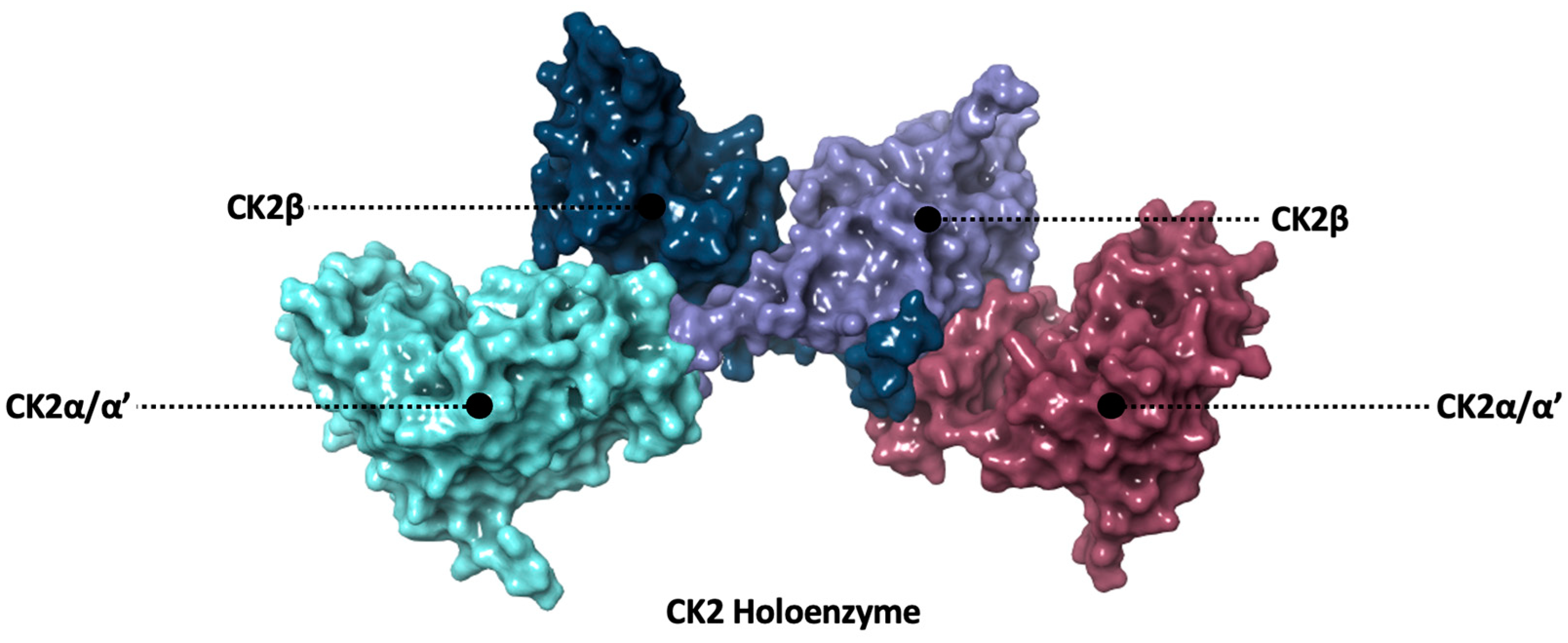
1.2. Structure of CK2
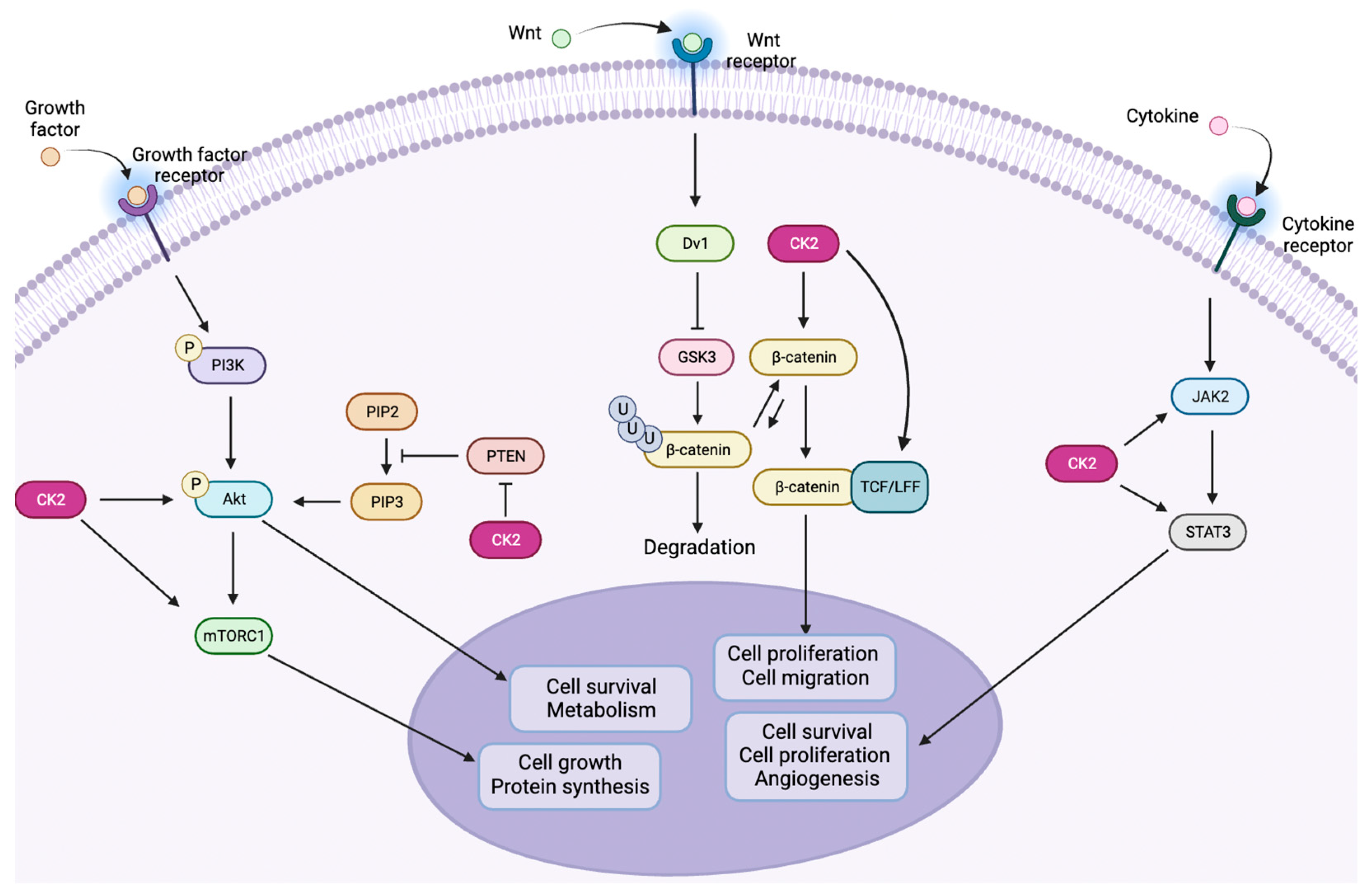
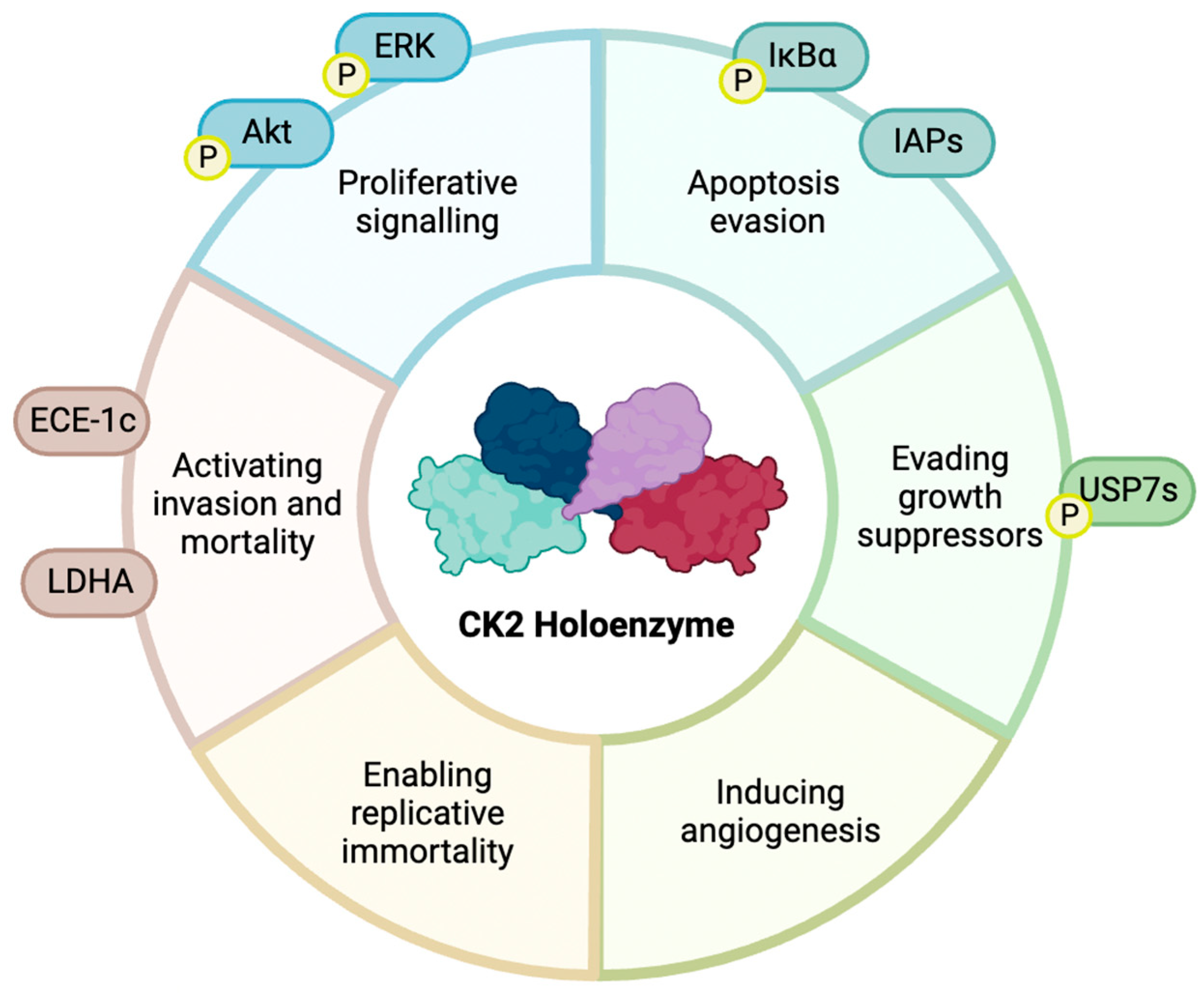
The ATP site comprises a deep hydrophobic binding pocket flanked by the hinge region and a catalytic Lys68 residue, making it an ideal location for aromatic compounds that contain hydrogen bonding substituents [8]. The Lys68 residue is essential for catalytic activity as it aids in the binding of the phosphates of ATP in the active site; this residue is conserved across all human protein kinases [30]. The structure of the ATP site is shown in Figure 3.
The α and α’ subunits are encoded by different genes, CSNK2A1 and CSNK2A2, respectively [31]. CK2α contains 391 amino acid residues and CK2α’ contains 350 amino acid residues; both isoforms share 90% sequence identity [10]. Both catalytic subunits adopt a bilobal structure that contains a lysine-rich N-terminus, and a C-terminus rich in α-helices. The ATP site is situated between the N-terminal and C-terminal domains, linked by the αD region, a region of the protein that has been exploited in the development of selective CK2 inhibitors [32]. The N-terminal segment contains five β-sheets, named β1-5, and one α-helix, named αC. The αC helix establishes substrate contact and, owing to its abundance of lysine residues, confers a tendency to favor acidic substrates. The C-terminal segment comprises seven α-helices and one β-sheet, which collectively form the structural foundation for both the ATP-binding site and activation loop. The activation loop is conformationally fixed by numerous stabilizing interactions between itself and the N-terminal segment. These stable interactions essentially “lock” CK2 in an active conformation, making it constitutively active without the need for an upstream phosphorylation event [8,33].
Many of CK2’s substrates do not require the presence of β-subunits for phosphorylation [34,35]. The β-subunits, each containing 215 amino acids, are encoded by the CSNK2B gene [36]. Although the presence of CK2β is not fundamental for substrate phosphorylation, the subunit plays a crucial role in enhancing the tetrameric complex’s thermostability, thus enhancing catalytic activity. Additionally, it aids in substrate recruitment by establishing both polar and hydrophobic interactions with the substrate, essentially acting as a “docking station” [37]. In this respect, substrates such as p53, nuclear phosphoprotein 140 (Nopp140), Fas-associated Factor 1 (FAS1), and topoisomerase II interact with CK2 via CK2β [28]. CK2β also regulates kinase activity by facilitating the shuttling of the kinase between the intracellular compartments, enabling the holoenzyme to enter the nucleus and gain access to most of CK2’s substrates [38,39].

1.3. CK2 in Cancer

The disease type in which the role of CK2 is most well-documented is cancer [40,41]. There is evidence to suggest that CK2 is directly involved in each of Hanahan and Weinberg’s six hallmarks of cancer: proliferative signaling, resisting cell death, evading growth suppressors, inducing angiogenesis, enabling replicative immortality, and activating invasion and mortality [42,43]. This involvement is highlighted in Figure 4.
The upregulation of CK2 has been found within the nucleus of a multitude of different cancer cell types, such as lung, breast, colorectal, and head and neck carcinomas [14,44,45,46]. Therefore, elevated levels of CK2 serve as a prognostic marker in cancer progression. However, CK2 is not considered an oncogene itself as it is ubiquitously expressed within healthy cells. Cancer cells exhibit a “non-oncogene addiction” to CK2, heavily relying on the kinase for survival [22,47].
Both CK2α and CK2α’ subunits are implicated in promoting tumor progression more than CK2β. The question of which isoform contributes most to CK2’s oncogenic potential remains the subject of ongoing debate [40]. However, the involvement of CK2α is more commonly reported than CK2α’. A study by Zou et al. showed that CK2α plays an essential role in the progression of colorectal carcinoma [48]. The suppression of CK2α via siRNA in LoVo cells led to G0/G1 phase arrest, induced cell senescence, elevated expression of p53/p21, and decreased expression of c-Myc. In 2015, Zhang et al. showed that silencing CK2α in hepatocellular carcinoma (HCC) induced HCC cell apoptosis and inhibited HCC cell migration, proliferation, and angiogenesis both in vitro and in vivo [49]. In line with more of a “tumor suppressor-like” role, CK2β prevents the phosphorylation of substrates vital for apoptosis evasion, such as caspase-3 [50]. Additionally, the role of CK2 in drug resistance has been linked to upregulation of CK2α, not CK2β [51]. However, CK2α’ has been shown to reduce migration in the mouse gonadotropin-releasing hormone (GnRH) neuronal cell line GN11, and CK2β promotes migration [36]. Furthermore, Takahashi et al. determined that the expression of CK2α and CK2β is elevated in human osteosarcoma, and siRNA knockdown of either CK2α or CK2β inhibited human osteosarcoma cell proliferation [52].
The pro-oncogenic function of each subunit cannot be objectively determined as this is dependent on cancer type, which CK2 substrates are present, and which isoform is needed to phosphorylate the substrates. However, CK2α/α’ and CK2β have differing roles in cancer progression, which is supportive of monomeric CK2 being more related to CK2’s oncogenic phenotype than tetrameric CK2.

2. Targeting CK2 within the ATP Site

2.1. ATP-Competitive Inhibition

The ATP site of CK2 is a primary target for inhibition as it is a highly ligandable pocket. As this site is conserved across the human kinome, selectivity issues often arise with ATP site binders.
Small-molecule ATP-competitive inhibitors of CK2 can be divided into four main classes: polyhalogenated benzimidazole and benzotriazole derivatives; anthraquinone derivatives; pyrazolo-triazines and pyrazolo-pyrimidines; and indoloquinazolines such as CX-4945. Some details of these classes of inhibitors are outlined below. However, for further details, the reader is directed towards an up-to-date and comprehensive review on ATP-competitive CK2 inhibitors [53].

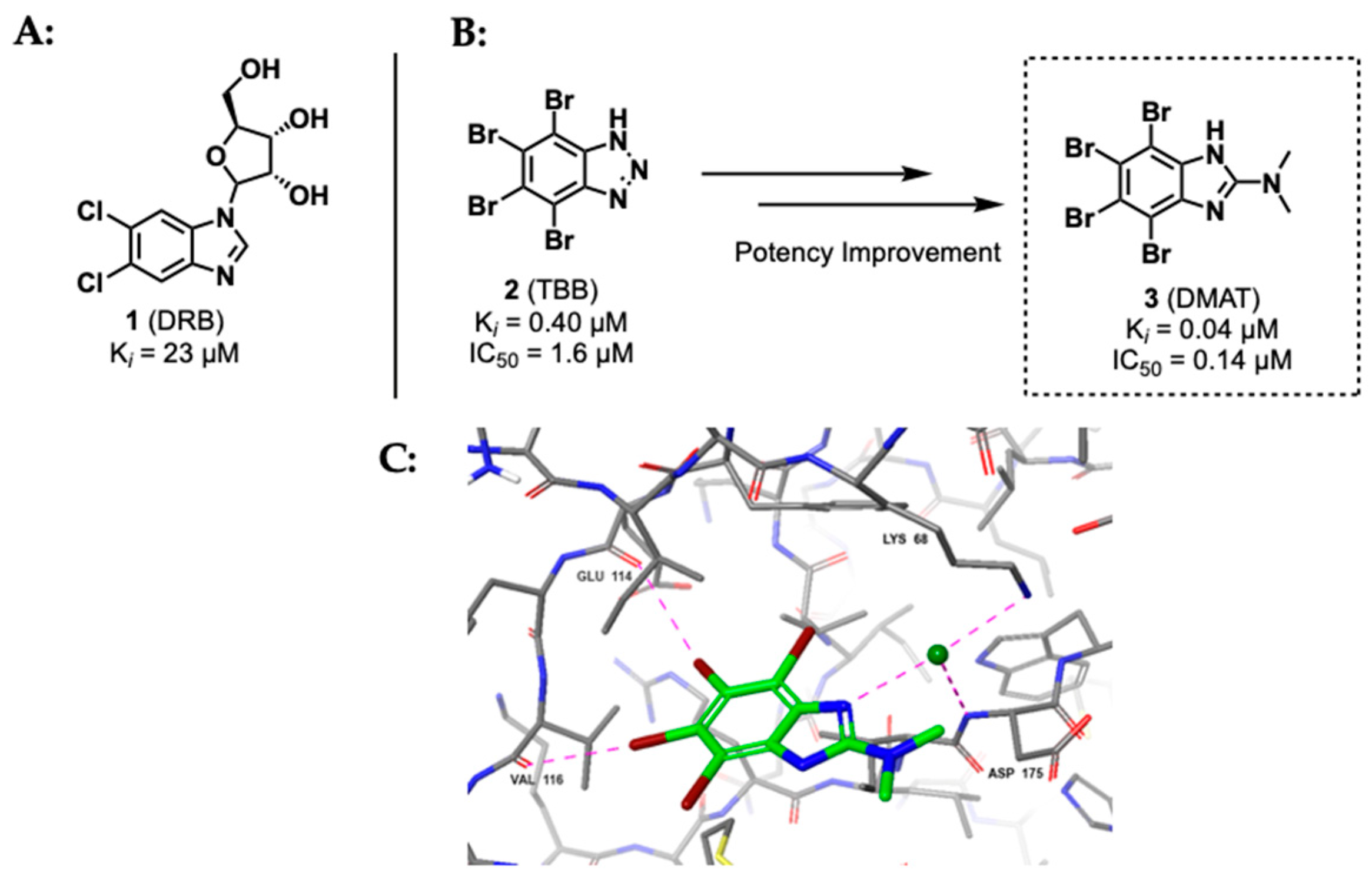
2.1.1. Polyhalogenated Benzimidazole and Benzotriazole Derivatives

The nucleoside analogue 5,6-dichloro-1-β-D-ribofuranosylbenzimidazole (DRB) 1, Figure 5, was initially recognized for its biological activity in 1954 [54]. However, it was not until 1986 when Zandomeni et al. reported inhibition of CK2 by DRB, although its activity against CK2 is only moderate (IC50 = 15 μM, Ki = 23 μM)* [55]. DRB has been found to be active against several other kinases (CK1, for example) with similar affinities to CK2. Additionally, it is also capable of binding to the CK2α/β interface. Despite its promiscuity, DRB has served as a foundation for many studies that have led to the development of numerous potent CK2 inhibitors that are in use today.
The first significant improvement regarding DRB stemmed from two key modifications: the elimination of the sugar moiety and the substitution of chlorine atoms with bulkier bromines. This resulted in the formation of 4,5,6,7-tetrabromo-1H-benzotriazole TBB 2, Figure 5, (IC50 = 0.14 μM, Ki = 0.40), and exhibited substantially improved cell permeability compared to DRB [56,57]. A screening of 10 μM TBB against 70 kinases unfortunately led to significant inhibition of over 90% of them, again highlighting the promiscuous nature of ATP-competitive kinase inhibitors.
Following the development of 4,5,6,7-tetrabromo-benzotriazole (TBB), subsequent structure–activity relationship studies led to the development of 2-dimethylamino-4,5,6,7-tetrabromo-benzimidazole (DMAT) 3, Figure 5, which exhibited even greater potency towards CK2 (Ki = 0.04 μM) [58]. However, DMAT was found to be less selective for CK2 compared to TBB; ten kinases were more than 90% inhibited by 10 μM DMAT, and eight kinases were more than 90% inhibited by 10 μM TBB [59]. Both TBB and DMAT have been used extensively and in vivo [60,61,62].

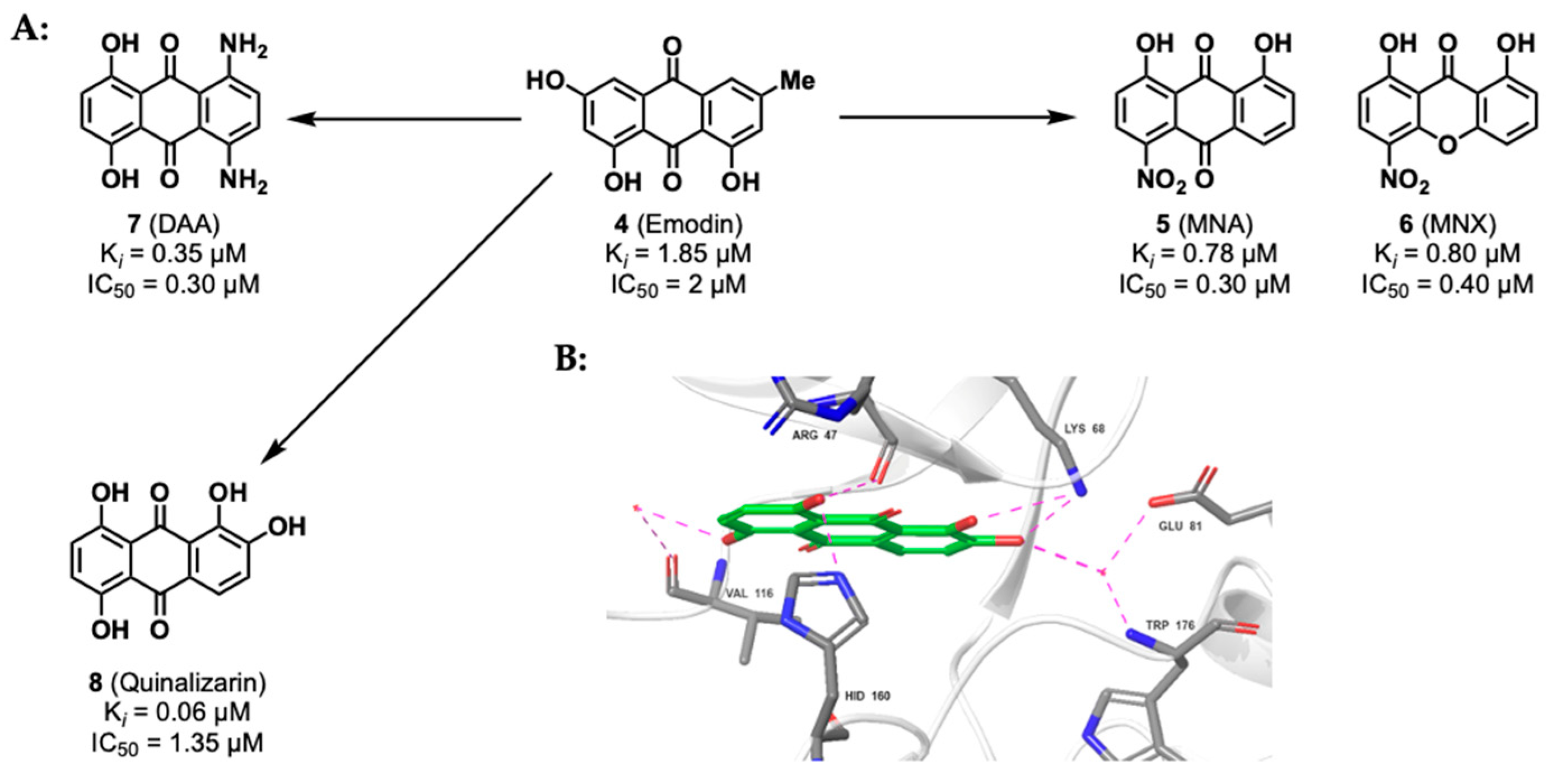
2.1.2. Anthraquinone Derivatives

Anthraquinones constitute a class of natural products containing phenolic substituents [63]. An important example is emodin 4, Figure 6, which is found in rhubarb rhizomes and inhibits numerous targets, including CK2 (IC50 = 2 μM, Ki = 7.2 μM) [64]. Several studies have been conducted to enhance anthraquinone-based inhibition of CK2, resulting in the development of MNA 5 (IC50 = 0.30 μM, Ki = 0.78 μM) and MNX 6 (IC50 = 0.40 μM, Ki = 0.80 μM), Figure 6 [65,66]. The presence of a nitro group in these derivatives increases polarization of the para-hydroxyl substituent through the electron-withdrawing effect. On the other hand, an alternative emodin derivative DAA 7, Figure 6, lacks a nitro group yet exhibits a lower Ki value (Ki = 0.35 μM, IC50 = 0.30 μM) than MNA and MNX [67]. This is attributed to the formation of additional hydrogen bonds to Glu114 and Val116.
Among further emodin derivatives is quinalizarin 8 (1,2,5,8-tetrahydroxyanthraquinone) (Ki = 60 nm), discovered via virtual screening [68]. Although structurally like emodin, quinalizarin is particularly potent and selective for CK2, more so than its predecessor. A selectivity profile of 1 μM quinalizarin was performed on a panel of 140 protein kinases, which found only 8% residual CK2 activity, and none of the other 139 kinases displayed residual activity of less than 50%. Moreover, quinalizarin showed the ability to differentiate between isolated CK2α and the CK2 holoenzyme [69]. Quinalizarin was found to be more active against the CK2 holoenzyme (IC50 = 0.15 μM and IC50 = 1.35 μM for CK2 holoenzyme and CK2α, respectively) and, therefore, could be used as a tool to quantify the presence of intracellular CK2 holoenzyme and CK2α.

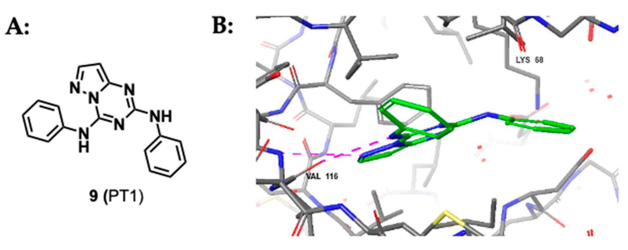
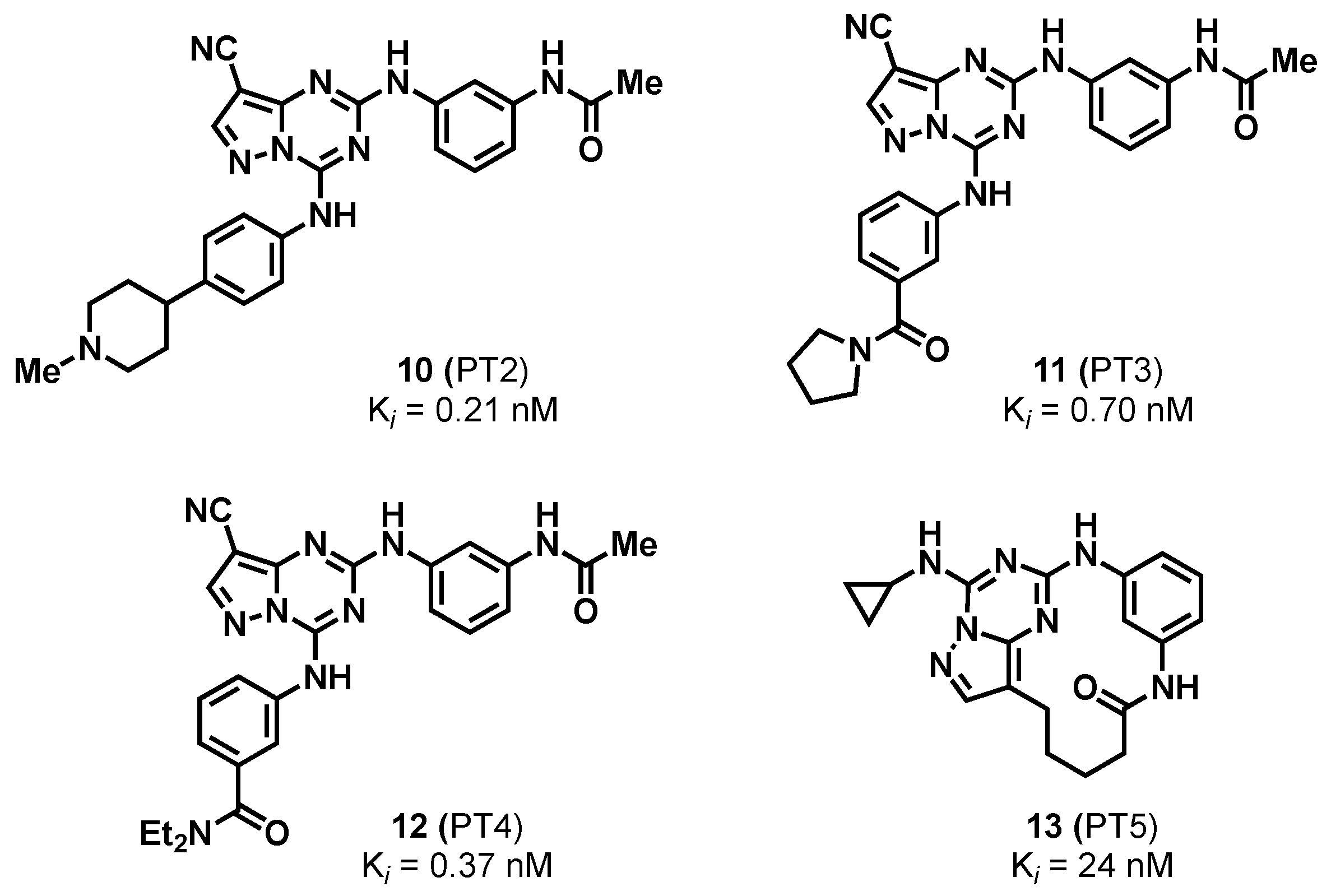
2.1.3. Pyrazolo-Triazines and Pyrazolo-Pyrimidines

A series of pyrazolo-triazines (PT) were designed for CK2 inhibition by Nie et al. in 2007 after a co-crystal structure of CK2 and pyrazolo-triazine 9 was obtained, Figure 7 [70]. The co-crystal structure revealed that the pyrazolo-triazine core occupied the adenine pocket within the ATP site and was anchored in place via two hydrogen bonds in the hinge region.
Subsequent structure–activity relationship studies led to the development of three compounds that differed at the phenyl substituent (PT2 10, PT3 11, and PT4 12, Figure 8) [70]. Although all the compounds displayed nanomolar Ki for CK2, they failed to exhibit potent inhibition of cell growth in the human colorectal carcinoma cell line HCT116 and human prostate cancer PC3 cell lines when tested using an MTT assay. Therefore, further improvement in physicochemical properties was required, resulting in the development of a second generation of pyrazolo-triazines, for example, PT5 13 [71]. Efforts to increase cell permeability resulted in a reduction in planarity in the second generation through macrocyclization with an alkyl side chain. Although this successfully increased cell permeability, a 10-fold increase in Ki was observed and PT5 had comparable cell growth inhibition to the first-generation derivatives. Pyrazolo-triazines have the potential to be developed into successful CK2 inhibitors; however, binding affinity must first be optimized. To date, no such optimization of pyrazolo-triazines has been published, and there are no data detailing the selectivity of this class of compounds.
Dowling et al. identified a related group of compounds known as pyrazolo-pyrimidines. In their work, kinase-focused subset screening methods, structure–activity relationship studies, and crystallography were employed [72]. Using these methods ultimately led to the identification of a lead compound AZ-7h 14, Figure 9, which has picomolar binding affinity to CK2 (Kd = 6.33 pM), and nanomolar growth inhibition activity in HCT116 (GI50 = 10 nM, HCT116) [73]. The compound’s selectivity was tested at 0.1 μM against a panel of 402 kinases, which showed only 12 kinases being inhibited by more than 50%, of which all were CMGC family members. Although limited off-target activity was observed, AZ-7h showed nanomolar activity for death-associated protein kinase 2 (DAPK2) and DAPK3 (IC50 = 8.0 nM and IC50 = 18 nM, respectively). In vivo mouse PK studies showed poor oral bioavailability of AZ-7h [73]. However, dose-dependent tumor growth inhibition was achieved through intravenous and intraperitoneal injection. This suggests that further optimization of AZ-7h could yield a promising CK2 inhibitor; however, activity against DAPK2 and DAPK3 should be reduced.
Through optimization of the pyrazolopyrimidine scaffold of AZ-7h, the Structural Genomics Consortium (SGC-UNC) developed SGC-CK2-1 15, Figure 10, (IC50 = 4.2 nM, and IC50 = 2.3 nM for CK2α and CK2α’, respectively) [74]. Sub-micromolar target engagement of CK2 was determined using nanoBRET assays in human embryonic kidney 293 (HEK-239) cells (IC50 = 36 nM and IC50 = 16 nM for CK2α and CK2α’, respectively), and, when tested against a panel of 403 wild-type human protein kinases at 1 μM, only three kinases were found to be inhibited by more than 90%. The authors claim that, unexpectedly, SGC-CK2-1 “does not demonstrate significant antiproliferative activity against a panel of 140 different cancer cell lines”. This claim sparked premature conclusions that the broad cancer essentiality of CK2 had therefore been disproved [75]. However, further investigation is needed to determine the potential effects of SGC-CK2-1 on cancer given that CK2 is implicated in all the recognized cancer hallmarks [44] beyond just influencing cell proliferation. The reader is directed towards a comprehensive review regarding this argument [76]. It is important to note that SGC-CK2-1 is an exceptional tool and represents the most selective CK2 inhibitor to date and would therefore be a first-choice compound for the exploration of CK2 cellular functions. A further review, which provides further details on the development and uses of SGC-CK2-1, is brought to the attention of the reader [77].

2.1.4. Indoloquinazolines Such as CX-4945

CX-4945 16, Figure 11, an indoloquinazoline, also known as silmitasertib, was developed by Siddiqui-Jain et al. in 2010 and is a potent orally bioavailable ATP-competitive inhibitor of CK2 (IC50 = 1 nM, Ki = 0.38 nM) [78]. CX-4945 is characterized by its comparatively small polar surface area, few rotatable bonds, and low aqueous solubility. It possesses a single carboxylic acid and two aromatic nitrogen heterocycles, which are both weakly basic. CX-4945′s potent binding affinity is attributed to the formation of an ionic bridge with Lys68, in addition to hydrophobic interactions and hydrogen bonding with the hinge region of the ATP site [27,28,73].
CX-4945 was granted orphan drug designation by the FDA for advanced cholangiocarcinoma in 2017 [79]. In 2021, CX-4945 demonstrated excellent preclinical anti-tumor activity and strong synergism with gemcitabine and cisplatin in phase Ib/II clinical studies (ClinicalTrials.gov identifier: NCT02128282) [80,81]. This combined therapeutic approach has the potential to be a promising first-line treatment against locally advanced and metastatic cholangiocarcinoma.
When screened in a selectivity panel of 238 kinases, CX-4945 was shown to inhibit a further seven kinases by over 90% at a concentration of 500 nM. The most significant inhibition was against dual-specificity tyrosine regulated kinases DRYK1A and DRYK1B; proviral integration site for Moloney murine leukemia virus kinase 1 (PIM-1), and CLK2 [27]. Despite suboptimal selectivity, CX-4945 is currently viewed as the gold standard of CK2 inhibitors due to its strong inhibitory activity and oral bioavailability. This illustrates a potential shift in perspective regarding off-target kinase activity as only moderate adverse side effects have been observed in CX-4945 clinical trials, and targeting multiple kinases with related substrate phosphorylation patterns could be considered advantageous [82]. Nonetheless, the development of more selective inhibitors is crucial for the advancement of CK2 chemical probes.
Recently, Menyhart et al. evaluated the selectivity of CX-4945 and SGC-CK2-1 via triple SILAC quantitative phosphoproteomics using human osteosarcoma U2OS cells expressing exogenous wild-type CSNK2A1 or an inhibitor-resistant triple mutant (TM, V66A/H160D/I174A) [83]. Only a minority of the phosphosites that were downregulated in response to CX-4945 treatment (15% at 4 h and 5% at 24 h) were found to be CSNK2A1-dependent. However, the majority of the phosphosites downregulated in response to SGC-CK2-1 treatment (more than 55% after 4 h and 24 h) were found to be CSNK2A1-dependent. Thus, SGC-CK2-1 is significantly more selective for CK2 than CX-4945.
Following the development of CX-4945 16, structure–activity relationship studies led to the development of CX-5011 17 and CX-5279 18, (Ki = 0.18 nM and Ki = 0.22 nM for CX-5011 and CX-5279, respectively) [84], Figure 12. Both new derivatives contain a pyrimidine ring instead of pyridine and showed better specificity for CK2 than CX-4945 (the Gini coefficients were 0.735, 0.755, and 0.615 for CX-5011, CX-5279, and CX-4945, respectively [85]), which improves upon one of the main downfalls of CX-4945 [69,84].

3. Extending beyond the ATP Site

3.1. Dual-Binding Ligands

The development of dual-binding therapeutics, especially those that inhibit synergistic targets, has received increasing attention in anticancer drug development. Many dual-binding bromodomain and extra-terminal domain (BET)/kinase inhibitors have been described, which simultaneously regulate multiple targets in disease pathways and have synergistic anti-tumor effects [86,87,88]. A dual-binding ligand approach could be a more effective therapeutic strategy than single-target therapeutics. To date, several CK2-based dual-binding ligands have been reported in the literature that have the potential to be successful clinical candidates.

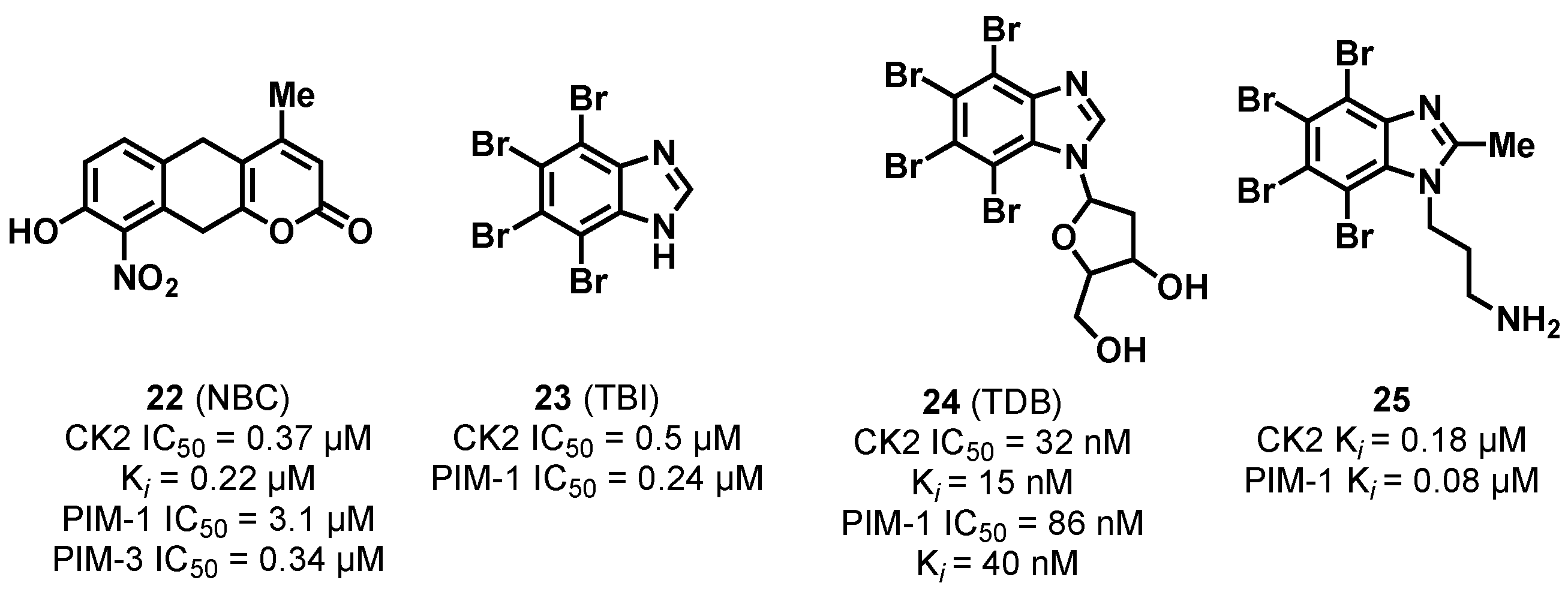
3.1.1. CK2/PIM Dual-Binding Ligands

Proviral integration site for Moloney murine leukemia virus (PIM) kinases are serine/threonine kinases, much like CK2, which interact with c-Myc [89,90]. PIM kinases are attractive targets in diseases that have high PIM expression, such as leukemia and prostate cancer [91,92]. CK2 and PIM both act on similar pathways, which, when dysregulated, result in tumorigenesis [93,94]. Off-target PIM inhibition has been observed in selectivity studies of CK2 inhibitors, making dual-binding CK2/PIM kinases a feasible therapeutic strategy [27].
CK2/PIM dual-binding inhibitors were first reported in the literature by Lopez-Ramos et al. in 2010 [95]. A series of CK2/PIM inhibitors (CPA 19, CPB 20, and AMR 21, Figure 13), which had nanomolar IC50 values for CK2 and PIM kinases, were developed through automated in vitro screening and hit expansion techniques. Unfortunately, these CK2/PIM dual-binding inhibitors are not suitable for use in the clinic due to poor cell permeability.
NBC 22, Figure 14, was initially developed as a CK2 inhibitor alongside anthraquinone derivative MNX 6 by Meggio et al. in 2004 [65]. NBC displayed potent inhibitory activity against CK2 (IC50 = 0.37 μM, Ki = 0.22 μM) but was found to inhibit PIM kinases, particularly PIM-1 and PIM-3 (IC50 = 3.1 μM and 0.34 μM, respectively). Furthermore, NBC induced apoptosis in Jurkat cells and displayed DC50 values around 15 μM, making NBC a promising CK2/PIM dual-binding ligand [96].
The inhibitory potential of the known CK2α inhibitor TBI 23, Figure 14, was explored by Cozza at al., where significant off-target activity against PIM-1 was identified [97]. The selectivity of TBI was then tuned towards CK2 and PIM-1 via inclusion of a deoxyribose to yield TDB (CK2 IC50 = 32 nM, PIM-1 IC50 = 86 nM) [98]. TDB 24, Figure 14, was found to show permanent inhibition of cell survival and migration of U2OS cells to a greater extent than “single-target” CK2 inhibitors, such as CX-4945, which is thought to be attributed to the synergistic effect of simultaneous CK2 and PIM-1 inhibition. However, CX-4945 displayed greater in vitro potency than TBI despite lower cytotoxicity [99].
Further analogues of CK2/PIM-1 dual-binding inhibitors were developed by Chojnaki et al. in 2018 based on the structure of TBI [100]. Aminopropyl-substituted derivative 25 was identified, which exhibits potent inhibition of CK2 and PIM-1 (Ki = 0.18 and 0.08 μM for CK2 and PIM-1, respectively). In vitro studies showed that 25, Figure 14, inhibited cell proliferation in human T-lymphoblasts CCRF-CEM, human breast cancer MCF-7, and PC3 cancer cell lines, and cell viability was reduced more significantly when compared with TBI and CX-4945, proving the synergistic effect of CK2/PIM-1 that dual-binding inhibitors could have in terms of a clinical benefit over ‘single-target’ CK2 inhibition.

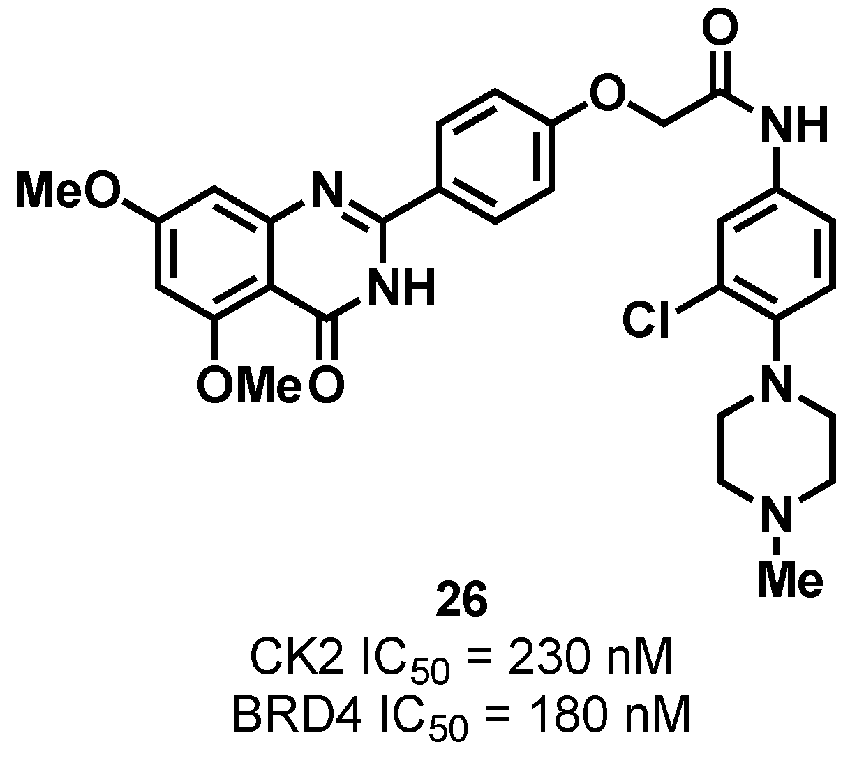
3.1.2. CK2/BRD4 Dual-Binding Ligands

Bromodomain-containing protein 4 (BRD4), a member of BET family, is a histone acetyltransferase (HAT) that plays a pivotal role in embryogenesis and cancer development [101,102]. Studies show that CK2 is responsible for the phosphorylation of BRD4 [101,103,104]. Hyperphosphorylation of BRD4 by CK2 is associated with drug resistance in triple-negative breast cancer (TNBC), making CK2/BRD4 dual-binding ligands a feasible therapeutic approach in breast cancer treatment [14,44,105,106].
Moreover, 26, Figure 15, is a CK2/BRD4 dual-binding inhibitor, first described by Zhang et al. in 2021, and was developed via rational drug design and structure–activity relationship studies [107]. Furthermore, 26 was found to possess potent and similar inhibition against CK2 (IC50 = 230 nM) and BRD4 (IC50 = 180 nM). In in vitro experiments, 26 was shown to inhibit proliferation and induce dose-dependent apoptosis and autophagy-related cell death in human TNBC MDA-MB-231 and MDA-MB-468 cells. In vivo experiments further validated a therapeutic effect, with potent anticancer activity being observed without any obvious toxicities. Hence, CK2/BRD4 dual-binding ligands have emerged as an appealing strategy for treating TNBC.

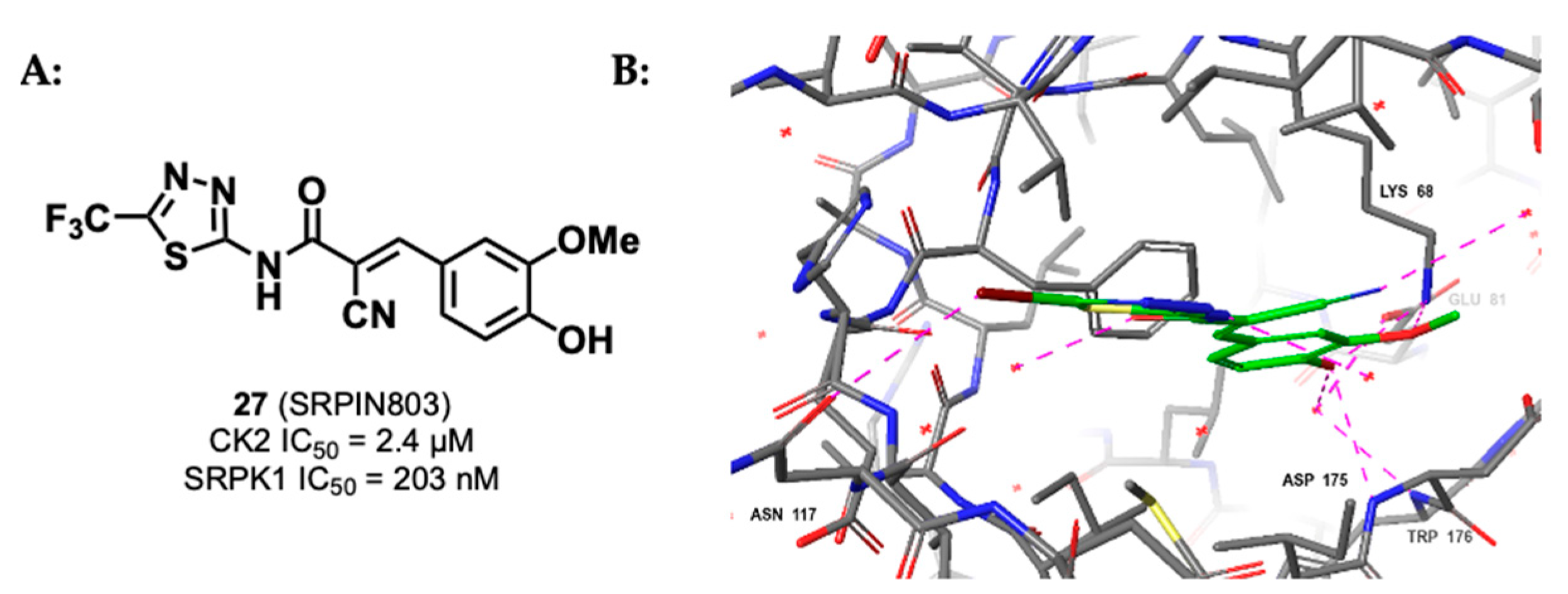
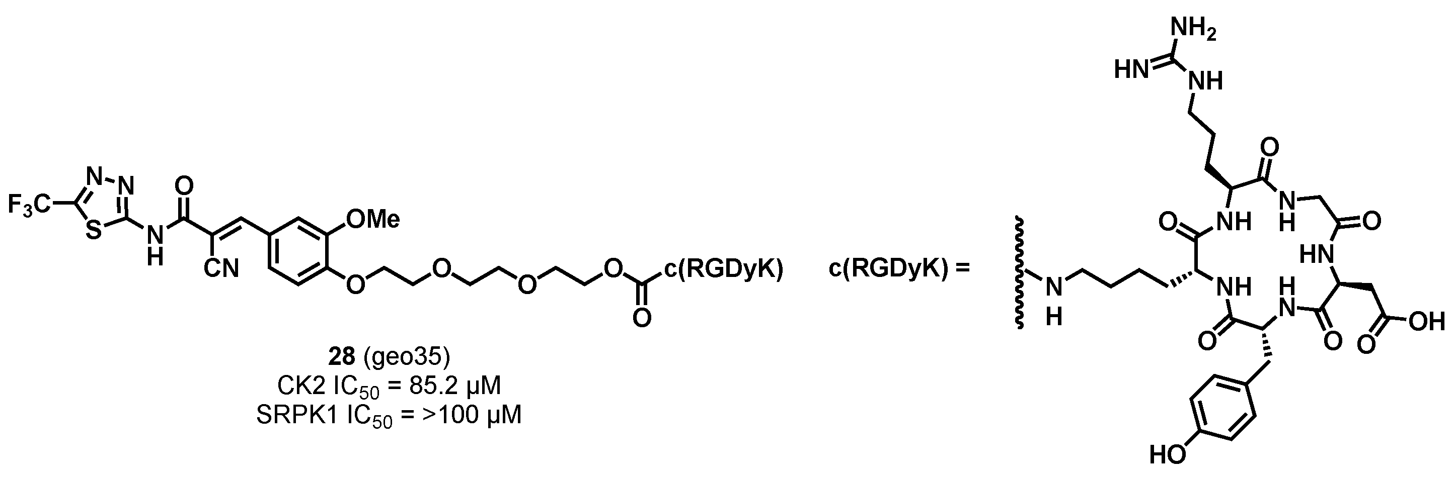
3.1.3. CK2/SRPK1 Dual Inhibitor

Serine-arginine protein kinase 1 (SRPK1) is an oncoprotein that plays a key role in the epidermal growth factor (EGFR)/Akt pathway, contributing to tumorigenesis [108]. SRPK1 is involved in angiogenesis and is overexpressed in numerous cancer types, such as prostate, breast and lung cancers, and glioma [109,110]. Furthermore, CK2 has been found to be the main kinase that phosphorylates SRPK1 [111]. Therefore, CK2/SRPK1 dual inhibitors could be used as anti-angiogenic therapies.
SRPIN803 27, Figure 16, was identified by Morooka et al. as a CK2/SRPK1 dual inhibitor (IC50 = 2.4 μM and 203 nM for CK2 and SRPK1, respectively) through pharmacophore docking models followed by in vitro kinase assays [112]. Exposure of SRPIN803 to Jurkat cells resulted in a significant reduction in cell viability, and suppression of vascular endothelial growth factor (VEGF) production was observed in a dose- and time-dependent manner in human retinal pigment epithelial ARPE-19 cells. Furthermore, genes, including IL8, HMOX1, and HK2, involved in angiogenesis were downregulated according to gene-expression array data. In an in vivo mouse model of age-related macular degeneration (AMD), SRPIN803, administered in the form of topical eye ointment, significantly inhibited choroidal neovascularization. Thus, SRPIN803 merits further investigation as an inhibitor of VEGF and as a topical ointment for ocular neovascularization in a clinical setting.
In 2021, Leonidis et al. developed a series of c(RGDyK) peptide conjugates of SRPIN803, of which geo35 28, Figure 17, was the most cytotoxic against MC7 and MRC5 cell lines (IC50 = 61 and 63 μM, respectively) [113]. Furthermore, the most active compound, geo35 (CK2 IC50 = 85.2 μM and SRPK1 IC50 = >100 μM), induced antiangiogenic activity in zebrafish embryos in a dose-dependent fashion. However, SRPIN803 alone was found to be more potent than geo35. Furthermore, geo35 displayed weak binding affinity to SRPK1, and it is unknown whether the observed cytotoxicity can be attributed to inhibition of both CK2 and SRPK1 or just CK2 alone. However, the development of an active peptide conjugate suggests that this approach could be adopted to develop a new class of antiangiogenic therapeutics.

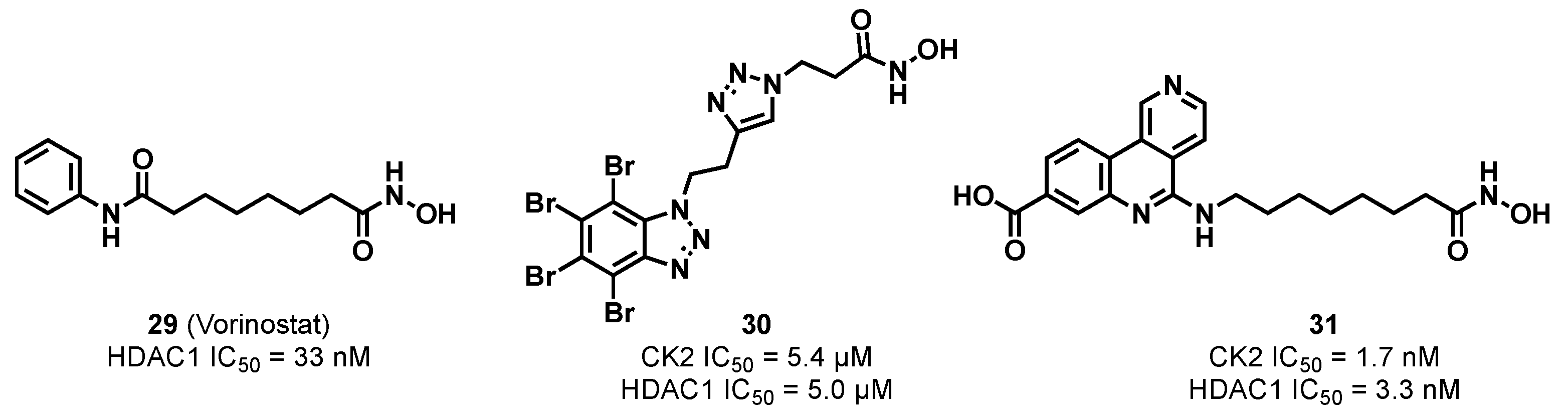
3.1.4. CK2/HDAC1 Dual Inhibitor

Histone deacetylases (HDACs) are a family of epigenetic proteins that control gene transcription and regulation, alongside cell proliferation, differentiation, migration, death, and angiogenesis [114]. As CK2 and HDAC are both related in similar cancer-related biological pathways, it is speculated that simultaneous inhibition of these targets by a dual inhibitor should improve efficacy compared to single-target therapeutics [115].
Recently, the Ramos group developed a series of CK2/HDAC1 inhibitors through combining the structures of vorinostat 29, Figure 18, a known HDAC1 inhibitor, and TBB or DMAT, known CK2 inhibitors [116,117,118,119]. The series, to which compound 30, Figure 18, belongs, had low micromolar activity in enzymatic assays and low micromolar LC50 values across numerous cell lines. In 2020, the Ramos group reported another series of CK2/HDAC1 dual-binding inhibitors in which CX-4945 replaced the DMAT-derived portion [120]. The lead compound, 31, Figure 19, displayed 3.0- and 3.5-times higher activity against recombinant CK2 and HDAC1 than CX-4945 and vorinostat, respectively. However, the in vitro inhibition observed with 31 in human lymph node carcinoma of the prostate (LNCaP) cells (IC50 = 16.31 μM) was lower than the individual parent compounds, which could be attributed to poorer cell permeability of the lead compound. Therefore, further optimization of the pharmacokinetic properties of 31 is needed before it could be a potential therapeutic.

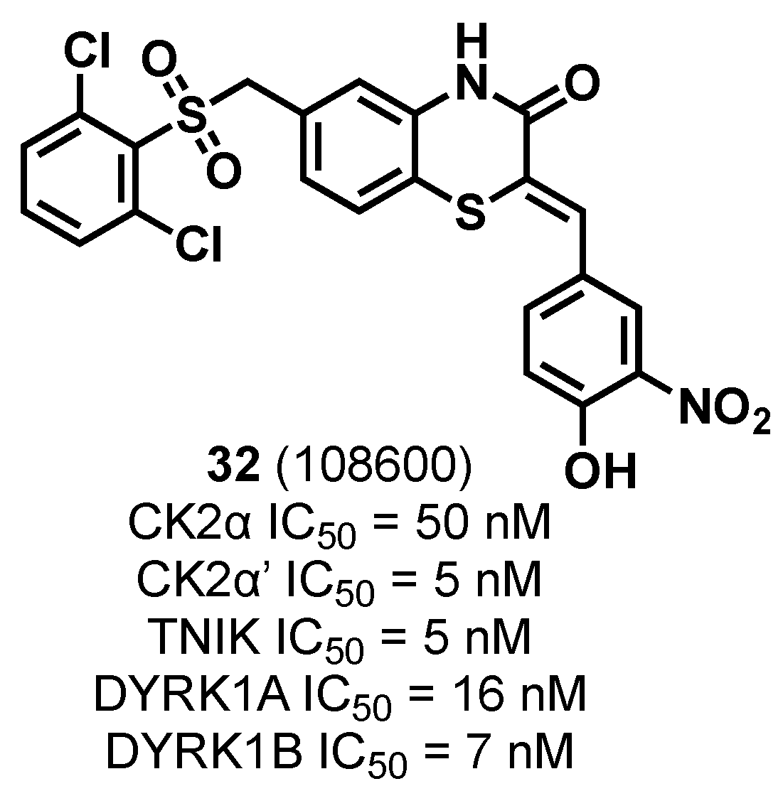
3.1.5. CK2/TNIK/DYRK1 Multiple Inhibitor

Dual-specificity tyrosine-regulated class 1 kinases (DYRK1) have multiple implications in cell differentiation and proliferation pathways via phosphorylation of key cell cycle regulators, such as CYCLIN D1 and p21, and regulate their degradation [98,121]. DYRK1 kinases also regulate chromatin modification and gene expression [122]. TRAF2- and NCK-interacting kinase (TNIK) has emerged as a kinase that is critical in the regulation of Wnt/β-catenin cell signaling pathway [123]. Collectively, CK2, DYRK1, and TNIK kinases activate STAT3, Wnt/β-catenin, PI3K/Akt, Hedgehog, and Notch 1 signaling molecules. Therefore, simultaneous inhibition of these three kinases, and their associated cancer-promoting pathways, could sensitize malignant drug-resistant cancer cells to standard-of-care chemotherapy.
A novel multi-kinase inhibitor, 108600 32, Figure 19, inhibited CK2, TNIK, and DYRK1 activity in a dose-dependent manner in triple-negative breast cancer cell lines [124]. Moreover, 108600 displayed IC50 values for CK2α, CK2α’, TNIK, DYRK1A, and DYRK1B of 50, 5, 5, 16, and 7 nM. Surprisingly, 108600’s inhibitory activity against the CK2α isoform is 10-fold higher than the CK2α’ isoform, indicating potential for isoform-selective derivatives to be developed. An unfavorable conformational change in CK2α was observed upon binding of 108600, which is not conductive with holoenzyme formation [124]. 108600 could, therefore, be acting as an ATP-competitive inhibitor and as an allosteric inhibitor. Treatment of LM2-4 cells, a highly metastatic variant of MDA-MB-231, showed that 108600 was an effective suppressor of established metastases when used in combination with paclitaxel. In an in vivo patient-derived xenograft (PDX) mouse model, 108600 successfully acted synergistically with paclitaxel, even when tumors were non-responsive to paclitaxel as a single agent. This may prove to be highly advantageous in a clinical setting and could lead to improvements in morbidity and mortality rates associated with advanced TNBC, and those that are resistant to chemotherapy [125].

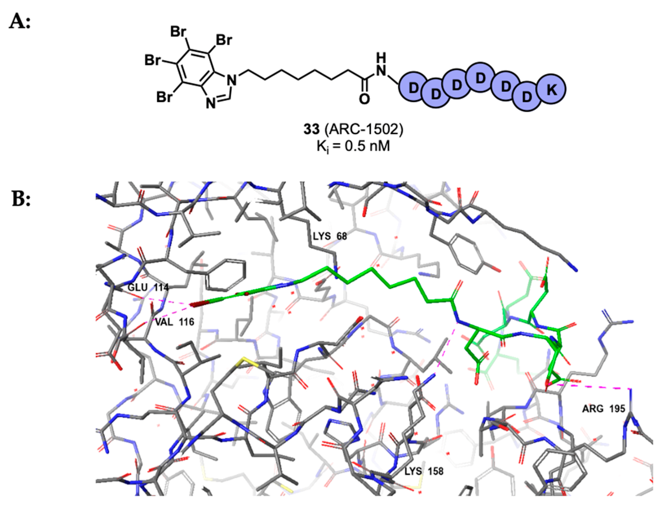
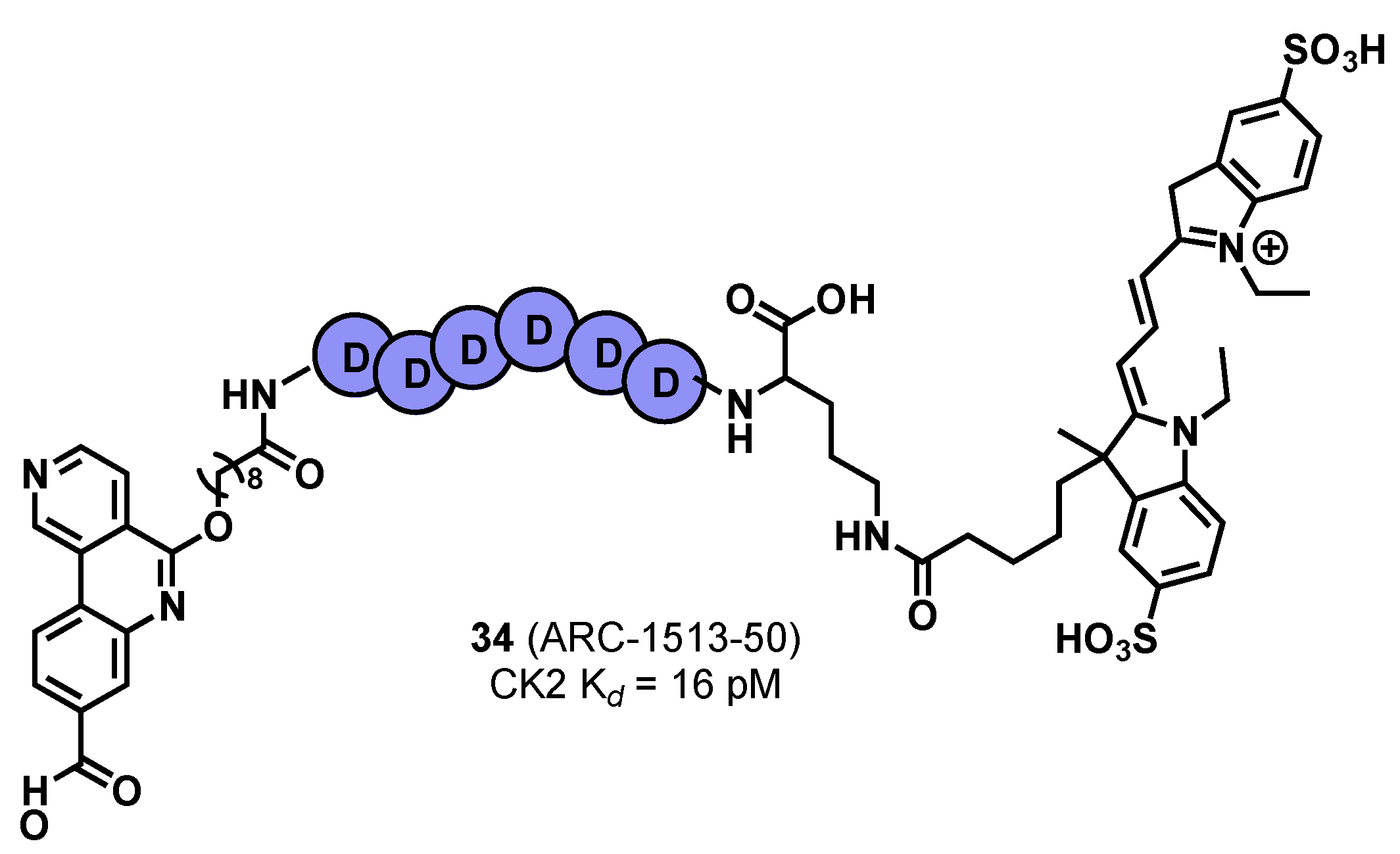
3.2. Substrate Binding Site Inhibition

An alternative approach for CK2 inhibition is to target the substrate binding site and therefore inhibit the binding of CK2 substrates. One defining feature of CK2 is that it solely phosphorylates acidophilic substrates [10]. Therefore substrate-competitive inhibitors with great selectivity could be developed to regulate CK2’s activity.
Polyglutamyl peptides were first identified as substrate-competitive inhibitors by Meggio et al. in 1983 [126]. (Glu)70 was found to inhibit CK2 in vitro (Ki = 0.11 μM) and did not have activity against closely related proteins, such as CK1. This highlighted that naturally occurring polypeptides that contain long stretches of acidic units could act as substrate-competitive CK2 inhibitors.
Perea et al. identified peptide P15 (WMSPRHLGT) through screening of a random cyclic peptide phage display library [127]. P15 was found to inhibit CK2 substrate phosphorylation in vitro in a competitive manner. The known cell-penetrating peptide TAT was attached to the N-terminus of P15, along with two cysteine residues attached to either end of P15. Cyclization of the modified peptide via a disulfide bridge afforded the cell-permeable peptide CIGB-325 (formerly known as P15-TAT and CIGB-300), which displayed moderate inhibition of cell viability across different cancerous cell lines. Additionally, intra-tumoral administration of CIGB-325 into TC-1 lung epithelial tumors in C57BL6 mice led to a significant reduction in tumor growth [127].
In 2008, CIGB-325 was administered to patients with cervical cancer in its first-in-man clinical trial [128]. It was observed that 75% of the patients had significant lesion reduction, and 19% of the patients showed full histological regression. In 2020, a preclinical trail was conducted to evaluate the potential of CIGB-325 as a treatment for breast cancer [14]. CIGB-325 demonstrated reduced breast cancer cell growth across the MDA-MB-231, MCF-7, and sarcomatoid mammary carcinoma F3II cell lines, and exhibited apoptosis and cell cycle arrest in vitro. In malignant cells, cell adhesion, migration, and clonogenic capacity were reduced. Promisingly, in an orthotopic model, treatment of CIGB-325 resulted in the inhibition of breast cancer colonization of the lung as the size and number of metastatic lesions was reduced. These encouraging results propose CIGB-325 as an adjuvant therapy post-surgery to limit tumor metastasis and recurrence.
In 2020, an exploratory clinical trial of CIGB-325 was conducted to investigate the use of the synthetic peptide against COVID-19 [129]. CIGB-325 was administered intravenously to COVID-19 patients, along with standard-of-care treatment, over a period of 15 days. Significantly fewer pulmonary lesions were observed at day 7 in patients who had received CIGB-325 and standard-of-care treatment, indicating that CK2 inhibitors could be used in combinatorial therapy against COVID-19.

3.3. Bi-Specific ATP/Substrate Competitive Inhibition

The substrate binding channel of CK2 has been defined based on the substrate binding channels identified in other kinases [130]. However, to date, there are no published crystal structure data to show a substrate of CK2α, or substrate channel inhibitor, bound in this channel. Bi-specific ATP/substrate-competitive inhibitors have emerged as a therapeutic strategy for selective CK2 inhibitors. These inhibitors incorporate both an ATP-competitive binder and an acidophilic peptide to bind in an ATP- and substrate-competitive manner. Due to the lack of crystal structure data, it is difficult to predict how much binding in the substrate channel contributes to the overall binding affinity of these inhibitors.
The first bi-specific ATP/substrate-competitive inhibitor was ARC-1502 33, Figure 20, developed by Enkvist et al. in 2012 [131]. ARC-1502 (Ki = 0.5 nM) contained the ATP-competitive inhibitor TBI conjugated to an aspartic acid-rich peptide. Alongside a strong binding affinity for CK2α, ARC-1502 exhibited good selectivity: when tested at 1 μM against a panel of one-hundred-forty kinases, only nine other kinases were inhibited at more than 50%. Unfortunately, ARC-1502 is not cell-permeable and is susceptible to proteolytic degradation; therefore, its uses as a chemical probe are limited.
Following ARC-1502, Viht et al. developed a cell-permeable and stable derivative of ARC-1502, named ARC-1859 [132]. This improved bi-specific inhibitor was designed as a pro-drug: the acidic aspartic acid side chains were ‘masked’ with cell-cleavable acetoxymethyl esters. The stability of the peptide was also improved through the replacement of amino acids with a poly-N-substituted glycine peptoid chain. In vitro, ARC-1502 was found to inhibit the phosphorylation of CK2α substrates Cdc37 and NFκB in a concentration-dependent manner.
In 2017, Vahter et al. developed a series of bi-specific ATP/substrate-competitive inhibitors using CX-4945 as the ATP site binder [133]. The most promising inhibitors of the series, ARC-1424-50 and its fluorescently labelled counterpart ARC-1513-50 34, Figure 21, showed outstanding binding affinity for CK2 (Kd = 37 and 16 pM, respectively). When tested at 1 μM in a panel of one-hundred-forty kinases, twenty-three kinases were inhibited at more than 50% and six were strongly inhibited at more than 90%. Therefore, ARC-1424-50 is one of the best chemical tools for competition experiments to measure binding affinities of other CK2α inhibitors due to its picomolar activity.

3.4. Inhibitors Acting in the αD Site

A pocket adjacent to the ATP site, named the αD site, was discovered by Brear et al. in 2016 [134]. The site was found through high-concentration fragment screening and is unique to CK2α/α’. Therefore, targeting this site could possibly lead to the creation of highly selective CK2 inhibitors. Most reported inhibitors that bind to the αD site simultaneously bind to the ATP site. There is one reported compound that is proposed to bind to the αD site and allosterically inhibit CK2; however, there are no structural data to support this hypothesis, and it remains unclear whether the αD site can act as an allosteric site alone [135].
Through crystallographic fragment screening efforts, De Fusco et al. successfully identified fragments 35 and 36 that bind to the ATP site, shown in Figure 22 [32]. Remarkably, several cell-permeable small molecules with picomolar affinity to the ATP site have been discovered. These fragments are ATP-competitive as they compete with endogenous ATP and thus prevent phosphorylation of CK2’s substrates.
A fragment-based drug discovery approach, via collaboration of the Spring and Hyvönen groups, resulted in a series of benzylamine biaryl molecules that bind in the αD site [32,134]. Due to the proximity of the ATP and αD sites, a weak ATP site fragment 36, Figure 22, was linked to an αD fragment. It was hypothesized that linking fragments that bind either the αD or ATP site would result in a selective CK2 inhibitor; selectivity would be driven by binding to the αD pocket. This proved true for the resulting inhibitor CAM4066 37, Figure 23, which exhibited subnanomolar affinity in enzymatic assays and low micromolar cellular activity (Kd = 320 nM, IC50 = 370 nM, GI50 = 8.8 μM for pro-CAM4066). Excitingly, CAM4066 possessed a Gini coefficient of 0.82 (52 kinase panel, 2 μM CAM4066), making CAM4066 the most selective CK2 inhibitor to date. The promising selectivity and biological data suggest that the unique binding mode of CAM4066 serves as an effective strategy for achieving potent selectivity and good inhibition when developing kinase inhibitors.
Bancet et al. recently discovered AB668 38 (Ki = 41 nM, IC50 = 65 nM), Figure 23, which also binds to the αD pocket and ATP site [136]. AB668 was screened against a panel of 468 kinases at a concentration of 2 μM, and only one other kinase (RPS6KA5) displayed a percentage inhibition above 50%. Additionally, AB668 was found to induce apoptotic cell death in 786-O renal carcinoma (at a concentration of 4 μM) and A375 melanoma (at a concentration of 5 μM) cell lines after 48 h, detected via live cell imaging. Interestingly, AB668 was better at inducing apoptosis in both cell lines than CX-4945 and SGC-CK2-1. Furthermore, AB668 did not induce cytotoxicity in extracts of non-cancerous HEK-293 cells, and the viability of normal human breast epithelial cells (MCF10A) was unchanged at high concentrations of AB668. These results suggest that only cancerous cell lines are sensitive to AB668. However, further in vitro and in vivo studies would be needed to better understand this anticancer effect.

3.5. Holoenzyme Assembly Inhibition

Targeting the CK2α/β interface is an alternative strategy of CK2 inhibition and disrupts formation of the holoenzyme complex.
One notable peptide that targets the CK2α/β protein-protein interaction (PPI) is CAM7117 39, Figure 24, developed by the Spring and Hyvönen groups [137]. CAM7117 was developed through the optimization of peptide Pc (IC50 = 3.0 μM, Kd = 1 μM), previously identified by Laudet et al. [138]. Pc contains the central interacting region of the CK2β C-terminal loop cyclized via a disulfide bridge. In the development of CAM7117, a structure-based approach was adopted to determine which covalent constraint would hold the peptide in an optimal conformation. Additionally, molecular modeling and X-ray crystallography of the peptide sequence revealed a single point mutation within the central region of Pc, namely Ile192 to Trp. Using copper-catalyzed azide-alkyne cycloaddition (CUAAC) chemistry and the movement of one of the cyclizing residues by one position yielded CAM7117 (Kd = 0.2 μM, GI50 = 33 μM). Unfortunately, CAM7117 contains unnatural amino acids, and its size limits any further optimization attempts.
In 2022, the Spring group developed another peptide-based CK2α/β PPI inhibitor, P8C9 40, Figure 25, through an iterative cycle of enzymatic assays, X-ray crystallography, molecular modelling, and cellular assays [139]. The peptide successfully binds CK2α at the CK2α/β PPI interaction site and is easily functionalized, highly stable in serum, and small enough to be further optimized. Cell-permeable analogues were synthesized, TAT-P8C9 and R3-P8C9, which successfully inhibited cell proliferation. These analogues can serve as chemical probes to aid the development of novel CK2α/β PPI inhibitors that can be used as therapeutics.
In addition to peptides targeting the CK2α/β interface, Kufareva et al. developed a small molecule probe 41, Figure 26, that suppressed subunit association in vitro [140]. The procedure of fumigation was applied to the CK2β-binding interface of CK2α, followed by the ICM PocketFinder algorithm, which identified druggable conformations of the target pocket. The best conformations were then subjected to virtual screening, which evaluated 40 as a hit compound. Compound 41 was found to selectively inhibit the phosphorylation of a CK2β-dependent peptide, with an estimated IC50 of 50 μM. Additionally, 50 μM of 41 selectively inhibited phosphorylation of a CK2β-dependent protein, namely Olig-2 transcription factor, by ~50% as compared to the same experiment without the inhibitor. An in vitro CK2α/β interaction assay showed that 41 inhibited the CK2α/β subunit interaction in a dose-dependent manner. Finally, 100 μM of 41 was screened against a panel of 45 Ser/Thr kinases and only one other kinase was inhibited, namely PIM-1 at 36%. Furthermore, the Gini coefficient was calculated to be 0.81, comparable with the Gini coefficient of CAM4066, which is 0.82. These results highlight the promise of targeting the CK2α/β interface in the development of highly selective CK2 inhibitors.
Compound 41 was used as a starting point in subsequent structure–activity relationship studies, which resulted in the development of compounds 42 (Kd = 43 μM) and 43 (Kd = 30 μM, IC50 = 22 μM), Figure 26, which inhibited phosphorylation of Olgio-2 by 66% and 80%, respectively [141]. In addition to interaction with the CK2α/β interface, an allosteric effect was observed: the electron density of ATP in crystal structures that have 42 or 43 bound is very weak compared to that observed in the apo form. This suggests that ATP is being destabilized by the binding of 42 or 43. Unfortunately, these inhibitors were not subjected to the 45-kinase selectivity panel and their inhibitory activities against other kinases is currently unknown.

3.6. CK2 Proteolysis-Targeting Chimeras

Proteolysis-targeting chimeras (PROTACs) are heterobifunctional molecules that have emerged as an alternative therapeutic strategy to overcome limitations often encountered when using small molecule inhibitors [141,142]. PROTACs consist of an E3-ubiquitin ligase binder and a protein of interest binder (in this case CK2), connected by a linker. PROTACs hijack the cell’s ubiquitin–proteasome system (UPS) to induce proteasomal degradation of the target protein. This occurs via formation of a ternary complex, consisting of an E3-ubiquitin ligase, PROTAC, and the protein of interest, which holds the E3-ubiquitin ligase and protein of interest in proximity for the transfer of ubiquitin to the protein of interest [143]. After several iterations of ubiquitin transfer, a polyubiquitin chain forms on the protein of interest, which is recognized by the 26S proteasome and results in degradation of the protein of interest.
In 2018, Chen et al. incorporated known small-molecule CK2α inhibitor CX-4945 into a series of cereblon (CRBN)-recruiting PROTACs, using pomalidomide as the E3-ubiquitin ligase binder [144]. Degradation of CK2 in a time-dependent manner was found with 10 μM of one PROTAC 44, Figure 27, (IC50 = 17 μM) in MDA-MB-231 cells after 24 h. It remains unclear whether the α, α’ and/or β subunits were equally degraded. Promisingly, the PROTAC also upregulated p53 and inhibited phosphorylation of Akt, a known CK2α substrate. Furthermore, apoptotic analysis of MDA-MB-231 cells showed the rate of apoptosis of the PROTAC (26.2%, 17.3 μM) was higher than that of CX-4945 (16.8%, 15.7 μM) after 24 h. After a 24 h incubation with 35 μM of both compounds, the rate of apoptosis was still higher with the PROTAC (31.0%) than compared with CX-4945 (19.3%). The faster rate of apoptosis with the PROTAC could be advantageous over the original binder; however, more in vitro and in vivo studies are needed to determine any potential clinical advantages.

4. Conclusions

The main aim of this review was to provide an update regarding CK2 inhibition strategies. In this review, we have discussed important features of the protein kinase CK2 and its role in numerous diseases, particularly cancer. We hope to have provided the reader with an overview of different ATP-competitive therapeutics, as well as their limitations. Furthermore, we hope to have provided insight into the main developments of inhibitors that act outside the ATP site.
There have been numerous efforts to develop effective and clinically useful CK2 inhibitors, and one small molecule, CX-4945, and one peptide, CIGB-325, have entered clinical trials. However, due to selectivity issues associated with CX-4945 and poor oral bioavailability encountered with peptides, we are still a long way from seeing CK2 as an established target in clinical oncology. Therefore, there is emphasis on developing selective CK2 inhibitors, which has driven researchers to explore CK2 beyond the ATP site. Two sites external to the ATP site have emerged, namely the αD site and the CK2α/β PPI interface, as valid sites for selective inhibition. Furthermore, new strategies have emerged to develop selective CK2 therapeutics: dual-functional inhibitors, bi-substrate inhibitors, and PROTACs. It is reasonable to hypothesize that continued research in this field will result in the development of potent, selective, and cell-permeable chemical probes that could be optimized to yield a clinically useful CK2 therapeutic. It is possible to also foresee, with continued chemical and biochemical developments, development of covalent inhibitors and small molecule/peptide hybrids that target multiple sites simultaneously.
Due to significant developments in CK2 inhibition, especially those discussed in this review, one can speculate that CK2 inhibition will remain at the forefront of kinase research over the coming years, especially in the pursuit of selective kinase inhibitors. The development of selective CK2 inhibitors will have major implications in a multitude of disease types, especially cancer and COVID-19.

Author Contributions

Conceptualization, S.D.-R.; software, R.M.W.; formal analysis, R.M.W. and S.D.-R.; investigation, S.D.-R.; data curation, S.D.-R.; writing—original draft preparation, S.D.-R.; writing—review and editing, S.D.-R., R.M.W., P.D.B., M.H. and D.R.S.; visualization, S.D.-R.; supervision, D.R.S. and M.H.; funding acquisition D.R.S. All authors have read and agreed to the published version of the manuscript.

Funding

S.D.-R. thanks AstraZeneca (AZ) for a studentship and financial support. D.R.S acknowledges support from the EPSRC, BBSRC, MRC, AZ, and the Cystic Fibrosis Trust UK.

Conflicts of Interest

The authors declare no conflicts of interest.

References

  1. Hou, Z.; Liu, H. Mapping the Protein Kinome: Current Strategy and Future Direction. Cells 2023, 12, 925. [Google Scholar] [CrossRef]
  2. Obsilova, V.; Obsil, T. The 14-3-3 Proteins as Important Allosteric Regulators of Protein Kinases. Int. J. Mol. Sci. 2020, 21, 8824. [Google Scholar] [CrossRef]
  3. Roskoski, R., Jr. Small Molecule Protein Kinase Inhibitors Approved by Regulatory Agencies Outside of the United States. Pharmacol. Res. 2023, 194, 106847. [Google Scholar] [CrossRef] [PubMed]
  4. Montenarh, M. Cellular Regulators of Protein Kinase CK2. Cell Tissue Res. 2010, 342, 139. [Google Scholar] [CrossRef]
  5. Kannan, N.; Neuwald, A.F. Evolutionary Constraints Associated with Functional Specificity of the CMGC Protein Kinases MAPK, CDK, GSK, SRPK, DYRK, and CK2α. Protein Sci. 2004, 13, 2059. [Google Scholar] [CrossRef]
  6. Burnett, G.; Kennedy, E.P. The Enzymatic Phosphorylation of Proteins. J. Biol. Chem. 1954, 211, 969. [Google Scholar] [CrossRef]
  7. Venerando, A.; Ruzzene, M.; Pinna, L.A. Casein Kinase: The Triple Meaning of a Misnomer. Biochem. J. 2014, 460, 141. [Google Scholar] [CrossRef] [PubMed]
  8. Niefind, K.; Guerra, B.; Ermakowa, I.; Issinger, O.G. Crystal structure of human protein kinase CK2: Insights into basic properties of the CK2 holoenzyme. EMBO J. 2001, 20, 5320. [Google Scholar] [CrossRef]
  9. Pinna, L.A. Protein Kinase CK2: A Challenge to Canons. J. Cell Sci. 2002, 115, 3873. [Google Scholar] [CrossRef]
  10. Roffey, S.E.; Litchfield, D.W. CK2 Regulation: Perspectives in 2021. Biomedicines 2021, 9, 1361. [Google Scholar] [CrossRef]
  11. Chen, Y.; Wang, Y.; Wang, J.; Zhou, Z.; Cao, S.; Zhang, J. Strategies of Targeting CK2 in Drug Discovery: Challenges, Opportunities, and Emerging Prospects. J. Med. Chem. 2023, 66, 2257. [Google Scholar] [CrossRef] [PubMed]
  12. Sarno, S.; Ghisellini, P.; Pinna, L.A. Unique activation mechanism of protein kinase CK2. J. Biol. Chem. 2002, 277, 22509. [Google Scholar] [CrossRef] [PubMed]
  13. Núñez de Villavicencio-Díaz, T.; Mazola, Y.; Perera Negrín, Y.; Cruz García, Y.; Guirola Cruz, O.; Perea Rodríguez, S.E. Predicting CK2 Beta-Dependent Substrates Using Linear Patterns. Biochem. Biophys. Rep. 2015, 4, 20. [Google Scholar] [CrossRef] [PubMed]
  14. Gottardo, M.F.; Capobianco, C.S.; Sidabra, J.E.; Garona, J.; Perera, Y.; Perea, S.E.; Alonso, D.F.; Farina, H.G. Preclinical Efficacy of CIGB-300, an Anti-CK2 Peptide, on Breast Cancer Metastasic Colonization. Sci. Rep. 2020, 10, 14689. [Google Scholar] [CrossRef] [PubMed]
  15. Guerra, B. Protein Kinase CK2 Subunits Are Positive Regulators of AKT Kinase. Int. J. Oncol. 2006, 28, 685. [Google Scholar] [CrossRef] [PubMed]
  16. Yu, M.; Yeh, J.; Van Waes, C. Protein Kinase CK2 Mediates Inhibitor-Kappa B Kinase and Aberrant Nuclear Factor-κB Activation by Serum Factor(s) in Head and Neck Squamous Carcinoma Cells. Cancer Res. 2006, 66, 6722. [Google Scholar] [CrossRef] [PubMed]
  17. Zheng, Y.; Qin, H.; Frank, S.J.; Deng, L.; Litchfield, D.W.; Tefferi, A.; Pardanani, A.; Lin, F.-T.; Li, J.; Sha, B.; et al. A CK2-Dependent Mechanism for Activation of the JAK-STAT Signaling Pathway. Blood 2011, 118, 156. [Google Scholar] [CrossRef] [PubMed]
  18. Gao, Y.; Wang, H. Casein Kinase 2 Is Activated and Essential for Wnt/β-Catenin Signaling. J. Biol. Chem. 2006, 281, 18394. [Google Scholar] [CrossRef] [PubMed]
  19. Jia, H.; Liu, Y.; Xia, R.; Tong, C.; Yue, T.; Jiang, J.; Jia, J. Casein Kinase 2 Promotes Hedgehog Signaling by Regulating Both Smoothened and Cubitus Interruptus. J. Biol. Chem. 2010, 285, 37218. [Google Scholar] [CrossRef]
  20. Zhang, S.; Long, H.; Yang, Y.-L.; Wang, Y.; Hsieh, D.; Li, W.; Au, A.; Stoppler, H.J.; Xu, Z.; Jablons, D.M.; et al. Inhibition of CK2α Down-Regulates Notch1 Signalling in Lung Cancer Cells. J. Cell. Mol. Med. 2013, 17, 854. [Google Scholar] [CrossRef]
  21. Borgo, C.; D’Amore, C.; Sarno, S.; Salvi, M.; Ruzzene, M. Protein kinase CK2: A potential therapeutic target for diverse human diseases. Sig. Transduct. Target Ther. 2021, 6, 183. [Google Scholar] [CrossRef] [PubMed]
  22. Ruzzene, M.; Pinna, L.A. Addiction to Protein Kinase CK2: A Common Denominator of Diverse Cancer Cells? Biochim. Biophys. Acta BBA—Proteins Proteom. 2010, 1804, 499. [Google Scholar] [CrossRef]
  23. Faust, M.; Montenarch, M. Subcellular localisation of protein kinase CK2. A key to its function? Cell Tissue Res. 2000, 301, 329. [Google Scholar] [CrossRef] [PubMed]
  24. Trembley, J.H.; Li, B.; Kren, B.T.; Gravely, A.A.; Caicedo-Granados, E.; Klein, M.A.; Ahmed, K. CX-4945 and siRNA-Mediated Knockdown of CK2 Improves Cisplatin Response in HPV(+) and HPV(−) HNSCC Cell Lines. Biomedicines 2021, 9, 571. [Google Scholar] [CrossRef] [PubMed]
  25. Meza, C.P.Q.; Ruzeene, M. Protein Kinase CK2 and SARS-CoV-2: An Expected Interplay Story. Kinases Phosphatases 2023, 1, 141. [Google Scholar] [CrossRef]
  26. Breen, M.E.; Soellner, M.B. Small Molecule Substrate Phosphorylation Site Inhibitors of Protein Kinases: Approaches and Challenges. ACS Chem. Biol. 2015, 10, 175. [Google Scholar] [CrossRef]
  27. Grygier, P.; Pustelny, K.; Nowak, J.; Golik, P.; Popowicz, G.M.; Plettenburg, O.; Dubin, G.; Menezes, F.; Czarna, A. Silmitasertib (CX-4945), a Clinically Used CK2-Kinase Inhibitor with Additional Effects on GSK3β and DYRK1A Kinases: A Structural Perspective. J. Med. Chem. 2023, 66, 4009. [Google Scholar] [CrossRef] [PubMed]
  28. Lee, J.Y.; Yun, J.-S.; Kim, W.-K.; Chun, H.-S.; Jin, H.; Cho, S.; Chang, J.H. Structural Basis for the Selective Inhibition of Cdc2-Like Kinases by CX-4945. BioMed Res. Int. 2019, 2019, 6125068. [Google Scholar] [CrossRef]
  29. Iegre, J.; Atkinson, E.L.; Brear, P.D.; Cooper, B.M.; Hyvönen, M.; Spring, D.R. Chemical Probes Targeting the Kinase CK2: A Journey Outside the Catalytic Box. Org. Biomol. Chem. 2021, 19, 4380. [Google Scholar] [CrossRef]
  30. Schwartz, P.A.; Murray, B.W. Protein Kinase Biochemistry and Drug Discovery. Bioorg. Chem. 2011, 39, 192. [Google Scholar] [CrossRef]
  31. Nuñez de Villavicencio-Diaz, T.; Rabalski, A.J.; Litchfield, D.W. Protein Kinase CK2: Intricate Relationships within Regulatory Cellular Networks. Pharmaceuticals 2017, 10, 27. [Google Scholar] [CrossRef] [PubMed]
  32. De Fusco, C.; Brear, P.; Iegre, J.; Georgiou, K.H.; Sore, H.F.; Hyvönen, M.; Spring, D.R. A Fragment-Based Approach Leading to the Discovery of a Novel Binding Site and the Selective CK2 Inhibitor CAM4066. Bioorg. Med. Chem. 2017, 25, 3471. [Google Scholar] [CrossRef]
  33. Niefind, K.; Guerra, B.; Pinna, L.A.; Issinger, O.-G.; Schomburg, D. Crystal Structure of the Catalytic Subunit of Protein Kinase CK2 from Zea Mays at 2.1 Å Resolution. EMBO J. 1998, 17, 2451. [Google Scholar] [CrossRef] [PubMed]
  34. Papinutto, E.; Ranchio, A.; Lolli, G.; Pinna, L.A.; Battistutta, R. Structural and Functional Analysis of the Flexible Regions of the Catalytic α-Subunit of Protein Kinase CK2. J. Struct. Biol. 2012, 177, 382. [Google Scholar] [CrossRef] [PubMed]
  35. Niefind, K.; Raaf, J.; Issinger, O.-G. Protein kinase CK2 in health and disease: Protein kinase CK2: From structures to insights. Cell Mol. Life Sci. 2009, 66, 1800. [Google Scholar] [CrossRef] [PubMed]
  36. Lettieri, A.; Borgo, C.; Zanieri, L.; D’Amore, C.; Oleari, R.; Paganoni, A.; Pinna, L.A.; Cariboni, A.; Salvi, M. Protein Kinase CK2 Subunits Differentially Perturb the Adhesion and Migration of GN11 Cells: A Model of Immature Migrating Neurons. Int. J. Mol. Sci. 2019, 20, 5951. [Google Scholar] [CrossRef] [PubMed]
  37. Bibby, A.C.; Litchfield, D.W. The Multiple Personalities of the Regulatory Subunit of Protein Kinase CK2: CK2 Dependent and CK2 Independent Roles Reveal a Secret Identity for CK2beta. Int. J. Biol. Sci. 2005, 1, 67. [Google Scholar] [CrossRef] [PubMed]
  38. Litchfield, D.W. Protein kinase CK2: Structure, regulation, and role in cellular decisions of life and death. Biochem. J. 2003, 369, 1–5. [Google Scholar] [CrossRef]
  39. Filhol, O.; Martiel, J.-L.; Cochet, C. Protein Kinase CK2: A New View of an Old Molecular Complex. EMBO Rep. 2004, 5, 351. [Google Scholar] [CrossRef]
  40. Trembley, J.H.; Wang, G.; Unger, G.; Slaton, J.; Ahmed, K. Protein Kinase CK2 in Health and Disease. Cell. Mol. Life Sci. 2009, 66, 1858. [Google Scholar] [CrossRef]
  41. Chua, M.M.J.; Ortega, C.E.; Sheikh, A.; Lee, M.; Abdul-Rassoul, H.; Hartshorn, K.L.; Dominguez, I. CK2 in Cancer: Cellular and Biochemical Mechanisms and Potential Therapeutic Target. Pharmaceuticals 2017, 10, 18. [Google Scholar] [CrossRef] [PubMed]
  42. Hanahan, D.; Weinberg, R.A. The Hallmarks of Cancer. Cell 2000, 100, 57. [Google Scholar] [CrossRef] [PubMed]
  43. Hanahan, D.; Weinberg, R.A. Hallmarks of Cancer: The next Generation. Cell 2011, 144, 646. [Google Scholar] [CrossRef] [PubMed]
  44. Trembley, J.H.; Kren, B.T.; Afzal, M.; Scaria, G.A.; Klein, M.A.; Ahmed, K. Protein Kinase CK2—Diverse Roles in Cancer Cell Biology and Therapeutic Promise. Mol. Cell. Biochem. 2023, 478, 899. [Google Scholar] [CrossRef]
  45. Faust, R.A.; Gapany, M.; Tristani, P.; Davis, A.; Adams, G.L.; Ahmed, K. Elevated Protein Kinase CK2 Activity in Chromatin of Head and Neck Tumors: Association with Malignant Transformation. Cancer Lett. 1996, 101, 31. [Google Scholar] [CrossRef]
  46. Yenice, S.; Davis, A.T.; Goueli, S.A.; Akdas, A.; Limas, C.; Ahmed, K. Nuclear casein kinase 2 (CK-2) activity in human normal, benign hyperplastic, and cancerous prostate. Prostate 1994, 24, 11–16. [Google Scholar] [CrossRef] [PubMed]
  47. Mandato, E.; Manni, S.; Zaffino, F.; Semenzato, G.; Piazza, F. Targeting CK2-Driven Non-Oncogene Addiction in B-Cell Tumors. Oncogene 2016, 35, 6045. [Google Scholar] [CrossRef]
  48. Zou, J.; Luo, H.; Zeng, Q.; Dong, Z.; Wu, D.; Liu, L. Protein Kinase CK2α Is Overexpressed in Colorectal Cancer and Modulates Cell Proliferation and Invasion via Regulating EMT-Related Genes. J. Transl. Med. 2011, 9, 97. [Google Scholar] [CrossRef]
  49. Zhang, H.-X.; Jiang, S.-S.; Zhang, X.-F.; Zhou, Z.-Q.; Pan, Q.-Z.; Chen, C.-L.; Zhao, J.-J.; Tang, Y.; Xia, J.-C.; Weng, D.-S. Protein Kinase CK2α Catalytic Subunit Is Overexpressed and Serves as an Unfavorable Prognostic Marker in Primary Hepatocellular Carcinoma. Oncotarget 2015, 6, 34800. [Google Scholar] [CrossRef]
  50. Turowec, J.P.; Vilk, G.; Gabriel, M.; Litchfield, D.W. Characterizing the Convergence of Protein Kinase CK2 and Caspase-3 Reveals Isoform-Specific Phosphorylation of Caspase-3 by CK2α’: Implications for Pathological Roles of CK2 in Promoting Cancer Cell Survival. Oncotarget 2013, 4, 560. [Google Scholar] [CrossRef]
  51. Zhou, B.; Ritt, D.A.; Morrison, D.K.; Der, C.J.; Cox, A.D. Protein Kinase CK2α Maintains Extracellular Signal-Regulated Kinase (ERK) Activity in a CK2α Kinase-Independent Manner to Promote Resistance to Inhibitors of RAF and MEK but Not ERK in BRAF Mutant Melanoma. J. Biol. Chem. 2016, 291, 17804. [Google Scholar] [CrossRef] [PubMed]
  52. Takahashi, K.; Setoguchi, T.; Tsuru, A. Inhibition of casein kinase 2 prevents growth of human osteosarcoma. Oncol. Rep. 2017, 37, 1141. [Google Scholar] [CrossRef] [PubMed]
  53. Atkinson, E.L.; Iegre, J.; Brear, P.D.; Zhabina, E.A.; Hyvönen, M.; Spring, D.R. Downfalls of Chemical Probes Acting at the Kinase ATP-Site: CK2 as a Case Study. Molecules 2021, 26, 1977. [Google Scholar] [CrossRef] [PubMed]
  54. Tamm, I.; Folkers, K.; Shunk, C.H.; Horsfall, F.L. Inhibition of Influenza Virus Multiplication by N-Glycosides of Benzimidazoles. J. Exp. Med. 1954, 99, 227. [Google Scholar] [CrossRef] [PubMed]
  55. Zandomeni, R.; Zandomeni, M.C.; Shugar, D.; Weinmann, R. Casein Kinase Type II Is Involved in the Inhibition by 5,6-Dichloro-1-Beta-D-Ribofuranosylbenzimidazole of Specific RNA Polymerase II Transcription. J. Biol. Chem. 1986, 261, 3414. [Google Scholar] [CrossRef]
  56. Szyszka, R.; Grankowski, N.; Felczak, K.; Shugar, D. Halogenated Benzimidazoles and Benzotriazoles as Selective Inhibitors of Protein Kinases CK-I and CK-II from Saccharomyces Cerevisiae and Other Sources. Biochem. Biophys. Res. Commun. 1995, 208, 418. [Google Scholar] [CrossRef]
  57. Wiley, R.H.; Hussung, K.F. Halogenated Benzotriazoles. J. Am. Chem. Soc. 1957, 79, 4395. [Google Scholar] [CrossRef]
  58. Pagano, M.A.; Meggio, F.; Ruzzene, M.; Andrzejewska, M.; Kazimierczuk, Z.; Pinna, L.A. 2-Dimethylamino-4,5,6,7-Tetrabromo-1H-Benzimidazole: A Novel Powerful and Selective Inhibitor of Protein Kinase CK2. Biochem. Biophys. Res. Commun. 2004, 321, 1040. [Google Scholar] [CrossRef] [PubMed]
  59. Pagano, M.A.; Bain, J.; Kazimierczuk, Z.; Sarno, S.; Ruzzene, M.; Di Maira, G.; Elliott, M.; Orzeszko, A.; Cozza, G.; Meggio, F.; et al. The Selectivity of Inhibitors of Protein Kinase CK2: An Update. Biochem. J. 2008, 415, 353. [Google Scholar] [CrossRef]
  60. Mishra, S.; Pertz, V.; Zhang, B.; Kaur, P.; Shimada, H.; Groffen, J.; Kazimierczuk, Z.; Pinna, L.A.; Heisterkamp, N. Treatment of P190 Bcr/Abl Lymphoblastic Leukemia Cells with Inhibitors of the Serine/Threonine Kinase CK2. Leukemia 2007, 21, 178. [Google Scholar] [CrossRef]
  61. Siddiqui, Y.H.; Kershaw, R.M.; Humphreys, E.H.; Assis Junior, E.M.; Chaudhri, S.; Jayaraman, P.-S.; Gaston, K. CK2 Abrogates the Inhibitory Effects of PRH/HHEX on Prostate Cancer Cell Migration and Invasion and Acts through PRH to Control Cell Proliferation. Oncogenesis 2017, 6, e293. [Google Scholar] [CrossRef]
  62. Ulges, A.; Witsch, E.J.; Pramanik, G.; Klein, M.; Birkner, K.; Bühler, U.; Wasser, B.; Luessi, F.; Stergiou, N.; Dietzen, S.; et al. Protein Kinase CK2 Governs the Molecular Decision between Encephalitogenic TH17 Cell and Treg Cell Development. Proc. Natl. Acad. Sci. USA 2016, 113, 10145. [Google Scholar] [CrossRef]
  63. Gartman, J.A.; Tambar, U.K. Recent Total Syntheses of Anthraquinone-Based Natural Products. Tetrahedron 2022, 105, 132501. [Google Scholar] [CrossRef] [PubMed]
  64. Janeczko, M.; Masłyk, M.; Kubiński, K.; Golczyk, H. Emodin, a Natural Inhibitor of Protein Kinase CK2, Suppresses Growth, Hyphal Development, and Biofilm Formation of Candida Albicans. Yeast 2017, 34, 253. [Google Scholar] [CrossRef]
  65. Meggio, F.; Pagano, M.A.; Moro, S.; Zagotto, G.; Ruzzene, M.; Sarno, S.; Cozza, G.; Bain, J.; Elliott, M.; Deana, A.D.; et al. Inhibition of Protein Kinase CK2 by Condensed Polyphenolic Derivatives: An In Vitro and In Vivo Study. Biochemistry 2004, 43, 12931. [Google Scholar] [CrossRef] [PubMed]
  66. Cozza, G. The Development of CK2 Inhibitors: From Traditional Pharmacology to in Silico Rational Drug Design. Pharmaceuticals 2017, 10, 26. [Google Scholar] [CrossRef] [PubMed]
  67. De Moliner, E.; Moro, S.; Sarno, S.; Zagotto, G.; Zanotti, G.; Pinna, L.A.; Battistutta, R. Inhibition of Protein Kinase CK2 by Anthraquinone-Related Compounds: A Structural Insight. J. Biol. Chem. 2003, 278, 1831. [Google Scholar] [CrossRef]
  68. Cozza, G.; Mazzorana, M.; Papinutto, E.; Bain, J.; Elliott, M.; di Maira, G.; Gianoncelli, A.; Pagano, M.A.; Sarno, S.; Ruzzene, M.; et al. Quinalizarin as a Potent, Selective and Cell-Permeable Inhibitor of Protein Kinase CK2. Biochem. J. 2009, 421, 387. [Google Scholar] [CrossRef]
  69. Cozza, G.; Venerando, A.; Sarno, S.; Pinna, L.A. The Selectivity of CK2 Inhibitor Quinalizarin: A Reevaluation. BioMed Res. Int. 2015, 2015, e734127. [Google Scholar] [CrossRef]
  70. Nie, Z.; Perretta, C.; Erickson, P.; Margosiak, S.; Almassy, R.; Lu, J.; Averill, A.; Yager, K.M.; Chu, S. Structure-Based Design, Synthesis, and Study of Pyrazolo[1,5-a][1,3,5]Triazine Derivatives as Potent Inhibitors of Protein Kinase CK2. Bioorg. Med. Chem. Lett. 2007, 17, 4191. [Google Scholar] [CrossRef]
  71. Nie, Z.; Perretta, C.; Erickson, P.; Margosiak, S.; Lu, J.; Averill, A.; Almassy, R.; Chu, S. Structure-Based Design and Synthesis of Novel Macrocyclic Pyrazolo[1,5-a] [1,3,5]Triazine Compounds as Potent Inhibitors of Protein Kinase CK2 and Their Anticancer Activities. Bioorg. Med. Chem. Lett. 2008, 18, 619. [Google Scholar] [CrossRef]
  72. Dowling, J.E.; Chuaqui, C.; Pontz, T.W.; Lyne, P.D.; Larsen, N.A.; Block, M.H.; Chen, H.; Su, N.; Wu, A.; Russell, D.; et al. Potent and Selective Inhibitors of CK2 Kinase Identified through Structure-Guided Hybridization. ACS Med. Chem. Lett. 2012, 3, 278. [Google Scholar] [CrossRef]
  73. Dowling, J.E.; Alimzhanov, M.; Bao, L.; Chuaqui, C.; Denz, C.R.; Jenkins, E.; Larsen, N.A.; Lyne, P.D.; Pontz, T.; Ye, Q.; et al. Potent and Selective CK2 Kinase Inhibitors with Effects on Wnt Pathway Signaling in Vivo. ACS Med. Chem. Lett. 2016, 7, 300. [Google Scholar] [CrossRef] [PubMed]
  74. Wells, C.I.; Drewry, D.H.; Pickett, J.E.; Tjaden, A.; Krämer, A.; Müller, S.; Gyenis, L.; Menyhart, D.; Litchfield, D.W.; Knapp, S.; et al. Development of a Potent and Selective Chemical Probe for the Pleiotropic Kinase CK2. Cell Chem. Biol. 2021, 28, 546.e10. [Google Scholar] [CrossRef] [PubMed]
  75. Licciardello, M.P.; Workman, P. A New Chemical Probe Challenges the Broad Cancer Essentiality of CK2. Trends Pharmacol. Sci. 2021, 42, 313. [Google Scholar] [CrossRef]
  76. Salvi, M.; Borgo, C.; Pinna, L.A.; Ruzzene, M. Targeting CK2 in Cancer: A Valuable Strategy or a Waste of Time? Cell Death Discov. 2021, 7, 1. [Google Scholar] [CrossRef] [PubMed]
  77. Ong, H.W.; Drewry, D.H.; Axtman, A.D. CK2 Chemical Probes: Past, Present, and Future. Kinases Phosphatases 2023, 1, 288. [Google Scholar] [CrossRef]
  78. Siddiqui-Jain, A.; Drygin, D.; Streiner, N.; Chua, P.; Pierre, F.; O’Brien, S.E.; Bliesath, J.; Omori, M.; Huser, N.; Ho, C.; et al. CX-4945, an orally bioavailable selective inhibitor of protein kinase CK2, inhibits prosurvival and angiogenic signaling and exhibits antitumor efficacy. Cancer Res. 2010, 70, 10288. [Google Scholar] [CrossRef]
  79. Senhwa Biosciences CX-4945 Granted Orphan Drug Designation by the US FDA in Cholangiocarcinoma. News Release. Senhwa Biosciences, Inc. January 4 2017. Available online: https://www.prnewswire.com/news-releases/senhwa-biosciences-cx-4945-granted-orphan-drug-designation-by-the-us-fda-in-cholangiocarcinoma-300385278.html (accessed on 14 February 2024).
  80. D’Amore, C.; Borgo, C.; Sarno, S.; Salvi, M. Role of CK2 Inhibitor CX-4945 in Anti-Cancer Combination Therapy—Potential Clinical Relevance. Cell. Oncol. Dordr. 2020, 43, 1003. [Google Scholar] [CrossRef]
  81. Chon, H.J.; Bae, K.J.; Lee, Y.; Kim, J. The Casein Kinase 2 Inhibitor, CX-4945, as an Anti-Cancer Drug in Treatment of Human Hematological Malignancies. Front. Pharmacol. 2015, 6, 70. [Google Scholar] [CrossRef]
  82. Garuti, L.; Roberti, M.; Bottegoni, G. Multi-Kinase Inhibitors. Curr. Med. Chem. 2015, 22, 695. [Google Scholar] [CrossRef]
  83. Menyhart, D.; Gyenis, L.; Jurcic, K.; Roffey, S.E.; Puri, A.; Jovanovic, P.; Szkop, K.J.; Pittock, P.; Lajoie, G.; Axtman, A.D.; et al. Comparison of CX-4945 and SGC-CK2-1 as Inhibitors of CSNK2 Using Quantitative Phosphoproteomics: Triple SILAC in Combination with Inhibitor-Resistant CSNK2. Curr. Res. Chem. Biol. 2023, 3, 100041. [Google Scholar] [CrossRef]
  84. Battistutta, R.; Cozza, G.; Pierre, F.; Papinutto, E.; Lolli, G.; Sarno, S.; O’Brien, S.E.; Siddiqui-Jain, A.; Haddach, M.; Anderes, K.; et al. Unprecedented Selectivity and Structural Determinants of a New Class of Protein Kinase CK2 Inhibitors in Clinical Trials for the Treatment of Cancer. Biochemistry 2011, 50, 8478. [Google Scholar] [CrossRef]
  85. Ursu, A.; Childs-Disney, J.L.; Angelbello, A.J.; Costales, M.G.; Meyer, S.M.; Disney, M.D. Gini Coefficients as a Single Value Metric to Define Chemical Probe Selectivity. ACS Chem. Biol. 2020, 15, 2031. [Google Scholar] [CrossRef]
  86. Ember, S.W.; Lambert, Q.T.; Berndt, N.; Gunawan, S.; Ayaz, M.; Tauro, M.; Zhu, J.-Y.; Cranfill, P.J.; Greninger, P.; Lynch, C.C.; et al. Potent Dual BET Bromodomain-Kinase Inhibitors as Value Added Multi-Targeted Chemical Probes and Cancer Therapeutics. Mol. Cancer Ther. 2017, 16, 1054. [Google Scholar] [CrossRef]
  87. Ciceri, P.; Müller, S.; O’Mahony, A.; Fedorov, O.; Filippakopoulos, P.; Hunt, J.P.; Lasater, E.A.; Pallares, G.; Picaud, S.; Wells, C.; et al. Dual Kinase-Bromodomain Inhibitors for Rationally Designed Polypharmacology. Nat. Chem. Biol. 2014, 10, 305. [Google Scholar] [CrossRef] [PubMed]
  88. Karim, R.M.; Bikowitz, M.J.; Chan, A.; Zhu, J.-Y.; Grassie, D.; Becker, A.; Berndt, N.; Gunawan, S.; Lawrence, N.J.; Schönbrunn, E. Differential BET Bromodomain Inhibition by Dihydropteridinone and Pyrimidodiazepinone Kinase Inhibitors. J. Med. Chem. 2021, 64, 15772. [Google Scholar] [CrossRef]
  89. Zhang, Y.; Wang, Z.; Li, X.; Magnuson, N.S. Pim kinase-dependent inhibition of c-Myc degradation. Oncogene 2008, 27, 4809. [Google Scholar] [CrossRef] [PubMed]
  90. Panchal, N.K.; Sabina, E.P. A Serine/Threonine Protein PIM Kinase as a Biomarker of Cancer and a Target for Anti-Tumor Therapy. Life Sci. 2020, 255, 117866. [Google Scholar] [CrossRef] [PubMed]
  91. Decker, S.; Finter, J.; Forde, A.J.; Kissel, S.; Schwaller, J.; Mack, T.S.; Kuhn, A.; Gray, N.; Follo, M.; Jumaa, H.; et al. PIM Kinases Are Essential for Chronic Lymphocytic Leukemia Cell Survival (PIM2/3) and CXCR4-Mediated Microenvironmental Interactions (PIM1). Mol. Cancer Ther. 2014, 13, 1231. [Google Scholar] [CrossRef]
  92. Luszczak, S.; Kumar, C.; Sathyadevan, V.K.; Simpson, B.S.; Gately, K.A.; Whitaker, H.C.; Heavey, S. PIM Kinase Inhibition: Co-Targeted Therapeutic Approaches in Prostate Cancer. Signal Transduct. Target. Ther. 2020, 5, 7. [Google Scholar] [CrossRef] [PubMed]
  93. Blanco-Aparicio, C.; Carnero, A. Pim Kinases in Cancer: Diagnostic, Prognostic and Treatment Opportunities. Biochem. Pharmacol. 2013, 85, 629. [Google Scholar] [CrossRef] [PubMed]
  94. Arrouchi, H.; Lakhlili, W.; Ibrahimi, A. A Review on PIM Kinases in Tumors. Bioinformation 2019, 15, 40. [Google Scholar] [CrossRef] [PubMed]
  95. López-Ramos, M.; Prudent, R.; Moucadel, V.; Sautel, C.; Barette, C.; Lafanechère, L.; Mouawad, L.; Grierson, D.; Schmidt, F.; Florent, J.; et al. New Potent Dual Inhibitors of CK2 and Pim Kinases: Discovery and Structural Insights. FASEB J. 2010, 24, 3171. [Google Scholar] [CrossRef] [PubMed]
  96. Sarno, S.; Mazzorana, M.; Traynor, R.; Ruzzene, M.; Cozza, G.; Pagano, M.A.; Meggio, F.; Zagotto, G.; Battistutta, R.; Pinna, L.A. Structural Features Underlying the Selectivity of the Kinase Inhibitors NBC and dNBC: Role of a Nitro Group That Discriminates between CK2 and DYRK1A. Cell. Mol. Life Sci. 2012, 69, 449. [Google Scholar] [CrossRef] [PubMed]
  97. Cozza, G.; Sarno, S.; Ruzzene, M.; Girardi, C.; Orzeszko, A.; Kazimierczuk, Z.; Zagotto, G.; Bonaiuto, E.; Di Paolo, M.L.; Pinna, L.A. Exploiting the Repertoire of CK2 Inhibitors to Target DYRK and PIM Kinases. Biochim. Biophys. Acta BBA—Proteins Proteom. 2013, 1834, 1402. [Google Scholar] [CrossRef] [PubMed]
  98. Cozza, G.; Girardi, C.; Ranchio, A.; Lolli, G.; Sarno, S.; Orzeszko, A.; Kazimierczuk, Z.; Battistutta, R.; Ruzzene, M.; Pinna, L.A. Cell-Permeable Dual Inhibitors of Protein Kinases CK2 and PIM-1: Structural Features and Pharmacological Potential. Cell. Mol. Life Sci. 2014, 71, 3173. [Google Scholar] [CrossRef]
  99. Girardi, C.; Ottaviani, D.; Pinna, L.A.; Ruzzene, M. Different Persistence of the Cellular Effects Promoted by Protein Kinase CK2 Inhibitors CX-4945 and TDB. BioMed Res. Int. 2015, 2015, 185736. [Google Scholar] [CrossRef]
  100. Chojnacki, K.; Wińska, P.; Wielechowska, M.; Łukowska-Chojnacka, E.; Tölzer, C.; Niefind, K.; Bretner, M. Biological Properties and Structural Study of New Aminoalkyl Derivatives of Benzimidazole and Benzotriazole, Dual Inhibitors of CK2 and PIM1 Kinases. Bioorg. Chem. 2018, 80, 266. [Google Scholar] [CrossRef]
  101. Donati, B.; Lorenzini, E.; Ciarrocchi, A. BRD4 and Cancer: Going beyond Transcriptional Regulation. Mol. Cancer 2018, 17, 164. [Google Scholar] [CrossRef]
  102. Di Micco, R.; Fontanals-Cirera, B.; Low, V.; Ntziachristos, P.; Yuen, S.K.; Lovell, C.D.; Dolgalev, I.; Yonekubo, Y.; Zhang, G.; Rusinova, E.; et al. Control of Embryonic Stem Cell Identity by BRD4-Dependent Transcriptional Elongation of Super-Enhancer-Associated Pluripotency Genes. Cell Rep. 2014, 9, 234. [Google Scholar] [CrossRef] [PubMed]
  103. Wu, S.-Y.; Lee, A.-Y.; Lai, H.-T.; Zhang, H.; Chiang, C.-M. Phospho Switch Triggers BRD4 Chromatin Binding and Activator Recruitment for Gene-Specific Targeting. Mol. Cell 2013, 49, 843. [Google Scholar] [CrossRef]
  104. Malvezzi, F.; Stubbs, C.J.; Jowitt, T.A.; Dale, I.L.; Guo, X.; DeGnore, J.P.; Degliesposti, G.; Skehel, J.M.; Bannister, A.J.; McAlister, M.S. Phosphorylation-Dependent BRD4 Dimerization and Implications for Therapeutic Inhibition of BET Family Proteins. Commun. Biol. 2021, 4, 1. [Google Scholar] [CrossRef]
  105. Wu, S.-Y.; Lee, C.-F.; Lai, H.-T.; Yu, C.-T.; Lee, J.-E.; Zuo, H.; Tsai, S.Y.; Tsai, M.-J.; Ge, K.; Wan, Y.; et al. Opposing Functions of BRD4 Isoforms in Breast Cancer. Mol. Cell 2020, 78, 1114.e10. [Google Scholar] [CrossRef]
  106. Sanz-Alvarez, M.; Cristobal, I.; Luque, M.; Santos, A.; Zazo, S.; Madoz-Gurpide, J.; Carames, C.; Chiang, C.M.; Garcia-Foncillas, J.; Eroles, P.; et al. Expression of Phosphorylated BRD4 is Markedly Associated with the Activation Status of the PP2A Pathway and Shows a Strong Prognostic Value in Triple Negative Breast Cancer Patients. Cancers 2021, 31, 1246. [Google Scholar] [CrossRef]
  107. Zhang, J.; Tang, P.; Zou, L.; Zhang, J.; Chen, J.; Yang, C.; He, G.; Liu, B.; Liu, J.; Chiang, C.-M.; et al. Discovery of Novel Dual-Target Inhibitor of Bromodomain-Containing Protein 4/Casein Kinase 2 Inducing Apoptosis and Autophagy-Associated Cell Death for Triple-Negative Breast Cancer Therapy. J. Med. Chem. 2021, 64, 18025. [Google Scholar] [CrossRef]
  108. Huang, J.-Q.; Duan, L.-X.; Liu, Q.-Y.; Li, H.-F.; Hu, A.-P.; Song, J.-W.; Lin, C.; Huang, B.; Yao, D.; Peng, B.; et al. Serine-Arginine Protein Kinase 1 (SRPK1) Promotes EGFR-TKI Resistance by Enhancing GSK3β Ser9 Autophosphorylation Independent of Its Kinase Activity in Non-Small-Cell Lung Cancer. Oncogene 2023, 42, 1233. [Google Scholar] [CrossRef] [PubMed]
  109. Nikas, I.P.; Themistocleous, S.C.; Paschou, S.A.; Tsamis, K.I.; Ryu, H.S. Serine-Arginine Protein Kinase 1 (SRPK1) as a Prognostic Factor and Potential Therapeutic Target in Cancer: Current Evidence and Future Perspectives. Cells 2019, 9, 19. [Google Scholar] [CrossRef]
  110. Yi, N.; Xiao, M.; Jiang, F.; Liu, Z.; Ni, W.; Lu, C.; Ni, R.; Chen, W. SRPK1 Is a Poor Prognostic Indicator and a Novel Potential Therapeutic Target for Human Colorectal Cancer. OncoTargets Ther. 2018, 11, 5359. [Google Scholar] [CrossRef]
  111. Mylonis, I.; Giannakouros, T. Protein kinase CK2 phosphorylates and activates the SR protein-specific kinase 1. Biochem. Biophys. Res. Commun. 2003, 301, 650. [Google Scholar] [CrossRef]
  112. Morooka, S.; Hoshina, M.; Kii, I.; Okabe, T.; Kojima, H.; Inoue, N.; Okuno, Y.; Denawa, M.; Yoshida, S.; Fukuhara, J.; et al. Identification of a Dual Inhibitor of SRPK1 and CK2 That Attenuates Pathological Angiogenesis of Macular Degeneration in Mice. Mol. Pharmacol. 2015, 88, 316. [Google Scholar] [CrossRef] [PubMed]
  113. Leonidis, G.; Dalezis, P.; Trafalis, D.; Beis, D.; Giardoglou, P.; Koukiali, A.; Sigala, I.; Nikolakaki, E.; Sarli, V. Synthesis and Biological Evaluation of a c(RGDyK) Peptide Conjugate of SRPIN803. ACS Omega 2021, 6, 28379. [Google Scholar] [CrossRef] [PubMed]
  114. Seto, E.; Yoshida, M. Erasers of Histone Acetylation: The Histone Deacetylase Enzymes. Cold Spring Harb. Perspect. Biol. 2014, 6, a018713. [Google Scholar] [CrossRef] [PubMed]
  115. Tsai, S.C.; Seto, E. Regulation of histone deacetylase 2 by protein kinase CK2. J. Biol. Chem. 2002, 277, 31826. [Google Scholar] [CrossRef] [PubMed]
  116. Bubna, A.K. Vorinostat—An Overview. Indian J. Dermatol. 2015, 60, 419. [Google Scholar] [CrossRef]
  117. Ruzzene, M.; Penzo, D.; Pinna, L.A. Protein Kinase CK2 Inhibitor 4,5,6,7-Tetrabromobenzotriazole (TBB) Induces Apoptosis and Caspase-Dependent Degradation of Haematopoietic Lineage Cell-Specific Protein 1 (HS1) in Jurkat Cells. Biochem. J. 2002, 364, 41. [Google Scholar] [CrossRef] [PubMed]
  118. Schneider, C.C.; Hessenauer, A.; Götz, C.; Montenarh, M. DMAT, an Inhibitor of Protein Kinase CK2 Induces Reactive Oxygen Species and DNA Double Strand Breaks. Oncol. Rep. 2009, 21, 1593. [Google Scholar] [CrossRef] [PubMed]
  119. Purwin, M.; Hernández-Toribio, J.; Coderch, C.; Panchuk, R.; Skorokhyd, N.; Filipiak, K.; de Pascual-Teresa, B.; Ramos, A. Design and Synthesis of Novel Dual-Target Agents for HDAC1 and CK2 Inhibition. RSC Adv. 2016, 6, 66595. [Google Scholar] [CrossRef]
  120. Rangasamy, L.; Ortín, I.; Zapico, J.M.; Coderch, C.; Ramos, A.; de Pascual-Teresa, B. New Dual CK2/HDAC1 Inhibitors with Nanomolar Inhibitory Activity against Both Enzymes. ACS Med. Chem. Lett. 2020, 11, 713. [Google Scholar] [CrossRef]
  121. Deboever, E.; Fistrovich, A.; Hulme, C.; Dunckley, T. The Omnipresence of DYRK1A in Human Diseases. Int. J. Mol. Sci. 2022, 23, 9355. [Google Scholar] [CrossRef]
  122. Di Vona, C.; Daniela Bezdan, D.; Islam, A.; Salichs, E.; López-Bigas, N.; Ossowski, S.; de la Luna, S. Chromatin-wide Profiling of DYRK1A Reveals a Role as a Gene-Specific RNA Polymerase II CTD Kinase. Mol. Cell 2015, 57, 506. [Google Scholar] [CrossRef] [PubMed]
  123. Mahmoudi, T.; Li, V.S.; Ng, S.S.; Taouatas, N.; Vries, R.G.; Mohammed, S.; Heck, A.J.; Clevers, H. The kinase TNIK is an essential activator of Wnt target genes. EMBO J. 2009, 28, 3329. [Google Scholar] [CrossRef] [PubMed]
  124. Sato, K.; Padgaonkar, A.A.; Baker, S.J.; Cosenza, S.C.; Rechkoblit, O.; Subbaiah, D.V.; Domingo-Domenech, J.; Bartkowski, A.; Port, E.R.; Aggarwal, A.K.; et al. Simultaneous CK2/TNIK/DYRK1 inhibition by 108600 suppresses triple negative breast cancer stem cells and chemotherapy-resistant disease. Nat. Commun. 2021, 12, 4671. [Google Scholar] [CrossRef]
  125. Sato, K.; Baker, S.J.; Reddy, E.P.; Irie, H.Y. Abstract 308: Novel Cancer Stem Cell Inhibitor 108600 Modulates Tumor Immunomicroenvironment of Triple Negative Breast Cancer (TNBC). Cancer Res. 2022, 82, PD3-08. [Google Scholar] [CrossRef]
  126. Meggio, F.; Pinna, L.A.; Marchiori, F.; Borin, G. Polyglutamyl Peptides: A New Class of Inhibitors of Type-2 Casein Kinases. FEBS Lett. 1983, 162, 235. [Google Scholar] [CrossRef] [PubMed]
  127. Perea, S.E.; Reyes, O.; Puchades, Y.; Mendoza, O.; Vispo, N.S.; Torrens, I.; Santos, A.; Silva, R.; Acevedo, B.; López, E.; et al. Antitumor Effect of a Novel Proapoptotic Peptide that Impairs the Phosphorylation by the Protein Kinase 2 (Casein Kinase 2). Cancer Res. 2004, 64, 7127. [Google Scholar] [CrossRef] [PubMed]
  128. Perea, S.E.; Baladron, I.; Garcia, Y.; Perera, Y.; Lopez, A.; Soriano, J.L.; Batista, N.; Palau, A.; Hernández, I.; Farina, H.; et al. CIGB-300, a Synthetic Peptide-Based Drug That Targets the CK2 Phosphoaceptor Domain. Translational and Clinical Research. Mol. Cell. Biochem. 2011, 356, 45. [Google Scholar] [CrossRef]
  129. Cruz, L.R.; Baladrón, I.; Rittoles, A.; Díaz, P.A.; Valenzuela, C.; Santana, R.; Vázquez, M.M.; García, A.; Chacón, D.; Thompson, D.; et al. Treatment with an Anti-CK2 Synthetic Peptide Improves Clinical Response in COVID-19 Patients with Pneumonia. A Randomized and Controlled Clinical Trial. ACS Pharmacol. Transl. Sci. 2020, 4, 206. [Google Scholar] [CrossRef] [PubMed]
  130. Cozza, G.; Zanin, S.; Sarno, S.; Costa, E.; Girardi, C.; Ribaudo, G.; Salvi, M.; Zagotto, G.; Ruzzene, M.; Pinna, L.A. Design, Validation and Efficacy of Bisubstrate Inhibitors Specifically Affecting Ecto-CK2 Kinase Activity. Biochem. J. 2015, 471, 415. [Google Scholar] [CrossRef]
  131. Enkvist, E.; Viht, K.; Bischoff, N.; Vahter, J.; Saaver, S.; Raidaru, G.; Issinger, O.-G.; Niefind, K.; Uri, A. A Subnanomolar Fluorescent Probe for Protein Kinase CK2 Interaction Studies. Org. Biomol. Chem. 2012, 10, 8645. [Google Scholar] [CrossRef]
  132. Viht, K.; Saaver, S.; Vahter, J.; Enkvist, E.; Lavogina, D.; Sinijärv, H.; Raidaru, G.; Guerra, B.; Issinger, O.-G.; Uri, A. Acetoxymethyl Ester of Tetrabromobenzimidazole-Peptoid Conjugate for Inhibition of Protein Kinase CK2 in Living Cells. Bioconjug. Chem. 2015, 26, 2324. [Google Scholar] [CrossRef]
  133. Vahter, J.; Viht, K.; Uri, A.; Enkvist, E. Oligo-Aspartic Acid Conjugates with Benzo[c][2,6]Naphthyridine-8-Carboxylic Acid Scaffold as Picomolar Inhibitors of CK2. Bioorg. Med. Chem. 2017, 25, 2277. [Google Scholar] [CrossRef] [PubMed]
  134. Brear, P.; De Fusco, C.; Hadje Georgiou, K.; Francis-Newton, N.J.; Stubbs, C.J.; Sore, H.F.; Venkitaraman, A.R.; Abell, C.; Spring, D.R.; Hyvönen, M. Specific Inhibition of CK2α from an Anchor Outside the Active Site. Chem. Sci. 2016, 7, 6839. [Google Scholar] [CrossRef]
  135. Li, C.; Zhang, X.; Zhang, N.; Zhou, Y.; Sun, G.; Zhao, L.; Zhong, R. Identification and Biological Evaluation of CK2 Allosteric Fragments through Structure-Based Virtual Screening. Molecules 2020, 25, 237. [Google Scholar] [CrossRef]
  136. Bancet, A.; Frem, R.; Jeanneret, F.; Mularoni, A.; Bazelle, P.; Roelants, C.; Delcros, J.G.; Guichou, J.F.; Pillet, C.; Coste, I.; et al. Cancer Selective Cell Death Induction by a Bivalent CK2 Inhibitor Targeting the ATP Site and the Allosteric αD Pocket. iScience. 2024, 27, 108903. [Google Scholar] [CrossRef] [PubMed]
  137. Iegre, J.; Brear, P.; Baker, D.J.; Tan, Y.S.; Atkinson, E.L.; Sore, H.F.; Donovan, D.H.O.; Verma, C.S.; Hyvönen, M.; Spring, D.R. Efficient Development of Stable and Highly Functionalised Peptides Targeting the CK2α/CK2β Protein–Protein Interaction. Chem. Sci. 2019, 10, 5056. [Google Scholar] [CrossRef]
  138. Laudet, B.; Barette, C.; Dulery, V.; Renaudet, O.; Dumy, P.; Metz, A.; Prudent, R.; Deshiere, A.; Dideberg, O.; Filhol, O.; et al. Structure-Based Design of Small Peptide Inhibitors of Protein Kinase CK2 Subunit Interaction. Biochem. J. 2007, 408, 363. [Google Scholar] [CrossRef] [PubMed]
  139. Atkinson, E.; Iegre, J.; D’Amore, C.; Brear, P.; Salvi, M.; Hyvonen, M.; Spring, D. Development of Small Cyclic Peptides Targeting the CK2α/β Interface. ChemRxiv 2022. [Google Scholar] [CrossRef]
  140. Kufareva, I.; Bestgen, B.; Brear, P.; Prudent, R.; Laudet, B.; Moucadel, V.; Ettaoussi, M.; Sautel, C.F.; Krimm, I.; Engel, M.; et al. Discovery of Holoenzyme-Disrupting Chemicals as Substrate-Selective CK2 Inhibitors. Sci. Rep. 2019, 9, 15893. [Google Scholar] [CrossRef]
  141. Békés, M.; Langley, D.R.; Crews, C.M. PROTAC Targeted Protein Degraders: The Past Is Prologue. Nat. Rev. Drug Discov. 2022, 21, 181. [Google Scholar] [CrossRef]
  142. Sun, X.; Gao, H.; Yang, Y.; He, M.; Wu, Y.; Song, Y.; Tong, Y.; Rao, Y. PROTACs: Great Opportunities for Academia and Industry. Signal Transduct. Target. Ther. 2019, 4, 1. [Google Scholar] [CrossRef] [PubMed]
  143. Ciechanover, A.; Schwartz, A.L. The Ubiquitin-Proteasome Pathway: The Complexity and Myriad Functions of Proteins Death. Proc. Natl. Acad. Sci. USA 1998, 95, 2727. [Google Scholar] [CrossRef] [PubMed]
  144. Chen, H.; Chen, F.; Liu, N.; Wang, X.; Gou, S. Chemically Induced Degradation of CK2 by Proteolysis Targeting Chimeras Based on a Ubiquitin–Proteasome Pathway. Bioorg. Chem. 2018, 81, 536. [Google Scholar] [CrossRef] [PubMed]
Figure 1. The heterotetrameric structure of protein kinase CK2. The catalytic α/α’ subunits are shown in cyan and burgundy; the regulatory β subunits are shown in navy and lilac (PDB: 1JWH).
Kinasesphosphatases 02 00007 g001
Figure 2. A schematic diagram depicting several biological pathways that CK2 is known to be involved in that are implicated in cancer progression. Pathways shown from right to left are PI3k/Akt, Wnt/β-catenin, and JAK2/STAT3. Created with BioRender.com (Accessed on 23 October 2023).
Kinasesphosphatases 02 00007 g002
Figure 3. The crystal structure of adenosine triphosphate (ATP) bound to CK2; key interactions are shown in magenta; red points indicate the presence of a water molecule.
Kinasesphosphatases 02 00007 g003
Figure 4. A schematic diagram showing CK2’s involvement in the six hallmarks of cancer, and examples of proteins directly regulated by CK2. Substrates marked with a P indicate they are phosphorylated by CK2. CK2 is involved in the stabilization of endothelin converting enzyme 1c (ECE-1c) and inhibitor of apoptosis proteins (IAPs) and increases expression of lactate dehydrogenase A (LDHA). Despite known CK2 involvement in enabling replicative immortality and inducing angiogenesis, the specific pathways that CK2 is involved in are unknown. Created with BioRender.com (Accessed on 23 October 2023).
Kinasesphosphatases 02 00007 g004
Figure 5. (A) Structure of CK2α inhibitor DRB 1; (B) structures of CK2α inhibitors TBB 2 and DMAT 3; (C) crystal structure of DMAT bound to CK2α; the green sphere represents a chloride; bonding interactions are shown in magenta (PDB 1ZOE).
Kinasesphosphatases 02 00007 g005
Figure 6. (A) Structures of CK2α inhibitors DAA 7, Emodin 4, MNA 5, MNX 6, and quinalizarin 8; (B) crystal structure of quinalizarin 8 bound to CK2α; bonding interactions are shown in magenta; red points indicate the presence of a water molecule (PDB 3FL5).
Kinasesphosphatases 02 00007 g006
Figure 7. (A) Structure of CK2α inhibitor PT1 9; (B) crystal structure of PT1 bound to CK2α; bonding interactions are shown in magenta; red points indicate presence of a water molecule (PDB 2PVH).
Kinasesphosphatases 02 00007 g007
Figure 8. Structures of CK2α inhibitors PT2 10, PT3 11, PT4 12, and PT5 13.
Kinasesphosphatases 02 00007 g008
Figure 9. (A) Structure of CK2α inhibitor AZ-7h 14; (B) crystal structure of AZ-7h bound to CK2α; residues that are involved in binding are labelled; red points indicate presence of water molecule (PDB 5H8E).
Kinasesphosphatases 02 00007 g009
Figure 10. Structure of CK2α/α’ inhibitor SGC-CK2-1 15.
Kinasesphosphatases 02 00007 g010
Figure 11. (A) Structure of CK2α inhibitor CX-4945 16; (B) crystal structure of CX-4945 bound to CK2α; bonding interactions are shown in magenta; red points indicate presence of a water molecule (PDB 3NGA).
Kinasesphosphatases 02 00007 g011
Figure 12. Structures of CX-4945 16 derivatives CX-5011 17 and CX-5279 18.
Kinasesphosphatases 02 00007 g012
Figure 13. Structures of CK2/PIM dual-binding inhibitors CPA 19, CPB 20, and AMR 21.
Kinasesphosphatases 02 00007 g013
Figure 14. Structures of CK2/PIM dual-binding inhibitors NBC 22, TBI 23, TDB 24, and 25.
Kinasesphosphatases 02 00007 g014
Figure 15. Structure of CK2/BRD4 dual-binding ligand 26.
Kinasesphosphatases 02 00007 g015
Figure 16. (A) Structure of CK2/SRPK1 dual inhibitor SRPIN803 27; (B) crystal structure of SRPIN803 bound to CK2α; bonding interactions are shown in magenta; red points indicate the presence of a water molecule (PDB 6RFE).
Kinasesphosphatases 02 00007 g016
Figure 17. Structure of CK2/SRPK1 dual inhibitor geo35 28 and peptide c(RGDyK).
Kinasesphosphatases 02 00007 g017
Figure 18. Structures of HDAC1 inhibitor vorinostat 29 and CK2/HDAC1 dual inhibitors 30 and 31.
Kinasesphosphatases 02 00007 g018
Figure 19. Structure of CK2/TNIK/DYRK1 multiple inhibitor 108600 32.
Kinasesphosphatases 02 00007 g019
Figure 20. (A) Structure of the first bi-specific ATP/substrate competitive inhibitor ARC-1502 33; (B) crystal structure of ARC-1502 bound to CK2α; bonding interactions are shown in magenta; red points indicate the presence of a water molecule (PDB 6SPX).
Kinasesphosphatases 02 00007 g020
Figure 21. Structure of ARC-1513-50 34, the fluorescently labelled counterpart of CK2α inhibitor ARC-1424-50.
Kinasesphosphatases 02 00007 g021
Figure 22. Structures of ATP site fragments 35 and 36; adjacent are their respective ribbon crystal structures; residues involved in binding are labelled ((A): PDB 5CSP and (B): PDB 5CSV).
Kinasesphosphatases 02 00007 g022
Figure 23. (A) Structure of CAM4066 37; the ATP site binder is highlighted in green and the αD site binder is highlighted in blue; (B) crystal structure of CAM4066 bound to CK2α ATP and αD sites; bonding interactions are shown in magenta; red points indicate the presence of a water molecule (PDB 5CU3); (C) structure of AB668 38; the ATP site binder is highlighted in green and the αD site binder is highlighted in blue.
Kinasesphosphatases 02 00007 g023
Figure 24. (A) Structure of CK2α/β interface binder CAM7117 39; (B) crystal structure of CAM7117 bound to CK2α/β interface (PDB 6Q4Q).
Kinasesphosphatases 02 00007 g024
Figure 25. Structure of CK2α/β PPI inhibitor P8C9 40, where R = cell-penetrating peptide.
Kinasesphosphatases 02 00007 g025
Figure 26. Structures of CK2α/β interface inhibitors 41, 42, and 43.
Kinasesphosphatases 02 00007 g026
Figure 27. Structure of CK2/CRBN recruiting PROTAC 44, which contains CX-4945 as the CK2 binder and pomalidomide as the CRBN binder.
Kinasesphosphatases 02 00007 g027
Disclaimer/Publisher’s Note: The statements, opinions and data contained in all publications are solely those of the individual author(s) and contributor(s) and not of MDPI and/or the editor(s). MDPI and/or the editor(s) disclaim responsibility for any injury to people or property resulting from any ideas, methods, instructions or products referred to in the content.

