Article

A New Genus of Ectinosomatidae (Copepoda, Harpacticoida) Symbiont in the Digestive Tract of Eudistoma vannamei Millar, 1977 (Ascidia, Polycitoridae) †

1
LABIMAR—Crustacea, Cnidaria & Associated Fauna, Biology Institute, Federal University of Bahia, Salvador 40170-110, Brazil
2
INES Integrated Environmental Solutions UG c/o Senckenberg Am Meer Wilhelmshaven, German Center for Marine Biodiversity Research (DZMB), Südstrand 44, 26382 Wilhelmshaven, Germany
3
Department of Marine Sciences, University of Puerto Rico at Mayagüez, Call Box 9000, Mayagüez, PR 00681, USA
*
Authors to whom correspondence should be addressed.
urn:lsid:zoobank.org:pub:45962C51-F016-40F2-B8EF-4EE8B5965F27; urn:lsid:zoobank.org:act:2C41560D-8072-409B-9A88-D53573794230; urn:lsid:zoobank.org:act:0F6C0864-9F04-480A-87AD-1638B7544EC4.
Arthropoda 2025, 3(2), 8; https://doi.org/10.3390/arthropoda3020008
Submission received: 29 March 2025 / Revised: 15 May 2025 / Accepted: 16 May 2025 / Published: 21 May 2025
(This article belongs to the Special Issue Integrative Taxonomy of Cladocera and Copepoda)

Abstract

:
A new genus of Ectinosomatidae is recorded in association with an Ascidia from a permanent submerged coral community in Barra Beach, Salvador City, Bahia State (Brazil). The new taxon belongs to a smaller group of genera in the family Ectinosomatidae based on the geniculate maxilla and the prehensile first swimming leg. This group is composed of Bradiellopsis, Chaulionyx, Halophytophilus, and Sigmatidium. The new genus differs from the others due to distinct aspects of the mandible gnathobasis, which features sharp processes on the pars incisiva and pars molaris followed by a short spinulated area and a serrulated seta; the presence of a two-segmented endopod of the first swimming leg, with the first endopodal segment longer than all exopodites combined; and a short, quadratic second endopodal segment with two strong curved claws. In addition, it differs from other genera due to the distinct armature of the first to fourth swimming legs and the presence of fused exopod and baseoendopod on the fifth swimming leg. Cruscollatus gen.nov. lives specifically within the digestive tract of Eudistoma vannamei Millar, 1977, an Ascidia species endemic to northeastern Brazil. This study reports the first documented association between a harpacticoid copepod of the family Ectinosomatidae (order Harpacticoida) and ascidian hosts (Ascidia).

1. Introduction

The Ectinosomatidae Sars, 1903 is a family of harpacticoid copepods primarily inhabiting marine meiobenthic environments [1,2]. In 1903, Ectinosomidae was established as a family [3]. More than seven decades later, the family was revised and renamed to Ectinosomatidae [4], following articles 29.3 and 35.4.1 of the International Code of Zoological Nomenclature. The family comprises 21 valid genera, with approximately 300 valid species, including the doubtful genus Tetanopsis Brady, 1910. Due to the morphological variability within the group, including species inadequately described, the family faces substantial taxonomic problems [2]. The observation of mouth parts is essential to the description of the group. However, interpreting these morphological traits remains challenging, leading to frequent misidentifications. For example, distinguishing between the genera Noodtiella and Lineosoma based on P1–P4 endopod segmentation is complicated by species with intermediate morphology shared among Noodtiella, Bradya, and Parabradya. Additionally, the purported fusion of the Mxp’s endopod and basis in Bradya introduces further taxonomic uncertainty [2]. The Ectinosomatidae primarily inhabit continental shelves and intertidal zone sediments, but they are also found in the deep sea, giving them a wide global distribution.
In 2001, Ho commented on the importance of the association studies, since five major Copepoda orders have symbiotic species [5]. Many studies have reported that copepod species can be found associated with the main marine invertebrate taxa such as poriferans, cnidarians, polychaetes, mollusks, other crustaceans, bryozoans, echinoderms, and ascidians [6,7,8,9,10]; and recently, Huys reviewed the symbiotic relationships involving Harpacticoida and other metazoans [6].
The relationship between ascidians and copepods is well-documented, particularly within the order Cyclopoida [7], with Ascidicolidae Thorell, 1859 comprising commensal or parasitic species on ascidians. Members of eight families within this order are known to live as parasites or commensals in both colonial and solitary ascidians. In other orders, this association is less common but has been noted in Harpacticoida [6] and Siphonostomatoida [8]. The latter is an order primarily comprising fish parasites or invertebrate associates. Four siphonostomatoid species were mentioned as being found in different unidentified ascidians [11]. However, they occur in low abundance, indicating a possible occasional association or even intraspecific competition. The relationship between harpacticoids and Ascidians, although uncommon, has been observed in various families [6]. The relationship between ectinosomatids and hosts like ascidians has not been documented yet.
The Ascidian genus Eudistoma Caullery, 1909 is typically found in tropical seas, and E. vannamei Millar, 1977 is an endemic colonial species of the Northeastern region of Brazil, formed by zooids of approximately 20 mm and observed on the intertidal zone [12]. No documented associations exist between the species E. vannamei and copepods in scientific literature, nor have any such relationships been reported for other members of the genus Eudistoma.
For Ectinosomatidae, there are few published records of the association of this family with invertebrate hosts, such as Peltobradya bryozoophila Médioni and Soyer, 1968 in bryozoans [13], Pseudobradya Sars, 1904 within a hermatypic coral [14], and Parahalectinosoma maiorovae George and Schwabe, 2019 commensal with Echiura [15]. Unpublished associations with serpulid polychaetes and Ascidians were also reported [6].
This research presents the first documented case of a harpacticoid copepod from the Ectinosomatidae family residing as an internal commensal of the digestive tract of the ascidian Eudistoma vannamei.

2. Materials and Methods

Ten colonies of E. vannamei were hand-collected from submerged coral reefs in depths ranging from 3 to 7 m at Barra Beach (13°00’37” S, 38°31’58” W) on the 10th of December 2012 in Salvador City, Bahia State (Brazil) (Figure 1). Each colony was conditioned in plastic bags with seawater and brought to the lab. The hosts had their tunics examined in search of external symbiont copepods. After a thorough examination, the tunicate colonies were placed, individually, in small aquariums containing seawater and oxygenated using air pumps. After four hours of relaxation, a 4% magnesium chloride solution was slowly added to the aquarium to anesthetize the hosts for dissection. Twelve copepods were found in the digestive tracts of nine E. vannamei colonies and fixed in 70% alcohol.
For better observation of morphological structures under the microscope, a subsample of copepod specimens was immersed in lactic acid to transparentize; later measured, stained with Chlorazol Black E, dissected, and mounted in permanent slides. All illustrations were prepared using a Leica DM2500 microscope equipped with differential interference contrast (DIC) optics and a camera lucida drawing attachment, followed by digital vectorization in CorelDRAW 2021 (Version 23.1.0.389, Corel Corporation, Ottawa, ON, Canada). The overall body measurement was recorded from the distal edge of the cephalosome to the distal edge of the furca.
For Confocal Laser Scanning Microscopy, two specimens were stained with 1:1 solution of Congo Red and Acid Fuchsin overnight using procedures adapted from the literature [16]. The whole specimens were temporarily mounted in glycerin, and self-adhesive plastic reinforcement rings were used to support the coverslip [16]. The material was examined using a confocal microscope Leica TCS SP5 equipped with a Leica DM5000 B upright microscope and 3 visible-light lasers (DPSS 10 mW 561 nm; HeNe 10 mW 633 nm; Ar 100 mW 458, 476, 488, and 514 nm), combined with the software LAS AF 2.2.1. (Leica Application Suite Advanced Fluorescence). Images were obtained using 561 nm excitation wavelength with 80% acousto-optic tunable filter (AOTF). Series of stacks were obtained, collecting overlapping optical sections throughout the whole preparation with optimal number of sections according to the software. The acquisition resolution was 2048 × 2048 pixels and the settings applied for the preparations are given in Table 1. Final images were obtained by maximum projection. To obtain a three-dimensional representation from selected body parts, the data produced during the CLSM scanning was processed with the free software Drishti (http://sf.anu.edu.au/Vizlab/drishti/; accessed on 23 September 2024). Final plates were composed and adjusted for contrast and brightness using the software Adobe Photoshop CS4 (Version 11.0.2, Adobe Inc., San Jose, CA, USA).
Terminology and homologization of maxillary and maxillipedal structures follows the literature [17]. Therefore, by the application of serial homology, the nomenclature for maxilla is modified as follows: praecoxa of maxilla is recognized as syncoxa (praecoxa and coxa), coxa is considered as the basis, and the basis is recognized as the first endopodal segment with claw. Abbreviations in the text are R (rostrum), A1 (antennule), A2 (antenna), Md (mandible), Mx1 (maxillule), Mx2 (maxilla), Mxp (maxilliped), and ae (aesthetasc). P1–P6 stands for the swimming legs 1–6; benp, exp and enp are abbreviations of baseoendopod, exopod, and endopod, respectively; exp (enp)−1 (−2, −3) denotes the proximal (middle, distal) segments of a ramus. Swimming leg armature, adapted from Sewell [18], uses Roman numerals for spines and Arabic numbers for setae, with zero indicating no element is present. Apomorphies are identified as “ap”. Type specimens are deposited at the Museu de História Natural da Bahia of the Universidade Federal da Bahia (UFBA).

3. Results

Order HARPACTICOIDA Sars, 1903
Family ECTINOSOMATIDAE Sars, 1903
Cruscollatus gen. nov.
Zoobank: urn:lsid:zoobank.org:act:2C41560D-8072-409B-9A88-D53573794230
Type-species by monotipy: Cruscollatus pedefusilia gen. et sp. nov.

3.1. Etymology

The generic name comes from the Latin words crus (leg) and collatus (brought together), masculine, nominative singular. The genus name is an allusion to the legs with both sides’ rami closely together.

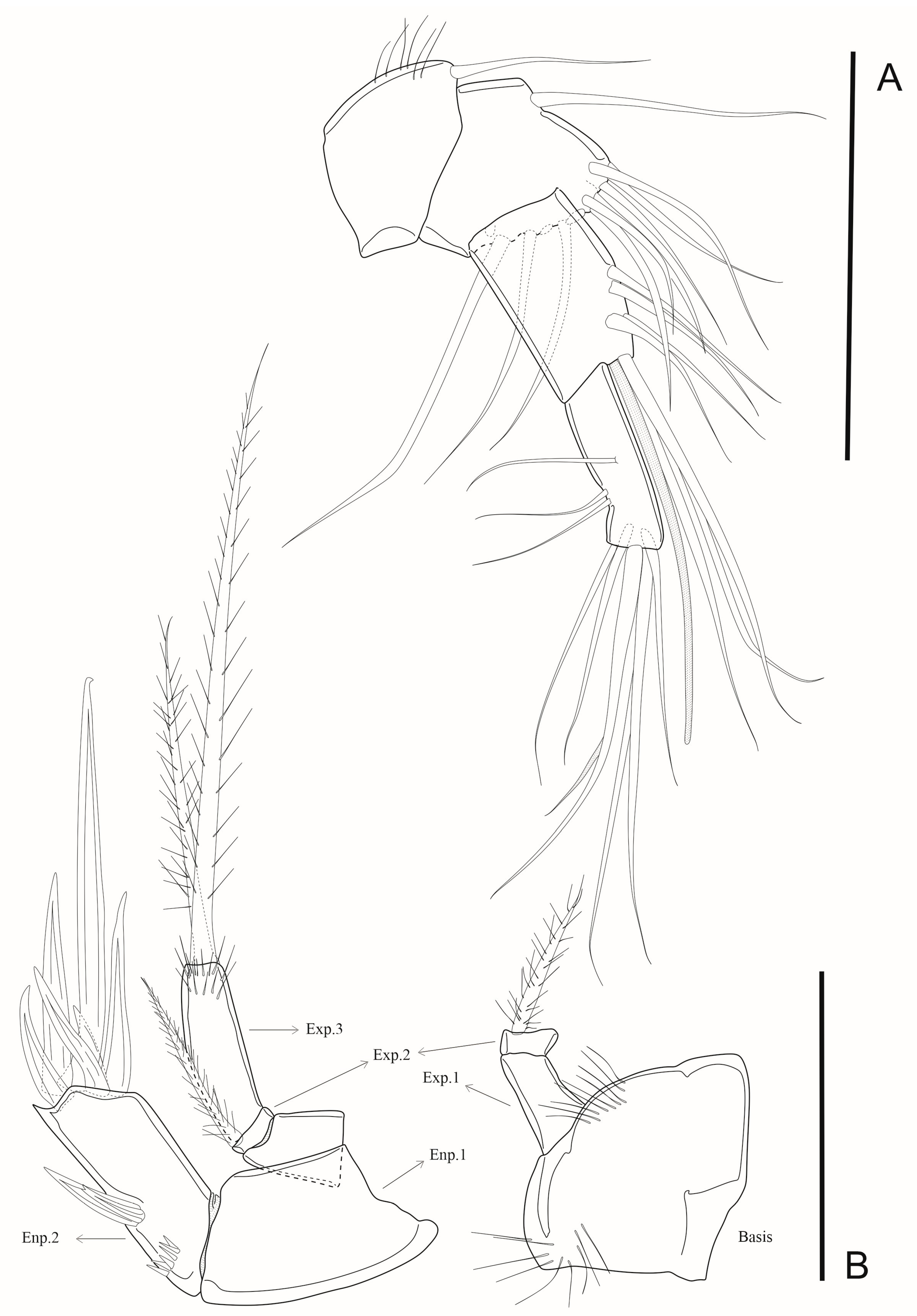
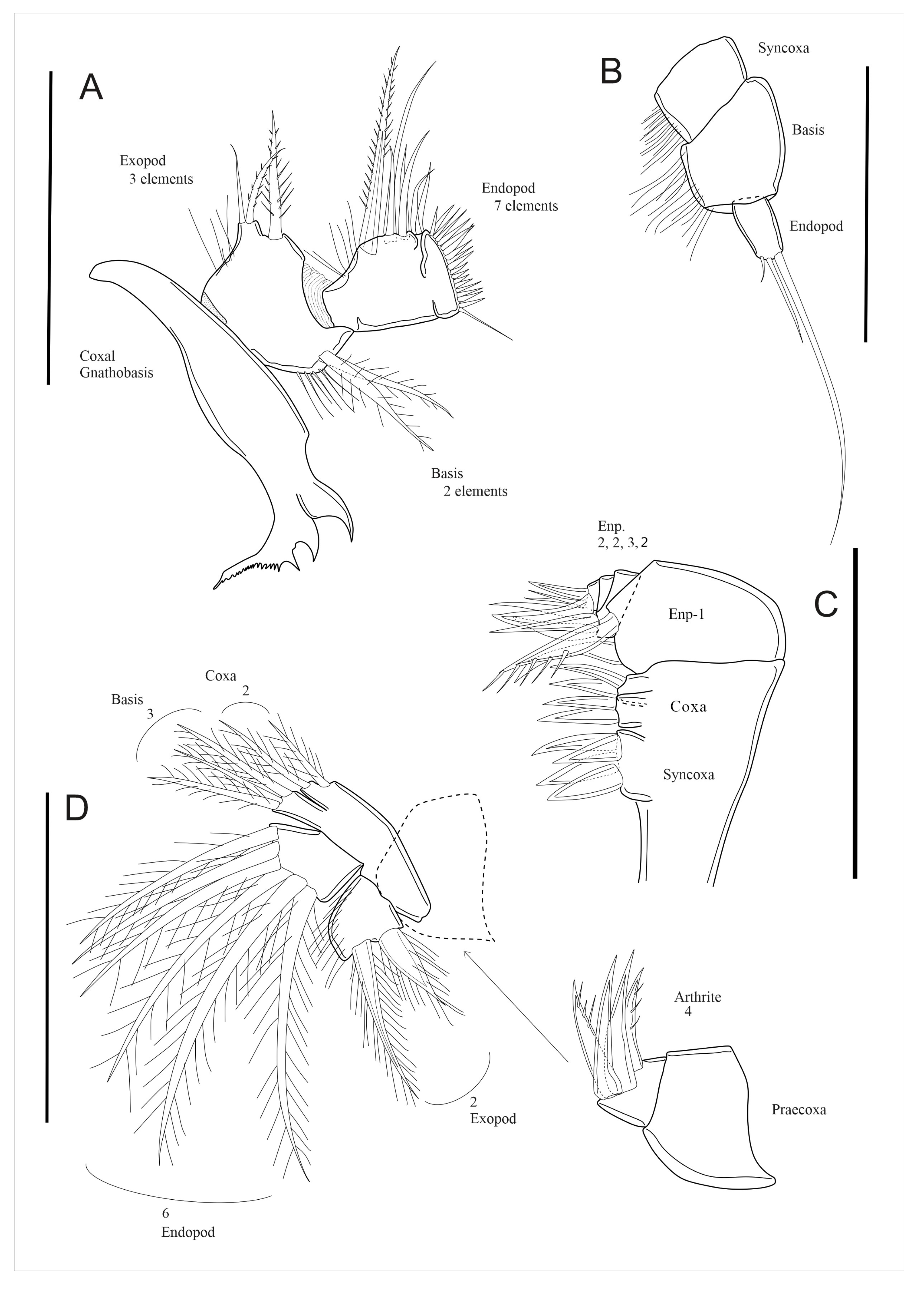
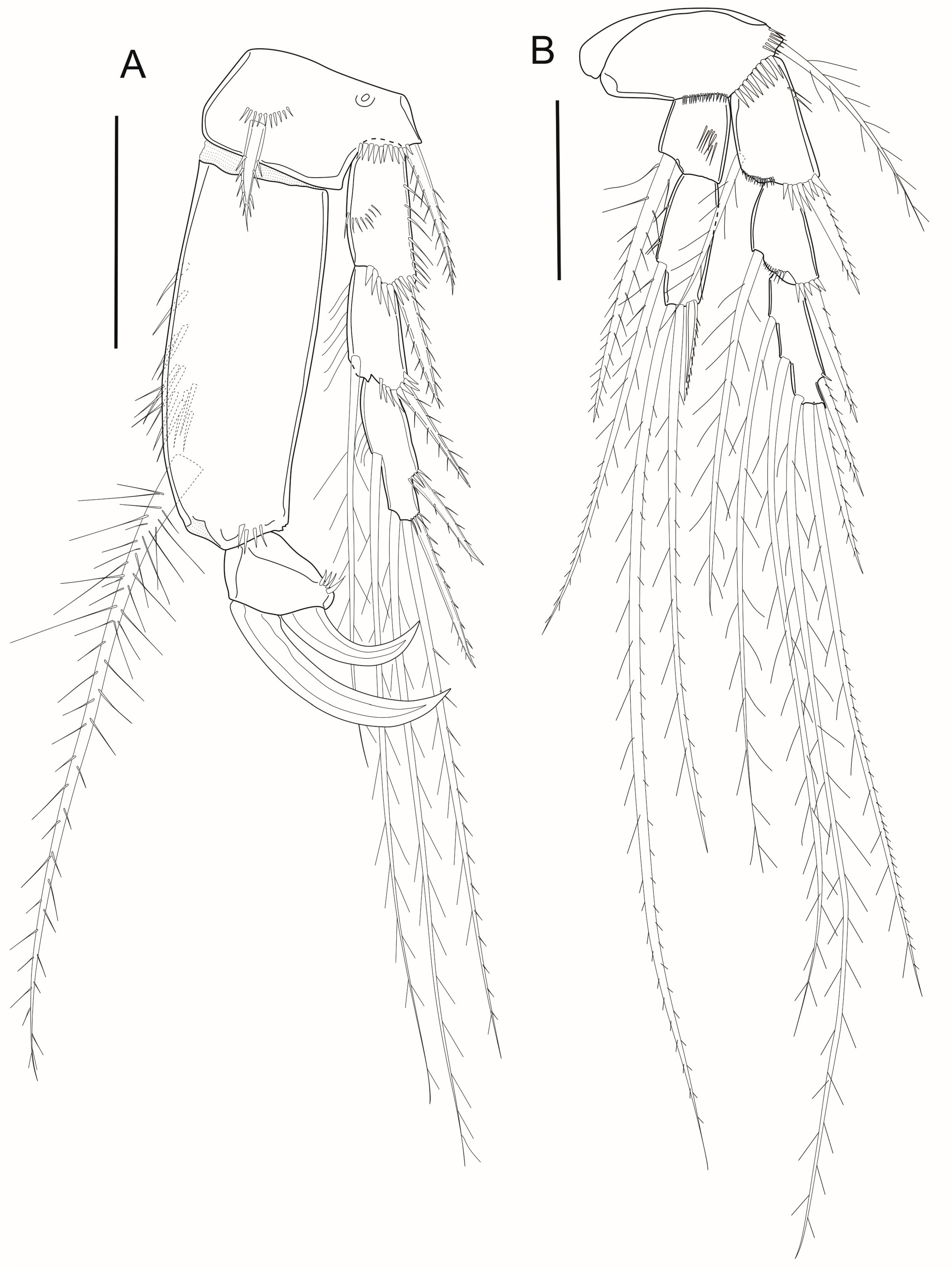
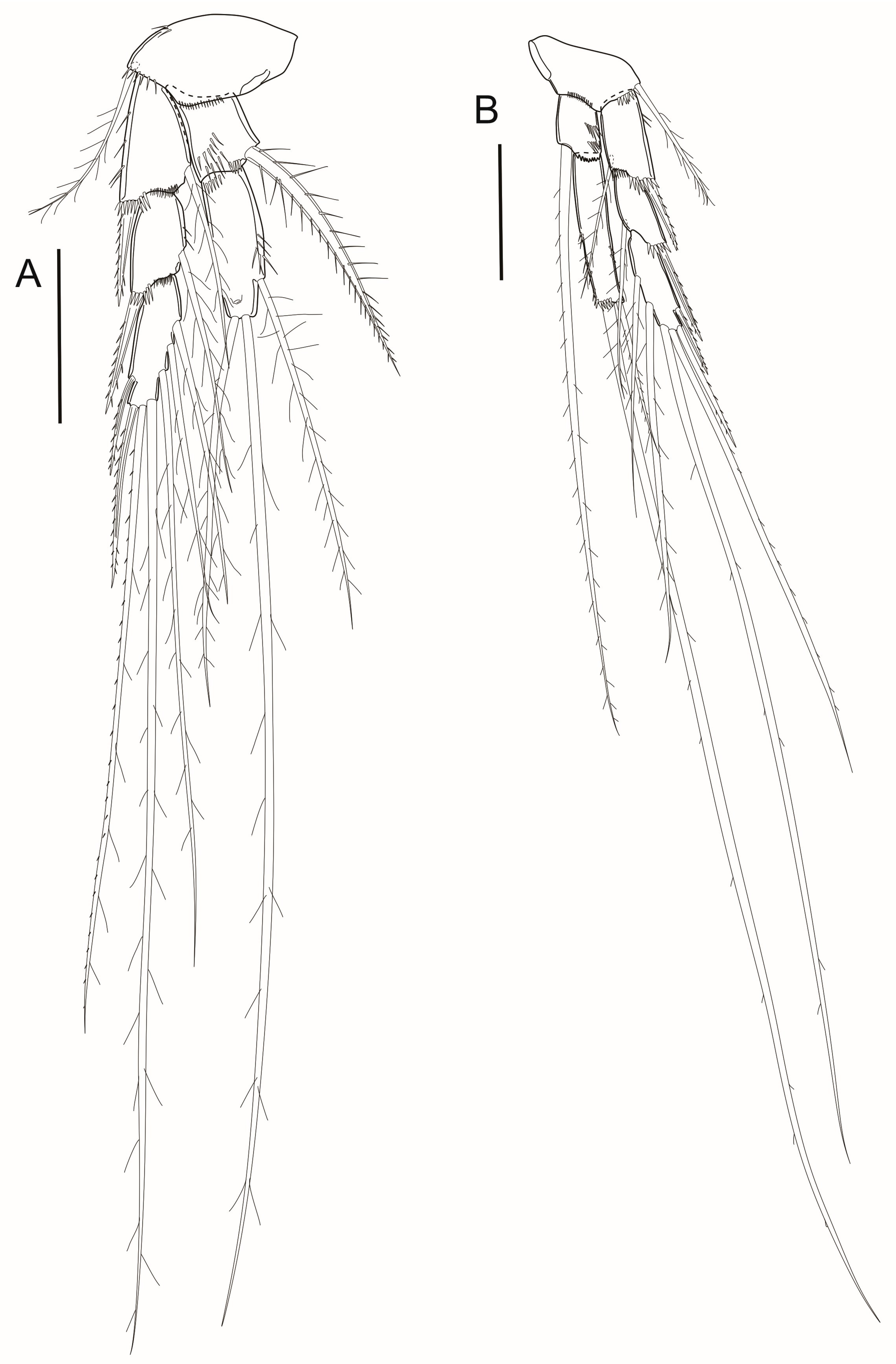
3.2. Genus Diagnosis

Body fusiform, tapering anteriorly and posteriorly; A1 four-segmented; A2 exp three-segmented; Md basis one-segmented, gnathobasis with sharp and curved pars incisive, pars molaris consisting of two sharp processes and a row of 7 slender teeth and fused seta; Mx1 coxal epipodite absent, Mx2 syncoxa with one endite, basis with two endites, enp four-segmented, enp-1 endite with a claw and a strong accessory spine; Mxp stenopodial; coxa and basis subequal in size and shape. P1–P4 exp three-segmented, enp two-segmented (Table 1); P1–P4 with large seta on end-1. P1 enp two-segmented, enp-1 exceptionally long, longer than exp-1 to 3 combined, enp-2 short, quadratic, and prehensile, with two claw-like curved spines distally. Exp-3 of P1 with two outer spines and three distal and inner setae. Exp-3 of P2, P3, and P4 with two outer spines and four, five, and four long distal and inner setae, respectively. P5 with exp and benp fused, with seven elements. Genital field consisting of a median genital slit, flanked by a pair of P6 with two setae each; midventral copulatory pore large.
Table 1. Armature formula of P1 to P4.
ExopodEndopod
P1I-0, I-1, II + 2 + 10, 0-II-0
P2I-1, I-1, II + 2 + 20, I, 2, 1
P3I-1, I-1, II + 2 + 30, 0, 2, 1
P4I-1, I-1, II + 2 + 20, 0, 2, 0

3.3. Putative Apomorphies of the Genus

Md gnathobasis with a row of slender teeth on the pars molaris, before the fused seta (ap). Enp of P1 2-segmented (ap); enp-1 exceedingly long, longer than exp-1 to 3 combined; enp-2 short, quadratic, and prehensile, with 2 claw-like curved spines distally (ap). P5 with benp and exp fused (ap).
Cruscollatus pedefusilia gen. et sp. nov.
Zoobank: urn:lsid:zoobank.org:act:0F6C0864-9F04-480A-87AD-1638B7544EC4

3.4. Etymology

The specific epithet is a noun in apposition; pedefusilia, derived from the latin pede (leg) and fusilia (fused), refers to the modified P5 with exp and enp fused.

3.5. Type Locality

Barra beach (13°00′37″ S, 38°31′58″ W) in Salvador city, Bahia State (Brazil).

3.6. Material Examined
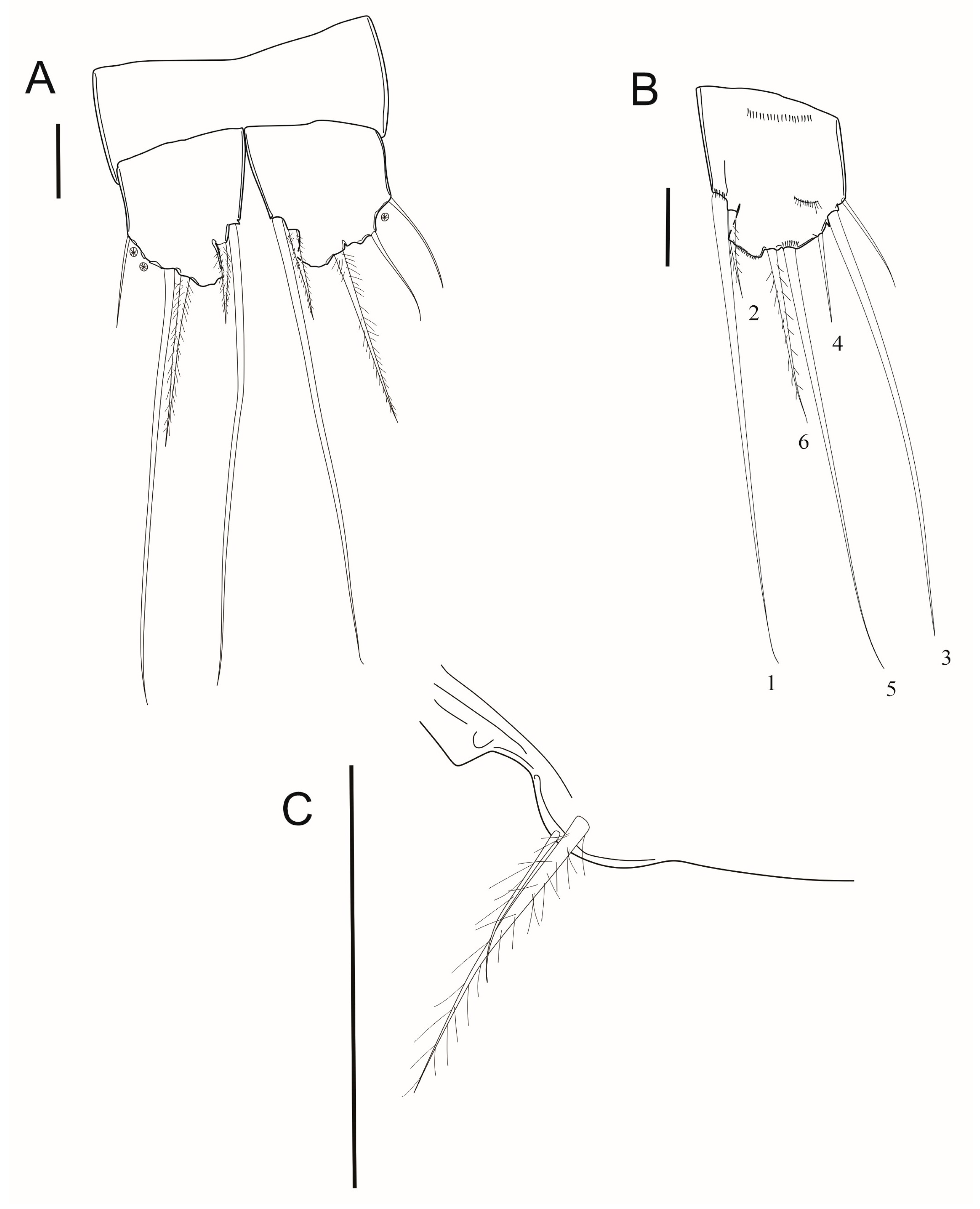
Holotype ♀ (UFBA 1725) and two paratypes ♀ (UFBA 1726); ♀ (UFBA 1727), Barra Beach, Salvador, Bahia State, collected by LABIMAR on 10th December 2012. All specimens were removed from the stomach and proximal intestine of nine colonies of Eudistoma vannamei Millar, 1977 that were hand-collected from submerged coral reefs, in depths ranging from 3 to 7 m.

3.7. Description

Female body length 884 µm (type series only), without furcal setae (Figure 2A and Figure 3A–D). Body fusiform, tapering anteriorly and posteriorly; anterior margin of cephalothorax slightly pointed; cephalic shield with sparse sensillae. All pedigerous somites with pointed epimera; posterior margin ornamented with row of setules followed by row of spinules, except in telson and furca. R (Figure 2B) large, square-shaped, rounded anteriorly.
Urosome (Figure 2C and Figure 4A–C) five-segmented, formed by fifth pedigerous somite, genital double-somite, and three free abdominal somites. Genital double-somite, including genital and first abdominal somites fused, wider than long; posterior margin in dorsal view with row of spinules and small sensillae. Genital field consisting of median genital slit; midventral copulatory pore large, transversal row of spinules inserted posteriorly to genital openings. Post-genital abdominal somites with row of setules close to posterior margin. First and second abdominal somites (Figure 2C and Figure 4) with row of spinules followed by setules in posterior margin. Anal somite with medial cleft (Figure 2C and Figure 4A,B), wider than long, with row of spinules along posterior margin and row of setules medially. Furca (Figure 2C–E and Figure 4A–C) longer than wide, with seven naked setae and extensions on lateral margins in dorsal view. Seta I short, inserted close to seta II; setae III and VI located distally, with basis covered by lateral projections of furca; setae IV and V long and located between lateral projections; and seta VII inserted on internal margin (Figure 2D–E). In dorsal view, cuticular process partially covering the ramus.
A1 (Figure 5A and Figure 6A,B) short, four-segmented, with armature formula: I-1, II-10, III-7 + ae, IV-10.
A2 (Figure 5B and Figure 6A,B) with short, naked coxa; basis with row of setules close to exp and end. Exp three-segmented, with armature formula 0,1,2; all plumose; exp-2 smaller than exp-1 and 3. Enp two-segmented. Enp-1 unarmed and fused to basis; enp-2 with eight setae, two medially located along outer margin, remaining setae inserted distally, one longer than the others.
Md (Figure 6B and Figure 7A). Gnathobasis with sharp pars incisive, pars molaris consisting of two sharp processes, row of 7 slender teeth, and fused seta. Basis with two plumose setae on inner side and row of setules. Enp one-segmented, with seven setae on outer margin, one of them bipinnate; distally, showing row of spinules. Exp fused to basis, with three setae unequal in length, two of them bipinnate, one smooth.
Mx1 (Figure 7D) praecoxa unarmed; praecoxal arthrite with four robust spines distally, two unipinnate and two smooth spines. Coxal endite with two plumose setae, basal endite with three plumose setae. Enp with six long plumose setae. Exp with row of long setules on inner margin, two large and robust plumose setae distally.
Mx2 (Figure 6B and Figure 7C) syncoxa with one endite, basis with two endites, enp four-segmented, and enp-1 endite with claw and strong accessory spine. Syncoxa endite with four robust spines and basis with two endites, each with two strong spines. Enp-1 with endite with claw and large accessory spine. Enp-2 and 3 with two naked spines; ep-4 with 3 spines.
Mxp (Figure 6B and Figure 7B) stenopodial. Syncoxa and basis with subequal in shape and length. Syncoxa with row of setules on inner margin; basis unarmed, with row of setules on inner margin; enp small, rectangular, longer than wide, one-segmented, with three setae, a long outer seta, a medium distal seta, and an inner thin seta.
P1 (Figure 8A, Table 2) with armed basis with two bipinnate spines on the inner and outer margins. Basis with row of tiny spinules close to inner spine and second row of spinules close to exp. Exp three-segmented, with bipinnate outer spines. Exp-1 without inner seta; exp-2 with long, plumose inner seta; exp-3 with long, plumose distal and inner seta. Enp two-segmented; enp-1 robust and longer than exp-1 to 3 combined, with long, plumose inner seta; enp-2 short, with two strong, curved distal claws.
P2 (Figure 8B, Table 2) basis with plumose outer seta with row of spinules near exp and enp insertions. Exp three-segmented. Exp-1 and 2 with outer bipinnate spine and inner plumose seta each; exp-3 with two outer bipinnate spines and distal and inner plumose setae. Enp two-segmented; enp-1 with plumose spinulose seta on inner margin; enp-2 with one inner and two distal plumose setae, unequal in length; inner seta about three times as long as outer one.
P3 (Figure 9A, Table 2) basis with plumose outer seta and row of spinules on outer margin close to insertion of exp. Exp-1 and 2 with bipinnate spine on outer margin and plumose setae on inner margin; exp-3 with two outer bipinnate spines, plumose distal and inner setae. Enp two-segmented; enp-1 with plumose spinulose seta on inner margin; enp-2 with one inner and two distal plumose setae, unequal in length; inner distal seta the longest.
P4 (Figure 9B, Table 2) basis with plumose outer seta and row of spinules near the insertion of exp and enp. Exp-1 and 2 with outer bipinnate spine and an inner plumose seta; enp-3 with two outer pinnate spines and two distal and two inner plumose setae. Enp-1 with seta longer than the whole enp and exp combined; enp-2 with two plumose setae distally, unequal in length; inner distal seta about five times as long as outer distal one.
P5 (Figure 10A,B) benp and exp fused, without visible articulation mark, with tiny external basal setae, one outer seta, three distal setae, and two inner setae close to inner margin, with remarkable rounded projections with serrated edges.
P6 flanking genital field (Figure 10C), with two setae each.

4. Discussion

The shape and armature of mouth parts is essential to indicate the phylogenetic relationships within Ectinosomatidae. The morphology of the Mx2 is a key feature that helps to define putative monophyletic groups within the family. These specimens can be categorized into two groups, based on whether the Mx2 is prehensile [Kihara & Huys, 2009]. The new genus has a naked syncoxa, an allobasis with two endites, and a four-segmented enp, the first segment with an endite with a claw and a strong accessory spine. This distinctive prehensile Mx2 is found in Cruscollatus gen. nov. and the following Ectinosomatidae genera: Bradya Boeck, 1819, Bradyellopsis Brian, 1925, Chaulionyx Kihara and Huys, 2009, Ectinosomoides Nicholls, 1945, Hastigerella Nicholls, 1935, Halophytophilus Brian, 1917, Kliesoma Hicks and Schiriever, 1985, Lineosoma Wells, 1965, Peltobradya Médione and Soyer, 1968, Pseudobradya Sars,1904, Pseudectinosoma Kunz, 1935, Nooditiella Wells, 1965, and Sigmatidium Giesbrecht, 1881 [1,2,19,20].
Among these Ectinosomatidae genera, only Bradiellopsis, Chaulionyx, Halophytophilus, Lineosoma, Nooditiella, and Pseudectinosoma, have P1 with a two-segmented enp [1,2] as in Cruscollatus gen. nov. The enp of P1 is also prehensile in Bradyellopsis, Chaulionyx, Halophytophilus, and Sigmatidium [1,2,20,21]. Cruscollatus gen. nov. P1 enp-1 is exceedingly long, longer than exp-1 to 3 combined, armed with a long plumose seta on the inner margin. The enp-2 of P1 is short, quadratic, and has two claw-like curved spines distally. Contrarily, the P1 enp-2 of Bradyellopsis has up to five elements, with at least two long spines [22,23]. Chaolionyx has six elements (five setae and one bifid spine) [2]; Halophytophilus bears three setae [1,20] or has the enp-2 absent, as in Halophytophilus lopheliae Gheerardyn et al., 2008 [1]. Sigmatidium typically has six elements but can reach up to eight, as in Sigmatidium difficile Giesbrecht, 1881 [1]. In the new genus, the armature of exp-3 consists of two spines on the outer margin instead of three spines, as in Halophytophilus [1,20]. In contrast, Sigmatidium and Chaolionix have two outer spines, as in the new genus [1,2]. The P2 exp-3 to P4 in Cruscollatus gen. nov. has two outer spines instead of three, as in Halophytophilus [1,20]. The new genus also has five and four setae in P3 exp-3 and P4 exp-3, respectively, whereas in Chaolionyx, exp-3 of P3 and P4 have four and five setae, respectively [2]. The new genus has a three-segmented A2 exp, and in Peltobradya, it is one-segmented [1].
The Mx2 of Cruscollatus gen. nov. bears three endites and a four-segmented enp, whereas Halophytophilus has only one endite, and Chaulionyx shows a rudimentary middle endite and a three-segmented enp [1,2,20]. The Mxp of Cruscollatus gen. nov. has syncoxa and a quadratic basis, of similar shape and sizes, and a one-segmented enp armed with two distal setae and an accessory inner one, all naked. Chaulionyx has its syncoxa armed with a seta and an enp with three plumose setae [2].
The gnathobasis of the Md has a row of delicate teeth. In contrast, Chaulionyx exhibits a gnathobasis with robust teeth on the cutting margin [2]. However, Halophytophilus and Sigmatidium lack this diagnostic characteristic in the original descriptions. The fusion of the benp and exp of the P5 of Cruscollatus gen. nov. is not seen in any of the previously mentioned genera, as they exhibit P5 with separate benp and exp. Parabradya Lang, 1944 and Rangabradya (Karanovic and Pesce, 2001) are two Ectinosomatidae genera which, similarly Cruscollatus gen. nov., have P5 benp and exp fused. However, the armature of P1 –P4 exp-3 is different from what occurs in the new genus [1,2,18,21].
The putative apomorphies described above cannot be found in most of the remaining genera within the family. When it can, as in the case of the fused P5 benp and exp of Parabradya and Rangabradya, other characteristics of the new genus, such as the peculiar armature of the Md gnathobasis and the unique morphology of the P1 enp preclude the inclusion of the new species within other Ectinosomatidae genera.

5. Key to the Genera of Ectinosomatidae (Adapted from Kihara and Huys, 2009)

1.
Body cylindrical with cephalothorax rectangular in dorsal view; body the same width throughout its length ….……………………...…………………………………………2
-
Body fusiform with cephalothorax sub-triangular in dorsal view; greatest body width usually at posterior margin of cephalothorax; urosome gradually tapering towards the posterior end…………………………………………….……………………………7
-
Body with dorsoventrally depressed prosome, clearly wider than urosome .....................................................................……… Peltobradya Médioni and Soyer, 1968
2.
Antennary exp two-segmented; maxilla prehensile, with major articulation between elongate syncoxa and elongate allobasis .............……………… Noodtiella Wells, 1965
-
Antennary exp one or three-segmented; maxilla not prehensile, with at most a slight angle between syncoxa and allobasis ......................................................……………… 3
3.
Enps P2–P4 2-segmented ..................…………………… Ectinosomoides Nicholls, 1945
-
Enps P2–P4 3-segmented .....…..................................................................……………… 4
4.
Anal somite with dorsal armature of claws, lappets or spiniform processes around anal opening; P5 exp with three marginal and one surface seta ………… ………..............................................…………………………… Arenosetella Wilson, 1932
-
Anal somite without such ornamentation........................................................…………5
5.
Antennary exp one-segmented, enp one-segmented .................. Tetanopsis Brady, 1910
-
Antennary exp one-segmented, enp two-segmented…. Parahalectinosoma George and Schwabe, 2019
-
Antennary exp three-segmented.........................................................................………… 6
6.
Female P5 with foliaceous setae on exp and benp, exp with three marginal and no surface setae; male P5 exp with four normal marginal setae ………………………………………………………...……………… Oikopus Wells, 1967
-
P5 with normal setae on exp and benp in both sexes, exp with 3 marginal and typically a surface seta [absent in Hastigerella noodti Soyer, 1974 = G. soyeri (Bodin, 1976)] ................................……………………………………………… Glabrotelson Huys, 2009
7.
P1–P4 enps 2-segmented .............................……………… Pseudectinosoma Kunz, 1935
-
P1 enp 2- or 3-segmented, P2–P4 enps 3-segmented......................…………………… 8
8.
P1 enp prehensile ……….....................................................................…………………… 9
-
P1 enp not prehensile .........................................................................…………………… 12
9.
P1 enp 2-segmented............................................................................…………………… 10
-
P1 enp 3-segmented .....................…………………… Klieosoma Hicks & Schriever, 1985
10.
P1–P2 exp-3 with two outer elements.........................................................…………… 11
-
P1–P2 exp-3 with three outer elements ...............………… Halophytophilus Brian, 1919
11.
Antennule with large spine on segment 2 (and often segments 1 and 3); antennary exp rudimentary, with 1–3 small setae; P1 enp-2 with 4 elements (1–2 pinnate and claw-like) ........................................……………………………… Bradyellopsis Brian, 1925
-
Armature elements on antennulary segments 1–3 setiform; antennary exp well developed and 3-segmented; P1 enp-2 with six elements (outer one bifid and claw-like) ...........................................…………………………… Chaulionyx Kihara and Huys, 2009
-
P1 enp-2 P1 with two claws; P5 completely fused without evidence of the borders between exp and baseoendopod; marine, found in digestive tract of ascidian………... Cruscollatus gen. nov.
12.
Maxilla prehensile, with syncoxa and allobasis forming right angle; P5 exo-pod poorly developed, short, fused to benp in female and distinct in male, with 3 marginal and no surface setae; body very small (< 300 μm) .................... Sigmatidium Giesbrecht, 1881
-
These characters not combined..............................................................………………… 13
13.
P5 exp and benp fused, forming a single plate in both sexes….…………………..… 14
-
P5 exp and benp at least partly discrete............................……………………………… 15
14.
P1–P4 exp-3 with 5, 6, 6, 6 elements, respectively; male P6 unarmed; body of female small (< 400 μm); continental groundwater……….. Rangabradya Karanovic and Pesce, 2001
-
P1–P4 exp-3 with 6, 7, 8, 8 elements, respectively; male P6 with two setae; body of female large (≥1200 μm); marine, usually deepwater ....................................Parabradya Lang, 1944
15.
Integument of somites with distinctive subrectangular pores; P5 exp with four marginal setae…………………………..…………………………… Ectinosoma Boeck, 1865
-
Integument of somites without distinctive subrectangular pores; P5 exp with three marginal setae and one seta on anterior surface………… …………………………16
16.
Mandible with rudimentary gnathobasis, elongate basis and filiform exp and enp, each terminating in 2–3 setae; antennary exp without lateral spines ………… …………...........................……………………………………… Ectinosomella Sars, 1910
-
These characters not combined……....................................................……………… 17
17.
Third segment of female antennule three times as long as wide; mandibular enp with one strong seta laterally; P1–P4 exp-3 with 2 outer spines; planktonic (occasionally in sediment) ………………………………….… Microsetella Brady and Robertson, 1873
-
These characters not combined….........................................................……………… 18
18.
Body comparatively robust with prosome-urosome separation usually distinct (exception: B. kurtschminkei Seifried and Martínez Arbizu, 2008; with dorsoventrally flattened habitus); antenna with two setae on proximal exp segment and one seta on proximal enp segment; mandibular exp with at least five setae; maxilliped robust with short enp usually fused at an angle with basis and bearing four conspicuous setae ....…………………………………………………...………… Bradya Boeck, 1973
-
Body comparatively slender with no sharp separation between prosome and urosome; antenna with less than 2 setae on proximal exp segment (except Pseudobradya ambigua Sars, 1920 with 2) and no seta on proximal enp segment; mandibular exp generally with fewer than 5 setae; Maxilliped usually slender and straight with discrete enp bearing 1 small and 4 conspicuous setae…………………………………19
19.
Antennule with or without dark pigment spot within the proximal three segments: maxilla prehensile, allobasis usually truncate distally and carrying 3-segmented enp (although enp sometimes exceedingly small and segmentation difficult to discern; reduced to a narrow three-segmented cylinder in P. leptognatha Sars, 1920); maxilliped short and robust……………………………………...…… Pseudobradya Sars, 1904
-
Antennule without pigment spot; maxilla with at most a slight angle between syncoxa and allobasis, the latter attenuating distally, enp three-segmented but always small, its morphology not clearly discernible; maxilliped slender ……………………… ………………....………………… Halectinosoma Vervoort, 1962

Author Contributions

Conceptualization and methodology, R.J., P.H.C., E.N., and N.S.; validation, P.H.C. and T.C.K.; formal analysis, P.H.C. and R.J.; investigation, A.F. and R.J.; resources, R.J.; data curation, A.F.; writing—review and editing, P.H.C. and R.J.; visualization, A.F.; supervision, R.J.; project administration, R.J.; funding acquisition, E.N. All authors have read and agreed to the published version of the manuscript.

Funding

This research was funded by the project “Assessment and research of sun coral in Todos-os-Santos Bay”, a cooperation agreement between UFBA and PETROBRAS (N°5850.0107361.18.9) regulated by RD&I investments clauses of Brazilian Agency of Petroleum, Natural Gas, and Biofuels (ANP Resolution 05/2015).

Data Availability Statement

All data referring to the results are included in the present article.

Acknowledgments

The authors thank Bahia C.S. and the LABIMAR team for collecting and sorting out the specimens. The authors also thank “Programa de Pós-Graduação em Biodiversidade e Evolução” (UFBA) for the logistical support. We are also grateful to the Chico Mendes Institute for Biodiversity Conservation (ICMbio) for collecting permission (Sisbio No 15161-1). This is publication number 77 that uses data from the Senckenberg am Meer Confocal Laserscanning Microscope Facility (SGN-SaM-cLSM).

Conflicts of Interest

The authors declare no conflicts of interest.

References

  1. Huys, R.; Gee, J.M.; Moore, C.G.; Hamond, R. Marine and Brackish Water Harpacticoid Copepods; The Linnean Society of London and Estuarine and Coastal Sciences Association by Field Studies Council: Shrewsbury, UK; London, UK, 1996; 323p. [Google Scholar]
  2. Kihara, T.C.; Huys, R. A new genus of Ectinosomatidae (Copepoda, Harpacticoida) from sublittoral sediments in Ubatuba, São Paulo State (Brazil), an updated key to genera and notes on Noodtiella Wells, 1965. ZooKeys 2009, 17, 57–88. [Google Scholar] [CrossRef]
  3. Sars, G.O. Copepoda Harpacticoida. Parts III & IV. Ectinosomidae, Harpacticidae (Part). An Account of the Crustacea of Norway, with Short Descriptions and Figs. of All the Species 5; Bergen Museum: Bergen, Norway, 1904; pp. 29–56, pls. 17–32. [Google Scholar]
  4. Moore, C.G. An emendation of the family name Ectinosomidae Sars to Ectinosomatidae (Copepoda, Harpacticoida). Crustaceana 1978, 34, 111. [Google Scholar] [CrossRef]
  5. Ho, J.-S. Why do symbiotic copepods matter? Hydrobiologia 2001, 453/454, 1–7. [Google Scholar] [CrossRef]
  6. Huys, R. Harpacticoid copepods—Their symbiotic associations and biogenic substrata: A review. Zootaxa 2016, 4174, 448–729. [Google Scholar] [CrossRef] [PubMed]
  7. Illg, P.L.; Dudley, P.L. The family Ascidicolidae and its subfamilies (Copepoda, Cyclopoida), with descriptions of new species. Mém. Mus. Hist. Nat. Sér. A Zool. 1980, 117, 1–192. [Google Scholar]
  8. Johnsson, R.; Bahia, C.; Neves, E. A new genus of Asterocheridae (Copepoda: Siphonostomatoida) ectoassociate of the ascidian Eudistoma vannamei Millar, 1977 (Polycitoridae) from Brazil. Zootaxa 2016, 4114, 162–170. [Google Scholar] [CrossRef] [PubMed]
  9. Humes, A.G. Cnidarians and copepods: A success story. Trans. Am. Micros. Soc. 1985, 104, 313–320. [Google Scholar] [CrossRef]
  10. Huys, R.; Boxshall, G.A. Copepod Evolution; The Ray Society: London, UK, 1991; 468p. [Google Scholar]
  11. Johnsson, R.; Neves, E. Siphonostomatoid copepods (Crustacea) associated with marine invertebrates and algae in Brazil: A review and future considerations. Zoosymposia 2012, 8, 69–80. [Google Scholar] [CrossRef]
  12. Lotufo. T.M.C. Ascidiacea (Chordata: Tunicata) do Litoral Tropical Brasileiro. Ph.D. Thesis, Instituto de Biociências, Universidade de São Paulo, São Paulo, Brazil, 2002.
  13. Médioni, A.; Soyer, J. Copépodes Harpacticoïdes de Banyuls-sur-Mer. 6. Nouvelles formes associées à des Bryozoaires. Vie et Milieu 1968, 18, 317–343. [Google Scholar]
  14. Snelgrove, P.V.R.; Lewis, J.B. Response of a coral-associated crustacean community to eutrophication. Mar. Biol. 1989, 101, 249–257. [Google Scholar] [CrossRef]
  15. George, K.H.; Schwabe, E. A new species of Ectinosomatidae Sars (Copepoda, Harpacticoida) associated with Pseudoikedella achaeta (Zenkevitch, 1958) (Echiura, Bonelliida). Prog. Oceanogr. 2019, 173, 1–8. [Google Scholar] [CrossRef]
  16. Michels, J.; Büntzow, M. Assessment of Congo red as a fluorescence marker for the exoskeleton of small crustaceans and the cuticle of polychaetes. J. Microsc. 2010, 238, 95–101. [Google Scholar] [CrossRef] [PubMed]
  17. Ferrari, F.D.; Ivanenko, V.N. The identity of protopodal segments and the ramus of maxilla 2 of copepods (Copepoda). Crustaceana 2008, 81, 823–835. [Google Scholar] [CrossRef]
  18. Sewell, R.B.S. The littoral and semi-parasitic Cyclopoida, the Monstrilloida and Notodelphyoida. Sci. Rep. John Murray Exped. Publ. Br. Mus. 1949, 9, 17–199. [Google Scholar]
  19. Lang, K. Die während der schwedischen Expedition nach Spitzbergen 1898 und nach Grönland 1899 eingesammelten Harpacticiden. K. Sven. Vetensk. Hand. 1936, 15, 1–55. [Google Scholar]
  20. Klie, W. Harpacticoida (Cop.) aus dem Bereich von Helgoland und der Kieler Bucht. 1. Kiel. Meeresforsch. 1949, 6, 90–128. [Google Scholar]
  21. Karanovic, T.; Pesce, G.L. A new genus and species of the family Ectinosomatidae (Crustacea: Copepoda: Harpacticoida) from the groundwaters of India. Ann. Limnol. 2001, 37, 281–292. [Google Scholar] [CrossRef]
  22. Watkins, R.L. Description of new species of Bradyellopsis and Perissocope (Copepoda: Harpacticoida) from the California coast with revised keys to the genera. J. Crust. Biol. 1987, 7, 380–395. [Google Scholar] [CrossRef]
  23. Brian, A. Descrizione di un nuove genere di Copepodo Arpacticoide dell’ Adriatico. [Description of a new genus of Copepoda Harpacticoida from the Adriatic Sea.]. Atti Soc. Ligustica 1925, 4, 116–122. [Google Scholar]
Figure 1. Locus typicus of the new species in Todos-os-Santos Bay, Barra Beach, Salvador, Bahia, Brazil.
Arthropoda 03 00008 g001
Figure 2. Cruscollatus pedefusilia gen. et sp. nov., female (holotype: UFBA 1725). (A) habitus, dorsal view; (B) R; (C) urosome, ventral view; (D) detail of furca in dorsal view; (E) detail of furca in ventral view. Scale bars: (A) = 250 µm; (C) = 100 µm; (B), (D,E) = 50 µm.
Arthropoda 03 00008 g002
Figure 3. Cruscollatus pedefusilia gen. et sp. nov., female. Confocal microphotography of habitus (A) dorsal view; (B) lateral view; (C) and (D) ventral view. (D) ventral view of ovigerous female. Scale bars: (AC) = 25 µm, (D) = 75 µm.
Arthropoda 03 00008 g003
Figure 4. Cruscollatus pedefusilia gen. et sp. nov., female. Confocal microphotography of urosome (A) dorsal view; (B) ventral view; (C) lateral view. Scale bars: (AC) = 100 µm.
Arthropoda 03 00008 g004
Figure 5. Cruscollatus pedefusilia gen. et sp. nov., female (holotype: UFBA 1725). (A), (A1); (B), (A2). Scale bars: (A,B) = 50 µm.
Arthropoda 03 00008 g005
Figure 6. Cruscollatus pedefusilia gen. et sp. nov., female. Confocal microphotography of habitus (A) anterior ventral region; (B) detail of anterior ventral region. Scale bars: (A) = 25 µm, (B) = 100 µm.
Arthropoda 03 00008 g006
Figure 7. Cruscollatus pedefusilia gen. et sp. nov., female (holotype: UFBA 1725). (A) Md; (B) Mxp; (C) Mx2; (D) Mx1. Scale bars: (AD) = 50 µm.
Arthropoda 03 00008 g007
Figure 8. Cruscollatus pedefusilia gen. et sp. nov., female (holotype: UFBA 1725). (A) P1; (B) P2. Scale bars: (A,B) = 50 µm.
Arthropoda 03 00008 g008
Figure 9. Cruscollatus pedefusilia gen. et sp. nov., female (holotype: UFBA 1725). (A) P3; (B) P4. Scale bars: (A,B) = 50 µm.
Arthropoda 03 00008 g009
Figure 10. Cruscollatus pedefusilia gen. et sp. nov., female (holotype: UFBA 1725). (A) P5; (B) P5 with setation detailed; (C) P6. Scale bars: (AC) = 50 µm.
Arthropoda 03 00008 g010
Table 2. Armature formula of P1 to P4.
ExopodEndopod
P1I-0, I-1, II + 2 + 10, 0-II-0
P2I-1, I-1, II + 2 + 20, I, 2, 1
P3I-1, I-1, II + 2 + 30, 0, 2, 1
P4I-1, I-1, II + 2 + 20, 0, 2, 0
Disclaimer/Publisher’s Note: The statements, opinions and data contained in all publications are solely those of the individual author(s) and contributor(s) and not of MDPI and/or the editor(s). MDPI and/or the editor(s) disclaim responsibility for any injury to people or property resulting from any ideas, methods, instructions or products referred to in the content.

