1. Introduction
Accurate assessment of regional lymph node status is a fundamental component of oncological staging in dogs, as lymph node metastasis is an important prognostic indicator that directly influences surgical planning, therapeutic decision-making, and overall clinical management [
1,
2]. Among canine neoplasms, cutaneous and mucosal melanomas are particularly aggressive because of their early propensity for lymphatic dissemination, which may occur even in the absence of clinically enlarged regional lymph nodes [
3,
4]. Consequently, sentinel lymph node (SLN) biopsy has gained increasing acceptance in veterinary oncology as a reliable approach for detecting occult metastatic disease and improving staging accuracy [
1].
Several SLN mapping techniques have been validated in dogs, including planar lymphoscintigraphy using technetium-99m-labeled colloids, intradermal administration of vital dyes such as methylene blue or patent blue, and near-infrared fluorescence imaging with indocyanine green (ICG) [
5,
6,
7,
8]. Although these methods have demonstrated satisfactory diagnostic performance, their widespread clinical use remains limited by the need for specialized equipment, radiopharmaceutical licensing, or dedicated imaging systems that are unavailable in many veterinary institutions [
6,
7,
9].
Indirect radiographic lymphography using water-soluble iodinated contrast media has emerged as a practical and accessible alternative for SLN identification. Following intradermal peritumoral injection, the contrast agent enters the lymphatic vessels and opacifies afferent channels, allowing visualization of the first draining lymph node using conventional radiographic equipment. Iohexol, a low-osmolality non-ionic iodinated contrast medium routinely available in veterinary hospitals, has an established safety profile and does not require specific regulatory authorization for clinical application [
10]. Previous studies have demonstrated the feasibility of iohexol-based lymphography for SLN mapping in dogs with cutaneous mast cell tumors, soft tissue sarcomas, and oral neoplasms, reporting detection rates and anatomical agreement comparable to those achieved with lymphoscintigraphy [
11,
12].
Lymphatic drainage of the canine head and neck is highly variable, making prediction of sentinel lymph nodes based solely on anatomical landmarks unreliable [
13]. Individual differences in drainage patterns may result in metastatic spread to unexpected lymphatic basins, emphasizing the importance of patient-specific mapping techniques. This brief report describes the application of indirect radiographic lymphography with iohexol for preoperative SLN mapping in a dog with auricular melanoma and correlates the imaging findings with histopathological confirmation of nodal metastasis.
2. Results
A 12-year-old spayed female mixed-breed dog weighing 8.8 kg was presented for evaluation of a slowly progressive ulcerated nodular mass involving the left auricle. According to the owner, the lesion had first been noticed approximately six months before presentation. Physical examination revealed a firm, multinodular cutaneous mass measuring 3 × 4 × 2.5 cm. An additional small cutaneous nodule was incidentally identified on the ventral abdominal wall. Regional lymph nodes (mandibular, superficial cervical, and parotid) were bilaterally symmetrical, soft, and non-palpable as discrete enlarged structures on physical examination, with no evidence of increased firmness or fixation to adjacent tissue.
Three-view thoracic radiographs, abdominal ultrasonography, complete blood count (CBC), and serum biochemical analysis showed no evidence of distant metastasis or other clinically significant systemic abnormalities. Fine-needle aspiration cytology of the auricular lesion yielded a highly cellular sample composed of round-to-polygonal melanocytic cells containing variable amounts of intracytoplasmic melanin pigment, consistent with a melanocytic neoplasm. Preoperative electrocardiographic and echocardiographic evaluations were unremarkable.
Based on the preoperative diagnostic workup, the patient was clinically staged as cT2N0M0 according to the World Health Organization (WHO) TNM classification system for canine tumors, as no evidence of regional lymph node enlargement or distant metastasis was identified. Following histopathological evaluation confirming metastatic involvement of the regional lymph nodes, the final pathological stage was consistent with pT2N1M0. The T2 category corresponds to a primary tumor measuring 2–4 cm in greatest diameter, according to the World Health Organization TNM staging scheme for canine melanoma [
14]; the primary auricular mass (3 × 4 × 2.5 cm) met this criterion based on its largest dimension. N0 reflected the clinical absence of palpable or radiographically enlarged regional lymph nodes at initial staging, whereas N1 was assigned after histopathological confirmation of nodal metastasis, illustrating the distinction between clinical (c) and pathological (p) staging within the TNM system.
Six days after cytological diagnosis, the patient underwent surgical treatment under general anesthesia. Sentinel lymph node mapping using iohexol-based indirect radiographic lymphography was performed immediately before surgery, as described in the Materials and Methods Section.
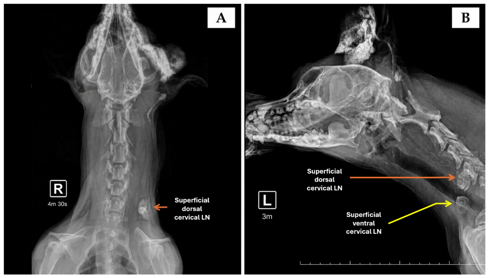
Radiographic examination demonstrated opacification of afferent lymphatic vessels draining toward both the superficial ventral cervical and superficial dorsal cervical lymph nodes (
Figure 1). Based on these findings, both lymph nodes were identified as sentinel lymph nodes. A lymph node was operationally defined as sentinel when direct and consistent convergence of contrasted afferent lymphatic vessels onto that node was observed within the initial imaging window (3 and 4.5 min after injection), before redistribution of contrast to secondary lymphatic stations.
Surgical treatment included vertical ear canal ablation, conchectomy, partial auriculectomy, excision of the superficial ventral and superficial dorsal cervical sentinel lymph nodes, removal of the left parotid lymph node, and excision of the abdominal cutaneous nodule. The procedure was completed without intraoperative complications, with a total anesthetic time of 95 min.
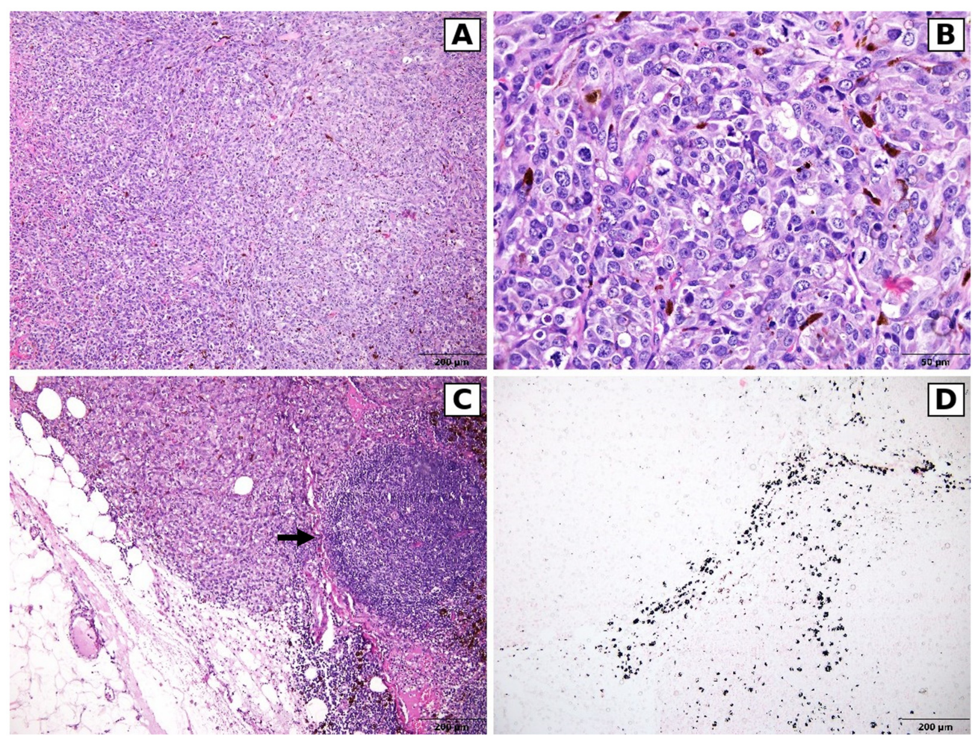
Histopathological examination confirmed a completely excised mixed-type cutaneous melanoma composed of epithelioid and spindle-cell populations. Surgical margins were free of neoplastic infiltration. The superficial ventral cervical sentinel lymph node contained macrometastatic melanoma (>2 mm), whereas the superficial dorsal cervical sentinel lymph node and the left parotid lymph node contained micrometastatic deposits (≤2 mm). In the macrometastatic node, neoplastic cells replaced approximately 30–40% of the nodal parenchyma. This 2 mm cut-off, adapted from human oncology staging criteria in the absence of a validated species-specific threshold for dogs, was used to distinguish micrometastatic from macrometastatic deposits, consistent with the adaptation of this criterion previously applied in canine mammary carcinoma staging [
15]. Histological grading of the primary tumor showed moderate anisokaryosis and anisocytosis, marked cellular pleomorphism, moderate nuclear pleomorphism, a mitotic index of 116 mitotic figures per 2.37 mm
2, and no lymphovascular invasion.
Although the left parotid lymph node was not clearly opacified or enlarged on radiographic examination, afferent lymphatic channels leading to this node were visible following contrast administration; based on the operational criterion described above, this node was therefore not classified as a sentinel lymph node, but was instead removed as part of an elective regional lymphadenectomy performed on the basis of its anatomical proximity to the primary tumor. Histopathological confirmation of metastatic involvement in this non-sentinel node indicates that incomplete or absent radiographic opacification does not exclude occult metastatic disease and may represent a false-negative finding of the lymphographic technique. This finding supports considering elective regional lymphadenectomy in combination with sentinel lymph node biopsy in similar cases of canine melanoma. Melanin pigment within metastatic foci was confirmed by Fontana–Masson staining (
Figure 2), and the melanocytic origin was further supported by positive immunolabeling for Melan-A and PNL2, performed on sections of the primary tumor. The abdominal lesion was diagnosed as a primary cutaneous hemangiosarcoma unrelated to the auricular melanoma and was excised with complete surgical margins.
Postoperative recovery was uneventful, and the patient was discharged 48 h after surgery. Because hematological evaluation revealed pre-existing bone marrow hypoplasia, adjuvant chemotherapy was not initiated. Although metronomic treatment with cyclophosphamide and piroxicam was considered, it was ultimately contraindicated because of the degree of myelosuppression.
Approximately two months after surgery, local recurrence developed at the previous cervical lymph node excision site. The recurrent lesion was surgically removed and processed for the production of an autologous tumor lysate vaccine through an institutional immunotherapy program at the University of São Paulo. The patient subsequently received four vaccine administrations at 21-day intervals.
Despite treatment, serial thoracic radiographs demonstrated progressive pulmonary metastatic disease. Systemic chemotherapy was attempted but discontinued after a single treatment because of severe myelosuppression associated with the pre-existing bone marrow disorder. The patient’s clinical condition progressively deteriorated, and euthanasia was elected approximately five months after the initial surgery. Necropsy confirmed disseminated metastatic melanoma involving the lungs, liver, and an intrathoracic lymph node, together with a recurrent cutaneous nodule at the cervical surgical site; the melanocytic origin of these lesions was confirmed by Fontana–Masson staining. Bone marrow hypoplasia was confirmed both grossly and microscopically. Incidental findings unrelated to the melanoma included a benign gastric leiomyoma and mild multinodular atrioventricular valve degeneration (endocardiosis).
No systemic adverse events associated with iohexol administration were observed during the perioperative period or subsequent follow-up. The only procedure-related adverse effect was transient mild-to-moderate localized edema at the injection sites, which resolved spontaneously without therapeutic intervention.
3. Discussion
This brief report demonstrates the successful application of indirect radiographic lymphography with iohexol for preoperative SLN mapping in a dog with auricular melanoma. Histopathological evaluation confirmed metastatic involvement of the mapped sentinel lymph nodes, supporting the clinical utility of this technique for lymphatic staging in canine melanoma. To the best of the authors’ knowledge, this is the first published report describing the use of iohexol-based indirect radiographic lymphography specifically for SLN mapping in canine auricular melanoma.
The correspondence between lymphographic findings and histopathological confirmation highlights the biological relevance of individualized lymphatic mapping in tumors of the head and neck. Cervical lymphatic drainage in dogs is characterized by considerable anatomical variability, and metastatic dissemination does not always follow predictable anatomical pathways [
13,
16]. Consequently, reliance on anatomical landmarks alone may result in inaccurate identification of regional draining lymph nodes and potential understaging of the disease.
In the present case, both the superficial ventral cervical and superficial dorsal cervical lymph nodes demonstrated contrast enhancement and were identified as sentinel lymph nodes. This drainage pattern differs from conventional anatomical descriptions of the canine pinna, which generally identify the superficial dorsal cervical lymph node as the primary drainage basin [
13]. The observed variation reinforces the importance of functional lymphatic mapping for individualized surgical planning rather than depending exclusively on anatomical expectations.
An additional finding of clinical relevance was the metastatic involvement of the left parotid lymph node. Although this node was neither enlarged nor clearly opacified during radiographic evaluation, visualization of afferent lymphatic vessels suggested lymphatic communication with the primary tumor. Histopathological examination subsequently confirmed micrometastatic disease, indicating that subtle lymphographic findings may still represent clinically significant metastatic pathways. This observation also suggests that incomplete contrast opacification should not be interpreted as evidence of the absence of metastatic involvement. The primary tumor also displayed a markedly elevated mitotic index (116 mitotic figures per 2.37 mm
2), far exceeding the threshold of approximately 3 mitotic figures per 10 high-power fields previously associated with reduced survival in cutaneous melanocytic neoplasms [
3], consistent with the aggressive clinical course observed in this case.
From a practical perspective, indirect radiographic lymphography using iohexol offers several advantages. The contrast agent is routinely available in veterinary hospitals, has an established safety profile, and can be administered without the regulatory requirements associated with radiopharmaceuticals [
10,
17]. Furthermore, image acquisition requires only conventional radiographic equipment, making the technique feasible in institutions that lack access to nuclear medicine facilities or near-infrared fluorescence imaging systems [
5,
7,
12,
18]. These reduced logistical and infrastructural requirements, rather than a formally demonstrated cost advantage, may facilitate broader implementation of sentinel lymph node mapping in routine veterinary oncology practice.
The timing of image acquisition represents an important technical consideration. Previous studies have demonstrated that optimal visualization of lymphatic vessels and sentinel lymph nodes occurs within the first few minutes after intradermal contrast administration, before redistribution to secondary lymphatic stations reduces image contrast [
12,
19]. In the present case, radiographs obtained at 3 and 4.5 min after injection provided clear delineation of both afferent lymphatic vessels and sentinel lymph nodes, emphasizing the importance of close coordination between the surgical and radiographic teams to ensure appropriate image timing.
Several limitations should be acknowledged. As a single-case report, this study does not allow assessment of the diagnostic accuracy, sensitivity, specificity, or false-negative rate of iohexol-based lymphography. In addition, no reference imaging modality, such as lymphoscintigraphy or indocyanine green fluorescence imaging, was available for direct comparison. Therefore, although the mapped lymph nodes proved metastatic, the relative performance of this technique cannot be established from the present report alone. Additional limitations include the absence of pre-contrast and post-excision radiographs, the acquisition of images at only two fixed time points, and the lack of an intraoperative confirmation method for the lymph nodes localized by preoperative imaging; these methodological constraints should be addressed in future prospective studies.
The incidental diagnosis of a primary cutaneous hemangiosarcoma at a distant anatomical site did not interfere with lymphatic mapping or interpretation of the imaging findings. Nevertheless, this observation illustrates the occurrence of synchronous primary neoplasms in geriatric dogs and emphasizes the importance of comprehensive clinical evaluation during oncological staging [
20].
Interpretation of the patient’s clinical outcome should also be approached with caution. Pre-existing bone marrow hypoplasia limited the use of systemic chemotherapy and substantially influenced postoperative therapeutic options. Consequently, disease progression cannot be attributed solely to the biological behavior of the melanoma, as the inability to administer standard adjuvant treatment likely contributed to the unfavorable outcome.
Despite these limitations, the present report expands the evidence supporting iohexol-based indirect radiographic lymphography as a feasible approach for sentinel lymph node mapping in dogs. Future research should prioritize prospective comparative studies against established reference techniques, particularly lymphoscintigraphy and near-infrared fluorescence imaging, to determine the diagnostic accuracy, concordance, and false-negative rates of iohexol-based mapping. Such investigations will be essential to define its role within standardized staging protocols and to support broader clinical adoption in veterinary surgical oncology.
4. Materials and Methods
Six days after cytological diagnosis, the dog underwent surgical treatment under general anesthesia. Premedication consisted of morphine (0.5 mg/kg IM), followed by induction with propofol (3 mg/kg IV) and ketamine (1 mg/kg IV). Anesthesia was maintained with isoflurane in oxygen. Regional analgesia was achieved using an infiltrative auricular base block with 0.25% bupivacaine (2 mg/kg) and local infiltration of the planned surgical incision with 1% lidocaine (5 mg/kg).
Sentinel lymph node mapping was performed immediately before tumor excision under aseptic conditions. With the patient positioned in lateral recumbency, a total of 9 mL of iohexol (Omnipaque
®, 300 mg I/mL; GE Healthcare, Chicago, IL, USA), corresponding to 0.75 mL per cm
2 of tumor surface area, was administered intradermally and equally distributed among four peritumoral injection sites using a 24-gauge needle. The tumor surface area was estimated as the product of the two largest perpendicular cutaneous diameters of the mass, without formal three-dimensional volumetric assessment or separate measurement of the ulcerated area; the dose of 0.75 mL per cm
2 was adapted from the protocol described by Brissot and Edery [
16]. Each injection site was gently massaged for approximately 60 s to facilitate lymphatic uptake of the contrast medium.
Radiographic evaluation of the ipsilateral cervical region was performed using conventional radiographic equipment (Siemens Multix B, Siemens Healthineers, Erlangen, Germany). The patient was already under general anesthesia at the time of image acquisition, immediately before tumor excision; no separate sedation protocol was used. Lateromedial images were acquired 3 min after contrast administration, followed by ventrodorsal projections at 4 min and 30 s, allowing visualization of afferent lymphatic vessels and identification of the corresponding sentinel lymph nodes. A lymph node was operationally defined as sentinel when direct and consistent convergence of contrasted afferent lymphatic vessels onto that node was observed within the initial imaging window (3 and 4.5 min after injection), before redistribution of contrast to secondary lymphatic stations. No pre-contrast or post-excision radiographs were obtained, and no additional sequential images beyond these two time points were acquired; these represent methodological limitations that are acknowledged in the Discussion.
After completion of lymphatic mapping, surgical excision included vertical ear canal ablation, conchectomy, and partial auriculectomy with macroscopically complete tumor removal. The superficial ventral cervical and superficial dorsal cervical sentinel lymph nodes identified during lymphography were excised, localized intraoperatively by correlating the preoperative radiographic findings with regional anatomical landmarks; no additional intraoperative confirmation method (e.g., detection probe or vital dye) was used. The left parotid lymph node was also removed as an elective regional lymphadenectomy because of its anatomical proximity to the primary lesion and the recognized variability of cervical lymphatic drainage in dogs, rather than on the basis of a positive sentinel lymphographic finding. The incidental abdominal cutaneous nodule was excised during the same anesthetic procedure.
All surgical specimens, including the primary tumor, regional lymph nodes, and abdominal cutaneous lesion, were fixed in 10% neutral buffered formalin and routinely processed for histopathological examination. Paraffin-embedded sections were stained with hematoxylin and eosin. Fontana–Masson histochemical staining was performed on metastatic lymph node sections to confirm the presence of melanin pigment. Lymph nodes were bisected along their longitudinal axis and serially sectioned; step sections were obtained at 3 µm intervals, with ten levels advanced between each section evaluated microscopically. Immunohistochemical labeling for Melan-A and PNL2 was performed on sections of the primary auricular tumor to confirm melanocytic differentiation according to established protocols [
3].
Immunohistochemistry for Melan-A and PNL2 was performed at an external reference laboratory; as is standard practice for melanocytic markers, a previously confirmed melanocytic neoplasm was used as the positive control, and a serial section processed without the primary antibody served as the negative control. This immunolabeling was not additionally applied to the metastatic lymph node sections. Iris fragments served as the positive control for Fontana–Masson histochemical staining.
5. Conclusions
Indirect radiographic lymphography using iohexol successfully identified the sentinel lymph nodes in a dog with auricular melanoma, and histopathological evaluation confirmed metastatic involvement of the mapped nodes. The technique was simple to perform, well tolerated, and relied on equipment and materials readily available in routine veterinary practice.
Although further prospective studies are needed to establish its diagnostic performance, iohexol-based indirect radiographic lymphography represents a practical and accessible option for sentinel lymph node mapping and may contribute to improved lymphatic staging in canine oncology, particularly in settings where advanced imaging modalities are unavailable.
Author Contributions
Conceptualization, R.C.B., E.A.R.S., L.A., B.M.d.A. and A.B.d.N.; methodology, R.C.B. and E.A.R.S.; investigation, R.C.B., E.A.R.S., L.A., B.M.d.A., L.S.G., S.P.d.S., J.R.E.d.M., P.C.M. and A.B.d.N.; resources, A.B.d.N.; data curation, R.C.B. and E.A.R.S.; writing—original draft preparation, R.C.B. and E.A.R.S.; writing—review and editing, R.C.B., E.A.R.S. and A.B.d.N.; visualization, R.C.B. and E.A.R.S.; supervision, A.B.d.N.; project administration, A.B.d.N. All authors have read and agreed to the published version of the manuscript.
Funding
This research received no external funding.
Institutional Review Board Statement
This study was approved by the Institutional Animal Care and Use Committee (IACUC) of the School of Agricultural and Veterinary Sciences, São Paulo State University (UNESP), Jaboticabal Campus, Brazil (Approval No. 5549/2024; Date: 18 December 2024).
Informed Consent Statement
Written informed consent was obtained from the animal’s owner prior to all procedures and for publication of this brief report. Iohexol-based indirect lymphography was performed as part of routine clinical management using a clinically approved contrast agent administered by a standard intradermal route, and was therefore conducted within standard veterinary clinical practice rather than as an experimental research protocol; accordingly, no additional institutional ethics committee approval was required beyond the owner’s written informed consent.
Data Availability Statement
The data supporting the findings of this study are available within the article. Additional data are available from the corresponding author upon reasonable request.
Conflicts of Interest
The authors declare no conflicts of interest.
Abbreviations
The following abbreviations are used in this manuscript: SLN, sentinel lymph node; LN, lymph node; CBC, complete blood count; WHO, World Health Organization; TNM, tumor-node-metastasis classification system; ICG, indocyanine green; H&E, hematoxylin and eosin.
References
- Beer, P.; Chiti, L.E.; Nolff, M.C. The Role of Sentinel Node Mapping and Lymphadenectomies in Veterinary Surgical Oncology. Lymphatics 2023, 1, 2–18. [Google Scholar] [CrossRef] [Scilit]
- Tuohy, J.L.; Milgram, J.; Worley, D.R.; Dernell, W.S. A Review of Sentinel Lymph Node Evaluation and the Need for Its Incorporation into Veterinary Oncology. Vet. Comp. Oncol. 2009, 7, 81–91. [Google Scholar] [CrossRef] [Scilit] [PubMed]
- Smedley, R.C.; Sebastian, K.; Kiupel, M. Diagnosis and Prognosis of Canine Melanocytic Neoplasms. Vet. Sci. 2022, 9, 175. [Google Scholar] [CrossRef] [Scilit] [PubMed]
- Smith, S.H.; Goldschmidt, M.H.; Mcmanus, P.M. A Comparative Review of Melanocytic Neoplasms. Vet. Pathol. 2002, 39, 651–678. [Google Scholar] [CrossRef] [Scilit] [PubMed]
- Favril, S.; Abma, E.; Stock, E.; Devriendt, N.; Van Goethem, B.; Blasi, F.; Brioschi, C.; Polis, I.; De Cock, H.; Miragoli, L.; et al. Fluorescence-Guided Surgery Using Indocyanine Green in Dogs with Superficial Solid Tumours. Vet. Rec. 2020, 187, 273. [Google Scholar] [CrossRef] [Scilit] [PubMed]
- Gariboldi, E.M.; Auletta, L.; Ferrari, R.; Ubiali, A.; Stefanello, D. When Blue Turns the Green Off: Implications of Methylene Blue Interference in Indocyanine Green Near-Infrared Fluorescence Imaging. Animals 2026, 16, 983. [Google Scholar] [CrossRef] [Scilit] [PubMed]
- Kim, S.; Lee, S. Utilization of Indocyanine Green for Intraoperative Sentinel Lymph Node Mapping in Canine Mammary Tumors. Sci. Rep. 2025, 15, 8315. [Google Scholar] [CrossRef] [Scilit] [PubMed]
- Mushtaq, S.; Bibi, A.; Park, J.E.; Jeon, J. Recent Progress in Technetium-99m-labeled Nanoparticles for Molecular Imaging and Cancer Therapy. Nanomaterials 2021, 11, 3022. [Google Scholar] [CrossRef] [Scilit] [PubMed]
- Gariboldi, E.M.; Ubiali, A.; Luconi, E.; Ferrari, R.; Boracchi, P.; Roccabianca, P.; Dell’Aere, S.; Giudice, C.; Recordati, C.; Grieco, V.; et al. Real-Time Quantification during Indocyanine Green Fluorescent-Guided Surgery in Canine Soft Tissue Sarcomas and Mast Cell Tumors. Sci. Rep. 2026, 16, 16178. [Google Scholar] [CrossRef] [Scilit] [PubMed]
- Kim, K.; Cheon, S.; Kang, K.; Hwang, Y.; Oh, D.; Yoon, J.; Choi, M. Computed Tomographic Lymphangiography of the Thoracic Duct by Subcutaneous Iohexol Injection into the Metatarsal Region. Vet. Surg. 2020, 49, 180–186. [Google Scholar] [CrossRef] [Scilit] [PubMed]
- Romańska, M.; Degórska, B.; Zabielska-Koczywąs, K.A. The Use of Sentinel Lymph Node Mapping for Canine Mast Cell Tumors. Animals 2024, 14, 1089. [Google Scholar] [CrossRef] [Scilit] [PubMed]
- Oblak, M.L.; Lu, H.Y.; Ram, A.S.; McKenna, C. Comparative Aspects of Targeted Sentinel Lymph Node Mapping in Veterinary and Human Medicine: Opportunities for Future Research. Front. Med. 2024, 11, 1342456. [Google Scholar] [CrossRef] [Scilit]
- Chiti, L.E.; Stefanello, D.; Manfredi, M.; Zani, D.D.; De Zani, D.; Boracchi, P.; Giudice, C.; Grieco, V.; Di Giancamillo, M.; Ferrari, R. To Map or Not to Map the CN0 Neck: Impact of Sentinel Lymph Node Biopsy in Canine Head and Neck Tumours. Vet. Comp. Oncol. 2021, 19, 661–670. [Google Scholar] [CrossRef] [Scilit] [PubMed]
- Polton, G.; Borrego, J.F.; Clemente-Vicario, F.; Clifford, C.A.; Jagielski, D.; Kessler, M.; Kobayashi, T.; Lanore, D.; Queiroga, F.L.; Rowe, A.T.; et al. Melanoma of the Dog and Cat: Consensus and Guidelines. Front. Vet. Sci. 2024, 11, 1359426, Erratum in Front. Vet. Sci. 2024, 11, 1442751. [Google Scholar] [CrossRef] [Scilit] [PubMed]
- Chocteau, F.; Abadie, J.; Loussouarn, D.; Nguyen, F. Proposal for a Histological Staging System of Mammary Carcinomas in Dogs and Cats. Part 1: Canine Mammary Carcinomas. Front. Vet. Sci. 2019, 6, 388. [Google Scholar] [CrossRef] [Scilit] [PubMed]
- Brissot, H.N.; Edery, E.G. Use of Indirect Lymphography to Identify Sentinel Lymph Node in Dogs: A Pilot Study in 30 Tumours. Vet. Comp. Oncol. 2017, 15, 740–753. [Google Scholar] [CrossRef] [Scilit] [PubMed]
- Fike, J.R.; Cann, C.E.; Turowski, K.; Norman, D.; Ax, L. Contrast Enhancement of Brain Tumors and Irradiated Normal Brain: A Comparison of Iohexol and Iothalamate. Neuroradiology 1986, 28, 61–64. [Google Scholar] [CrossRef] [Scilit] [PubMed]
- Hlusko, K.C.; Cole, R.; Tillson, D.M.; Boothe, H.W.; Almond, G.; Coggeshall, W.S.; Matz, B.M. Sentinel Lymph Node Detection Differs When Comparing Lymphoscintigraphy to Lymphography Using Water Soluble Iodinated Contrast Medium and Digital Radiography in Dogs. Vet. Radiol. Ultrasound 2020, 61, 659–666. [Google Scholar] [CrossRef] [Scilit] [PubMed]
- Grimes, J.A.; Reed, R.A.; Beale, C.; Secrest, S.A. Effect of Contrast Agent Viscosity and Massage on Success of Computed Tomography Lymphangiography with Aqueous Contrast for Sentinel Lymph Node Identification in Healthy Dogs. Vet. Comp. Oncol. 2021, 19, 587–592. [Google Scholar] [CrossRef] [Scilit] [PubMed]
- Rebhun, R.B.; Thamm, D.H. Multiple Distinct Malignancies in Dogs: 53 Cases. J. Am. Anim. Hosp. Assoc. 2010, 46, 20–30. [Google Scholar] [CrossRef] [Scilit] [PubMed]
| Disclaimer/Publisher’s Note: The statements, opinions and data contained in all publications are solely those of the individual author(s) and contributor(s) and not of MDPI and/or the editor(s). MDPI and/or the editor(s) disclaim responsibility for any injury to people or property resulting from any ideas, methods, instructions or products referred to in the content. |




 ,
,