Article

Deciphering Early Biostimulant-Induced Metabolic Reprogramming in Young Olive Trees (Olea europaea L.) Through GC/EI/MS Metabolomics


by
Stefanos Kolainis
†,
Christos N. Kerezoudis
†,
Nikolaos Barkolias
,
Sotirios Giannakaris
,
Ioannis F. Kalampokis
* and
Konstantinos A. Aliferis
*
Laboratory of Pesticide Science, Department of Crop Science, Agricultural University of Athens, Iera Odos 75, 118 55 Athens, Greece
*
Authors to whom correspondence should be addressed.
†
These authors contributed equally to this work.
Agrochemicals 2026, 5(3), 33; https://doi.org/10.3390/agrochemicals5030033
Submission received: 15 June 2026 / Revised: 14 July 2026 / Accepted: 17 July 2026 / Published: 20 July 2026
(This article belongs to the Section Pesticides)

Highlights

  • GC/EI/MS metabolomics revealed rapid metabolic reprogramming in olive leaves following biostimulant application.
  • Treatment with seaweed-based formulation enhanced the biosynthetic and growth-associated metabolism in olive plants.
  • Harpin-based formulation induced a stress-priming metabolic signature associated with osmoprotection, photoprotection, and enhanced abiotic stress priming.
  • Conventional spectrophotometric assessment of photosynthetic pigments detected no significant treatment effects, whereas untargeted metabolomics revealed extensive biochemical responses to biostimulant application.

Abstract

Olive tree (Olea europaea L.) cultivation is increasingly challenged by climate change and the necessity for more sustainable crop management strategies. In this context, plant biostimulants have emerged as inputs of high potential for improving crop performance and resilience. Nonetheless, knowledge of their underlying biochemical mechanisms remains largely fragmented. Here, an untargeted GC/EI/MS-based metabolomics approach was employed to investigate the early metabolic responses of young olive trees to foliar application of two commercial biostimulants: a seaweed (SE)- and a harpin (HRP)-based formulation. Leaf samples were collected seven days post-treatment and were subjected to photosynthetic-content and metabolomics analyses. The former revealed no statistically significant differences in chlorophyll and carotenoid contents among treatments, indicating that photosynthetic pigment homeostasis was maintained. In contrast, metabolomics analysis demonstrated extensive and coordinated metabolic reprogramming induced by both products. SE treatments resulted in a primarily promoted growth-oriented metabolic profile, characterized by enhanced carbon utilization, modulation of antioxidant metabolism, activation of plastidial isoprenoid biosynthesis, and remodeling of the shikimate–phenylpropanoid pathway. Conversely, HRP treatments elicited a distinct stress-priming signature involving the accumulation of α,α-trehalose, phytol, and α-ketoglutaric acid, together with membrane lipid remodeling associated with improved osmoprotection and stress acclimation. These findings provide novel insights into the mode(s) of action of olive biostimulants and highlight the importance of metabolomics for identifying biochemical markers associated with enhanced plant resilience and sustainable olive production.

1. Introduction

Olive tree (Olea europaea subsp. europaea) cultivation represents one of the most important perennial cropping systems in the Mediterranean basin, where it plays a fundamental economic, environmental, and cultural role [1,2,3]. Worldwide, more than 10 million hectares are devoted to the sector, with approximately 98% located in the Mediterranean region [4]. Greece is among the leading olive-producing countries, where olive cultivation contributes substantially to the national agricultural economy [5]. Despite its adaptation to the Mediterranean conditions, the plant is increasingly challenged by both biotic and abiotic stresses [6,7]. Several devastating pathogens threaten olive productivity and fruit quality, including Verticillium dahliae Kühn [8]; Colletotrichum spp. [9]; and the emerging bacterium Xylella fastidiosa [10,11]. In addition, pests such as the olive fruit fly (Bactrocera oleae) can cause severe economic losses by reducing yield and olive oil quality [12]. Climate change further exacerbates these challenges by increasing the frequency and severity of abiotic stresses [13]. Rising temperatures, prolonged drought, irregular rainfall, and extreme weather events impair physiological and reproductive processes, reducing productivity and threatening long-term orchard sustainability despite the relatively high drought tolerance of olive trees [6,14,15].
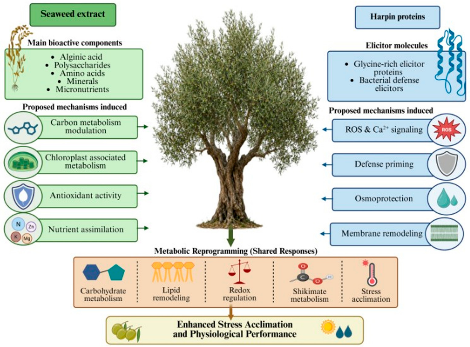
In response to these challenges, the European Union (EU) has promoted sustainable agricultural practices through initiatives such as the European Green Deal and the Farm to Fork Strategy [16,17], encouraging environmentally friendly and resource-efficient alternatives to conventional agrochemical inputs. Within this framework, plant biostimulants have emerged as promising tools for sustainable crop management [18,19]. They comprise a broad and heterogeneous group of bioactive products whose definition has historically varied [20,21]. The EU Fertilizing Products Regulation (FPR) 2019/1009 [22] has established a harmonized framework defining plant biostimulants as products that stimulate plant nutrition processes independently of their nutrient content to improve nutrient use efficiency, tolerance to abiotic stress, crop quality traits, and nutrient availability in the rhizosphere [23]. Among the various plant biostimulant categories, seaweed (SE)-based and harpin (HRP)-based biostimulants have attracted considerable scientific and commercial interest due to, among other factors, their multiple beneficial effects on plant physiology and stress responses [24,25,26] (Figure 1).
SE products, particularly those derived from the seaweed Ascophyllum nodosum (Fucaceae), contain a diverse array of bioactive compounds, such as phytohormone-like substances, polysaccharides, amino acids, and minerals [27]. These compounds have been associated with enhanced plant nutrient uptake, improved photosynthetic performance, root growth stimulation, improved antioxidant activity and tolerance to abiotic stresses such as drought and salinity [28,29,30]. Focusing on olive trees, SE-based formulations have been associated with enhanced vegetative growth, increased photosynthetic efficiency, and improved adaptation to drought stress [31,32]. On the other hand, HRP biostimulants represent an important category of elicitor compounds with demonstrated effects on plant growth and the activation of defense responses [33]. Harpins are glycine-rich proteins originally produced by phytopathogenic bacteria and secreted through the type III secretion system [34]. Despite their bacterial origin, harpins are non-toxic to plants and have been shown to trigger plant defense signaling pathways, including systemic acquired resistance (SAR) [35,36], while several studies have also reported positive effects of harpins on plant phenological and physiological processes [37,38]. In perennial tree crops, including olive, HRP products may contribute to enhanced stress resilience, improved physiological performance, and improved fruit quality [39,40].
Although numerous studies have investigated the effects of biostimulants on traits such as yield, growth, and physiological parameters [41,42], to date, comprehensive untargeted metabolomics characterization of biostimulant-induced metabolic responses in olive has been largely lacking [43]. Due to the complexity of biostimulant-induced plant responses, advanced bioanalytical tools are required to elucidate their effects on plant metabolism. In this context, metabolomics has emerged as a powerful approach for mining plant responses to bioactive agents [44,45,46,47]. Among the available platforms, GC/EI/MS remains widely used because of its high sensitivity, reproducibility, and broad coverage of primary metabolites, including sugars, amino acids, organic acids, fatty acids, and several derivatizable semi-polar compounds [38,48,49]. In the agri-food sector, metabolomics has become an increasingly important tool for investigating plant responses to abiotic and biotic stimuli [50,51,52]. In olive research, metabolomics could provide valuable insight into stress responses, fruit quality, and the biochemical mechanisms underlying plant performance and interactions with biostimulants [53,54]. Such approaches facilitate the identification of metabolite biomarkers and pathways associated with environmental adaptation, supporting the optimization of biostimulant applications in sustainable olive production.
Despite the increasing interest in plant biostimulants, important gaps remain in our understanding of their mode(s) of action (MoA) in olive. In particular, comparative metabolomics studies evaluating mechanistically distinct biostimulants under identical experimental conditions remain lacking, while the metabolic basis of early responses to biostimulants and stress acclimation is still poorly understood. Thus, the present study aimed to comparatively investigate the early metabolic responses of olive trees following treatment with an SE and an HRP biostimulant employing untargeted GC/EI/MS metabolomics. By integrating metabolite profiling with pathway-level biological interpretation, we sought to identify treatment-specific metabolite signatures and provide new insights into the mechanisms underlying biostimulant activity in olive trees.

2. Materials and Methods

2.1. Chemicals and Reagents

All chemicals and reagents used in the experimentation were of the highest commercially available grade. GC/EΙ/MS-grade ethyl acetate and methanol, used in leaf metabolite extraction, and dimethyl sulfoxide (DMSO) for the extraction of the photosynthetic pigments were purchased from Carlo Erba Reagents (Val de Reuil, France). Anhydrous pyridine (99.8%), methoxylamine hydrochloride (MeOX; 98% w/w), and N-methyl-N-(trimethylsilyl)trifluoroacetamide (MSTFA) were purchased from Sigma-Aldrich (Steinheim, Germany). Analytical standards of selected tentatively identified plant metabolites were obtained from Sigma-Aldrich (Steinheim, Germany) for absolute metabolite identification. A harpin-containing commercial product (HRP, Harpin αβ 1%) and a seaweed-based extract (SE) commercial biostimulant [1% w/w alginic acid (from A. nodosum seaweed extract), 0.35% w/w mannitol (from A. nodosum seaweed extract), 5% w/w urea, 1.5% w/w MgO, 3.5% w/w SO3, 1.2% w/w B, 1.0% w/w Mn, 0.1% w/w Mo, 0.5% w/w Zn] were used in the experiment.

2.2. Plant Material, Growth Conditions, Biostimulant Application, and Sample Collection

One-year-old olive trees (Olea europaea L. cv. Koroneiki), approximately 100 cm in height, were purchased from Olea Kostelenos Nurseries (Galatas, Greece) (Figure S1). To simulate standard commercial orchard conditions, the plants were established in a greenhouse at a spacing of 4 × 2 m, corresponding to a planting density of approximately 125 trees per acre. Throughout the experiment, the plants were monitored regularly for the presence of disease symptoms or pest infestations. Environmental conditions were maintained at 22 ± 2 °C under a 14 h light/10 h dark photoperiod. Each plant was irrigated with 100 mL of water every three days.
Sixty-three healthy and uniform plants based on their overall appearance were randomly assigned to three treatment groups: twenty-one were treated with HRP, twenty-one with SE, and twenty-one received sterile water and served as the untreated group. Spray solutions were prepared according to the manufacturers’ recommendations, and dosages were set at 12–15 g/acre (~0.105 g/tree) for HRP and 200–250 mL/100 L water (~1.75 mL/tree) for SE. To ensure thorough and uniform foliar coverage, 50 mL of the respective treatment solution was applied to each seedling by spraying from multiple directions at 20 cm.
The effects of the biostimulants on plants’ metabolism and photosynthetic pigment content were assessed seven days post-treatment. This timepoint was selected to investigate the early physiological and metabolic responses elicited by the biostimulants, as alterations in cellular metabolism and photosynthetic machinery are expected to occur before the manifestation of long-term growth and developmental effects. The first four fully expanded leaves from separate shoots immediately below the apex were collected from each plant, briefly rinsed with water, gently blotted dry, labeled, and immediately flash-frozen in liquid N2 to quench metabolic activity. Leaves from three plants were pooled to provide a pooled sample. In total, seven pooled samples were collected per treatment. The snap-frozen leaves were immediately pulverized into a fine powder using a mortar and pestle under liquid N2. The pulverized leaf tissues were then stored at −80 °C until further processing.

2.3. Assessment of the Effect of Biostimulants on Chlorophyll (Chl) and Carotenoid Concentrations in Olive Leaves

For the determination of the chlorophyll (Chl) and carotenoid content of Olive leaves using UV-Vis spectrophotometry at specific wavelengths, a uniSPEC 2 spectrophotometer (LLG Labware, Meckenheim, Germany) was used. An already established extraction protocol based on DMSO was employed with minor modifications [55,56]. Briefly, 20 mg of each of the pulverized samples was weighed and transferred into a labeled 2 mL Eppendorf tube. Following the addition of 1 mL of DMSO, the samples were incubated in a water bath at 64 °C for 60 min. Immediately after incubation, the tubes were briefly centrifuged, and the supernatants were carefully transferred to new tubes. For spectrophotometric analysis, a 0.5 mL aliquot of the resulting extract was diluted with 0.5 mL of DMSO and transferred into a cuvette. Absorbance (A) measurements were recorded at 470 nm, 648 nm, and 664 nm, with each sample measured in duplicate. Following acquisition of absorbance values, chlorophyll α (Chlα), chlorophyll β (Chlβ), and carotenoid concentrations were calculated, as previously described [57], expressed as μg mL−1. The calculated contents of the photosynthetic pigments were statistically analyzed using Tukey’s honestly significant difference (HSD) test at a significance level of α = 0.05 using the software JMP v.18.2.1 (SAS Institute, Cary, NC, USA).

2.4. Monitoring of the Biostimulant-Induced Metabolic Changes in Olive Plants Using GC/EI/MS-Based Metabolomics

Leaf metabolite extraction was performed by adding 100 mg of pulverized leaves into a 0.5 mL methanol–ethyl acetate solution (50:50, v/v), following a previously developed protocol [58]. The resulting suspensions were sonicated for 20 min (Ultrasonic cleaner 3200 EP S3, SOLTEC S.r.l, Milano, Italy), shaken for 45 min (GFL 3006, Geschacha für Labortechnik mbH, Burgwedel, Germany), and finally filtered through 0.2 μm PTFE filters (Macherey-Nagel, Duren, Germany) to remove debris. Extracts were evaporated to dryness using a vacuum concentrator (Eppendorf Concentrator Plus, Eppendorf, Hamburg, Germany). A two-step derivatization protocol was followed [59]. Briefly, methoximation was performed by adding 80 μL of MeOX solution (20 μL of 0.2 μg mL−1 in anhydrous pyridine) to the dried extracts and incubating at 30 °C for 2 h in a dry block incubator (Digital Cooling Bath, Thermo Fisher Scientific, Waltham, MA, USA). Subsequently, 80 μL MSTFA was added and the samples were incubated at 37 °C for 90 min for silylation. Experimental blanks were identically prepared to monitor background contaminants [60].
Metabolic profiling was conducted on an Agilent 6890 gas chromatograph coupled with a 5973 inert mass selective detector (Agilent Technologies Inc., Santa Clara, CA, USA). Samples were analyzed on an HP-5MS UI column (30 m × 0.25 mm × 0.25 μm). Helium carrier gas flowed at 1 mL min−1, and oven temperature was programmed from 70 °C (5 min hold) to 310 °C at 5 °C min−1, then held for 1 min. Electron ionization was set at 70 eV with full-scan MS data acquired between 50 and 800 Da at 4 scans sec−1. Source and quadrupole temperatures were 230 °C and 150 °C, respectively.
Chromatograms were deconvoluted with the software AMDIS v.2.73 (National Institute of Standards and Technology, Gaithersburg, MD, USA) and tentative metabolite identifications based on spectral matching to the NIST ‘23 library v.3.0 were performed. Peaks present in blanks were excluded. Absolute metabolite identification employed authentic standards analyzed on the same platform following the Metabolomics Standards Initiative guidelines [61]. Datasets were further processed in MS-DIAL v.5.5.26 [62] and cleaned by removing non-biological features in Excel v2605 (Microsoft, Redmond, WA, USA). Subsequently, multivariate analyses, including OPLS-DA (p < 0.05) and hierarchical clustering (HCA), were performed using SIMCA-P+ v.18.1.0 (Umetrics, Sartorius Stedim Data Analytics AB, Umeå, Sweden) to discover trends and biomarkers of the plant biostimulation effect [59,63].

3. Results and Discussion

3.1. Photosynthetic Pigment Homeostasis Is Maintained Following Seaweed- and Harpin-Based Biostimulant Applications

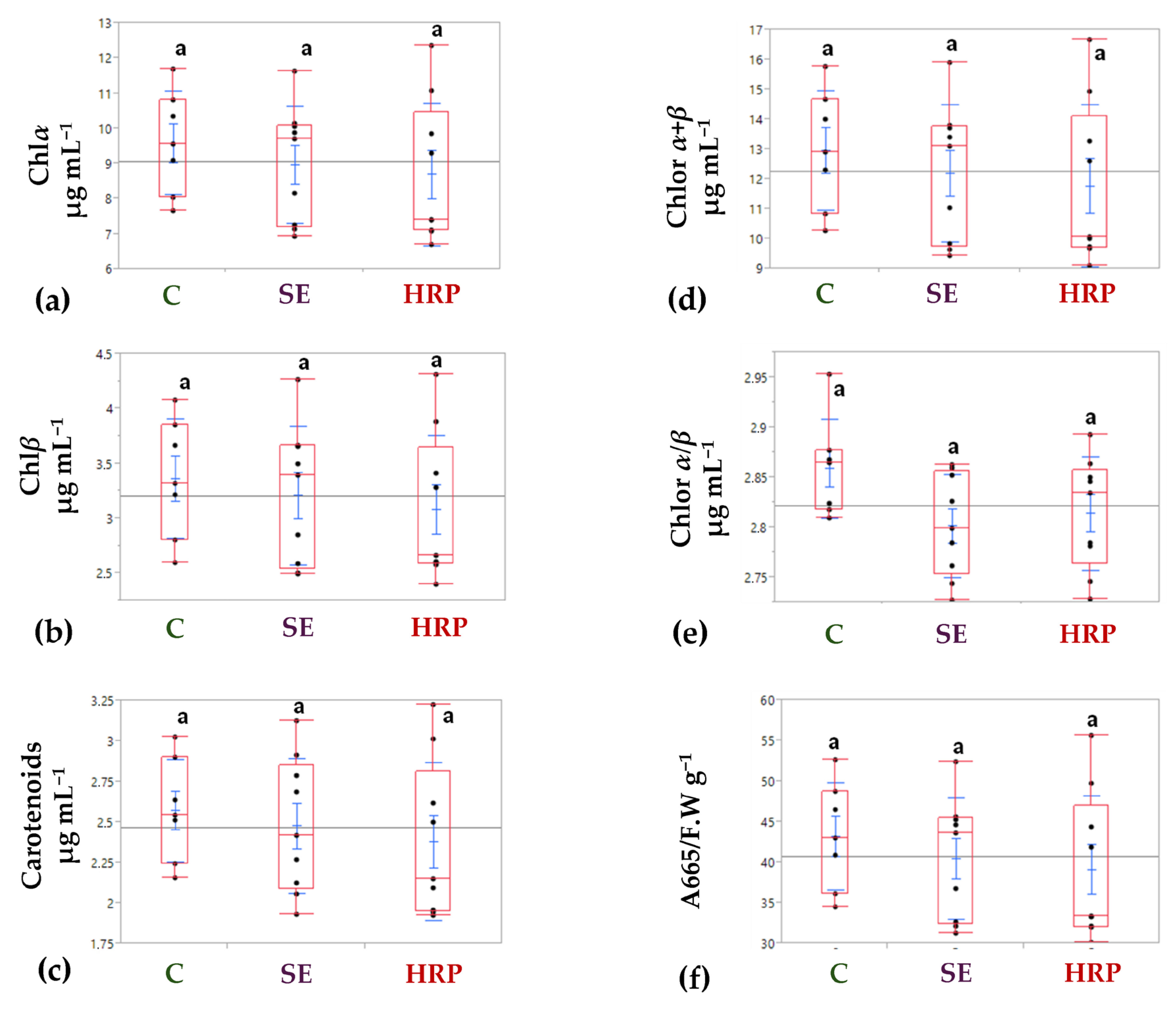
Measurements of the photosynthetic pigment content of olive leaves and their fluctuation in response to the biostimulants were performed 7 days post-treatment. In addition to the quantification of Chlα, Chlβ, and carotenoids, the total Chl content (Chlα + Chlβ) as well as the Chlα/Chlβ, and A665/F.W. ratios were calculated to provide a comprehensive assessment of the effects of the applied biostimulants on the photosynthetic and physiological status of the young olive plants. The analysis revealed that neither the SE nor the HRP treatment had a statistically significant effect on the photosynthetic pigment content of plants compared to the untreated ones (Figure 2). Mean total Chl concentrations were 12.96 μg mL−1 for the untreated, 12.20 μg mL−1 for the SE-treated, and 11.78 μg mL−1 for the HRP-treated plants (Figure 2d). Similarly, carotenoid concentrations exhibited no statistically significant variation among treatments (Figure 2c). Mean carotenoid contents were 2.57 μg mL−1 in the untreated, 2 Chl.48 μg mL−1 in the SE-treated, and 2.38 μg mL−1 in the HRP-treated plants. Additionally, no statistically significant differences were observed for the Chlα/Chlβ (Figure 2e) and A665/F.W. (Figure 2f) ratios. The Chlα/Chlβ ratio ranged from 2.86 in the untreated to 2.80 and 2.81 in the SE- and HRP-treated plants, respectively. Likewise, the A665/F.W. ratio varied between 43.2, 40.5, and 39.2 g−1 for the untreated, SE-, and HRP-treated leaves.
Leaves’ Chlα and Chlβ concentrations have long been associated with photosynthetic efficiency and overall plant vigor [64]. Additionally, the Chlα/Chlβ ratio is widely used as an indicator of photosynthetic performance and leaf physiological status, particularly in studies related to leaf senescence, since Chlα generally declines more rapidly than Chlβ during aging processes [65]. Furthermore, the A665/F.W. ratio has been proposed as an alternative rapid estimator of total Chl content and as an indicator of phytotoxicity and bioactivity without the need for mathematical conversion models [66]. Taken together, the absence of statistically significant differences in the Chl content, carotenoid concentration, and related physiological indices indicates that, under the experimental conditions set and the application dosages tested, both biostimulants did not significantly affect the photosynthetic activity or pigment-associated physiological status of olive plants.
The results obtained for the SE treatments cannot be directly compared to previous studies, since, to the best of our knowledge, no reports are currently available regarding the application of similar products to olive trees. In an earlier study [67], the effect of the application of different concentrations of extracts derived from the same seaweed species (A. nodosum) on cucumber (Cucumis sativus) seedlings under different experimental conditions was investigated. The results demonstrated an increase in the plants’ Chl concentration following treatment; however, no further enhancement was observed with increasing extract concentration. Increased Chl levels in several plant species following root application of a formulation comparable to that used in the present study have been reported [68]. Nonetheless, foliar application did not result in statistically significant elevation compared to the untreated plants, which is in line with results of the current study. In grapevine (Vitis vinifera L.), application of A. nodosum extract has also led to increased total Chl content [69]. Studies evaluating the independent effects of inorganic elements have nevertheless been conducted in other plant species. In a recent study [70], the effects of three foliar applications of Zn, B, and Cu on mandarin trees (Citrus reticulata) were investigated separately, revealing slight increases in Chl concentration, particularly following Zn application. Furthermore, sulfur application significantly increased Chl levels in soybean (Glycine max) plants [71]. Manganese (Mn), a micronutrient characterized by a narrow sufficiency–toxicity range, has been reported to initially increase Chl content at low application rates, whereas higher Mn concentrations eventually reduce Chl levels [72,73]. In contrast, molybdenum appears to exert minimal influence on Chl concentration even at elevated levels [74].
Regarding the second formulation used in the present study, no previous reports have examined the effect of HRP on Chl levels of olive trees, while the available literature in other crops remains limited. In pepper plants (Capsicum annuum), only a slight increase in Chl concentration was observed following three foliar applications of harpin [75]. In a subsequent study, no significant effects of harpin application on Chlα, Chlβ, and the Chlα/Chlβ ratio in pepper plants were reported [76], findings that are in agreement with those of the present study.
Overall, the available evidence, together with the findings of the present study, suggests that the effects of SE- and HRP-based formulations on Chl accumulation are highly dependent on plant species, formulation composition, and mode of application. Under the experimental conditions employed here, foliar application of both of the products tested did not significantly alter plants’ Chl content, indicating that their biostimulant activity is likely mediated through metabolic and signaling pathways rather than through direct stimulation of photosynthetic pigment accumulation of olive trees. These observations further support the importance of metabolomics-based approaches for uncovering the biochemical effects of biostimulants that may not be detectable using conventional physiological measurements alone. Consequently, further investigation involving multiple applications and different experimental conditions is required to further elucidate the effects of such biostimulants on the photosynthetic physiology of olive plants.

3.2. GC/EI/MS Metabolomics Suggests Broad and Treatment-Specific Metabolic Reprogramming

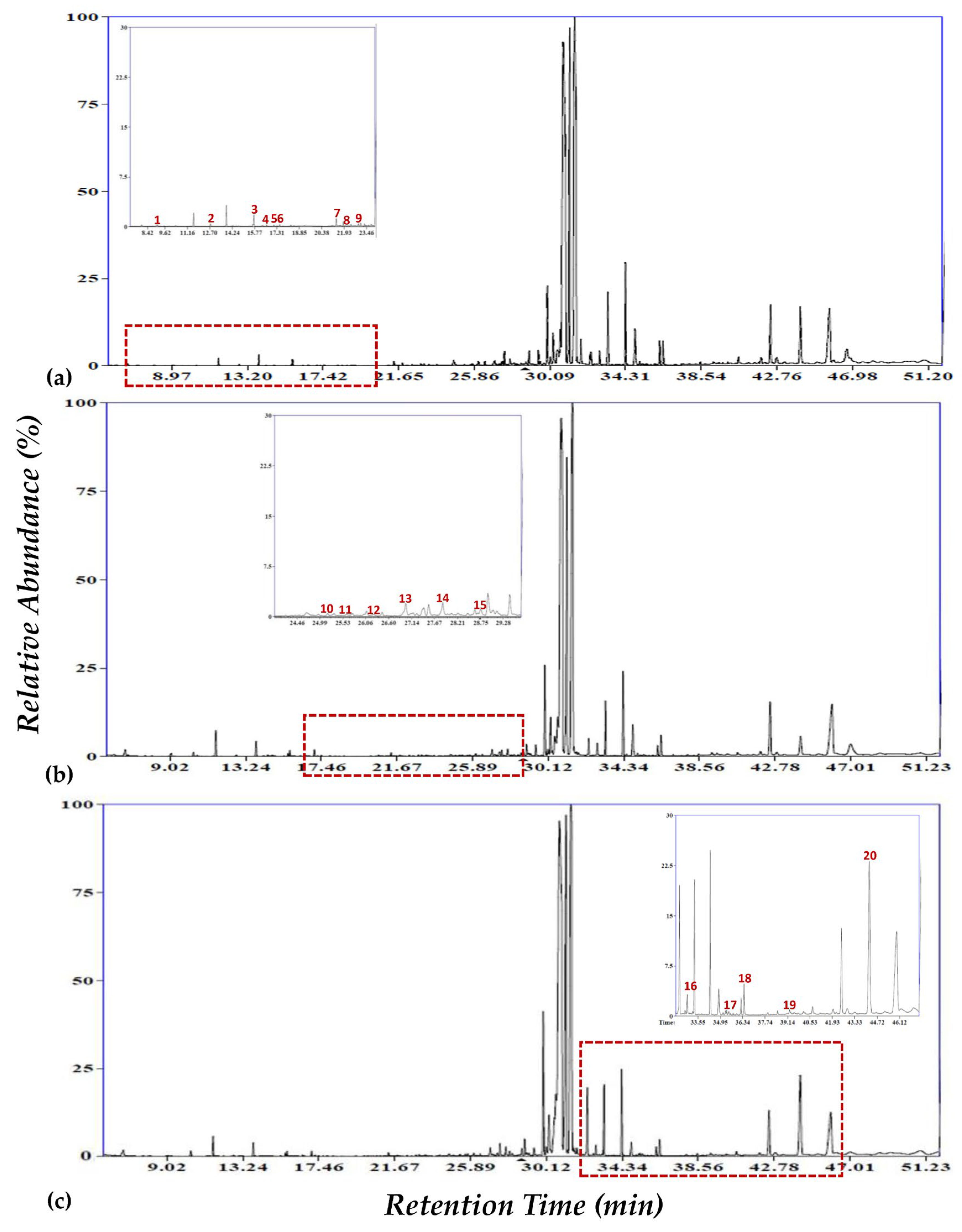
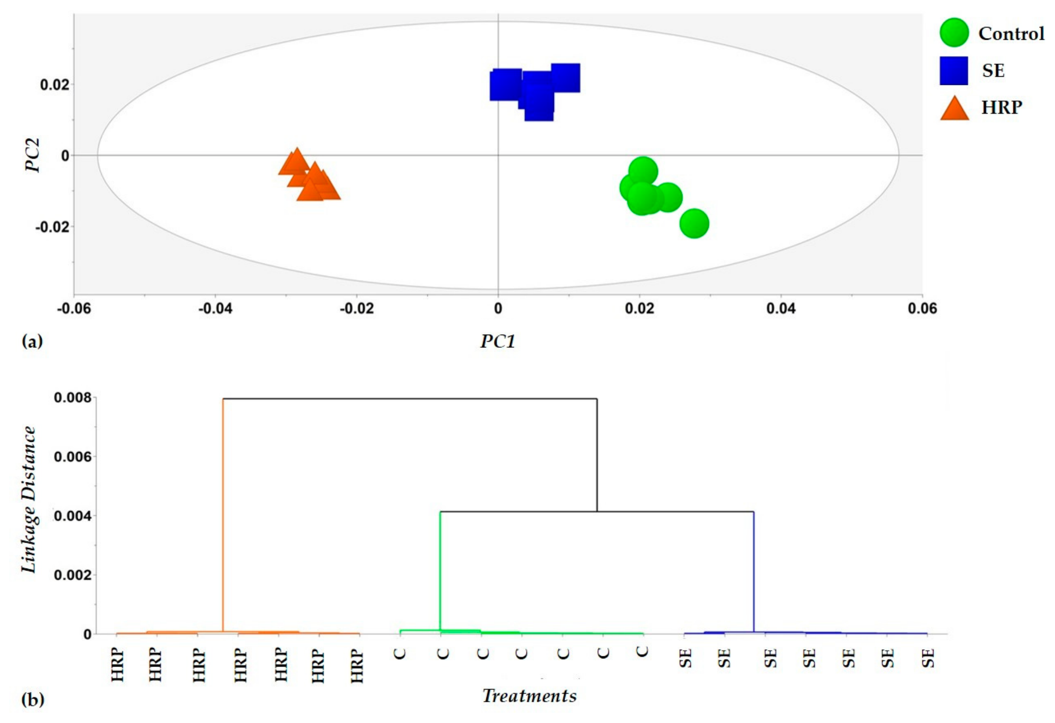
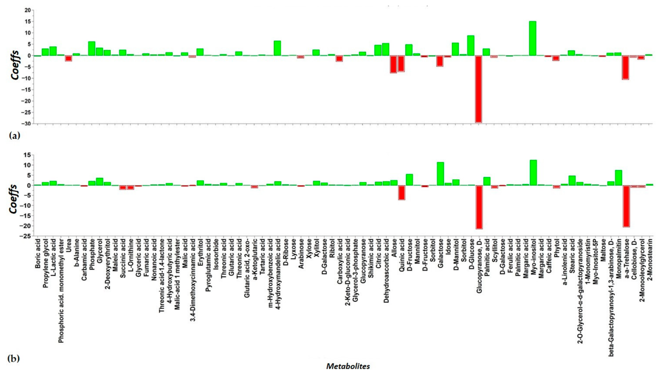
The robustness of the GC/EI/MS bioanalytical workflow employed is confirmed by the high quality of the acquired total ion chromatograms (TICs) (Figure 3), which exhibit well-resolved peaks, stable baselines, and highly reproducible chromatographic profiles across all treatment groups. The applied metabolomics pipeline successfully recorded the olive leaf metabolome, resulting in the reproducible detection of 179 metabolic features, out of which 88 were tentatively or absolutely annotated, corresponding to 57 unique metabolites. The identified metabolites consisted predominantly of carbohydrates, carboxylic acids, and fatty acids (~90%), with the rest comprising amino acids, alcohols, heterocyclic compounds, phosphoric acids, and vitamins (~10%) (Table S1). Multivariate analyses demonstrated tight clustering among biological replicates and trends, and the absence of outliers. OPLS-DA modeling (Figure 4a) distinguished between the recorded metabolite profiles according to biostimulant treatments (HRP, SE, Untreated). High values of the total explained variation in X [R2X(cum) = 0.71], Y [R2Y(cum) = 0.94], and predictive ability [Q2(cum) = 0.91], for 2 principal components, were calculated. HCA (Figure 4b) further resolved discrete groupings, notably highlighting the distinct metabolic impact of HRP. Metabolite contributions driving separation are displayed in Figure 5. OPLS-DA score plots of pairwise comparisons (Figure S2) confirmed these findings, revealing complete metabolic separation between the untreated and biostimulant-treated plants.

3.3. Biostimulant-Driven Metabolic Reprogramming and Physiological Adaptation in Young Olive Trees

Untargeted metabolomics analysis combined with OPLS-DA revealed that both SE and HRP biostimulants induced extensive and highly coordinated metabolic reprogramming in young olive leaf tissues. Rather than causing isolated perturbations of individual metabolites, both treatments elicited broad alterations across multiple interconnected metabolic pathways, resulting in distinct metabolic signatures relative to the untreated plants. Although several common responses were observed, the global metabolite profiles indicated partially distinct MoA for the two treatments. Similar metabolome-scale fluctuation has been repeatedly associated with biostimulant-induced physiological reprogramming and enhanced environmental adaptation in plants [77,78,79]. The observed metabolic alterations were primarily associated with central carbon metabolism, osmotic regulation, lipid remodeling, and redox-associated pathways. Additionally, amino acid metabolism and aromatic secondary metabolism were also substantially affected, collectively indicating that both treatments broadly modulated interconnected metabolic networks, acting as active metabolic modulators rather than simple nutritional inputs.
Based on the OPLS-DA coefficients (Figure 5), both formulations promoted a substantial reallocation of metabolic resources toward pathways associated with stress acclimation, cellular protection, and biosynthetic activity, consistent with a biostimulant-induced shift from basal metabolic maintenance toward an actively primed physiological state. Although both treatments induced partially overlapping responses, clear differences emerged in the metabolic strategies associated with each formulation, possibly indicating that SE and HRP engage partially distinct signaling and metabolic mechanisms to achieve their effects. The nature and magnitude of these treatment-specific metabolic shifts are examined in detail in the following sections. Collectively, these coordinated metabolic responses provide a biochemical framework for interpreting the early physiological responses induced by the biostimulants being tested, despite the absence of substantial changes in the plants’ photosynthetic pigments seven days post-treatment.

3.4. Integrated Remodeling of Carbohydrate, Antioxidant, and Isoprenoid Metabolism Following SE Application

One of the most prominent effects of the SE treatment was the extensive remodeling of plants’ carbohydrate metabolism. Several soluble sugars and carbohydrate-associated metabolites, including D-glucopyranose, galactose, allose, arabinose, fructose-related metabolites, and myo-inositol-associated compounds, exhibited substantially lower abundance in treated compared to the untreated plants (Figure 5a). Such coordinated reduction is consistent with enhanced metabolic turnover and redistribution of carbon toward biosynthetic and respiratory processes frequently associated with metabolically active physiological states [80,81]. Alternative explanations, including reduced carbohydrate biosynthesis, altered assimilate transport, or increased carbohydrate consumption associated with stress responses, cannot be excluded. However, the simultaneous depletion of multiple soluble sugars together with coordinated changes in glycolytic intermediates, compatible solutes, and central carbon metabolism is more consistent with enhanced metabolic utilization and carbon reallocation than a generalized suppression of carbohydrate metabolism. Collectively, these responses may indicate that the SE treatment promoted increased metabolic plasticity and carbon flux redistribution in olive tissues, characteristics commonly associated with biostimulant-induced metabolic activation [82,83].
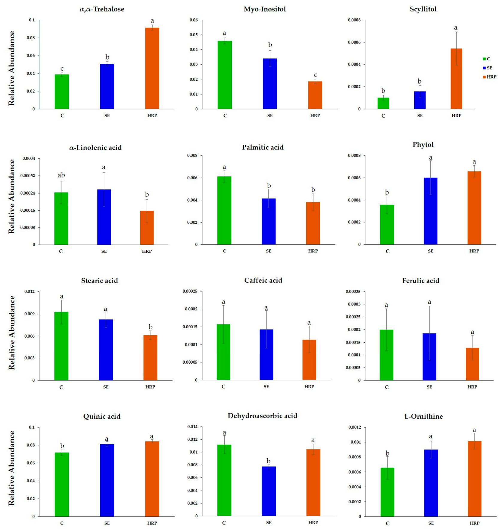
Of particular interest was the substantial alteration of α,α-trehalose metabolism (Figure 6). Beyond its role as a reserve carbohydrate, trehalose is a multifunctional signaling metabolite involved in sugar sensing, osmotic regulation, ROS mitigation, and stress adaptation [84,85]. Its altered accumulation under SE treatment may indicate a significant effect on carbon allocation, sugar sensing, and stress-associated signaling pathways, supporting the interpretation that SE promoted metabolic activation and physiological acclimation through extensive reconfiguration of carbon metabolism [86,87,88]. Similarly, osmoprotective polyols and sugar alcohols, including mannitol, erythritol, xylitol, and glycerol-related metabolites, were significantly affected, collectively suggesting major rewiring of osmoprotective metabolism and carbon partitioning.
The recorded metabolic response further suggested selective fluctuation of metabolites associated with the shikimate–phenylpropanoid pathway. Among those, quinic acid exhibited the most pronounced treatment-associated response, whereas for shikimic acid, caffeic acid, and ferulic acid, modest fluctuations were observed (Figure 6). In summary, these observations suggest selective modulation of shikimate-associated metabolism rather than broad activation of the entire shikimate–phenylpropanoid pathway. Quinic acid, which displayed the strongest SE-associated response, is a key intermediate of the shikimate pathway, supporting the biosynthesis of aromatic amino acids and downstream phenylpropanoid-derived compounds. In olive, these metabolites contribute to the biosynthesis of phenolic compounds and secoiridoids, which play important roles in antioxidant defense, stress adaptation, and pathogen resistance [89,90]. Their observed accumulation could suggest intensified metabolic channeling toward downstream antioxidant- and defense-associated pathways, a response frequently linked to biostimulant-induced physiological priming [91,92,93]. Complementing these findings, the pronounced reduction in myo-inositol (Figure 6), a primary metabolite central to phosphatidylinositol signaling, cell wall biosynthesis, and ascorbate metabolism, likely reflects increased metabolic flux through signaling-related and antioxidant pathways, reinforcing the interpretation of broad stress-acclimation reconfiguration following SE treatment [80].
An additional hallmark of the SE treatment was the modulation of redox-associated metabolism. The decrease in dehydroascorbic acid, the oxidized form of ascorbic acid, could indicate alterations in antioxidant turnover and ascorbate–glutathione cycle activity (Figure 6). This response may reflect improved antioxidant recycling efficiency, reduced oxidative burden, or enhanced utilization of antioxidant defense systems, collectively indicating a biostimulant-induced priming effect associated with improved oxidative stress management. Biostimulant-mediated regulation of reactive oxygen species (ROS) detoxification pathways is considered a central component of plant acclimation responses under environmental stress conditions and may contribute substantially to enhanced physiological resilience [94]. Improved antioxidant homeostasis may protect cellular membranes, proteins, chloroplasts, and photosynthetic machinery against oxidative damage during drought, heat, or transplant stress, conditions frequently encountered in olive cultivation systems. Additional evidence of extensive metabolic reorganization emerged from the central organic acid metabolism. Multiple TCA cycle intermediates, including citric acid, malic acid, fumaric acid, succinic acid, and α-ketoglutaric acid, were among the most discriminatory metabolites between treated and untreated plants (Figure 5). Because these intermediates connect respiration, amino acid biosynthesis, redox regulation, and anaplerotic metabolism [81], their coordinated modulation strongly suggest large-scale remodeling of respiratory carbon flux and enhanced carbon turnover and mobilization rather than reserve accumulation. This increased central metabolic activity was further mirrored at the level of membrane lipids.
Lipid-associated metabolites were also substantially influenced by the SE treatment. Saturated fatty acids and storage-related lipids, including palmitic acid, stearic acid, monopalmitin, and monostearin, accumulated preferentially in untreated plants, whereas SE-treated plants exhibited higher levels of metabolites associated with membrane turnover and glycerolipid remodeling, including 2-monooleoylglycerol (Figure 6). Such coordinated changes plausibly suggest active membrane restructuring and lipid turnover following biostimulant application [95,96]. In olive plants, membrane remodeling of this kind is closely associated with environmental acclimation and developmental signaling, particularly during root establishment and early canopy growth. The accumulation of phytol was also biologically meaningful because it serves as a precursor for Chl-associated metabolism and tocopherol (vitamin E) biosynthesis [97,98,99], which could possibly indicate enhanced chloroplast stability, reinforced antioxidant capacity, and support of photosynthetic function.
Beyond the observed changes in lipid remodeling and phytol accumulation, the SE treatment also affected metabolites associated with plastidial isoprenoid metabolism. A notable finding was the accumulation of 2-deoxyerythritol, a metabolite structurally related to 2-C-methyl-D-erythritol and associated with the methylerythritol phosphate (MEP) pathway (Figure 6). This observation may indicate increased engagement of plastidial isoprenoid biosynthesis, a pathway contributing to the synthesis of carotenoids, chlorophyll-associated compounds, terpenoids, and phytohormone precursors involved in stress adaptation and photosynthetic regulation [100]. Given the central role of isoprenoid-derived metabolites in photoprotection, membrane stabilization, antioxidant defense, and hormonal signaling, modulation of this pathway provides further evidence that SE promotes adaptive metabolic reprogramming linked to enhanced environmental resilience.
Collectively, the coordinated changes observed across carbohydrate, organic acid, lipid, antioxidant, and isoprenoid metabolism indicate that SE primarily acts through enhancement of carbon turnover, stimulation of central metabolic activity, and reinforcement of antioxidant and membrane-associated processes. These responses are consistent with the reported MoA of A. nodosum-derived biostimulants and are of particular relevance in perennial Mediterranean crops such as olive, where efficient management of metabolic resources directly underpins resilience to fluctuating environmental conditions [77,79]. Whether a complementary but mechanistically distinct pattern of metabolic reprogramming is elicited by HRP-based biostimulants is examined in the following section.

3.5. Metabolome-Level Evidence of Enhanced Stress Resilience Following HRP Application

Unlike the growth-oriented metabolic activation observed with SE, the HRP treatment elicited a metabolic signature more directly aligned with stress preparedness, osmoprotection, and membrane remodeling. Among the most discriminatory metabolites, galactose, fructose, myo-inositol, palmitic acid, stearic acid, and glycerol were significantly depleted, while α,α-trehalose, α-ketoglutaric acid, phytol, 2-monooleoylglycerol, and scyllitol were enriched, in HRP-treated plants. Taken together, this metabolic configuration is consistent with a shift toward stress-responsive physiological reprogramming, suggesting that treated plants may be metabolically preconditioned to better withstand subsequent environmental challenges [101,102,103,104].
One of the most significant metabolic responses following HRP application was the pronounced accumulation of α,α-trehalose in HRP-treated plants (Figure 6). In contrast to its role in SE-treated plants, where trehalose metabolism was broadly remodeled as part of carbon signaling reconfiguration, its enrichment in HRP-treated plants suggests a more direct osmoprotective function. Beyond osmotic adjustment, trehalose contributes to protein and membrane stabilization, participates in the regulation of ROS homeostasis, and activates downstream stress-signaling networks [105]. The accumulation of scyllitol could further indicate the activation of osmoprotective and stress-buffering mechanisms following HRP treatment (Figure 6). Cyclitols are widely recognized for their roles in osmotic adjustment, membrane stabilization, and protection against oxidative and dehydration-induced damage [106]. The increased abundance of scyllitol therefore provides complementary evidence for the establishment of a stress-primed metabolic state, reinforcing the interpretation that HRP promotes enhanced physiological readiness to cope with subsequent environmental challenges. A parallel shift in lipid metabolism further supported this observation. The observed substantial decreases in palmitic acid, stearic acid, and glycerol, combined with the accumulation of phytol and 2-monooleoylglycerol, are indicative of active change in membrane lipid composition (Figure 6). The elevation of phytol, a catabolite of Chl and precursor of tocopherol, is notable in this context as it signals enhanced photoprotective capacity and antioxidant reinforcement at the chloroplast level—metabolic adjustments that collectively support membrane integrity and photosynthetic resilience under Mediterranean field conditions [80,82].
The accumulation of α-ketoglutaric acid constitutes an additional key metabolic indicator of the HRP treatment’s impact (Figure 5). As a central intermediate linking carbon metabolism with nitrogen assimilation and amino acid biosynthesis, it plays a crucial role in metabolic flexibility and stress adaptation [107]. Its accumulation suggests enhanced integration between carbon and nitrogen metabolism, potentially supporting increased biosynthetic capacity, nutrient utilization efficiency, and metabolic adaptability under changing environmental conditions. Furthermore, increased levels of cellobiose and glucopyranose-related metabolites suggest modifications in carbohydrate turnover and cell wall-associated metabolism (Figure 6). These responses indicate enhanced structural adaptation, altered carbon partitioning, and dynamic remodeling of carbohydrate reserves. Similar metabolic rearrangement involving carbohydrates, amino acid precursors, and lipid metabolism have been repeatedly associated with abiotic stress acclimation and enhanced resilience mechanisms in plants [83,108].
These findings are collectively indicative of an MoA for HRP that is fundamentally distinct from the carbon-mobilization and growth-stimulatory effects observed for SE. Rather than accelerating metabolic turnover and biosynthetic activity, HRP appears to reconfigure plant physiology toward a defense-ready state, fortifying cellular and molecular protection systems in anticipation of abiotic challenges [37,109]. The resulting metabolite profiles following HRP application are consistent with a defense-priming MoA in which plants are physiologically and biochemically preconditioned before stress onset. This profile is complementary to, yet mechanistically distinct from, the growth-activating metabolic reprogramming described for SE, and together the two treatments illustrate how biostimulants can target different but equally relevant aspects of olive stress physiology.

3.6. Selective Preservation of Metabolites Associated with Signaling, Defense, and Redox Regulation

Equally informative to the metabolites that responded strongly to treatment were those whose levels remained unchanged despite their well-established roles in plant stress physiology and defense regulation. Among these, α-linolenic acid, caffeic acid, ferulic acid, and pyroglutamic acid exhibited no statistically significant fluctuations following treatment with SE and HRP (Figure 6). α-linolenic acid serves as the primary precursor of jasmonic acid biosynthesis and plays a pivotal role in membrane integrity, oxylipin signaling, and plant responses to environmental stress [110,111]. Similarly, caffeic acid and ferulic acid are key intermediates of the phenylpropanoid pathway, contributing to antioxidant defense, cell wall reinforcement, lignification processes, and protection against biotic and abiotic stressors [112,113] (Figure 5). Pyroglutamic acid is closely associated with glutathione turnover and cellular redox homeostasis, linking primary metabolism with antioxidant regulation and stress resilience [114]. The relative stability of these metabolites is noteworthy because they occupy strategic positions within several major defense- and stress-associated metabolic networks. Under conditions that strongly activate plant defense responses, substantial alterations in jasmonic acid precursors, phenylpropanoid intermediates, and glutathione-associated metabolites are frequently observed as resources redirected toward protective and adaptive processes. The absence of substantial alterations in the levels of these compounds could indicate that neither SE nor HRP elicited a generalized stress-like response or extensive activation of major plant defense pathways. Instead, the observed responses appear to reflect a more targeted physiological adjustment in which specific metabolic sectors were selectively modulated while key signaling and regulatory hubs remained comparatively stable.
This interpretation is consistent with the broader metabolic patterns observed throughout the present study. Although both biostimulants induced significant changes in carbohydrate utilization, osmoprotective metabolism, membrane remodeling, antioxidant-associated processes, and central carbon metabolism, these responses occurred without major perturbation of several metabolites traditionally associated with defense signaling and secondary metabolism. Such selective regulation may be particularly advantageous from a physiological perspective, as it allows plants to optimize metabolic performance and stress resilience while avoiding the substantial energetic costs frequently associated with large-scale activation of defense pathways. This pattern is consistent with our previous metabolomics study in Cannabis sativa L., where HRP application resulted in the substantial alteration of primary and secondary metabolism, while α-linolenic acid and caffeic acid remained largely unchanged [38]. The results of a recent study on Arabidopsis revealed extensive reconfiguration of primary and secondary metabolism, particularly pathways associated with carbon utilization, respiratory activity, lipid remodeling, and defense-related processes following SE application [115], which further supports our findings. Importantly, several oxylipin- and phenylpropanoid-associated metabolites remained stable or decreased in abundance, supporting the hypothesis that SE biostimulants act through selective metabolic reprogramming rather than generalized activation of defense metabolism. Collectively, these findings support the notion that biostimulant-mediated enhancement of plant performance is achieved through targeted metabolic adjustments and improved metabolic efficiency rather than widespread activation of canonical defense-related pathways.
While the present findings provide new insights into the selective olive metabolic responses induced by SE and HRP, several considerations should be taken into account when interpreting their broader biological significance. Metabolomics was performed at a single post-treatment sampling point, providing only a snapshot of the dynamic metabolic responses to biostimulant application. Earlier sampling times would likely capture rapid signaling events, whereas later sampling could reveal longer-term physiological acclimation and metabolic stabilization. Although GC/EI/MS provides excellent coverage of primary metabolism, complementary LC-MS/MS and NMR approaches would substantially expand metabolome coverage, particularly for olive-specific specialized metabolites such as secoiridoids and flavonoids. Even though the present study was conducted under controlled greenhouse conditions, the observed metabolite signatures are consistent with physiological responses previously associated with enhanced abiotic stress resilience. Nevertheless, these findings represent early biochemical indicators of physiological adaptation rather than direct evidence of improved agronomic performance. Future field validation could further validate whether these metabolic responses translate into improved crop performance.

4. Conclusions

The present metabolomics proof-of-concept investigation supports the hypothesis that both biostimulant formulations tested function as genuine metabolic modulators in young olive plants via partially complementary mechanisms. The SE primarily promoted metabolic responses consistent with enhanced carbon turnover, antioxidant regulation, membrane remodeling, and plastidial metabolism, whereas the HRP formulation elicited a metabolic profile consistent with osmoprotection, stress priming, and enhanced physiological preparedness. These findings provide mechanistic insights into the early biochemical olive tree responses associated with biostimulant application while simultaneously highlighting the value of metabolomics for elucidating biostimulant MoA and identifying candidate metabolic biomarkers. Nevertheless, the present findings represent early metabolic responses obtained under controlled greenhouse conditions and should not be interpreted as direct evidence of improved agronomic performance. Future investigations integrating time-course sampling, complementary LC-MS/MS metabolomics, physiological measurements, and multi-environment field validation will be essential to determine whether the observed metabolic reprogramming ultimately translates into enhanced stress resilience, crop productivity, and fruit quality under Mediterranean cultivation systems.

Supplementary Materials

The following supporting information can be downloaded at: https://www.mdpi.com/article/10.3390/agrochemicals5030033/s1, Figure S1: Plant material used in the biostimulant experiment. One-year-old olive trees (Olea europaea L. cv. Koroneiki) grown in pots under controlled conditions. (a) General view of the experimental plants and (b) close-up showing shoot architecture, leaf morphology, and early floral development; Table S1. Representative annotated olive metabolites; Figure S2. OPLS-DA PC1/PC2 score plots illustrating the metabolomic discrimination between control olive plantlets (C) and each biostimulant treatment based on GC/EI/MS metabolite profiling. Upper panel: control (C) versus S.E. Lower panel: control (C) versus HRP. Samples are displayed within the 95% confidence interval ellipse representing Hotelling’s T2. The clear separation between treated and untreated samples indicates substantial treatment-induced metabolic reprogramming associated with each biostimulant formulation.

Author Contributions

Conceptualization, S.K., I.F.K. and K.A.A.; methodology, S.K., N.B., C.N.K., S.G., I.F.K. and K.A.A.; software, C.N.K., I.F.K. and K.A.A.; validation, I.F.K. and K.A.A.; formal analysis, C.N.K., S.G., I.F.K. and K.A.A.; investigation, S.K., N.B., I.F.K. and K.A.A.; resources, I.F.K. and K.A.A.; data curation, C.N.K., S.G., I.F.K. and K.A.A.; writing—original draft preparation, S.K., C.N.K., S.G., I.F.K. and K.A.A.; writing—review and editing, S.K., N.B., C.N.K., S.G., I.F.K. and K.A.A.; visualization, C.N.K., S.G., I.F.K. and K.A.A.; supervision I.F.K. and K.A.A. All authors have read and agreed to the published version of the manuscript.

Funding

This research received no external funding.

Institutional Review Board Statement

Not applicable.

Informed Consent Statement

Not applicable.

Data Availability Statement

The data presented in this study are available in the website of the Biostimulant, Biocontrol, and Pesticide Metabolomics Group (B2PMG) at https://www.aua.gr/pesticide-metabolomicsgroup/Resources/default.html (accessed on 21 May 2025) [Olea europaea var Koroneiki (B2PMG-06-26)].

Conflicts of Interest

The authors declare no conflicts of interest.

Abbreviations

The following abbreviations are used in this manuscript:
Chlα, Chlorophyll α
ChlβChlorophyll β
F.W.Fresh Weight
HCAHierarchical Cluster Analysis
HRPHarpin
MoAMode(s) of Action
OPLSDAOrthogonal Partial Least Squares Discriminant Analysis
PCPrincipal Component
SESeaweed Extract

References

  1. Besnard, G.; Terral, J.-F.; Cornille, A. On the origins and domestication of the olive: A review and perspectives. Ann. Bot. 2018, 121, 385–403. [Google Scholar] [CrossRef] [Scilit] [PubMed]
  2. Langgut, D.; Cheddadi, R.; Carrión, J.S.; Cavanagh, M.; Colombaroli, D.; Eastwood, W.J.; Greenberg, R.; Litt, T.; Mercuri, A.M.; Miebach, A. The origin and spread of olive cultivation in the Mediterranean Basin: The fossil pollen evidence. Holocene 2019, 29, 902–922. [Google Scholar] [CrossRef] [Scilit]
  3. Palli, J.; Fiolna, S.; Bini, M.; Cappella, F.; Izdebski, A.; Masi, A.; Mensing, S.; Nigro, L.; Piovesan, G.; Sadori, L. The human-driven ecological success of olive trees over the last 3700 years in the Central Mediterranean. Quat. Sci. Rev. 2025, 356, 109313. [Google Scholar] [CrossRef] [Scilit]
  4. FAO. Crops and Livestock Products. Available online: https://www.fao.org/faostat/en/#data/QCL/visualize (accessed on 3 June 2026).
  5. Chartzoulakis, K. A Historical and Social Insight of Olive Groves and Landscapes in Greece. In The Olive Landscapes of the Mediterranean: Key Challenges and Opportunities for their Sustainability in the Early XXIst Century; Springer: Berlin/Heidelberg, Germany, 2024; pp. 209–221. [Google Scholar] [CrossRef] [Scilit]
  6. Brito, C.; Dinis, L.-T.; Moutinho-Pereira, J.; Correia, C.M. Drought stress effects and olive tree acclimation under a changing climate. Plants 2019, 8, 232. [Google Scholar] [CrossRef] [Scilit] [PubMed]
  7. Cardoni, M.; Mercado-Blanco, J. Confronting stresses affecting olive cultivation from the holobiont perspective. Front. Plant Sci. 2023, 14, 1261754. [Google Scholar] [CrossRef] [Scilit] [PubMed]
  8. Montes-Osuna, N.; Mercado-Blanco, J. Verticillium wilt of olive and its control: What did we learn during the last decade? Plants 2020, 9, 735. [Google Scholar] [CrossRef] [Scilit] [PubMed]
  9. Kolainis, S.; Koletti, A.; Lykogianni, M.; Karamanou, D.; Gkizi, D.; Tjamos, S.E.; Paraskeuopoulos, A.; Aliferis, K.A. An integrated approach to improve plant protection against olive anthracnose caused by the Colletotrichum acutatum species complex. PLoS ONE 2020, 15, e0233916. [Google Scholar] [CrossRef] [Scilit] [PubMed]
  10. Martelli, G.; Boscia, D.; Porcelli, F.; Saponari, M. The olive quick decline syndrome in south-east Italy: A threatening phytosanitary emergency. Eur. J. Plant Pathol. 2016, 144, 235–243. [Google Scholar] [CrossRef] [Scilit]
  11. Varikou, K.; Skouras, P.; Markakis, E. Pest and Diseases and Main Control Strategies of Greek Olive Groves. In The Olive Landscapes of the Mediterranean: Key Challenges and Opportunities for their Sustainability in the Early XXIst Century; Springer: Berlin/Heidelberg, Germany, 2024; pp. 245–253. [Google Scholar] [CrossRef] [Scilit]
  12. Lantero, E.; Matallanas, B.; Callejas, C. Current status of the main olive pests: Useful integrated pest management strategies and genetic tools. Appl. Sci. 2023, 13, 12078. [Google Scholar] [CrossRef] [Scilit]
  13. Michalopoulos, G.; Kasapi, K.; Koubouris, G.; Psarras, G.; Arampatzis, G.; Hatzigiannakis, E.; Kavvadias, V.; Xiloyannis, C.; Montanaro, G.; Malliaraki, S. Adaptation of Mediterranean olive groves to climate change through sustainable cultivation practices. Climate 2020, 8, 54. [Google Scholar] [CrossRef] [Scilit]
  14. Fraga, H.; Moriondo, M.; Leolini, L.; Santos, J.A. Mediterranean olive orchards under climate change: A review of future impacts and adaptation strategies. Agronomy 2021, 11, 56. [Google Scholar] [CrossRef] [Scilit]
  15. Parri, S.; Romi, M.; Hoshika, Y.; Giovannelli, A.; Dias, M.C.; Piritore, F.C.; Cai, G.; Cantini, C. Morpho-physiological responses of three italian olive tree (Olea europaea L.) cultivars to drought stress. Horticulturae 2023, 9, 830. [Google Scholar] [CrossRef] [Scilit]
  16. European Commisson. The European Green Deal. Available online: https://commission.europa.eu/strategy-and-policy/priorities-2019-2024/european-green-deal_en (accessed on 3 June 2026).
  17. Boix-Fayos, C.; De Vente, J. Challenges and potential pathways towards sustainable agriculture within the European Green Deal. Agric. Syst. 2023, 207, 103634. [Google Scholar] [CrossRef] [Scilit]
  18. Rouphael, Y.; Colla, G. Toward a sustainable agriculture through plant biostimulants: From experimental data to practical applications. Agronomy 2020, 10, 1461. [Google Scholar] [CrossRef] [Scilit]
  19. Khoulati, A.; Ouahhoud, S.; Taibi, M.; Ezrari, S.; Mamri, S.; Merah, O.; Hakkou, A.; Addi, M.; Maleb, A.; Saalaoui, E. Harnessing biostimulants for sustainable agriculture: Innovations, challenges, and future prospects. Discov. Agric. 2025, 3, 56. [Google Scholar] [CrossRef] [Scilit]
  20. Du Jardin, P. Plant biostimulants: Definition, concept, main categories and regulation. Sci. Hortic. 2015, 196, 3–14. [Google Scholar] [CrossRef] [Scilit]
  21. Du Jardin, P.; Brown, P.H.; Dejong, T.M.; Cassán, F.; Ferrante, A.; Fotopoulos, V.; Manganaris, G.A.; Carillo, P. Unlocking the black box of plant biostimulants. Sci. Hortic. 2025, 350, 114281. [Google Scholar] [CrossRef] [Scilit]
  22. European Union. Regulation (EY) 2019/1009 of the European Parliament and of the Council of 5 June 2019 Laying down Rules on the Making Available on the Market of EU Fertilising and Amending Regulations (EC) No 1069/2009 and (EC) No 2003/2003 (Text with EEA Relevance). 2019, pp. 1–114. Available online: https://eur-lex.europa.eu/eli/reg/2019/1009/oj/eng (accessed on 3 June 2026).
  23. Mannino, G. Plant-biostimulants interaction: Scientific trends, markets dynamics, and real-world implication. J. Plant Interact. 2025, 20, 2572668. [Google Scholar] [CrossRef] [Scilit]
  24. Mukherjee, A.; Patel, J. Seaweed extract: Biostimulator of plant defense and plant productivity. Int. J. Environ. Sci. Technol. 2020, 17, 553–558. [Google Scholar] [CrossRef] [Scilit]
  25. Raja, B.; Vidya, R. Application of seaweed extracts to mitigate biotic and abiotic stresses in plants. Physiol. Mol. Biol. Plants 2023, 29, 641–661. [Google Scholar] [CrossRef] [Scilit] [PubMed]
  26. Mughunth, R.; Velmurugan, S.; Mohanalakshmi, M.; Vanitha, K. A review of seaweed extract’s potential as a biostimulant to enhance growth and mitigate stress in horticulture crops. Sci. Hortic. 2024, 334, 113312. [Google Scholar] [CrossRef] [Scilit]
  27. Kumari, S.; Sehrawat, K.D.; Phogat, D.; Sehrawat, A.R.; Chaudhary, R.; Sushkova, S.N.; Voloshina, M.S.; Rajput, V.D.; Shmaraeva, A.N.; Marc, R.A. Ascophyllum nodosum (L.) Le Jolis, a pivotal biostimulant toward sustainable agriculture: A comprehensive review. Agriculture 2023, 13, 1179. [Google Scholar] [CrossRef] [Scilit]
  28. Goñi, O.; Quille, P.; O’Connell, S. Ascophyllum nodosum extract biostimulants and their role in enhancing tolerance to drought stress in tomato plants. Plant Physiol. Biochem. 2018, 126, 63–73. [Google Scholar] [CrossRef] [Scilit] [PubMed]
  29. Shukla, P.S.; Mantin, E.G.; Adil, M.; Bajpai, S.; Critchley, A.T.; Prithiviraj, B. Ascophyllum nodosum-based biostimulants: Sustainable applications in agriculture for the stimulation of plant growth, stress tolerance, and disease management. Front. Plant Sci. 2019, 10, 462648. [Google Scholar] [CrossRef] [Scilit] [PubMed]
  30. Attia, M.S.; Abdelaziz, A.M.; Elsayed, S.M.; Osman, M.S.; Ali, M.M. Protective role of Ascophyllum nodosum seaweed biomass conjugated organic minerals as therapeutic nutrients to enhance tomato plant grown under salinity stress. Biomass Convers. Biorefin. 2025, 15, 611–622. [Google Scholar] [CrossRef] [Scilit]
  31. Dias, M.C.; Figueiras, R.; Sousa, M.; Araújo, M.; de Oliveira, J.M.P.F.; Pinto, D.C.; Silva, A.M.; Santos, C. Ascophyllum nodosum extract improves olive performance under water deficit through the modulation of molecular and physiological processes. Plants 2024, 13, 2908. [Google Scholar] [CrossRef] [Scilit] [PubMed]
  32. Dias, M.C.; Araújo, M.; Pinto, D.C.; Silva, A.M. Ascophyllum nodosum extract enhances olive fruit quality under contrasting irrigation regimes. J. Appl. Phycol. 2026, 38, 765–777. [Google Scholar] [CrossRef] [Scilit]
  33. Liu, Y.; Zhou, X.; Liu, W.; Huang, J.; Liu, Q.; Sun, J.; Cai, X.; Miao, W. HpaXpm, a novel harpin of Xanthomonas phaseoli pv. manihotis, acts as an elicitor with high thermal stability, reduces disease, and promotes plant growth. BMC Microbiol. 2020, 20, 4. [Google Scholar] [CrossRef] [Scilit] [PubMed]
  34. Wei, Z.-M.; Laby, R.J.; Zumoff, C.H.; Bauer, D.W.; He, S.Y.; Collmer, A.; Beer, S.V. Harpin, Elicitor of the hypersensitive response produced by the plant pathogen Erwinia amylovora. Science 1992, 257, 85–88. [Google Scholar] [CrossRef] [Scilit] [PubMed]
  35. Dong, H.; Delaney, T.P.; Bauer, D.W.; Beer, S.V. Harpin induces disease resistance in Arabidopsis through the systemic acquired resistance pathway mediated by salicylic acid and the NIM1 gene. Plant J. 1999, 20, 207–215. [Google Scholar] [CrossRef] [Scilit] [PubMed]
  36. Patoliya, J.; Thaker, K.; Rabadiya, K.; Patel, J.; Trivedi, T.; Patel, S.; Jain, N.; Kumar, P.; Joshi, R. Distinct immune pathways activated by harpin proteins in Sorghum bicolor. Physiol. Mol. Biol. Plants 2025, 31, 785–793. [Google Scholar] [CrossRef] [Scilit] [PubMed]
  37. Patoliya, J.; Thaker, K.; Kumar, P.; Jain, N.; Joshi, R. From pathogen effectors to plant enhancers-Harpin proteins as novel biostimulants: A review. Int. J. Biol. Macromol. 2026, 365, 152410. [Google Scholar] [CrossRef] [Scilit] [PubMed]
  38. Kerezoudis, C.N.; Zervou, M.; Matzapetakis, M.; Bilalis, D.; Aliferis, K.A. Metabolomics-driven investigation of harpin αβ and laminarin effects on Cannabis sativa L. Employing GC/EI/MS and 1H NMR metabolomics. Agrochemicals 2025, 4, 16. [Google Scholar] [CrossRef] [Scilit]
  39. Li, M.; Wang, Y.; Dong, Y. Pre-harvest application of harpin β protein improves fruit on-tree and storage quality attributes of ‘Lapins’ and ‘Regina’sweet cherry (Prunus avium L.). Sci. Hortic. 2020, 263, 109115. [Google Scholar] [CrossRef] [Scilit]
  40. Karyda, A.-G.; Roubis, G.; Komninou, S.; Mpelimpasaki, A.; Zoti, M.; Roussos, P.A. Enhancing olive oil functional properties by pre-harvest foliar application of chitosan and harpin elicitors on ‘Megaritiki’ olive cultivar grown under rainfed conditions in Greece. Agronomy 2026, 16, 788. [Google Scholar] [CrossRef] [Scilit]
  41. Drobek, M.; Frąc, M.; Cybulska, J. Plant biostimulants: Importance of the quality and yield of horticultural crops and the improvement of plant tolerance to abiotic stress—A review. Agronomy 2019, 9, 335. [Google Scholar] [CrossRef] [Scilit]
  42. Li, J.; Van Gerrewey, T.; Geelen, D. A meta-analysis of biostimulant yield effectiveness in field trials. Front. Plant Sci. 2022, 13, 836702. [Google Scholar] [CrossRef] [Scilit] [PubMed]
  43. Baltazar, M.; Correia, S.; Guinan, K.J.; Sujeeth, N.; Bragança, R.; Gonçalves, B. Recent advances in the molecular effects of biostimulants in plants: An overview. Biomolecules 2021, 11, 1096. [Google Scholar] [CrossRef] [Scilit] [PubMed]
  44. Aliferis, K.A.; Chrysayi-Tokousbalides, M. Metabolomics in pesticide research and development: Review and future perspectives. Metabolomics 2011, 7, 35–53. [Google Scholar] [CrossRef] [Scilit]
  45. Nephali, L.; Piater, L.A.; Dubery, I.A.; Patterson, V.; Huyser, J.; Burgess, K.; Tugizimana, F. Biostimulants for plant growth and mitigation of abiotic stresses: A Metabolomics perspective. Metabolites 2020, 10, 505. [Google Scholar] [CrossRef] [Scilit] [PubMed]
  46. Aliferis, K.A. Metabolomics in plant protection product research and development: Discovering the mode (s)-of-action and mechanisms of toxicity. In Environmental Metabolomics; Elsevier: Amsterdam, The Netherlands, 2020; pp. 163–194. [Google Scholar] [CrossRef] [Scilit]
  47. Lephatsi, M.M.; Meyer, V.; Piater, L.A.; Dubery, I.A.; Tugizimana, F. Plant responses to abiotic stresses and rhizobacterial biostimulants: Metabolomics and epigenetics perspectives. Metabolites 2021, 11, 457. [Google Scholar] [CrossRef] [Scilit] [PubMed]
  48. Papadopoulou, E.-A.; Giaki, K.; Angelis, A.; Skaltsounis, A.-L.; Aliferis, K.A. A metabolomic approach to assess the toxicity of the olive tree endophyte Bacillus sp. PTA13 lipopeptides to the aquatic macrophyte Lemna minor L. Toxics 2022, 10, 494. [Google Scholar] [CrossRef] [Scilit] [PubMed]
  49. Papadopoulou, E.-A.; Angelis, A.; Skaltsounis, A.-L.; Aliferis, K.A. GC/EI/MS and 1H NMR metabolomics reveal the effect of an olive tree endophytic Bacillus sp. lipopeptide extract on the metabolism of Colletotrichum acutatum. Metabolites 2023, 13, 462. [Google Scholar] [CrossRef] [Scilit] [PubMed]
  50. Ntatsi, G.; Aliferis, K.A.; Rouphael, Y.; Napolitano, F.; Makris, K.; Kalala, G.; Katopodis, G.; Savvas, D. Salinity source alters mineral composition and metabolism of Cichorium spinosum. Environ. Exp. Bot. 2017, 141, 113–123. [Google Scholar] [CrossRef] [Scilit]
  51. Sudiro, C.; Guglielmi, F.; Hochart, M.; Senizza, B.; Zhang, L.; Lucini, L.; Altissimo, A. A phenomics and metabolomics investigation on the modulation of drought stress by a biostimulant plant extract in tomato (Solanum lycopersicum). Agronomy 2022, 12, 764. [Google Scholar] [CrossRef] [Scilit]
  52. Chatzigianni, M.; Savvas, D.; Papadopoulou, E.-A.; Aliferis, K.A.; Ntatsi, G. Combined effect of salt stress and nitrogen level on the primary metabolism of two contrasting hydroponically grown Cichorium spinosum L. ecotypes. Biomolecules 2023, 13, 607. [Google Scholar] [CrossRef] [Scilit] [PubMed]
  53. Claros, M.G.; Bullones, A.; Castro, A.J.; Lima-Cabello, E.; Viruel, M.Á.; Suárez, M.F.; Romero-Aranda, R.; Fernández-Pozo, N.; Veredas, F.J.; Belver, A. Multi-omic advances in olive tree (Olea europaea subsp. europaea L.) under salinity: Stepping towards ‘smart oliviculture’. Biology 2025, 14, 287. [Google Scholar] [CrossRef] [Scilit] [PubMed]
  54. Anwar, M.A.; Galal, D.; Khalifa, I.; Zahran, H.A.; Capanoglu, E.; Farag, M.A. Metabolomics: A compilation of applications for enhancing agricultural traits, disease resistance, biotic interaction, byproducts valorization, and quality control purposes of olive. Trends Food Sci. Technol. 2024, 143, 104311. [Google Scholar] [CrossRef] [Scilit]
  55. Hiscox, J.; Israelstam, G. A method for the extraction of chlorophyll from leaf tissue without maceration. Can. J. Bot. 1979, 57, 1332–1334. [Google Scholar] [CrossRef] [Scilit]
  56. Richardson, A.D.; Duigan, S.P.; Berlyn, G.P. An evaluation of noninvasive methods to estimate foliar chlorophyll content. New Phytol. 2002, 153, 185–194. [Google Scholar] [CrossRef] [Scilit]
  57. Chappelle, E.W.; Kim, M.S.; McMurtrey, J.E., III. Ratio analysis of reflectance spectra (RARS): An algorithm for the remote estimation of the concentrations of chlorophyll a, chlorophyll b, and carotenoids in soybean leaves. Remote Sens. Environ. 1992, 39, 239–247. [Google Scholar] [CrossRef] [Scilit]
  58. Kalampokis, I.F.; Kapetanakis, G.C.; Aliferis, K.A.; Diallinas, G. Multiple nucleobase transporters contribute to boscalid sensitivity in Aspergillus nidulans. Fungal Genet. Biol. 2018, 115, 52–63. [Google Scholar] [CrossRef] [Scilit] [PubMed]
  59. Kalampokis, I.F.; Erban, A.; Amillis, S.; Diallinas, G.; Kopka, J.; Aliferis, K.A. Untargeted metabolomics as a hypothesis-generation tool in plant protection product discovery: Highlighting the potential of trehalose and glycerol metabolism of fungal conidiospores as novel targets. Metabolomics 2020, 16, 79. [Google Scholar] [CrossRef] [Scilit] [PubMed]
  60. Sarmah, N.; Kaldis, A.; Kalampokis, I.; Aliferis, K.A.; Voloudakis, A.; Perdikis, D. Metabolomic and genomic approach to study defense induction by Nesidiocoris tenuis against Tuta absoluta and Tetranychus urticae in tomato plants. Metabolites 2022, 12, 838. [Google Scholar] [CrossRef] [Scilit] [PubMed]
  61. Fiehn, O.; Robertson, D.; Griffin, J.; van der Werf, M.; Nikolau, B.; Morrison, N.; Sumner, L.W.; Goodacre, R.; Hardy, N.W.; Taylor, C.; et al. The metabolomics standards initiative (MSI). Metabolomics 2007, 3, 175–178. [Google Scholar] [CrossRef] [Scilit]
  62. Lai, Z.; Tsugawa, H.; Wohlgemuth, G.; Mehta, S.; Mueller, M.; Zheng, Y.; Ogiwara, A.; Meissen, J.; Showalter, M.; Takeuchi, K.; et al. Identifying metabolites by integrating metabolome databases with mass spectrometry cheminformatics. Nat. Methods 2018, 15, 53–56. [Google Scholar] [CrossRef] [Scilit] [PubMed]
  63. Karamanou, D.A.; Aliferis, K.A. Yeast (Saccharomyces cerevisiae) GC/EI/MS metabolomics dataset. Data Brief 2020, 29, 105208. [Google Scholar] [CrossRef] [Scilit] [PubMed]
  64. Fleischer, W.E. The relation between chlorophyll content and rate of photosynthesis. J. Gen. Physiol. 1935, 18, 573. [Google Scholar] [CrossRef] [Scilit] [PubMed]
  65. Kura-Hotta, M.; Satoh, K.; Katoh, S. Relationship between photosynthesis and chlorophyll content during leaf senescence of rice seedlings. Plant Cell Physiol. 1987, 28, 1321–1329. [Google Scholar] [CrossRef] [Scilit]
  66. Kastanias, M.A.; Chrysayi-Tokousbalides, M. Bioactivity of the fungal metabolite (8 R, 16 R)-(−)-pyrenophorin on graminaceous plants. J. Agric. Food Chem. 2005, 53, 5943–5947. [Google Scholar] [CrossRef] [Scilit] [PubMed]
  67. Whapham, C.; Blunden, G.; Jenkins, T.; Hankins, S. Significance of betaines in the increased chlorophyll content of plants treated with seaweed extract. J. Appl. Phycol. 1993, 5, 231–234. [Google Scholar] [CrossRef] [Scilit]
  68. Blunden, G.; Jenkins, T.; Liu, Y.-W. Enhanced leaf chlorophyll levels in plants treated with seaweed extract. J. Appl. Phycol. 1996, 8, 535–543. [Google Scholar] [CrossRef] [Scilit]
  69. Sabir, S.; Arshad, M.; Chaudhari, S.K. Zinc oxide nanoparticles for revolutionizing agriculture: Synthesis and applications. Sci. World J. 2014, 2014, 925494. [Google Scholar] [CrossRef] [Scilit] [PubMed]
  70. Ilyas, R.; Sapuan, S.; Ishak, M. Isolation and characterization of nanocrystalline cellulose from sugar palm fibres (Arenga pinnata). Carbohydr. Polym. 2018, 181, 1038–1051. [Google Scholar] [CrossRef] [Scilit] [PubMed]
  71. Ganeshamurthy, A.; Sammi Reddy, K. Effect of integrated use of farmyard manure and sulphur in a soybean and wheat cropping system on nodulation, dry matter production and chlorophyll content of soybean on swell-shrink soils in Central India. J. Agron. Crop Sci. 2000, 185, 91–97. [Google Scholar] [CrossRef] [Scilit]
  72. Macfie, S.M.; Taylor, G.J. The effects of excess manganese on photosynthetic rate and concentration of chlorophyll in Triticum aestivum grown in solution culture. Physiol. Plant. 1992, 85, 467–475. [Google Scholar] [CrossRef] [Scilit]
  73. Arya, S.K.; Roy, B. Manganese induced changes in growth, chlorophyll content and antioxidants activity in seedlings of broad bean (Vicia faba L.). J. Environ. Biol. 2011, 32, 707. [Google Scholar] [PubMed]
  74. Brune, A.; Urbach, W.; Dietz, K.J. Differential toxicity of heavy metals is partly related to a loss of preferential extraplasmic compartmentation: A comparison of Cd-, Mo-, Ni-and Zn-stress. New Phytol. 1995, 129, 403–409. [Google Scholar] [CrossRef] [Scilit]
  75. Akbudak, B.; Tezcan, H.; Eris, A. Evaluation of messenger plant activator as a preharvest and postharvest treatment of sweet cherry fruit under a controlled atmosphere. Int. J. Food Sci. Nutr. 2009, 60, 374–386. [Google Scholar] [CrossRef] [Scilit] [PubMed]
  76. Akbudak, N.; Tezcan, H. Incidence of verticillium and plant development in pepper as affected by the bioactivator harpin. Int. J. Veg. Sci. 2009, 15, 253–263. [Google Scholar] [CrossRef] [Scilit]
  77. Calvo, P.; Nelson, L.; Kloepper, J.W. Agricultural uses of plant biostimulants. Plant Soil 2014, 383, 3–41. [Google Scholar] [CrossRef] [Scilit]
  78. Yakhin, O.I.; Lubyanov, A.A.; Yakhin, I.A.; Brown, P.H. Biostimulants in plant science: A global perspective. Front. Plant Sci. 2017, 7, 2049. [Google Scholar] [CrossRef] [Scilit] [PubMed]
  79. Rouphael, Y.; Colla, G. Biostimulants in agriculture. Front. Plant Sci. 2020, 11, 40. [Google Scholar] [CrossRef] [Scilit] [PubMed]
  80. Krasensky, J.; Jonak, C. Drought, salt, and temperature stress-induced metabolic rearrangements and regulatory networks. J. Exp. Bot. 2012, 63, 1593–1608. [Google Scholar] [CrossRef] [Scilit] [PubMed]
  81. Fernie, A.R.; Trethewey, R.N.; Krotzky, A.J.; Willmitzer, L. Metabolite profiling: From diagnostics to systems biology. Nat. Rev. Mol. Cell Biol. 2004, 5, 763–769. [Google Scholar] [CrossRef] [Scilit] [PubMed]
  82. Shulaev, V.; Cortes, D.; Miller, G.; Mittler, R. Metabolomics for plant stress response. Physiol. Plant. 2008, 132, 199–208. [Google Scholar] [CrossRef] [Scilit] [PubMed]
  83. Ghatak, A.; Chaturvedi, P.; Weckwerth, W. Metabolomics in plant stress physiology. Plant Gen. Mol. Biol. 2018, 164, 187–236. [Google Scholar] [CrossRef] [Scilit] [PubMed]
  84. Paul, M.J.; Primavesi, L.F.; Jhurreea, D.; Zhang, Y. Trehalose metabolism and signaling. Annu. Rev. Plant Biol. 2008, 59, 417–441. [Google Scholar] [CrossRef] [Scilit] [PubMed]
  85. Van Houtte, H.; Vandesteene, L.; López-Galvis, L.; Lemmens, L.; Kissel, E.; Carpentier, S.; Feil, R.; Avonce, N.; Beeckman, T.; Lunn, J.E. Overexpression of the trehalase gene AtTRE1 leads to increased drought stress tolerance in Arabidopsis and is involved in abscisic acid-induced stomatal closure. Plant Physiol. 2013, 161, 1158–1171. [Google Scholar] [CrossRef] [Scilit] [PubMed]
  86. Yang, Y.; Xie, J.; Li, J.; Zhang, J.; Zhang, X.; Yao, Y.; Wang, C.; Niu, T.; Bakpa, E.P. Trehalose alleviates salt tolerance by improving photosynthetic performance and maintaining mineral ion homeostasis in tomato plants. Front. Plant Sci. 2022, 13, 974507. [Google Scholar] [CrossRef] [Scilit] [PubMed]
  87. Han, Y.; Liang, A.; Xu, D.; Zhang, Y.; Shi, J.; Li, M.; Liu, T.; Qi, H. Versatile roles of trehalose in plant growth and development and responses to abiotic stress. Veg. Res. 2024, 4, e007. [Google Scholar] [CrossRef] [Scilit]
  88. Ibrahim, E.A. Review: Trehalose and its role in plant adaptation to salinity stress. Plant Sci. 2025, 357, 112533. [Google Scholar] [CrossRef] [Scilit] [PubMed]
  89. Alagna, F.; Mariotti, R.; Panara, F.; Caporali, S.; Urbani, S.; Veneziani, G.; Esposto, S.; Taticchi, A.; Rosati, A.; Rao, R.; et al. Olive phenolic compounds: Metabolic and transcriptional profiling during fruit development. BMC Plant Biol. 2012, 12, 162. [Google Scholar] [CrossRef] [Scilit] [PubMed]
  90. Rodríguez-López, C.E.; Hong, B.; Paetz, C.; Nakamura, Y.; Koudounas, K.; Passeri, V.; Baldoni, L.; Alagna, F.; Calderini, O.; O’Connor, S.E. Two bi-functional cytochrome P450 CYP72 enzymes from olive (Olea europaea) catalyze the oxidative C-C bond cleavage in the biosynthesis of secoxy-iridoids–flavor and quality determinants in olive oil. New Phytol. 2021, 229, 2288–2301. [Google Scholar] [CrossRef] [Scilit] [PubMed]
  91. Foyer, C.H.; Noctor, G. Redox homeostasis and antioxidant signaling: A metabolic interface between stress perception and physiological responses. Plant Cell 2005, 17, 1866–1875. [Google Scholar] [CrossRef] [Scilit] [PubMed]
  92. Cruz de Carvalho, M.H. Drought stress and reactive oxygen species: Production, scavenging and signaling. Plant Sign. Behav. 2008, 3, 156–165. [Google Scholar] [CrossRef] [Scilit] [PubMed]
  93. Baxter, A.; Mittler, R.; Suzuki, N. ROS as key players in plant stress signalling. J. Exp. Bot. 2014, 65, 1229–1240. [Google Scholar] [CrossRef] [Scilit] [PubMed]
  94. Ahanger, M.A.; Tomar, N.S.; Tittal, M.; Argal, S.; Agarwal, R. Plant growth under water/salt stress: ROS production; antioxidants and significance of added potassium under such conditions. Physiol. Mol. Biol. Plants 2017, 23, 731–744. [Google Scholar] [CrossRef] [Scilit] [PubMed]
  95. Suh, M.C.; Uk Kim, H.; Nakamura, Y. Plant lipids: Trends and beyond. J. Exp. Bot. 2022, 73, 2715–2720. [Google Scholar] [CrossRef] [Scilit] [PubMed]
  96. Lavell, A.A.; Benning, C. Cellular Organization and Regulation of Plant Glycerolipid Metabolism. Plant Cell Physiol. 2019, 60, 1176–1183. [Google Scholar] [CrossRef] [Scilit] [PubMed]
  97. Gutbrod, K.; Romer, J.; Dörmann, P. Phytol metabolism in plants. Prog. Lipid Res. 2019, 74, 1–17. [Google Scholar] [CrossRef] [Scilit] [PubMed]
  98. Vom Dorp, K.; Hölzl, G.; Plohmann, C.; Eisenhut, M.; Abraham, M.; Weber, A.P.; Hanson, A.D.; Dörmann, P. Remobilization of phytol from chlorophyll degradation is Essential for tocopherol synthesis and growth of arabidopsis. Plant Cell 2015, 27, 2846–2859. [Google Scholar] [CrossRef] [Scilit] [PubMed]
  99. Mach, J. Phytol from degradation of chlorophyll feeds biosynthesis of tocopherols. Plant Cell 2015, 27, 2676. [Google Scholar] [CrossRef] [Scilit] [PubMed]
  100. Bali, S.; Mohapatra, S.; Michael, R.; Arora, R.; Dogra, V. Plastidial metabolites and retrograde signaling: A case study of MEP pathway intermediate MEcPP that orchestrates plant growth and stress responses. Plant Physiol. Biochem. 2025, 222, 109747. [Google Scholar] [CrossRef] [Scilit] [PubMed]
  101. Sarkar, A.K.; Sadhukhan, S. Imperative role of trehalose metabolism and trehalose-6-phosphate signaling on salt stress responses in plants. Physiol. Plant. 2022, 174, e13647. [Google Scholar] [CrossRef] [Scilit] [PubMed]
  102. Alvarez, M.E.; Savouré, A.; Szabados, L. Proline metabolism as regulatory hub. Trends Plant Sci. 2022, 27, 39–55. [Google Scholar] [CrossRef] [Scilit] [PubMed]
  103. Mushtaq, N.U.; Saleem, S.; Rasool, A.; Shah, W.H.; Tahir, I.; Seth, C.S.; Rehman, R.U. Proline tagging for stress tolerance in plants. Int. J. Genom. 2025, 2025, 9348557. [Google Scholar] [CrossRef] [Scilit] [PubMed]
  104. Ishitani, M.; Majumder, A.L.; Bornhouser, A.; Michalowski, C.B.; Jensen, R.G.; Bohnert, H.J. Coordinate transcriptional induction of myo-inositol metabolism during environmental stress. Plant J. 1996, 9, 537–548. [Google Scholar] [CrossRef] [Scilit] [PubMed]
  105. Conrath, U.; Beckers, G.J.; Flors, V.; García-Agustín, P.; Jakab, G.; Mauch, F.; Newman, M.-A.; Pieterse, C.M.; Poinssot, B.; Pozo, M.J. Priming: Getting ready for battle. Mol. Plant-Microbe Interact. 2006, 19, 1062–1071. [Google Scholar] [CrossRef] [Scilit] [PubMed]
  106. Ghosh, U.K.; Islam, M.N.; Siddiqui, M.N.; Khan, M.A.R. Understanding the roles of osmolytes for acclimatizing plants to changing environment: A review of potential mechanism. Plant Signal. Behav. 2021, 16, 1913306. [Google Scholar] [CrossRef] [Scilit] [PubMed]
  107. Hildebrandt, T.M. Synthesis versus degradation: Directions of amino acid metabolism during Arabidopsis abiotic stress response. Plant Mol. Biol. 2018, 98, 121–135. [Google Scholar] [CrossRef] [Scilit] [PubMed]
  108. Valluru, R.; Van den Ende, W. Plant fructans in stress environments: Emerging concepts and future prospects. J. Exp. Bot. 2008, 59, 2905–2916. [Google Scholar] [CrossRef] [Scilit] [PubMed]
  109. Choi, M.-S.; Kim, W.; Lee, C.; Oh, C.-S. Harpins, multifunctional proteins secreted by gram-negative plant-pathogenic bacteria. Mol. Plant-Microbe Interact. 2013, 26, 1115–1122. [Google Scholar] [CrossRef] [Scilit] [PubMed]
  110. Griffiths, G. Jasmonates: Biosynthesis, perception and signal transduction. Essays Biochem. 2020, 64, 501–512. [Google Scholar] [CrossRef] [Scilit] [PubMed]
  111. Gfeller, A.; Dubugnon, L.; Liechti, R.; Farmer, E.E. Jasmonate biochemical pathway. Sci. Signal. 2010, 3, cm3. [Google Scholar] [CrossRef] [Scilit] [PubMed]
  112. Lopes, R.; Costa, M.; Ferreira, M.; Gameiro, P.; Fernandes, S.; Catarino, C.; Santos-Silva, A.; Paiva-Martins, F. Caffeic acid phenolipids in the protection of cell membranes from oxidative injuries. Interaction with the membrane phospholipid bilayer. Biochim. Biophys. Acta Biomembr. 2021, 1863, 183727. [Google Scholar] [CrossRef] [Scilit] [PubMed]
  113. Zi, X.; Zhou, S.; Wu, B. Alpha-linolenic acid mediates diverse drought responses in Maize (Zea mays L.) at seedling and flowering stages. Molecules 2022, 27, 771. [Google Scholar] [CrossRef] [Scilit] [PubMed]
  114. Lei, S.; Rossi, S.; Yang, Z.; Yu, J.; Huang, B. Metabolic regulation of 5-oxoproline for enhanced heat tolerance in perennial ryegrass. Stress Biol. 2024, 4, 46. [Google Scholar] [CrossRef] [Scilit] [PubMed]
  115. Tran, T.L.C.; Callahan, D.L.; Islam, M.T.; Wang, Y.; Arioli, T.; Cahill, D. Comparative metabolomic profiling of Arabidopsis thaliana roots and leaves suggests complex response mechanisms induced by a seaweed extract. Front. Plant Sci. 2023, 14, 1114172. [Google Scholar] [CrossRef] [Scilit] [PubMed]
Figure 1. Proposed mechanisms of action of seaweed- and harpin-based biostimulants in olive plants. Despite their distinct mechanism(s) of action, both biostimulant categories converge on shared metabolic reprogramming responses, including modulation of carbon and shikimate metabolism, lipid remodeling, redox regulation, and stress acclimation pathways, ultimately enhancing plant physiological performance.
Agrochemicals 05 00033 g001
Figure 2. Boxplots illustrating the effects of the three treatments (C, untreated; SE, seaweed extract; HRP, harpin) on the concentrations of chlorophyll a (Chlα) (a), chlorophyll β (Chlβ) (b), carotenoids (c), total chlorophyll (Chlα + Chlβ) (d), and the Chlα/Chlβ (e) and A665/F.W. (f) ratios in young olive plants, seven days post-treatment. Boxes represent the interquartile range (IQR), horizontal lines indicate the median, whiskers denote the minimum and maximum values, and black dots correspond to individual biological replicates. Different letters above the boxplots indicate statistically significant differences among treatments according to Tukey’s honestly significant difference (HSD) test (p < 0.05).
Agrochemicals 05 00033 g002
Figure 3. Representative GC/EI/MS total ion chromatograms (TICs) obtained from untreated (a), SE-treated (b), and HRP-treated (c) olive plants. Magnifications of the red dashed areas and annotations for representative discriminatory metabolites are displayed. Identified metabolites included: (1) L-lactic acid, (2) phosphoric Acid, (3) glycerol, (4) succinic acid, (5) glyceric acid, (6) fumaric acid, (7) malic acid, (8) pyroglutamic acid, (9) threonic acid, (10) lyxose, (11) arabinose, (12) xylose, (13) ribitol, (14) glycerol-3-phosphoric acid, (15) shikimic acid, (16) palmitic acid, (17) α-linolenic acid, (18) stearic acid, (19) 1-monomyristin, and (20) α,α-trehalose. Peak assignments were based on GC/EI/MS metabolite identification and correspond to representative metabolites contributing to the discrimination among biostimulant treatments.
Agrochemicals 05 00033 g003
Figure 4. Multivariate analysis of the metabolomic responses of young olive (Olea europaea L. cv. Koroneiki) plants following treatment with a seaweed extract (SE)- and a harpin (HRP)-based formulation, based on GC/EI/MS metabolite profiling. (a) OPLS-DA score plot (PC1 vs. PC2) showing the discrimination and clustering of the untreated control (C), SE-, and HRP-treated samples according to their metabolite profiles. Samples are displayed within the 95% confidence ellipse corresponding to Hotelling’s T2 statistic. PC, principal component; OPLS-DA, orthogonal partial least squares discriminant analysis. (b) Hierarchical cluster analysis (HCA) dendrogram generated using Ward’s linkage method, illustrating the relationships among biological replicates and treatment groups based on metabolomic similarity.
Agrochemicals 05 00033 g004
Figure 5. OPLS-DA coefficient plots depicting the discriminatory metabolites between untreated and biostimulant-treated young olive (Olea europaea L. cv. Koroneiki) plants based on GC/EI/MS metabolomic profiling. (a) Seaweed extract (SE)-treated versus untreated plants and (b) harpin (HRP)-based formulation-treated versus untreated plants. Positive CoeffCS values correspond to metabolites enriched in the untreated plants, whereas negative CoeffCS values correspond to metabolites enriched in the respective biostimulant-treated plants.
Agrochemicals 05 00033 g005
Figure 6. Relative abundances of selected discriminatory metabolites in leaves of young olive (Olea europaea L. cv. Koroneiki) plants following treatment with a seaweed extract (SE) and a harpin (HRP)-based formulation. Bars represent the mean ± standard deviation (SD) of the relative metabolite abundances (C, untreated; SE, seaweed extract; HRP, harpin). Different letters (a, ab, b, c) above the bars indicate statistically significant differences among treatments for each metabolite according to Tukey’s honestly significant difference (HSD) test (p < 0.05).
Agrochemicals 05 00033 g006
Disclaimer/Publisher’s Note: The statements, opinions and data contained in all publications are solely those of the individual author(s) and contributor(s) and not of MDPI and/or the editor(s). MDPI and/or the editor(s) disclaim responsibility for any injury to people or property resulting from any ideas, methods, instructions or products referred to in the content.