Article

Unit-Emitting Carbon Dots for Cell Imaging and Lipid Droplet Quantification

Anhui Province Key Laboratory of Biomedical Materials and Chemical Measurement, Key Laboratory of Functional Molecular Solids, Ministry of Education, College of Chemistry and Materials Science, Anhui Normal University, Wuhu 241000, China
*
Author to whom correspondence should be addressed.
Targets 2024, 2(2), 126-136; https://doi.org/10.3390/targets2020008
Submission received: 30 April 2024 / Revised: 21 May 2024 / Accepted: 3 June 2024 / Published: 11 June 2024
(This article belongs to the Special Issue Fluorescence Imaging of Disease Biomarkers)

Abstract

:
The interactions between carbon dots (C-dots) and cells and the corresponding subcellular organelle localization are both significant for bio-sensing and bio-imaging. In this study, we explore cellular uptake and internalization behaviors of two kinds of lipophilic unit-emitting C-dots for three different kinds of cells. It is found that both C-dots can localize in lipid droplets with high efficiency. Compared with commercial dyes, the imaged lipid droplets by the proposed C-dots possess well-defined outlines. Based on such superior imaging performances, the quantification of lipid droplets for cells pretreated by oleic acid stimulation and starvation is well achieved.

1. Introduction

Carbon dots (C-dots), also named carbon nanodots or carbogenic dots, are attractive fluorescent nanomaterials. C-dots possess a few advantages, including easy preparation, low cost, high stability, hypotoxicity, high biocompatibility, compact structure, and superior optical properties, compared with inorganic semiconductor nanocrystals and organic fluorophores [1,2,3]. As a result, fluorescent C-dots have great potential for applications in many fields, such as light-emitting diodes, information encryption, biomedicine, bio-sensing, bio-imaging, etc. [4,5,6,7].
In terms of bio-imaging applications, C-dots are rather appropriate for the image of various subcellular organelles based on the rational choice of C-dot precursors or particle surface chemical modification. For example, positively charged C-dots, often functionalized with ammonium groups by one-step fabrication, can be employed for mitochondria imaging and corresponding analyte detection [8,9]. L-cysteine-rich chiral C-dots have the capacity for long-time Golgi targeting [10]. By means of a two-step preparation method, namely, C-dot fabrication and subsequent surface modification, morpholine group-functionalized C-dots are obtained for highly selective lysosomal imaging [11]. Furthermore, several groups have employed lipophilic or amphipathic C-dots for lipid droplet localization in cells and in vivo [12,13,14].
Among various subcellular organelles, lipid droplets have been stressed recently. Scientists have gradually realized that the lipid droplet is the main organelle for lipid storage, which is composed of a lipophilic core and a phospholipid monolayer shell [15,16]. Furthermore, lipid droplets are highly dynamic and can interact with other subcellular compartments, for example, the endoplasmic reticulum, mitochondria, and lysosomes [17,18]. As a result, lipid droplets are associated with a few physiological activities, such as membrane transport, lipid metabolism, protein degradation, and signal transduction [19]. On the other hand, lipid droplet formation/evolution is related to some diseases, inflammation, atherosclerosis, and even cancer [20,21]. Therefore, it is crucial to track lipid droplet movement in live cells.
Fluorescent imaging is a popular technique for cell research. To date, several organic dyes, such as BODIPY 493/503, Nile Red, and AIE materials, have been designed for lipid droplet imaging [22,23,24,25]. Based on these probe tools, a deeper understanding of lipid droplets has been achieved. However, it is still difficult to quantify lipid droplets in living cells because of limited imaging performance.
In this study, we explore the cellular uptake and internalization behaviors of two kinds of lipophilic unit-emitting C-dots for three different cells. The two C-dots are fabricated by using ortho-aminophenol (o-Ap) as a sole precursor and are obtained by preparative thin-layer chromatography (PTLC) separation [26]. For simplicity, they are named C-dots-1 and C-dots-2. The two C-dots have almost identical properties, including components, size, absorption/emission spectra, and QYs; the only difference is their oxygen contents (5.43% vs. 17.33%, Table S1). It is found that the two C-dots can localize in lipid droplets with high efficiency. Compared with commercial dyes, the imaged lipid droplets by the proposed C-dots possess well-defined outlines. Based on such superior imaging performances, the quantification of lipid droplets for the cells pretreated by oleic acid stimulation and starvation is well achieved.

2. Materials and Methods

2.1. Reagents

o-AP, dimethylsulfoxide (DMSO), chlorpromazine (CPZ), and cytochalasin D (CytoD) were purchased from Aladdin Chemistry Co. Ltd. (China). N-hexane, and ethyl acetate were purchased from Titan Scientific Co., Ltd. (China). PTLC silica gel GF254 plates (20 cm × 20 cm) were purchased from Shanghai Shengya Chemical Co. Ltd. (Shanghai, China). CCK-8, MitoTracker Red ROS (MTR), and LysoTracker Red (LTR) were purchased from Beyotime Biotech Co., Ltd. (Shanghai, China). AIE-LD-B01 was obtained from AIE Institute (Guangzhou, China). Nystatin and oleic acid were bought from Shanghai Haoyuan Chemexpress Co., Ltd. (Shanghai, China). HeLa, 4T1, and NIH-3T3 cells were purchased from Procell Life Science & Technology Co., Ltd. (Wuhan, China). DMEM medium, RPMI 1640 medium, fetal bovine serum (FBS), penicillin–streptomycin solution (PS), trypsin, and PBS (pH 7.4) were purchased from Gibco Invitrogen Co., Ltd. (New York, NY, USA).

2.2. Apparatus and Measurements

Transmission electron microscope (TEM) images were taken using an HT7800 microscope at an accelerated voltage of 100 kV. High-resolution transmission electron microscope (HRTEM) images were taken by FEI Tecnai G2 20 S-Twin microscope at an accelerated voltage of 200 kV. Atomic Force microscope (AFM) images were conducted using Bruker Dimension Icon. Ultraviolet–vis absorption spectroscopy (UV-Vis) was measured by a UV-Vis spectrophotometer (Hitachi U-2910). Steady-state fluorescence spectra and time-scanning spectra were measured by a fluorescence spectrophotometer (Hitachi F-4600). The absolute fluorescence quantum yield and time-resolved fluorescence spectra were tested by a steady state/transient fluorescence spectrometer (FLS-1000). Cytotoxicity was measured by a multifunctional microplate reader (Synergy H T). Cell images were obtained by a confocal laser scanning microscope (TCS SP8).

2.3. C-Dot Fabrication and Separation

C-dots-1 and C-dots-2 were obtained based on our previous report [18]. First, 0.1091 g of o-AP was dissolved in 100 mL of ultrapure water by a vortex mixer, and the solution pH value was adjusted to 3.0 by sulfuric acid (10 mM). Then, the solution was averagely transferred into six poly(tetrafluoroethylene)-lined steel autoclaves (25 mL). The steel autoclaves were heated in a blast-style drying oven at 160 °C for 6 h.
The crude products were first filtrated by a 0.22 μm mixed cellulose esters filter. Then, the obtained solution was concentrated from ~100 to ~5 mL by rotary evaporation (60 °C). Next, 10–15 mL of n-hexane was added for extraction. After the extraction reaction reached the equilibrium (about 10 h), the upper C-dots containing the n-hexane solution were carefully removed. Then, fresh n-hexane (10–15 mL) was added again for another round of extraction. These procedures were conducted repeatedly three to four times. Finally, the obtained n-hexane solution was merged together and concentrated to ~10 mL by rotary evaporation (45 °C) for the following separation study.
Then, PTLC treatments were conducted. The developing solvent used was the mixture of n-hexane and ethyl acetate (the volume ratio was 7:1). The separation process took about 40 min. After separation, the two bands containing C-dots-1 and C-dots-2 were carefully scraped from the plate and then extracted by silica column chromatography (the eluent was a mixture of n-hexane and ethyl acetate with a 5:1 volume ratio). Finally, the resulting two solutions were filtrated with a 0.22 μm mixed cellulose esters filter to remove possible SiO2 powders, and the purified C-dots-1 and C-dots-2 were obtained.

2.4. Cell Culture

HeLa and 4T1 cells are cultured in RPMI 1640 supplemented with 10% FBS and 1% PS. NIH-3T3 cells are cultured in DMEM supplemented with 10% FBS and 1% PS. All the cells are cultured at 37 °C under a 5% CO2 humidified atmosphere.

2.5. Cytotoxicity Assay

The cytotoxicity of the C-dots was evaluated using a CCK-8 kit using Hela cells as a model. First, 4T1 cells were inoculated into 96-well plates so that the cells were fully adherent, and then a fresh RPMI 1640 medium (200 μL) containing a series of graded concentrations of the C-dots was added. Six groups were set for each concentration and incubated for 24 h. The old culture medium was sucked out and cleaned 3 times with PBS. Fresh 1640 medium containing 10 μL CCK-8 kit was added to each well and incubated for 2 h. Finally, the survival rate of the cells was measured by using an enzyme label (λ = 450 nm).

2.6. Subcellular Localization

HeLa cells (or other cells) were preincubated with C-dots-1 (or C-dots-2) (50 μg mL−1) for 6 h. Commercial dyes (MTR, LTR, AIE-LD-B01) were added to MTR (20 μL of various dyes diluted to 10 mL 1640 fresh medium, 90 min incubation) after washing three times with PBS. The dyes were removed and washed three times with PBS. The fluorescence images are then obtained on a confocal fluorescence microscope with a 64× objective lens using the sequential frame scanning mode (blue fluorescence channel, Ex/Em = 405/410 − 470 nm, green fluorescence channel, Ex/Em = 488/495 − 540 nm, red fluorescence channel, Ex/Em = 552/570 − 700 nm).

2.7. Cell Stimulation

Oleic acid stimulation was performed as follows: For the adherent Hela cells, 1640 medium (700 μL, containing 1 nM oleic acid) was added and cultured for 2 h at 37 °C under a 5% CO2 humidified atmosphere. After the cells were washed three times with PBS, C-dots-1 (50 μg mL−1, put in 1640 medium) were added and cultured for an additional 8 h. Finally, the cells were washed three times with PBS for CLSM imaging.
The starvation pretreatment was performed as follows: The adherent Hela cells were washed three times with PBS. Then, 1640 medium without FBS (700 μL) was added and cultured for 2 h at 37 °C under a 5% CO2 humidified atmosphere. After the cells were washed three times with PBS, C-dots-1 (50 μg mL−1, put in 1640 medium) were added and cultured for an additional 8 h. Finally, the cells were washed three times with PBS for CLSM imaging.

3. Results and Discussion

According to our previous studies, the two C-dots are highly lipophilic (Figure S1) [26]. As shown in Figure 1, C-dots-1 and C-dots-2 can disperse well on transmission electron microscopy (TEM) grids. Their size is about 3.5 nm. Based on atomic force microscope (AFM) measurement, the height of the C-dots is 2–4 nm, indicating their subsphaeroidal morphology. Figure 1h–j show the absorption, excitation, and emission spectra of the two C-dots. In terms of the first two spectra, the two C-dots have three peaks at 432, 464, and 492 nm. For the emission spectra, the three peaks are located at 501, 536, and 572 nm because of Stokes shift effects. Notably, for C-dots-1 and C-dots-2, all three kinds of spectra perfectly overlap each other, although they have different chemical compositions (Table S1). According to our previous study, the two C-dots have the same emitting units, which are responsible for the fluorescent emission. C-dots-1 and C-dots-2 are highly bright and emit green light under UV light (inset in Figure 1j), and their quantum yields are 64.98 and 63.52% (Table S1).
Next, the interactions between the two C-dots and cells were studied. As shown in Figure S2, both the C-dots exhibits stable fluorescent intensities in several mediums, such as DMEM and 1640. First, their cytotoxicity was tested by CCK-8 assay. As shown in Figure S3, the cell viability is always maintained at about 100% as the concentrations of the incubated C-dots are 0–50 μg mL−1, indicating that the two C-dots are low-toxic. As shown in the confocal fluorescent images, the fluorescence intensities in the cells become stronger and stronger with the enhancement in the incubated C-dot concentrations (Figures S4 and S5). Furthermore, as shown in Figures S6 and S7, for C-dots-1 and C-dots-2, the fluorescence signals of the incubated cells become evident and stable after an 8 h incubation. Based on these results, the corresponding experimental conditions (C-dots 50 μg mL−1; incubation time: 8 h) were adopted in the experiments.
We further studied the cell uptake behaviors of the two C-dots. As shown in Figures S8 and S9, for both C-dots-1 or C-dots-2, the fluorescence intensities of the incubated cells do not exhibit obvious differences at 4 and 37 °C, which implies that the cell uptake is mainly based on an energy-independent passive diffusion pathway [27]. Sequentially, the effects of several chemical inhibitors including chlorpromazine (CPZ, one of the inhibitors for clathrin-mediated endocytosis), cytoD (one of the inhibitors for micropinocytosis and phagocytosis), and nystatin (one of the inhibitors for caveolin-mediated endocytosis), were investigated [27]. As shown in Figures S10 and S11, after the inhibition processes, only the fluorescence intensities of the cells treated with nystatin decreased by 18–20%. These results indicate that the cell accumulation of the two C-dots is mainly based on a passive diffusion process, although clathrin-mediated cell endocytosis also has some effects on the particle interiorization.
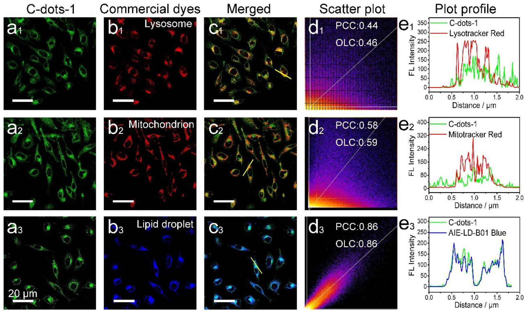
In the next experiments, the cell imaging and subcellular localization behaviors of the two C-dots were studied by comparison with several commercial organelle dyes through co-culture operation. As shown in Figure 2, for Hela cells, C-dots-1 tends to localize in lipid droplets, as reflected by a more than 85% Pearson’s correlation coefficient (PCC) and overlap coefficient (OLC), as compared with the corresponding commercial dyes (AIE-LD-B01). Furthermore, only very few C-dots pass into lysosomes (Figure 2a2–e2) and mitochondria (Figure 2a3–e3) because the corresponding PCC and OLC values are less than 0.60.
For 3T3 and HepG2 cells, the subcellular organelle localization behaviors of C-dots-1 are similar to those of Hela cells on the whole. As shown in Figures S12 and S13, C-dots-1 can also localize in lipid droplets with high efficiency, as indicated by greater than 0.85 PCC and OLC values. At the same time, only very few C-dots are internalized by lysosomes and mitochondria. Especially for HepG2 cells, the PCC values for these two organelles are only 0.18 and 0.32.
As shown in Figure 3, Figures S14 and S15, for C-dots-2, the localization performances for the three subcellular organelles are similar to those of C-dots-1. C-dots-2 can also lipid droplets with high efficiency, as reflected by ~0.85 PCC and OLC values compared with the corresponding commercial dyes. At the same time, only very small parts of C-dots-2 were internalized by the other two organelles. Based on the above, it is clear that both C-dots-1 and C-dots-2 can well target lipid droplets for the three kinds of studied cells. For simplicity, we next employed C-dots-1 for further research.
Because of the promising applications for lipid droplet imaging of the C-dots, we compared their imaging performances with the corresponding commercial dyes. Figure 4 shows lipid droplet images of the same cells by C-dots-1 and AIE-LD-B01. Figure 4b,d are the amplified parts for several targeted lipid droplets. As shown in the rectangular box (Figure 4b), for C-dots-1-stained cells, each lipid droplet is well-defined and easily numbered. In contrast, for AIE-LD-B01-stained cells, the same lipid droplets link together, and it is difficult to distinguish the exact numbers. According to the above, for lipid droplet co-localization, the PCC and OLC values of C-dots-1 and commercial AIE-LD-B01 are about 0.85. That is to say, the co-localization results exhibit evident relevance but are far from perfect. According to Figure 4, the lipid droplets exhibit spherical and subspherical morphology, which is closer to their true face (spherical particles instead of elongated ones). Based on these results, the present C-dots possess preferable localization and imaging performances for lipid droplets.
Because the proposed C-dot targeting method can obtain clear outlines for lipid droplets, we next employed it for the corresponding quantification efforts by means of a 3D confocal imaging technique. As shown in Figure 5, for the control group, each cell has 148 lipid droplets. After stimulation by oleic acid, the number of lipid droplets dramatically enhances to 486. In contrast, when the cells are starvation pretreated, lipid droplets are decreased by about 40% (to 106, as compared with the control). Even though previous studies have shown that oleic acid stimulation and starvation treatment can cause an increase and decrease in lipid droplets in living cells, respectively [28,29,30], quantitative research is still rather difficult because of the limited imaging performances of the fluorescent dyes. Furthermore, for oleic acid-stimulated cells, distinct lipid droplets are observed within the cell nucleus, which is an interesting phenomenon deserving further investigation.
Finally, we studied the organelle uptake behaviors of the C-dots by the pretreatments of oleic acid stimulation and starvation using Hela cells as an example. As shown in Figure 6, after oleic acid stimulation, the internalization behaviors of lysosomes and lipid droplets do not drastically change. However, for mitochondria, the targeting performances exhibit a dramatic decrease as compared with the untreated cells (0.11 vs. 0.58 for PCC values). For the starved cells (Figure 7), more C-dots obviously pass into lysosomes as compared with the control cells (0.77 vs. 0.44 for PCC values); Furthermore, the uptake of mitochondria remains almost unchanged. Under a starvation state, autophagy is activated and forms photophores, which can selectively recognize lipid droplets and form mature autophagosomes by the action of relevant proteins. Then, autophagosomes are transported into lysosomes and form autolysosomes by further fusion effects. Under the expression of relevant proteins, autolysosomes break down lipid droplets, which is called lipophagy. Because of the occurrence of lipophagy, the correlation between lysosomes and lipid droplets increases [31,32].

4. Conclusions

In summary, we show that unit-emitting C-dots can be employed for lipid droplet localization of different kinds of cells. Because of its superior imaging performance for lipid droplets, the proposed C-dot-based targeting can be employed for lipid droplet quantification. We expect that C-dots can play increasingly important roles in cell imaging.

Supplementary Materials

The following supporting information can be downloaded at: https://www.mdpi.com/article/10.3390/targets2020008/s1, Figure S1: Lipophilic property of the two C-dots. Between the n-hexane and water phases, the two C-dots always stay in n-hexane, indicating their high lipotropy; Figure S2: Fluorescence spectra of C-dots-1 (a,b) and C-dots-2 (c,d) in different mediums; Figure S3: Cytotoxicity experiments of C-dots-1 (a) and C-dots-2 (b); Figure S4: CLSM images of Hela cells incubated with different concentrations of C-dots-1 (5-50 μg mL−1) for 8 h, including brightfield and green channels; Figure S5: CLSM images of Hela cells incubated with different concentrations of C-dots-2 (5-50 μg mL−1) for 8 h, including brightfield and green channels; Figure S6: CLSM images of C-dots-1 (50 μg mL−1) incubated Hela cells for different times; Figure S7: CLSM images of C-dots-2 (50 μg mL−1) incubated Hela cells for different times; Figure S8: Fluorescent images of Hela cells incubated with 50 μg mL−1 C-dots-1 at 37 °C (a) and 4 °C. (c) Normalized fluorescence intensity; Figure S9: Fluorescent images of Hela cells incubated with 50 μg mL−1 C-dots-2 at 37 °C (a) and 4 °C. (c) Normalized fluorescence intensity; Figure S10: Fluorescent images of Hela cells incubated with 50 μg mL−1 C-dots-1 (a), treated with CPZ (b), CytoD (c), and nystatin (d), and then incubated with C-dots-1. (e) Normalized fluorescence intensity; Figure S11: Fluorescent images of Hela cells incubated with 50 μg mL−1 C-dots-2 (a), treated with CPZ (b), CytoD (c), and nystatin (d), and then incubated with C-dots-2. (e) Normalized fluorescence intensity; Figure S12: The internalization (3T3 cells) behaviors of C-dots-1. Fluorescent images of cells cocultured with C-dots-1 (a1,a2,a3) and commercial organelle probes for lysosomes (LTR, b1), mitochondria (MTR, b2), and lipid droplets (AIE-LD-B01, b3). Merged photos (c1,c2,c3). Co-located scatter plots (d1,d2,d3) and fluorescence intensities along the yellow lines in the corresponding merged photos (e1,e2,e3); Figure S13: The internalization (HepG2 cells) behaviors of C-dots-1. Fluorescent images of cells cocultured with C-dots-1 (a1,a2,a3) and commercial organelle probes for lysosomes (LTR, b1), mitochondria (MTR, b2), and lipid droplets (AIE-LD-B01, b3). Merged photos (c1,c2,c3). Co-located scatter plots (d1,d2,d3) and fluorescence intensities along the yellow lines in the corresponding merged photos (e1,e2,e3); Figure S14: The internalization (3T3 cells) behaviors of C-dots-2. Fluorescent images of cells cocultured with C-dots-2 (a1, a2, a3) and commercial organelle probes for lysosomes (LTR, b2), mitochondria (MTR, b2), and lipid droplets (AIE-LD-B01, b3). Merged photos (c1,c2,c3). Co-located scatter plots (d1,d2,d3) and fluorescence intensities along the yellow lines in the corresponding merged photos (e1,e2,e3); Figure S15: The internalization (HepG2 cells) behaviors of C-dots-2. Fluorescent images of cells cocultured with C-dots-2 (a1,a2,a3) and commercial organelle probes for lysosomes (LTR, b1), mitochondria (MTR, b2), and lipid droplets (AIE-LD-B01, b3). Merged photos (c1,c2,c3). Co-located scatter plots (d1,d2,d3) and fluorescence intensities along the yellow lines in the corresponding merged photos (e1,e2,e3); Table S1: Composition and QYs of C-dots-1 and C-dots-2.

Author Contributions

Y.X. (Yanli Xu) designed this study protocol and wrote this manuscript. Y.X. (Yunsheng Xia) performed the experiments. All authors have read and agreed to the published version of the manuscript.

Funding

This work was financially supported by the National Natural Science Foundation of China (No. 22274001).

Informed Consent Statement

Not applicable.

Data Availability Statement

The data that support the findings of this study are available from the College of Chemistry and Materials Science, Anhui Normal University. The data are also available from the authors upon reasonable request and with the permission of the College of Chemistry and Materials Science, Anhui Normal University.

Conflicts of Interest

The authors declare no conflicts of interest.

References

  1. Ðorđević, L.; Arcudi, F.; Cacioppo, M.; Prato, M. A multifunctional chemical toolbox to engineer carbon dots for biomedical and energy applications. Nat. Nanotechnol. 2022, 17, 112–130. [Google Scholar] [CrossRef] [PubMed]
  2. Fang, M.; Wang, B.; Qu, X.; Li, S.; Huang, J.; Li, J.; Lu, S.; Zhou, N. State-of-the-art of biomass-derived carbon dots: Preparation, properties, and applications. Chin. Chem. Lett. 2024, 25, 108423. [Google Scholar] [CrossRef]
  3. Wang, Y.; Xia, Y. Optical, electrochemical and catalytic methods for in-vitro diagnosis using carbonaceous nanoparticles: A review. Microchim. Acta 2019, 186, 50. [Google Scholar] [CrossRef] [PubMed]
  4. Khan, W.U.; Wang, D.; Wang, Y. Highly green emissive nitrogen-doped carbon dots with excellent thermal stability for bioimaging and solid-state LED. Inorg. Chem. 2018, 57, 15229–15239. [Google Scholar] [CrossRef] [PubMed]
  5. Kim, A.; Dash, J.K.; Kumar, P.; Patel, R. Carbon-based quantum dots for photovoltaic devices: A review. ACS Appl. Electron. Mater. 2022, 4, 27–58. [Google Scholar] [CrossRef]
  6. Wang, Y.; Li, Z.; Guo, G.; Xia, Y. Liver injury traceability: Spatiotemporally monitoring oxidative stress processes by unit-emitting carbon dots. Anal. Chem. 2023, 95, 2765–2773. [Google Scholar] [CrossRef] [PubMed]
  7. Li, Q.; Fan, J.; Mu, H.; Chen, L.; Yang, Y.; Yu, S. Nucleus-targeting orange-emissive carbon dots delivery adriamycin for enhanced anti-liver cancer therapy. Chin. Chem. Lett. 2024, 35, 108947. [Google Scholar] [CrossRef]
  8. Cheng, B.; Cao, L.; Li, C.; Huo, F.Y.; Meng, Q.F.; Tong, G.; Wu, X.; Bu, L.L.; Rao, L.; Wang, S. Fluorine-doped carbon quantum dots with deep-red emission for hypochlorite determination and cancer cell imaging. Chin. Chem. Lett. 2024, 35, 108969. [Google Scholar] [CrossRef]
  9. Xin, N.; Gao, D.; Su, B.; Zhou, T.; Zhu, Y.; Wu, C.; Wei, D.; Sun, J.; Fan, H. Orange-emissive carbon dots with high photostability for mitochondrial dynamics tracking in living cells. ACS Sens. 2023, 8, 1161–1172. [Google Scholar] [CrossRef]
  10. Li, R.S.; Gao, P.F.; Zhang, H.Z.; Zheng, L.L.; Li, C.M.; Wang, J.; Li, Y.F.; Liu, F.; Li, N.; Huang, C.Z. Chiral nanoprobes for targeting and long-term imaging of the Golgi apparatus. Chem. Sci. 2017, 8, 6829–6835. [Google Scholar] [CrossRef]
  11. Wu, L.; Li, X.; Ling, Y.; Huang, C.; Jia, N. Morpholine derivative-functionalized carbon dots-based fluorescent probe for highly selective lysosomal imaging in living cells. ACS Appl. Mater. Interfaces 2017, 9, 28222–28232. [Google Scholar] [CrossRef] [PubMed]
  12. Gao, D.; Zhang, Y.; Wu, K.; Min, H.; Wei, D.; Sun, J.; Yang, H.; Fan, H. One-step synthesis of ultrabright amphiphilic carbon dots for rapid and precise tracking lipid droplets dynamics in biosystems. Biosens. Bioelectron. 2022, 200, 113928. [Google Scholar] [CrossRef] [PubMed]
  13. Paul, S.; Bhattacharya, A.; Hazra, N.; Gayen, K.; Sen, P.; Banerjee, A. Yellow-emitting carbon dots for selective fluorescence imaging of lipid droplets in living cells. Langmuir 2022, 38, 8829–8836. [Google Scholar] [CrossRef] [PubMed]
  14. She, C.; Wang, Z.; Zeng, J.; Wu, F. G Orange/red dual-emissive boron- and nitrogen-codoped carbon dots for wash-free and selective staining of lipid droplets in live cells. Carbon 2022, 191, 636–645. [Google Scholar] [CrossRef]
  15. Ohsaki, Y.; Suzuki, M.; Fujimoto, T. Open questions in lipid droplet biology. Chem. Biol. 2014, 21, 86e96. [Google Scholar] [CrossRef] [PubMed]
  16. Thiam, A.R.; Dugail, I. Lipid droplet-membrane contact sites-from protein binding to function. J. Cell Sci. 2019, 132, jcs230169. [Google Scholar] [CrossRef]
  17. Welte, M.A.; Gould, A.P. Lipid droplet functions beyond energy storage. Biochim. Biophys. Acta, Mol. Cell Biol. Lipids 2017, 1862, 1260–1272. [Google Scholar] [CrossRef]
  18. Liang, T.; Wen, D.; Chen, G.; Chan, A.; Chen, Z.; Li, H.; Wang, Z.; Han, X.; Jiang, L.; Zhu, J.J.; et al. Adipocyte-derived anticancer lipid droplets. Adv. Mater. 2021, 33, 2100629. [Google Scholar] [CrossRef] [PubMed]
  19. Zhao, N.; Ma, C.; Yang, W.; Yin, W.; Wei, J.; Li, N. Facile construction of boranyl complexes with aggregation-induced emission characteristics and their specific lipid droplet imaging applications. Chem. Commun. 2019, 55, 8494–8497. [Google Scholar] [CrossRef]
  20. Xu, S.; Zhang, X.; Liu, P. Lipid Droplet Proteins and Metabolic Diseases. Biochim. Biophys. Acta, Mol. Basis Dis. 2018, 1864, 1968–1983. [Google Scholar] [CrossRef]
  21. Dalhaimer, P. Lipid droplets in disease. Cell 2019, 8, 974. [Google Scholar] [CrossRef] [PubMed]
  22. Sayed, S.M.; Li, X.F.; Jia, H.R.; Durrani, S.F.; Wu, G.; Lu, X. A dibenzothiophene core-based small-molecule AIE probe for wash-free and selective staining of lipid droplets in live mammalian and fungal cells. Sensor. Actuator. B Chem. 2021, 343, 130128. [Google Scholar] [CrossRef]
  23. Chen, J.; Liu, W.; Fang, X.; Qiao, Q.; Xu, Z. BODIPY 493 acts as a bright buffering fluorogenic probe for super-resolution imaging of lipid droplet dynamics. Chin. Chem. Lett. 2022, 33, 5042–5046. [Google Scholar] [CrossRef]
  24. Hornum, M.; Mulberg, M.W.; Szomek, M.; Reinholdt, P.; Brewer, J.R.; Wüstner, D.; Kongsted, J.; Nielsen, P. Substituted 9-diethylaminobenzo[a]phenoxazin-5-ones (Nile red analogues): Synthesis and photophysical properties. J. Org. Chem. 2021, 86, 1471–1488. [Google Scholar] [CrossRef] [PubMed]
  25. Yu, Y.; Xing, H.; Park, H.; Zhang, R.; Peng, C.; Sung, H.H.Y.; Williams, I.D.; Ma, C.; Wong, K.S.; Li, S.; et al. Deep-red aggregation-induced emission luminogen based on dithiofuvalene-fused benzothiadiazole for lipid droplet-specific imaging. ACS Mater. Lett. 2022, 4, 159–164. [Google Scholar] [CrossRef]
  26. Wang, C.; Guo, G.; Wang, Y.; Yin, X.; Zong, H.; Xia, Y. Twins of minimalistic carbon dots: Uniform emitting-units and molecular level repeatable photoluminescence. Adv. Opt. Mater. 2022, 10, 2102827. [Google Scholar] [CrossRef]
  27. Zhang, H.; Liu, J.; Sun, Y.Q.; Liu, M.; Guo, W. Carbon−dipyrromethenes: Bright cationic fluorescent dyes and potential application in revealing cellular trafficking of mitochondrial glutathione conjugates. J. Am. Chem. Soc. 2020, 142, 17069–17078. [Google Scholar] [CrossRef] [PubMed]
  28. Chao, T.Y.; Raines, R.T. Mechanism of ribonuclease an endocytosis: Analogies to cell-penetrating peptides. Biochemistry 2011, 50, 8374–8382. [Google Scholar] [CrossRef] [PubMed]
  29. Ujimoto, Y.F.; Noduka, J.O.; Omma, K.J.H.; Amaguchi, S.Y.; Ori, M.M.; Igashi, Y.H.; Akita, M.M.; Inoshita, T.K.; Oda, J.N.; Tabe, H.I.; et al. Long-chain fatty acids induce lipid droplet formation in a cultured human hepatocyte in a manner dependent of acyl-CoA synthetase. Biol. Pharm. Bull. 2006, 29, 2174–2180. [Google Scholar] [CrossRef]
  30. Wang, K.; Ma, S.; Ma, Y.; Zhao, Y.; Xing, M.; Zhou, L.; Cao, D.; Lin, W. Aurone derivative revealing the metabolism of lipid droplets and monitoring oxidative stress in living cells. Anal. Chem. 2020, 92, 6631–6636. [Google Scholar] [CrossRef]
  31. Nguyen, T.B.; Olzmann, J.A. Lipid droplets and lipotoxicity during autophagy. Autophagy 2017, 13, 2002–2003. [Google Scholar] [CrossRef] [PubMed]
  32. Barbosa, A.D.; Siniossoglou, S. Spatial distribution of lipid droplets during starvation: Implications for lipophagy. Commun. Integr. Bio. 2016, 9, e1183854. [Google Scholar] [CrossRef] [PubMed]
Figure 1. Characterizations of C-dots-1 and C-dots-2. TEM images of C-dots-1 (a) and C-dots-2 (c). HRETM images of C-dots-1 (b) and C-dots-2 (d). Size distribution of the two dots (e). AFM images of C-dots-1 (f) and C-dots-2 (g). Absorption (h), excitation (i), and emission (j) spectra of the two C-dots.
Targets 02 00008 g001
Figure 2. The internalization (Hela cells) behaviors of C-dots-1. Fluorescent images of cells cocultured with C-dots-1 (a1,a2,a3) and commercial organelle probes for lysosomes (LTR, b1), mitochondria (MTR, b2), and lipid droplets (AIE-LD-B01, b3). Merged photos (c1,c2,c3). Co-located scatter plots (d1,d2,d3) and fluorescence intensities along the yellow lines in the corresponding merged photos (e1,e2,e3). Scale: 20 μm.
Targets 02 00008 g002
Figure 3. The internalization (Hela cells) behaviors of C-dots-2. Fluorescent images of cells cocultured with C-dots-2 (a1,a2,a3) and commercial organelle probes for lysosomes (LTR, b1), mitochondria (MTR, b2), and lipid droplets (AIE-LD-B01, b3). Merged photos (c1,c2,c3). Co-located scatter plots (d1,d2,d3) and fluorescence intensities along the yellow lines in the corresponding merged photos (e1,e2,e3). Scale: 20 μm.
Targets 02 00008 g003
Figure 4. Comparison of lipid droplet imaging by using C-dots-1 (a,b) and commercial AIE-LD-B01 (c,d).
Targets 02 00008 g004
Figure 5. Two-dimensional (a,c,e) and three-dimensional (b,d,f) lipid droplet imaging and corresponding lipid droplet quantification (g) under different conditions by C-dots-1 localization.
Targets 02 00008 g005
Figure 6. The internalization behaviors of C-dots-1 for oleic acid-stimulated Hela cells. Fluorescent images of cells cocultured with C-dots-1 (a1,a2,a3) and commercial organelle probes for lysosomes (LTR, b1), mitochondria (MTR, b2), and lipid droplets (AIE-LD-B01, b3). Merged photos (c1,c2,c3). Co-located scatter plots (d1,d2,d3) and fluorescence intensities along the yellow lines in the corresponding merged photos (e1,e2,e3). Scale: 20 μm.
Targets 02 00008 g006
Figure 7. The internalization behaviors of C-dots-1 for starvation-treated Hela cells. Fluorescent images of cells cocultured with C-dots-1 (a1,a2,a3) and commercial organelle probes for lysosomes (LTR, b1), mitochondria (MTR, b2), and lipid droplets (AIE-LD-B01, b3). Merged photos (c1,c2,c3). Co-located scatter plots (d1,d2,d3) and fluorescence intensities along the yellow lines in the corresponding merged photos (e1,e2,e3). Scale: 20 μm.
Targets 02 00008 g007
Disclaimer/Publisher’s Note: The statements, opinions and data contained in all publications are solely those of the individual author(s) and contributor(s) and not of MDPI and/or the editor(s). MDPI and/or the editor(s) disclaim responsibility for any injury to people or property resulting from any ideas, methods, instructions or products referred to in the content.