Review

Potential of MMP-2 and MMP-9 Gelatinase Blockade as a Therapeutic Strategy in Fibrosarcoma Treatment: A Decadal Review

Department of Cancer Biology, Mayo Clinic, Jacksonville, FL 32224, USA
Targets 2024, 2(2), 104-125; https://doi.org/10.3390/targets2020007
Submission received: 21 May 2024 / Revised: 1 June 2024 / Accepted: 3 June 2024 / Published: 5 June 2024

Abstract

Fibrosarcoma represents a significant challenge in oncology, characterized by high invasiveness and a poor prognosis. Gelatinases, particularly matrix metalloproteinases MMP-2 and MMP-9, play a pivotal role in the degradation of the extracellular matrix, facilitating tumor invasion and metastasis. Inhibiting these enzymes has emerged as a promising therapeutic strategy. This review evaluates the progress in the development and therapeutic potential of gelatinase inhibitors as treatments for fibrosarcoma over the last decade, highlighting molecular mechanisms and future directions. A comprehensive literature review was conducted, focusing on studies published from 2013 to 2023. Research articles and review papers relevant to gelatinase inhibition and fibrosarcoma were examined to assess the efficacy and mechanisms of gelatinase inhibitors. Gelatinase inhibitors have shown the potential to reduce tumor progression, invasion, and metastasis in fibrosarcoma. Clinical trials, although limited, have indicated that these inhibitors can be effectively integrated into existing therapeutic regimens, offering a reduction in metastatic spread and potentially improving patient survival rates. Mechanistic studies suggest that the inhibition of MMP-2 and MMP-9 disrupts critical pathways involved in tumor growth and cell invasion. Gelatinase inhibition represents a viable and promising approach to fibrosarcoma treatment. Future research should focus on developing more specific inhibitors, understanding long-term outcomes, and integrating gelatinase inhibition into multimodal treatment strategies to enhance efficacy.

1. Fibrosarcoma

Fibrosarcoma is a rare type of cancer that arises from fibrous connective tissue, primarily affecting the fibroblasts, which are the cells that form the collagen-rich fibrous tissue throughout the body [1]. This type of cancer belongs to a larger group of cancers known as soft-tissue sarcomas. It primarily develops in the fibroblasts and is characterized by the formation of malignant fibrous tumors [2]. Fibrosarcoma can occur anywhere in the body but is most commonly found in the legs, arms, and trunk. It can also appear in the bones and is occasionally found in the lungs or other internal organs [3]. While it can affect individuals of any age, there are variations such as infantile fibrosarcoma that occurs in children, typically presenting a different, often less aggressive, behavior compared to adult forms [4].
The symptoms of fibrosarcoma depend on the tumor’s location but typically include a noticeable lump or swelling that may be painful, pain or soreness caused by compressed nerves or muscles, and limited movement in the nearby joints if the tumor is located close to joints [5]. Diagnosing fibrosarcoma involves a combination of medical imaging and a tissue biopsy. Imaging techniques such as MRI, CT scans, and X-rays are used to visualize the tumor’s size, location, and impact on the surrounding structures. A biopsy, where a sample of the tumor is removed and examined microscopically, is essential for a definitive diagnosis [6].
A complete surgical excision of the tumor is the primary treatment approach. Efforts are made to remove the tumor with some surrounding tissue to minimize the risk of recurrence [7]. Radiation therapy can be used post-surgery to eliminate residual cancer cells or in cases where surgery is not fully possible [8]. The role of chemotherapy is more limited but can be considered depending on the case, particularly for more advanced or metastatic disease [9]. The prognosis for fibrosarcoma varies widely based on factors such as the tumor’s size, its location, the extent of surgical removal, the presence of metastasis, and the patient’s overall health. Early detection and complete surgical removal typically offer the best outcomes [10]. Recurrence is possible, which requires ongoing monitoring. Understanding fibrosarcoma and its treatment options is crucial for effective management and improving patient outcomes. Regular follow-ups and imaging are often recommended as part of the post-treatment care plan to monitor for any signs of recurrence [11].
Fibrosarcoma is considered a rare type of cancer, with its prevalence being relatively low compared to other types of cancer. Fibrosarcoma accounts for approximately 1% of all soft-tissue sarcomas. The incidence rate varies, but, typically, it is estimated to affect around 0.2 to 0.3 people per 100,000 in the population annually [12]. Although fibrosarcoma can occur at any age, it generally has two peak incidences. In infants (as infantile fibrosarcoma, a less aggressive form) and in adults aged 30–55 years. The adult form tends to be more aggressive [13]. There is a slight male predominance in the incidence of fibrosarcoma [14]. Data on racial disparities are less clear, but like many soft-tissue sarcomas, there is no significant evidence of a racial predilection. There is no significant geographic variation noted with fibrosarcoma; it appears to affect individuals worldwide without major differences in incidence rates across different regions [15].
There are no well-established risk factors for fibrosarcoma except for a history of previous radiation therapy, which has been linked to a slightly increased risk of developing this and other types of sarcomas [16]. The survival rates for fibrosarcoma vary significantly depending on factors like tumor size, location, grade, and the success of surgical removal. Generally, the 5-year survival rate for localized fibrosarcoma can be quite high (approximately 50–70%), but this decreases significantly if the cancer is high-grade or has metastasized [16]. Understanding the prevalence and epidemiology of fibrosarcoma helps guide research and treatment strategies. Despite its rarity, the impact on affected individuals can be significant, highlighting the need for ongoing research and tailored treatment approaches.

2. Matrix Metalloproteinases (MMPs)

MMPs are a group of enzymes crucial to the regulation of extracellular matrix (ECM) components such as collagen, elastin, and gelatin, which play a fundamental role in tissue remodeling, wound healing, and other physiological and pathological processes [17]. Due to their significant involvement in these biological processes, MMPs are studied extensively in the context of diseases such as cancer, where they can facilitate tumor invasion and metastasis [18,19]. MMPs are characterized by several structural domains that are common across the different enzymes in this family, including the signal peptide; the propeptide domain, which is approximately 80 amino acids long and contains a conserved cysteine switch motif, typically PRCGXPD; the catalytic domain; the hinge region (also known as the linker peptide); and the hemopexin-like domain, which is involved in substrate recognition and specificity (Figure 1) [20].
MMPs can be classified based on their substrate specificity, domain organization, and sequence homology into multiple major groups, including the following: collagenases (e.g., MMP-1, MMP-8, and MMP-13), which primarily degrade fibrillar collagens, which are critical components of the extracellular matrix; gelatinases (e.g., MMP-2 and MMP-9), which can degrade gelatin and denatured collagens, playing significant roles in cancer metastasis; stromelysins, which can degrade a variety of ECM proteins but have broad specificity (e.g., MMP-3 and MMP-10); and membrane-type MMPs (MT-MMPs), which are membrane-bound and participate in pericellular proteolysis, crucial for cell migration and other functions [21]. MMPs are typically secreted as inactive proenzymes and need to be activated extracellularly. This activation can occur through various mechanisms, such as autoactivation, activation by other MMPs, or disruption of the cysteine switch by chemical agents or reactive oxygen species [22].
MMP activity is tightly regulated at multiple levels, such as gene transcription, zymogen activation, and inhibition by tissue inhibitors of metalloproteinases (TIMPs), which bind to the active sites of MMPs and block their enzymatic activity [23].
Understanding the structure, classification, and regulation of MMPs is critical in the context of physiological processes and in the development of therapeutic agents, especially for diseases characterized by excessive ECM degradation, such as arthritis, fibrosis, and cancer. MMPs play a significant and complex role in cancer progression. These enzymes are capable of degrading various components of the ECM, which not only influences tumor growth and development but also impacts metastasis, the process by which cancer spreads to new areas of the body [24]. Understanding the function of MMPs in cancer can provide insights into potential therapeutic targets and strategies for inhibiting tumor progression and metastasis.

3. MMP Roles in Fibrosarcoma

MMPs play a critical role in the progression and behavior of fibrosarcoma [25]. Understanding the interaction between MMPs and the tumor microenvironment in fibrosarcoma provides insights into the aggressive nature of this cancer and potential therapeutic targets. Fibrosarcoma cells produce MMPs that degrade the ECM, facilitating not only the local invasion of the tumor into surrounding tissues but also supporting tumor expansion. This ECM breakdown is pivotal in tumor progression because it liberates space for growing tumor cells and modifies the biomechanical properties of the tissue matrix [26]. The degradation of ECM components by MMPs releases bound growth factors such as transforming growth factor-beta (TGF-β) and fibroblast growth factors (FGFs), which are involved in promoting cell proliferation and angiogenesis and are essential for tumor growth and survival [27]. MMPs clear a path through the ECM, enabling fibrosarcoma cells to invade adjacent tissues and, potentially, enter the bloodstream or lymphatic system, leading to metastasis [28]. Additionally, they can induce epithelial-to-mesenchymal transition (EMT)-like processes in tumor cells, increasing their motility and invasive capabilities [20]. This is particularly relevant in fibrosarcoma, given its mesenchymal origin. By altering chemokine and cytokine profiles through ECM remodeling, MMPs can modulate the immune microenvironment around fibrosarcomas. This can lead to reduced effectiveness of the immune response against the tumor, aiding in tumor survival and progression [29].
In fibrosarcoma, certain MMPs are often upregulated. For example, the gelatinases MMP-2 (gelatinase A) and MMP-9 (gelatinase B) are frequently found to be elevated and are associated with poor prognostic outcomes. Their expression levels can correlate with tumor grade, size, metastatic potential, and the overall aggressiveness of the disease. MMP-2 and MMP-9 are instrumental in angiogenesis within fibrosarcoma tumors [30]. They degrade the basement membrane and interstitial collagens, facilitating endothelial cell migration and the formation of new blood vessels. This neovascularization is crucial for providing the tumor with nutrients and oxygen and for metabolic waste removal. The ability of these enzymes to break down the structural barriers in the ECM and basement membranes is particularly significant in the context of tumor invasion, metastasis, and angiogenesis [31].
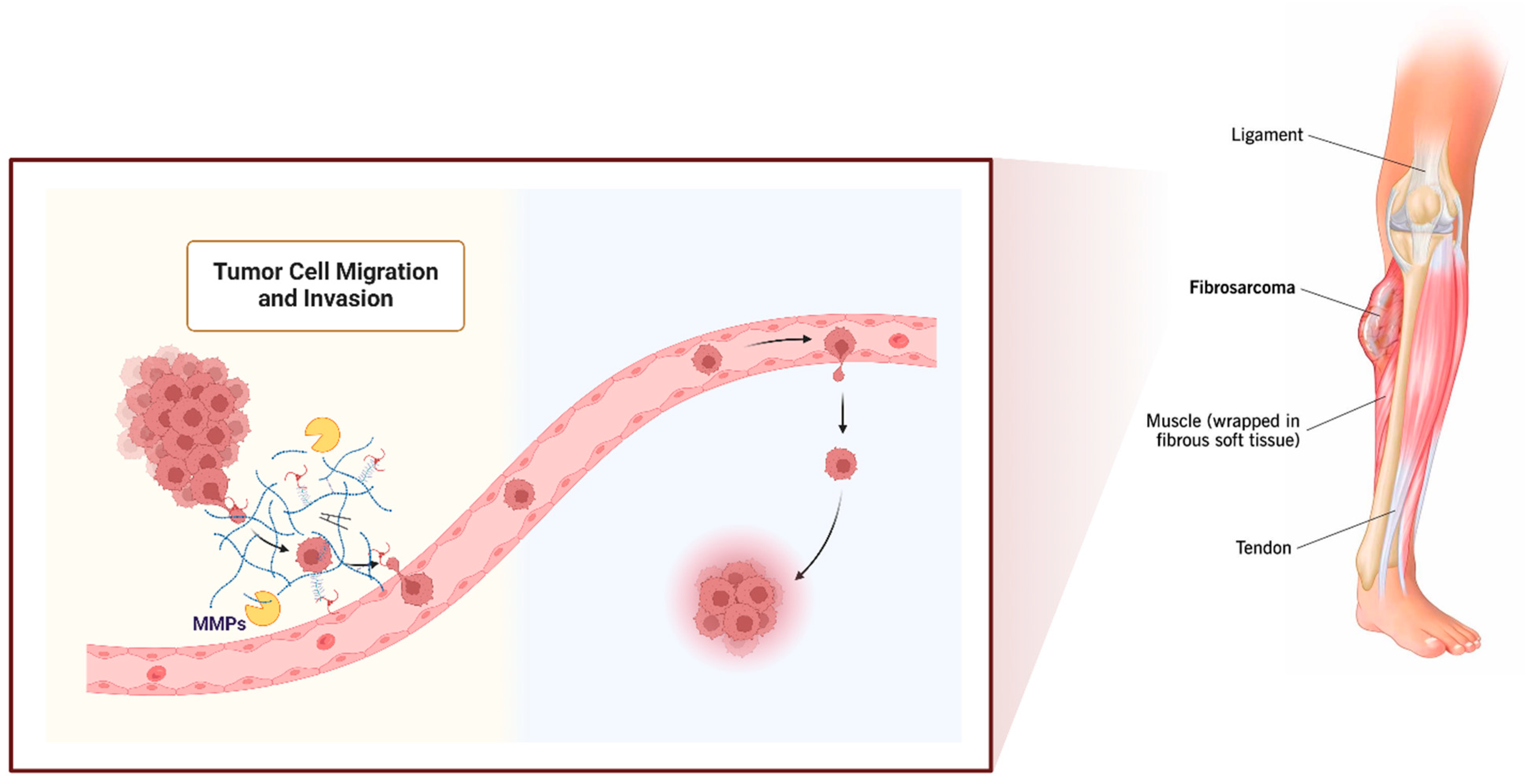
Gelatinases degrade type IV collagen, which is a major component of basement membranes. This degradation is crucial for the disruption of these membranes and may allow fibrosarcoma cells to escape from their original site and invade surrounding tissues [30] (Figure 2). By breaking down the physical barriers in the ECM and basement membranes, gelatinases clear a path for tumor cells to migrate. This enhanced migratory capability is essential for local invasion and distant metastasis, the latter being a leading cause of cancer-related mortality [32]. The activity of gelatinases, particularly MMP-9, is associated with the mobilization of VEGF (vascular endothelial growth factor) from the ECM. VEGF is a potent pro-angiogenic factor that stimulates the formation of new blood vessels within the tumor, facilitating tumor growth and providing a means for cancer cells to access the systemic circulation [33]. The degradation products of basement membranes, generated by gelatinase activity, have been shown to promote the migration and organization of endothelial cells, which are critical steps in the formation of new blood vessels [34]. Beyond their roles in structural ECM degradation, gelatinases can process various bioactive molecules, including cytokines and growth factors. This processing can activate these molecules or modulate their activity, thus influencing inflammatory responses, tumor growth, and immune surveillance within the tumor microenvironment [35]. By modifying the ECM and releasing sequestered growth factors, gelatinases may also directly or indirectly promote tumor cell proliferation and survival. This effect supports not only primary tumor growth but also the establishment and expansion of metastases [36].

4. Therapeutic Implications of Gelatinase Inhibition in Fibrosarcoma

Given their crucial roles in fibrosarcoma progression, gelatinases represent valuable targets for therapeutic intervention. Several inhibitors that specifically target MMP-2 and MMP-9 have been investigated for their potential to halt tumor progression by preventing ECM degradation, invasion, and angiogenesis [37]. These include both small-molecule inhibitors and biological agents like monoclonal antibodies. Integrating gelatinase inhibitors with conventional therapies such as chemotherapy and radiation might enhance the overall treatment efficacy, particularly by preventing tumor spread and overcoming resistance mechanisms [38].
However, the therapeutic targeting of gelatinases comes with challenges. Specific inhibitors that can selectively target MMP-2 or MMP-9 without affecting other MMPs are needed to minimize potential side effects, as broad-spectrum MMP inhibitors have been associated with adverse clinical outcomes [39]. The redundancy within the MMP family and the compensatory pathways involving other proteases can undermine the effectiveness of gelatinase-specific inhibitors [40].
HT-1080 cells are widely used in cancer research, particularly for studying the biological mechanisms and therapeutic strategies related to fibrosarcoma and other similar cancers. HT-1080 is a cell line derived from a human fibrosarcoma that was established from a 35-year-old male patient [41]. These cells are known for their ability to invade and migrate, making them mainly useful for studies on cancer metastasis, tumor progression, and drug testing, particularly in fibrosarcoma-related studies [42].
Ongoing research on fibrosarcoma is focused on improving the specificity of gelatinase inhibitors and understanding their role in the tumor microenvironment. This includes studying the interplay between gelatinases and other cell types within the tumor, such as immune cells and fibroblasts, to develop more comprehensive therapeutic strategies. Gelatinases are pivotal in the progression of fibrosarcoma through their involvement in ECM remodeling, invasion, angiogenesis, and the modulation of the tumor microenvironment. The effective targeting of these enzymes holds promise for improving clinical outcomes in fibrosarcoma treatment. In the following paragraphs, I will concentrate on the potential of gelatinase inhibitors as a therapeutic approach for treating fibrosarcoma.

5. Natural-Based Inhibitors

Natural-based anticancer compounds are substances derived from natural sources—such as plants, fungi, bacteria, and marine organisms—that exhibit potential anticancer properties. These compounds often play a pivotal role in the development of new cancer therapies, offering alternatives to synthetic drugs. The exploration of natural compounds is driven by their diverse chemical structures and mechanisms of action, which can target various cellular pathways involved in cancer progression [43]. Natural anticancer compounds can exert their effects through various mechanisms, including inducing apoptosis, inhibiting angiogenesis, suppressing metastasis, and modulating the immune system to enhance the body’s ability to fight cancer [44]. Research into natural-based anticancer compounds is significant due to their potential to offer more targeted, less toxic treatment options and their ability to overcome resistance to current therapies [45]. Ongoing research focuses on isolating new compounds, understanding their mechanisms, and developing ways to synthesize them efficiently and test them in clinical settings. This field holds a promising avenue for the discovery of novel therapies that can contribute to cancer treatment [46]. Natural-based anticancer compounds have garnered interest for their potential in treating various types of cancer, including fibrosarcoma (Table 1). In the following paragraphs, there are some notable natural compounds that have shown potential in research for treating fibrosarcoma in the last decade.
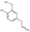
Eugenol is a naturally occurring compound commonly found in clove oil, though it is also present in other essential oils like cinnamon, nutmeg, and bay leaf. It is a phenylpropene, which is a type of aromatic compound that gives cloves their distinctive smell and flavor [61]. Eugenol has a variety of uses, both in traditional medicine and in commercial applications. Eugenol has been studied for its potential anticancer properties, and some research suggests it might have beneficial effects in fighting various types of cancer [62]. In 2013, Nam and Kim investigated the potential of eugenol to suppress the expression and activity of MMPs, as well as its antioxidant effects. They specifically examined the impact of eugenol on MMP-9, which is associated with metastasis, using gelatin zymography and Western blot analysis. Their findings revealed that eugenol effectively inhibited MMP-9 activity in HT-1080 cells, a human fibrosarcoma cell line, stimulated by PMA (phorbol 12-myristate 13-acetate). Additionally, the study indicated that the inhibitory effects of eugenol on MMP-9 are mediated through the inactivation of the ERK (Extracellular signal-Regulated Kinase) pathway [47]. In the same year, Park et al. evaluated fisetin as an anticancer compound. Fisetin is a plant flavonol, a type of flavonoid, which is found in many fruits and vegetables. Fisetin inhibited MMP-9 more effectively than tetracycline, a naturally occurring MMP inhibitor. It also suppressed the proliferation of fibrosarcoma HT-1080 cells and human umbilical vascular endothelial cells (HUVECs) in a dose-dependent manner. Additionally, fisetin reduced the invasiveness of HT-1080 cells and the ability of HUVECs to form tubes in vitro [48].
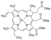
In a further study in 2013 by Roomi and colleagues, they evaluated a nutrient mixture (NM) on different adult human sarcoma cell lines. The NM, prepared by VitaTech (Hayward, CA, USA), was composed of the following ingredients in the relative amounts indicated: vitamin C (as ascorbic acid and as Mg, Ca, and palmitate ascorbate) 700 mg; L-lysine 1000 mg; standardized green tea extract (80% polyphenol) 1000 mg; L-proline 750 mg; L-arginine 500 mg; N-acetyl cysteine 200 mg; selenium 30 μg; copper 2 mg; and manganese 1 mg. In gelatinase zymography, HT-1080 cells showed strong expression of inactive MMP-2 and weak expression of active MMP-2, both more pronounced than MMP-9. These were inhibited by the NM in a dose-dependent manner, achieving almost complete inhibition of MMP-9 at 100 μg/mL and MMP-2 at 250 μg/mL. Treatment with PMA (100 ng/mL) increased the expression of active MMP-2 and MMP-9 in HT-1080 cells; the NM effectively blocked both MMP-2 and MMP-9 in a dose-dependent manner, completely inhibiting active MMP-2 at 500 μg/mL and nearly completely inhibiting MMP-9 at 1000 μg/mL. The activity of TIMPs was upregulated by the NM in the HT-1080 cell line in a dose-dependent manner [63]. In 2018, this group also explored the effects of 10, 25, and 50 μM of chlorophyllin on HT-1080 cell proliferation and migration. Cell proliferation in HT-1080 cells was significantly reduced at a 50 μM dose of chlorophyllin. The expression of MMP-2 and MMP-9 also decreased in a dose-dependent manner, with significant inhibition observed at 25 μM and levels becoming virtually undetectable at 50 μM. Additionally, cell invasion through Matrigel and cell migration were reduced as the concentrations of chlorophyllin increased, with complete inhibition of invasion at 50 μM. H&E staining at 10 μM of chlorophyllin revealed some cellular changes indicative of apoptosis, while more significant apoptotic morphological changes were observed at higher doses of chlorophyllin [55].
Quercetin is a type of flavonoid antioxidant that is found in a variety of fruits, vegetables, and grains. Some research has indicated that quercetin may help slow cancer cell growth and induce apoptosis in various types of cancer cells [64]. Lee et al. conducted research to determine if quercetin could suppress the activities of MMP-2 and MMP-9 by reducing the formation of reactive oxygen species (ROS), a process anticipated to diminish cell motility. To trigger prolonged formation of ROS, cells were exposed to phenazine methosulfate (PMS; 1 μM). Quercetin, at noncytotoxic levels, was found to inhibit the increase in cell motility induced by PMS in HT-1080 cells. After 24 h of PMS treatment, nearly all cells demonstrated migration; however, quercetin significantly (p < 0.01) reduced this effect. Additionally, quercetin at concentrations up to 10 μg/mL was observed to decrease PMS-induced MMP-2 activation. Quercetin treatments significantly reduced the formation of ROS induced by PMS (p < 0.01) and led to decreased cell motility, accompanied by a decrease in MMP-2 and -9 activity in HT-1080 cells, even without PMS treatment [49].
One year later, in 2014, Filipiak et al. explored whether the inhibitory effect of gallic acid on gelatinases correlated with its cytotoxic activity in HT-1080 cells and assessed whether delphinidin-3-glucoside exhibited similar properties. Gallic acid and delphinidin-3-glucoside, which are prevalent bioactive compounds in many fruits and vegetables, are particularly concentrated in berries. Gallic acid is noted for its cytotoxic effects across various cancer cell lines and has demonstrated the ability to inhibit carcinogenesis in animal studies. Gallic acid and delphinidin-3-glucoside have exhibited selective cytotoxicity toward HT-1080 cells. In migration and invasion assays, both gallic acid and delphinidin-3-glucoside demonstrated anti-invasive properties. A zymographic analysis revealed that gallic acid inhibits both secreted and activated gelatinases. Additionally, gallic acid was shown to suppress the proteolytic activities of MMP-2 and MMP-9 with similar effectiveness. Nuclear magnetic resonance (NMR) and molecular modeling studies have confirmed that gallic acid interacts with MMP-2, likely at the catalytic center [50].
Scutellarein is a flavone, a type of flavonoid compound that naturally occurs in certain plants. It can be found in plants like Scutellaria lateriflora, and it has been the subject of research for its various biological activities. Preliminary studies have indicated that scutellarein may have anticancer effects, including inhibiting the proliferation of certain types of cancer cells and inducing apoptosis [65]. Shi et al.’s analysis showed that scutellarein significantly inhibited the growth of HT-1080 by inducing apoptosis. Additionally, an in vivo experiment using Balb/c nude mice demonstrated that both the volume and weight of the tumors were significantly decreased after treatment with scutellarein. The further outcomes revealed that scutellarein potently blocked cell migration, invasion, and the expression and activity of MMP-2 and MMP-9 [51].
Marine plants, including algae, seaweeds, and other aquatic vegetation, have garnered significant interest in the field of cancer research due to their unique bioactive compounds [66]. In an interesting study, eight different species of brown algae were collected, and their extracts were analyzed for their anti-MMP effects. Based on the gelatin zymography results, Ecklonia cava, Ecklonia bicyclis, and Ishige okamurae demonstrated stronger inhibitory effects on MMP-2 and MMP-9 activities at concentrations of 10, 50, and 100 µg/mL, compared to the other samples tested. All samples effectively prevented cell migration in a dose-dependent manner after 24 h of incubation. Among all the samples tested, Ecklonia cava and Ishige okamurae were the most effective at inhibiting the invasion of HT-1080 cells [67]. In a similar study, Barletta et al. evaluated the anticancer effect of a hydrophilic extract from Posidonia oceanica. Posidonia oceanica, commonly known as Neptune grass or Mediterranean tapeweed, is a seagrass species that forms extensive meadows in the Mediterranean Sea. Their results showed for the first time in the highly invasive HT-1080 human fibrosarcoma cell line that hydrophilic extract from P. oceanica significantly reduced both gene and protein expressions of the gelatinases MMP-2 and MMP-9. Additionally, it directly inhibited gelatinolytic activity in a dose-dependent manner in vitro. Furthermore, their findings exhibited that this extract strongly suppressed the migration and invasion of HT-1080 cells [68]. Limonium tetragonum, commonly referred to as sea lavender, is a species of flowering plant in the Plumbaginaceae family. In traditional medicine, particularly in Korea, Limonium tetragonum has been used to treat various ailments due to its supposed anti-inflammatory and hepatoprotective properties. In an interesting study, Limonium tetragonum was investigated for its capability to inhibit MMP-2 and MMP-9 in HT-1080 fibrosarcoma cells. The study found that extracts from L. tetragonum effectively reduced the enzymatic activity and mRNA expression of MMP-2 and MMP-9, as evidenced by gelatin zymography and RT-PCR assays, respectively [69].
In a study in 2017, Lee et al. investigated the inhibitory effect of Hizikia fusiformis solvent-partitioned fractions on the invasion and MMP activity of HT-1080 cells. Hizikia fusiformis, a type of brown algae, has been found to exhibit various bioactivities such as antiviral, antimicrobial, and anti-inflammatory effects. These properties are attributed, in part, to its content of bioactive polysaccharides. H. fusiformis crude extract was fractionated with organic solvents, H2O, n-BuOH, 85% aqueous MeOH, and n-hexane (n-Hex). All fractions successfully inhibited the enzymatic activities of MMP-2 and MMP-9, as evidenced by the gelatin zymography assay. The n-Hex fraction also significantly curtailed cell migration. These fractions reduced the mRNA and protein levels of MMP-2 and MMP-9 while increasing those of TIMP-1 and TIMP-2. Among the fractions, the H2O fraction was the least effective, whereas the n-Hex fraction was the most effective [70]. In a similar study one year later, the effect of Sargassum horneri, a species of brown algae, was examined by researchers on HT-1080 cells. All fractions inhibited the enzymatic activities of MMP-2 and MMP-9, as demonstrated by gelatin zymography. Apart from the H2O fraction, all fractions significantly hindered cell migration. Additionally, every tested fraction suppressed the mRNA and protein levels of MMP-2, MMP-9, TIMP-1, and TIMP-2 [71]. In a further study by this group, Ishige okamurae, a brown alga, blocked the enzymatic activities of MMP-2 and MMP-9. Additionally, cell migration was significantly reduced by the 85% aqueous MeOH fraction. The fractions decreased the mRNA and protein levels of MMP-2 and MMP-9 while increasing the levels of TIMP-1 and TIMP-2. Among these, the H2O fraction was the least effective, while the 85% aq. MeOH fraction was the most effective [72].
Galangin and kaempferol are both naturally occurring flavonoids widely recognized for their potent antioxidant and anti-inflammatory properties. They are found in a variety of plants and are commonly consumed through dietary sources [73,74]. In a study in 2015, Choi and colleagues showed that galangin and kaempferol effectively reduced MMP-9 secretion in HT-1080 cells, while fisetin had only a slight effect on decreasing its secretion. Galangin and kaempferol did not impact cell viability at concentrations up to 30 µM. Luciferase reporter assays indicated that galangin and kaempferol reduced the transcription of MMP-9 mRNA [52].
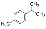
p-Cymene, also known as 4-isopropyltoluene, is a naturally occurring aromatic organic compound. It is classified as a monoterpene and is a constituent of a variety of essential oils derived from plants, particularly cumin and thyme [75]. A study by Li et al. showed that p-cymene inhibited the production and gene expression of MMP-9 in HT-1080 cells in a dose-dependent manner when augmented by 12-O-tetradecanoylphorbol 13-acetate (TPA). Conversely, p-cymene increased the TPA-augmented production and gene expression of TIMP-1 in these cells. However, the baseline levels of MMP-9 and TIMP-1 mRNAs, as well as TIMP-1 protein, remained unchanged in the cells treated with p-cymene. Additionally, the study found that p-cymene dose-dependently reduced the TPA-enhanced invasiveness of HT-1080 cells in vitro [53].
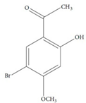
The hippocampus, used in traditional Chinese medicine, is known for treating conditions such as tumors, aging, thrombosis, inflammation, hypertension, and prostatic hyperplasia [76]. 1-(5-Bromo-2-hydroxy-4-methoxyphenyl) ethanone (SE1), derived from the seahorse (Hippocampus kuda Bleeler), has demonstrated efficacy in suppressing proinflammatory responses. In a study by Gong et al., SE1 significantly inhibited gelatin digestion by MMP-9, which was induced by PMA, and reduced the migration of HT-1080 cells in a dose-dependent manner. The molecular docking results revealed that SE1 interacts with Tyr245 and His226 of MMP-9 through hydrogen and Pi-Pi bonds, leading to the inhibition of MMP-9 activity [54].
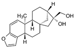
Kahweol acetate (KA), a coffee-specific diterpene, has shown antitumor properties in human tumor cells [77]. In a study by Choi and colleagues, KA significantly reduced cell proliferation induced by PMA in human fibrosarcoma cells. It also decreased PMA-induced cell migration and invasion in a concentration-dependent manner. KA inhibited the activation of MMP-9, primarily through the suppression of nuclear factor kappa B (NF-κB) activation. Additionally, KA hindered the PMA-induced phosphorylation of p38 MAP kinase, c-Jun N-terminal kinase (JNK) 1/2, and Akt, which are key signaling molecules upstream of MMP-9 expression [56].
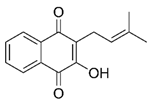
Lapachol is a natural chemical compound with the molecular formula C15H14O3. It is classified as a naphthoquinone and is primarily found in the bark of lapacho trees, which are native to South America. Lapachol has been studied for its potential medicinal properties, including its antibacterial, antifungal, and anti-inflammatory effects [78]. In HT-1080 cells, lapachol was found to inhibit the activation of MMP-2 and MMP-9 when stimulated by PMA at concentrations above 0.5 μM. Notably, both the protein and gene expression levels of MMP-2, when stimulated by PMA, were significantly reduced with 1 μM of lapachol compared to the group treated with PMA alone. Furthermore, lapachol also elevated the expression level of TIMP-1 compared to the PMA-only treatment group [57].
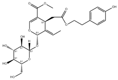
In a study by Kim et al., a phenyl ethanoid, salidroside (SAL), and two secoiridoids, 8(E)-nuezhenide (NZD) and ligustroside (LIG), were isolated from the fruits of Ligustrum japonicum. In fibrosarcoma cells, all the compounds substantially reduced the release of MMP-2 and MMP-9, as confirmed by gelatin zymography and ELISA tests. Moreover, the mRNA and protein expression levels of MMP-2 and MMP-9 were significantly decreased, as evidenced by RT-PCR and Western blot analyses. The Western blot assays revealed that SAL and LIG effectively diminished the expression of MMP-2 in a dose-dependent fashion. Similarly, NZD decreased the expression of MMP-9 [58].
β-Caryophyllene oxide (CPO) is a natural organic compound classified as a sesquiterpene oxide. It is a derivative of β-caryophyllene, a major component in many essential oils found in spices and aromatic herbs. In terms of biological activity, β-caryophyllene oxide has attracted interest for its potential therapeutic properties. It has been studied for its anti-inflammatory, anticancer, and analgesic properties [79]. The study by Jo et al. found that β-caryophyllene oxide (CPO) at a concentration of 32 μM decreased MMP-9 activity by 28% and MMP-2 activity by 60%. In the presence of 32 μM CPO, MMP-2 expression was reduced by 45%. Additionally, CPO lowered the expression levels of MMP-2 and MMP-9 in the immunofluorescence staining assay. An invasion assay on PMA-treated fibrosarcoma cells demonstrated that CPO reduced cell invasion in a dose-dependent manner, starting at a concentration of 2 μM [59].
Recently, in 2023, the antitumor effect of silibinin was explored on fibrosarcoma cells. Silibinin, also known as silybin, is a bioactive compound extracted from the seeds of the milk thistle plant (Silybum marianum). Recent studies have explored its effectiveness in inhibiting the growth of cancer cells in the liver, prostate, skin, and breast [80]. Silibinin, at concentrations above 20 μM, significantly reduced the activation levels of MMP-2 and MMP-9 when stimulated by PMA. Additionally, at a concentration of 25 μM, silibinin decreased the expression levels of MMP-2, IL-1β, ERK-1/2, and phosphorylated p38. Furthermore, silibinin at concentrations exceeding 10 μM blocked cell invasion in HT-1080 cells [60].
When considering natural-based medications for treating conditions like fibrosarcoma, the disadvantages can be particularly significant. For serious conditions like cancer, it is crucial to use treatments that have been proven effective through rigorous clinical trials. Natural-based treatments often lack sufficient scientific evidence supporting their efficacy in treating cancer, which can make them unreliable and potentially dangerous as a primary treatment option. Due to variations in the source and preparation of natural products, their potency and effectiveness can fluctuate significantly [81]. This inconsistency can be particularly problematic when treating something as aggressive and life-threatening as fibrosarcoma. Natural products can interfere with conventional cancer treatments, such as chemotherapy and radiation. They might either potentiate or inhibit the effects of these treatments, leading to increased toxicity or reduced efficacy [82]. While conventional cancer treatments undergo extensive safety testing, the safety profile of many natural medications is not as well established, especially in the context of cancer. This uncertainty can pose additional risks to patients. Relying on unproven natural treatments might cause delays in receiving more conventional therapies that have a proven track record of success. This delay can be critical, especially in fast-growing tumors like fibrosarcoma. For patients considering natural-based medications for fibrosarcoma, it is important to discuss these options thoroughly with oncologists and healthcare providers. Integrating such treatments into a comprehensive care plan should be carried out cautiously, ensuring that they do not undermine standard, evidence-based oncological therapies.

6. Synthetic Small-Molecule Inhibitors

Chemical and small-molecule medications are a cornerstone of cancer treatment. These drugs, designed to interact with specific molecular targets within cancer cells or related pathways, offer a broad range of therapeutic approaches [83]. Traditional chemotherapy drugs are small molecules that interfere with cell division. They target rapidly dividing cells, a hallmark of cancer cells, but can also affect normal cells, leading to side effects. These are designed to specifically target and inhibit cancer-specific molecules and signaling pathways, which are essential for tumor growth and survival [84]. Targeted therapies tend to have fewer side effects compared to traditional chemotherapy as they are designed to be more selective for cancer cells. Many targeted therapies inhibit specific enzymes that are involved in cancer cell signaling pathways, which are crucial for many cell functions, including division and survival [85]. Several chemical and small-molecule inhibitors have been developed, such as non-peptide small-molecule inhibitors. These newer inhibitors are designed to be more selective, aiming to reduce side effects and increase efficacy by specifically targeting MMP-2 and MMP-9. While the primary focus has been on the metastatic potential of cancers, MMP inhibitors also have the potential to improve outcomes by reducing angiogenesis and tumor growth [21]. In fibrosarcoma, where rapid and invasive growth is a major concern, inhibiting gelatinases can be particularly beneficial.
The treatment of HT-1080 cells with doxycycline resulted in a dose-dependent reduction in MMP-2 and MMP-9, with linear-trend R-squared values of 0.780 and 0.798, respectively. When HT-1080 cells were treated with PMA, there was a dose-dependent decrease in MMP-9 (R2 = 0.543) observed with the doxycycline treatment compared to the control, while MMP-2 levels remained unchanged. Actinomycin-D administration also led to a dose-dependent reduction in MMP-2, showing a strong linear trend (R2 = 0.978). Dexamethasone at a concentration of 50 μM had no impact on MMP-9 levels but reduced MMP-2 levels by 38% [86].
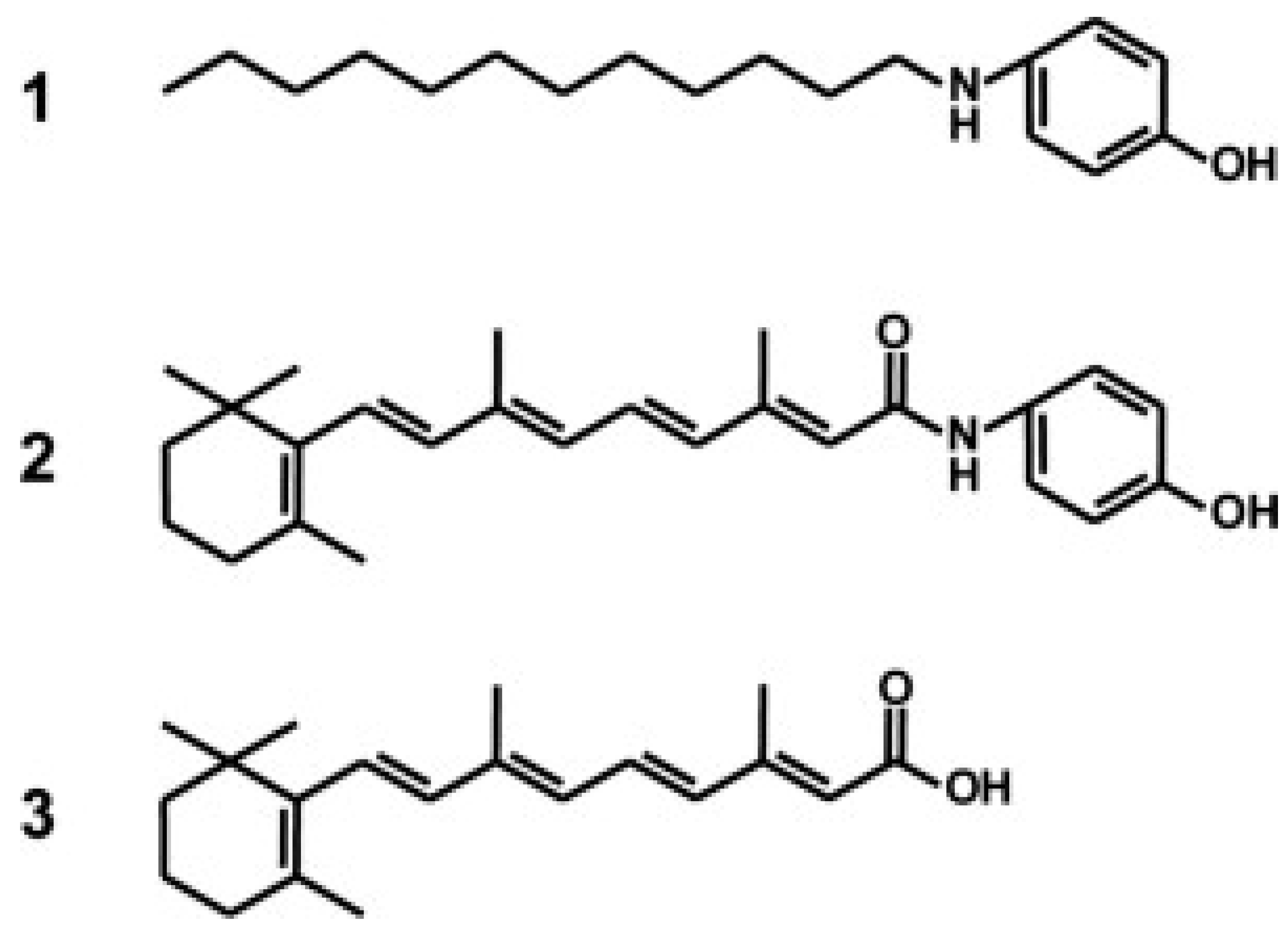
p-Dodecylaminophenol (p-DDAP) is a synthetic compound derived from fenretinide, which itself is a derivative of all-trans-retinoic acid used in cancer treatment. This compound has been developed to offer potent anticancer effects with fewer side effects than fenretinide, has exhibited strong antiproliferative properties against various cancers, including prostate cancer, and has shown effectiveness both in vitro and in vivo [87]. Takahashi et al. explored the anti-invasive effects of p-DDAP (1), which was produced based on N-(4-hydroxyphenyl)retinamide (2), a synthetic amide of all-trans-retinoic acid (3) (Figure 3). In HT-1080 cells, p-DDAP inhibited growth, induced apoptosis, and arrested the cell cycle in the S phase in a dose-dependent manner. Additionally, p-DDAP significantly suppressed cell invasion as well as the activity and mRNA expression of MMP-9. Moreover, the expression of the reversion-inducing cysteine-rich protein with Kazal motifs (RECK), a negative regulator of MMP-9, was increased following treatment. These results suggested that p-DDAP could be an effective anticancer agent, not only suppressing cell growth through apoptosis induction and cell cycle arrest but also inhibiting cell invasion by decreasing MMP-9 expression due to an increase in RECK expression [88].
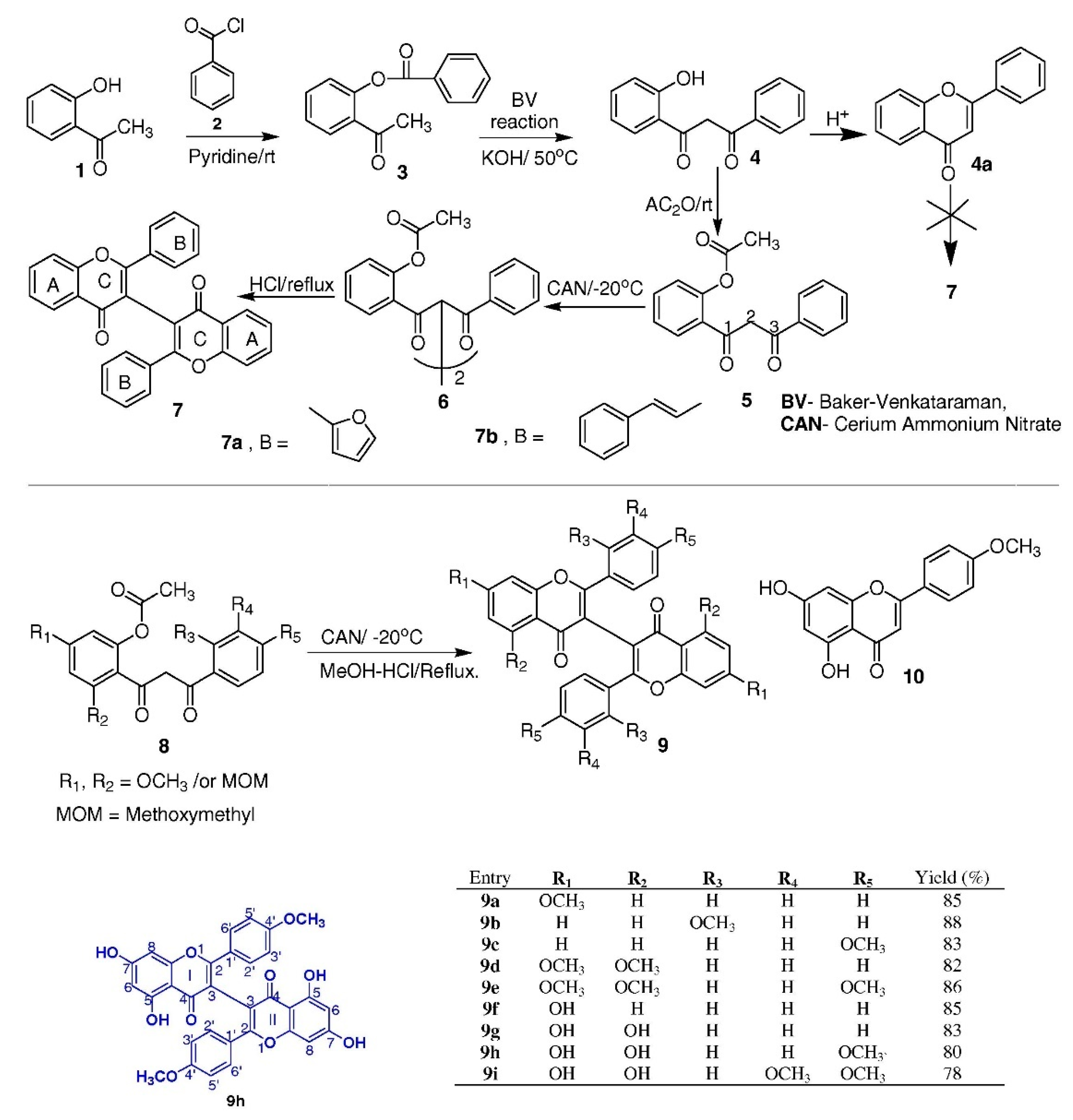
In an interesting study, eleven biflavones, labeled 7a–b and 9a–i, were synthesized using a streamlined and effective method and evaluated for their inhibitory effects on MMP-2 and MMP-9 enzymes. Notably, the compound known as (I-3,II-3)-biacacetin (9h), resembling natural products, emerged as the most effective inhibitor (Figure 4). Unlike many conventional inhibitors, molecular docking studies revealed that 9h inhibits MMP-2 and MMP-9 in fibrosarcoma cells through interactions that do not involve zinc binding [89].
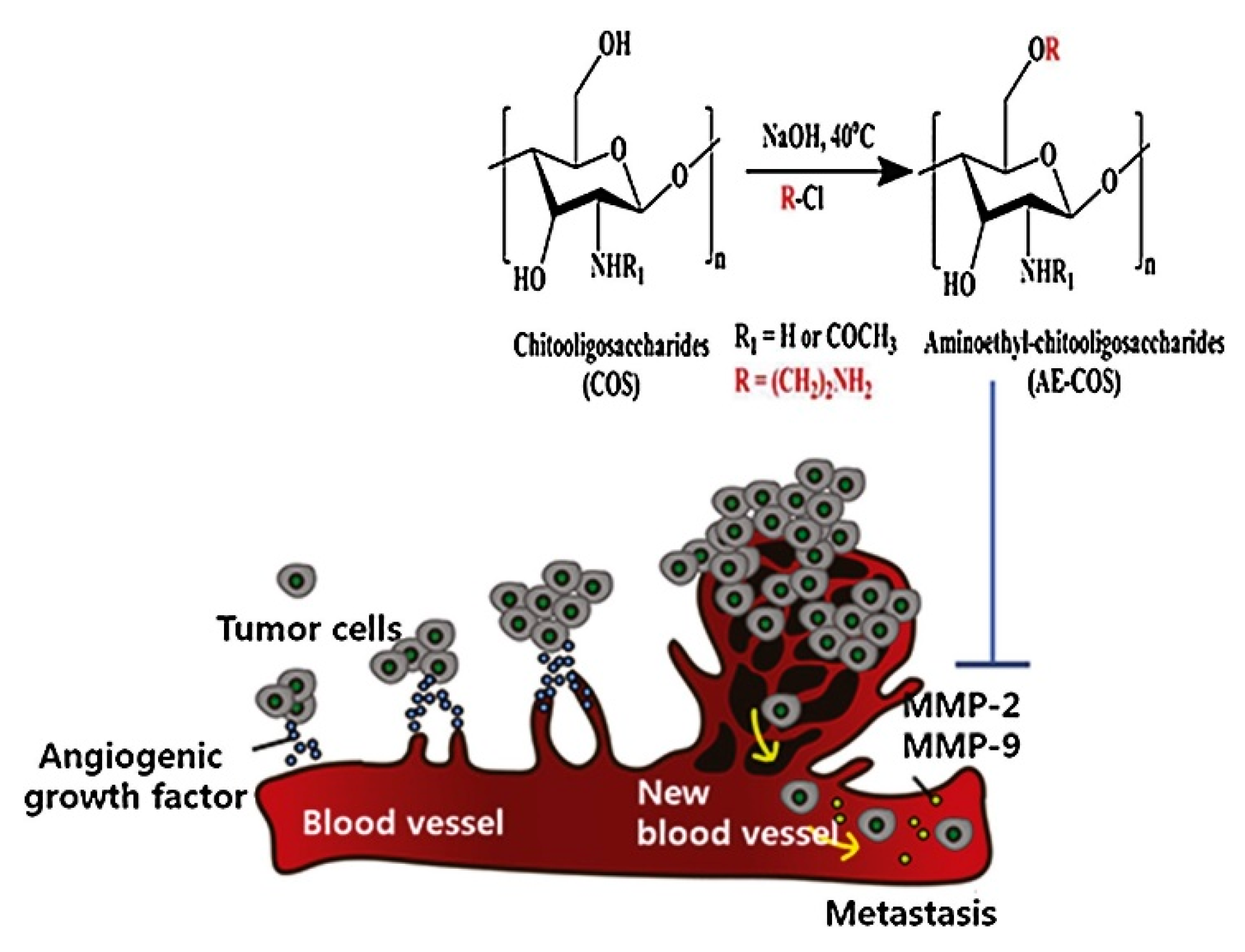
Aminoethyl-chitooligosaccharides (AE-COSs) are chemically modified derivatives of chitooligosaccharides, where hydroxyl groups are substituted with aminoethyl groups at specific positions, commonly the C-6 position. This modification enhances their biological activities, making AE-COSs particularly useful in various biomedical applications. Research has demonstrated the effectiveness of AE-COSs in several areas. Notably, AE-COSs have shown significant potential for inhibiting cancer cell proliferation and invasion [90]. The results from MTT assays, gelatin zymography, and Western blot analyses, which measure MMP-2 and MMP-9, indicate that aminoethyl-chitooligosaccharides (AE-COSs) (Figure 5) can inhibit the invasive and metastatic potential of HT-1080 cells by blocking MMP-2 and MMP-9 activity and expression [91].
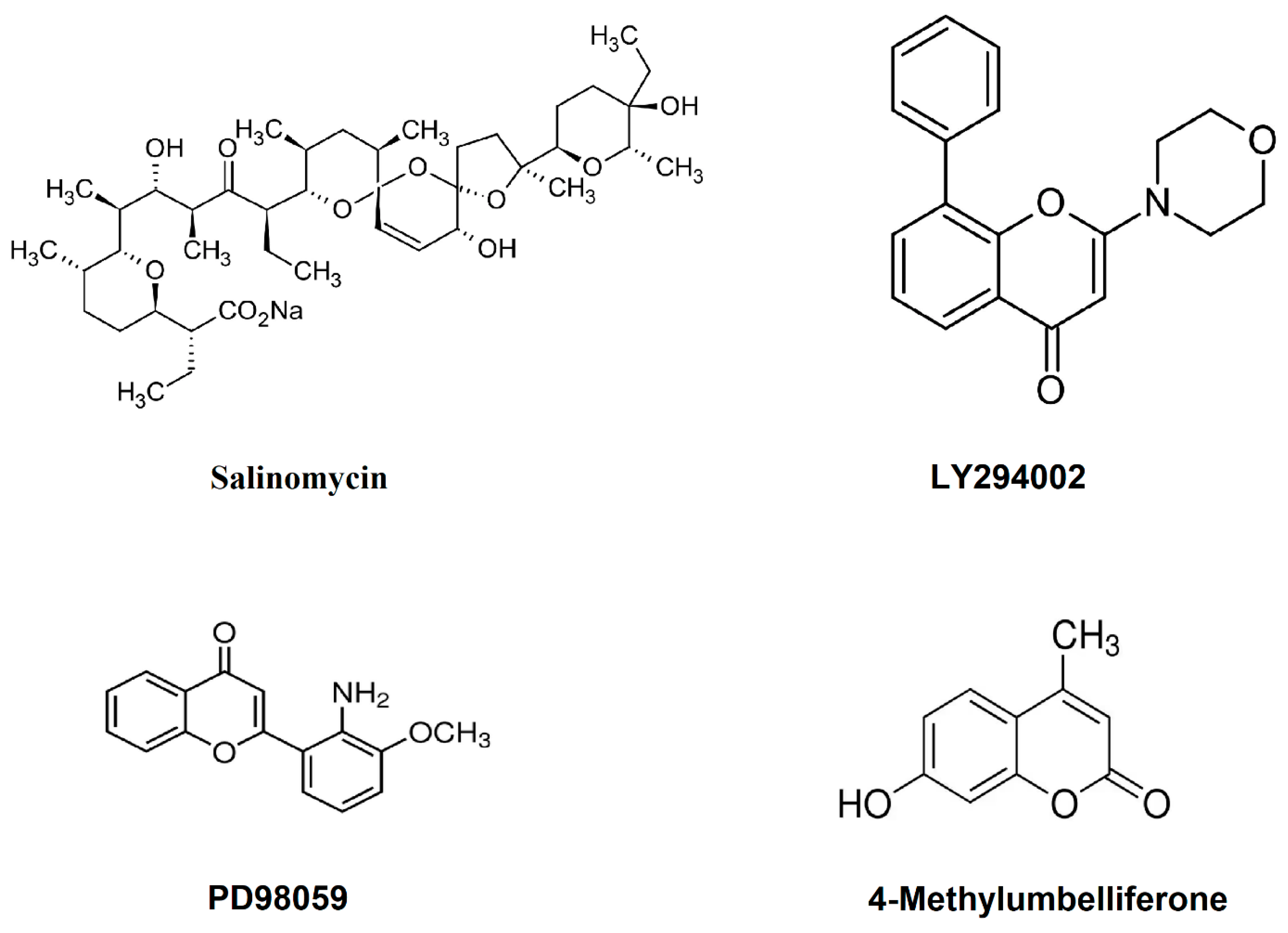
LY294002, PD98059, and SB203580 are all chemical inhibitors used in biochemical research to study signaling pathways. LY294002 is a specific inhibitor of phosphoinositide 3-kinases (PI3Ks) [92], and PD98059 is an inhibitor of mitogen-activated protein kinase (MEK), which is a part of the MAPK/ERK pathway [93]. SB203580 primarily inhibits p38 MAP kinase, which is involved in inflammatory responses and stress [94]. A study by Yu and Kim revealed that when these inhibitors were applied along with salinomycin (Figure 6), there was a suppression in metastatic potential through the reduction in MMP-2 expression as well as migration and invasion in HT-1080 cells. In a further study, they also found that 5-azacytidine (5-aza C), a methyltransferase inhibitor, can induce MMP-9 activity in HT-1080 cells and promote migration and invasion significantly, which can be inhibited by its inhibitors (LY294002 and PD98059) (Figure 6) [95].
Combination therapy that uses radiation along with small-molecule inhibitors is a promising approach for enhancing cancer treatment effectiveness. This strategy aims to target cancer cells more precisely and reduce the side effects typically associated with conventional therapies [96]. 4-Methylumbelliferone (4-MU) (Figure 6) is a derivative of coumarin, primarily known for its use as a hyaluronic acid synthesis inhibitor. A study showed that there was a higher reduction in the viability of HT-1080 cells cultured with a combination of 2 Gy IR and 100 μM 4-MU compared with untreated control cells. Moreover, this treatment led to a substantial decline in the invasion rate and MMP-2 and MMP-9 expression in the fibrosarcoma cell line [97].
In a study by Alford et al., they developed a series of compounds derived from the lead molecule N-[4-(difluoromethoxy)phenyl]-2-[(4-oxo-6-propyl-1H-pyrimidin-2-yl)sulfanyl]-acetamide, aimed at targeting the hemopexin-like domain of MMP-9. This group identified N-(4-fluorophenyl)-4-(4-oxo-3,4,5,6,7,8-hexahydroquinazolin-2-ylthio)butanamide, or 3c, as a potent lead compound with a dissociation constant (Kd) of 320 nM, exhibiting specificity for the proMMP-9 hemopexin-like domain. They showed that 3c inhibits MMP-9 homodimerization, and 500 nM of 3c effectively blocks fibrosarcoma cell invasion through the basement membrane and decreases angiogenesis [98].
The synthetic chemical small molecules used in cancer therapy often offer significant advantages, such as the ability to target specific molecular pathways involved in cancer progression. However, they also come with several disadvantages. Many synthetic small molecules can be highly toxic, not only to cancer cells but also to normal cells, leading to various side effects. For example, they might cause nausea, vomiting, hair loss, and a decrease in blood cell counts, which can lead to increased susceptibility to infections [99]. Cancer cells can develop resistance to these treatments over time. This resistance can occur through various mechanisms, such as mutations in the target molecule, increased drug efflux, or compensatory activation of alternative pathways. This makes the treatment less effective over time and necessitates the use of additional or alternative therapies [100]. While designed to be specific, some synthetic small molecules may still affect other cellular targets, leading to off-target effects and unintended consequences. This lack of absolute specificity can compromise their effectiveness and safety [101]. The development, production, and formulation of synthetic small molecules can be expensive, which may make these drugs less accessible to patients, especially in low-income regions [102]. Issues related to the absorption, distribution, metabolism, and excretion of these drugs can affect their efficacy and safety. For instance, a drug might not reach the tumor in sufficient concentrations to be effective or could be metabolized too quickly to have a therapeutic effect [103]. Some tumors, such as fibrosarcoma, have a microenvironment that hinders the effective penetration and distribution of these drugs, which can limit their efficacy in killing cancer cells throughout the tumor. These challenges highlight the need for continuous research to improve the specificity, effectiveness, and safety of synthetic chemical small molecules in cancer therapy.

7. Protein- and Peptide-Based Inhibitors

Protein- and peptide-based therapies offer a promising approach to cancer treatment, focusing on using peptides and proteins to target specific biological processes involved in cancer growth and progression. Proteins and peptides can be designed to target specific receptors, enzymes, or other proteins associated with cancer cells with high specificity, potentially reducing the risk of harming normal cells [104]. These molecules can be engineered to carry out a variety of functions, such as directly inhibiting tumor growth, blocking signals that promote cancer cell survival, or delivering cytotoxic agents specifically to cancer cells [18,105]. Compared to traditional chemotherapy, peptide and protein therapies often have lower toxicity, leading to fewer and less severe side effects [106]. Being natural biological molecules, proteins and peptides are biodegradable and less likely to accumulate in the body, reducing long-term side effects [107]. The development of protein- and peptide-based therapies for fibrosarcoma is still in the experimental stages, with many potential treatments undergoing preclinical trials or early clinical testing. The complexity of fibrosarcoma’s biological mechanisms often necessitates combination therapies, where protein- or peptide-based therapies might be used in conjunction with other treatments like chemotherapy or radiation.
In 2014, Kim et al. developed a carnosine–gallic acid peptide (CGP) to explore a more effective MMP inhibitor than carnosine. They examined the inhibitory impacts of CGP on MMP-2 and MMP-9 within the human fibrosarcoma cell line. CGP significantly reduced the expression levels of MMP-2 and MMP-9 without causing cytotoxic effects. Furthermore, CGP potentially hinders migration and invasion in HT-1080 cells by targeting the urokinase plasminogen activator (uPA)–uPA receptor signaling pathways, thereby suppressing MMP-2 and MMP-9 activity [108].
CD97 is a tumor-associated adhesion-class G-protein-coupled receptor that plays a role in regulating cell migration. Hsiao et al.’s findings indicated that CD97 expression in HT-1080 fibrosarcoma cells increases the secretion of TIMP-2, which subsequently reduces the activity of MMP-MT1 and MMP-2. This reduction in enzyme activity hampers cell migration and invasion in vitro [109].
In a study by Ko and colleagues in 2018, they isolated a heptameric peptide (Leu-Leu- Ala-Pro-Pro-Glu-Arg) from the marine microalga Pavlova lutheri and studied its inhibitory influence and mechanism of action on MMP-9 in HT-1080 cells. The outcomes exhibited that MMP-9’s mRNA and protein expression levels were decreased in the presence of peptides. The activity of MMP-9 when stimulated by PMA was similar to that of the control group; however, treatment with the peptide considerably reduced MMP-9 activity at concentrations of 250 and 500 μg/mL−1 (p < 0.05) [110].
Phage display is a versatile tool for the development of protease inhibitors. This technology involves the expression of peptide or protein libraries on the surface of phages. The displayed peptides or proteins can be engineered to bind to specific targets, including proteases [111]. In our two related studies in 2019 and 2022, we found that a phage display-isolated linear and cyclic type of peptide (MHPNAGHGSLMR) can significantly block the gelatinolytic activity of MMP-2 (pro- and active form) and proMMP-9 in a dose-dependent manner in HT-1080 cells. The results from cell invasion also showed inhibition of cell invasion at a concentration of 50 μM of peptide [112,113].
Currently, there are no antibodies specifically approved for the treatment of fibrosarcoma. However, research is ongoing to explore various antibodies that might target molecules associated with fibrosarcoma cells. Antibodies that inhibit growth factor receptors, which are often overexpressed in various cancers, including sarcomas, could potentially be effective. Research may focus on developing monoclonal antibodies that target specific antigens expressed in fibrosarcoma cells. These could include surface proteins unique to fibrosarcoma or associated with aggressive tumor behavior. Patients with fibrosarcoma might be eligible to participate in clinical trials testing new antibody therapies or combinations of existing treatments. These trials are crucial for discovering effective therapies for rare cancers like fibrosarcoma.
Proteins and peptides offer promising therapeutic potential for cancer treatment due to their specificity and ability to target key molecular pathways. However, their use as therapeutic agents also presents several disadvantages and challenges. Proteins and peptides are generally less stable in the body compared to small-molecule drugs. They are prone to degradation by proteases and other enzymes in the bloodstream and digestive tract, which can significantly reduce their effective lifespan and activity. Effective delivery of proteins and peptides to the target site within the body is challenging. Their large size and complex structure can hinder their ability to cross cellular membranes, and they may be poorly absorbed when administered orally. This often necessitates intravenous or subcutaneous administration, which can be less convenient and more painful for patients. Proteins and peptides can potentially be recognized as foreign substances by the patient’s immune system, leading to immune responses that can neutralize the therapeutic agents or cause adverse reactions. Managing immunogenicity is a major concern, particularly for repeated administrations. Due to issues with bioavailability and degradation, high doses of protein and peptide drugs may be required to achieve therapeutic effects, which can increase the risk of side effects and further escalate treatment costs. The effective penetration of proteins and peptides into solid tumors is often limited, which can reduce their efficacy against certain types of cancer.

8. Conclusions

Over the past decade, the study of the gelatinases MMP-2 and MMP-9 as therapeutic targets in fibrosarcoma treatment has revealed their significant potential in inhibiting tumor progression and metastasis. Blockade of these enzymes can disrupt the extracellular matrix remodeling that fibrosarcomas exploit for growth and dissemination [33]. Clinical and experimental research into MMP inhibitors has uncovered promising approaches to curbing these processes, with several inhibitors showing efficacy in reducing tumor size and improving survival rates in preclinical models [114]. However, challenges remain in translating these findings into clinical successes due to issues such as side effects, resistance, and the specificity of inhibitors. The therapeutic potential of gelatinase blockade in fibrosarcoma treatment remains substantial but requires further refinement and investigation to optimize dosing, minimize adverse effects, and enhance delivery mechanisms.
The potential of targeting gelatinases as a therapeutic strategy in the treatment of fibrosarcoma is promising, as highlighted in this decadal review of inhibitors categorized into natural, chemical, and protein- and peptide-based groups. Natural inhibitors have shown significant potential due to their biocompatibility and fewer side effects, making them an appealing option for long-term therapy. Chemical inhibitors, on the other hand, are often more potent and can be designed to selectively target MMP-2 and MMP-9, minimizing off-target effects and enhancing therapeutic efficacy. Protein- and peptide-based inhibitors provide a unique advantage due to their specificity and ability to disrupt enzyme–substrate interactions directly at the molecular level. While each category of inhibitors has its advantages and limitations, the integration of these strategies could lead to more effective and personalized therapeutic approaches in fibrosarcoma treatment. Future research should focus on overcoming the challenges related to the resistance, delivery, and stability of these inhibitors to fully harness their potential in clinical settings.
Future research should also explore combination therapies that may enhance the effectiveness of gelatinase inhibitors and reduce the likelihood of tumor resistance. Continued exploration in this field holds promise for more effective fibrosarcoma treatments, potentially improving outcomes for patients suffering from this aggressive cancer.

Funding

This research received no external funding.

Data Availability Statement

Not applicable.

Conflicts of Interest

The author declares no conflicts of interest.

References

  1. Augsburger, D.; Nelson, P.J.; Kalinski, T.; Udelnow, A.; Knosel, T.; Hofstetter, M.; Qin, J.W.; Wang, Y.; Gupta, A.S.; Bonifatius, S.; et al. Current diagnostics and treatment of fibrosarcoma -perspectives for future therapeutic targets and strategies. Oncotarget 2017, 8, 104638–104653. [Google Scholar] [CrossRef] [PubMed]
  2. Genadry, K.C.; Pietrobono, S.; Rota, R.; Linardic, C.M. Soft Tissue Sarcoma Cancer Stem Cells: An Overview. Front. Oncol. 2018, 8, 475. [Google Scholar] [CrossRef] [PubMed]
  3. Huvos, A.G.; Higinbotham, N.L. Primary fibrosarcoma of bone. A clinicopathologic study of 130 patients. Cancer 1975, 35, 837–847. [Google Scholar] [CrossRef] [PubMed]
  4. Atalay, I.B.; Togral, G. Unusual localization and aggressive progression of large infantile fibrosarcoma. Acta Orthop. Traumatol. Turc. 2019, 53, 507–511. [Google Scholar] [CrossRef]
  5. Scott, S.M.; Reiman, H.M.; Pritchard, D.J.; Ilstrup, D.M. Soft tissue fibrosarcoma. A clinicopathologic study of 132 cases. Cancer 1989, 64, 925–931. [Google Scholar] [CrossRef] [PubMed]
  6. Ferreira, C.R.; da Fonseca, L.G.; Piotto, G.H.M.; Geyer, F.C.; de Alcantara, P.S.M. Fibrosarcoma: A challenging diagnosis. Autops. Case Rep. 2013, 3, 21–29. [Google Scholar] [CrossRef] [PubMed]
  7. Dernell, W.S.; Withrow, S.J.; Kuntz, C.A.; Powers, B.E. Principles of treatment for soft tissue sarcoma. Clin. Tech. Small Anim. Pract. 1998, 13, 59–64. [Google Scholar] [CrossRef] [PubMed]
  8. Windeyer, B.; Dische, S.; Mansfield, C.M. The place of radiotherapy in the management of fibrosarcoma of the soft tissues. Clin. Radiol. 1966, 17, 32–40. [Google Scholar] [CrossRef]
  9. Chew, W.; Benson, C.; Thway, K.; Hayes, A.; Miah, A.; Zaidi, S.; Lee, A.T.J.; Messiou, C.; Fisher, C.; van der Graaf, W.T.; et al. Clinical Characteristics and efficacy of chemotherapy in sclerosing epithelioid fibrosarcoma. Med. Oncol. 2018, 35, 138. [Google Scholar] [CrossRef]
  10. Zhang, P.; Zhao, L.; Zhu, Y.J.; Qiu, B.; Guo, S.P.; Li, Y.; Liu, Q.; Liu, M.Z.; Xi, M. Prognosis of Fibrosarcoma in Patients With and Without a History of Radiation for Nasopharyngeal Carcinoma. Ann. Surg. Oncol. 2017, 24, 434–440. [Google Scholar] [CrossRef]
  11. Cantin, J.; McNeer, G.P.; Chu, F.C.; Booher, R.J. The problem of local recurrence after treatment of soft tissue sarcoma. Ann. Surg. 1968, 168, 47–53. [Google Scholar] [CrossRef]
  12. Wibmer, C.; Leithner, A.; Zielonke, N.; Sperl, M.; Windhager, R. Increasing incidence rates of soft tissue sarcomas? A population-based epidemiologic study and literature review. Ann. Oncol. 2010, 21, 1106–1111. [Google Scholar] [CrossRef] [PubMed]
  13. Coffin, C.M.; Dehner, L.P. Fibroblastic-myofibroblastic tumors in children and adolescents: A clinicopathologic study of 108 examples in 103 patients. Pediatr. Pathol. 1991, 11, 569–588. [Google Scholar] [CrossRef] [PubMed]
  14. Bahrami, A.; Folpe, A.L. Adult-type fibrosarcoma: A reevaluation of 163 putative cases diagnosed at a single institution over a 48-year period. Am. J. Surg. Pathol. 2010, 34, 1504–1513. [Google Scholar] [CrossRef] [PubMed]
  15. Burningham, Z.; Hashibe, M.; Spector, L.; Schiffman, J.D. The epidemiology of sarcoma. Clin. Sarcoma Res. 2012, 2, 14. [Google Scholar] [CrossRef] [PubMed]
  16. Folpe, A.L. Fibrosarcoma: A review and update. Histopathology 2014, 64, 12–25. [Google Scholar] [CrossRef]
  17. Wang, X.; Khalil, R.A. Matrix Metalloproteinases, Vascular Remodeling, and Vascular Disease. Adv. Pharmacol. 2018, 81, 241–330. [Google Scholar] [CrossRef] [PubMed]
  18. Shoari, A.; Khalili-Tanha, G.; Coban, M.A.; Radisky, E.S. Structure and computation-guided yeast surface display for the evolution of TIMP-based matrix metalloproteinase inhibitors. Front. Mol. Biosci. 2023, 10, 1321956. [Google Scholar] [CrossRef]
  19. Radisky, E.S. Extracellular proteolysis in cancer: Proteases, substrates, and mechanisms in tumor progression and metastasis. J. Biol. Chem. 2024, 300, 107347. [Google Scholar] [CrossRef]
  20. Radisky, E.S.; Radisky, D.C. Matrix metalloproteinase-induced epithelial-mesenchymal transition in breast cancer. J. Mammary Gland. Biol. Neoplasia 2010, 15, 201–212. [Google Scholar] [CrossRef]
  21. Almutairi, S.; Kalloush, H.M.; Manoon, N.A.; Bardaweel, S.K. Matrix Metalloproteinases Inhibitors in Cancer Treatment: An Updated Review (2013–2023). Molecules 2023, 28, 5567. [Google Scholar] [CrossRef]
  22. Klein, T.; Bischoff, R. Physiology and pathophysiology of matrix metalloproteases. Amino Acids 2011, 41, 271–290. [Google Scholar] [CrossRef]
  23. Cabral-Pacheco, G.A.; Garza-Veloz, I.; Castruita-De la Rosa, C.; Ramirez-Acuna, J.M.; Perez-Romero, B.A.; Guerrero-Rodriguez, J.F.; Martinez-Avila, N.; Martinez-Fierro, M.L. The Roles of Matrix Metalloproteinases and Their Inhibitors in Human Diseases. Int. J. Mol. Sci. 2020, 21, 9739. [Google Scholar] [CrossRef] [PubMed]
  24. Walker, C.; Mojares, E.; Del Rio Hernandez, A. Role of Extracellular Matrix in Development and Cancer Progression. Int. J. Mol. Sci. 2018, 19, 3028. [Google Scholar] [CrossRef] [PubMed]
  25. Xu, J.; Du, Y.; Liu, W.J.; Li, L.; Li, Y.; Wang, X.F.; Yi, H.F.; Shan, C.K.; Xia, G.M.; Liu, X.J.; et al. Intensive fibrosarcoma-binding capability of the reconstituted analog and its antitumor activity. Drug Deliv. 2018, 25, 102–111. [Google Scholar] [CrossRef]
  26. Benassi, M.S.; Gamberi, G.; Magagnoli, G.; Molendini, L.; Ragazzini, P.; Merli, M.; Chiesa, F.; Balladelli, A.; Manfrini, M.; Bertoni, F.; et al. Metalloproteinase expression and prognosis in soft tissue sarcomas. Ann. Oncol. 2001, 12, 75–80. [Google Scholar] [CrossRef]
  27. Lu, P.; Takai, K.; Weaver, V.M.; Werb, Z. Extracellular matrix degradation and remodeling in development and disease. Cold Spring Harb. Perspect. Biol. 2011, 3, a005058. [Google Scholar] [CrossRef]
  28. Elgundi, Z.; Papanicolaou, M.; Major, G.; Cox, T.R.; Melrose, J.; Whitelock, J.M.; Farrugia, B.L. Cancer Metastasis: The Role of the Extracellular Matrix and the Heparan Sulfate Proteoglycan Perlecan. Front. Oncol. 2019, 9, 1482. [Google Scholar] [CrossRef] [PubMed]
  29. Niland, S.; Riscanevo, A.X.; Eble, J.A. Matrix Metalloproteinases Shape the Tumor Microenvironment in Cancer Progression. Int. J. Mol. Sci. 2021, 23, 146. [Google Scholar] [CrossRef]
  30. Yang, H.K.; Jeong, K.C.; Kim, Y.K.; Jung, S.T. Role of matrix metalloproteinase (MMP) 2 and MMP-9 in soft tissue sarcoma. Clin. Orthop. Surg. 2014, 6, 443–454. [Google Scholar] [CrossRef]
  31. Roebuck, M.M.; Helliwell, T.R.; Chaudhry, I.H.; Kalogrianitis, S.; Carter, S.; Kemp, G.J.; Ritchie, D.A.; Jane, M.J.; Frostick, S.P. Matrix metalloproteinase expression is related to angiogenesis and histologic grade in spindle cell soft tissue neoplasms of the extremities. Am. J. Clin. Pathol. 2005, 123, 405–414. [Google Scholar] [CrossRef] [PubMed]
  32. Di Martino, J.S.; Akhter, T.; Bravo-Cordero, J.J. Remodeling the ECM: Implications for Metastasis and Tumor Dormancy. Cancers 2021, 13, 4916. [Google Scholar] [CrossRef] [PubMed]
  33. Partridge, J.J.; Madsen, M.A.; Ardi, V.C.; Papagiannakopoulos, T.; Kupriyanova, T.A.; Quigley, J.P.; Deryugina, E.I. Functional analysis of matrix metalloproteinases and tissue inhibitors of metalloproteinases differentially expressed by variants of human HT-1080 fibrosarcoma exhibiting high and low levels of intravasation and metastasis. J. Biol. Chem. 2007, 282, 35964–35977. [Google Scholar] [CrossRef]
  34. Nguyen, M.; Arkell, J.; Jackson, C.J. Human endothelial gelatinases and angiogenesis. Int. J. Biochem. Cell Biol. 2001, 33, 960–970. [Google Scholar] [CrossRef] [PubMed]
  35. Farina, A.R.; Mackay, A.R. Gelatinase B/MMP-9 in Tumour Pathogenesis and Progression. Cancers 2014, 6, 240–296. [Google Scholar] [CrossRef] [PubMed]
  36. Bjorklund, M.; Koivunen, E. Gelatinase-mediated migration and invasion of cancer cells. Biochim. Biophys. Acta 2005, 1755, 37–69. [Google Scholar] [CrossRef] [PubMed]
  37. Augoff, K.; Hryniewicz-Jankowska, A.; Tabola, R.; Stach, K. MMP9: A Tough Target for Targeted Therapy for Cancer. Cancers 2022, 14, 1847. [Google Scholar] [CrossRef] [PubMed]
  38. Hodge, J.W.; Ardiani, A.; Farsaci, B.; Kwilas, A.R.; Gameiro, S.R. The tipping point for combination therapy: Cancer vaccines with radiation, chemotherapy, or targeted small molecule inhibitors. Semin. Oncol. 2012, 39, 323–339. [Google Scholar] [CrossRef] [PubMed]
  39. Vandenbroucke, R.E.; Libert, C. Is there new hope for therapeutic matrix metalloproteinase inhibition? Nat. Rev. Drug Discov. 2014, 13, 904–927. [Google Scholar] [CrossRef]
  40. Moliere, S.; Jaulin, A.; Tomasetto, C.L.; Dali-Youcef, N. Roles of Matrix Metalloproteinases and Their Natural Inhibitors in Metabolism: Insights into Health and Disease. Int. J. Mol. Sci. 2023, 24, 10649. [Google Scholar] [CrossRef]
  41. Rasheed, S.; Nelson-Rees, W.A.; Toth, E.M.; Arnstein, P.; Gardner, M.B. Characterization of a newly derived human sarcoma cell line (HT-1080). Cancer 1974, 33, 1027–1033. [Google Scholar] [CrossRef] [PubMed]
  42. Zhang, L.; Hill, R.P. Hypoxia enhances metastatic efficiency in HT1080 fibrosarcoma cells by increasing cell survival in lungs, not cell adhesion and invasion. Cancer Res. 2007, 67, 7789–7797. [Google Scholar] [CrossRef] [PubMed]
  43. Wang, J.; Jiang, Y.F. Natural compounds as anticancer agents: Experimental evidence. World J. Exp. Med. 2012, 2, 45–57. [Google Scholar] [CrossRef] [PubMed]
  44. Nan, Y.; Su, H.; Zhou, B.; Liu, S. The function of natural compounds in important anticancer mechanisms. Front. Oncol. 2022, 12, 1049888. [Google Scholar] [CrossRef] [PubMed]
  45. Lin, S.R.; Chang, C.H.; Hsu, C.F.; Tsai, M.J.; Cheng, H.; Leong, M.K.; Sung, P.J.; Chen, J.C.; Weng, C.F. Natural compounds as potential adjuvants to cancer therapy: Preclinical evidence. Br. J. Pharmacol. 2020, 177, 1409–1423. [Google Scholar] [CrossRef] [PubMed]
  46. Chunarkar-Patil, P.; Kaleem, M.; Mishra, R.; Ray, S.; Ahmad, A.; Verma, D.; Bhayye, S.; Dubey, R.; Singh, H.N.; Kumar, S. Anticancer Drug Discovery Based on Natural Products: From Computational Approaches to Clinical Studies. Biomedicines 2024, 12, 201. [Google Scholar] [CrossRef] [PubMed]
  47. Nam, H.; Kim, M.M. Eugenol with antioxidant activity inhibits MMP-9 related to metastasis in human fibrosarcoma cells. Food Chem. Toxicol. 2013, 55, 106–112. [Google Scholar] [CrossRef] [PubMed]
  48. Park, J.H.; Jang, Y.J.; Choi, Y.J.; Jang, J.W.; Kim, J.H.; Rho, Y.K.; Kim, I.J.; Kim, H.J.; Leem, M.J.; Lee, S.T. Fisetin inhibits matrix metalloproteinases and reduces tumor cell invasiveness and endothelial cell tube formation. Nutr. Cancer 2013, 65, 1192–1199. [Google Scholar] [CrossRef] [PubMed]
  49. Lee, D.E.; Chung, M.Y.; Lim, T.G.; Huh, W.B.; Lee, H.J.; Lee, K.W. Quercetin suppresses intracellular ROS formation, MMP activation, and cell motility in human fibrosarcoma cells. J. Food Sci. 2013, 78, H1464–H1469. [Google Scholar] [CrossRef]
  50. Filipiak, K.; Hidalgo, M.; Silvan, J.M.; Fabre, B.; Carbajo, R.J.; Pineda-Lucena, A.; Ramos, A.; de Pascual-Teresa, B.; de Pascual-Teresa, S. Dietary gallic acid and anthocyanin cytotoxicity on human fibrosarcoma HT1080 cells. A study on the mode of action. Food Funct. 2014, 5, 381–389. [Google Scholar] [CrossRef]
  51. Shi, X.; Chen, G.; Liu, X.; Qiu, Y.; Yang, S.; Zhang, Y.; Fang, X.; Zhang, C.; Liu, X. Scutellarein inhibits cancer cell metastasis in vitro and attenuates the development of fibrosarcoma in vivo. Int. J. Mol. Med. 2015, 35, 31–38. [Google Scholar] [CrossRef] [PubMed]
  52. Choi, Y.J.; Lee, Y.H.; Lee, S.T. Galangin and kaempferol suppress phorbol-12-myristate-13-acetate-induced matrix metalloproteinase-9 expression in human fibrosarcoma HT-1080 cells. Mol. Cells 2015, 38, 151–155. [Google Scholar] [CrossRef] [PubMed]
  53. Li, J.; Liu, C.; Sato, T. Novel Antitumor Invasive Actions of p-Cymene by Decreasing MMP-9/TIMP-1 Expression Ratio in Human Fibrosarcoma HT-1080 Cells. Biol. Pharm. Bull. 2016, 39, 1247–1253. [Google Scholar] [CrossRef]
  54. Gong, F.; Zhang, Y.; Lin, J.; Li, C.; Zhou, C.; Hong, P.; Ryu, B.; Qian, Z.J. 1-(5-Bromo-2-hydroxy-4-methoxyphenyl)ethanone [SE1] Inhibits MMP-9 Expression by Regulating NF-kappaB and MAPKs Signaling Pathways in HT1080 Human Fibrosarcoma Cells. Evid. Based Complement. Alternat Med. 2018, 2018, 5639486. [Google Scholar] [CrossRef] [PubMed]
  55. Roomi, M.W.; Bhanap, B.; Niedzwiecki, A.; Rath, M. Chlorophyllin suppresses growth, mmp secretion, invasion and cell migration of fibrosarcoma cell line ht-1080. Med. Res. Arch. 2018, 6. [Google Scholar] [CrossRef]
  56. Choi, J.H.; Hwang, Y.P.; Jin, S.W.; Lee, G.H.; Kim, H.G.; Han, E.H.; Kim, S.K.; Kang, K.W.; Chung, Y.C.; Jeong, H.G. Suppression of PMA-induced human fibrosarcoma HT-1080 invasion and metastasis by kahweol via inhibiting Akt/JNK1/2/p38 MAPK signal pathway and NF-kappaB dependent transcriptional activities. Food Chem. Toxicol. 2019, 125, 1–9. [Google Scholar] [CrossRef]
  57. Kim, J.; Kim, M.M. Effect of Lapachol on the Inhibition of Matrix Metalloproteinase Related to the Invasion of Human Fibrosarcoma Cells. Curr. Mol. Pharmacol. 2021, 14, 620–626. [Google Scholar] [CrossRef] [PubMed]
  58. Kim, H.; Kong, C.S.; Seo, Y. Salidroside, 8(E)-Nuezhenide, and Ligustroside from Ligustrum japonicum Fructus Inhibit Expressions of MMP-2 and -9 in HT 1080 Fibrosarcoma. Int. J. Mol. Sci. 2022, 23, 2660. [Google Scholar] [CrossRef]
  59. Jo, H.W.; Kim, M.M. beta-Caryophyllene oxide inhibits metastasis by downregulating MMP-2, p-p38 and p-ERK in human fibrosarcoma cells. J. Food Biochem. 2022, 46, e14468. [Google Scholar] [CrossRef]
  60. Jo, A.I.; Kim, M.M. Silibinin Inhibits Cell Invasion through the Inhibition of MMPs, p-p38, and IL-1beta in Human Fibrosarcoma Cells. Front. Biosci. (Landmark Ed.) 2023, 28, 64. [Google Scholar] [CrossRef]
  61. Zari, A.T.; Zari, T.A.; Hakeem, K.R. Anticancer Properties of Eugenol: A Review. Molecules 2021, 26, 7407. [Google Scholar] [CrossRef] [PubMed]
  62. Ulanowska, M.; Olas, B. Biological Properties and Prospects for the Application of Eugenol-A Review. Int. J. Mol. Sci. 2021, 22, 3671. [Google Scholar] [CrossRef] [PubMed]
  63. Roomi, M.W.; Kalinovsky, T.; Niedzwiecki, A.; Rath, M. Modulation of u-PA, MMPs and their inhibitors by a novel nutrient mixture in adult human sarcoma cell lines. Int. J. Oncol. 2013, 43, 39–49. [Google Scholar] [CrossRef] [PubMed]
  64. Jeong, J.H.; An, J.Y.; Kwon, Y.T.; Rhee, J.G.; Lee, Y.J. Effects of low dose quercetin: Cancer cell-specific inhibition of cell cycle progression. J. Cell Biochem. 2009, 106, 73–82. [Google Scholar] [CrossRef] [PubMed]
  65. Ma, H.; Yue, G.G.; Lee, J.K.; Gao, S.; Yuen, K.K.; Cheng, W.; Li, X.; Lau, C.B. Scutellarin, a flavonoid compound from Scutellaria barbata, suppresses growth of breast cancer stem cells in vitro and in tumor-bearing mice. Phytomedicine 2024, 128, 155418. [Google Scholar] [CrossRef] [PubMed]
  66. Sithranga Boopathy, N.; Kathiresan, K. Anticancer drugs from marine flora: An overview. J. Oncol. 2010, 2010, 214186. [Google Scholar] [CrossRef] [PubMed]
  67. Bae, M.J.; Karadeniz, F.; Ahn, B.N.; Kong, C.S. Evaluation of Effective MMP Inhibitors from Eight Different Brown Algae in Human Fibrosarcoma HT1080 Cells. Prev. Nutr. Food Sci. 2015, 20, 153–161. [Google Scholar] [CrossRef] [PubMed]
  68. Barletta, E.; Ramazzotti, M.; Fratianni, F.; Pessani, D.; Degl’Innocenti, D. Hydrophilic extract from Posidonia oceanica inhibits activity and expression of gelatinases and prevents HT1080 human fibrosarcoma cell line invasion. Cell Adh. Migr. 2015, 9, 422–431. [Google Scholar] [CrossRef]
  69. Bae, M.J.; Karadeniz, F.; Lee, S.G.; Seo, Y.; Kong, C.S. Inhibition of MMP-2 and MMP-9 Activities by Limonium tetragonum Extract. Prev. Nutr. Food Sci. 2016, 21, 38–43. [Google Scholar] [CrossRef]
  70. Lee, S.G.; Karadeniz, F.; Oh, J.H.; Yu, G.H.; Kong, C.S. Inhibitory Effect of Hizikia fusiformis Solvent-Partitioned Fractions on Invasion and MMP Activity of HT1080 Human Fibrosarcoma Cells. Prev. Nutr. Food Sci. 2017, 22, 184–190. [Google Scholar] [CrossRef]
  71. Karadeniz, F.L.S.; Oh, J.H.; Kim, J.A.; Kong, C.S. Inhibition of MMP-2 and MMP-9 activities by solvent-partitioned Sargassum horneri extracts. Fish. Aquat. Sci. 2018, 21, 1–7. [Google Scholar] [CrossRef]
  72. Karadeniz, F.L.S.; Oh, J.H.; Yu, G.H.; Jang, M.S.; Seo, Y.; Kong, C.S. Solvent-partitioned fractions from Ishige okamurae extract inhibit MMP-2 and MMP-9 activities in human fibrosarcoma cells in vitro. J. Appl. Phycol. 2018, 30, 121–127. [Google Scholar] [CrossRef]
  73. Al-Khayri, J.M.; Sahana, G.R.; Nagella, P.; Joseph, B.V.; Alessa, F.M.; Al-Mssallem, M.Q. Flavonoids as Potential Anti-Inflammatory Molecules: A Review. Molecules 2022, 27, 2901. [Google Scholar] [CrossRef] [PubMed]
  74. Tuli, H.S.; Sak, K.; Adhikary, S.; Kaur, G.; Aggarwal, D.; Kaur, J.; Kumar, M.; Parashar, N.C.; Parashar, G.; Sharma, U.; et al. Galangin: A metabolite that suppresses anti-neoplastic activities through modulation of oncogenic targets. Exp. Biol. Med. 2022, 247, 345–359. [Google Scholar] [CrossRef]
  75. Balahbib, A.; El Omari, N.; Hachlafi, N.E.; Lakhdar, F.; El Menyiy, N.; Salhi, N.; Mrabti, H.N.; Bakrim, S.; Zengin, G.; Bouyahya, A. Health beneficial and pharmacological properties of p-cymene. Food Chem. Toxicol. 2021, 153, 112259. [Google Scholar] [CrossRef] [PubMed]
  76. Chen, L.; Wang, X.; Huang, B. The genus Hippocampus—A review on traditional medicinal uses, chemical constituents and pharmacological properties. J. Ethnopharmacol. 2015, 162, 104–111. [Google Scholar] [CrossRef] [PubMed]
  77. Makino, T.; Izumi, K.; Hiratsuka, K.; Kano, H.; Shimada, T.; Nakano, T.; Kadomoto, S.; Naito, R.; Iwamoto, H.; Yaegashi, H.; et al. Anti-proliferative and anti-migratory properties of coffee diterpenes kahweol acetate and cafestol in human renal cancer cells. Sci. Rep. 2021, 11, 675. [Google Scholar] [CrossRef] [PubMed]
  78. Mendes Miranda, S.E.; Alcantara Lemos, J.; Fernandes, R.S.; Silva, J.O.; Ottoni, F.M.; Townsend, D.M.; Rubello, D.; Alves, R.J.; Cassali, G.D.; Ferreira, L.A.M.; et al. Enhanced antitumor efficacy of lapachol-loaded nanoemulsion in breast cancer tumor model. Biomed. Pharmacother. 2021, 133, 110936. [Google Scholar] [CrossRef]
  79. Fidyt, K.; Fiedorowicz, A.; Strzadala, L.; Szumny, A. beta-caryophyllene and beta-caryophyllene oxide-natural compounds of anticancer and analgesic properties. Cancer Med. 2016, 5, 3007–3017. [Google Scholar] [CrossRef]
  80. Ray, P.P.; Islam, M.A.; Islam, M.S.; Han, A.; Geng, P.; Aziz, M.A.; Mamun, A.A. A comprehensive evaluation of the therapeutic potential of silibinin: A ray of hope in cancer treatment. Front. Pharmacol. 2024, 15, 1349745. [Google Scholar] [CrossRef]
  81. Tavakoli, J.; Miar, S.; Majid Zadehzare, M.; Akbari, H. Evaluation of effectiveness of herbal medication in cancer care: A review study. Iran. J. Cancer Prev. 2012, 5, 144–156. [Google Scholar] [PubMed]
  82. Fasinu, P.S.; Rapp, G.K. Herbal Interaction With Chemotherapeutic Drugs-A Focus on Clinically Significant Findings. Front. Oncol. 2019, 9, 1356. [Google Scholar] [CrossRef] [PubMed]
  83. Hoelder, S.; Clarke, P.A.; Workman, P. Discovery of small molecule cancer drugs: Successes, challenges and opportunities. Mol. Oncol. 2012, 6, 155–176. [Google Scholar] [CrossRef] [PubMed]
  84. Zhong, L.; Li, Y.; Xiong, L.; Wang, W.; Wu, M.; Yuan, T.; Yang, W.; Tian, C.; Miao, Z.; Wang, T.; et al. Small molecules in targeted cancer therapy: Advances, challenges, and future perspectives. Signal Transduct. Target. Ther. 2021, 6, 201. [Google Scholar] [CrossRef] [PubMed]
  85. Anand, U.; Dey, A.; Chandel, A.K.S.; Sanyal, R.; Mishra, A.; Pandey, D.K.; De Falco, V.; Upadhyay, A.; Kandimalla, R.; Chaudhary, A.; et al. Cancer chemotherapy and beyond: Current status, drug candidates, associated risks and progress in targeted therapeutics. Genes. Dis. 2023, 10, 1367–1401. [Google Scholar] [CrossRef] [PubMed]
  86. Roomi, M.W.; Kalinovsky, T.; Monterrey, J.; Rath, M.; Niedzwiecki, A. In vitro modulation of MMP-2 and MMP-9 in adult human sarcoma cell lines by cytokines, inducers and inhibitors. Int. J. Oncol. 2013, 43, 1787–1798. [Google Scholar] [CrossRef]
  87. Takahashi, N.; Watanabe, Y.; Maitani, Y.; Yamauchi, T.; Higashiyama, K.; Ohba, T. p-Dodecylaminophenol derived from the synthetic retinoid, fenretinide: Antitumor efficacy in vitro and in vivo against human prostate cancer and mechanism of action. Int. J. Cancer 2008, 122, 689–698. [Google Scholar] [CrossRef] [PubMed]
  88. Takahashi, N.; Takeda, K.; Imai, M. Inhibitory effects of p-dodecylaminophenol on the invasiveness of human fibrosarcoma cell line HT1080. Bioorg Med. Chem. 2013, 21, 6015–6021. [Google Scholar] [CrossRef] [PubMed]
  89. Nanjan, P.; Nambiar, J.; Nair, B.G.; Banerji, A. Synthesis and discovery of (I-3,II-3)-biacacetin as a novel non-zinc binding inhibitor of MMP-2 and MMP-9. Bioorg Med. Chem. 2015, 23, 3781–3787. [Google Scholar] [CrossRef]
  90. Liang, S.; Sun, Y.; Dai, X. A Review of the Preparation, Analysis and Biological Functions of Chitooligosaccharide. Int. J. Mol. Sci. 2018, 19, 2197. [Google Scholar] [CrossRef]
  91. Hong, S.; Ngo, D.N.; Kim, M.M. Inhibitory effect of aminoethyl-chitooligosaccharides on invasion of human fibrosarcoma cells. Environ. Toxicol. Pharmacol. 2016, 45, 309–314. [Google Scholar] [CrossRef] [PubMed]
  92. Bar, J.; Lukaschuk, N.; Zalcenstein, A.; Wilder, S.; Seger, R.; Oren, M. The PI3K inhibitor LY294002 prevents p53 induction by DNA damage and attenuates chemotherapy-induced apoptosis. Cell Death Differ. 2005, 12, 1578–1587. [Google Scholar] [CrossRef] [PubMed]
  93. Wang, K.; Hou, Y.; Gu, C.; Zhao, D.; Duan, Y.; Ran, Z.; Li, Q.; Li, X. Inhibitory effect of the mitogen activated protein kinase specific inhibitor PD98059 on Mtb-Ag-activated gammadeltaTau cells. Int. J. Clin. Exp. Pathol. 2017, 10, 9644–9648. [Google Scholar]
  94. Sreekanth, G.P.; Chuncharunee, A.; Sirimontaporn, A.; Panaampon, J.; Noisakran, S.; Yenchitsomanus, P.T.; Limjindaporn, T. SB203580 Modulates p38 MAPK Signaling and Dengue Virus-Induced Liver Injury by Reducing MAPKAPK2, HSP27, and ATF2 Phosphorylation. PLoS ONE 2016, 11, e0149486. [Google Scholar] [CrossRef]
  95. Yu, S.M.; Kim, S.J. Salinomycin causes migration and invasion of human fibrosarcoma cells by inducing MMP-2 expression via PI3-kinase, ERK-1/2 and p38 kinase pathways. Int. J. Oncol. 2016, 48, 2686–2692. [Google Scholar] [CrossRef]
  96. Bayat Mokhtari, R.; Homayouni, T.S.; Baluch, N.; Morgatskaya, E.; Kumar, S.; Das, B.; Yeger, H. Combination therapy in combating cancer. Oncotarget 2017, 8, 38022–38043. [Google Scholar] [CrossRef]
  97. Saga, R.; Monzen, S.; Chiba, M.; Yoshino, H.; Nakamura, T.; Hosokawa, Y. Anti-tumor and anti-invasion effects of a combination of 4-methylumbelliferone and ionizing radiation in human fibrosarcoma cells. Oncol. Lett. 2017, 13, 410–416. [Google Scholar] [CrossRef]
  98. Alford, V.M.; Kamath, A.; Ren, X.; Kumar, K.; Gan, Q.; Awwa, M.; Tong, M.; Seeliger, M.A.; Cao, J.; Ojima, I.; et al. Targeting the Hemopexin-like Domain of Latent Matrix Metalloproteinase-9 (proMMP-9) with a Small Molecule Inhibitor Prevents the Formation of Focal Adhesion Junctions. ACS Chem. Biol. 2017, 12, 2788–2803. [Google Scholar] [CrossRef]
  99. Basak, D.; Arrighi, S.; Darwiche, Y.; Deb, S. Comparison of Anticancer Drug Toxicities: Paradigm Shift in Adverse Effect Profile. Life 2021, 12, 48. [Google Scholar] [CrossRef]
  100. Lei, Z.N.; Tian, Q.; Teng, Q.X.; Wurpel, J.N.D.; Zeng, L.; Pan, Y.; Chen, Z.S. Understanding and targeting resistance mechanisms in cancer. MedComm (2020) 2023, 4, e265. [Google Scholar] [CrossRef]
  101. Beck, H.; Harter, M.; Hass, B.; Schmeck, C.; Baerfacker, L. Small molecules and their impact in drug discovery: A perspective on the occasion of the 125th anniversary of the Bayer Chemical Research Laboratory. Drug Discov. Today 2022, 27, 1560–1574. [Google Scholar] [CrossRef]
  102. Newman, D.J.; Cragg, G.M. Natural Products as Sources of New Drugs over the Nearly Four Decades from 01/1981 to 09/2019. J. Nat. Prod. 2020, 83, 770–803. [Google Scholar] [CrossRef]
  103. Li, Y.; Meng, Q.; Yang, M.; Liu, D.; Hou, X.; Tang, L.; Wang, X.; Lyu, Y.; Chen, X.; Liu, K.; et al. Current trends in drug metabolism and pharmacokinetics. Acta Pharm. Sin. B 2019, 9, 1113–1144. [Google Scholar] [CrossRef] [PubMed]
  104. Vadevoo, S.M.P.; Gurung, S.; Lee, H.S.; Gunassekaran, G.R.; Lee, S.M.; Yoon, J.W.; Lee, Y.K.; Lee, B. Peptides as multifunctional players in cancer therapy. Exp. Mol. Med. 2023, 55, 1099–1109. [Google Scholar] [CrossRef]
  105. Ebrahimi, S.B.; Samanta, D. Engineering protein-based therapeutics through structural and chemical design. Nat. Commun. 2023, 14, 2411. [Google Scholar] [CrossRef]
  106. Mirzaei, H.; Kazemi, B.; Bandehpour, M.; Shoari, A.; Asgary, V.; Ardestani, M.S.; Madadkar-Sobhani, A.; Cohan, R.A. Computational and nonglycosylated systems: A simpler approach for development of nanosized PEGylated proteins. Drug Des. Devel Ther. 2016, 10, 1193–1200. [Google Scholar] [CrossRef] [PubMed]
  107. Bruno, B.J.; Miller, G.D.; Lim, C.S. Basics and recent advances in peptide and protein drug delivery. Ther. Deliv. 2013, 4, 1443–1467. [Google Scholar] [CrossRef] [PubMed]
  108. Kim, S.R.; Eom, T.K.; Byun, H.G. Inhibitory effect of the carnosine-gallic acid synthetic peptide on MMP-2 and MMP-9 in human fibrosarcoma HT1080 cells. J. Pept. Sci. 2014, 20, 716–724. [Google Scholar] [CrossRef]
  109. Hsiao, C.C.; Wang, W.C.; Kuo, W.L.; Chen, H.Y.; Chen, T.C.; Hamann, J.; Lin, H.H. CD97 inhibits cell migration in human fibrosarcoma cells by modulating TIMP-2/MT1- MMP/MMP-2 activity--role of GPS autoproteolysis and functional cooperation between the N- and C-terminal fragments. FEBS J. 2014, 281, 4878–4891. [Google Scholar] [CrossRef]
  110. Ko, S.C.; Heo, S.Y.; Choi, S.W.; Qian, Z.J.; Heo, S.J.; Kang, D.H.; Kim, N.; Jung, W.K. A heptameric peptide isolated from the marine microalga suppresses PMA-induced secretion of matrix metalloproteinase-9 through the inactivation of the JNK, p38, and NF-κB pathways in human fibrosarcoma cells. J. Appl. Phycol. 2018, 30, 2367–2378. [Google Scholar] [CrossRef]
  111. Zani, M.L.; Moreau, T. Phage display as a powerful tool to engineer protease inhibitors. Biochimie 2010, 92, 1689–1704. [Google Scholar] [CrossRef] [PubMed]
  112. Shoari, A.; Rasaee, M.J.; Kanavi, M.R.; Daraei, B. Functional mimetic peptide discovery isolated by phage display interacts selectively to fibronectin domain and inhibits gelatinase. J. Cell Biochem. 2019, 120, 19699–19711. [Google Scholar] [CrossRef] [PubMed]
  113. Shoari, A.; Khalili, S.; Rasaee, M.J.; Löwik, D.W.P.M. A Phage Display Derived Cyclized Peptide Inhibits Fibrosarcoma Cells Invasion via Suppression of MMP-9 Activity. Int. J. Pept. Res. Ther. 2022, 28, 136. [Google Scholar] [CrossRef]
  114. Winer, A.; Adams, S.; Mignatti, P. Matrix Metalloproteinase Inhibitors in Cancer Therapy: Turning Past Failures Into Future Successes. Mol. Cancer Ther. 2018, 17, 1147–1155. [Google Scholar] [CrossRef]
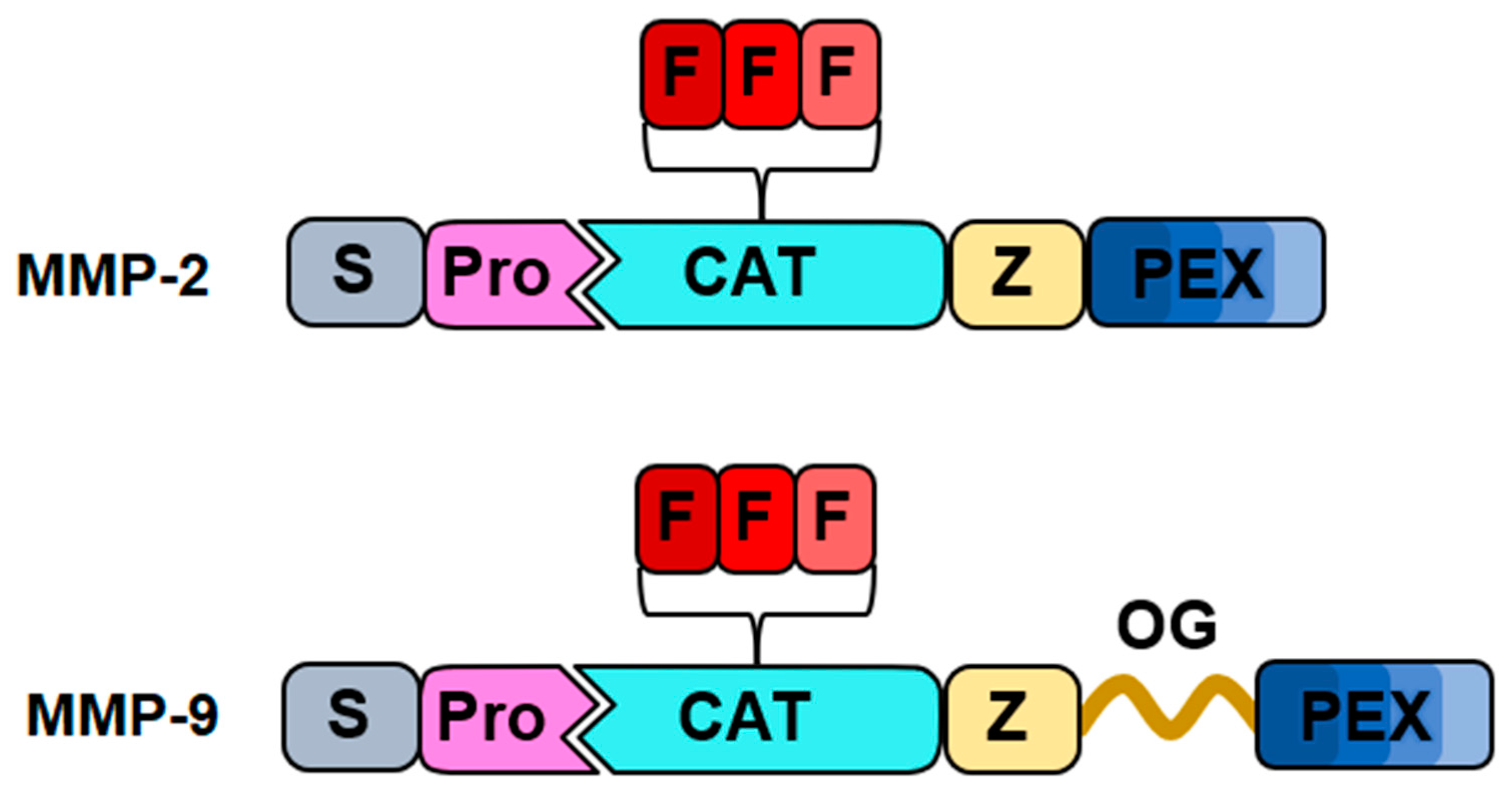
Figure 1. The multidomain structure of MMP-2 and MMP-9. The following domains are shown: the signal peptide (silver), the propeptide (pink), the catalytic domain (cyan), the three fibronectin repeats (red), the metal binding site (beige), the OG domain (brown), and the PEX domain (blue).
Targets 02 00007 g001
Figure 2. Gelatinases facilitate the migration and invasion of tumor cells by degrading the ECM barrier and promoting the remodeling of the surrounding tissue. This allows cancer cells to escape from the primary tumor site and invade adjacent tissues, contributing to the aggressiveness and metastatic potential of fibrosarcoma. (Illustration created with BioRender.com) (https://app.biorender.com accessed on 4 June 2024).
Targets 02 00007 g002
Figure 3. Chemical structures of p-DDAP (1), which was produced based on N-(4-hydroxyphenyl) retinamide (2), a synthetic amide of all-trans-retinoic acid (3). Adapted with permission from [88], Elsevier, 2013.
Targets 02 00007 g003
Figure 4. Retro-synthetic analysis of (I-3,II-3)-biflavones. (compound in blue). Adapted with permission from [89], Elsevier, 2015.
Targets 02 00007 g004
Figure 5. Diagram depicting the process by which AE-COS inhibits the invasion of human fibrosarcoma cells. AE-COS hinders the infiltration of human fibrosarcoma cells by obstructing the activity of MMP-2 and MMP-9. Adapted with permission from [91], Elsevier, 2016.
Targets 02 00007 g005
Figure 6. Chemical structure of salinomycin, LY294002, PD98059, and 4-Methylumbelliferone.
Targets 02 00007 g006
Table 1. List of natural-based compounds explored as gelatinase inhibitors in fibrosarcoma cells.
NameSourceStructureFunctionRef.
EugenolClovesTargets 02 00007 i001Inhibited MMP-9 activities related to metastasis in PMA-stimulated HT-1080 cells[47]
FisetinDalbergia odoriferaTargets 02 00007 i002Inhibited MMP-9 more efficiently than a naturally occurring MMP inhibitor, tetracycline; dose-dependently
inhibits proliferation of fibrosarcoma HT-1080 cells
[48]
QuercetinPolyphenol found abundantly in a variety of foodsTargets 02 00007 i003Inhibited PMS-induced increases in cell motility in HT-1080 cells; attenuated PMS-induced MMP-2 activation[49]
Gallic acid,
delphinidin-3-glucoside
Polyphenols found in a variety of foodsTargets 02 00007 i004Anti-invasive activity; inhibitory activity on secreted and activated gelatinases; inhibited MMP-2 and MMP-9 proteolytic activity[50]
ScutellareinScutellaria laterifloraTargets 02 00007 i005Proliferation rate of cells was significantly suppressed; volume and weight of the tumors were markedly reduced; potently inhibited cell migration, invasion, and the expression and activity of MMP-2 and MMP-9[51]
Galangin, kaempferolFlavonoids Targets 02 00007 i006Efficiently decreased MMP-9 secretion; decreased transcription of MMP-9 mRNA[52]
p- CymenePlantsTargets 02 00007 i007Dose-dependently inhibited the TPA-augmented production and gene expression of MMP-9; enhanced the TPA-augmented production and gene expression of TIMP-1; TPA-augmented invasiveness was inhibited [53]
SE1Hippocampus kuda BleelerTargets 02 00007 i008Potently inhibited gelatin digestion by MMP-9 induced by PMA; inhibited migration of HT-1080 cells in a dose-dependent manner[54]
ChlorophyllinPlants and some algaeTargets 02 00007 i009Cell proliferation was significantly decreased; expression of MMP-2 and MMP-9 decreased; cell invasion through Matrigel and cell migration were also reduced[55]
KahweolCoffeeTargets 02 00007 i010Inhibited cell proliferation enhanced by PMA; attenuated PMA-induced cell migration; suppressed PMA-enhanced activation of MMP-9[56]
LapacholBignoniaceaeTargets 02 00007 i011Inhibited the activation of MMP-2 and MMP-9 stimulated by PMA; protein and gene expression levels of MMP-2 remarkably decreased; increased
the expression level of TIMP-1
[57]
Salidroside, 8(E)-nuezhenide, ligustrosideLigustrum japonicumTargets 02 00007 i012Significantly lowered the amount of MMP-2 and MMP-9 released; RNA and protein expression levels of MMP-2 and MMP-9 were notably suppressed[58]
β-Caryophyllene oxidePiper nigrumTargets 02 00007 i013Reduced MMP-2 and MMP-9 activity; expression level of MMP-2 was reduced; reduced cell invasion [59]
SilibininSilybum marianumTargets 02 00007 i014Showed growth inhibitory effects on fibrosarcoma cells; remarkably inhibited the levels of MMP-2 and MMP-9 activation; inhibited cell invasion on HT-1080 cells[60]
Disclaimer/Publisher’s Note: The statements, opinions and data contained in all publications are solely those of the individual author(s) and contributor(s) and not of MDPI and/or the editor(s). MDPI and/or the editor(s) disclaim responsibility for any injury to people or property resulting from any ideas, methods, instructions or products referred to in the content.