Review

MicroRNA as a Potential Biomarker for Amyotrophic Lateral Sclerosis (ALS)

by
José Augusto Nogueira-Machado
*,
Amanda Tábita da Silva Albenaz
and
Fabiana Rocha-Silva
Programa de Pós-Graduação em Medicina/Biomedicina, Faculdade Santa Casa de Belo Horizonte, Belo Horizonte 30150-240, MG, Brazil
*
Author to whom correspondence should be addressed.
Sclerosis 2025, 3(2), 16; https://doi.org/10.3390/sclerosis3020016
Submission received: 18 January 2025 / Revised: 22 April 2025 / Accepted: 26 April 2025 / Published: 1 May 2025

Abstract

:
Background: Amyotrophic lateral sclerosis (ALS) is a rare, incurable, and fatal neurodegenerative disease that affects the muscles and results in paralysis. The onset and development of ALS involve complex interactions among metabolic signaling, genetic pathways, and external factors (epigenetics). New biomarkers and alternative therapeutic targets have been suggested; nonetheless, the results have been unsatisfactory. Mutations in SOD1, fused in sarcoma (FUS), and TAR DNA-binding protein 43 (TDP-43) have been identified in sporadic amyotrophic lateral sclerosis and approximately 12–20% of familial amyotrophic lateral sclerosis (fALS). Aim: This review analyzes dysregulated microRNA signaling pathways and their interactions with metabolic pathways in the context of ALS progression. Significance: Despite this, biomarkers remain unreliable, and the current medications prolong life without providing a cure. Some proposed approaches to control ALS progression include balancing autophagy and apoptosis, eliminating aggregated proteins, addressing mitochondrial dysfunction, and reducing inflammation. There is a need for studies on new biomarkers, medications, and therapeutic targets. In this context, deregulated circulating microRNAs are attracting attention for new studies on ALS at various phases of the disease. Despite the extensive literature on microRNAs as potential biomarkers for ALS, the proposition for translational clinical use remains limited. Studies have indicated a significant downregulation or upregulation of microRNAs in the motor neurons of ALS patients compared with those with other neurodegenerative disorders and healthy controls. The microRNA biogenesis highlights the importance of this study. MicroRNAs regulate protein synthesis (translation); all human cells express many microRNAs. The complementary structures of microRNA sequences and their mRNA targets allow them to significantly alter cellular and physiological processes. Studies have examined these microRNAs as potential biomarkers for several physiological states and diseases. Comments: The success of these studies may lead to simple, low-cost, and efficient solutions for controlling the progression of ALS and other degenerative diseases. As a result, it is challenging to identify a specific biomarker with total reliability, as a specific microRNA that is increased in one disease phase can decrease in another. These points require careful consideration. They exhibit several complexities and varied interactions, focusing on mRNA targets. The current critical review highlights the potential of microRNAs as biomarkers for diagnosis, prognosis, and therapeutic options in ALS, and raises several points for discussion. Conclusions: The current critical review highlights the potential of microRNAs as biomarkers for diagnosis, prognosis, and therapeutic options in ALS, and raises several points for discussion.

1. Introduction

Amyotrophic lateral sclerosis (ALS) is a rare progressive neurological disorder characterized by the gradual loss of muscle control. The pathophysiological mechanisms behind the development of ALS have been identified as complex and multifactorial. It may be familial (fALS), showing direct transmission from parent to offspring, or sporadic (sALS), resulting from environmental factors that induce epigenetic mutations. The most important symptoms are fatigue and reduced exercise capacity, muscular weakness, respiratory compromise, dyspnea at rest, and dysphagia [1,2]. The gradual decline in respiratory muscle strength can lead to respiratory failure and ultimately, death [3,4,5].
It is important to note that several mutations have been identified in both sALS and fALS. Mutations are commonly found in genes such as SOD1, FUS (fused in sarcoma/translocated in liposarcoma), and TDP-43 (TAR DNA-binding protein 43). These mutations are also present in fALS, with a prevalence ranging from approximately 12% to 20% [6,7]. The consequences of these mutations are directly linked to the progression of the disease [6,7,8,9,10]. For example, the mutated C9ORF72 gene produces harmful dipeptide repeat proteins. In contrast, SOD1, an antioxidant protein, contributes to the formation of aggregated proteins and the eventual death of motor neurons. TDP-43 and FUS are RNA-binding proteins that can aggregate. When FUS and TDP-43 are mutated, they lead to cytoplasmic mislocalization and aggregation, which are associated with the progression of the disease. The mutations observed in sALS are also found in specific subtypes in familial form [11,12,13]. SOD1 and TDP-43 can serve as targets for the movement of toxic, altered proteins between cells through extracellular vesicles (EVs), worsening the disease. Prion-like domains have been identified in both FUS and TDP-43, and evidence suggests that these domains play a crucial role in aggregation [13].
These mutations represent only a small part of our understanding, and due to the complexity of the disease, our knowledge remains limited and we still do not fully understand its many facets. Consequently, medical treatment options for both forms of the disease, including over 30 subtypes of familial ALS, are restricted and largely ineffective. Recent studies have explored several approaches, some focusing on new mutations, which account for just a tiny fraction of our insights into the disease. Other studies have aimed to develop new drugs, new biomarkers and potential therapeutic targets. Furthermore, medical therapies, primarily riluzole and edaravone, have shown limited effectiveness, extending life only briefly without providing a cure. However, ongoing research may soon lead to the testing of new medications. Studies should focus on identifying new, easily quantifiable biomarkers that can aid in clinical diagnosis and potentially serve as therapeutic targets. These biomarkers should be derived from key proteins involved in metabolic signaling and genetic findings. While some proposals for these biomarkers exist, many are still in the early stages of development. Research on biomarkers for amyotrophic lateral sclerosis (ALS) and other neurodegenerative diseases has been ongoing for decades; however, advancements have not yet led to definitive solutions. Currently, neurofilaments (NfLs) are used clinically to indicate neural injury and neurodegeneration, but not all of the proposed biomarkers have demonstrated the same level of efficacy. For ALS, various biomarkers have been suggested. In a comprehensive review by Irwin et al. [14], the authors assessed fluid biomarkers specifically for ALS including cerebrospinal fluid (CSF), plasma, and blood. Some proposed biomarkers include TDP-43, which can be detected in both CSF and plasma. NfL is present in both blood and CSF. Using fluid biomarkers can improve the diagnosis of ALS, facilitating both disease identification and prognostic predictions. This approach is generally straightforward, easy, and fast. microRNAs, among several potential biomarkers, have attracted interest due to a possible association with ALS, and some microRNAs can target predisposed neurons associated with ALS and other neurodegenerative disorders. It has been suggested that a complex interaction involves several elements in the development of the disease. These factors include neuron-specific impairment, cellular stress, proteostasis processes, mitochondrial dysfunction, oxidative damage, compromised growth factor signaling, inflammation (potentially exacerbated by activated astrocytes and microglia), glutamatergic excitotoxicity, advanced age, genetic susceptibility, environmental influences, interactions between motor neurons and muscle or neighboring cells, and protein misfolding. The unique features of microRNAs make them potentially valuable biomarkers as they are highly sensitive, specific, and accurate [9,10,11,12,15,16].
Additionally, compared with other RNA classes, microRNAs demonstrate reliability and stability when quantified from plasma, tears, saliva, or CSF [16]. They also resist extreme conditions such as boiling, freeze–thaw cycles, pH variations, chemical agents, and enzymes [16,17,18]. Reliable biomarker identification could improve ALS diagnostics, facilitating early diagnosis and treatment. Disease biomarkers can aid in medical decision-making by predicting outcomes and monitoring disease progression [19]. Deregulated circulating microRNAs have been found in patients in different disease stages [20,21,22,23]. However, studies of microRNA as potential biomarkers are still scarce. Studies have reported a significant downregulation of miRNAs in motor neurons in patients compared with those with other neurodegenerative diseases and healthy controls [24,25]. The typical delay in diagnosing ALS before the onset of symptoms is approximately one year. Thus, identifying a specific disease biomarker could reduce this period. Reports suggest a possible association between dysregulated functions in the pathophysiological mechanisms of ALS and microRNA levels [26]. Dysregulation of RNA metabolism plays a significant role in the pathogenesis of the disease. In ALS, the processing of miRNAs in both the nucleus and cytoplasm involves TDP-43 and FUS [27]. Several excellent reviews are currently available on miRNAs and ALS. However, this mini-review critically examines the potential of miRNA as a biomarker and therapeutic target, addressing various doubts and uncertainties related to this approach.

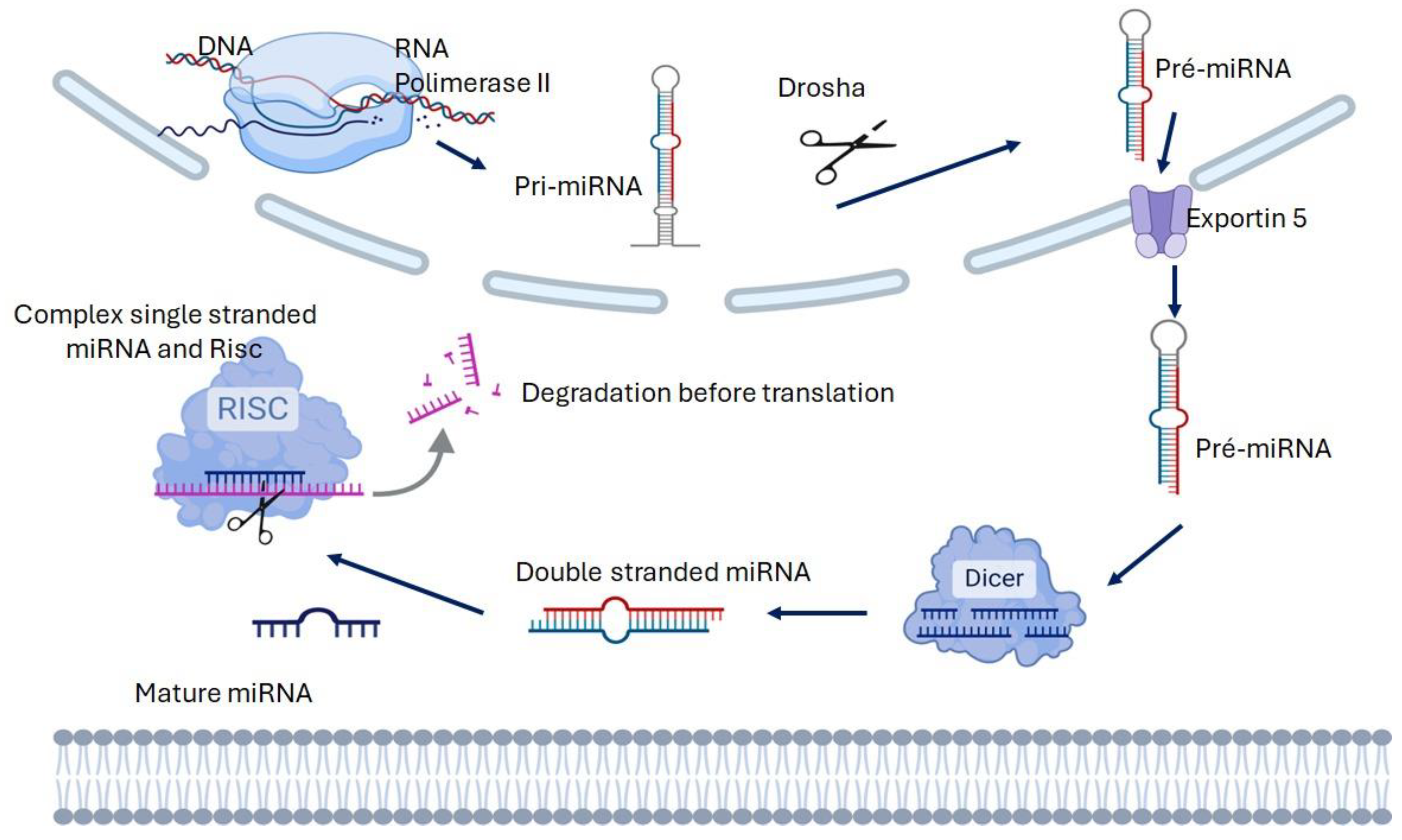
2. Biogenesis and Function of microRNA
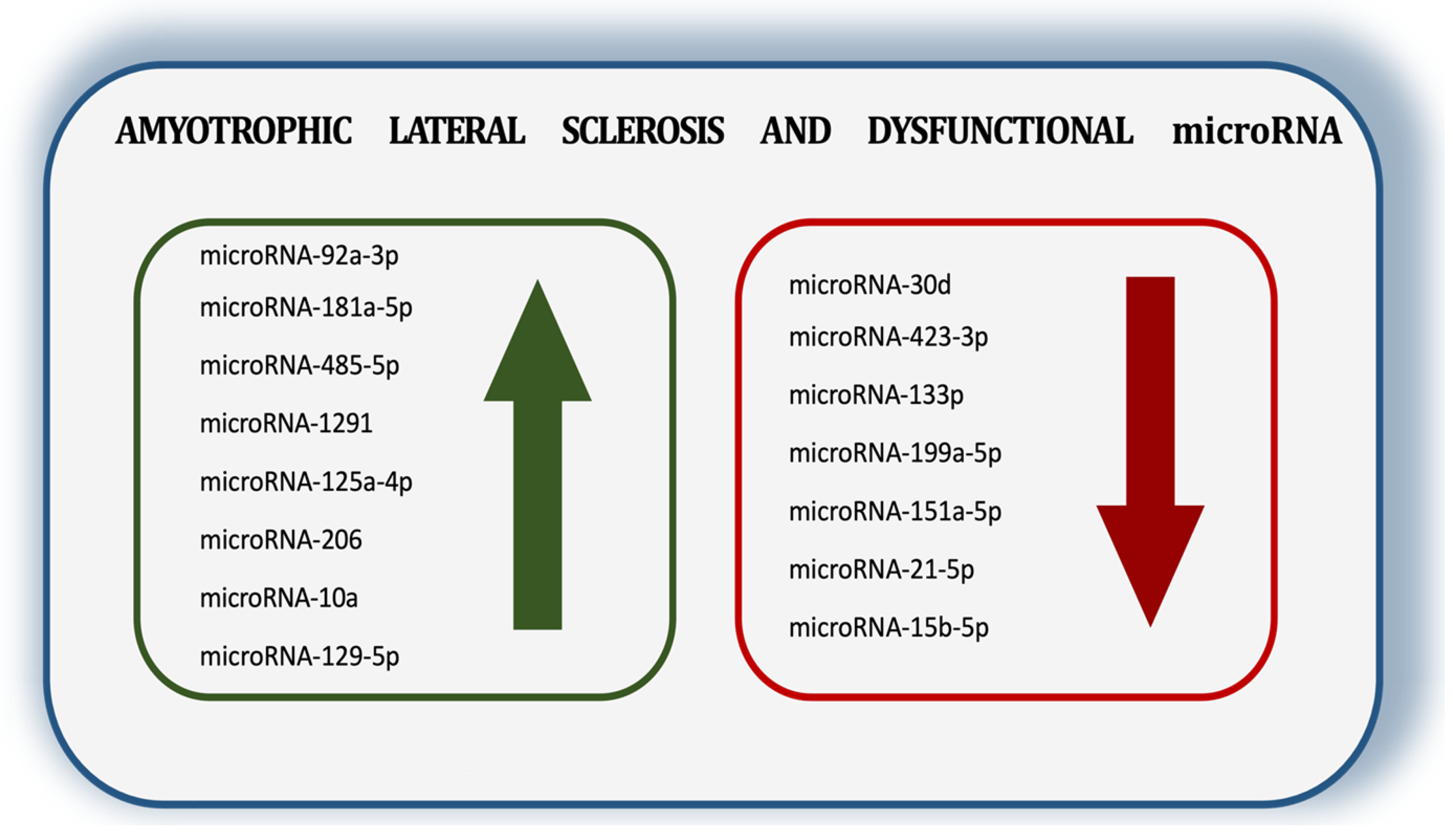
Circulating microRNAs in ALS have been investigated and reported as important biomarkers. They have been found in serum, plasma, CSF fluid, and extracellular vesicles (EVs) [11]. Several studies have focused on the dysregulation of microRNAs, suggesting that they could be a novel resource for identifying biomarkers and therapeutic targets in ALS [18]. Extensive studies on its reliability demonstrate promising prospects for the near future. Small, non-protein-coding RNA molecules (microRNAs) regulate messenger RNAs (mRNAs) by controlling gene expression. They inhibit the translation process by degrading mRNA, thereby preventing the formation of proteins [28]. A single microRNA can regulate multiple mRNAs, maintaining cellular homeostasis [29]. MicroRNAs range in length from 17 to 25 nucleotides and target mRNAs, thereby regulating protein synthesis (translation). All human cells express a significant number of microRNAs. Because the sequences of miRNAs and their mRNA targets are complementary, they can influence cellular functions differently. As a result, these molecules have been investigated as potential biomarkers for various physiological conditions and diseases. Their interactions with targets are complex and multifaceted [30]. The biogenesis process starts in the nucleus, where RNA polymerase II transcribes a long primary microRNA (pri-mRNA) from genomic DNA. After that, Drosha (RNase III) cleaves the pri-microRNA, creating a precursor microRNA (pre-microRNA). Exportin-5 transports it from the nucleus to the cytoplasm. Dicer, another RNase III enzyme, binds to pre-miRNA in the cytoplasm and cleaves it into double-stranded microRNA. The guide strand of the duplex is taken in by the RNA-induced silencing complex (RISC), which produces a mature single-stranded microRNA. The mature single-strand and RISC complexes interact with the 3′ UTR of the target mRNA to regulate gene expression. Conversely, the complementary strand usually breaks down [31,32,33,34] (Figure 1).
MicroRNAs promote intercellular communication [35,36,37,38,39]. Around 90% of circulating miRNAs are believed to interact with proteins or lipoproteins. The remaining 10% is secreted as microvesicles (exosomes), which are formed by the fusion of the plasma membrane [40]. When associated with lipoproteins or inside exosomes, microRNAs are protected from RNase degradation, thereby enhancing their stability. The mechanisms that underlie the processing of miRNAs in exosomes remain poorly understood. Some microRNA sequences can dictate their incorporation into exosomes. Exosomal microRNAs can potentially suppress gene expression at other locations through a mechanism similar to that of intracellular microRNAs. Extracellular microRNAs may act as ligands that regulate immune system activity by activating or suppressing it. In humans, more than 2000 microRNAs regulate around 60 percent of human genes [41]. Some of them can be transferred between cells and across tissues [42,43,44,45]. Studies have identified endogenous and extracellular microRNAs as important regulators of intracellular processes. Dietary (vegetable and animal) products can transfer microRNAs between species [46,47]. Studies have shown that people may absorb exosomes from their food, which carry microRNA-carrying vesicles to different peripheral tissues. Studies have reported the cross-species transfer of microRNA involving both eukaryotes and prokaryotes [43,48,49]. For instance, miRNA-21 and microRNA-29a can affect immune cells through exosomes, which bind to and activate TLR8, thereby activating the NF-κB signaling pathway [41].

3. ALS and microRNA

ALS is associated with more than 40 distinct genes and potentially 100 mutations. The genes most commonly impacted include those located on chromosome 9 open reading frame 72 (C9orf72), superoxide dismutase 1 (SOD1), TAR DNA-binding protein (TARDBP), and fused in sarcoma (FUS) [42,43,44,45,46,50,51,52,53,54]. Identifying specific microRNAs associated with regulating the translation phase for C9orf72, SOD1, TARDBP, or fused in sarcoma (FUS) would provide a promising biomarker and an additional tool for diagnosing sporadic ALS. However, dealing with familial forms, which include over 30 subtypes, will be a challenging task. Several studies suggest that microRNAs are dysregulated, reinforcing their potential as important biomarkers and therapeutic targets for ALS. Some of them are downregulated, such as microRNA-335-5p in autophagy and microRNA-183-5p in apoptosis, while microRNA-129-5p and microRNA-142-3p are upregulated in neuronal differentiation, neuromuscular junction, and neuroinflammation, respectively [47,48,49,50,51,52,55,56,57,58,59]. In an excellent review, Nguyen et al. demonstrate that multiple microRNAs are imbalanced in ALS, thereby disrupting synapse and neuromuscular junction function, neurofilament dynamics, neurogenesis, and RNA/protein metabolism [58]. In ALS, an imbalance of miRNAs affects several aspects of pathophysiology including the neuromuscular junction, neurogenesis, and RNA/protein metabolism [47,48,49,50,51,52,53,54]. Studies have shown that serum from patients with ALS contains decreased levels of specific microRNAs, namely miRNA-335-5p, microRNA-183-5p, and microRNA-218. In particular, microRNA-335-5p is known to target caspases 3 and 7, which play a role in regulating apoptosis. This reduction in microRNA-335-5p could influence the balance between autophagy and apoptosis, potentially impacting the disease. Transfection of microRNA-183-5p protects against cell death, while its inhibition enhances cell death. microRNA-183-5p affects apoptosis and necroptosis [48,49,50,51,52,53,54,55]. Tissue-specific microRNAs, usually confined to their expression tissue, may be released into the circulatory system under pathological conditions. As a result, there is growing interest in determining their role in the pathogenesis of various degenerative diseases, intending to develop minimally invasive and low-cost disease biomarkers [11]. Recent studies have identified potential circulating microRNAs, specifically microRNA-4649 5p, microRNA-424, microRNA-133a, and microRNA-206, as serum biomarkers for diagnosing ALS [56,57,58,59,60,61,62,63,64,65,66,67]. Dobrowolny et al measured the microRNAs as the disease progressed and found that patients with ALS with greater levels of microRNA-206, microRNA-133a, and microRNA-151a-5p experienced slower clinical aggravation [64]. More specifically, during the mild stage of ALS disease, the serum levels of microRNA-206 and microRNA-151a-5p increased, but during both the mild and severe phases, they decreased. However, the levels of microRNA-133a and microRNA-199a-5p remained low throughout the entire disease duration. On the other hand, the disease downregulated microRNA-423-3p and microRNA-151a-5p in the mild and terminal stages, respectively. ALS pathology appears to originate at a single site and spread prion-like via EVs that transport cargo molecules (misfolded and aggregated proteins and microRNAs) related to ALS [66]. Thus, studying microRNAs in serum and other biological fluids could be an important tool for evaluating biomarkers for the disease and its progression stages [68]. Lo et al. proposed a comparison of microRNAs from the extracellular vesicle-derived cargo in ALS between the patients and controls [65]. The ALS frontal cortex and spinal cord showed a decrease in microRNAs, while the serum levels increased. MicroRNA-342-3p was detected to be upregulated, while microRNA-1254 was downregulated. Furthermore, different tissues exhibited overlapping microRNAs including microRNA-587, microRNA-298, microRNA-4443, and microRNA-450a-3p [69]. The authors suggest the role of microRNA cargo from circulating EVs in the pathogenesis of ALS and demonstrate the potential of EVs as biomarkers for ALS. MicroRNA-206, a muscle-specific microRNA, reinforces the role of muscle in affecting disease progression in ALS, and was significantly higher in fast-progressing ALS than in slow-progressing ALS [70]. Patients with ALS showed a downregulation of microRNA-183-5p. Interestingly, the early symptomatic stages of ALS correlated with an increase in microRNA-183-5p in response to stress in motor neurons. Conversely, during the late-symptomatic stage, a decrease in microRNA-183-5p was observed, which was associated with the degeneration and death of motor neurons [71,72]. Therefore, either its downregulation or its increase could indicate distinct pathophysiologic roles, making the interpretation of quantification data more complex. Furthermore, it is essential to recognize that microRNAs can concurrently regulate multiple pathways and interact with genes beyond the disease context, influencing important mechanisms and affecting signaling. As an important site for disease onset/progression, muscle tissue needs to be considered. Changes in muscle function and intrinsic alterations in myocytes can be observed in motor neuron diseases, such as ALS [9,10,11,12,13,14,15,16,17,18,19,72,73,74,75], prior to the development of muscle dysfunction. ALS is more associated with neuromuscular dysfunction than with the degeneration of motor neurons and death. Neuromuscular junction and muscle dysfunction have been observed in the early stages of the disease in both human and mouse models before motor neuron death. This suggests that this may be a key factor in the development of ALS in the G93A-SOD1 animal model [17,18,72,74]. Muscle loss and weakness are among the first symptoms observed. This supports the idea that skeletal muscle plays a pivotal role in ALS pathology and is not merely a consequence of motor neuron dysfunction [73,75,76]. The dysregulation of myomicroRNAs and their targets in ALS, SMA, and SBMA mouse models indicates an interconnected pathogenic component associated with muscle dysfunction. Patients’ sera exhibit a similar myomicroRNA fingerprint. Specifically, both murine muscle and human serum increase microRNA-206 [77]. Nonetheless, the upregulation of microRNA-206 is correlated with a diminished rate of clinical deterioration [68]. Is microRNA-206 protective of muscle tissue? Casado et al. conducted a meta-analysis on ALS, focusing on the role of microRNAs in various body fluids. They found that microRNAs in the serum, blood, CSF, plasma, and neural-enriched extracellular vesicles were consistently dysregulated. The study identified key dysregulated homo sapiens (hsa) microRNAs such as hsa-microRNA-206, hsa-microRNA-338-3p, hsa-microRNA-146a-5p, and hsa-microRNA-151a-5p, all of which showed elevated levels in ALS cases, while hsa-microRNA-183-5p, hsa-microRNA-10b-5p, hsa-microRNA-29b-3p, and hsa-microRNA-4454 showed downregulation [76]. However, the functions of these microRNAs are complex. They may have different roles in specific tissues; hsa-microRNA-206 is notably linked to muscle function in ALS [78,79], an important approach for ALS symptoms and progression studies. Casado et al. conducted a meta-analysis on amyotrophic lateral sclerosis, focusing on the role of microRNAs in various body fluids and found that microRNAs in the serum, blood, CSF, plasma, and neural-enriched extracellular vesicles were consistently dysregulated [76]. Key dysregulated microRNAs identified included hsa-microRNA-206, hsa-microRNA-338-3p, hsa-microRNA-146a-5p, and hsa-microRNA-151a-5p, which were elevated in ALS cases, whereas hsa-microRNA-183-5p, hsa-microRNA-10b-5p, hsa-microRNA-29b-3p, and hsa-microRNA-4454 were downregulated. An exhaustive study proposed eight microRNAs as potential biomarkers for ALS. The miRNAs under consideration were microRNA-10b-5p, microRNA-4454, microRNA-199a-3p, microRNA-151a-3p, microRNA-151-5p, microRNA-199a-5p, microRNA-146-5p, and microRNA-29b-3p (Figure 2). Their sensitivity and specificity were approximately 96% and 97%, respectively, meeting the FDA approval criteria. These criteria include four main requirements: (i) an evaluation of needs; (ii) a reason for use; (iii) a benefit/risk evaluation; and (iv) data confirming eligibility. In this study, the authors utilized EVs derived from the plasma of ALS patients.
The functions of these microRNAs are complex, as they may have different roles in specific tissues, with hsa-microRNA-206 notably linked to muscle function in ALS. Gascón et al. (2024) applied computational and bioinformatics analyses to identify 12 microRNAs connected to upregulated genes and five microRNAs associated with downregulated genes [77]. The functions of certain microRNAs in these groups include hsa-microRNA-100-5p, which has been associated with neuronal apoptosis in the central nervous system and the neurodegeneration of motor neuron G376D-TARDBP, in conjunction with hsa-microRNA-133a-3p, is considered a potential preclinical biomarker for ALS progression. Additionally, hsa-microRNA-206 and hsa-microRNA-133b have been identified as promising biomarkers for ALS [80,81,82,83,84,85]. Both the TDP-43 and C9orf72 genes seem to be targets of miR-142-3p in ALS. Additionally, miR-142-3p is linked to microglial activation and neuroinflammation. According to clinical studies, microRNA-142-3p is elevated in patients with mutations in SOD1986R and TDP-43A315T. In contrast, miRNA-200c-3p and miR-10a-3p are downregulated [83]. Musaro et al. suggest that patients with ALS who have symptoms of muscle weakness present an increase in miR423-3p and a decrease in microRNA-206 [84]. This may indicate that the levels of microRNA-206, a typical muscle-specific microRNA, reflect the role of muscle in controlling the progression of ALS. The results on the microRNA levels are sometimes contradictory because they can vary depending on the type of liquid used for quantification, the disease phase, and other parameters. As a result, it is very challenging to identify a specific biomarker with absolute certainty. A specific microRNA that is increased in one disease phase can decrease in another, so these points require careful consideration. Maity and Kaundal, studying dysregulated microRNA, observed the upregulation of hsa-microRNA-206, hsa-microRNA-133b, and hsa-miR-23a, along with the downregulation of hsa-miRNA-338-3p, hsa-miRNA-218, hsa-microRNA-21-5p, and hsa-let-7b-5p in ALS patients [85]. These microRNAs play an important role in ALS progression. The complexity of microRNAs can be seen in their role in regulating gene expression at post-transcriptional stages, inhibiting translation, and promoting the degradation of molecules. Moreover, one or several microRNAs can control, interfere with, or orchestrate the expression of one gene or an intricate network of genes. Thus, microRNAs can participate in the development of several diseases, including cancer and neurodegenerative diseases, and interfere with various metabolic signaling pathways. In this way, microRNAs are among the most extensively studied non-coding RNAs and are being thoroughly investigated as potential biomarkers for diagnosis, prognosis, severity assessment, prediction, and monitoring responses to treatments. Its role in understanding the mechanism and control of ALS is essential. Both sALS and fALS often exhibit mutations in certain genes that likely disrupt microRNAs. The dysregulation of these miRNAs is recognized as a potential biomarker for the disease and its stages. Additionally, these genes are involved in various pathways and processes that contribute to the onset and progression of the disease. Thus, studying them is essential for clarifying and discovering new ways to research ALS and other neurodegenerative disease conditions. In and of itself, ALS is a complex disease with many manifestations and an intricate, unknown pathophysiology. Several non-coding RNAs, particularly microRNAs, and metabolic pathways are involved. The microRNA levels may change as the disease progresses, and the dynamic molecular environment of ALS shows alterations over time and across different phases. This complicates the approach, even though it presents a valuable opportunity to discover biomarkers. It has been suggested that several microRNAs might regulate the neuronal susceptibility associated with ALS and also possibly influence muscle alterations. Muscle weakness is the main symptom of ALS, and it makes sense to consider muscle metabolic lesions in addition to neuronal factors, particularly in neuronal plaques. It is expected that a disparity between studies is a concern in microRNA research, which can be attributed mainly to liquids, tissues—whether they are from EVs or not—the methods for isolation, sample size, and research focus, resulting in a multiplicity of details that can alter the results between studies [85]. These details regarding microRNA isolation are crucial. As a result, several manuscripts have led to different conclusions regarding microRNA as biomarkers for ALS. Despite these challenges, this remains the most effective method for identifying a non-invasive biomarker that can help clinicians initiate treatment promptly. The eight microRNAs proposed by Banack et al. [75] affect several signaling pathways associated with oxidative stress, cell viability, motor neuron loss, synaptic transmission, neuronal regeneration, neural inflammation, and other processes. Furthermore, it is important to consider that miRNAs can have multiple and differing targets, which indicates their complexity and the need for further studies.
It is worth noting that a single microRNA exhibits contradictory effects in different systems, implying that microRNA interactions depend on the pathology and the site of action. Another relevant point is that most studies have examined microRNA levels without considering their specific functions in particular diseases. This complicates the study of microRNAs, necessitating additional research to confirm their validity as reliable biomarkers or therapeutic resources for ALS. Despite the difficulties and lack of definitive functional analyses in ALS, this approach appears highly beneficial and promising.

4. Conclusions

The main symptom of amyotrophic lateral sclerosis, an incurable disease, is muscle weakness, which progresses to respiratory muscle paralysis, and eventually to death. It suggests a problem involving neurons and muscles. There is no cure for ALS, and most medications, including riluzole and edaravone, only extend life for a short time. The sporadic form of ALS has received the most attention, however, the familial form, in which the genetic heritage is identified, has over 30 subtypes, each defined by a mutation in a determining gene. Some fALS cases have mutations in SOD1, FUS, and other genes also found in sALS. However, not all fALS subtypes, within a spectrum of over 30 subtypes, contain these mutations. Research on signaling and identifying new therapeutic targets is extremely difficult because the target for one form may not be the same for all subtypes. ALS requires extensive research into genetics, metabolic signaling, environmental factors, and epigenetics. Many questions remain unanswered. However, there is hope in the potential of CRISPR and stem cells. Successful embryo selection has been performed; however, this latter option only affects the next generation and does not directly address the disease. Due to the cost, this last option may not be accessible to all individuals Nevertheless, all options represent a promising direction for future research. The first gene associated with familial amyotrophic lateral sclerosis (fALS) was identified in 1993. Each subtype of fALS is designated with a number (e.g., ALS1, ALS2) assigned sequentially. Initially, 23 subtypes were documented based on the classification established by Online Mendelian Inheritance in Man (OMIM). More than thirty genes and loci have been linked to fALS. At present, fALS includes autosomal dominant, autosomal recessive, X-linked (XL), and subtypes associated with frontotemporal dementia (FTD). The solution for sALS will undoubtedly differ from those for fALS, ALS1, ALS2, ALS3, ALS8, and several other subtypes because there are more than 30 additional subtypes of fALS aside from sALS. The common symptom among them is muscular dysfunction. A faster way to define new therapeutic targets or even a therapeutic solution, although no less complex, would be the identification of microRNAs and their effects on the mRNA of mutated proteins. However, identifying one or a group of microRNAs and their interconnections with signaling is quite challenging. Likewise, considering our current scientific knowledge, a one-size-fits-all solution for ALS is not feasible, as it would be impossible to extend it to all forms and subtypes of ALS. As some authors have proposed, viewing ALS as a dysfunctional neuromuscular process could help identify microRNAs that correct neuronal and muscular dysfunctions.

Author Contributions

J.A.N.-M. wrote, reviewed, and edited the manuscript; F.R.-S. and A.T.d.S.A. drew the figures and participated in the revisions of the manuscript. All authors have read and agreed to the published version of the manuscript.

Funding

The authors thank FAPEMIG (Fundação de Amparo à Pesquisa do Estado de Minas Gerais) for providing financial support for the research, with process numbers APQ 03767-23 and 000097-21.

Conflicts of Interest

The authors declare no conflicts of interest, financial or otherwise.

References

  1. Pasinelli, P.; Brown, R.H. Molecular biology of amyotrophic lateral sclerosis: Insights from genetics. Nat. Rev. Neurosci. 2006, 7, 710–723. [Google Scholar] [CrossRef] [PubMed]
  2. Neusch, C.; Bähr, M.; Schneider-Gold, C. Glia cells in amyotrophic lateral sclerosis: New clues to understanding an old disease? Muscle Nerve. 2007, 35, 712–724. [Google Scholar] [CrossRef] [PubMed]
  3. Stambler, N.; Charatan, M.; Cedarbaum, J.M. Prognostic indicators of survival in ALS. Neurology 1998, 50, 66–72. [Google Scholar] [CrossRef] [PubMed]
  4. Ramirez, C.; Piemonte, M.E.P.; Callegaro, D.; Da Silva, H.C. Fatigue in amyotrophic lateral sclerosis: Frequency and associated factors. Amyotroph. Lateral Scler. 2008, 9, 75–80. [Google Scholar] [CrossRef]
  5. Leigh, P.N.; Abrahams, S.; Al-Chalabi, A.; Ampong, M.A.; Goldstein, L.H.; Johnson, J.; Lyall, R.; Moxham, J.; Mustfa, N.; Rio, A.; et al. The management of motor neurone disease. J. Neurol. Neurosurg. Psychiatry 2003, 74, S32–S47. [Google Scholar] [CrossRef]
  6. Mejzini, R.; Flynn, L.L.; Pitout, I.L.; Fletcher, S.; Wilton, S.D.; Akkari, P.A. ALS Genetics, Mechanisms, and Therapeutics: Where Are We Now? Front Neurosci. 2019, 6, 1310. [Google Scholar] [CrossRef]
  7. Chiò, A.; Logroscino, G.; Traynor, B.J.; Collins, J.; Simeone, J.C.; Goldstein, L.A.; White, L.A. Global epidemiology of amyotrophic lateral sclerosis: A systematic review of the published literature. Neuroepidemiology 2013, 41, 118–130. [Google Scholar] [CrossRef]
  8. Talbott, E.O.; Malek, A.M.; Lacomis, D. The epidemiology of amyotrophic lateral sclerosis. Handb. Clin. Neurol. 2016, 138, 225–238. [Google Scholar]
  9. Peggion, C.; Scalcon, V.; Massimino, M.L.; Nies, K.; Lopreiato, R.; Rigobello, M.P.; Bertoli, A. SOD1 in ALS: Taking Stock in Pathogenic Mechanisms and the Role of Glial and Muscle Cells. Antioxidants 2022, 23, 614. [Google Scholar] [CrossRef]
  10. Balendra, R.; Isaacs, A.M. C9orf72-mediated ALS and FTD: Multiple pathways to disease. Nat. Rev. Neurol. 2018, 14, 544–558. [Google Scholar] [CrossRef]
  11. Banack, S.A.; Dunlop, R.A.; Cox, P.A. An miRNA fingerprint using neural-enriched extracellular vesicles from blood plasma: Towards a biomarker for amyotrophic lateral sclerosis/motor neuron disease. Open Biol. 2020, 10, 200116. [Google Scholar] [CrossRef] [PubMed]
  12. Hardiman, O.; Al-Chalabi, A.; Chio, A.; Corr, E.M.; Logroscino, G.; Robberecht, W.; Shaw, P.J.; Simmons, Z.; van den Berg, L.H. Amyotrophic lateral sclerosis. Nat. Rev. Dis. Primers. 2017, 5, 17071. [Google Scholar] [CrossRef] [PubMed]
  13. Gitler, A.D.; Shorter, J. RNA-binding proteins with prion-like domains in ALS and FTLD-U. Prion 2011, 5, 179–187. [Google Scholar] [CrossRef] [PubMed]
  14. Mitchell, P.S.; Parkin, R.K.; Kroh, E.M.; Fritz, B.R.; Wyman, S.K.; Pogosova-Agadjanyan, E.L.; Peterson, A.; Noteboom, J.; O’Briant, K.C.; Allen, A.; et al. Circulating microRNAs as stable blood-based markers for cancer detection. Proc. Natl. Acad. Sci. USA 2008, 105, 10513–10518. [Google Scholar] [CrossRef]
  15. Cortez, M.A.; Bueso-Ramos, C.; Ferdin, J.; Lopez-Berestein, G.; Sood, A.K.; Calin, G.A. MicroRNAs in body fluids-the mix of hormones and biomarkers. Nat. Rev. Clin. Oncol. 2011, 8, 467–477. [Google Scholar] [CrossRef]
  16. Mo, M.H.; Chen, L.; Fu, Y.; Wang, W.; Fu, S.W. Cell-free circulating miRNA biomarkers in cancer. J. Cancer 2012, 3, 432–448. [Google Scholar] [CrossRef]
  17. Mayeux, R. Biomarkers: Potential uses and limitations. NeuroRx 2004, 1, 182–188. [Google Scholar] [CrossRef]
  18. Kmetzsch, V.; Latouche, M.; Saracino, D.; Rinaldi, D.; Camuzat, A.; Gareau, T.; Le Ber, I.; Colliot, O.; Becker, E. MicroRNA signatures in genetic frontotemporal dementia and amyotrophic lateral sclerosis. Ann. Clin. Transl. Neurol. 2022, 9, 1778–1791. [Google Scholar] [CrossRef]
  19. Gentile, G.; Morello, G.; La Cognata, V.; Guarnaccia, M.; Conforti, F.L.; Cavallaro, S. Dysregulated miRNAs as Biomarkers and Therapeutical Targets in Neurodegenerative Diseases. J. Pers. Med. 2022, 10, 770. [Google Scholar] [CrossRef]
  20. Panio, A.; Cava, C.; D’Antona, S.; Bertoli, G.; Porro, D. Diagnostic Circulating miRNAs in Sporadic Amyotrophic Lateral Sclerosis. Front. Med. 2022, 6, 861960. [Google Scholar] [CrossRef]
  21. Taylor, J.P.; Brown, R.H., Jr.; Cleveland, D.W. Decoding ALS: From genes to mechanism. Nature. 2016, 10, 197–206. [Google Scholar] [CrossRef] [PubMed]
  22. Rinchetti, P.; Rizzuti, M.; Faravelli, I.; Corti, S. MicroRNA Metabolism and Dysregulation in Amyotrophic Lateral Sclerosis. Mol. Neurobiol. 2018, 55, 2617–2630. [Google Scholar] [CrossRef] [PubMed]
  23. Gomes, B.C.; Peixinho, N.; Pisco, R.; Gromicho, M.; Pronto-Laborinho, A.C.; Rueff, J.; de Carvalho, M.; Rodrigues, A.S. Differential Expression of miRNAs in Amyotrophic Lateral Sclerosis Patients. Mol. Neurobiol. 2023, 60, 7104–7117. [Google Scholar] [CrossRef] [PubMed]
  24. Koike, Y.; Onodera, O. Implications of miRNAs dysregulation in amyotrophic lateral sclerosis: Challenging for clinical applications. Front. Neurosci. 2023, 21, 1131758. [Google Scholar] [CrossRef]
  25. Dharap, A.; Pokrzywa, C.; Murali, S.; Pandi, G.; Vemuganti, R. MicroRNA miR-324-3p induces promoter-mediated expression of RelA gene. PLoS ONE 2013, 8, e79467. [Google Scholar] [CrossRef]
  26. Basak, I.; Patil, K.S.; Alves, G.; Larsen, J.P.; Møller, S.G. microRNAs as neuroregulators, biomarkers and therapeutic agents in neurodegenerative diseases. Cell Mol. Life Sci. 2016, 73, 811–827. [Google Scholar] [CrossRef]
  27. Hu, W.; Coller, J. What comes first: Translational repression or mRNA degradation? The deepening mystery of microRNA function. Cell Res. 2012, 22, 1322–1324. [Google Scholar] [CrossRef]
  28. Cassidy, J.J.; Bernasek, S.M.; Bakker, R.; Giri, R.; Peláez, N.; Eder, B.; Bobrowska, A.; Bagheri, N.; Nunes Amaral, L.A.; Carthew, R.W. Repressive Gene Regulation Synchronizes Development with Cellular Metabolism. Cell 2019, 178, 980–992.e17. [Google Scholar] [CrossRef]
  29. Chen, X.; Xie, D.; Zhao, Q.; You, Z.H. MicroRNAs and complex diseases: From experimental results to computational models. Brief. Bioinform. 2019, 22, 515–539. [Google Scholar] [CrossRef]
  30. Pu, M.; Chen, J.; Tao, Z.; Miao, L.; Qi, X.; Wang, Y.; Ren, J. Regulatory network of miRNA on its target: Coordination between transcriptional and post-transcriptional regulation of gene expression. Cell Mol. Life Sci. 2019, 76, 441–451. [Google Scholar] [CrossRef]
  31. Saliminejad, K.; Khorram Khorshid, H.R.; Soleymani Fard, S.; Ghaffari, S.H. An overview of microRNAs: Biology, functions, therapeutics, and analysis methods. J. Cell Physiol. 2019, 234, 5451–5465. [Google Scholar] [CrossRef] [PubMed]
  32. Arroyo, J.D.; Chevillet, J.R.; Kroh, E.M.; Ruf, I.K.; Pritchard, C.C.; Gibson, D.F.; Mitchell, P.S.; Bennett, C.F.; Pogosova-Agadjanyan, E.L.; Stirewalt, D.L.; et al. Argonaute2 complexes carry a population of circulating microRNAs independent of vesicles in human plasma. Proc. Natl. Acad. Sci. USA 2011, 22, 5003–5008. [Google Scholar] [CrossRef] [PubMed]
  33. Vickers, K.C.; Palmisano, B.T.; Shoucri, B.M.; Shamburek, R.D.; Remaley, A.T. MicroRNAs are transported in plasma and delivered to recipient cells by high-density lipoproteins. Nat. Cell Biol. 2011, 13, 423–433. [Google Scholar] [CrossRef] [PubMed]
  34. Théry, C. Exosomes: Secreted vesicles and intercellular communications. F1000 Biol. 2011, 3, 15. [Google Scholar] [CrossRef]
  35. Kogure, T.; Lin, W.L.; Yan, I.K.; Braconi, C.; Patel, T. Intercellular nanovesicle-mediated microRNA transfer: A mechanism of environmental modulation of hepatocellular cancer cell growth. Hepatology 2011, 54, 1237–1248. [Google Scholar] [CrossRef]
  36. Weber, J.A.; Baxter, D.H.; Zhang, S.; Huang, D.Y.; Huang, K.H.; Lee, M.J.; Galas, D.J.; Wang, K. The microRNA spectrum in 12 body fluids. Clin. Chem. 2010, 56, 1733–1741. [Google Scholar] [CrossRef]
  37. Théry, C.; Zitvogel, L.; Amigorena, S. Exosomes: Composition, biogenesis and function. Nat. Rev. Immunol. 2002, 2, 569–579. [Google Scholar] [CrossRef]
  38. Friedman, R.C.; Farh, K.K.; Burge, C.B.; Bartel, D.P. Most mammalian mRNAs are conserved targets of microRNAs. Genome Res. 2009, 19, 92–105. [Google Scholar] [CrossRef]
  39. Zhou, G.; Zhou, Y.; Chen, X. New insight into inter-kingdom communication: Horizontal transfer of mobile small RNAs. Front. Microbiol. 2017, 8, 768. [Google Scholar] [CrossRef]
  40. Simons, M.; Raposo, G. Exosomes–vesicular carriers for intercellular communication. Curr. Opin. Cell Biol. 2009, 21, 575–581. [Google Scholar] [CrossRef]
  41. Vickers, K.C.; Remaley, A.T. Lipid-based carriers of microRNAs and intercellular communication. Curr. Opin. Lipidol. 2012, 23, 91–97. [Google Scholar] [CrossRef] [PubMed]
  42. Zhang, L.; Hou, D.; Chen, X.; Li, D.; Zhu, L.; Zhang, Y.; Li, J.; Bian, Z.; Liang, X.; Cai, X.; et al. Exogenous plant MIR168a specifically targets mammalian LDLRAP1: Evidence of cross-kingdom regulation by microRNA. Cell Res. 2012, 22, 107–126. [Google Scholar] [CrossRef]
  43. Baier, S.R.; Nguyen, C.; Xie, F.; Wood, J.R.; Zempleni, J. MicroRNAs are absorbed in biologically meaningful amounts from nutritionally relevant doses of cow milk and affect gene expression in peripheral blood mononuclear cells, HEK-293 kidney cell cultures, and mouse livers. J. Nutr. 2014, 144, 1495–1500. [Google Scholar] [CrossRef] [PubMed]
  44. Kusuma, R.J.; Manca, S.; Friemel, T.; Sukreet, S.; Nguyen, C.; Zempleni, J. Human vascular endothelial cells transport foreign exosomes from cow’s milk by endocytosis. Am. J. Physiol. Cell Physiol. 2016, 310, C800–C807. [Google Scholar] [CrossRef] [PubMed]
  45. Li, J.; Zhang, Y.; Li, D.; Liu, Y.; Chu, D.; Jiang, X.; Hou, D.; Zen, K.; Zhang, C.Y. Small non-coding RNAs transfer through mammalian placenta and directly regulate fetal gene expression. Protein Cell. 2015, 6, 391–396. [Google Scholar] [CrossRef]
  46. Fabbri, M.; Paone, A.; Calore, F.; Galli, R.; Gaudio, E.; Santhanam, R.; Lovat, F.; Fadda, P.; Mao, C.; Nuovo, G.J.; et al. MicroRNAs bind to Toll-like receptors to induce prometastatic inflammatory response. Proc. Natl. Acad. Sci. USA 2012, 31, E2110–E2116. [Google Scholar] [CrossRef]
  47. Ueno, Y.; Morishima, Y.; Hata, T.; Shindo, A.; Murata, H.; Saito, T.; Nakamura, Y.; Shindo, K. Current progress in microRNA profiling of circulating extracellular vesicles in amyotrophic lateral sclerosis: A systematic review. Neurobiol. Dis. 2024, 200, 106639. [Google Scholar] [CrossRef]
  48. Turner, M.R.; Al-Chalabi, A.; Chio, A.; Hardiman, O.; Kiernan, M.C.; Rohrer, J.; Rowe, J.; Seeley, W.; Talbot, K. Genetic screening in sporadic ALS and FTD. J. Neurol. Neurosurg. Psychiatry 2017, 88, 1042–1044. [Google Scholar] [CrossRef]
  49. Vijayakumar, U.G.; Milla, V.; Cynthia Stafford, M.Y.; Bjourson, A.J.; Duddy, W.; Duguez, S.M.-R. A Systematic Review of Suggested Molecular Strata, Biomarkers and Their Tissue Sources in ALS. Front. Neurol. 2019, 10, 400. [Google Scholar] [CrossRef]
  50. Volk, A.E.; Weishaupt, J.H.; Andersen, P.M.; Ludolph, A.C.; Kubisch, C. Current knowledge and recent insights into the genetic basis of amyotrophic lateral sclerosis. Med. Genet. 2018, 30, 252–258. [Google Scholar] [CrossRef]
  51. Chia, R.; Chiò, A.; Traynor, B.J. Novel genes associated with amyotrophic lateral sclerosis: Diagnostic and clinical implications. Lancet Neurol. 2018, 17, 94–102. [Google Scholar] [CrossRef] [PubMed]
  52. Dilmaghani, N.A.; Hussen, B.M.; Nateghinia, S.; Taheri, M.; Ghafouri-Fard, S. Emerging role of microRNAs in the pathogenesis of amyotrophic lateral sclerosis. Metab. Brain Dis. 2021, 36, 737–749. [Google Scholar] [CrossRef] [PubMed]
  53. De Luna, N.; Turon-Sans, J.; Cortes-Vicente, E.; Carrasco-Rozas, A.; Illán-Gala, I.; Dols-Icardo, O.; Clarimón, J.; Lleó, A.; Gallardo, E.; Illa, I.; et al. Downregulation of miR-335-5P in Amyotrophic Lateral Sclerosis Can Contribute to Neuronal Mitochondrial Dysfunction and Apoptosis. Sci. Rep. 2020, 9, 4308. [Google Scholar] [CrossRef] [PubMed]
  54. Li, C.; Chen, Y.; Chen, X.; Wei, Q.; Ou, R.; Gu, X.; Cao, B.; Shang, H. MicroRNA-183-5p is stress-inducible and protects neurons against cell death in amyotrophic lateral sclerosis. J. Cell. Mol. Med. 2020, 24, 8614–8622. [Google Scholar] [CrossRef]
  55. Reichenstein, I.; Eitan, C.; Diaz-Garcia, S.; Haim, G.; Magen, I.; Siany, A.; Hoye, M.L.; Rivkin, N.; Olender, T.; Toth, B.; et al. Human genetics and neuropathology suggest a link between miR-218 and amyotrophic lateral sclerosis pathophysiology. Sci. Transl. Med. 2019, 11, eaav5264. [Google Scholar] [CrossRef]
  56. Loffreda, A.; Nizzardo, M.; Arosio, A.; Ruepp, M.-D.; Calogero, R.; Volinia, S.; Galasso, M.; Bendotti, C.; Ferrarese, C.; Lunetta, C.; et al. miR-129-5p: A key factor and therapeutic target in amyotrophic lateral sclerosis. Prog. Neurobiol. 2020, 190, 101803. [Google Scholar] [CrossRef]
  57. Matamala, J.M.; Arias-Carrasco, R.; Sanchez, C.; Uhrig, M.; Bargsted, L.; Matus, S.; Maracaja-Coutinho, V.; Abarzua, S.; van Zundert, B.; Verdugo, R.; et al. Genome-wide circulating microRNA expression profiling reveals potential biomarkers for amyotrophic lateral sclerosis. Neurobiol. Aging 2018, 64, 123–138. [Google Scholar] [CrossRef]
  58. Nguyen, T.P.N.; Kumar, M.; Fedele, E.; Bonanno, G.; Bonifacino, T. MicroRNA Alteration, Application as Biomarkers, and Therapeutic Approaches in Neurodegenerative Diseases. Int. J. Mol. Sci. 2022, 25, 4718. [Google Scholar] [CrossRef]
  59. Wang, L.; Zhang, L. MicroRNAs in amyotrophic lateral sclerosis: From pathogenetic involvement to diagnostic biomarker and therapeutic agent development. Neurol. Sci. 2020, 41, 3569–3577. [Google Scholar] [CrossRef]
  60. Cacchiarelli, D.; Legnini, I.; Martone, J.; Cazzella, V.; D’Amico, A.; Bertini, E.; Bozzoni, I. miRNAs as serum biomarkers for Duchenne muscular dystrophy. EMBO Mol. Med. 2011, 3, 258–265. [Google Scholar] [CrossRef]
  61. Kovanda, A.; Leonardis, L.; Zidar, J.; Koritnik, B.; Dolenc-Groselj, L.; Ristic Kovacic, S.; Curk, T.; Rogelj, B. Differential expression of microRNAs and other small RNAs in muscle tissue of patients with ALS and healthy age-matched controls. Sci. Rep. 2018, 4, 5609. [Google Scholar] [CrossRef] [PubMed]
  62. Takahashi, I.; Hama, Y.; Matsushima, M.; Hirotani, M.; Kano, T.; Hohzen, H.; Yabe, I.; Utsumi, J.; Sasaki, H. Identification of plasma microRNAs as a biomarker of sporadic Amyotrophic Lateral Sclerosis. Mol. Brain 2015, 24, 67. [Google Scholar] [CrossRef] [PubMed]
  63. de Andrade, H.M.; de Albuquerque, M.; Avansini, S.H.; de S Rocha, C.; Dogini, D.B.; Nucci, A.; Carvalho, B.; Lopes-Cendes, I.; França, M.C., Jr. MicroRNAs-424 and 206 are potential prognostic markers in spinal onset amyotrophic lateral sclerosis. J. Neurol. Sci. 2016, 15, 19–24. [Google Scholar] [CrossRef] [PubMed]
  64. Dobrowolny, G.; Martone, J.; Lepore, E.; Casola, I.; Petrucci, A.; Inghilleri, M.; Morlando, M.; Colantoni, A.; Scicchitano, B.M.; Calvo, A.; et al. A longitudinal study defined circulating microRNAs as reliable biomarkers for disease prognosis and progression in ALS human patients. Cell Death Discov. 2021, 11, 4. [Google Scholar] [CrossRef]
  65. Lo, T.W.; Figueroa-Romero, C.; Hur, J.; Pacut, C.; Stoll, E.; Spring, C.; Lewis, R.; Nair, A.; Goutman, S.A.; Sakowski, S.A.; et al. Extracellular Vesicles in Serum and Central Nervous System Tissues Contain microRNA Signatures in Sporadic Amyotrophic Lateral Sclerosis. Front. Mol. Neurosci. 2021, 29, 739016. [Google Scholar] [CrossRef]
  66. Chen, Y.; Wei, Q.; Chen, X.; Li, C.; Cao, B.; Ou, R.; Hadano, S.; Shang, H.F. Aberration of miRNAs Expression in Leukocytes from Sporadic Amyotrophic Lateral Sclerosis. Front. Mol. Neurosci. 2016, 17, 69. [Google Scholar] [CrossRef]
  67. Bilsland, L.G.; Sahai, E.; Kelly, G.; Golding, M.; Greensmith, L.; Schiavo, G. Deficits in axonal transport precede ALS symptoms in vivo. Proc. Natl. Acad. Sci. USA 2010, 23, 20523–20528. [Google Scholar] [CrossRef]
  68. Rusmini, P.; Polanco, M.J.; Cristofani, R.; Cicardi, M.E.; Meroni, M.; Galbiati, M.; Piccolella, M.; Messi, E.; Giorgetti, E.; Lieberman, A.P.; et al. Aberrant Autophagic Response in The Muscle of A Knock-in Mouse Model of Spinal and Bulbar Muscular Atrophy. Sci. Rep. 2015, 22, 15174. [Google Scholar] [CrossRef]
  69. Galbiati, M.; Crippa, V.; Rusmini, P.; Cristofani, R.; Cicardi, M.E.; Giorgetti, E.; Onesto, E.; Messi, E.; Poletti, A. ALS-related misfolded protein management in motor neurons and muscle cells. Neurochem. Int. 2014, 79, 70–78. [Google Scholar] [CrossRef]
  70. Marcuzzo, S.; Zucca, I.; Mastropietro, A.; de Rosbo, N.K.; Cavalcante, P.; Tartari, S.; Bonanno, S.; Preite, L.; Mantegazza, R.; Bernasconi, P. Hind limb muscle atrophy precedes cerebral neuronal degeneration in G93A-SOD1 mouse model of amyotrophic lateral sclerosis: A longitudinal MRI study. Exp. Neurol. 2011, 231, 30–37. [Google Scholar] [CrossRef]
  71. Marcuzzo, S.; Bonanno, S.; Figini, M.; Scotti, A.; Zucca, I.; Minati, L.; Riva, N.; Domi, T.; Fossaghi, A.; Quattrini, A.; et al. A longitudinal DTI and histological study of the spinal cord reveals early pathological alterations in G93A-SOD1 mouse model of amyotrophic lateral sclerosis. Exp. Neurol. 2017, 293, 43–52. [Google Scholar] [CrossRef] [PubMed]
  72. Dobrowolny, G.; Aucello, M.; Rizzuto, E.; Beccafico, S.; Mammucari, C.; Bonconpagni, S.; Belia, S.; Wannenes, F.; Nicoletti, C.; Del Prete, Z.; et al. Skeletal Muscle Is a Primary Target of SOD1G93A-Mediated Toxicity. Cell Metab. 2008, 8, 425–436. [Google Scholar] [CrossRef] [PubMed]
  73. Loeffler, J.P.; Picchiarelli, G.; Dupuis, L.; De Aguilar, J.L.G. The Role of Skeletal Muscle in Amyotrophic Lateral Sclerosis. Brain Pathol. 2016, 26, 227–236. [Google Scholar] [CrossRef] [PubMed]
  74. Malacarne, C.; Galbiati, M.; Giagnorio, E.; Cavalcante, P.; Salerno, F.; Andreetta, F.; Cagnoli, C.; Taiana, M.; Nizzardo, M.; Corti, S.; et al. Dysregulation of Muscle-Specific MicroRNAs as Common Pathogenic Feature Associated with Muscle Atrophy in ALS, SMA and SBMA: Evidence from Animal Models and Human Patients. Int. J. Mol. Sci. 2021, 26, 5673. [Google Scholar] [CrossRef]
  75. Banack, S.A.; Dunlop, R.A.; Mehta, P.; Mitsumoto, H.; Wood, S.P.; Han, M.; Cox, P.A. A microRNA diagnostic biomarker for amyotrophic lateral sclerosis. Brain Commun. 2024, 6, fcae268. [Google Scholar] [CrossRef]
  76. Casado Gama, H.; Amorós, M.A.; Andrade de Araújo, M.; Sha, C.M.; Vieira, M.P.S.; Torres, R.G.D.; Souza, G.F.; Junkes, J.A.; Dokholyan, N.V.; Leite Góes Gitaí, D.; et al. Systematic review and meta-analysis of dysregulated microRNAs derived from liquid biopsies as biomarkers for amyotrophic lateral sclerosis. Noncoding RNA Res. 2024, 6, 523–535. [Google Scholar] [CrossRef]
  77. Gascón, E.; Zaragoza, P.; Calvo, A.C.; Osta, R. Sporadic Amyotrophic Lateral Sclerosis Skeletal Muscle Transcriptome Analysis: A Comprehensive Examination of Differentially Expressed Genes. Biomolecules 2024, 20, 377. [Google Scholar] [CrossRef]
  78. Wallach, T.; Mossmann, Z.J.; Szczepek, M.; Wetzel, M.; Machado, R.; Raden, M.; Miladi, M.; Kleinau, G.; Kruger, C.; Dembny, P.; et al. Microrna-100-5p and Microrna-298-5p Released from Apoptotic Cortical Neurons Are Endogenous Toll-Like Receptor 7/8 Ligands That Contribute to Neurodegeneration. Mol. Neurodegener. 2021, 16, 80. [Google Scholar] [CrossRef]
  79. Ruffo, P.; Catalano, S.; La Bella, V.; Conforti, F.L. Deregulation of Plasma Microrna Expression in a Tardbp-Als Family. Biomolecules 2023, 13, 706. [Google Scholar] [CrossRef]
  80. Cheng, J.; Ho, W.K.; Wu, B.T.; Liu, H.P.; Lin, W.Y. Mirna Profiling as a Complementary Diagnostic Tool for Amyotrophic Lateral Sclerosis. Sci. Rep. 2023, 13, 13805. [Google Scholar] [CrossRef]
  81. Toivonen, J.M.; Manzano, R.; Oliván, S.; Zaragoza, P.; García-Redondo, A.; Osta, R. Microrna-206: A Potential Circulating Biomarker Candidate for Amyotrophic Lateral Sclerosis. PLoS ONE 2014, 9, e89065. [Google Scholar] [CrossRef] [PubMed]
  82. Liu, H.; Lan, S.; Shi, X.J.; Fan, F.C.; Liu, Q.S.; Cong, L.; Cheng, Y. Systematic Review and Meta-Analysis on Micrornas in Amyotrophic Lateral Sclerosis. Brain Res. Bull. 2023, 194, 82–89. [Google Scholar] [CrossRef] [PubMed]
  83. Kmetzsch, V.; Anquetil, V.; Saracino, D.; Rinaldi, D.; Camuzat, A.; Gareau, T.; Jornea, L.; Forlani, S.; Couratier, P.; Wallon, D.; et al. Plasma microRNA signature in presymptomatic and symptomatic subjects with C9orf72-associated frontotemporal dementia and amyotrophic lateral sclerosis. J. Neurol. Neurosurg. Psychiatry 2021, 92, 485–493. [Google Scholar] [CrossRef] [PubMed]
  84. Musarò, A.; Dobrowolny, G.; Cambieri, C.; Libonati, L.; Moret, F.; Casola, I.; Laurenzi, G.; Garibaldi, M.; Inghilleri, M.; Ceccanti, M. MiR206 and 423-3p Are Differently Modulated in Fast and Slow-Progressing Amyotrophic Lateral Sclerosis Patients. Neuromolecular Med. 2024, 15, 5. [Google Scholar] [CrossRef]
  85. Maity, D.; Kaundal, R.K. Exploring dysregulated miRNAs in ALS: Implications for disease pathogenesis and early diagnosis. Neurol. Sci. 2024, 21, 1661–1686. [Google Scholar] [CrossRef]
Figure 1. MicroRNA(miRNA) biogenesis. Phases of biogenesis occur in the nucleus with the transcription of pri-RNA by RNA polymerase II. The role of Drosha, an RNase III, is to produce the precursor miRNA (pre-miRNA). Exit from the nucleus occurs via exportin-5 to the cytoplasm, followed by the action of another RNase III enzyme (Dicer), which cleaves it into a double-stranded miRNA. The RNA-induced silencing complex (RISC) produces a mature, single-stranded miRNA that acts on mRNA (messenger RNA), interfering with the translation process. Figure is created in BioRender.com: https://www.biorender.com/ (accessed on 20 December 2024).
Sclerosis 03 00016 g001
Figure 2. Amyotrophic lateral sclerosis and dysfunctional microRNAs. The microRNAs indicated by the green arrow are upregulated in ALS, while those indicated by the red arrow are downregulated in ALS.
Sclerosis 03 00016 g002
Disclaimer/Publisher’s Note: The statements, opinions and data contained in all publications are solely those of the individual author(s) and contributor(s) and not of MDPI and/or the editor(s). MDPI and/or the editor(s) disclaim responsibility for any injury to people or property resulting from any ideas, methods, instructions or products referred to in the content.

