
Harnessing Artificial Intelligence for the Diagnosis, Treatment and Research of Multiple Sclerosis

 ,
, 
Abstract
1. Introduction
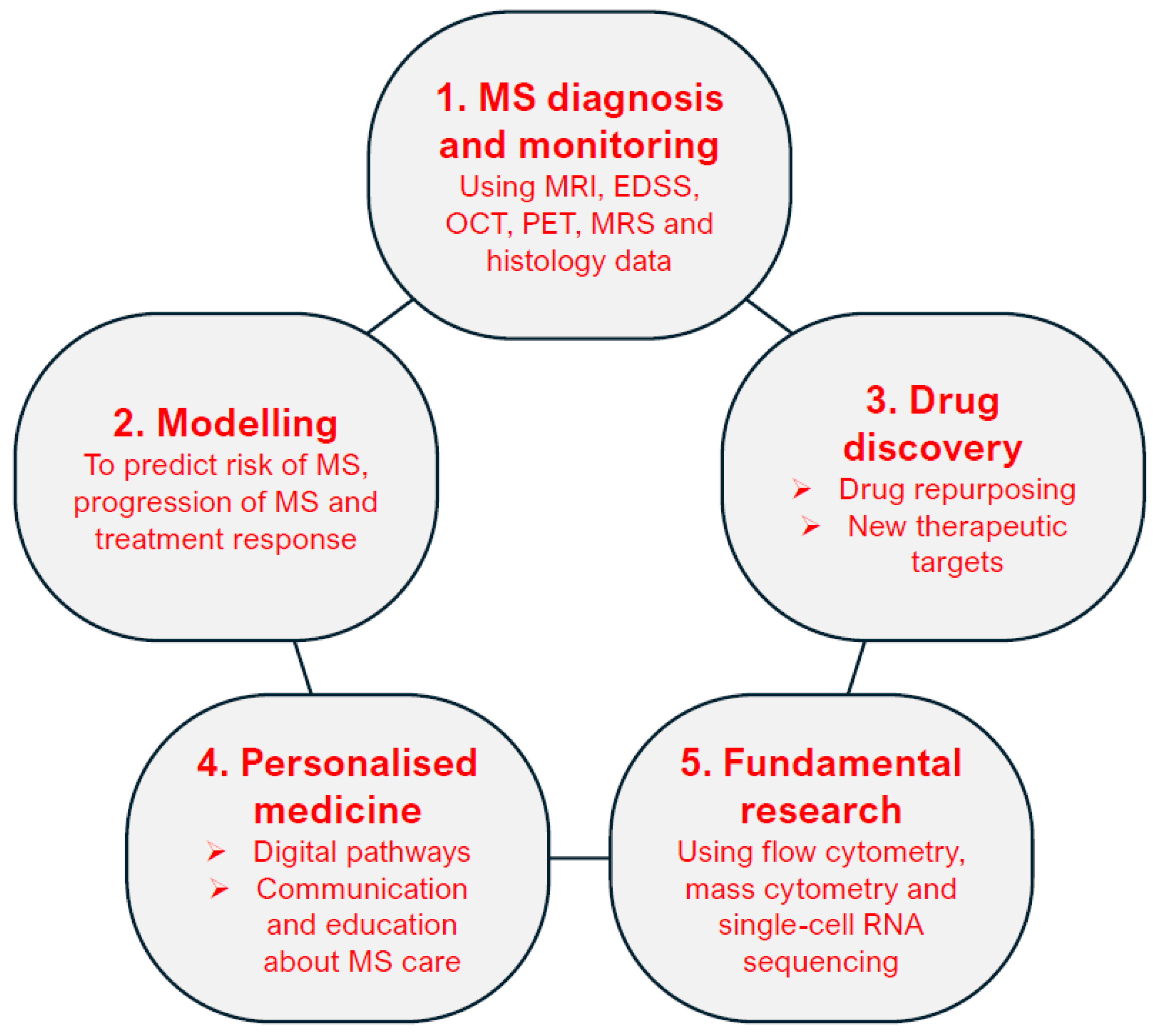
2. AI-Assisted Medical Data Analysis for MS Diagnosis and Monitoring
2.1. AI Analysis of MRI Data
2.2. EDSS, OCT and PET Data Analysis
2.3. Future Directions for MS Diagnosis
3. AI-Modelling to Predict Risk of MS, Progression of MS and Treatment Response
3.1. AI Assists in Diagnostic Assessments and Prevents Diagnostic Delays in MS Diagnosis
3.2. Different ML Techniques Available to Analyse Clinical Datasets for MS
3.3. Combination of ML and Clinicians Enhances the Prediction of MS Progression
3.4. Monitoring Treatment Response with ML Models
3.5. Utilisation of AI in MS Prognosis
3.6. Applications of ML Models to MRI
3.7. Statistical Modelling Improves Prediction of Disability Progression in MS
4. AI-Assisted Drug Discovery
4.1. Deep Learning and ML Methods for Drug Repurposing and New Therapeutic Targets
4.2. Future Directions for Drug Discovery
5. AI-Assisted Personalised Medicine Approach to MS Care
5.1. AI-Assisted Digital Patient Pathways and Digital Twins
5.2. Using AI to Estimate DMT Response
5.3. AI-Facilitated Communication and Education in MS Care
6. AI-Assisted Fundamental Research Using Single-Cell Data
6.1. AI-Assisted Flow Cytometry Analysis
6.2. AI Analysis of Mass Cyotmetry Data
6.3. Single-Cell RNA Sequencing Analysis by AI
6.4. Future Directions for Fundamental Research
7. Discussion
8. Conclusions
Author Contributions
Funding
Conflicts of Interest
References
- Jakimovski, D.; Bittner, S.; Zivadinov, R.; Morrow, S.A.; Benedict, R.H.; Zipp, F.; Weinstock-Guttman, B. Multiple sclerosis. Lancet 2024, 403, 183–202. [Google Scholar] [CrossRef]
- Portaccio, E.; Magyari, M.; Havrdova, E.K.; Ruet, A.; Brochet, B.; Scalfari, A.; Di Filippo, M.; Tur, C.; Montalban, X.; Amato, M.P. Multiple sclerosis: Emerging epidemiological trends and redefining the clinical course. Lancet Reg. Health Eur. 2024, 44, 100977. [Google Scholar] [CrossRef] [PubMed]
- Vargas, D.L.; Tyor, W.R. Update on disease-modifying therapies for multiple sclerosis. J. Investig. Med. 2017, 65, 883–891. [Google Scholar] [CrossRef] [PubMed]
- Collins, C.; Dennehy, D.; Conboy, K.; Mikalef, P. Artificial intelligence in information systems research: A systematic literature review and research agenda. Int. J. Inform. Manag. 2021, 60, 102383. [Google Scholar] [CrossRef]
- Tafti, D.; Ehsan, M.; Xixis, K.L. Multiple Sclerosis. In StatPearls; StatPearls Publishing: Treasure Island, FL, USA, 2025. [Google Scholar]
- Gustavsen, S.; Olsson, A.; Sondergaard, H.B.; Andresen, S.R.; Sorensen, P.S.; Sellebjerg, F.; Oturai, A. The association of selected multiple sclerosis symptoms with disability and quality of life: A large Danish self-report survey. BMC Neurol. 2021, 21, 317. [Google Scholar] [CrossRef]
- Khattap, M.G.; Abd Elaziz, M.; Hassan, H.; Elgarayhi, A.; Sallah, M. AI-based model for automatic identification of multiple sclerosis based on enhanced sea-horse optimizer and MRI scans. Sci. Rep. 2024, 14, 12104. [Google Scholar] [CrossRef] [PubMed]
- Zhang, Y.-D.; Pan, C.; Sun, J.; Tang, C. Multiple sclerosis identification by convolutional neural network with dropout and parametric ReLU. J. Comput. Sci. 2018, 28, 1–10. [Google Scholar] [CrossRef]
- Wang, S.H.; Tang, C.; Sun, J.; Yang, J.; Huang, C.; Phillips, P.; Zhang, Y.D. Multiple Sclerosis Identification by 14-Layer Convolutional Neural Network With Batch Normalization, Dropout, and Stochastic Pooling. Front. Neurosci. 2018, 12, 818. [Google Scholar] [CrossRef]
- Cunill, V.; Massot, M.; Clemente, A.; Calles, C.; Andreu, V.; Núñez, V.; López-Gómez, A.; Díaz, R.M.; Jiménez, M.L.R.; Pons, J.; et al. Relapsing-Remitting Multiple Sclerosis Is Characterized by a T Follicular Cell Pro-Inflammatory Shift, Reverted by Dimethyl Fumarate Treatment. Front. Immunol. 2018, 9, 1097. [Google Scholar] [CrossRef]
- Gabr, R.E.; Coronado, I.; Robinson, M.; Sujit, S.J.; Datta, S.; Sun, X.; Allen, W.J.; Lublin, F.D.; Wolinsky, J.S.; Narayana, P.A. Brain and lesion segmentation in multiple sclerosis using fully convolutional neural networks: A large-scale study. Mult. Scler. 2020, 26, 1217–1226. [Google Scholar] [CrossRef]
- Coronado, I.; Gabr, R.E.; Narayana, P.A. Deep learning segmentation of gadolinium-enhancing lesions in multiple sclerosis. Mult. Scler. 2021, 27, 519–527. [Google Scholar] [CrossRef] [PubMed]
- La Rosa, F.; Beck, E.S.; Maranzano, J.; Todea, R.A.; van Gelderen, P.; de Zwart, J.A.; Luciano, N.J.; Duyn, J.H.; Thiran, J.P.; Granziera, C.; et al. Multiple sclerosis cortical lesion detection with deep learning at ultra-high-field MRI. NMR Biomed. 2022, 35, e4730. [Google Scholar] [CrossRef] [PubMed]
- Gentile, G.; Jenkinson, M.; Griffanti, L.; Luchetti, L.; Leoncini, M.; Inderyas, M.; Mortilla, M.; Cortese, R.; De Stefano, N.; Battaglini, M. BIANCA-MS: An optimized tool for automated multiple sclerosis lesion segmentation. Hum. Brain Mapp. 2023, 44, 4893–4913. [Google Scholar] [CrossRef] [PubMed]
- Valverde, S.; Cabezas, M.; Roura, E.; Gonzalez-Villa, S.; Pareto, D.; Vilanova, J.C.; Ramio-Torrenta, L.; Rovira, A.; Oliver, A.; Llado, X. Improving automated multiple sclerosis lesion segmentation with a cascaded 3D convolutional neural network approach. Neuroimage 2017, 155, 159–168. [Google Scholar] [CrossRef]
- Eshaghi, A.; Young, A.L.; Wijeratne, P.A.; Prados, F.; Arnold, D.L.; Narayanan, S.; Guttmann, C.R.G.; Barkhof, F.; Alexander, D.C.; Thompson, A.J.; et al. Identifying multiple sclerosis subtypes using unsupervised machine learning and MRI data. Nat. Commun. 2021, 12, 2078. [Google Scholar] [CrossRef]
- Rostami, A.; Robatjazi, M.; Dareyni, A.; Ghorbani, A.R.; Ganji, O.; Siyami, M.; Raoofi, A.R. Enhancing classification of active and non-active lesions in multiple sclerosis: Machine learning models and feature selection techniques. BMC Med. Imaging 2024, 24, 345. [Google Scholar] [CrossRef]
- Amini, A.; Shayganfar, A.; Amini, Z.; Ostovar, L.; HajiAhmadi, S.; Chitsaz, N.; Rabbani, M.; Kafieh, R. Deep learning for discrimination of active and inactive lesions in multiple sclerosis using non-contrast FLAIR MRI: A multicenter study. Mult. Scler. Relat. Disord. 2024, 87, 105642. [Google Scholar] [CrossRef]
- Shekari, F.; Vard, A.; Adibi, I.; Danesh-Mobarhan, S. Investigating the feasibility of differentiating MS active lesions from inactive ones using texture analysis and machine learning methods in DWI images. Mult. Scler. Relat. Disord. 2024, 82, 105363. [Google Scholar] [CrossRef] [PubMed]
- Dwyer, M.; Lyman, C.; Ferrari, H.; Bergsland, N.; Fuchs, T.A.; Jakimovski, D.; Schweser, F.; Weinstock-Guttmann, B.; Benedict, R.H.B.; Riolo, J.; et al. DeepGRAI (Deep Gray Rating via Artificial Intelligence): Fast, feasible, and clinically relevant thalamic atrophy measurement on clinical quality T2-FLAIR MRI in multiple sclerosis. Neuroimage Clin. 2021, 30, 102652. [Google Scholar] [CrossRef]
- Peters, S.; Kellermann, G.; Watkinson, J.; Gartner, F.; Huhndorf, M.; Sturner, K.; Jansen, O.; Larsen, N. AI supported detection of cerebral multiple sclerosis lesions decreases radiologic reporting times. Eur. J. Radiol. 2024, 178, 111638. [Google Scholar] [CrossRef]
- Barnett, M.; Wang, D.; Beadnall, H.; Bischof, A.; Brunacci, D.; Butzkueven, H.; Brown, J.W.L.; Cabezas, M.; Das, T.; Dugal, T.; et al. A real-world clinical validation for AI-based MRI monitoring in multiple sclerosis. npj Digit. Med. 2023, 6, 196. [Google Scholar] [CrossRef] [PubMed]
- Roca, P.; Attye, A.; Colas, L.; Tucholka, A.; Rubini, P.; Cackowski, S.; Ding, J.; Budzik, J.F.; Renard, F.; Doyle, S.; et al. Artificial intelligence to predict clinical disability in patients with multiple sclerosis using FLAIR MRI. Diagn. Interv. Imaging 2020, 101, 795–802. [Google Scholar] [CrossRef] [PubMed]
- Optic Neuritis Study, G. Multiple sclerosis risk after optic neuritis: Final optic neuritis treatment trial follow-up. Arch. Neurol. 2008, 65, 727–732. [Google Scholar] [CrossRef]
- Dongil-Moreno, F.J.; Ortiz, M.; Pueyo, A.; Boquete, L.; Sanchez-Morla, E.M.; Jimeno-Huete, D.; Miguel, J.M.; Barea, R.; Vilades, E.; Garcia-Martin, E. Diagnosis of multiple sclerosis using optical coherence tomography supported by explainable artificial intelligence. Eye 2024, 38, 1502–1508. [Google Scholar] [CrossRef]
- Ortiz, M.; Mallen, V.; Boquete, L.; Sanchez-Morla, E.M.; Cordon, B.; Vilades, E.; Dongil-Moreno, F.J.; Miguel-Jimenez, J.M.; Garcia-Martin, E. Diagnosis of multiple sclerosis using optical coherence tomography supported by artificial intelligence. Mult. Scler. Relat. Disord. 2023, 74, 104725. [Google Scholar] [CrossRef]
- Kenney, R.C.; Liu, M.; Hasanaj, L.; Joseph, B.; Abu Al-Hassan, A.; Balk, L.J.; Behbehani, R.; Brandt, A.; Calabresi, P.A.; Frohman, E.; et al. The Role of Optical Coherence Tomography Criteria and Machine Learning in Multiple Sclerosis and Optic Neuritis Diagnosis. Neurology 2022, 99, e1100–e1112. [Google Scholar] [CrossRef]
- Hernandez, M.; Ramon-Julvez, U.; Vilades, E.; Cordon, B.; Mayordomo, E.; Garcia-Martin, E. Explainable artificial intelligence toward usable and trustworthy computer-aided diagnosis of multiple sclerosis from Optical Coherence Tomography. PLoS ONE 2023, 18, e0289495. [Google Scholar] [CrossRef] [PubMed]
- Wei, W.; Poirion, E.; Bodini, B.; Durrleman, S.; Ayache, N.; Stankoff, B.; Colliot, O. Predicting PET-derived demyelination from multimodal MRI using sketcher-refiner adversarial training for multiple sclerosis. Med. Image Anal. 2019, 58, 101546. [Google Scholar] [CrossRef]
- Tolkach, Y.; Wolgast, L.M.; Damanakis, A.; Pryalukhin, A.; Schallenberg, S.; Hulla, W.; Eich, M.L.; Schroeder, W.; Mukhopadhyay, A.; Fuchs, M.; et al. Artificial intelligence for tumour tissue detection and histological regression grading in oesophageal adenocarcinomas: A retrospective algorithm development and validation study. Lancet Digit. Health 2023, 5, e265–e275. [Google Scholar] [CrossRef] [PubMed]
- Gehrung, M.; Crispin-Ortuzar, M.; Berman, A.G.; O’Donovan, M.; Fitzgerald, R.C.; Markowetz, F. Triage-driven diagnosis of Barrett’s esophagus for early detection of esophageal adenocarcinoma using deep learning. Nat. Med. 2021, 27, 833–841. [Google Scholar] [CrossRef]
- Lu, M.Y.; Chen, T.Y.; Williamson, D.F.K.; Zhao, M.; Shady, M.; Lipkova, J.; Mahmood, F. AI-based pathology predicts origins for cancers of unknown primary. Nature 2021, 594, 106–110. [Google Scholar] [CrossRef] [PubMed]
- Nagpal, K.; Foote, D.; Liu, Y.; Chen, P.C.; Wulczyn, E.; Tan, F.; Olson, N.; Smith, J.L.; Mohtashamian, A.; Wren, J.H.; et al. Development and validation of a deep learning algorithm for improving Gleason scoring of prostate cancer. NPJ Digit. Med. 2019, 2, 48. [Google Scholar] [CrossRef] [PubMed]
- Swiderska-Chadaj, Z.; Ma, Z.; Ing, N.; Markiewicz, T.; Lorent, M.; Cierniak, S.; Walts, A.E.; Knudsen, B.S.; Gertych, A. Contextual Classification of Tumor Growth Patterns in Digital Histology Slides; Springer: Cham, Switzerland, 2019; pp. 13–25. [Google Scholar]
- Compston, A. Occasional essay: Multiple sclerosis in the digital age: ‘seeing through a glass darkly’. J. Neurol. Neurosurg. Psychiatry 2020, 91, 1017–1023. [Google Scholar] [CrossRef] [PubMed]
- Kilic, A.K.; Kurne, A.T.; Oguz, K.K.; Soylemezoglu, F.; Karabudak, R. Mass lesions in the brain: Tumor or multiple sclerosis? Clinical and imaging characteristics and course from a single reference center. Turk. Neurosurg. 2013, 23, 728–735. [Google Scholar] [CrossRef]
- Friedrich, M.; Struffert, T.; Dohmen, H.; Uhl, E. Two patients with cerebral lesions: Is it tumor or multiple sclerosis? Illustrative cases J. Neurosurg. Case Lessons 2022, 4, 8. [Google Scholar] [CrossRef]
- Plantone, D.; Renna, R.; Sbardella, E.; Koudriavtseva, T. Concurrence of multiple sclerosis and brain tumors. Front. Neurol. 2015, 6, 40. [Google Scholar] [CrossRef]
- Rocca, M.A.; Anzalone, N.; Storelli, L.; Del Poggio, A.; Cacciaguerra, L.; Manfredi, A.A.; Meani, A.; Filippi, M. Deep Learning on Conventional Magnetic Resonance Imaging Improves the Diagnosis of Multiple Sclerosis Mimics. Investig. Radiol. 2021, 56, 252–260. [Google Scholar] [CrossRef]
- Llufriu, S.; Kornak, J.; Ratiney, H.; Oh, J.; Brenneman, D.; Cree, B.A.; Sampat, M.; Hauser, S.L.; Nelson, S.J.; Pelletier, D. Magnetic resonance spectroscopy markers of disease progression in multiple sclerosis. JAMA Neurol. 2014, 71, 840–847. [Google Scholar] [CrossRef]
- Arnold, D.L.; Wolinsky, J.S.; Matthews, P.M.; Falini, A. The use of magnetic resonance spectroscopy in the evaluation of the natural history of multiple sclerosis. J. Neurol. Neurosurg. Psychiatry 1998, 64 (Suppl. 1), S94–S101. [Google Scholar]
- Eksi, Z.; Ozcan, M.E.; Cakiroglu, M.; Oz, C.; Aralasmak, A. Differentiation of multiple sclerosis lesions and low-grade brain tumors on MRS data: Machine learning approaches. Neurol. Sci. 2021, 42, 3389–3395. [Google Scholar] [CrossRef]
- Hurwitz, B.J. The diagnosis of multiple sclerosis and the clinical subtypes. Ann. Indian. Acad. Neurol. 2009, 12, 226–230. [Google Scholar] [CrossRef] [PubMed]
- Alowais, S.A.; Alghamdi, S.S.; Alsuhebany, N.; Alqahtani, T.; Alshaya, A.I.; Almohareb, S.N.; Aldairem, A.; Alrashed, M.; Bin Saleh, K.; Badreldin, H.A.; et al. Revolutionizing healthcare: The role of artificial intelligence in clinical practice. BMC Med. Educ. 2023, 23, 689. [Google Scholar] [CrossRef] [PubMed]
- Khalifa, M.; Albadawy, M. Artificial Intelligence for Clinical Prediction: Exploring Key Domains and Essential Functions. Comput. Methods Programs Biomed. Update 2024, 5, 100148. [Google Scholar] [CrossRef]
- Matinfar, F.; Tavakoli Golpaygani, A. A Fuzzy Expert System for Early Diagnosis of Multiple Sclerosis. J. Biomed. Phys. Eng. 2022, 12, 181–188. [Google Scholar] [CrossRef]
- Brownlee, W.J.; Miller, D.H. Clinically isolated syndromes and the relationship to multiple sclerosis. J. Clin. Neurosci. 2014, 21, 2065–2071. [Google Scholar] [CrossRef]
- Darvishi, S.; Hamidi, O.; Poorolajal, J. Prediction of Multiple sclerosis disease using machine learning classifiers: A comparative study. J. Prev. Med. Hyg. 2021, 62, E192–E199. [Google Scholar] [CrossRef]
- Lötsch, J.; Schiffmann, S.; Schmitz, K.; Brunkhorst, R.; Lerch, F.; Ferreiros, N.; Wicker, S.; Tegeder, I.; Geisslinger, G.; Ultsch, A. Machine-learning based lipid mediator serum concentration patterns allow identification of multiple sclerosis patients with high accuracy. Sci. Rep. 2018, 8, 14884. [Google Scholar] [CrossRef] [PubMed]
- Berek, K.; Bsteh, G.; Auer, M.; Di Pauli, F.; Zinganell, A.; Berger, T.; Deisenhammer, F.; Hegen, H. Cerebrospinal Fluid Findings in 541 Patients With Clinically Isolated Syndrome and Multiple Sclerosis: A Monocentric Study. Front. Immunol. 2021, 12, 675307. [Google Scholar] [CrossRef]
- Schreiner, T.G.; Romanescu, C.; Popescu, B.O. The Blood-Brain Barrier-A Key Player in Multiple Sclerosis Disease Mechanisms. Biomolecules 2022, 12, 538. [Google Scholar] [CrossRef]
- Gopalan, R. B-134 Artificial Intelligence (AI)-Driven Clinical Decision Support: Potential to Predict the Risk for Multiple Sclerosis. Clin. Chem. 2023, 69, hvad097-468. [Google Scholar] [CrossRef]
- Tacchella, A.; Romano, S.; Ferraldeschi, M.; Salvetti, M.; Zaccaria, A.; Crisanti, A.; Grassi, F. Collaboration between a human group and artificial intelligence can improve prediction of multiple sclerosis course: A proof-of-principle study. F1000Research 2017, 6, 2172. [Google Scholar] [CrossRef] [PubMed]
- Bonacchi, R.; Filippi, M.; Rocca, M.A. Role of artificial intelligence in MS clinical practice. Neuroimage Clin. 2022, 35, 103065. [Google Scholar] [CrossRef] [PubMed]
- Kanber, B.; Nachev, P.; Barkhof, F.; Calvi, A.; Cardoso, J.; Cortese, R.; Prados, F.; Sudre, C.H.; Tur, C.; Ourselin, S.; et al. High-dimensional detection of imaging response to treatment in multiple sclerosis. NPJ Digit. Med. 2019, 2, 49. [Google Scholar] [CrossRef] [PubMed]
- Praet, J.; Anderhalten, L.; Comi, G.; Horakova, D.; Ziemssen, T.; Vermersch, P.; Lukas, C.; van Leemput, K.; Steppe, M.; Aguilera, C.; et al. A future of AI-driven personalized care for people with multiple sclerosis. Front. Immunol. 2024, 15, 1446748. [Google Scholar] [CrossRef]
- Lesjak, Ž.; Galimzianova, A.; Koren, A.; Lukin, M.; Pernuš, F.; Likar, B.; Špiclin, Ž. A Novel Public MR Image Dataset of Multiple Sclerosis Patients With Lesion Segmentations Based on Multi-rater Consensus. Neuroinformatics 2018, 16, 51–63. [Google Scholar] [CrossRef]
- Muslim, A.M.; Mashohor, S.; Gawwam, G.A.; Mahmud, R.; Hanafi, M.B.; Alnuaimi, O.; Josephine, R.; Almutairi, A.D. Brain MRI dataset of multiple sclerosis with consensus manual lesion segmentation and patient meta information. Data Brief. 2022, 42, 108139. [Google Scholar] [CrossRef]
- Amin, M.; Martínez-Heras, E.; Ontaneda, D.; Prados Carrasco, F. Artificial Intelligence and Multiple Sclerosis. Curr. Neurol. Neurosci. Rep. 2024, 24, 233–243. [Google Scholar] [CrossRef]
- Placido, D.; Yuan, B.; Hjaltelin, J.X.; Zheng, C.; Haue, A.D.; Chmura, P.J.; Yuan, C.; Kim, J.; Umeton, R.; Antell, G.; et al. A deep learning algorithm to predict risk of pancreatic cancer from disease trajectories. Nat. Med. 2023, 29, 1113–1122. [Google Scholar] [CrossRef]
- Kohli, M.D.; Summers, R.M.; Geis, J.R. Medical Image Data and Datasets in the Era of Machine Learning-Whitepaper from the 2016 C-MIMI Meeting Dataset Session. J. Digit. Imaging 2017, 30, 392–399. [Google Scholar] [CrossRef]
- Pontillo, G.; Tommasin, S.; Cuocolo, R.; Petracca, M.; Petsas, N.; Ugga, L.; Carotenuto, A.; Pozzilli, C.; Iodice, R.; Lanzillo, R.; et al. A Combined Radiomics and Machine Learning Approach to Overcome the Clinicoradiologic Paradox in Multiple Sclerosis. AJNR Am. J. Neuroradiol. 2021, 42, 1927–1933. [Google Scholar] [CrossRef]
- Ma, X.; Zhang, L.; Huang, D.; Lyu, J.; Fang, M.; Hu, J.; Zang, Y.; Zhang, D.; Shao, H.; Ma, L.; et al. Quantitative radiomic biomarkers for discrimination between neuromyelitis optica spectrum disorder and multiple sclerosis. J. Magn. Reson. Imaging 2019, 49, 1113–1121. [Google Scholar] [CrossRef] [PubMed]
- Luo, X.; Piao, S.; Li, H.; Li, Y.; Xia, W.; Bao, Y.; Liu, X.; Geng, D.; Wu, H.; Yang, L. Multi-lesion radiomics model for discrimination of relapsing-remitting multiple sclerosis and neuropsychiatric systemic lupus erythematosus. Eur. Radiol. 2022, 32, 5700–5710. [Google Scholar] [CrossRef] [PubMed]
- Inglese, F.; Kant, I.M.J.; Monahan, R.C.; Steup-Beekman, G.M.; Huizinga, T.W.J.; van Buchem, M.A.; Magro-Checa, C.; Ronen, I.; de Bresser, J. Different phenotypes of neuropsychiatric systemic lupus erythematosus are related to a distinct pattern of structural changes on brain MRI. Eur. Radiol. 2021, 31, 8208–8217. [Google Scholar] [CrossRef] [PubMed]
- Magro Checa, C.; Cohen, D.; Bollen, E.L.E.M.; van Buchem, M.A.; Huizinga, T.W.J.; Steup-Beekman, G.M. Demyelinating disease in SLE: Is it multiple sclerosis or lupus? Best. Pract. Res. Clin. Rheumatol. 2013, 27, 405–424. [Google Scholar] [CrossRef]
- Cesar, B.; Dwyer, M.G.; Shucard, J.L.; Polak, P.; Bergsland, N.; Benedict, R.H.B.; Weinstock-Guttman, B.; Shucard, D.W.; Zivadinov, R. Cognitive and White Matter Tract Differences in MS and Diffuse Neuropsychiatric Systemic Lupus Erythematosus. Am. J. Neuroradiol. 2015, 36, 1874–1883. [Google Scholar] [CrossRef]
- Gauthier, S.A.; Mandel, M.; Guttmann, C.R.G.; Glanz, B.I.; Khoury, S.J.; Betensky, R.A.; Weiner, H.L. Predicting short-term disability in multiple sclerosis. Neurology 2007, 68, 2059–2065. [Google Scholar] [CrossRef]
- Kurtzke, J.F. Rating neurologic impairment in multiple sclerosis: An expanded disability status scale (EDSS). Neurology 1983, 33, 1444–1452. [Google Scholar] [CrossRef]
- Piena, M.A.; Kroep, S.; Simons, C.; Fenwick, E.; Harty, G.T.; Wong, S.L.; van Hout, B.A. An Innovative Approach to Modelling the Optimal Treatment Sequence for Patients with Relapsing-Remitting Multiple Sclerosis: Implementation, Validation, and Impact of the Decision-Making Approach. Adv. Ther. 2022, 39, 892–908. [Google Scholar] [CrossRef]
- Jackson, C.H.; Sharples, L.D.; Thompson, S.G.; Duffy, S.W.; Couto, E. Multistate Markov Models for Disease Progression with Classification Error. J. R. Stat. Soc. Ser. D Stat. 2003, 52, 193–209. [Google Scholar] [CrossRef]
- Zeng, X.; Zhu, S.; Lu, W.; Liu, Z.; Huang, J.; Zhou, Y.; Fang, J.; Huang, Y.; Guo, H.; Li, L.; et al. Target identification among known drugs by deep learning from heterogeneous networks. Chem. Sci. 2020, 11, 1775–1797. [Google Scholar] [CrossRef]
- Gönen, M. Predicting drug-target interactions from chemical and genomic kernels using Bayesian matrix factorization. Bioinformatics 2012, 28, 2304–2310. [Google Scholar] [CrossRef]
- Xia, Z.; Wu, L.Y.; Zhou, X.; Wong, S.T. Semi-supervised drug-protein interaction prediction from heterogeneous biological spaces. BMC Syst. Biol. 2010, 4 (Suppl. 2), S6. [Google Scholar] [CrossRef] [PubMed]
- deAndrés-Galiana, E.J.; Bea, G.; Fernández-Martínez, J.L.; Saligan, L.N. Analysis of defective pathways and drug repositioning in Multiple Sclerosis via machine learning approaches. Comput. Biol. Med. 2019, 115, 103492. [Google Scholar] [CrossRef] [PubMed]
- Palacios, R.; Goni, J.; Martinez-Forero, I.; Iranzo, J.; Sepulcre, J.; Melero, I.; Villoslada, P. A Network Analysis of the Human T-Cell Activation Gene Network Identifies Jagged1 as a Therapeutic Target for Autoimmune Diseases. PLoS ONE 2007, 2, e1222. [Google Scholar] [CrossRef] [PubMed]
- Agarwal, S.; Dugar, D.; Sengupta, S. Ranking Chemical Structures for Drug Discovery: A New Machine Learning Approach. J. Chem. Inf. Model. 2010, 50, 716–731. [Google Scholar] [CrossRef]
- Khamis, M.A.; Gomaa, W.; Ahmed, W.F. Machine learning in computational docking. Artif. Intell. Med. 2015, 63, 135–152. [Google Scholar] [CrossRef]
- Inojosa, H.; Voigt, I.; Wenk, J.; Ferber, D.; Wiest, I.; Antweiler, D.; Weicken, E.; Gilbert, S.; Kather, J.N.; Akgun, K.; et al. Integrating large language models in care, research, and education in multiple sclerosis management. Mult. Scler. 2024, 30, 1392–1401. [Google Scholar] [CrossRef]
- Wenk, J.; Voigt, I.; Inojosa, H.; Schlieter, H.; Ziemssen, T. Building digital patient pathways for the management and treatment of multiple sclerosis. Front. Immunol. 2024, 15, 1356436. [Google Scholar] [CrossRef]
- Palaniappan, R.; Siva, R. Revolutionizing MS Rehabilitation with Digital Twins and Machine Learning: A Promising Path to Precision Medicine; Springer: Cham, Switzerland, 2024; pp. 182–192. [Google Scholar]
- Falet, J.R.; Durso-Finley, J.; Nichyporuk, B.; Schroeter, J.; Bovis, F.; Sormani, M.P.; Precup, D.; Arbel, T.; Arnold, D.L. Estimating individual treatment effect on disability progression in multiple sclerosis using deep learning. Nat. Commun. 2022, 13, 5645. [Google Scholar] [CrossRef]
- Inojosa, H.; Gilbert, S.; Kather, J.N.; Proschmann, U.; Akgün, K.; Ziemssen, T. Can ChatGPT explain it? Use of artificial intelligence in multiple sclerosis communication. Neurol. Res. Pract. 2023, 5, 48. [Google Scholar] [CrossRef]
- Maida, E.; Moccia, M.; Palladino, R.; Borriello, G.; Affinito, G.; Clerico, M.; Repice, A.M.; Di Sapio, A.; Iodice, R.; Spiezia, A.L.; et al. ChatGPT vs. neurologists: A cross-sectional study investigating preference, satisfaction ratings and perceived empathy in responses among people living with multiple sclerosis. J. Neurol. 2024, 271, 4057–4066. [Google Scholar] [CrossRef]
- Balas, M.; Kaplan, A.J.; Esmail, K.; Saleh, S.; Sharma, R.A.; Yan, P.; Arjmand, P. Translating ophthalmic medical jargon with artificial intelligence: A comparative comprehension study. Can. J. Ophthalmol. 2024. [Google Scholar] [CrossRef] [PubMed]
- Gross, C.C.; Schulte-Mecklenbeck, A.; Steinberg, O.V.; Wirth, T.; Lauks, S.; Bittner, S.; Schindler, P.; Baranzini, S.E.; Groppa, S.; Bellmann-Strobl, J.; et al. Multiple sclerosis endophenotypes identified by high-dimensional blood signatures are associated with distinct disease trajectories. Sci. Transl. Med. 2024, 16, eade8560. [Google Scholar] [CrossRef] [PubMed]
- Park, L.M.; Lannigan, J.; Jaimes, M.C. OMIP-069: Forty-Color Full Spectrum Flow Cytometry Panel for Deep Immunophenotyping of Major Cell Subsets in Human Peripheral Blood. Cytom. A 2020, 97, 1044–1051. [Google Scholar] [CrossRef] [PubMed]
- Van Gassen, S.; Callebaut, B.; Van Helden, M.J.; Lambrecht, B.N.; Demeester, P.; Dhaene, T.; Saeys, Y. FlowSOM: Using self-organizing maps for visualization and interpretation of cytometry data. Cytom. A 2015, 87, 636–645. [Google Scholar] [CrossRef]
- Garcia, A.; Dugast, E.; Shah, S.; Morille, J.; Lebrun-Frenay, C.; Thouvenot, E.; De Sèze, J.; Le Page, E.; Vukusic, S.; Maurousset, A.; et al. Immune Profiling Reveals the T-Cell Effect of Ocrelizumab in Early Relapsing-Remitting Multiple Sclerosis. Neurol. Neuroimmunol. Neuroinflamm. 2023, 10, e200091. [Google Scholar] [CrossRef]
- Waede, M.; Voss, L.F.; Kingo, C.; Moeller, J.B.; Elkjaer, M.L.; Illes, Z. Longitudinal analysis of peripheral immune cells in patients with multiple sclerosis treated with anti-CD20 therapy. Ann. Clin. Transl. Neurol. 2024, 11, 2657–2672. [Google Scholar] [CrossRef]
- McInnes, L.; Healy, J. UMAP: Uniform Manifold Approximation and Projection for Dimension Reduction. arXiv 2018, arXiv:1802.03426. [Google Scholar] [CrossRef]
- Samusik, N.; Good, Z.; Spitzer, M.H.; Davis, K.L.; Nolan, G.P. Automated mapping of phenotype space with single-cell data. Nat. Methods 2016, 13, 493–496. [Google Scholar] [CrossRef]
- Levine, J.H.; Simonds, E.F.; Bendall, S.C.; Davis, K.L.; Amir, E.A.D.; Tadmor, M.D.; Litvin, O.; Fienberg, H.G.; Jager, A.; Zunder, E.R.; et al. Data-Driven Phenotypic Dissection of AML Reveals Progenitor-like Cells that Correlate with Prognosis. Cell 2015, 162, 184–197. [Google Scholar] [CrossRef]
- Diebold, M.; Galli, E.; Kopf, A.; Sanderson, N.S.R.; Callegari, I.; Benkert, P.; Gonzalo Núñez, N.; Ingelfinger, F.; Herms, S.; Cichon, S.; et al. High-dimensional immune profiling identifies a biomarker to monitor dimethyl fumarate response in multiple sclerosis. Proc. Natl. Acad. Sci. USA 2022, 119, e2205042119. [Google Scholar] [CrossRef] [PubMed]
- Abdelaal, T.; Michielsen, L.; Cats, D.; Hoogduin, D.; Mei, H.; Reinders, M.J.T.; Mahfouz, A. A comparison of automatic cell identification methods for single-cell RNA sequencing data. Genome Biol. 2019, 20, 194. [Google Scholar] [CrossRef] [PubMed]
- Esaulova, E.; Cantoni, C.; Shchukina, I.; Zaitsev, K.; Bucelli, R.C.; Wu, G.F.; Artyomov, M.N.; Cross, A.H.; Edelson, B.T. Single-cell RNA-seq analysis of human CSF microglia and myeloid cells in neuroinflammation. Neurol. Neuroimmunol. Neuroinflamm. 2020, 7, e732. [Google Scholar] [CrossRef] [PubMed]
- Sankowski, R.; Böttcher, C.; Masuda, T.; Geirsdottir, L.; Sagar Sindram, E.; Seredenina, T.; Muhs, A.; Scheiwe, C.; Shah, M.J.; Heiland, D.H. Mapping microglia states in the human brain through the integration of high-dimensional techniques. Nat. Neurosci. 2019, 22, 2098–2110. [Google Scholar] [CrossRef]
- Ostkamp, P.; Deffner, M.; Schulte-Mecklenbeck, A.; Wünsch, C.; Lu, I.N.; Wu, G.F.; Goelz, S.; De Jager, P.L.; Kuhlmann, T.; Gross, C.C.; et al. A single-cell analysis framework allows for characterization of CSF leukocytes and their tissue of origin in multiple sclerosis. Sci. Transl. Med. 2022, 14, eadc9778. [Google Scholar] [CrossRef]
- Barone, S.M.; Paul, A.G.; Muehling, L.M.; Lannigan, J.A.; Kwok, W.W.; Turner, R.B.; Woodfolk, J.A.; Irish, J.M. Unsupervised machine learning reveals key immune cell subsets in COVID-19, rhinovirus infection, and cancer therapy. Elife 2021, 10, e64653. [Google Scholar] [CrossRef]
- Arvaniti, E.; Claassen, M. Sensitive detection of rare disease-associated cell subsets via representation learning. Nat. Commun. 2017, 8, 14825. [Google Scholar] [CrossRef]
- Fišer, K.; Sieger, T.; Schumich, A.; Wood, B.; Irving, J.; Mejstříková, E.; Dworzak, M.N. Detection and monitoring of normal and leukemic cell populations with hierarchical clustering of flow cytometry data. Cytom. A 2012, 81, 25–34. [Google Scholar] [CrossRef]
- Ford, R.K.; Juillard, P.; Hawke, S.; Grau, G.E.; Marsh-Wakefield, F. Cladribine Reduces Trans-Endothelial Migration of Memory T Cells across an In Vitro Blood-Brain Barrier. J. Clin. Med. 2022, 11, 6006. [Google Scholar] [CrossRef]
- Patel, M.A.; Villalobos, F.; Shan, K.; Tardo, L.M.; Horton, L.A.; Sguigna, P.V.; Blackburn, K.M.; Munoz, S.B.; Moog, T.M.; Smith, A.D.; et al. Generative artificial intelligence versus clinicians: Who diagnoses multiple sclerosis faster and with greater accuracy? Mult. Scler. Relat. Disord. 2024, 90, 105791. [Google Scholar] [CrossRef]
- Lee, J. Is Artificial Intelligence Better Than Human Clinicians in Predicting Patient Outcomes? J. Med. Internet Res. 2020, 22, e19918. [Google Scholar] [CrossRef] [PubMed]
- Bradshaw, A.; Hughes, N.; Vallez-Garcia, D.; Chokoshvili, D.; Owens, A.; Hansen, C.; Emmert, K.; Maetzler, W.; Killin, L.; Barnes, R.; et al. Data sharing in neurodegenerative disease research: Challenges and learnings from the innovative medicines initiative public-private partnership model. Front. Neurol. 2023, 14, 1187095. [Google Scholar] [CrossRef] [PubMed]
- Giehl, K.; Mutsaerts, H.-J.; Aarts, K.; Barkhof, F.; Caspers, S.; Chetelat, G.; Colin, M.-E.; Düzel, E.; Frisoni, G.B.; Ikram, M.A.; et al. Sharing brain imaging data in the Open Science era: How and why? Lancet Digit. Health 2024, 6, e526–e535. [Google Scholar] [CrossRef] [PubMed]
- Sonicki, Z. Large multi-modal models—The present or future of artificial intelligence in medicine? Croat. Med. J. 2024, 65, 1–2. [Google Scholar] [CrossRef]
- Ciccarelli, O.; Barkhof, F.; Calabrese, M.; De Stefano, N.; Eshaghi, A.; Filippi, M.; Gasperini, C.; Granziera, C.; Kappos, L.; Rocca, M.A.; et al. Using the Progression Independent of Relapse Activity Framework to Unveil the Pathobiological Foundations of Multiple Sclerosis. Neurology 2024, 103, e209444. [Google Scholar] [CrossRef]
- Lublin, F.D.; Häring, D.A.; Ganjgahi, H.; Ocampo, A.; Hatami, F.; Čuklina, J.; Aarden, P.; Dahlke, F.; Arnold, D.L.; Wiendl, H.; et al. How patients with multiple sclerosis acquire disability. Brain 2022, 145, 3147–3161. [Google Scholar] [CrossRef]
- Rocca, M.A.; Preziosa, P.; Barkhof, F.; Brownlee, W.; Calabrese, M.; De Stefano, N.; Granziera, C.; Ropele, S.; Toosy, A.T.; Vidal-Jordana, A.; et al. Current and future role of MRI in the diagnosis and prognosis of multiple sclerosis. Lancet Reg. Health Eur. 2024, 44, 100978. [Google Scholar] [CrossRef]
Disclaimer/Publisher’s Note: The statements, opinions and data contained in all publications are solely those of the individual author(s) and contributor(s) and not of MDPI and/or the editor(s). MDPI and/or the editor(s) disclaim responsibility for any injury to people or property resulting from any ideas, methods, instructions or products referred to in the content. |
© 2025 by the authors. Licensee MDPI, Basel, Switzerland. This article is an open access article distributed under the terms and conditions of the Creative Commons Attribution (CC BY) license (https://creativecommons.org/licenses/by/4.0/).
Share and Cite
Patil, M.S.; Lin, L.Y.; Ford, R.K.; James, E.J.; Morton, S.; Marsh-Wakefield, F.; Hawke, S.; Grau, G.E. Harnessing Artificial Intelligence for the Diagnosis, Treatment and Research of Multiple Sclerosis. Sclerosis 2025, 3, 15. https://doi.org/10.3390/sclerosis3020015
Patil MS, Lin LY, Ford RK, James EJ, Morton S, Marsh-Wakefield F, Hawke S, Grau GE. Harnessing Artificial Intelligence for the Diagnosis, Treatment and Research of Multiple Sclerosis. Sclerosis. 2025; 3(2):15. https://doi.org/10.3390/sclerosis3020015
Chicago/Turabian StylePatil, Manisha S., Linda Y. Lin, Rachel K. Ford, Elizaveta J. James, Stella Morton, Felix Marsh-Wakefield, Simon Hawke, and Georges E. Grau. 2025. "Harnessing Artificial Intelligence for the Diagnosis, Treatment and Research of Multiple Sclerosis" Sclerosis 3, no. 2: 15. https://doi.org/10.3390/sclerosis3020015
APA StylePatil, M. S., Lin, L. Y., Ford, R. K., James, E. J., Morton, S., Marsh-Wakefield, F., Hawke, S., & Grau, G. E. (2025). Harnessing Artificial Intelligence for the Diagnosis, Treatment and Research of Multiple Sclerosis. Sclerosis, 3(2), 15. https://doi.org/10.3390/sclerosis3020015