Review

Advancing Viral Defense: Unravelling the Potential of Host-Directed Antivirals Against SARS-CoV-2

by
Zheng Yao Low
1,
Siau Wui Chin
1,
Sharifah Syed Hassan
2 and
Wee Sim Choo
1,*
1
School of Science, Monash University Malaysia, Jalan Lagoon Selatan, Bandar Sunway 47500, Selangor, Malaysia
2
Jeffrey Cheah School of Medicine and Health Sciences, Monash University Malaysia, Jalan Lagoon Selatan, Bandar Sunway 47500, Selangor, Malaysia
*
Author to whom correspondence should be addressed.
Drugs Drug Candidates 2025, 4(2), 13; https://doi.org/10.3390/ddc4020013
Submission received: 4 March 2025 / Revised: 24 March 2025 / Accepted: 26 March 2025 / Published: 28 March 2025
(This article belongs to the Special Issue Fighting SARS-CoV-2 and Related Viruses)

Abstract

:
The COVID-19 pandemic, driven by the high transmissibility and immune evasion caused by SARS-CoV-2 and its variants (e.g., Alpha, Delta, Omicron), has led to massive casualties worldwide. As of November 2024, the International Committee on Taxonomy of Viruses (ICTV) has identified 14,690 viral species across 3522 genera. The increasing infectious and resistance to FDA and EMA-approved antivirals, such as 300-fold efficacy reduction in Nirmatrelvir against the SARS-CoV-2 3CLpro, highlight the need for mutation-stable antivirals, likewise targeting the essential host proteins like kinases, heat shock proteins, lipid metabolism proteins, immunological pathway proteins, etc. Unlike direct-acting antivirals, HDAs reduce the risk of resistance by targeting conserved host proteins essential for viral replication. The proposal for repurposing current FDA-approved drugs for host-directed antiviral (HDA) approach is not new, such as the Ouabain, a sodium-potassium ATPase inhibitor for herpes simplex virus (HSV) and Verapamil, a calcium channel blocker for influenza A virus (IAV), to name a few. Given the colossal potential of the mutation-stable HDA approach to exterminate the virus infection, it has been increasingly studied on SARS-CoV-2. This review aims to unravel the interaction between viruses and human hosts and their successfully proposed host-directed antiviral approach to provide insight into an alternative treatment to the rampant mutation in SARS-CoV-2. The benefits, limitations, and potential of host protein-targeted antiviral therapies and their prospects are also covered in this review.

1. Introduction

Viruses are small, non-cellular microorganisms that have constantly threatened health security globally. As of November 2024, the International Committee on Taxonomy of Viruses (ICTV) has recorded 14,690 viral species across 3522 genera under 314 families [1]. A prime example of the occasion would be the disastrous episodes of the coronavirus disease 2019 (COVID-19) pandemic attributed to the severe acute respiratory syndrome coronavirus-2 (SARS-CoV-2). In the goal of exterminating the ongoing pandemic, it is crucial to revisit the fundamentals of viruses and how they interact and infect the host cells. Essentially, viruses are classified into seven different groups in accordance with the Baltimore classification (BC), namely BCI: dsDNA, BCII: ssDNA, BCIII: dsRNA, BCIV: (+) ssRNA, BCV: (−) ssRNA, BCVI: reverse-transcribing RNA (ssRNA-RT), and BCVII: reverse-transcribing DNA (dsDNA-RT) [2]. In general, to support the viral life cycle, translating the viral genomic mRNA to the essential structural and non-structural proteins (nsp) is the foremost, most crucial step and requires a positive sense (+) ssRNA. Thus, for the (−) ssRNA viruses, the ssRNA will first be converted to positive sense by RNA polymerases such as the RNA-dependent RNA polymerase (RdRp) found in the SARS-CoV-2 [3]. In the past 2 decades, we have witnessed major outbreaks of the SARS-CoV (2002), H1N1 Influenza (2009), MERS-CoV (2012), Ebola (2014), Zika Virus (2015), and the current COVID-19 (2019) [4]. While most pandemics revealed much vulnerability in humans, there has been active pursuit and introduction of vaccines and antivirals. However, despite the small genomic size, the immense capacity conferred to the viruses to mutate and hijack host metabolic pathways for persistent viral replication has rendered many virus-targeting antivirals and vaccines ineffective.
For instance, the current SARS-CoV-2 has mutated rapidly, giving rise to variants such as alpha, beta, gamma, delta, mu, lambda, and omicron, accounting for 700 million infected individuals and 7 million deaths [5,6]. Currently, there are a few FDA- and EMA-approved drugs against SARS-CoV-2. There were mainly direct-acting antiviral agents, namely Veklury (Remdesivir), Paxlovid (Nirmatrelvir and Ritonavir), Lagevrio (Molnupiravir), Ronapreve (Casirivimab/Imdevimab), and Regkirona (Regdanvimab) [7,8]. Apart from antivirals, FDA- and EMA-approved vaccines for COVID-19 application include Pfizer-BioNTech (Comirnaty BNT162b2), Moderna (Spikevax mRNA-1273), and Novavax (NVX-CoV2373), to name a few [9]. Despite this, the Omicron subvariant has continued to evolve rapidly, further developing immune evasion and drug resistance. As such, the BA.1 variant has demonstrated increased infectivity and immune evasion against the BNT162b2 and mRNA-1273 vaccines, contributing to an 8.7-fold and 5.07-fold reduction in neutralisation titers, respectively [10]. Adding fuel to the fire, studies have reported a staggering increase in resistance against antivirals. Of these, the different mutations of SARS-CoV-2 3-chymotrypsin-like cysteine protease 3CLpro have contributed to resistance against Nirmatrelvir, ranging from 2–300 fold [11]. Apart from that, an in silico study has also recorded a great potential of favipiravir resistance in RdRP mutations in SARS-CoV-2, such as H439D, C622R, and T680A, to name a few [11]. However, a report suggests the current resistance against Nirmatrelvir and Ensitrelvir are still relatively low in prevalence at 2070/million for G15S and 1386/million for T21I, respectively, representing the two most frequent mutations in almost all SARS-CoV-2 variants and lineages [12]. Still, the invariable loss of current vaccine and antiviral efficacy poses a serious concern to many and has driven scientists to look for alternative antivirals, particularly a stable drug that would not be affected by the rampant viral mutation. To no stranger, the replication of a virus requires numerous host factors or host proteins, and thus host-driven targeted antiviral drugs such as small interfering RNA (siRNA) and clustered regularly interspaced short palindromic repeats (CRISPR) may pave the way to a better therapeutic approach, especially considering the slow or mutation stable host factors. Kinases, heat shock proteins (HSPs), lipid metabolism, proteins endosomal sorting complex required for transport (ESCRT), nuclear transport proteins, and immunological pathways, to name a few, are currently garnering increased traction to be targeted for viral replication. Despite the benefits such as reduced viral resistance and broad-spectrum potential from targeting the host protein in viral replication, the developments are, however, limited due to a slew of concerns such as the risk of host toxicity, challenge in specificity, and potential adverse immune reactions, to name a few.
Having explained the incongruous situation of antiviral usage concerning the increased risk of resistance, this review aims to emphasise the high fidelity of host receptor-virus interaction as a mode of efficacious drug development to combat the rampant increase of COVID-19 mutation and infection. The life cycle and current therapeutics for SARS-CoV-2 are briefly discussed in this context, along with potential host proteins and the mode of mechanism listed. This review covers the benefits, limitations, and prospects of targeting host proteins in viral replication to provide insight into how to counteract SARS-CoV-2.

2. Virus Diversity and Replication Strategies

Viruses are obligate intracellular parasites that depend on host cellular machinery for replication and survival. They possess diverse genomes requiring different replication strategies, such as RNA-dependent RNA or RNA-dependent DNA synthesis. Generally, viruses are classified into seven groups (Table 1). Unlike cellular genomes, which universally consist of double-stranded DNA (dsDNA), viral genomes can be composed of either DNA or RNA. Furthermore, viral genomes may be single- or double-stranded (ss or ds) DNA and RNA, exist in sense (+) or antisense (−) ssRNA, or reverse-transcribing viruses such as +ssRNA retroviruses (+ssRNA-RT) and dsDNA-RT [13]. A virus’s life cycle consists of three stages: entry, genome replication, and exit. A virion, the complete virus particle, carries genetic material (DNA or RNA) within a protective capsid. Upon attaching to a host cell, it sheds the capsid and releases its genome for replication. This is followed by intracellular trafficking, including viral genome replication, viral protein translation, viral components assembly and transportation to envelopment sites (for enveloped viruses) [14]. Depending on the virus type, replication occurs in either the cytosol (most RNA viruses) or the nucleus (most DNA viruses) [14]. The cycle completes with the virion buds off the cell and proceeds to another round of viral infection [15].
Of the various groups of viruses, RNA viruses are the common culprits in respiratory infections due to their rapid mutation rates and high infection capacities, which is evident by their rapid widespread and immune evasion incidence like the influenza A infection [24]. Consequently, RNA viruses are, of no doubt, a culprit for several pandemics, including the current coronavirus outbreak and seasonal influenza. Unlike DNA viruses, which rely on host polymerases, RNA viruses employ their RNA-dependent RNA polymerase (RdRp) for genome replication, which has a higher error rate that drives frequent mutations and immune escape. Hence, the RNA virus poses a significant concern to public health, especially amid the recent widespread fear instilled by the COVID-19 pandemic led by SARS-CoV-2. Therefore, understanding SARS-CoV-2’s interactions with the host may unravel potential antiviral strategies in addressing the ongoing and future pandemics and shall be the central focus of this review to provide crucial insights into viral pathogenesis, immune responses, and therapeutic developments, contributing to alternative antiviral drugs and pandemic preparedness.

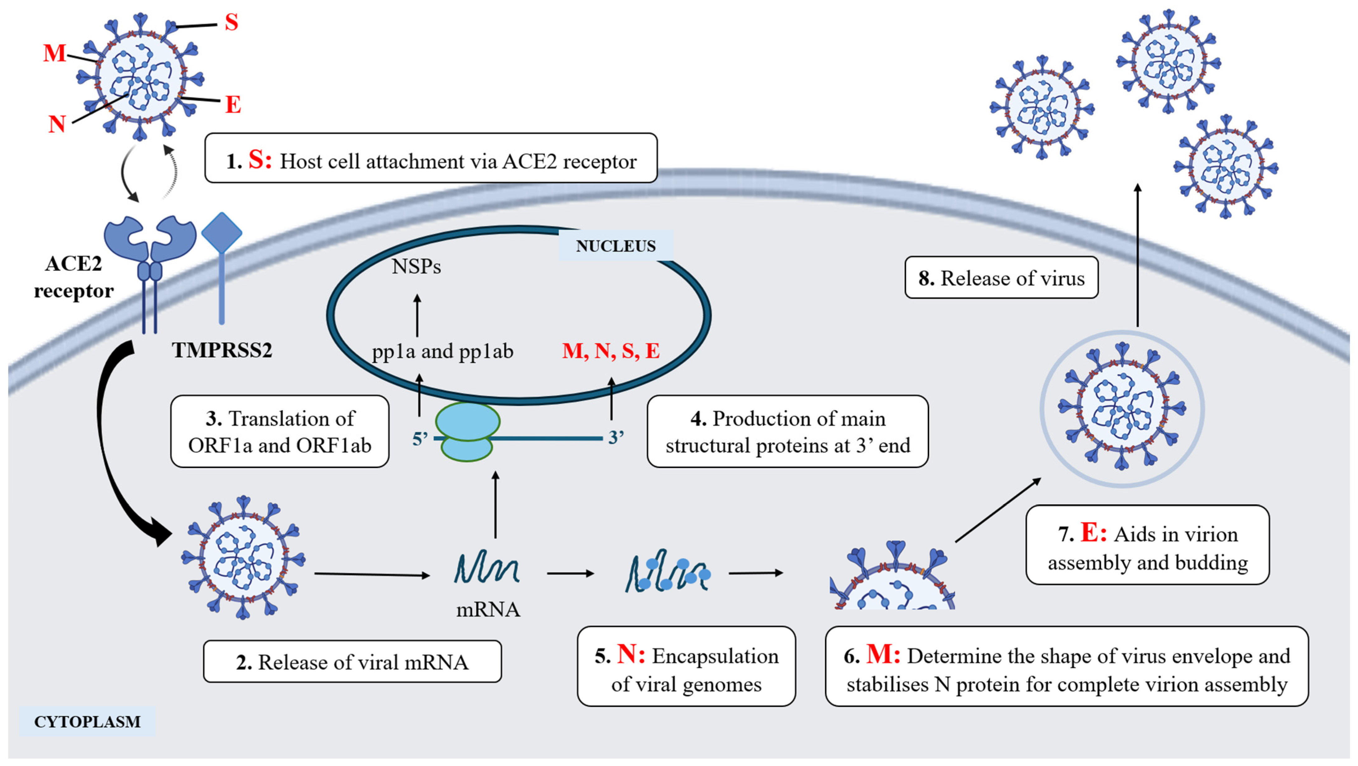
3. Viral-Host Interaction in Virus Infection: SARS-CoV-2

SARS-CoV-2 is a well-known etiological agent of the pandemic outbreak COVID-19. It is an enveloped, non-segmented and +ssRNA virus that exists as a sphere with crown-like spikes on the outer surface. The four main structural proteins that makeup SARS-CoV-2 are spike (S) glycoprotein, envelope (E) glycoprotein, membrane (M) glycoprotein, and nucleocapsid (N) protein [25]. The entry of SARS-CoV-2 into host cells is facilitated by the S protein that attaches to the host cell receptor, angiotensin-converting enzyme 2 (ACE2). Subsequently, the S protein is then cleaved by the widely expressed host furin-like protease into S1 (binds ACE2) and S2 (mediate virus fusion) subunits (Figure 1) [25]. Upon virus-host fusion, the host cell type II transmembrane serine protease (TMPRSS2) further primes the S2 subunit to induce conformational change to facilitate viral entry to the host cells, making both the TMPRSS2 and ACE2 the main determinants of the virus entry [25]. In the event of insufficient or absent TMPRSS2, cathepsin L protease, under an acidic condition, takes over the activation of S protein in endolysosomes where the virus-ACE2 complex is internalised [26]. SARS-CoV-2 then releases its mRNA in the cytoplasm, translating into viral proteins in the host nuclei. The genome of SARS-CoV-2 possesses a 5′ cap containing the open reading frame (ORF) 1a and 1b encoding polyproteins, pp1a and pp1ab, that are cleaved by viral proteases into non-structural proteins (NSPs) 1–11 and 12–16, respectively, and poly(A) 3′ tail encoding four main structural proteins [27]. In addition, variable ORFs can also be found alternating at the 3′-end, such as ORF3a, ORF3d, ORF6, ORF7a, ORF7b, and ORF8, to name a few, for the production of accessory proteins [28]. The viral proteins involved in cleaving the polyproteins are papain-like proteases (PLpro) and 3-chymotrypsin-like proteases (3CLpro), encoded in NSP3 and NSP5, respectively [29]. NSPs play crucial roles in viral replication, host immune evasion, and viral enzyme production, which was thoroughly reviewed [30].
Viruses infect species across all three domains of life: Archaea, Bacteria, and Eukarya; some viruses are host-specific, and others infect multiple species [31]. A host supports the virus’s life cycle and greatly depends on the host’s susceptibility to infection. Among these, cell tropism will be highlighted in this context, which means the virus’s specificity to a cell carrying a specific receptor [15]. For instance, HIV predominantly targets CD4+ T-helper lymphocytes as these cells express CD4 receptors and co-receptors (CCR5 and CXCR4) for HIV attachment [32]. SARS-CoV-2, on the other hand, utilises viral S protein to bind to host ACE2 receptors for cell entry. The first thing that comes to mind regarding COVID-19 is the human upper respiratory tract, which is highly ACE2 expressed. However, the respiratory tract is not the sole target as there is increasing evidence of cardiac infection, testicular infection, and gastrointestinal and renal complications from SARS-CoV-2, which coincidentally have high ACE2 expressions, explaining other possible reservoirs for the virus infection [26,33,34]. Unfortunately, ACE2 is not qualified as a game changer, given its abundance and low specificity against the SARS-CoV-2 variants and the potential interference with the renin-angiotensin-aldosterone system (RAAS) that regulates vascular function [35]. This prompts the researchers to explore other host factors, with the hope of developing host-directed antivirals that are highly conserved and stable with minimal host toxicity against SARS-CoV-2 to bring an end to the COVID-19 era.

4. Current Antiviral Therapeutics

The current antiviral therapies are none other than viral-directed antiviral (VDA) agents and host-directed antiviral (HDA) agents. As the name suggests, the former directs viral protein, and the latter directs host factor proteins to exterminate the viral infection.

4.1. Viral-Directed Antiviral (VDA)

Antiviral drug development has predominantly focused on inhibiting the viral life cycle via VDA agents. However, antiviral drug resistance has rapidly emerged. For instance, the FDA-approved Tamiflu (Oseltamivir), Relenza (Zanamivir), and Rapivab (peramivir) functioned to inhibit neuraminidase (NA) have replaced the older M2 ion channel inhibitors, the amantadine and rimantadine for Influenza A virus due to resistance in circulating strains, including the A(H1N1)pdm09 pandemic strain, which has continuously evolved and contributed to the recurrent human seasonal Influenza [36,37]. Similarly, Vidarabine, an FDA-approved nucleoside analogue for inhibiting HSV DNA synthesis, was replaced by alternatives like Acyclovir, Famciclovir, and Foscarnet due to resistance and toxicity [38,39]. Resistance continues to challenge the efficacy of antiviral treatments (Table 2).
The rapidly mutating nature of viruses, such as SARS-CoV-2, further complicates antiviral efficacy. Taking the etiological agent of the current COVID-19 pandemic, SARS-CoV-2 has shown how mutations enhance the virus’s infection capacity and reduce vaccine efficacy. For instance, the N501Y mutation found in multiple variants (alpha, beta, gamma, mu, omicron) enhances host ACE-2 receptor binding affinity, increasing infection capacity [6]. The E484K mutation found in alpha, beta, gamma, and mu has also contributed to the loss of neutralising activity of convalescent sera and monoclonal antibodies (mAB) along with increased ACE2 binding [40,41]. Deep mutational scanning of SARS-CoV-2 variants further revealed mutations (I358F, Y365F, Y365W, V367F, F392W and Q493, Q498, N501) that enhances RBD expression and ACE-2 binding affinity, respectively [42].
The effect of antiviral resistance heightened with the introduction of the Omicron variant with more than 30 mutations in spike protein alone [3]. Mutations like R346T, R346K, F486S, and G446S have conferred neutralisation resistance and reduced neutralising efficacy of vaccine-generated antibodies (Abs) [43,44]. The loss of neutralising titres in current antivirals such as Casirivimab, Imdevimab, Bamlanivimab, Etesevimab, Sotrovimab, and Evusheld cocktails for monoclonal antibody therapy against Omicron has been well-reported [45]. Among these, Sotrovimab and Evusheld neutralising titters were reported to significantly decrease up to 19-fold and 80-fold, respectively, compared to the D614G mutation found in earlier variants [45,46]. To complicate the situation, the omicron spike protein is reported to be 26–34 fold neutralisation resistant against Pfizer BNT162b2 and Moderna vaccine-elicited antibody sera from recovered donors, further highlighting the challenge posed by antiviral resistance in VDA [45].
Table 2. Examples of current FDA/EMA-approved virus-directed antivirals (VDA) and their mode of action against influenza A virus and SARS-CoV-2.
VirusApproved Antiviral DrugMode of ActionReferences
SARS-CoV-2
(COVID-19)
Paxlovid (Nirmatrelvir and Ritonavir)Inhibits SARS-CoV-2 main protease (Mpro)[47]
Veklury (Remdesivir)Inhibits RNA-dependent RNA polymerase[48]
Lagevrio (Molnupiravir)Introducing errors into viral RNA[48]
Bamlanivimab and EtesevimabMonoclonal antibodies targeting spike protein[49]
Casirivimab and Imdevimab (Ronapreve)Monoclonal antibodies targeting spike protein[50]
Sotrovimab **Monoclonal antibodies targeting spike protein[51,52]
Evusheld (tixagevimab and cligavimab) **Monoclonal antibodies targeting spike protein[53]
Influenza A virus (IAV)Tamiflu (Oseltamivir)Inhibit neuraminidase (NA)[54,55]
Relenza (Zanamivir)Inhibit neuraminidase (NA)[54,55]
Rapivab (Peramivir)Inhibit neuraminidase (NA)[54,55]
Xofluza (Baloxavir Marboxil)Inhibit cap-dependent endonuclease[54,55]
Amantadine **M2 ion channel inhibitors[56,57]
Rimantadine **M2 ion channel inhibitors[56,57]
** Not recommended or discontinued due to resistance and reduced efficacy.

4.2. Host-Directed Antivirals (HDA)

The need for alternative antiviral agents is an understatement, especially during a global pandemic, COVID-19. Since viruses often exploit a broad amount of essential host protein for viral life cycle and replication, developing HDA may offer a potent antiviral drug that is less prone to viral resistance, which may be a key to exterminating and containing epidemics [58,59,60,61]. During infection, the virus induces extensive immune signalling response upon recognising the pathogen-associated molecular patterns (PAMPs), a conserved virus or bacterial motifs that are not found in the human immune system via the host’s pattern recognition receptors (PRR) such as Toll-like receptors (TLRs): TLR3, 4, 7, 8, Retinoic acid-inducible gene I-like receptors (RLRs): RIG-1 (Retinoic acid-inducible gene I), MDA5 (melanoma differentiation-associated protein 5), and the LGP2 (Laboratory of Genetics and Physiology 2) co-factor [62]. The activation of PRR subsequently triggers the phosphorylation of IFN-regulatory factors (IRFs) such as IRF3 and IRF7, which in turn promotes the production of interferons (IFNs) such as Type I and II IFN [37]. These IFNs activate the JAK-STAT pathway that upregulates the antiviral IFN-stimulated genes (ISGs) such as the 2′,5–oligoadenylate synthetase (OAS) and protein kinase R (PKR) to facilitate suppression of viral replication [30].
Things are a little different in HDA. One might perceive HDA as a direct target of only host protein interacting with viral proteins. However, HDA typically modulates the host’s immune system and cellular machinery to fend off or prevent virus infection. For instance, human interferons, part of the cytokine family, are essential signalling proteins in the innate immune response that activate immune cells such as macrophage and natural killer cells and heightened antiviral response for counteracting and the eradication of viral infection and hold a significant target to suppress viral replication [63]. The PEGylated interferon alfa-2a and alfa-2b bind to the human type I IFN, activating its JAK-STAT pathway to clear the virus. Apart from that, these PEGylated interferons induce early activation of dendritic cells, which in turn produces cytokines that enhance the activation of CD56bright natural killer cells (CD56bright NK), Th17-CD4+ T-helper cells and Th1-CD8+ T lymphocytes/cytotoxic T cells, exterminating the HCV via both innate and adaptive immune response [64,65]. Notably, despite the PEGylated interferon alfa-2a and alfa-2b having been reported to have promising antiviral responsiveness against HCV, the latter is preferred due to greater exposure to patients, ultimately carrying greater antiviral activity [66,67].
Another notable example would be the Maraviroc, an approved chemokine receptor type 5 (CCR5) antagonist for the treatment of HIV-1 [68]. CCR5 is a human cell surface G-protein-coupled receptor (GPCR) that responds to RANTES, macrophage inflammatory protein–1α (MIP-1α), MIP-1β, and monocyte chemotactic protein–2 (MCP-2), which in turn regulates the trafficking of leucocyte [69]. CCR5 and another chemokine receptor, the C-X-C chemokine receptor type 4 (CXCR4), have been well-established as a co-receptor to the HIV-1 envelope glycoprotein gp120 and human CD4 complex (gp120-CD4) to facilitate the entry and fusion of HIV-1 [70]. Notably, the modified Maraviroc, PF-232798, has demonstrated superior efficacy against the maraviroc-resistant HIV-1 strain with no adverse effects observed at 250 mg in phase II clinical trials [71]. Other notable examples of current circulating viruses and the proposed FDA/EMA-approved host-directed drugs for drug repositioning have also been included (Table 3).
As mentioned earlier, the most significant advantages conferred by targeting the host proteins in viral replication would be reduced viral resistance, broad-spectrum potential, and conserved targets. For instance, Camostat mesylate and Nafamostat mesylate, human Transmembrane Protease Serine 2 (TMPRSS2) inhibitors that have proven to suppress prostate cancer, have exhibited strong antiviral activities against IAV, SARS-CoV, MERS-CoV, and the current SARS-CoV-2 [72,73,74]. In addition, the combination of HDA and VDA regimes has resulted in strong synergistic antiviral activity. For instance, the triple combination of TMPRSS2 inhibitors (HDA) such as camostat, nafamostat, and avoralstat to inhibit SARS-CoV-2 entry, dihydroorotate dehydrogenase inhibitors (HDA) such as brequinar that depletes human intracellular pyrimidines which are required for viral RNA synthesis and the molnupiravir (DAA), a prodrug of N-hydroxycytidine (NHC) which incorporates incorrect nucleotides during the RNA synthesis to halt the SARS-CoV-2 replication, have synergistically suppressed infection by SARS-CoV-2 and its variants by up to 95–100% compared to 80% when administered alone [75].
Table 3. Examples of current FDA/EMA-approved host-directed drugs and their proposed mode of action (drug repositioning) against circulating viruses.
VirusApproved Host-Directed DrugProposed Antiviral ActionReferences
SARS-CoV-2 (COVID-19)Imatinib
  • Bcr-Abl tyrosine kinase inhibitor that phosphorylates tyrosine to treat chronic myeloid leukaemia (CML).
  • Inhibit SARS-CoV-2 entry and spike-host ACE2 membrane fusion by inhibiting Abl1 and Abl2 kinases.
  • May inhibit SARS-CoV-2 by blocking the epidermal growth factor receptor (EGFR) pathway, which was exploited to promote virus growth, such as the negative regulation of SOCS protein required for healthy antiviral response.
[76,77,78,79]
Clofazimine
  • Anti-leprosy drug and is currently being considered for non-tuberculous mycobacterial (NTM) infections.
  • Inhibit cell fusion mediated by SARS-CoV-2 S protein and inhibit the unwinding activity of nsp13 helicase via double-stranded DNA or RNA substrate.
  • Reduced SARS-CoV-2 titre by more than 3 log10 and reverted 90% of human genes that were altered post 6 h SARS-CoV-2 infection.
  • It may also serve as a prophylaxis based on the reported diminished SARS-CoV-2 titre by up to a twofold reduction in the Syrian hamster model post 4 days’ infection.
[80,81]
Herpes Simplex Virus (HSV)Ouabain
  • Na+/K+-ATPase inhibitor for arrhythmia and heart failure patients.
  • Inhibit fusion of host cells and HSV, resulting in at least fivefold viral gene reduction in HSV.
  • It may also inhibit the activity of host topoisomerase II, which is required for the cleavage of progeny viral DNA.
[82,83,84]
Influenza A virus (IAV)Verapamil
  • An FDA-approved voltage-gated calcium channel blocker (CCB) for cardiac arrhythmia and high blood pressure.
  • IAV induces Ca2+ influx, which favours endocytic uptake of IAV by the host cell.
  • CCB prevents calcium influx, inhibiting IAV-host cell fusion and assembly and halting viral replication.
[85,86,87]

5. Harnessing Host Proteins to Combat SARS-CoV-2

5.1. Kinases

Kinase proteins are critical enzymes that regulate cellular functions via protein phosphorylation, altering the protein conformation and modulating their binding affinity with other molecules. Protein phosphorylation, a key post-translational modification (PTM), is crucial in regulating a wide range of cellular processes such as cell growth, signal transduction, and protein synthesis [88]. Kinase activities are tightly controlled, and dysregulation of their activities often accompanies various diseases, such as cancer, diabetes, neurological disorders, and infectious diseases [89]. A few prominent kinase families include serine/threonine kinases, phosphoinositide 3-kinases/protein kinase B/mammalian target of rapamycin (PI3K-AKT-mTOR) pathway, JAK-STAT and IκB Kinase (IKK), and NF-κB pathway. These kinases play different roles, with the familiar roles of regulating stress responses, cell survival, and metabolism. Although kinases are more extensively studied in cancer, their roles in viral infections cannot be overlooked, given that viruses often hijack host kinases to facilitate their infection cycle, making them the potential antiviral target by repurposing the small molecules kinase inhibitors for viral infections.
To list a few, HIV hijacks cyclin-dependent kinases (CDKs) 1, 2 and 6 to inactivate an antiviral phosphohydrolase (SAMHD1), which in turn facilitates the reverse transcription of HIV in activated CD4+ T cells [90]. The host cell phosphoinositide 3-kinases/protein kinase B (PI3K/Akt) signalling pathway is utilised by influenza A virus, Hepatitis C virus and African swine fever virus to facilitate the endosomal entry of the virus to the host cells, in turn delaying the host’s immune recognition [91]. Notably, the JAK-STAT and IκB Kinase (IKK) and NF-κB pathways are exploited by SARS-CoV-2 and HIV to suppress the antiviral responses by regulating the host immune system [92]. Similarly, the SARS-CoV-2 also relies on the host kinases for heavy phosphorylation events to have successful infection. SARS-CoV-2 has been shown to hijack several of the CMGC kinases, consisting of the cyclin-dependent kinases (CDKs), mitogen-activated protein kinases (MAPKs), glycogen synthase kinases (GSKs), and CDC-like kinases (CLKs), which all fall under the family of serine/threonine kinase [93]. Evidence has pointed towards identifying several serine or threonine phosphorylation sites (phosphosites) in SARS-CoV-2 proteins, such as the N, M, S, NSP3, and ORF9b proteins [94]. The phosphosites such as Casein kinases 1 and 2 (CK1 and CK2), G protein-coupled receptor kinases (GRK), Cdc2-like kinases (CLK), and P38 group kinase, to name a few, were reported to interact with various host kinases [95]. While there were mixed reports, phosphosites S23, S26, S79, S176, S197, T198, T205, and T206 in N protein, and S173, S211, S212, and S214 in M protein of SARS-CoV-2 have been suggested as critical sites for phosphorylation events in viral-host interaction, warranting further investigation for potential therapeutic targets [94].
To elaborate the interaction of CMGC kinases with SARS-CoV-2, glycogen synthase kinase-3 (GSK3), a conserved serine/threonine kinase, is found activated by SARS-CoV-2 N protein, leading to host oxidative stress and inflammation via degrading nuclear factor erythroid 2-related factor (Nrf2) and phosphorylating nuclear factor-κB (NF-κB), respectively [96]. While the host p38/MAPK pathway is crucial to mediating the cellular stress and inflammatory responses, SARS-CoV-2 exploits this pathway to promote viral replication and modulate the host immune response by producing cytokines, a hallmark of severe COVID-19 pathogenesis [97]. Diving further, the nsps of SARS-CoV-2 have been reported to interact with CDK2, particularly the nsp12, a key component of the RdRp complex phosphorylated at T20. This phosphorylation event facilitates the assembly of the RdRp complex that consists of nsp12, nsp7, and nsp8, eventually promoting the synthesis of viral RNA for viral genome replication [98]. Besides CMGC kinases, SARS-CoV-2 triggers the phosphorylation of Eukaryotic Translation Initiation Factor 2 Alpha Kinase 2 (EIF2AK2), a host antiviral kinase also known as PKR, activating the inflammasome and disrupting the host cell translational machinery by phosphorylating eukaryotic translation initiation factor 2 alpha (eIF2), eventually leading to cytokine storm and limited host protein synthesis [99].
The convoluted plot in kinase pathways is no stranger due to extensive crosstalk in nature to regulate physiological and pathological processes. Nonetheless, given how crucial host kinases are for viral infection, they present a prospective therapeutic target against SARS-CoV-2. GSK3 inhibitors, such as lithium chloride and kenpaullone, have been shown to reduce the GSK-3-directed N protein phosphorylation, reducing SARS-CoV-2 viral titre and cytopathic effect up to 85% in infected VeroE6 cells, subsequently reducing the severity of infection [100]. The imidazole-oxindole C16, a PKR antagonist, significantly inhibited viral proliferation in SARS-CoV-2-infected cells [99]. Notably, C16 also binds to the SARS-CoV-2 N protein, which is often crucial in viral RNA synthesis and suppressing host IFNs, thereby preventing host translational shut-off and contributing to a potent antiviral activity [99]. To recap, the host p38/MAPK is often exploited by SARS-CoV-2 to promote viral replication and induce host cytokines production in severe COVID-19 pathogenesis; p38 inhibitors, such as losmapimod and dilmapimod in clinical trials for other indications, could be repositioned for COVID-19 [101]. Another study demonstrated the significant potential of CDK2 inhibitor, SNS-032, in combating SARS-CoV-2, as evidenced by the SARS-CoV-2 RdRp inhibitory activity at an EC50 of 73nM and SARS-CoV-2 infection inhibition at EC50 of 84nM [98]. In the same study, SNS-032 was also shown to inhibit RdRp complex formation by halting the phosphorylation of T20 of nsp12 [98].

5.2. Heat Shock Proteins (HSPs)

Cellular protein homeostasis, or proteostasis, is crucial for cell survival and preventing various diseases’ development. To maintain proteostasis, HSPs are necessary molecular chaperones that come into play, mainly to regulate the folding of newly synthesised polypeptides and degrading misfolded proteins [102]. HSPs are divided into HSP100, HSP90, HSP70, HSP60, and small HSPs based on their molecular weight and functions [103]. Besides being constitutively expressed under normal cellular conditions, HSPs are markedly activated under extreme conditions to buffer stresses such as heat, oxidative stress, and toxins to prevent protein misfolding and aggregation [104]. Beyond their protective roles, HSPs are implicated in diseases like cancer, neurodegenerative disorders, and autoimmune conditions. For instance, HSPs promote resistance to therapy against cancer by contributing to the tumour progression and inhibiting apoptosis [105]. Impaired HSPs can lead to the accumulation of misfolded proteins and cellular dysfunction, leading to neurodegenerative diseases such as Alzheimer’s and Parkinson’s [106]. The involvement of HSPs extends to viral infections where host HSPs were found to support viral replicase or macromolecular protein complexes in completing a virus life cycle ranging from viral replication and assembly to virion production [103]. In addition, HSP90 also partook in stabilising various virus proteins, including paramyxoviruses polymerase and L protein, chikungunya virus nsp, and the protease of HCV NSP to facilitate virus replication [103]. HSP70 and HSP90 were demonstrated to synergistically promote hepatitis B virus (HBV) capsid assembly [107].
Highlighting SARS-CoV-2 viruses, the elevation of body temperature is a natural fallout of the virus infection, which is also a result of the overexpressed HSPs. HSP70 was reported to interact with ACE2 and spike protein’s RBD domain to stabilise and protect their structures under febrile conditions, subsequently allowing viral entry into the host cells [108]. The exploitation of HSP70 by SARS-CoV-2 is further explained when the introduction of an HSP70 inhibitor, PES,2-(3-chlorophenyl) ethynesulfonamide (PES-Cl) and siRNA restricted SARS-CoV-2 infection in Vero-E6 cells [108]. Another chaperone, HSP90, and its isoform HSP90β, also reported to interact with SARS-CoV-2 proteins-N, M, ORF3, ORF7a, and ORF7b proteins, where the downregulation of the five mentioned viral proteins occurred in the event of HSP90β knock-out via CRISPR-Cas9 [109]. HSP90 was reported to regulate the M and N protein levels in SARS-CoV-2 virus assembly and protect the N proteins from proteasome-mediated degradation [109]. Significant reduction of lung lesions in SARS-CoV-2 virus-infected hamsters was reported upon Alvespimycin (17-DMAG) administration, an HSP90 inhibitor, making HSP90 a promising therapeutic target [109]. Apart from that, other in silico studies showed that HSP27 (sHSP) and HSPA8 (HSP70 cognate) could also be targets of SARS-CoV-2 spike proteins to facilitate viral entry and survival within the host [110]. In contrast to the PAMPs, damage-associated molecular patterns (DAMPs) are endogenous molecules released during stress to alert the immune system of tissue damage or trauma [111]. The plasma glucose-regulated protein gp96, an HSP, is significantly elevated in COVID-19 patients as it can behave as DAMP, in which it will activate the innate immune responses and interact with TLR, eventually stimulating the secretion of proinflammatory cytokines, leading to the severe stage of COVID-19 [112].
Despite the pathological roles in COVID-19, the protective role of HSPs is also highlighted for their antiviral potential. Oncogenic HSP90 and HSP70 inhibitors have been extensively investigated, with many ongoing clinical trial stages, such as the geldanamycin (HSP90 inhibitor) for advanced solid tumours or non-Hodgkin’s lymphoma, 17-DMAG for relapsed chronic lymphocytic leukaemia, and autologous HSP70-peptide complex (AG-858) for chronic myeloid leukaemia [104]. Unfortunately, they are yet to be approved by the FDA. Despite that, these drugs were tabled to be repurposed for antiviral treatment, including the current COVID-19. Nonetheless, there are reports on hepatotoxicity at high concentrations, and the focus on these inhibitors, such as geldanamycin with broad antiviral activities, has now been shifted to its safer analogues or derivatives [113,114]. An in silico study showed strong binding of Ignaciomycin (geldanamycin analogue) with the mutated sites in the RBD of Delta and Omicron variants via hydrogen bonding (H-bonding) interaction, inhibiting RBD interaction with ACE2 receptor, thus preventing viral entry and replication [113]. SNX-5422, an orally bioavailable HSP90 inhibitor which is currently under clinical trials for anticancer treatment, has also demonstrated effective inhibition of SARS-CoV-2 replication in vitro at a high selectivity index at the initial infection stage, indicating a potential in reducing COVID-19 disease severity [115]. Another HSP90 inhibitor, 17-AAG, was reported to substantially suppress SARS-CoV-2 propagation in vitro [112]. Despite many reports on HSP90 inhibitors against SARS-CoV-2, one notable HSP70 inhibitor, the PES-Cl, has been reported to be more potent than Remdesivir in the quest for inhibiting SARS-CoV-2 viral replication [108]. Owing to the reported toxicity, using HSPs for COVID-19 requires further investigation concerning their specificity to achieve minimal cytotoxicity without disrupting normal cellular functions.

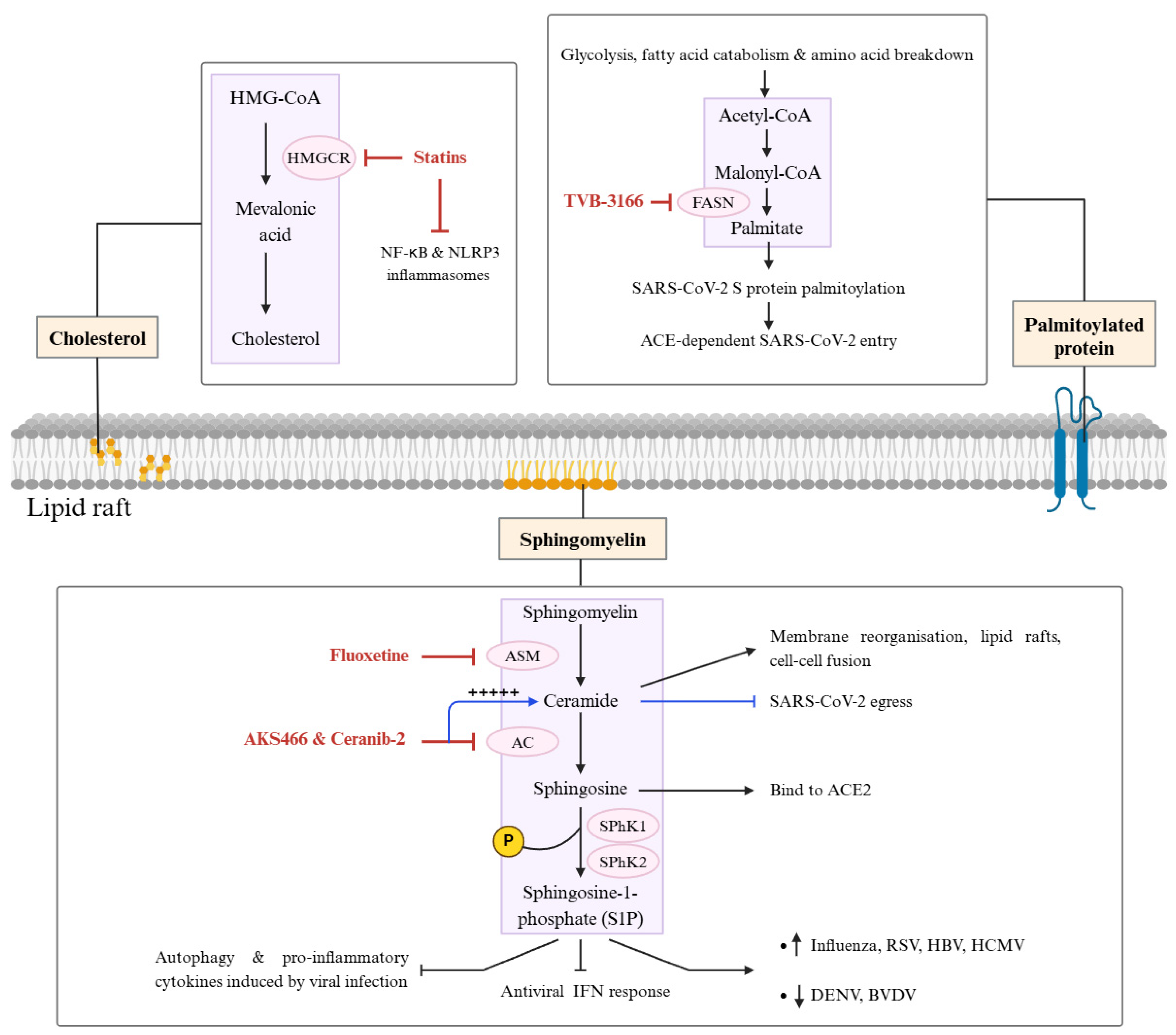
5.3. Lipid Metabolism Proteins in Viral Replication

Viruses are obligate intracellular parasites that hijack host cellular machinery, such as ribosomes, DNA and RNA polymerases, ATP, cell membrane and cellular pathways, to replicate and produce new virions. Among these, host cell membranes, primarily composed of lipids, play a key role in virus replication [116]. Lipids encompass diverse hydrophobic molecules such as phospholipids, glycerolipids (triacylglycerol (TAG), diacylglycerol (DAG)), sphingolipids (sphingomyelin (SM), ceramide, glycosphingolipids), sterols (cholesterol and ergosterol), fatty acids (FAs), glycerophospholipids (phosphotidylcholine (PC), phosphatidylethanolamine (PE), phosphatidylserine (PS), phosphatidylinositol (PI)), saccharolipids (glucosylceramide, lactosylceramide), polyketides (erythromycin, tetracyline) and prenol (dolichol, ubiquinone) lipids [117]. These molecules are crucial in energy storage, signalling, protection, and cellular structure formation [118,119]. Given the convoluted roles in virus replication and host immune response, lipids present potential targets for antiviral therapy.
Viruses exploit host lipids for endocytosis, membrane remodeling, replication, assembly, egress, and immune evasion [117]. Generally, there are four major lipid roles for viral entry: (1) direct viral receptors, where non-enveloped viruses such as polyomaviruses directly bind to the lipids (glycosphingolipids) of host cell surface; (2) indirect viral receptors, where viruses like HCV associate with host low-density lipoprotein receptor (LDL-R) or utilise clathrin-mediated endocytosis to promote viral endocytosis; (3) cofactors, where viruses like influenza and HIV uses host lipid raft (cholesterol-rich domains) to enhance viral entry or to trigger the alternative endocytic pathways (caveolin-mediated/macropinocytosis) and (4) fusion assisting, where viruses like the DENV utilises certain host lipid to promote fusion of host cell membrane and viral envelop for internalisation while some viruses like the SARS-CoV-2 facilitate lipid-dependent pH changes and enzymatic escape from endosomes [120,121,122].
Phospholipids, sphingolipids, and sterols largely maintain the structural integrity of human cell membranes. The internalisation, replication, and egress of the virus requires the remodelling of human cell membranes to induce membrane curvature that is essential to the formation of viral replication complexes (VRCs) that either bend outwards (positive curvature) or inwards (negative curvature), which is vital for the budding and assembly, and protected environment for viral RNA synthesis, respectively [119]. The cell membrane curvature can be achieved via different combination and synthesis of various shaped and charged PC (neutral cylindrical), PE (neutral cone), PS (negative cylindrical), SM (neutral cylindrical), PI (negative cone), phosphatidylinositol-4-phosphate (PI4P) (negative inverted cone), phosphatidylinositol-4,5-bisphosphate (PI(4,5)P2) (negative inverted cone), and phosphatidylinositol-3,4,5-trisphosphate (PI(3,4,5)P3) (negative inverted cone) [123].
SARS-CoV and SARS-CoV-2 require palmitoylation of their S protein to interact with host membranes and promote fusion [124]. This process depends on fatty acid synthesis, specifically palmitic acid produced from converting cytosolic acetyl-CoA and malonyl-CoA by fatty acid synthase (FASN) [118]. Additionally, the SARS-CoV-2 S protein binds high-density lipoprotein (HDL) particles through scavenger receptor class B type 1 (SR-B1), enhancing ACE2-dependent viral entry [125]. SARS-CoV-2 also exploits host cells’ intracellular membranes, such as the phospholipase A2α enzyme (cPLA2α), which catalyses the glycerophospholipids conversion to free fatty acid and a lysophospholipid that is essential for forming double-membrane vesicles (DMVs), which is critical for coronavirus’ replication [126]. Notably, the ageing-associated phospholipase A2 (PLA2) group IID (PLA2G2D) correlated with severe SARS-CoV infection in mice models, and the inhibition significantly improved survival at a striking 80% [127].
Interestingly, polyunsaturated fatty acids such as linoleic acid was reported to inhibit SARS-CoV-2 attachment and replication by interacting with the receptor-binding domain of the S protein and RdRp [128,129]. Besides that, the SARS-CoV-2 infection has been found to increase lipid droplet formation and upregulated lipid metabolism such as CD36, PPAR-γ, SREBP-1, and diacylglycerol acyltransferase-1, further affirming the role of lipids in SARS-CoV-2 replication [124].
Given the intertwined relationship between host lipid-protein and viral replication, targeting the lipid pathway may help counteract SARS-CoV-2. SARS-CoV-2 utilises human ACE2 and TMPRSS2/4 receptors at cholesterol-rich lipid raft for entry. Cholesterol-lowering drugs, such as FDA-approved statins, inhibit the HMG-CoA reductase (HMGCR) enzyme, which catalyses the conversion of HMG-CoA to mevalonic acid for cholesterol synthesis, limiting severe viral infection such as SARS-CoV [130]. Statins also limit cytokine complications from the NF-κB and NLRP3 inflammasomes in coronavirus infection, making cholesterol synthesis a potential target for SARS-CoV-2 inhibition [131]. On a side note, fatty acids are essential for producing the host cell membrane’s phospholipid bilayer, while cholesterol regulates membrane fluidity through inserts. While HMGCR is crucial for cholesterol synthesis, fatty acid anabolism drives the formation of the mentioned cell membrane bilayer. They share the same building block, the Acetyl-CoA produced via glycolysis, fatty acid catabolism, and amino acid breakdown [132]. The fatty acid synthesis catalysed by FASN has been reported to be vital for the acylation (palmitoylation) of SARS-CoV-2 S protein to enhance the binding of host ACE2 receptors for viral fusion. FASN inhibitors such as TVB-3166 have been reported to effectively inhibit the acylation of SARS-CoV-2 S protein, hampering the viral spread in vitro [133].
The SARS-CoV-2 S protein was revealed to interact with gangliosides, a sphingolipid, to enhance viral attachment, entry, and replication by activating Acid Sphingomyelinase (ASM) that hydrolyses sphingomyelin into ceramide that promotes membrane reorganisation, producing lipid rafts and enhancing cell-cell fusion, to name a few [134,135]. Fluoxetine, a selective serotonin reuptake inhibitor (SSRI) that inhibits ASM, has been reported to suppress SARS-CoV-2 replication in addition to reducing IL-6/NF-κB pro-inflammatory signalling in Vero and Caco-2 cells (Figure 2) [136]. When the ceramide level rises, Acid Ceramidase (AC) will be activated as homeostatic countermeasures to break down the ceramide to sphingosine, which either competitively binds to ACE2 or is phosphorylated into sphingosine-1-phosphate (S1P) by sphingosine kinases 1 and 2 (SPhK1 and SPhK2) [137]. To date, five known S1P receptors (S1P1-S1P5) have been recorded to play a critical role in intracellular signalling, lymphocyte trafficking, immune modulation, vascular homeostasis, cell survival and death, DNA synthesis, and many more [138,139].
S1P has dual effects on viral infections, either enhancing or inhibiting replication depending on the virus and infection stage [140]. While it suppresses autophagy and pro-inflammatory cytokines induced by viral infection, it was reported to promote replication in influenza, RSV, HBV, and HCMV [141]. Conversely, it inhibits DENV and BVDV replication [141]. S1P also suppresses antiviral IFN response, promoting inflammation and membrane fluidity [134]. Although sphingosine production may reduce the affinity for SARS-CoV-2 S protein and halt viral entry and replication, it may also reverse the effect to promote viral replication depending on the condition and stage of infection [142]. Apart from that, despite the reported roles of ceramide in enhancing viral fusion and replication, AC inhibitors such as the AKS466 and Ceranib-2 have been reported to cause SARS-CoV-2 particle enrichment in endo-lysosome via ceramide accumulation, which inhibits SARS-CoV-2 virus egress, where the author suggests AC inhibitor may retain SARS-CoV-2 in the lysosome, halting its replication cycle (Figure 2) [143]. While the lipid metabolism certainly paves the way for alternative antiviral therapy, the intricate relationship between the lipid-protein in the host cellular machinery, and the various contradicting reports from the interaction with different viruses, further clinical studies have to be carried out to conclude the use of anti-lipid related drugs against the SARS-CoV-2.

5.4. Immunological Pathway Proteins

The innate immune system is the first host defence against viral infection. Five major types of protein domains of host-pathogen recognition receptors (PRRs), toll-like receptors (TLRs), and retinoic acid-inducible gene-I (RIG-I)-like receptors (RLRs) have been recognised as the most critical recognition receptors for viruses and facilitate antiviral signalling [62]. TLRs were first identified to be an essential host defence receptor against fungal infection in Drosophila; the mammalian homologue was also discovered later to induce inflammatory responses, namely the TLR 1-13, albeit the last three were only found in mice but not humans with TLR10 cited as non-functional [144,145]. The activation of TLR signalling begins with the dimerisation of TLRs, and different TIR domain-containing adaptor proteins strictly regulate this to relay the downstream signalling which entails the Toll/IL-1R homology domain-containing adaptor molecule 1 (TICAM-1/TRIF) for TLR3; the universal myeloid differentiation primary-response gene 88 (MyD88) and MyD88-adaptor-like protein (MAL) (all TLRs except TLR3); the TRIF-related adaptor molecule (TRAM) for TLR4 [146]. Following the binding of the TLRs to the mentioned adaptor proteins, the IKK complex (IKKα and β, γ) induces phosphorylation of the α subunit of the inhibitor of kappa B (IκB) (consisting of IκBα, IκBβ, and IκBε subunits) that naturally exist to prevent NF-κB from entering the nucleus (inactivated state). The phosphorylation triggers ubiquitination of IκBα, subsequently releasing and allowing the binding of NF-κB with specific promoters in the nucleus to generate various pro-inflammatory cytokines (such as IL-1, IL-6 and TNF-α). RLRs, on the other hand, include three main family members, namely the RIG-I (Retinoic acid-inducible gene I), MDA5 (Melanoma Differentiation-Associated protein 5), and LGP2 (Laboratory of Genetics and Physiology 2) (mainly a co-factor). The former two share many similarities, which are activated via the recognition of dsRNA. Similar to TLRs, the RLRs are regulated by the adaptor proteins such as the mitochondrial antiviral-signalling (MAVS) kinase that entails the TANK binding kinase 1 (TBK1) and inhibitor of κ-B kinase ε (IKKε), which subsequently activates the production of interferon-regulatory factors or IRFs (such as IRF3 and IRF7), which in turn activates the synthesis of IFNs—type I (IFN-α, IFN-β, IFN-ε, IFN-κ, IFN-ω) and type III (IFN-λ) [147]. These IFNs bind to their respective IFN receptors, type I IFN to IFN-α/β receptor (IFNAR1/IFNAR2) and type III IFNs to IFN-λ (IFNLR) receptors. These, in turn, trigger the associated downstream JAK1 and TYK2 molecules to phosphorylate the cytoplasmic STAT1 and STAT2 transcription factors to form a transcription factor complex (STAT1/STAT2/IRF9) that would ultimately form the IFN-stimulated gene factor 3 (ISGF3) that upregulate antiviral interferon-stimulated genes (ISGs), such as the 2′−5′–oligoadenylate synthetase (OAS) and protein kinase R (PKR) (Figure 3).
Having to detail the pathway above, there is no doubt that viruses like the current SARS-CoV-2 exploit the human immunological pathway to confer viral survival and replicative advantage. Unfortunately, the proteins in the immunological pathway are a dual-edged sword for COVID-19 infection, causing immunotherapy in virus infection challenging. For instance, TLR agonists and antagonists can be employed depending on the stage and severity of the disease. TLR agonists such as Pam3CSK4, L-PAMPO, Imiquimod (R837), and Resiquimod (R848) can be given in the early stages of infection as to activate the release of type I IFNs and cytokines, just sufficient to inhibit further SARS-CoV-2 infection [148]. The agonist can also be a vaccine adjuvant to enhance the immune response. On the other hand, for the severe or later stage of virus infection, partial TLR antagonists such as oxPAPC, Eritoran (E5564), CQ, and HCQ can be employed to prevent cytokine storm, leading to immune damage and organ failure [148]. However, TLR drugs require further clinical studies before they are approved for clinical use.
Besides targeting the primary proteins like TLR and RLR, regulatory or accessory proteins such as Suppressor of Cytokine Signalling (SOCS) and 14-3-3 protein family can also be the focus of the search for HDA alternatives for SARS-CoV-2. SOCS proteins mediate the negative regulation of the JAK/STAT pathway to prevent cytokine storms. SOCS1 and SOCS3 bind to JAK1, JAK2, and TYK2 kinases to inhibit tyrosine phosphorylation of STAT1 and STAT3, which eventually reduce the expression of antiviral ISGs and subsequently reduce the production of pro-inflammatory cytokines [146]. Given that SOCS proteins are easily hijacked and upregulated by viruses in the early stage of infection for viral replication, SOCS inhibitors were proposed to serve as early-infection-stage anti-COVID-19 therapy to activate the JAK/STAT pathway for the release of cytokine to halt further viral replication [146]. However, using SOCS inhibitors requires detailed study and proceed with caution, as persistent administration can cause detrimental cytokine storms. 14-3-3 proteins, consisting of seven isoforms (β, γ, ε, η, σ, τ, and ζ), are regulatory proteins that have various biological functions, including protein trafficking, cell cycle control, apoptosis, and cell signalling pathways, which primarily regulate TLR and RLR signalling pathways to initiate host immune response against viral infection [149]. 14-3-3 proteins can also be hijacked by SARS-CoV-2 via the binding of phosphorylated N protein to 14-3-3 at the phosphorylated site (Ser197 as the key site) to allow the nucleocytoplasmic N shuttling to facilitate viral replication [149].
Due to the complexity of immune signalling pathways, focusing on a single protein HDA target for COVID-19 has become increasingly challenging, as many of these proteins exhibit dual roles in both immune response and viral pathogenesis, necessitating more comprehensive research to fully understand their functions and therapeutic potential. Nevertheless, it is undeniable that the regulation of the mentioned proteins holds significant potential as a promising HDA alternative.

6. Future Prospects of Targeting Host Proteins Against SARS-CoV-2

This review presents a slew of different host protein-based targets, such as the other kinases, heat shock proteins, immunological pathways, and lipid metabolism-related proteins to exterminate SARS-CoV-2 infection. To date, several ways exist to regulate host proteins to achieve the said viral inhibition, such as gene-targeted therapies (siRNA and CRISPR-Cas9) and small-molecule inhibitors. In general, gene-targeted therapies have distinct advantages over small-molecule inhibitors, as the former completes Watson–Crick base pairing with mRNA, enabling high specificity in the regulation of gene expression [150]. In contrast, the latter recognises and interacts with the complex spatial conformation of specific proteins, which can be more challenging and less specific. However, exceptions were made to unmodified siRNA, which has unstable and poor pharmacokinetic behaviour as it is prone to degradation due to the functional characteristics of nucleases in the body [150]. The use of the CRISPR-Cas9 system, however, is not without caveats. Several reports have pointed towards off-target effects, DNA-damage toxicity, and immunotoxicity where events like unintended gene editing, cellular repair processes, and interaction with the host’s immune response on pre-existing anti-Cas9 antibodies, respectively, have posed a serious concern for wide usage [151]. Despite several successes in gene-targeting therapies, regulating the host gene of interest concerning SARS-CoV-2 is far from being tabled and established due to regulation restrictions, ethical and safety concerns, and the high cost and limited human clinical trials. On the other hand, the use of small-molecule inhibitors to regulate host proteins that support viral infection cycle have been actively studied by many, such as the mentioned kenpaullone (GSK3 inhibitor), geldanamycin (HSP90 inhibitor), TVB-3166 (FASN inhibitor), and oxPAPC (TLR inhibitor). However, none of them is clinically approved.
To complicate things further, host proteins such as the 14-3-3 and PABP proteins, which play essential roles in normal cellular function (mRNA stability, cell translation, cell death, and signal transduction) and are often associated with diseases such as cancer and neurodegenerative, were also reported to interact with SARS-CoV-2 viral proteins [5,152,153]. While results pointed towards the potential SARS-CoV-2 inhibition upon regulating the 14-3-3 protein, it may hamper the cell apoptosis cycle and cause possible harm to patients with pre-existing illnesses such as cancer and neurodegenerative diseases [5]. Given the strategies’ limitations of targeting the host protein, one should consider the downsides and the role of the host protein when considering such an approach for antivirals. Nonetheless, tissue-targeting delivery, such as nanoparticles, short half-life small-molecule inhibitors or a dual-targeting approach with the combination of a partial host protein inhibition and a viral targeting drug may be the solution to mitigate the risks and enhance therapeutic efficacy against virus infection. In summary, while these host-protein targeting approaches have their limitations, they still hold much potential as alternative therapeutic options for COVID-19.

7. Conclusions

Exploring host-directed antivirals (HDAs) presents a promising alternative to traditional virus-targeting therapies for SARS-CoV-2. Given the rapid mutation and resistance development observed in viral-directed antivirals, targeting host proteins is critical in providing a more stable and potentially effective antiviral strategy. Among the mentioned host-targeting approaches, inhibitors such as imidazole-oxindole C16 have demonstrated potent antiviral activity by interfering with key host pathways, including PKR inhibition, in addition to direct interaction with the SARS-CoV-2 N viral protein. This synergism enhances viral suppression and mitigates potential viral immune evasion. Despite these advantages, there are downsides to using HDAs, including potential off-target effects with adverse cellular consequences beyond accountability due to the delicate balance required to modulate host pathways, rendering the development of HDAs elusive. Additionally, combining different therapies, such as C16, with established antiviral drugs like remdesivir may also provide a synergistic effect, enhancing therapeutic efficacy. In summary, although HDAs have their own challenges, they represent a viable, potential, and innovative strategy to deliver drugs with broad-spectrum antiviral activity and reduce resistance development and synergism to current drugs to fight against SARS-CoV-2 and other emerging viral threats. Future research should prioritize optimizing host-targeting drugs through precision medicine and targeted drug delivery, paving the way for next-generation antiviral therapies to overcome the growing challenge of drug resistance.

Author Contributions

Z.Y.L. and S.W.C.: Conceptualization, writing—original draft preparation; S.S.H. and W.S.C.: writing review and editing. All authors have read and agreed to the published version of the manuscript.

Funding

This research was supported by the School of Science, Monash University Malaysia.

Conflicts of Interest

All authors declare that the review has no commercial or financial relationships that could be construed as a potential conflict of interest.

References

  1. ICTV Current ICTV Taxonomy Release. 2024. Available online: https://ictv.global/taxonomy (accessed on 1 January 2025).
  2. Koonin, E.V.; Krupovic, M.; Agol, V.I. The Baltimore Classification of Viruses 50 Years Later: How Does It Stand in the Light of Virus Evolution? Microbiol. Mol. Biol. Rev. 2021, 85, 10–1128. [Google Scholar] [CrossRef]
  3. Low, Z.Y.; Yip, A.J.W.; Lal, S.K. The Molecular Biology of SARS-CoV-2 and Its Evolving Variants. In Learning from the COVID-19 Pandemic; CRC Press: Boca Raton, FL, USA, 2023; pp. 67–82. ISBN 978-1-00-335890-9. [Google Scholar]
  4. Bhadoria, P.; Gupta, G.; Agarwal, A. Viral Pandemics in the Past Two Decades: An Overview. J. Fam. Med. Prim. Care 2021, 10, 2745–2750. [Google Scholar] [CrossRef]
  5. Low, Z.Y.; Yip, A.J.W.; Chan, A.M.L.; Choo, W.S. 14-3-3 Family of Proteins: Biological Implications, Molecular Interactions, and Potential Intervention in Cancer, Virus and Neurodegeneration Disorders. J. Cell. Biochem. 2024, 125, e30624. [Google Scholar] [CrossRef]
  6. Zabidi, N.Z.; Liew, H.L.; Farouk, I.A.; Puniyamurti, A.; Yip, A.J.W.; Wijesinghe, V.N.; Low, Z.Y.; Tang, J.W.; Chow, V.T.K.; Lal, S.K. Evolution of SARS-CoV-2 Variants: Implications on Immune Escape, Vaccination, Therapeutic and Diagnostic Strategies. Viruses 2023, 15, 944. [Google Scholar] [CrossRef] [PubMed]
  7. FDA Coronavirus (COVID-19). Drugs: FDA Approved or Authorized Drugs to Reduce the Risk of Hospitalization or Death for Patients with Mild to Moderate COVID-19 2023; FDA: Silver Spring, MD, USA, 2023.
  8. European Centre for Disease Prevention and Control. SARS-CoV-2 Variant Mutations Conferring Reduced Susceptibility to Antiviral Drugs and Monoclonal Antibodies: A Non Systematic Literature Review for Surveillance Purposes; Publications Office: Luxembourg, 2023.
  9. Yang, J.; Hong, W.; Shi, H.; Wang, Z.; He, C.; Lei, H.; Yan, H.; Alu, A.; Ao, D.; Chen, Z.; et al. A Recombinant Protein Vaccine Induces Protective Immunity against SARS-CoV-2 JN.1 and XBB-Lineage Subvariants. Sig. Transduct. Target. Ther. 2025, 10, 58. [Google Scholar] [CrossRef]
  10. Shi, H.; Sun, J.; Zeng, Y.; Wang, X.; Liu, S.; Zhang, L.; Shao, E. Immune Escape of SARS-CoV-2 Variants to Therapeutic Monoclonal Antibodies: A System Review and Meta-Analysis. Virol. J. 2023, 20, 266. [Google Scholar] [CrossRef]
  11. Lopez, U.M.; Hasan, M.M.; Havranek, B.; Islam, S.M. SARS-CoV-2 Resistance to Small Molecule Inhibitors. Curr. Clin. Micro. Rpt. 2024, 11, 127–139. [Google Scholar] [CrossRef]
  12. Ip, J.D.; Wing-Ho Chu, A.; Chan, W.-M.; Cheuk-Ying Leung, R.; Umer Abdullah, S.M.; Sun, Y.; To, K.K.-W. Global Prevalence of SARS-CoV-2 3CL Protease Mutations Associated with Nirmatrelvir or Ensitrelvir Resistance. eBioMedicine 2023, 91, 104559. [Google Scholar] [CrossRef]
  13. Mahmoudabadi, G.; Phillips, R. A Comprehensive and Quantitative Exploration of Thousands of Viral Genomes. eLife 2018, 7, e31955. [Google Scholar] [CrossRef]
  14. Robinson, M.; Schor, S.; Barouch-Bentov, R.; Einav, S. Viral Journeys on the Intracellular Highways. Cell. Mol. Life Sci. 2018, 75, 3693–3714. [Google Scholar] [CrossRef]
  15. Ryu, W.-S. Virus Life Cycle. In Molecular Virology of Human Pathogenic Viruses; Elsevier: Amsterdam, The Netherlands, 2017; pp. 31–45. ISBN 978-0-12-800838-6. [Google Scholar]
  16. Rampersad, S.; Tennant, P. Replication and Expression Strategies of Viruses. In Viruses; Elsevier: Amsterdam, The Netherlands, 2018; pp. 55–82. ISBN 978-0-12-811257-1. [Google Scholar]
  17. Louten, J. Virus Replication. In Essential Human Virology; Elsevier: Amsterdam, The Netherlands, 2016; pp. 49–70. ISBN 978-0-12-800947-5. [Google Scholar]
  18. Weber, F.; Wagner, V.; Rasmussen, S.B.; Hartmann, R.; Paludan, S.R. Double-Stranded RNA Is Produced by Positive-Strand RNA Viruses and DNA Viruses but Not in Detectable Amounts by Negative-Strand RNA Viruses. J. Virol. 2006, 80, 5059–5064. [Google Scholar] [CrossRef]
  19. Low, Z.Y.; Yip, A.J.W.; Sharma, A.; Lal, S.K. SARS Coronavirus Outbreaks Past and Present—A Comparative Analysis of SARS-CoV-2 and Its Predecessors. Virus Genes 2021, 57, 307–317. [Google Scholar] [CrossRef] [PubMed]
  20. Payne, S. Introduction to RNA Viruses. In Viruses; Elsevier: Amsterdam, The Netherlands, 2017; pp. 97–105. ISBN 978-0-12-803109-4. [Google Scholar]
  21. Hughes, S.H. Reverse Transcription of Retroviruses and LTR Retrotransposons. Microbiol. Spectr. 2015, 3, 1051–1077. [Google Scholar] [CrossRef]
  22. Wei, L.; Ploss, A. Mechanism of Hepatitis B Virus cccDNA Formation. Viruses 2021, 13, 1463. [Google Scholar] [CrossRef] [PubMed]
  23. Zhao, Q.; Chang, J.; Rijnbrand, R.; Lam, A.M.; Sofia, M.J.; Cuconati, A.; Guo, J.-T. Pregenomic RNA Launch Hepatitis B Virus Replication System Facilitates the Mechanistic Study of Antiviral Agents and Drug-Resistant Variants on Covalently Closed Circular DNA Synthesis. J. Virol. 2022, 96, e01150-22. [Google Scholar] [CrossRef] [PubMed]
  24. Santiago-Olivares, C.; Martínez-Alvarado, E.; Rivera-Toledo, E. Persistence of RNA Viruses in the Respiratory Tract: An Overview. Viral Immunol. 2023, 36, 3–12. [Google Scholar] [CrossRef]
  25. Astuti, I.; Ysrafil. Severe Acute Respiratory Syndrome Coronavirus 2 (SARS-CoV-2): An Overview of Viral Structure and Host Response. Diabetes Metab. Syndr. Clin. Res. Rev. 2020, 14, 407–412. [Google Scholar] [CrossRef]
  26. Jackson, C.B.; Farzan, M.; Chen, B.; Choe, H. Mechanisms of SARS-CoV-2 Entry into Cells. Nat. Rev. Mol. Cell Biol. 2022, 23, 3–20. [Google Scholar] [CrossRef]
  27. Subissi, L.; Imbert, I.; Ferron, F.; Collet, A.; Coutard, B.; Decroly, E.; Canard, B. SARS-CoV ORF1b-Encoded Nonstructural Proteins 12–16: Replicative Enzymes as Antiviral Targets. Antivir. Res. 2014, 101, 122–130. [Google Scholar] [CrossRef]
  28. Yadav, R.; Chaudhary, J.K.; Jain, N.; Chaudhary, P.K.; Khanra, S.; Dhamija, P.; Sharma, A.; Kumar, A.; Handu, S. Role of Structural and Non-Structural Proteins and Therapeutic Targets of SARS-CoV-2 for COVID-19. Cells 2021, 10, 821. [Google Scholar] [CrossRef]
  29. Tam, D.; Lorenzo-Leal, A.C.; Hernández, L.R.; Bach, H. Targeting SARS-CoV-2 Non-Structural Proteins. Int. J. Mol. Sci. 2023, 24, 13002. [Google Scholar] [CrossRef]
  30. Low, Z.Y.; Zabidi, N.Z.; Yip, A.J.W.; Puniyamurti, A.; Chow, V.T.K.; Lal, S.K. SARS-CoV-2 Non-Structural Proteins and Their Roles in Host Immune Evasion. Viruses 2022, 14, 1991. [Google Scholar] [CrossRef] [PubMed]
  31. Fermin, G. Host Range, Host–Virus Interactions, and Virus Transmission. In Viruses; Elsevier: Amsterdam, The Netherlands, 2018; pp. 101–134. ISBN 978-0-12-811257-1. [Google Scholar]
  32. Masenga, S.K.; Mweene, B.C.; Luwaya, E.; Muchaili, L.; Chona, M.; Kirabo, A. HIV–Host Cell Interactions. Cells 2023, 12, 1351. [Google Scholar] [CrossRef] [PubMed]
  33. Costa, G.M.J.; Lacerda, S.M.S.N.; Figueiredo, A.F.A.; Wnuk, N.T.; Brener, M.R.G.; Andrade, L.M.; Campolina-Silva, G.H.; Kauffmann-Zeh, A.; Pacifico, L.G.G.; Versiani, A.F.; et al. High SARS-CoV-2 Tropism and Activation of Immune Cells in the Testes of Non-Vaccinated Deceased COVID-19 Patients. BMC Biol. 2023, 21, 36. [Google Scholar] [CrossRef]
  34. Lindner, D.; Fitzek, A.; Bräuninger, H.; Aleshcheva, G.; Edler, C.; Meissner, K.; Scherschel, K.; Kirchhof, P.; Escher, F.; Schultheiss, H.-P.; et al. Association of Cardiac Infection with SARS-CoV-2 in Confirmed COVID-19 Autopsy Cases. JAMA Cardiol. 2020, 5, 1281. [Google Scholar] [CrossRef]
  35. Beyerstedt, S.; Casaro, E.B.; Rangel, É.B. COVID-19: Angiotensin-Converting Enzyme 2 (ACE2) Expression and Tissue Susceptibility to SARS-CoV-2 Infection. Eur. J. Clin. Microbiol. Infect. Dis. 2021, 40, 905–919. [Google Scholar] [CrossRef]
  36. FDA. Influenza (Flu) Antiviral Drugs and Related Information: FDA Approved Drugs for Influenza; FDA: Silver Spring, MD, USA, 2022.
  37. Amarelle, L.; Lecuona, E.; Sznajder, J.I. Anti-Influenza Treatment: Drugs Currently Used and Under Development. Arch. Bronconeumol. 2017, 53, 19–26. [Google Scholar] [CrossRef]
  38. Piperi, E.; Papadopoulou, E.; Georgaki, M.; Dovrat, S.; Bar Illan, M.; Nikitakis, N.G.; Yarom, N. Management of Oral Herpes Simplex Virus Infections: The Problem of Resistance. A narrative review. Oral. Dis. 2024, 30, 877–894. [Google Scholar] [CrossRef]
  39. Frejborg, F.; Kalke, K.; Hukkanen, V. Current Landscape in Antiviral Drug Development against Herpes Simplex Virus Infections. Smart Med. 2022, 1, e20220004. [Google Scholar] [CrossRef]
  40. Lim, H.; Baek, A.; Kim, J.; Kim, M.S.; Liu, J.; Nam, K.-Y.; Yoon, J.; No, K.T. Hot Spot Profiles of SARS-CoV-2 and Human ACE2 Receptor Protein Protein Interaction Obtained by Density Functional Tight Binding Fragment Molecular Orbital Method. Sci. Rep. 2020, 10, 16862. [Google Scholar] [CrossRef]
  41. Halfmann, P.J.; Kuroda, M.; Armbrust, T.; Theiler, J.; Balaram, A.; Moreno, G.K.; Accola, M.A.; Iwatsuki-Horimoto, K.; Valdez, R.; Stoneman, E.; et al. Characterization of the SARS-CoV-2 B.1.621 (Mu) Variant. Sci. Transl. Med. 2022, 14, eabm4908. [Google Scholar] [CrossRef] [PubMed]
  42. Starr, T.N.; Greaney, A.J.; Hilton, S.K.; Ellis, D.; Crawford, K.H.D.; Dingens, A.S.; Navarro, M.J.; Bowen, J.E.; Tortorici, M.A.; Walls, A.C.; et al. Deep Mutational Scanning of SARS-CoV-2 Receptor Binding Domain Reveals Constraints on Folding and ACE2 Binding. Cell 2020, 182, 1295–1310.e20. [Google Scholar] [CrossRef] [PubMed]
  43. Cao, Y.; Jian, F.; Wang, J.; Yu, Y.; Song, W.; Yisimayi, A.; Wang, J.; An, R.; Chen, X.; Zhang, N.; et al. Imprinted SARS-CoV-2 Humoral Immunity Induces Convergent Omicron RBD Evolution. Nature 2022, 614, 521–529. [Google Scholar] [CrossRef]
  44. Shaheen, N.; Mohamed, A.; Soliman, Y.; Abdelwahab, O.A.; Diab, R.A.; Desouki, M.T.; Rababah, A.A.; Khaity, A.; Hefnawy, M.T.; Swed, S.; et al. Could the New BA.2.75 Sub-Variant Lead to Another COVID-19 Wave in the World?—Correspondence. Int. J. Surg. 2022, 105, 106861. [Google Scholar] [CrossRef] [PubMed]
  45. Tada, T.; Zhou, H.; Dcosta, B.M.; Samanovic, M.I.; Chivukula, V.; Herati, R.S.; Hubbard, S.R.; Mulligan, M.J.; Landau, N.R. Increased Resistance of SARS-CoV-2 Omicron Variant to Neutralization by Vaccine-Elicited and Therapeutic Antibodies. eBioMedicine 2022, 78, 103944. [Google Scholar] [CrossRef]
  46. Mader, A.-L.; Tydykov, L.; Glück, V.; Bertok, M.; Weidlich, T.; Gottwald, C.; Stefl, A.; Vogel, M.; Plentz, A.; Köstler, J.; et al. Omicron’s Binding to Sotrovimab, Casirivimab, Imdevimab, CR3022, and Sera from Previously Infected or Vaccinated Individuals. iScience 2022, 25, 104076. [Google Scholar] [CrossRef]
  47. Bege, M.; Borbás, A. The Design, Synthesis and Mechanism of Action of Paxlovid, a Protease Inhibitor Drug Combination for the Treatment of COVID-19. Pharmaceutics 2024, 16, 217. [Google Scholar] [CrossRef]
  48. Vangeel, L.; Chiu, W.; De Jonghe, S.; Maes, P.; Slechten, B.; Raymenants, J.; André, E.; Leyssen, P.; Neyts, J.; Jochmans, D. Remdesivir, Molnupiravir and Nirmatrelvir Remain Active against SARS-CoV-2 Omicron and Other Variants of Concern. Antivir. Res. 2022, 198, 105252. [Google Scholar] [CrossRef]
  49. Chen, P.; Behre, G.; Hebert, C.; Kumar, P.; Farmer Macpherson, L.; Graham-Clarke, P.L.; De La Torre, I.; Nichols, R.M.; Hufford, M.M.; Patel, D.R.; et al. Bamlanivimab and Etesevimab Improve Symptoms and Associated Outcomes in Ambulatory Patients at Increased Risk for Severe Coronavirus Disease 2019: Results From the Placebo-Controlled Double-Blind Phase 3 BLAZE-1 Trial. Open Forum Infect. Dis. 2022, 9, ofac172. [Google Scholar] [CrossRef]
  50. Miyashita, N.; Nakamori, Y.; Ogata, M.; Fukuda, N.; Yamura, A.; Ishiura, Y. Clinical Efficacy of Casirivimab-Imdevimab Antibody Combination Treatment in Patients with COVID-19 Delta Variant. J. Infect. Chemother. 2022, 28, 1344–1346. [Google Scholar] [CrossRef]
  51. Heo, Y.-A. Sotrovimab: First Approval. Drugs 2022, 82, 477–484. [Google Scholar] [CrossRef] [PubMed]
  52. Butzer, S.K.; Habbig, S.; Mehler, K.; Haumann, S.; Meyer, M.; Jung, N.; Oberthür, A. Use of Sotrovimab in 14 Children with COVID-19: A Single-Center Experience. Pediatr. Infect. Dis. J. 2023, 42, e61–e63. [Google Scholar] [CrossRef] [PubMed]
  53. FDA. FDA Announces Evusheld Is Not Currently Authorized for Emergency Use in the U.S.; FDA: Silver Spring, MD, USA, 2023.
  54. Eichberg, J.; Maiworm, E.; Oberpaul, M.; Czudai-Matwich, V.; Lüddecke, T.; Vilcinskas, A.; Hardes, K. Antiviral Potential of Natural Resources against Influenza Virus Infections. Viruses 2022, 14, 2452. [Google Scholar] [CrossRef] [PubMed]
  55. FDA Treating Flu with Antiviral Drugs; FDA: Silver Spring, MD, USA, 2024.
  56. Batool, S.; Chokkakula, S.; Song, M.-S. Influenza Treatment: Limitations of Antiviral Therapy and Advantages of Drug Combination Therapy. Microorganisms 2023, 11, 183. [Google Scholar] [CrossRef]
  57. Li, Y.; Huo, S.; Yin, Z.; Tian, Z.; Huang, F.; Liu, P.; Liu, Y.; Yu, F. Retracted and republished from: “The current state of research on influenza antiviral drug development: Drugs in clinical trial and licensed drugs”. mBio 2024, 15, e00175-24. [Google Scholar] [CrossRef]
  58. Chitalia, V.C.; Munawar, A.H. A Painful Lesson from the COVID-19 Pandemic: The Need for Broad-Spectrum, Host-Directed Antivirals. J. Transl. Med. 2020, 18, 390. [Google Scholar] [CrossRef]
  59. Jans, D.A.; Wagstaff, K.M. Ivermectin as a Broad-Spectrum Host-Directed Antiviral: The Real Deal? Cells 2020, 9, 2100. [Google Scholar] [CrossRef]
  60. Karim, M.; Lo, C.-W.; Einav, S. Preparing for the next Viral Threat with Broad-Spectrum Antivirals. J. Clin. Investig. 2023, 133, e170236. [Google Scholar] [CrossRef]
  61. Martin, D.E.; Tripp, R.A. Host-Directed Antiviral Therapeutics and the NIAID Research Agenda: An Underexplored Frontier. Front. Virol. 2024, 4, 1458112. [Google Scholar] [CrossRef]
  62. Li, D.; Wu, M. Pattern Recognition Receptors in Health and Diseases. Sig. Transduct. Target. Ther. 2021, 6, 291. [Google Scholar] [CrossRef]
  63. Fensterl, V.; Sen, G.C. Interferons and Viral Infections. BioFactors 2009, 35, 14–20. [Google Scholar] [CrossRef] [PubMed]
  64. Bruder Costa, J.; Dufeu-Duchesne, T.; Leroy, V.; Bertucci, I.; Bouvier-Alias, M.; Pouget, N.; Brevot-Lutton, O.; Bourliere, M.; Zoulim, F.; Plumas, J.; et al. Pegylated Interferon α-2a Triggers NK-Cell Functionality and Specific T-Cell Responses in Patients with Chronic HBV Infection without HBsAg Seroconversion. PLoS ONE 2016, 11, e0158297. [Google Scholar] [CrossRef]
  65. Liu, N.; Liu, B.; Zhang, L.; Li, H.; Chen, Z.; Luo, A.; Chen, M.; Peng, M.; Yin, W.; Ren, H.; et al. Recovery of Circulating CD56dim NK Cells and the Balance of Th17/Treg after Nucleoside Analog Therapy in Patients with Chronic Hepatitis B and Low Levels of HBsAg. Int. Immunopharmacol. 2018, 62, 59–66. [Google Scholar] [CrossRef]
  66. Silva, M.; Poo, J.; Wagner, F.; Jackson, M.; Cutler, D.; Grace, M.; Bordens, R.; Cullen, C.; Harvey, J.; Laughlin, M. A Randomised Trial to Compare the Pharmacokinetic, Pharmacodynamic, and Antiviral Effects of Peginterferon Alfa-2b and Peginterferon Alfa-2a in Patients with Chronic Hepatitis C (COMPARE). J. Hepatol. 2006, 45, 204–213. [Google Scholar] [CrossRef]
  67. Jaeckel, E.; Cornberg, M.; Wedemeyer, H.; Santantonio, T.; Mayer, J.; Zankel, M.; Pastore, G.; Dietrich, M.; Trautwein, C.; Manns, M.P. Treatment of Acute Hepatitis C with Interferon Alfa-2b. N. Engl. J. Med. 2001, 345, 1452–1457. [Google Scholar] [CrossRef] [PubMed]
  68. He, Y.; Zhou, J.; Gao, H.; Liu, C.; Zhan, P.; Liu, X. Broad-Spectrum Antiviral Strategy: Host-Targeting Antivirals against Emerging and Re-Emerging Viruses. Eur. J. Med. Chem. 2024, 265, 116069. [Google Scholar] [CrossRef] [PubMed]
  69. Tan, Q.; Zhu, Y.; Li, J.; Chen, Z.; Han, G.W.; Kufareva, I.; Li, T.; Ma, L.; Fenalti, G.; Li, J.; et al. Structure of the CCR5 Chemokine Receptor–HIV Entry Inhibitor Maraviroc Complex. Science 2013, 341, 1387–1390. [Google Scholar] [CrossRef]
  70. Choi, W.-T.; An, J. Biology and Clinical Relevance of Chemokines and Chemokine Receptors CXCR4 and CCR5 in Human Diseases. Exp. Biol. Med. 2011, 236, 637–647. [Google Scholar] [CrossRef]
  71. Kim, M.B.; Giesler, K.E.; Tahirovic, Y.A.; Truax, V.M.; Liotta, D.C.; Wilson, L.J. CCR5 Receptor Antagonists in Preclinical to Phase II Clinical Development for Treatment of HIV. Expert. Opin. Investig. Drugs 2016, 25, 1377–1392. [Google Scholar] [CrossRef]
  72. Yamaya, M.; Shimotai, Y.; Hatachi, Y.; Lusamba Kalonji, N.; Tando, Y.; Kitajima, Y.; Matsuo, K.; Kubo, H.; Nagatomi, R.; Hongo, S.; et al. The Serine Protease Inhibitor Camostat Inhibits Influenza Virus Replication and Cytokine Production in Primary Cultures of Human Tracheal Epithelial Cells. Pulm. Pharmacol. Ther. 2015, 33, 66–74. [Google Scholar] [CrossRef]
  73. Hoffmann, M.; Hofmann-Winkler, H.; Smith, J.C.; Krüger, N.; Arora, P.; Sørensen, L.K.; Søgaard, O.S.; Hasselstrøm, J.B.; Winkler, M.; Hempel, T.; et al. Camostat Mesylate Inhibits SARS-CoV-2 Activation by TMPRSS2-Related Proteases and Its Metabolite GBPA Exerts Antiviral Activity. EBioMedicine 2021, 65, 103255. [Google Scholar] [CrossRef]
  74. Yamamoto, M.; Matsuyama, S.; Li, X.; Takeda, M.; Kawaguchi, Y.; Inoue, J.; Matsuda, Z. Identification of Nafamostat as a Potent Inhibitor of Middle East Respiratory Syndrome Coronavirus S Protein-Mediated Membrane Fusion Using the Split-Protein-Based Cell-Cell Fusion Assay. Antimicrob. Agents Chemother. 2016, 60, 6532–6539. [Google Scholar] [CrossRef] [PubMed]
  75. Wagoner, J.; Herring, S.; Hsiang, T.-Y.; Ianevski, A.; Biering, S.B.; Xu, S.; Hoffmann, M.; Pöhlmann, S.; Gale, M.; Aittokallio, T.; et al. Combinations of Host- and Virus-Targeting Antiviral Drugs Confer Synergistic Suppression of SARS-CoV-2. Microbiol. Spectr. 2022, 10, e03331-22. [Google Scholar] [CrossRef]
  76. Sacha, T. Imatinib in Chronic Myeloid Leukemia: An Overview. Mediterr. J. Hematol. Infect. Dis. 2013, 6, e2014007. [Google Scholar] [CrossRef] [PubMed]
  77. Strobelt, R.; Adler, J.; Paran, N.; Yahalom-Ronen, Y.; Melamed, S.; Politi, B.; Shulman, Z.; Schmiedel, D.; Shaul, Y. Imatinib Inhibits SARS-CoV-2 Infection by an off-Target-Mechanism. Sci. Rep. 2022, 12, 5758. [Google Scholar] [CrossRef] [PubMed]
  78. Han, Y.; Kim, S.; Park, T.; Hwang, H.; Park, S.; Kim, J.; Pyun, J.; Lee, M. Reduction of Severe Acute Respiratory Syndrome Coronavirus 2 (SARS-CoV-2) Variant Infection by Blocking the Epidermal Growth Factor Receptor (EGFR) Pathway. Microbiol. Spectr. 2024, 12, e01583-24. [Google Scholar] [CrossRef]
  79. Razzaq, A.; Disoma, C.; Zhou, Y.; Tao, S.; Chen, Z.; Liu, S.; Zheng, R.; Zhang, Y.; Liao, Y.; Chen, X.; et al. Targeting Epidermal Growth Factor Receptor Signalling Pathway: A Promising Therapeutic Option for COVID-19. Rev. Med. Virol. 2024, 34, e2500. [Google Scholar] [CrossRef]
  80. Stadler, J.A.M.; Maartens, G.; Meintjes, G.; Wasserman, S. Clofazimine for the Treatment of Tuberculosis. Front. Pharmacol. 2023, 14, 1100488. [Google Scholar] [CrossRef]
  81. Yuan, S.; Yin, X.; Meng, X.; Chan, J.F.-W.; Ye, Z.-W.; Riva, L.; Pache, L.; Chan, C.C.-Y.; Lai, P.-M.; Chan, C.C.-S.; et al. Clofazimine Broadly Inhibits Coronaviruses Including SARS-CoV-2. Nature 2021, 593, 418–423. [Google Scholar] [CrossRef]
  82. Ebert, S.N.; Subramanian, D.; Shtrom, S.S.; Chung, I.K.; Parris, D.S.; Muller, M.T. Association between the P170 Form of Human Topoisomerase II and Progeny Viral DNA in Cells Infected with Herpes Simplex Virus Type 1. J. Virol. 1994, 68, 1010–1020. [Google Scholar] [CrossRef]
  83. Dodson, A.W.; Taylor, T.J.; Knipe, D.M.; Coen, D.M. Inhibitors of the Sodium Potassium ATPase That Impair Herpes Simplex Virus Replication Identified via a Chemical Screening Approach. Virology 2007, 366, 340–348. [Google Scholar] [CrossRef] [PubMed]
  84. Boff, L.; Schneider, N.F.Z.; Munkert, J.; Ottoni, F.M.; Ramos, G.S.; Kreis, W.; Braga, F.C.; Alves, R.J.; De Pádua, R.M.; Simões, C.M.O. Elucidation of the Mechanism of Anti-Herpes Action of Two Novel Semisynthetic Cardenolide Derivatives. Arch. Virol. 2020, 165, 1385–1396. [Google Scholar] [CrossRef] [PubMed]
  85. Zimmermann, P.; Aberer, F.; Eckstein, M.L.; Haupt, S.; Erlmann, M.P.; Moser, O. Verapamil and Its Role in Diabetes. Diabetology 2022, 3, 393–406. [Google Scholar] [CrossRef]
  86. Chen, X.; Cao, R.; Zhong, W. Host Calcium Channels and Pumps in Viral Infections. Cells 2019, 9, 94. [Google Scholar] [CrossRef]
  87. Jayaseelan, V.P.; Paramasivam, A. Repurposing Calcium Channel Blockers as Antiviral Drugs. J. Cell Commun. Signal. 2020, 14, 467–468. [Google Scholar] [CrossRef]
  88. Ardito, F.; Giuliani, M.; Perrone, D.; Troiano, G.; Muzio, L.L. The Crucial Role of Protein Phosphorylation in Cell Signaling and Its Use as Targeted Therapy (Review). Int. J. Mol. Med. 2017, 40, 271–280. [Google Scholar] [CrossRef]
  89. Kawahata, I.; Fukunaga, K. Protein Kinases and Neurodegenerative Diseases. Int. J. Mol. Sci. 2023, 24, 5574. [Google Scholar] [CrossRef]
  90. Rice, A.P. Cyclin-Dependent Kinases as Therapeutic Targets for HIV-1 Infection. Expert Opin. Ther. Targets 2016, 20, 1453–1461. [Google Scholar] [CrossRef]
  91. Diehl, N.; Schaal, H. Make Yourself at Home: Viral Hijacking of the PI3K/Akt Signaling Pathway. Viruses 2013, 5, 3192–3212. [Google Scholar] [CrossRef]
  92. Guo, Q.; Jin, Y.; Chen, X.; Ye, X.; Shen, X.; Lin, M.; Zeng, C.; Zhou, T.; Zhang, J. NF-κB in Biology and Targeted Therapy: New Insights and Translational Implications. Sig. Transduct. Target. Ther. 2024, 9, 53. [Google Scholar] [CrossRef]
  93. Chowdhury, I.; Dashi, G.; Keskitalo, S. CMGC Kinases in Health and Cancer. Cancers 2023, 15, 3838. [Google Scholar] [CrossRef] [PubMed]
  94. Chatterjee, B.; Thakur, S.S. SARS-CoV-2 Infection Triggers Phosphorylation: Potential Target for Anti-COVID-19 Therapeutics. Front. Immunol. 2022, 13, 829474. [Google Scholar] [CrossRef]
  95. Bouhaddou, M.; Memon, D.; Meyer, B.; White, K.M.; Rezelj, V.V.; Correa Marrero, M.; Polacco, B.J.; Melnyk, J.E.; Ulferts, S.; Kaake, R.M.; et al. The Global Phosphorylation Landscape of SARS-CoV-2 Infection. Cell 2020, 182, 685–712.e19. [Google Scholar] [CrossRef]
  96. Rana, A.K.; Rahmatkar, S.N.; Kumar, A.; Singh, D. Glycogen Synthase Kinase-3: A Putative Target to Combat Severe Acute Respiratory Syndrome Coronavirus 2 (SARS-CoV-2) Pandemic. Cytokine Growth Factor. Rev. 2021, 58, 92–101. [Google Scholar] [CrossRef]
  97. Higgins, C.A.; Nilsson-Payant, B.E.; Bonaventure, B.; Kurland, A.P.; Ye, C.; Yaron, T.M.; Johnson, J.L.; Adhikary, P.; Golynker, I.; Panis, M.; et al. SARS-CoV-2 Hijacks P38β/MAPK11 to Promote Virus Replication. mBio 2023, 14, e01007-23. [Google Scholar] [CrossRef]
  98. Guo, S.; Lei, X.; Chang, Y.; Zhao, J.; Wang, J.; Dong, X.; Liu, Q.; Zhang, Z.; Wang, L.; Yi, D.; et al. SARS-CoV-2 Hijacks Cellular Kinase CDK2 to Promote Viral RNA Synthesis. Sig. Transduct. Target. Ther. 2022, 7, 400. [Google Scholar] [CrossRef]
  99. Jain, S.; Rego, S.; Park, S.; Liu, Y.; Parn, S.; Savsani, K.; Perlin, D.S.; Dakshanamurthy, S. RNASeq Profiling of COVID19-infected Patients Identified an EIF2AK2 Inhibitor as a Potent SARS-CoV-2 Antiviral. Clin. Transl. Med. 2022, 12, e1098. [Google Scholar] [CrossRef] [PubMed]
  100. Wu, C.-H.; Yeh, S.-H.; Tsay, Y.-G.; Shieh, Y.-H.; Kao, C.-L.; Chen, Y.-S.; Wang, S.-H.; Kuo, T.-J.; Chen, D.-S.; Chen, P.-J. Glycogen Synthase Kinase-3 Regulates the Phosphorylation of Severe Acute Respiratory Syndrome Coronavirus Nucleocapsid Protein and Viral Replication. J. Biol. Chem. 2009, 284, 5229–5239. [Google Scholar] [CrossRef] [PubMed]
  101. Grimes, J.M.; Grimes, K.V. P38 MAPK Inhibition: A Promising Therapeutic Approach for COVID-19. J. Mol. Cell. Cardiol. 2020, 144, 63–65. [Google Scholar] [CrossRef]
  102. Dubey, A.; Prajapati, K.S.; Swamy, M.; Pachauri, V. Heat Shock Proteins: A Therapeutic Target Worth to Consider. Vet. World 2015, 8, 46–51. [Google Scholar] [CrossRef]
  103. Wu, S.; Zhao, Y.; Wang, D.; Chen, Z. Mode of Action of Heat Shock Protein (HSP) Inhibitors against Viruses through Host HSP and Virus Interactions. Genes 2023, 14, 792. [Google Scholar] [CrossRef] [PubMed]
  104. Hu, C.; Yang, J.; Qi, Z.; Wu, H.; Wang, B.; Zou, F.; Mei, H.; Liu, J.; Wang, W.; Liu, Q. Heat Shock Proteins: Biological Functions, Pathological Roles, and Therapeutic Opportunities. MedComm 2022, 3, e161. [Google Scholar] [CrossRef] [PubMed]
  105. Yun, C.W.; Kim, H.J.; Lim, J.H.; Lee, S.H. Heat Shock Proteins: Agents of Cancer Development and Therapeutic Targets in Anti-Cancer Therapy. Cells 2019, 9, 60. [Google Scholar] [CrossRef]
  106. Campanella, C.; Pace, A.; Caruso Bavisotto, C.; Marzullo, P.; Marino Gammazza, A.; Buscemi, S.; Palumbo Piccionello, A. Heat Shock Proteins in Alzheimer’s Disease: Role and Targeting. Int. J. Mol. Sci. 2018, 19, 2603. [Google Scholar] [CrossRef]
  107. Seo, H.W.; Seo, J.P.; Jung, G. Heat Shock Protein 70 and Heat Shock Protein 90 Synergistically Increase Hepatitis B Viral Capsid Assembly. Biochem. Biophys. Res. Commun. 2018, 503, 2892–2898. [Google Scholar] [CrossRef]
  108. Joshi, P.; Garg, S.; Mani, S.; Shoaib, R.; Jakhar, K.; Almuqdadi, H.T.A.; Sonar, S.; Marothia, M.; Behl, A.; Biswas, S.; et al. Targeting Host Inducible-Heat Shock Protein 70 with PES-Cl Is a Promising Antiviral Strategy against SARS-CoV-2 Infection and Pathogenesis. Int. J. Biol. Macromol. 2024, 279, 135069. [Google Scholar] [CrossRef]
  109. Zhao, Z.; Xu, L.-D.; Zhang, F.; Liang, Q.-Z.; Jiao, Y.; Shi, F.-S.; He, B.; Xu, P.; Huang, Y.-W. Heat Shock Protein 90 Facilitates SARS-CoV-2 Structural Protein-Mediated Virion Assembly and Promotes Virus-Induced Pyroptosis. J. Biol. Chem. 2023, 299, 104668. [Google Scholar] [CrossRef]
  110. Navhaya, L.T.; Blessing, D.M.; Yamkela, M.; Godlo, S.; Makhoba, X.H. A Comprehensive Review of the Interaction between COVID-19 Spike Proteins with Mammalian Small and Major Heat Shock Proteins. Biomol. Concepts 2024, 15, 20220027. [Google Scholar] [CrossRef]
  111. Tang, D.; Kang, R.; Coyne, C.B.; Zeh, H.J.; Lotze, M.T. PAMPs and DAMPs: Signal 0s That Spur Autophagy and Immunity. Immunol. Rev. 2012, 249, 158–175. [Google Scholar] [CrossRef]
  112. Wei, R.; Zhou, B.; Li, S.; Zhong, D.; Li, B.; Qin, J.; Zhao, L.; Qin, L.; Hu, J.; Wang, J.; et al. Plasma Gp96 Is a Novel Predictive Biomarker for Severe COVID-19. Microbiol. Spectr. 2021, 9, e00597-21. [Google Scholar] [CrossRef]
  113. Stalin, A.; Saravana Kumar, P.; Senthamarai Kannan, B.; Saravanan, R.; Ignacimuthu, S.; Zou, Q. Potential Inhibition of SARS-CoV-2 Infection and Its Mutation with the Novel Geldanamycin Analogue: Ignaciomycin. Arab. J. Chem. 2024, 17, 105493. [Google Scholar] [CrossRef]
  114. Wang, Z.; Pan, Q.; Ma, L.; Zhao, J.; McIntosh, F.; Liu, Z.; Ding, S.; Lin, R.; Cen, S.; Finzi, A.; et al. Anthracyclines Inhibit SARS-CoV-2 Infection. Virus Res. 2023, 334, 199164. [Google Scholar] [CrossRef] [PubMed]
  115. Goswami, R.; Russell, V.S.; Tu, J.J.; Thomas, C.; Hughes, P.; Kelly, F.; Langel, S.N.; Steppe, J.; Palmer, S.M.; Haystead, T.; et al. Oral Hsp90 Inhibitor SNX-5422 Attenuates SARS-CoV-2 Replication and Dampens Inflammation in Airway Cells. iScience 2021, 24, 103412. [Google Scholar] [CrossRef]
  116. Lorizate, M.; Krausslich, H.-G. Role of Lipids in Virus Replication. Cold Spring Harb. Perspect. Biol. 2011, 3, a004820. [Google Scholar] [CrossRef]
  117. Sumbria, D.; Berber, E.; Mathayan, M.; Rouse, B.T. Virus Infections and Host Metabolism—Can We Manage the Interactions? Front. Immunol. 2021, 11, 594963. [Google Scholar] [CrossRef]
  118. Abu-Farha, M.; Thanaraj, T.A.; Qaddoumi, M.G.; Hashem, A.; Abubaker, J.; Al-Mulla, F. The Role of Lipid Metabolism in COVID-19 Virus Infection and as a Drug Target. Int. J. Mol. Sci. 2020, 21, 3544. [Google Scholar] [CrossRef]
  119. Zhang, Z.; He, G.; Filipowicz, N.A.; Randall, G.; Belov, G.A.; Kopek, B.G.; Wang, X. Host Lipids in Positive-Strand RNA Virus Genome Replication. Front. Microbiol. 2019, 10, 286. [Google Scholar] [CrossRef]
  120. Heaton, N.S.; Randall, G. Multifaceted Roles for Lipids in Viral Infection. Trends Microbiol. 2011, 19, 368–375. [Google Scholar] [CrossRef]
  121. Cosset, F.-L.; Denolly, S. Lipoprotein receptors: A little grease for enveloped viruses to open the lock? J. Biol. Chem. 2024, 300, 107849. [Google Scholar] [CrossRef]
  122. Palacios-Rápalo, S.N.; De Jesús-González, L.A.; Cordero-Rivera, C.D.; Farfan-Morales, C.N.; Osuna-Ramos, J.F.; Martínez-Mier, G.; Quistián-Galván, J.; Muñoz-Pérez, A.; Bernal-Dolores, V.; Del Ángel, R.M.; et al. Cholesterol-Rich Lipid Rafts as Platforms for SARS-CoV-2 Entry. Front. Immunol. 2021, 12, 796855. [Google Scholar] [CrossRef]
  123. Cockcroft, S. Mammalian Lipids: Structure, Synthesis and Function. Essays Biochem. 2021, 65, 813–845. [Google Scholar] [CrossRef] [PubMed]
  124. Theken, K.N.; Tang, S.Y.; Sengupta, S.; FitzGerald, G.A. The Roles of Lipids in SARS-CoV-2 Viral Replication and the Host Immune Response. J. Lipid Res. 2021, 62, 100129. [Google Scholar] [CrossRef] [PubMed]
  125. Wei, C.; Wan, L.; Yan, Q.; Wang, X.; Zhang, J.; Yang, X.; Zhang, Y.; Fan, C.; Li, D.; Deng, Y.; et al. HDL-Scavenger Receptor B Type 1 Facilitates SARS-CoV-2 Entry. Nat. Metab. 2020, 2, 1391–1400. [Google Scholar] [CrossRef]
  126. Müller, C.; Hardt, M.; Schwudke, D.; Neuman, B.W.; Pleschka, S.; Ziebuhr, J. Inhibition of Cytosolic Phospholipase A2 α Impairs an Early Step of Coronavirus Replication in Cell Culture. J. Virol. 2018, 92, e01463-17. [Google Scholar] [CrossRef]
  127. Vijay, R.; Hua, X.; Meyerholz, D.K.; Miki, Y.; Yamamoto, K.; Gelb, M.; Murakami, M.; Perlman, S. Critical Role of Phospholipase A2 Group IID in Age-Related Susceptibility to Severe Acute Respiratory Syndrome–CoV Infection. J. Exp. Med. 2015, 212, 1851–1868. [Google Scholar] [CrossRef] [PubMed]
  128. Goc, A.; Sumera, W.; Rath, M.; Niedzwiecki, A. Linoleic Acid Binds to SARS-CoV-2 RdRp and Represses Replication of Seasonal Human Coronavirus OC43. Sci. Rep. 2022, 12, 19114. [Google Scholar] [CrossRef]
  129. Goc, A.; Niedzwiecki, A.; Rath, M. Polyunsaturated ω-3 Fatty Acids Inhibit ACE2-Controlled SARS-CoV-2 Binding and Cellular Entry. Sci. Rep. 2021, 11, 5207. [Google Scholar] [CrossRef]
  130. Fernández-Oliva, A.; Ortega-González, P.; Risco, C. Targeting Host Lipid Flows: Exploring New Antiviral and Antibiotic Strategies. Cell. Microbiol. 2019, 21, e12996. [Google Scholar] [CrossRef]
  131. Sorice, M.; Misasi, R.; Riitano, G.; Manganelli, V.; Martellucci, S.; Longo, A.; Garofalo, T.; Mattei, V. Targeting Lipid Rafts as a Strategy Against Coronavirus. Front. Cell Dev. Biol. 2021, 8, 618296. [Google Scholar] [CrossRef]
  132. Zhuang, X.-Y.; Zhang, Y.-H.; Xiao, A.-F.; Zhang, A.-H.; Fang, B.-S. Key Enzymes in Fatty Acid Synthesis Pathway for Bioactive Lipids Biosynthesis. Front. Nutr. 2022, 9, 851402. [Google Scholar] [CrossRef]
  133. Mekhail, K.; Lee, M.; Sugiyama, M.; Astori, A.; St-Germain, J.; Latreille, E.; Khosraviani, N.; Wei, K.; Li, Z.; Rini, J.; et al. FASN Inhibitor TVB-3166 Prevents S-Acylation of the Spike Protein of Human Coronaviruses. J. Lipid Res. 2022, 63, 100256. [Google Scholar] [CrossRef]
  134. Niort, K.; Dancourt, J.; Boedec, E.; Al Amir Dache, Z.; Lavieu, G.; Tareste, D. Cholesterol and Ceramide Facilitate Membrane Fusion Mediated by the Fusion Peptide of the SARS-CoV-2 Spike Protein. ACS Omega 2023, 8, 32729–32739. [Google Scholar] [CrossRef]
  135. Cesar-Silva, D.; Pereira-Dutra, F.S.; Giannini, A.L.M.; Maya-Monteiro, C.M.; De Almeida, C.J.G. Lipid Compartments and Lipid Metabolism as Therapeutic Targets against Coronavirus. Front. Immunol. 2023, 14, 1268854. [Google Scholar] [CrossRef] [PubMed]
  136. Zimniak, M.; Kirschner, L.; Hilpert, H.; Geiger, N.; Danov, O.; Oberwinkler, H.; Steinke, M.; Sewald, K.; Seibel, J.; Bodem, J. The Serotonin Reuptake Inhibitor Fluoxetine Inhibits SARS-CoV-2 in Human Lung Tissue. Sci. Rep. 2021, 11, 5890. [Google Scholar] [CrossRef] [PubMed]
  137. Törnquist, K.; Asghar, M.Y.; Srinivasan, V.; Korhonen, L.; Lindholm, D. Sphingolipids as Modulators of SARS-CoV-2 Infection. Front. Cell Dev. Biol. 2021, 9, 689854. [Google Scholar] [CrossRef] [PubMed]
  138. Arish, M.; Husein, A.; Kashif, M.; Saleem, M.; Akhter, Y.; Rub, A. Sphingosine-1-Phosphate Signaling: Unraveling Its Role as a Drug Target against Infectious Diseases. Drug Discov. Today 2016, 21, 133–142. [Google Scholar] [CrossRef]
  139. Mohammed, S.; Bindu, A.; Viswanathan, A.; Harikumar, K.B. Sphingosine 1-Phosphate Signaling during Infection and Immunity. Prog. Progress. Lipid Res. 2023, 92, 101251. [Google Scholar] [CrossRef]
  140. Wang, L.; Jiang, H.; Shen, S.; Wen, C.; Xing, Z.; Shi, Y. Inhibition of Autophagy and Chemokine Induction by Sphingosine 1-Phosphate Receptor 1 through NF-κB Signaling in Human Pulmonary Endothelial Cells Infected with Influenza A Viruses. PLoS ONE 2018, 13, e0205344. [Google Scholar] [CrossRef]
  141. Zhang, L.; Liu, J.; Xiao, E.; Han, Q.; Wang, L. Sphingosine-1-phosphate Related Signalling Pathways Manipulating Virus Replication. Rev. Med. Virol. 2023, 33, e2415. [Google Scholar] [CrossRef]
  142. Edwards, M.J.; Becker, K.A.; Gripp, B.; Hoffmann, M.; Keitsch, S.; Wilker, B.; Soddemann, M.; Gulbins, A.; Carpinteiro, E.; Patel, S.H.; et al. Sphingosine Prevents Binding of SARS–CoV-2 Spike to Its Cellular Receptor ACE2. J. Biol. Chem. 2020, 295, 15174–15182. [Google Scholar] [CrossRef]
  143. Geiger, N.; Kersting, L.; Schlegel, J.; Stelz, L.; Fähr, S.; Diesendorf, V.; Roll, V.; Sostmann, M.; König, E.-M.; Reinhard, S.; et al. The Acid Ceramidase Is a SARS-CoV-2 Host Factor. Cells 2022, 11, 2532. [Google Scholar] [CrossRef] [PubMed]
  144. Takeda, K. Toll-like Receptors in Innate Immunity. Int. Immunol. 2004, 17, 1–14. [Google Scholar] [CrossRef]
  145. Sameer, A.S.; Nissar, S. Toll-Like Receptors (TLRs): Structure, Functions, Signaling, and Role of Their Polymorphisms in Colorectal Cancer Susceptibility. BioMed Res. Int. 2021, 2021, 1157023. [Google Scholar] [CrossRef]
  146. Low, Z.Y.; Wen Yip, A.J.; Chow, V.T.K.; Lal, S.K. The Suppressor of Cytokine Signalling Family of Proteins and Their Potential Impact on COVID-19 Disease Progression. Rev. Med. Virol. 2022, 32, e2300. [Google Scholar] [CrossRef]
  147. Park, A.; Iwasaki, A. Type I and Type III Interferons—Induction, Signaling, Evasion, and Application to Combat COVID-19. Cell Host Microbe 2020, 27, 870–878. [Google Scholar] [CrossRef]
  148. Liu, Z.-M.; Yang, M.-H.; Yu, K.; Lian, Z.-X.; Deng, S.-L. Toll-like Receptor (TLRs) Agonists and Antagonists for COVID-19 Treatments. Front. Pharmacol. 2022, 13, 989664. [Google Scholar] [CrossRef] [PubMed]
  149. Tugaeva, K.V.; Hawkins, D.E.D.P.; Smith, J.L.R.; Bayfield, O.W.; Ker, D.-S.; Sysoev, A.A.; Klychnikov, O.I.; Antson, A.A.; Sluchanko, N.N. The Mechanism of SARS-CoV-2 Nucleocapsid Protein Recognition by the Human 14-3-3 Proteins. J. Mol. Biol. 2021, 433, 166875. [Google Scholar] [CrossRef]
  150. Hu, B.; Zhong, L.; Weng, Y.; Peng, L.; Huang, Y.; Zhao, Y.; Liang, X.-J. Therapeutic siRNA: State of the Art. Sig. Transduct. Target. Ther. 2020, 5, 101. [Google Scholar] [CrossRef]
  151. Uddin, F.; Rudin, C.M.; Sen, T. CRISPR Gene Therapy: Applications, Limitations, and Implications for the Future. Front. Oncol. 2020, 10, 1387. [Google Scholar] [CrossRef]
  152. Farouk, I.A.; Low, Z.Y.; Yip, A.J.W.; Lal, S.K. PABPC1: A Novel Emerging Target for Cancer Prognostics and Anti-Cancer Therapeutics. In Molecular Biomarkers for Cancer Diagnosis and Therapy; Sobti, R.C., Sugimura, H., Sobti, A., Eds.; Springer Nature Singapore: Singapore, 2024; pp. 619–636. ISBN 978-981-9937-45-5. [Google Scholar]
  153. Lei, J.; Ma-Lauer, Y.; Han, Y.; Thoms, M.; Buschauer, R.; Jores, J.; Thiel, V.; Beckmann, R.; Deng, W.; Leonhardt, H.; et al. The SARS-unique Domain (SUD) of SARS-CoV and SARS-CoV-2 Interacts with Human Paip1 to Enhance Viral RNA Translation. EMBO J. 2021, 40, e102277. [Google Scholar] [CrossRef]
Figure 1. A diagram illustrates the roles of the SARS-CoV-2 structural proteins in the viral life cycle and their contributions to the viral infection pathophysiology. E: Envelope protein; M: Membrane protein; N: Nucleocapsid protein; S: Spike protein.
Ddc 04 00013 g001
Figure 2. An overview of potential lipid modulators to inhibit viral replication. +++++ denoted as the cause for accumulation.
Ddc 04 00013 g002
Figure 3. The innate immunological pathways involved in antiviral signaling are a common pathway hijacked by many viruses, including the SARS-CoV-2 virus.
Ddc 04 00013 g003
Table 1. Summary of different types of viruses and their mode of replication.
Type of VirusesVirus ClassExamplesModes of ReplicationReferences
DNA virusesdsDNAHerpesvirus
  • Employs the host’s DNA polymerase to replicate the dsDNA.
  • Directly transcribes the dsDNA to mRNA via the host’s RNA polymerase. The viral mRNA is then transported and translated to protein by the host’s translational machinery in the ribosomes.
[16]
ssDNAParvovirus
  • Employs the host’s DNA polymerase to convert viral ssDNA to dsDNA intermediate, which serves as an intermediate for both viral replication and transcription.
  • The dsDNA intermediate synthesises new ssDNA via the host’s DNA polymerase.
  • Concurrently, the dsDNA intermediate is transcribed to mRNA via the host’s DdRp.
  • The viral mRNA is then transported and translated to protein by the host’s translational machinery in the ribosomes.
[16]
RNA virusesdsRNARotavirus, Bluetongue virus
  • Utilises RdRp to transcribe one of their RNA strands to +ssRNA, which serves as viral mRNA.
  • The mRNA is translated to new RdRp, structural and non-structural viral proteins, by the host cell’s ribosome.
  • The replication comes after, where the +ssRNA strands (mRNA) serve as templates to synthesise new −ssRNA strands by the RdRp.
  • The new +ssRNA and −ssRNA fuse to form a new dsRNA genome.
[17,18]
+ssRNACoronavirus, Picornavirus, Flavivirus, Togavirus
  • The +ssRNA strand acts as the mRNA to be translated directly to viral protein by the ribosome’s host cell translation machinery.
  • Concurrently, the viral RdRp synthesises the complementary −ssRNA to serve as a template for producing more +ssRNA.
[19,20]
−ssRNAInfluenza virus, Rabies virus, Ebola virus, Measles virus, Mumps virus, Hantavirus
  • The −ssRNA must first be transcribed into +ssRNA by RdRp (which also acts as an mRNA) before being translated to viral protein by the host cell translation machinery.
  • At the same time, the +ssRNA will serve as a template to produce more −ssRNA.
[20]
Reverse-transcribing viruses/retrovirusesssRNA-RTHIV
  • Viral RT was used to convert ssRNA into dsDNA via three steps:
    • Synthesising the complementary DNA strand (ssRNA → RNA: DNA hybrid),
    • RNA degradation (RNase H degrade the RNA from the RNA: DNA hybrid) and
    • synthesising the second DNA strand via viral RT.
  • The new viral dsDNA is then integrated into the host genome by integrase.
  • The integrated viral DNA (proviral) is then transcribed into mRNA by the host’s RNA polymerase and subsequently translated by host ribosomes.
[21]
dsDNA-RTHepadnavirus
  • The viral dsDNA is first converted into a covalently closed circular DNA (cccDNA), which serves as a template for transcription into mRNA and pregenomic RNA (pgRNA) via the host’s RNA polymerase.
  • Viral mRNA is then translated into viral proteins such as core protein, surface protein, and polymerase protein (viral reverse transcriptase) by the host ribosomes.
  • The pgRNA is then encapsidated by the viral core and reverse transcribed into minus-strand DNA by the polymerase protein via three steps:
    • Priming and initiation (the reverse transcriptase utilises specific RNA sequence to synthesise minus-strand DNA from the pgRNA’s 3′ end)
    • RNA: DNA hybridisation (the pgRNA’s 5′ end is then used to synthesise complementary minus-strand DNA to form RNA: DNA hybrid)
    • RNA degradation (at the same time, the reverse transcriptase degrades the RNA strand, leaving only the minus-strand DNA)
  • The new minus-strand DNA now serves as a template to synthesise plus-strand DNA, forming a partially double-stranded relaxed circular DNA (rcDNA).
[22,23]
DdRp, DNA-dependent RNA polymerase; ds, double-stranded; HIV, Human Immunodeficiency Virus; RdRp, RNA-dependent RNA polymerase; RT, Reverse transcription; ss, single-stranded.
Disclaimer/Publisher’s Note: The statements, opinions and data contained in all publications are solely those of the individual author(s) and contributor(s) and not of MDPI and/or the editor(s). MDPI and/or the editor(s) disclaim responsibility for any injury to people or property resulting from any ideas, methods, instructions or products referred to in the content.

