Review

GABA and GABA Receptors in Insects: Lessons from Periplaneta americana and Drosophila melanogaster


Institute of Biology, Leipzig University, 04103 Leipzig, Germany
Receptors 2026, 5(3), 29; https://doi.org/10.3390/receptors5030029
Submission received: 29 January 2026 / Revised: 15 June 2026 / Accepted: 1 September 2026 / Published: 15 September 2026

Abstract

GABA (γ-aminobutyric acid) is the most abundant inhibitory neurotransmitter in the central nervous system of insects. GABAergic signal transduction is involved in the control and modulation of various vital functions such as salivation, locomotion, vision and olfaction, circadian timekeeping and sleep, arousal as well as learning and memory. GABA exerts its effects by binding to specific receptors, which are divided into GABAA and GABAB receptors. GABAA receptors are ligand-gated Cl channels and molecular targets for a variety of small-molecule insecticides (polychlorocycloalkanes such as dieldrin, phenylpyrazoles such as fipronil, isoxazolines, and meta-diamides) that are widely used in agriculture. GABAB receptors are seven-transmembrane G-protein-coupled receptors. GABAB receptors function as obligate heteromers consisting of the two subunits, GABAB-R1 and GABAB-R2, whereby GABAB-R1 binds the ligand and GABAB-R2 is coupled to the G protein. Usually, adenylyl cyclase activity is inhibited via the Gαi subunits of heterotrimeric G proteins; this leads to a reduction in the intracellular cAMP level. This review summarizes the current knowledge on the molecular and pharmacological properties of insect GABA receptors. Finally, two established model organisms for studying the effects of GABA in insects are presented as examples. The American cockroach (Periplaneta americana) has a long tradition as an object to study GABAergic circuits in the olfactory pathway and the role of GABA in the control of salivary secretion. The fruit fly (Drosophila melanogaster) has proven to be an unsurpassed model system to study the effects of GABA and specific GABA receptors on a variety of physiological functions and behavioral processes. In particular, D. melanogaster is used as a disease model for several human diseases in which GABAergic signaling is involved.

1. Introduction

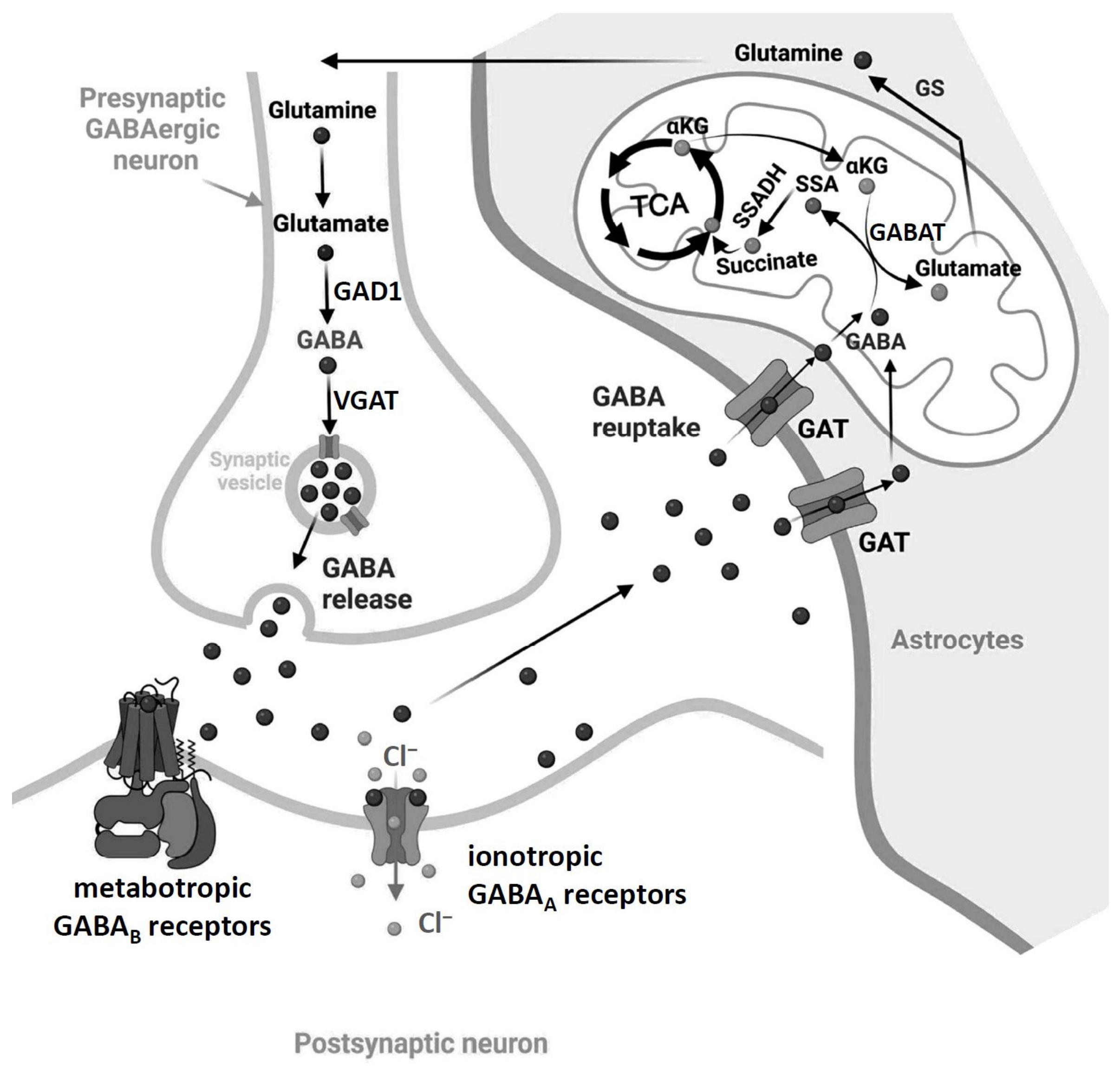
In both deuterostomes and protostomes, γ-aminobutyric acid (GABA) is the predominant inhibitory neurotransmitter in the central nervous system (CNS). It plays a central role in controlling the activity of most neural networks and consequently influences a wide range of behaviors. In humans, impaired GABA signaling is associated with sleep disorders, such as insomnia and narcolepsy, making GABA signaling a key pharmacological target to treat sleep disorders. GABA is also an important mediator of rapid inhibitory synaptic signaling in the nervous system of insects. Its main task is to dampen the excitability of nerve cells and regulate signal transmission. Without GABA, the nervous system would collapse due to overexcitement. This review article covers the latest research developments in the field of GABA research in insects, including biosynthesis, reuptake, and its receptors (Figure 1). In addition, the review summarizes the physiological and behavioral effects of GABA, particularly in the American cockroach (Periplaneta americana) and the fruit fly (Drosophila melanogaster).

2. Biosynthesis of GABA

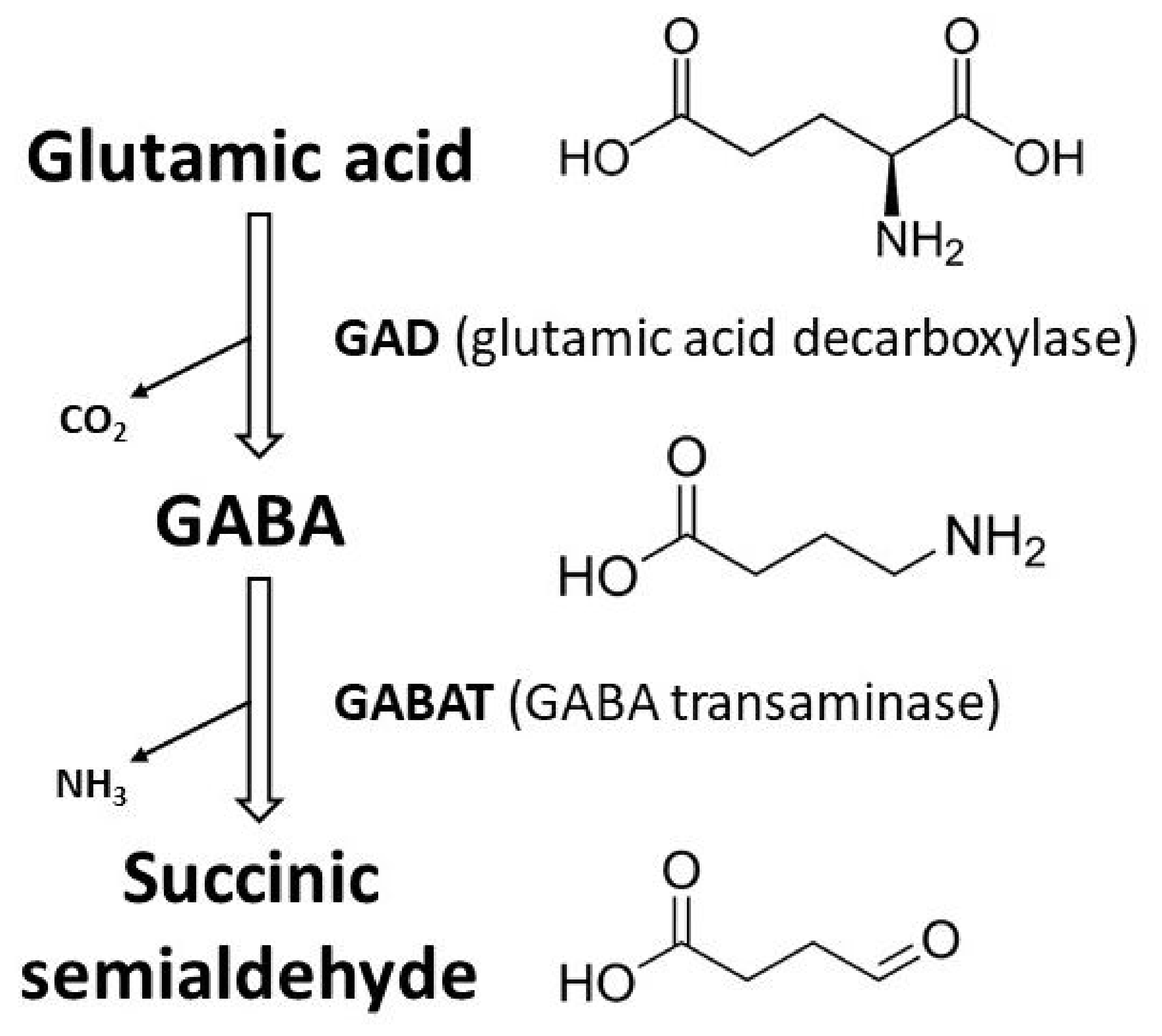
In D. melanogaster, GABA is synthesized by glutamic acid decarboxylase (GAD; Figure 2) enzymes, including DmGAD1 (expressed exclusively in neurons) and DmGAD2 (expressed exclusively in glia) [2,3]. Dmgad1 encodes a 57 kDa protein that is exclusively distributed in the nervous system during embryogenesis and in adult flies [4]. Biochemical studies have shown that DmGAD1, like GAD in mammals, catalyzes the formation of GABA from glutamate with similar efficiency and kinetics [5]. DmGad2 (aka Adc) is a second D. melanogaster gene with high similarity to mammalian GAD and encodes a similarly sized (58 kDa) amino acid decarboxylase [6]. However, adult heterozygotes with DmGad2 deletions show wild-type GAD activity, and the expression pattern of DmGAD2 suggests a role in glia rather than neurons [6]. Thus, DmGAD1 appears to be fully responsible for the enzymatic conversion of glutamate to GABA in D. melanogaster neurons. DmGAD1 expression can be detected for the first time approximately 16 h after egg laying [7]. The expression of DmGAD1 and DmGAD2 in the antennal lobe was mapped. It was found that only a few neurons release GABA, while most neurons in the antennal lobe receive inhibitory signals [8]. The expression level of DmGAD1 is regulated by TBPH, the homolog of the human TAR DNA binding protein 43 (TDP-43), by facilitating GAD1 pre-mRNA splicing [9,10]. Consequently, the GABA level is significantly reduced in brains of TBPH-null flies compared to wild type controls [10].
Interestingly, DmGAD1 is involved in the formation of synapses at the neuromuscular junction (NMJ) and is involved in postsynaptic glutamate receptor field formation at NMJ synapses [11]. Thus, DmGAD1 in the presynaptic motor neuron appears to regulate the glutamate level locally, which in turn regulates the level of receptors on the postsynaptic muscle cell [11].

3. Uptake Mechanisms

Neurotransmitter transporters are responsible for the movement of neurotransmitters across biological membranes. Vesicular and plasma membrane transporters for neurotransmitters represent two different activities (for reviews, see: [12,13,14]). Vesicular neurotransmitter transporters are localized in the membrane of secretory vesicles and are responsible for the transport of neurotransmitters into the vesicle lumen for storage. Plasma membrane neurotransmitter transporters are responsible for the termination of synaptic transmission and the recycling of neurotransmitters after their release. Both the vesicular and the plasma membrane transporters belong to the Solute Carrier (SLC) family of proteins and share several characteristic features (for reviews, see: [12,14]).

3.1. The Vesicular GABA Transporter (VGAT)

Vesicular transporters utilize an “antiport” mechanism in which a proton gradient is used to direct the movement of the neurotransmitter in the opposite direction and into the lumen of the secretory vesicle. Vesicular transporter subfamilies include SCL32, of which the vesicular GABA transporter (VGAT) is the lone member [13]. The D. melanogaster VGAT (DmVGAT) appears to be expressed in all GABAergic neurons in the larva, as it co-localizes precisely with GABA in the ventral nerve cord, and is also expressed in most, if not all, adult GABAergic neurons [2,15]. Mutation of DmVGAT leads to developmental lethality [15]. However, inducible expression of a DmVGAT transgene has been used to restore DmVGAT function during development, thereby enabling behavioral studies in adults. One adult phenotype detected in the conditional rescue line was a surprisingly specific defect in the detection of small objects in the fly’s visual field [15].

3.2. The Putative Vesicular Amino Acid Transporter Mahogany (mah)

The D. melanogaster gene mahogany (mah, CG13646), which was originally identified as an eye color mutant, encodes a putative amino acid transporter belonging to the SLC38 protein family involved in the transfer of amino acids to granules [16,17]. In humans, there are 11 members of this family, and they are all Na+-dependent amino acid transporters that are widely distributed in many tissues of the body, including the nervous system [18]. The SLC38A8 member of this family is present in both inhibitory and excitatory neurons in the mouse brain and preferentially transports glutamine, alanine, arginine, histidine and aspartate into neurons [19]. Of particular interest is that this transporter is thought to play an important role in the glutamate/GABA–glutamine cycle in the brain [20], in which glutamine, the amidated form of glutamic acid, serves to regulate glutamate and GABA stores in nerve terminals. Most interestingly, mah is consistently and reproducibly downregulated twofold in a hyperaggressive line of fruit flies, the so-called bully line [21]. Furthermore, an experimental reduction of mah expression by about half resulted in a hyperaggressive phenotype that partially resembles that of bully flies [21].

3.3. The GABA Transporter (GAT)

Neurons have long been recognized to be capable of “importing” a variety of substrates by way of specific, high affinity, Na+-dependent, plasma membrane transporters. These proteins overcome the energetic barrier for substrate transport by using the electrochemical gradient and, simultaneously with the substrate, pass Na+ ions into the cell (for a review, see: [14]). Since both Na+ and the neurotransmitter move in the same direction across the plasma membrane, this is by definition a “symport” mechanism. Plasma membrane transporters for GABA (GATs) are members of the SLC6 family, which share a common architecture with 12 transmembrane domains (TMDs) [14].
GATs have been identified in the tobacco hornworm Manduca sexta (MsGAT; [22]), in D. melanogaster (DmGAT; [23]) and in the cabbage looper Trichoplusia ni (TnGAT; [24]). All three insect GATs are similar in their primary structure to mammalian GAT-1. Both MsGAT and TnGAT are sensitive to the mammalian GAT inhibitor DL-2,4-diaminobutyric acid (DABA) [22,24]. In contrast to mammalian GATs, neither MsGAT nor TnGAT are significantly inhibited by β-alanine. Membrane preparations of D. melanogaster embryos, larvae, pupae and adults showed saturable uptake of tritiated GABA and inhibition by DABA and nipecotic acid [25,26]. However, the required concentrations of inhibitors were higher than those used in the tests with heterologously expressed transporters.
DmGAT is expressed exclusively in astrocytes, so that glial cells play an important role in GABA clearance and recycling (Figure 1; [27,28,29]). MsGAT is localized to neuronal processes early in development [30], but in adults the labeling also appears to be restricted to glia. Feeding DABA to larval and adult D. melanogaster flies or feeding nipecotic acid to adult flies disrupts various aspects of motor behavior [25,26]. Knockdown of DmGAT during development also leads to severe locomotor defects in both the larval and adult stages [28]. In addition, depletion of DmGAT suppresses genetically induced seizures in adult flies [27,28]. Reduced GAT levels in astrocytes increase sleep [31]. Interestingly, despite the brain-wide expression of GAT in astrocytes, this hypersomnolence can be largely attributed to increased GABAergic tone in a small group of circadian neurons that promote arousal: the large ventral lateral neurons (l-LNvs; [31]).
The excitatory amino acid transporter 2 (EAAT2; Table 1), which translocates taurine (in addition to aspartate) into the glial sheath, could also modulate GABA signaling. Taurine is a GABAA receptor agonist that accumulates during wakefulness in mammals, and loss of EAAT2 leads to increased sleep in fruit flies [32]. Interestingly, feeding flies taurine also promotes sleep [33].

4. Degradation of GABA by γ-Aminobutyric Acid Transaminase (GABAT)

In glia, GABA is metabolized via the GABA shunt, centered on the GABA transaminase GABAT (Table 1; Figure 1 and Figure 2; [34]). GABAT is a mitochondrial enzyme, which breaks down GABA into the products succinic semialdehyde and glutamate. Both succinic semialdehyde and glutamate can enter the citric acid cycle via independent pathways. The glutamate produced by GABAT can also be converted to glutamine by glutamine synthetase, and glutamine is transferred to neurons for resynthesis of GABA.
In D. melanogaster, gabat is expressed in neurons and non-GABAergic glial cells [35]. Loss of GABAT leads to an accumulation of GABA in the brain, increases sleep [35] and affects metabolism, such that flies without GABAT cannot survive on carbohydrate media [34]. In contrast to the metabolic effects, the effects of GABAT on sleep are not dependent on glutamate, suggesting that GABAT regulates metabolic and sleep homeostasis through independent mechanisms [34].

5. GABAA Receptors Are GABA-Gated Cl Channels

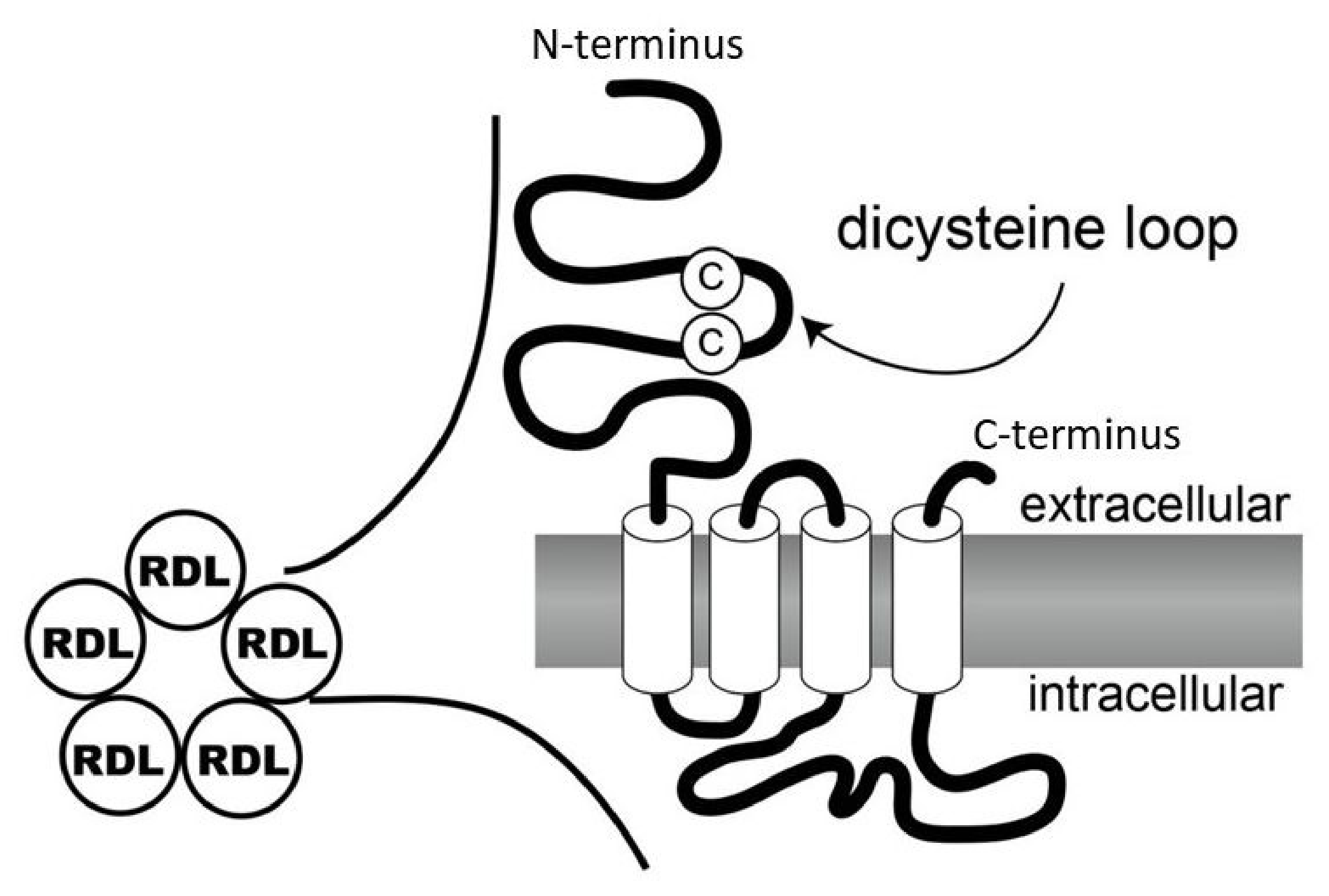
Once released from the presynaptic terminal, GABA causes signals either by stimulating fast-acting ligand-gated ion channels (LGICs; ionotropic GABAA receptors) or more slowly acting G-protein-coupled receptors (GPCRs; metabotropic GABAB receptors) (Table 2; [36,37,38]). Ionotropic GABAA receptors belong to the ligand-gated Cl channel (LCCH) superfamily and are composed of five subunits that facilitate the passage of Cl through a central pore upon activation with an agonist [39,40,41]. Conserved structural features of the LCCH protein family include four hydrophobic TMDs (M1-M4), a highly divergent intracellular loop (IL) between M3 and M4, and a large extracellular N-terminal domain containing the dicysteine loop formed by the covalent linkage of two cysteine residues separated by 13 amino acid residues (Figure 3; [39]). This dicysteine loop is the hallmark of all known members of the superfamily of LGICs.
A segment of the extracellular domain is bounded by two conserved tetrapeptide elements (binding domains BD1 and BD2). These are considered to be determinants of ligand binding to mammalian GABAA receptor β subunits [43]. Here, BD1 and BD2 each contain conserved amino acid motifs and are separated by 41–43 amino acid residues. Only three LCCH proteins from D. melanogaster (Rdl, Lcch3 and Lcch-14A; see below) show a high degree of conservation of the mammalian GABAA receptor consensus structure in this region [39]. The Lcch-47C sequence is also similar, lacking the second conserved tyrosine in BD1, but otherwise matching the GABA receptor consensus structure.
In mammals, these channels are targets of important drugs with sedative and CNS-depressant activity. In arthropods, GABA-gated Cl channels are the targets of an expanding number and variety of acaricides and insecticides, including cyclodienes, phenylpyrazoles, avermectins, and isoxazolines (see: Section 5.5; [44,45,46,47]). It has been experimentally proven that GABAA receptor subunits and their splice variants differ in developmental expression and in their cellular and subcellular distribution [48,49].

5.1. Resistant to Dieldrin (Rdl, Lcch1, CG10537)

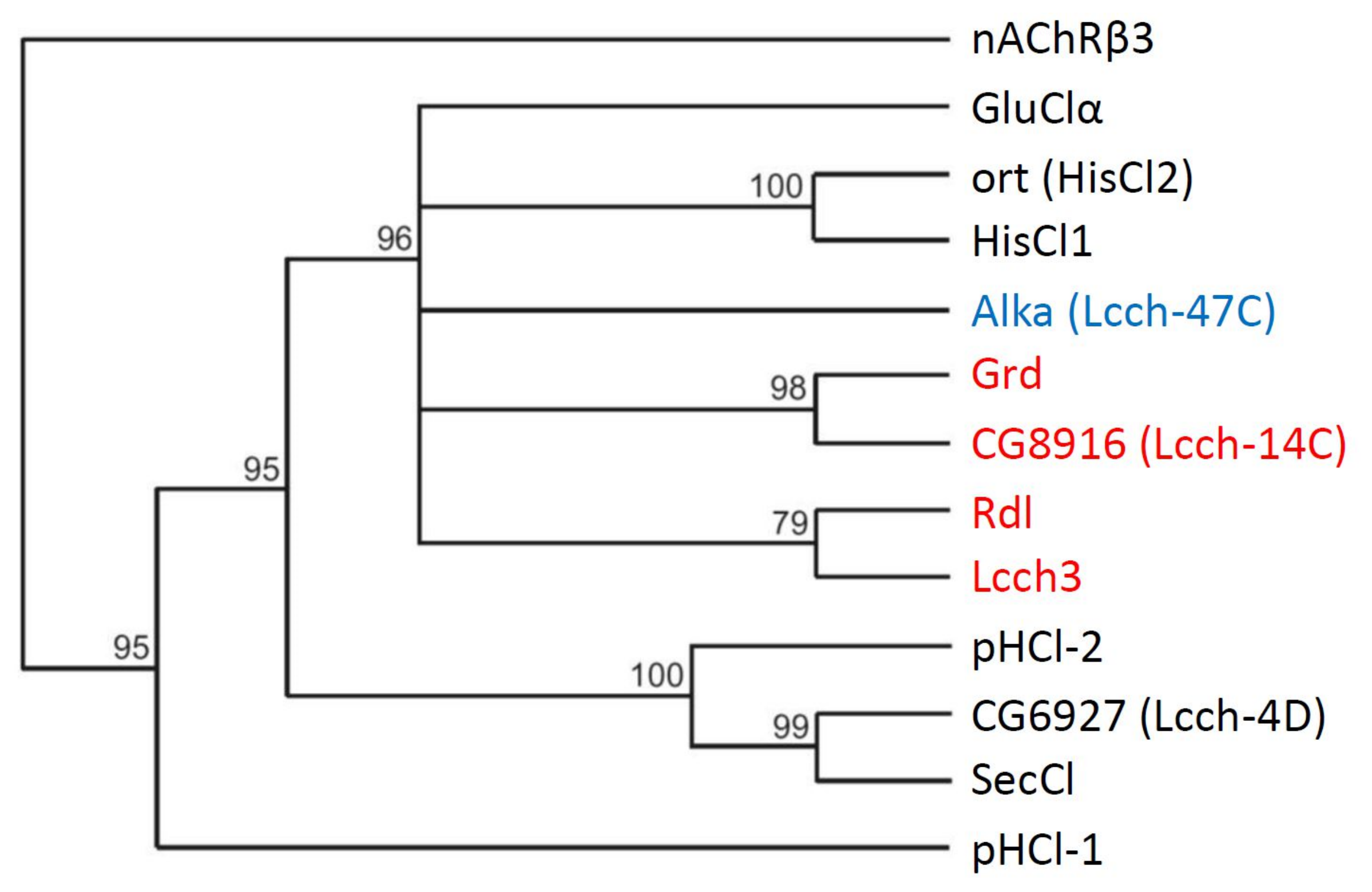
A D. melanogaster strain with resistance to dieldrin [50] and reduced neuronal sensitivity to dieldrin and picrotoxin (PTX; [51]), which were known to block GABA receptors, provided a starting point for the identification of putative genes for GABA receptor subunits in this species. The resistance gene, designated Resistance to dieldrin (Rdl; Table 2; Figure 4), encodes a protein with a high degree of amino acid sequence similarity to the β-subunits of mammalian GABA receptors [52]. The restoration of insecticide susceptibility by transformation of dieldrin-resistant flies with the wild-type Rdl gene provided genetic proof that resistance is indeed mediated by a mutation in this GABA receptor subunit [53]. At wild-type receptors, dieldrin acts as a non-competitive antagonist, directly blocking the channel pore. This blockade by dieldrin prevents GABA from exerting its inhibitory effect, leading to sustained over-excitation of the CNS, convulsions, and ultimately death. Alternative splicing of the transcript derived from the Rdl gene can give rise to different isoforms of this subunit [54,55]. The Rdl genes of other studied insect species such as Apis mellifera [56], Tribolium casteneum [57], Acyrthosiphon pisum [58], Blatta germanica and P. americana [59] also show alternative splicing. An additional level of structural diversification may result from adenosine-to-inosine pre-mRNA editing, which can lead to amino acid substitutions with important functional consequences [60]. The functional expression of transcripts modified by both alternative splicing and RNA editing shows that the combination of the two editing methods produces a number of different subtypes with differing sensitivities to the agonist GABA [61].
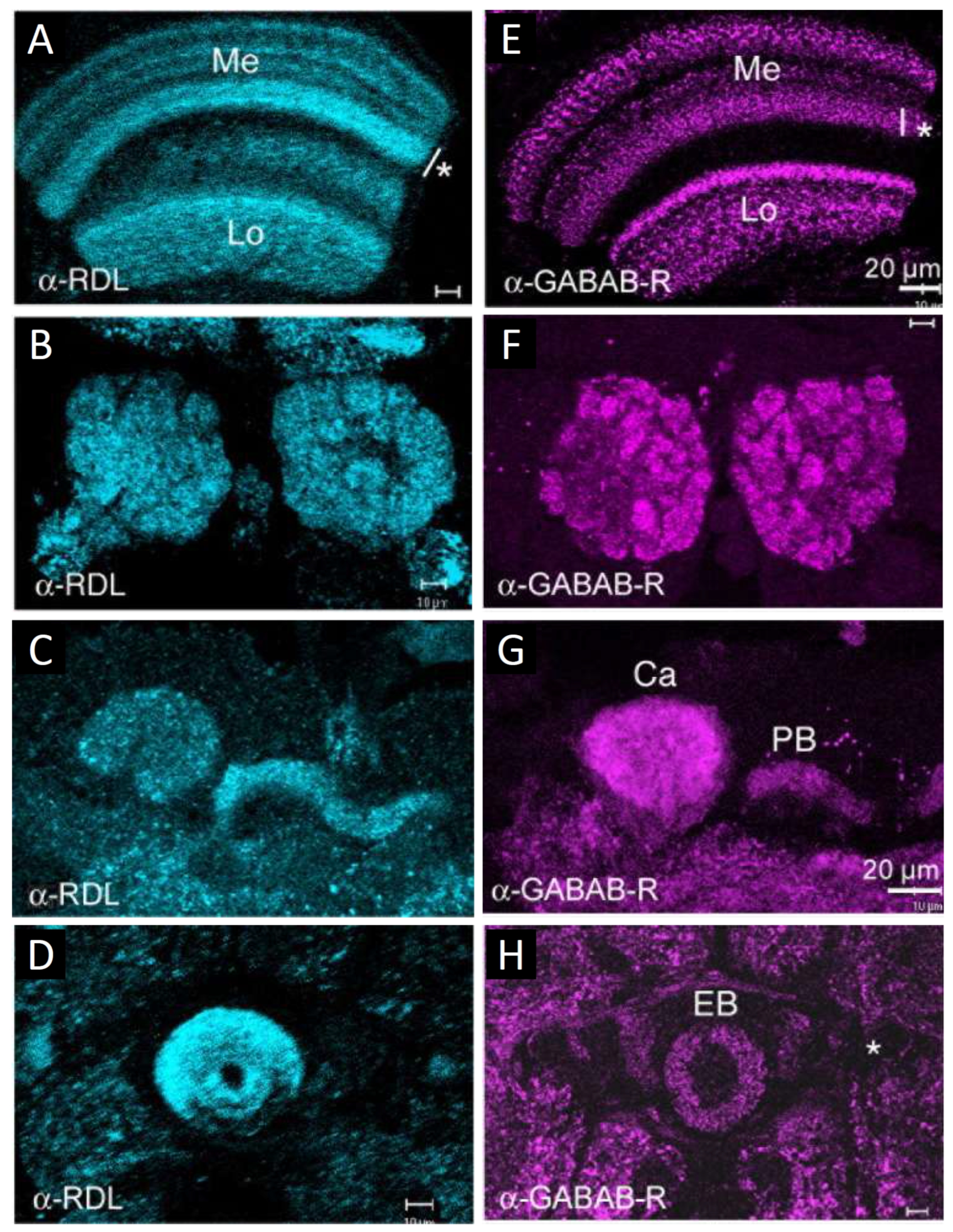
In D. melanogaster, the expression pattern of the Rdl subunit has been well characterized by various methods [62,63,64]. Rdl is mainly expressed in the nervous system of the fly throughout development [64]. Distinct expression of Rdl has been found in the developing nervous system of late-stage embryos (stages 14–16; [63]). In adult flies, Rdl is highly expressed in the antennal lobes, the mushroom bodies, the optic lobes, the ventrolateral protocerebrum and the central complex (Figure 5; [2,63]). In the lobula plate of the optic lobes, both horizontal and vertical tangential cells express the Rdl receptor, and both are adjacent to GABAergic terminals (Figure 5; [2,65,66,67]). Overall, the expression pattern suggests a widespread presence of Rdl-containing GABA receptors in the nervous system of D. melanogaster.

5.2. Glycine Receptor (Grd, Lcch2, CG7446)

The cDNA for Grd, GABA and glycine receptor-like subunit of D. melanogaster (Table 2; Figure 4), was isolated by Harvey and colleagues [68]. Interestingly, the Grd sequence contains a 73-amino-acid insertion in the extracellular domain that is not present in any other member of the protein family. The Grd protein differs greatly from the consensus structure of mammalian GABAA receptors, as it has a divergent BD2 sequence and an extremely long segment (117 amino acid residues) between BD1 and BD2 [39].

5.3. Ligand-Gated Chloride Channel Homolog 3 (Lcch3, CG17336)

A PCR-based strategy to isolate LCCH sequences from D. melanogaster utilized a conserved amino acid sequence “signature motif” found in the TMD M2 of mammalian GABA and glycine receptor subunits and yielded three genomic DNA fragments, designated Lcch1, Lcch2, and Lcch3 [69]. Lcch1 is identical to Rdl, while Lcch2 is identical to Grd. The cDNA for Lcch3 (Table 2; Figure 4) encodes another protein with strong sequence similarity to the mammalian GABAA receptor β subunits [69].
In contrast to Rdl expression in embryonic development, Lcch3 appears very early (stage 11) in the developing neuroblasts before neuronal differentiation [62]. Later, Lcch3 is present in the neuronal cell bodies of the embryonic nerve cord and brain as well as in neuronal cell bodies surrounding the optic neuropil of adult flies.
Two other putative LCCH subunits of D. melanogaster, Lcch-14A (CG8916) and Alkaliphile (Alka, Lcch-47C, CG12344) have been suggested by several authors as possible candidates for GABAA receptor subunits (Table 2; Figure 4; [39,70,71]). The CG8916 ortholog from the Asiatic rice borer Chilo suppressalis (Cs8916) was recently studied to determine whether it can form part of a functional ionotropic GABA receptor [70]. Cs8916 does not form a functional ion channel when expressed alone. However, Cs8916 can form heteromeric ion channels that respond to GABA or β-alanine when expressed in combination with either CsLcch3 or CsRdl1 (see: Section 5.4; [70]). The CG8916 ortholog of the silk moth Bombyx mori is expressed in Corazonin neurons and participates in progeny diapause determination [72]. Alka subunits have recently been shown to selectively form a Cl channel that is opened by high pH values and expressed in specific gustatory receptor neurons [73]. Thus, the Alka channel enables aversive taste responses to basic food.

5.4. Composition of Functional GABAA Receptors

The expression of functional GABAA receptors in Xenopus laevis oocytes or in stably transformed insect cells provided important confirmation that the Rdl protein is a component of GABAA receptors of D. melanogaster [52]. Subsequent research using this system showed that the pharmacological properties of homomultimeric Rdl receptors in oocytes faithfully reproduce most of the properties of native GABAA receptors in the insect nervous system (for reviews, see: [39,45,48]). For example, native neuronal GABAA receptors and homomultimeric Rdl receptors in oocytes are highly sensitive to activation by muscimol and isoguvacin, but insensitive to the antagonist bicuculline. Rdl homomultimers can also be inhibited by the application of dieldrin or PTX [52]. Patch-clamp recordings of single ion channels in D. melanogaster neurons showed that most native GABA-gated Cl channels can also be blocked by both dieldrin and PTX [74]. The close correspondence of the pharmacological properties of Rdl homomultimers with those of native insect GABAA receptors has led to their widespread use as substitutes for native insect neuronal GABAA receptors, particularly in insecticide efficacy studies.
Attempts to reconstitute heteromultimeric GABAA receptors containing Rdl and any of the other putative GABAA receptor subunits have long met with limited success. Injection of synthetic Rdl and Lcch3 mRNAs into oocytes did not result in functional receptors with properties other than those attributable to homomultimeric Rdl receptors [39]. However, coexpression of Rdl and Lcch3 subunits in insect cells resulted in a population of receptors that exhibited altered kinetics of channel activation and inactivation following GABA exposure and increased sensitivity to bicuculline compared to homomultimeric Rdl receptors in the same system [75]. The properties of these putative heteromultimeric receptors differ from those of native GABAA receptors, which may indicate that heteromultimers containing Rdl and Lcch3 are not present in insects. This conclusion is supported by the observation that the expression patterns of the Rdl and Lcch3 genes hardly overlap in the CNS of D. melanogaster [62]. However, this view has been challenged by a more recent in situ hybridization study, which showed that Rdl- and Lcch3-expressing cells are widely distributed throughout the brain, with no significant differences in their expression patterns [8].
Surprisingly, D. melanogaster Grd and Lcch3 form cation-permeable channels when expressed in Xenopus laevis oocytes [49]. Co-expression studies have shown that heteromeric receptors composed of Grd1 and Lcch3 subunits of the human louse (Pediculus humanus) are also permeable to Na+ [76]. Huang and colleagues [70] studied the Cs8916 subunit of C. suppressalis by co-expressing it with CsRdl1 or CsLcch3 in Xenopus oocytes. When expressed alone, Cs8916 does not form functional ion channels. However, Cs8916 can form heteromeric ion channels when expressed with either CsLcch3 or CsRdl1 [70]. Interestingly, the recombinant heteromeric Cs8916/Lcch3 channel is a cation-selective channel that is sensitive to GABA or β-alanine [70]. In contrast, the heteromeric Cs8916/Rdl1 channel responded to GABA and was Cl-selective, but differed physiologically from CsRdl1 homomers [70].
Despite the general pharmacological similarity of Rdl homomultimers to native receptors, other evidence suggests that at least some native insect GABAA receptors are heteromultimeric complexes of Rdl and at least one other, as yet unidentified subunit [48]. For example, in contrast to neuronal GABA receptors and Rdl homomultimers, GABA receptors on insect neuromuscular endplates are insensitive to activation by isoguvacin. Furthermore, native GABA receptors, both in nerve and muscle, show patterns of sensitivity to allosteric modulation by benzodiazepines that are not found in heterologously expressed Rdl homomultimers. Finally, GABAA receptors on cultured neurons of Rdl flies show a modest reduction in sensitivity to PTX, but not the profound resistance to PTX found in Rdl homomultimers [77].
A series of immunoprecipitation experiments with antibodies against the Rdl or glutamate-gated Cl channel (GluClα) subunits led to the surprising result that some LCCHs in head preparations of D. melanogaster are heteromultimers containing both Rdl and GluClα [78]. In these experiments, individual antibodies were characterized with respect to their ability to precipitate the ivermectin or nodulosporic acid binding activity of solubilized fly head membranes. Antibodies against GluClα precipitated all binding activity for both radioligands. In contrast, antibodies against Rdl precipitated all of the nodulosporic acid binding activity, but only about 70% of the ivermectin binding activity. The simplest interpretation of these data is that both GluClα and Rdl are components of some ivermectin and all nodulosporic acid receptors in the fly head and that there is a second population of ivermectin receptors that contains GluClα but not Rdl. Such a potential presence of hybrid channels could also explain the higher in vivo sensitivity to PTX in some cell types of the medulla of the fly’s visual system [79]. However, attempts to express heteromultimeric GluClα/Rdl receptors by injecting mixtures of synthetic mRNAs into oocytes were unsuccessful [78]. Another study using the orthologs of these two subunits from the house fly Musca domestica showed that small amounts of MdGluClα mRNA can enhance the expression of MdRdl GABAA receptors, but provided no evidence for a population of heteromultimeric MdGluClα/MdRdl receptors with unique properties [80]. Currently, apart from the results of the immunoprecipitation experiments, there is no evidence that GluClα and Rdl are components of a single receptor, and therefore no information is available on the agonist gating and other pharmacological properties of these putative receptors.
In summary, the role of other (putative) GABAA receptor-like subunits (in particular Grd and Lcch3) and their ability to co-assemble with Rdl remains poorly characterized and the exact subunit composition of native GABAA receptors containing Rdl subunits is therefore far from understood.

5.5. GABAA Receptors as Insecticide Targets

Rdl receptors, named after their original identification in mutant D. melanogaster strains that had developed genetic insensitivity to the insecticide dieldrin, are widely distributed in the insect CNS and are the target of numerous insecticides, including cylcodiene insecticides such as dieldrin, aldrin and endrin, as well as phenylpyrazoles such as the now widely used fipronils [42,45]. By blocking GABA-gated Cl channels, these substances cause an indirect excitation of the nervous system by the removal of inhibitory input. Sequencing of several dieldrin-resistant strains of D. melanogaster has shown that a single alanine residue (A301 in D. melanogaster) within the predicted ion channel lining of the receptor (at the beginning of M2) may be replaced by either a serine or a glycine residue to render the strains resistant to dieldrin [45,52]. Replacement of this alanine in heterologously expressed Rdl homomultimers also confers resistance to dieldrin and PTX on the associated GABA-gated Cl fluxes. Importantly, replacement of both A301S and A301G (which are both semidominant) also confer resistance when expressed in vivo in transformed flies [81].
The critical importance of A301 was confirmed by the discovery of another replacement of this residue, in this case with asparagine (A301N), in the fipronil-resistant grasshopper Laodelphax striatellus [82]. In a strain of Drosophila simulans selected for fipronil resistance in the laboratory, both the original replacement A301G and a second replacement T350M were found in M3 [83]. T350M enhances the effect of A301G in heterologously expressed channels [83]. Recent structural modeling studies have shown that both A301 and T350 are close to the proposed cyclodiene/fipronil site within the channel pore [44,81], suggesting that the mutations directly affect binding to this site. These insecticides preferentially bind to the desensitized state of the receptor via allosteric effects [74]. Finally, it is worth noting that the mutations present do not appear to affect the binding and activity of two newer classes of GABAA receptor blockers, the isoxazolines and meta-diamides. According to modeling studies, these substances interact with other residues within the channel pore [44]. They act by allosteric inhibition of the channel, which leads to hyperexcitation and convulsions [84,85]. The effects of specific mutations in the Rdl subunit on the inhibitory effect of isoxazolines and meta-diamides have been investigated and provided valuable information regarding the location and structure of the binding pocket and potential resistance mechanisms [86,87,88,89]. Recently, Zhou et al. [90] used fruit flies with different variants of the Rdl gene to test insecticides from the isoxazoline (i.e., isocycloseram, fluxametamide and fluralaner) and meta-diamide (i.e., broflanilide) classes. While some mutations made the flies less sensitive to certain insecticides, the G335M mutation led to super-resistance to all tested insecticides from these substance groups [90]. The authors hypothesize that residues within the TMDs M1 and M3 of adjacent subunits form the binding pocket for meta-diamide and isoxazoline insecticides [90]. Since the G335M mutation causes homozygous lethality in adult flies, G335 probably plays a crucial role in insect survival. Therefore, it seems unlikely that resistance mutations at this amino acid residue evolve naturally in the field [90]. Labouré and colleagues [41] recently investigated the honeybee Rdl receptor using structural biology methods. The authors identified three specific allosteric binding sites. One of these sites is a previously unknown allosteric site in the TMD region to which the fungal meroterpenoid chrodrimanin B binds [41]. Furthermore, it was shown that the avermectin insecticide abamectin stabilizes a closed-pore conformation [41]. A review by Zhang and colleagues [91] provides a comprehensive overview of recent advances (2021–2025) in the development of GABAA receptor-targeted insecticides, with a focus on design strategies and structure-activity relationships.
In D. melanogaster, the Rdl gene has also been found as duplicated alleles in which two copies of the gene coding for the receptor are located next to each other, one resistant and one susceptible [92]. This ‘forced’ (compound) heterozygosity allows the persistence of a wild-type (susceptible) copy of the gene encoding the Rdl subunit and thus facilitates the persistence of susceptible Rdl subunits in the associated native receptor. The maintenance of wild-type receptor subunits therefore potentially offsets any fitness costs associated with receptors consisting only of resistant subunits [45,93].

6. GABAB Receptors Are GPCR Heterodimers

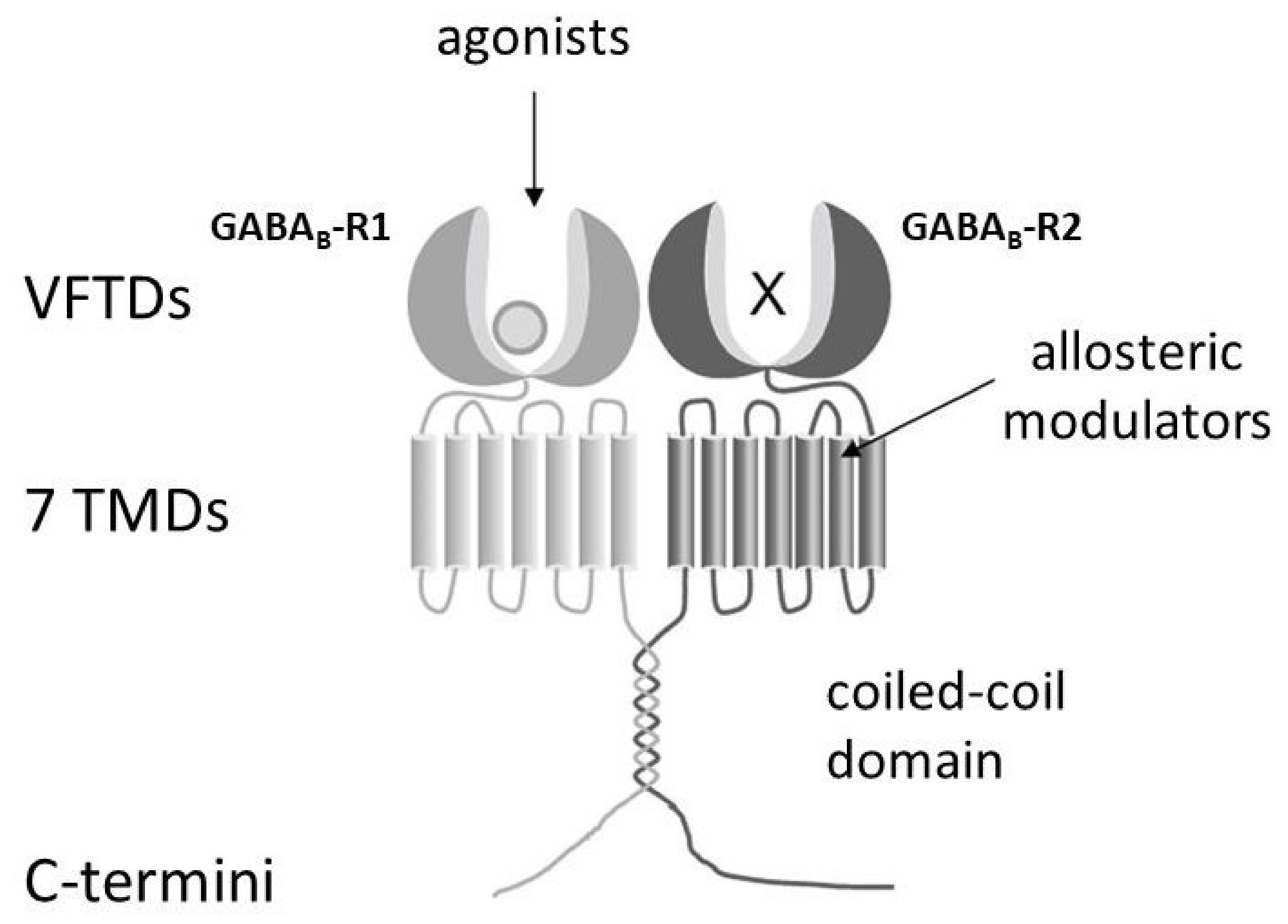
GABAB receptors are class C (or family 3) GPCRs. This group also includes metabotropic glutamate receptors, Ca2+-sensing receptors, sweet and umami taste receptors [38]. Within the GPCR superfamily, class C GPCRs stand out with their large N-terminal Venus flytrap domain (VFTD). To form a functional receptor, two structurally related GABAB receptor subunits, GABAB-R1 and GABAB-R2, have to form obligate heterodimers (Figure 6; [94,95,96]). It is experimentally well established that the VFTD of GABAB-R1 contains the agonist binding site, whereas GABAB-R2 provides coupling to a G protein and allosterically increases agonist affinity at GABAB-R1 [95,97]. Both subunits interact with each other via the extracellularly exposed N-terminal domains [98], the TMDs [99], and a coiled-coil domain formed by the intracellular located C termini [96,100]. Heterodimers may dynamically self-assemble into tetramers (dimers of heterodimers) or even larger complexes, and associate with a range of trafficking, effector, and regulatory proteins ([101,102,103]; for a review, see: [38]). Thus, the molecular complexity of GABAB receptors can be increased on different routes making GABAB receptors highly dynamic structures. Intracellular trafficking of the D. melanogaster subunits DmGABAB-R1 and DmGABAB-R2 was recently investigated [104]. Interestingly, DmGABAB-R1 can reach the cell surface on its own, while DmGABAB-R2 remains in the endoplasmic reticulum unless it interacts with DmGABAB-R1. Retention in the endoplasmic reticulum involves the IL2 of DmGABAB-R2. The intracellular coiled-coil domain enables transport of the DmGABAB-R1/R2 heterodimer to the cell surface [104].
Activated GABAB-R1/2 heteromers inhibit adenylyl cyclase activity via Gαi-subunits of heterotrimeric G proteins. This leads to reduced levels of intracellular cAMP ([cAMP]i). In addition to Gαi-dependent signaling, Gβγ-subunits may mediate inhibition of N-, P- or Q-type Ca2+ channels [106,107] or activation of K+ channels, mainly inward rectifying Kir3 channels, upon GABAB receptor activation [96]. Especially the ability of GABAB receptors to inhibit Ca2+ channels has a tremendous impact in controlling neurotransmitter release.
Three GABAB receptor subunits (DmGABAB-R1, DmGABAB-R2 and DmGABAB-R3) have been molecularly characterized from D. melanogaster (Table 2; [37]). Recently, Hou and colleagues [108] determined the cryo-EM structures of the D. melanogaster GABAB-R1/2 heteromer both in the inactive state (with an antagonist bound) and in the active state (with the agonist GABA bound). Like its human homolog, the D. melanogaster GABAB receptor exhibits asymmetric activation. However, a larger inactive interface spanning TMDs 3–5 of the two subunits is responsible for preventing constitutive activity of the D. melanogaster GABAB receptor [108]. The ordered C-terminus of DmGABAB-R2, together with IL1, IL3, and TMD3, forms a pocket that enables coupling to the Gi protein, whereas IL2 of DmGABAB-R2 is less involved [108]. This contrasts with the situation in most class C GPCRs, in which IL2 is crucial for Gi protein coupling [109,110].
Physiologically, D. melanogaster GABAB receptors are necessary for normal development [111], the regulation of carbohydrate and lipid metabolism [112], the modulation of olfactory and gustatory processing [113,114,115,116], the regulation of circadian activity [117], sleep maintenance [118], and the behavior-impairing effects of ethanol [119]. Interestingly, the distribution of the DmGABAB-R2 protein matches closely that of GABAA receptors (Rdl immunoreactivity) in most areas of the fly brain (Figure 5; [2]). However, differences exist especially in the mushroom body lobes [2]. Recently, Nakamizo-Dojo and colleagues [120] identified a small group of GABAergic neurons in the brain of D. melanogaster larvae, termed subesophageal-zone-localized descending GABAergic neurons (SDGs), which connect to the axonal terminals of a specific class of peripheral nociceptive neurons (C4da) in the ventral nerve cord. These SDGs inhibit the synaptic activity of C4da neurons via GABAB receptors, thereby terminating escape rolling behavior [120].
In D. melanogaster, astrocytes show an increase in DmGAT concurrent with synaptogenesis, which is fine-tuned through astrocytic GABAB-R1/2 signaling [27]. This means that astrocytes regulate DmGAT levels by directly measuring extracellular GABA. Furthermore, inhibition of astrocytic GABAB-R1/2 signaling suppresses seizure activity in mutants with hyperexcitable neurons (see: Section 7.2.5; [27]), and knockdown of the GABAB receptor alters the morphology of the tracheae of D. melanogaster [111].
Not much is known yet about the significance of the third GABAB receptor subtype of D. melanogaster, DmGABAB-R3, which is expressed in a similar, albeit slightly different, spatiotemporal pattern to Rdl [8]. When DmGABAB-R3 is genetically ablated from a specific set of clock neurons, the period of the fly’s locomotor activity increases (see: Section 7.2.1; [121]).
Meanwhile, the molecular structure and protein localization of two cockroach GABAB receptor subunits, PaGABAB-R1 and PaGABAB-R2, have been elucidated [122]. Activation of heterologously expressed PaGABAB-R1/PaGABAB-R2 heteromers results in specific inhibition of adenylyl cyclase activity [122]. The affinity of the heteromeric receptor is quite high, with an EC50 for GABA of about 85 nM (Table 3; [122]). The effect of GABA is mimicked by two synthetic agonists, namely SKF97541 and 3-APPA with EC50 values between 72 and 93 nM. CGP 55845 and CGP 54626 have been identified as potent competitive antagonists of the PaGABAB-R1/2 receptors (Table 3; [122]).
Blankenburg et al. [123] also investigated the functionality and pharmacological profile of a chimeric GABAB receptor consisting of the GABAB-R1 subunit from P. americana and the GABAB-R2 subunit from D. melanogaster. Interestingly, activation of these chimeric heteromers also led to inhibition of adenylyl cyclase activity [123]. Thus, individual subunits of obligate heteromeric GABAB receptor proteins can be replaced by orthologous and possibly even distantly related GABAB receptor subunits [123,124].

7. Insect Models for Studying Physiological and Behavioral Effects of GABA

The available literature on physiological and behavioral processes in which GABA and GABA receptors are involved in insects is almost unmanageable. Therefore, I would like to limit myself to two insect species in the following, the American cockroach (P. americana) and the fruit fly (D. melanogaster). As far as the assignment of individual receptor subtypes to such effects is concerned, D. melanogaster has once again proven to be an unsurpassed model system.

7.1. Periplaneta americana (Blattodea)

The American cockroach, P. americana, is an established model in insect neurobiology that is easily accessible for physiological and pharmacological experiments [125,126,127]. For example, biochemical and electrophysiological examinations can be performed on the same tissue [126]. Thus, the use of cockroach neurons has resulted in many instructive studies investigating the actions of insecticides on LGICs as well as GPCRs in the insect nervous system.

7.1.1. Receptors

Studies on the fast coxal depressor motor neuron in P. americana [128] have shown that GABA activates a Cl channel. The use of this and other identified neurons has enabled systematic analysis of the response to GABA and a range of ligands (for a review, see [126]). For example, Wafford and colleagues [129] demonstrated that the cyclodiene endrin non-competitively blocks the GABA response of the fast coxal depressor motor neuron. Using a preparation from nerve cords of cockroaches, endrin was able to completely block GABA-stimulated 36Cl uptake. These results showed that cyclodienes can block native neuronal GABAA receptors in insects. At around the same time, the conductance properties of GABA-gated Cl channels in dissociated adult cockroach neurons were reported [130]. Later, GABA-mediated currents were studied in short-term cultured dorsal unpaired median (DUM) neurons of the terminal ganglion of P. americana using the whole-cell patch-clamp technique [131,132]. The current–voltage curve was found to be biphasic [131,132]. This bimodal dose–response curve reflects the presence of two distinct GABA receptor subtypes in DUM neuron somata, designated GABAR1 and GABAR2. GABAR2 is positively regulated by Ca2+ influx via a channel containing a TRPγ protein, which activates Ca2+/calmodulin-dependent protein kinase (CaMKinase) II [131,133]. In contrast, Cl currents through GABAR2 are selectively blocked by activation of protein kinase C (PKC) and potentiated by PKC inhibition [132]. The authors suggest that the observed positive effect of CaMKinase II could occur through an inhibition of PKC activity [132]. Interestingly, the two GABA receptor subtypes respond differently to the phenylpyrazole insecticide fipronil, with GABAR1 being less sensitive to fipronil than GABAR2 and the sensitivity of GABAR2 to fipronil decreasing when PKC is inhibited [132]. The genes of the GABAA receptor subunits Rdl, Grd, Lcch3, and other putative subunits of LGICs of P. americana have now been annotated [59].
The existence of GABAB receptors in insects was first demonstrated in P. americana after application of the GABAB receptor agonist 3-aminopropylphosphinic acid (3-APPA) into the abdominal ganglion [134]. This treatment resulted in a hyperpolarization of the membrane potential and was resistant to the specific GABAA receptor antagonist PTX. Similarly, the GABAB receptor agonists 3-APPA and SKF 97541 induced hyperpolarization of the fast coxal depressor motor neuron [135].
As mentioned above (see: Section 6), the molecular structure and protein localization of two GABAB receptor subunits of the cockroach, PeaGABAB-R1 and PeaGABAB-R2, have been elucidated, and the pharmacological profile of the heterologously expressed heteromer has been investigated [122].

7.1.2. Olfaction

Groundbreaking studies on cockroaches using various approaches have contributed significantly to our understanding of the general mechanisms and principles of olfactory information processing from the outset (e.g., [136]). In cockroaches, as in all insects, the detection of odor signals takes place in various types of antennal olfactory sensillae, which contain the olfactory sensor neurons (=first-order neurons). The initial synaptic processing of odor information in the antennal lobe glomeruli is carried out by two important types of neurons: local interneurons and projection neurons (for a review, see: [125]). The former mediate complex inhibitory and excitatory interactions between and within the glomeruli to form an olfactory representation in the antennal lobe [137]. The latter transmit the processed information to higher-level processing regions of the protocerebrum, in particular the mushroom bodies. It has long been known that a specific type of local interneurons, the spiking type I local interneurons, and some projection neurons of P. americana are GABAergic [137,138,139,140]. Synaptic inputs induced by spike trains in presynaptic local interneurons trigger inhibitory postsynaptic potentials (IPSPs) in both projection neurons and local interneurons [141]. These consist of both slow, sustained components and fast, transient components. The fast IPSPs are mediated by GABAA receptors, while the slow, sustained IPSPs are mediated by GABAB receptors [141].
There are essentially two main types of projection neurons (=second-order neurons) that form the main output pathway from the antennal lobe. These are uniglomerular projection neurons, which receive synaptic inputs exclusively in one glomerulus, and multiglomerular projection neurons, which integrate synaptic inputs from many glomeruli (for reviews, see: [125,142]). The projection neurons typically respond to a variety of odors with a series of impulses, as do the olfactory sensory neurons. Two subtypes of uniglomerular projection neurons can be distinguished, which receive input from different groups of glomeruli and thus from different classes of olfactory sensory cells housed in different types of antennal sensillae [143]. They are referred to as type 1 and type 2 projection neurons [143,144,145,146,147]. These two classes of projection neurons terminate in different areas of the mushroom bodies, specifically in the basal and lip regions of the mushroom body calyces, where class I and class II Kenyon cells (intrinsic neurons of the mushroom bodies = third-order neurons) extend dendrites, respectively [143,148]. In contrast to projection neurons, Kenyon cells respond only to certain odors with a few impulses, thus achieving sparse odor coding. One mechanism that contributes to the formation of spatially and temporally sparse odor representations in populations of Kenyon cells is widespread and broadly coordinated GABAergic inhibition, which feeds back output signals from the mushroom bodies to Kenyon cells. In fact, the basal and lip regions of the mushroom body calyces also contain terminals of various classes of GABAergic interneurons known as calycal giants [149,150,151]. These GABAergic interneurons apparently have a feedback function [150]. It can therefore be concluded that dual parallel processing of odors is not only maintained from the olfactory sensory neurons to the Kenyon cells, but is likely to continue up to the feedback circuits of the mushroom bodies [150].

7.1.3. Salivary Gland Physiology

The salivary glands of P. americana are a well-established model system for investigating the role of neuroactive substances in epithelial physiology and stimulus-secretion coupling in exocrine glands (for reviews, see: [127,152]). The salivary duct nerve (SDN), which descends from the subesophageal ganglion, innervates the paired salivary glands. In addition to serotonergic fibers, the SDN contains two relatively thick axons, the dopaminergic salivary neuron (SN)1 and the GABAergic SN2 [153,154]. Pharmacological experiments have provided evidence that GABAB receptors are involved in saliva production and/or secretion [154]. When GABA is applied during electrical stimulation of the SDN, the electrical and secretory response of the exocrine cells is enhanced, whereas GABA has no apparent effect without simultaneous electrical stimulation. Because GABAB receptor agonists mimicked and GABAB receptor antagonists blocked these effects, GABA has been suggested to act presynaptically via GABAB receptors either on serotonergic and/or dopaminergic nerve fibers [154]. Meanwhile, PeaGABAB-R1-like immunoreactivity has been detected in GABAergic fibers innervating the salivary glands, suggesting that GABAB receptors act as autoreceptors in SN2 of P. americana [122].

7.2. Drosophila melanogaster (Diptera)

D. melanogaster has a history of more than a century in genetic research and has been intensively studied for almost half a century as a versatile model organism for behavioral genetics and neurogenetics. The fly has biological characteristics that are advantageous, such as its small body size, ease of breeding and handling, high offspring production, short life cycle, and only four sets of chromosomes. The genome of D. melanogaster was completely sequenced in 2000 [155]. Another advantage is the availability of valuable resources, including a genome-wide transgenic RNA interference (RNAi) library and many mutants, such as insertion mutants with various types of transposable elements, as well as highly sophisticated genetic tools. Its relatively simple CNS is another advantageous biological feature for uncovering the molecular and neuronal basis of various behaviors through neurogenetic studies.
In D. melanogaster, although GABA appears in detectable amounts relatively late in development, it is distributed throughout the nervous system, and about 20% of neurons show GABA immunoreactivity [7]. Based on single-cell transcriptomics, it is estimated that the central brain of D. melanogaster contains approximately 10% (10,000) GABAergic neurons [156]. Various modulatory effects of GABAergic signaling on, e.g., olfactory perception [157], olfactory memory [158], locomotion [26], sleep [1,159], male aggression [160], and feeding [161] have been reported in this genetic model organism. In the following, I will limit myself to functional circuits: circadian timekeeping, sleep, vision and olfaction, as well as to a few disease models in D. melanogaster in which GABAergic signaling plays a central role.

7.2.1. Circadian Timekeeping

The central clock network in D. melanogaster consists of approximately 150 neurons, which include the small and large ventral lateral neurons (s-LNvs and l-LNvs, respectively), the dorsal lateral neurons (LNds), the dorsal neurons (DNs) and the lateral posterior neurons (for recent reviews, see: [162,163,164]). Of the metabotropic GABAB receptor subunits, expression of GABAB-R2 and -R3 has been detected in the LNvs of both adult and larval brains [117,121]. GABA has been shown to act on the LNvs and functions as a slow-acting inhibitory neurotransmitter [117,165]. Administration of GABA to dissociated larval s-LNvs or intact adult s-LNvs attenuates intracellular Ca2+ release via activation of metabotropic GABAB receptors, resulting in blockade of spontaneous Ca2+ oscillations [117,165]. Genetic ablation of GABAB-R3 in the LNvs increases the period of locomotor rhythms in adult flies, while hyperexcitatory GABAergic neurons elicited behavioral arrhythmias associated with perturbations in the cyclic expression of the clock genes period and vrille [121]. At the molecular level, GABAB-R3 signaling in the LNvs appears to involve the parallel activation of Gs and Go signal transduction pathways, which have additive effects on period length in adult flies [121,165]. In contrast, GABAB-R2 does not appear to be critical for the maintenance of circadian rhythmicity, as genetic manipulations that downregulate the expression of GABAB-R2 do not alter the locomotor rhythms of adult flies [118]. Interestingly, RNAi-mediated knockdown of the LCCH subunits Grd and Alka disrupts circadian rhythmicity, possibly by altering the glycinergic inhibitory tone of the network [166].
Although GABA is typically considered to be inhibitory, its effects depend entirely on the intracellular Cl concentration ([Cl]i) of the receiving neuron (inhibitory-to-excitatory GABA switch; [167]). The balance between two opposing transporters determines this concentration: The Na-K-Cl cotransporter (NKCC) pumps Cl into the cell. High NKCC activity thus increases [Cl]i, making GABA excitatory and depolarizing. The K-Cl symporter (KCC) pumps Cl out of the cell. High KCC activity thus lowers [Cl]i, making GABA inhibitory and hyperpolarizing. A recent study by Eick and colleagues [168] used D. melanogaster to uncover how this cellular mechanism affects behavior under changing seasonal day lengths. Constant light normally disrupts the circadian clock and causes flies to become arrhythmic [169]. However, altered KCC expression in l-LNvs restores rhythmic locomotor behavior under constant light [168]. The Cl transport balance is tightly regulated by a Cl-sensitive Wnk/Fray kinase feedback loop. When KCC, NKCC, Wnk, or Fray are downregulated, the normal rhythmic breakdown caused by constant light is bypassed. During long summer days, the flies’ morning activity is reciprocally adjusted. Knocking down KCC reduces morning behavioral activity, whereas knocking down NKCC increases morning behavioral activity [168]. The light-sensitive, clock-regulated protein Quasimodo interacts directly with NKCC and suppresses its activity to inhibit excessive locomotor behavior in the morning of long days [168,170]. Thus, the precise control of [Cl]i and GABA polarity by the KCC/NKCC/Wnk/Fray network acts as a molecular regulator that enables the brain’s circadian clock to adjust behavioral timing to seasonal fluctuations in daylight.

7.2.2. Sleep

D. melanogaster is a powerful model for understanding the fundamental neuronal and molecular basis of sleep [1,171,172]. In D. melanogaster, sleep is defined as periods of inactivity lasting at least 5 min [173]. These periods of inactivity are associated with an increased arousal threshold and decreased local field potentials in the brain [174]. Depriving the flies of resting periods leads to increased sleep in the following days, which is characteristic of homeostatic sleep rebound [173]. D. melanogaster passes through distinct sleep stages, with shorter sleep bouts corresponding to lighter sleep and longer resting periods characterizing deeper sleep [175]. In D. melanogaster, sleep plays a crucial role in memory consolidation and waste clearance [176].
GABAergic signaling has been linked to the rhythmic coupling of the circadian clock to sleep–wake cycles. In particular, the modulation of l-LNv’s activity by GABA has received much attention. Indeed, sleep is markedly decreased when the function of the ionotropic GABAA receptor subunit Rdl is reduced in these cells. This can be achieved either by direct downregulation [177,178,179] or by manipulating regulators of its transport to the plasma membrane (WAKE; [180]), its clustering (DNlg4; [181]) and its stability (Fblx4; [182]). On the other hand, pharmacological administration of the GABAA receptor agonist gaboxadol induces sleep behavior in D. melanogaster [183]. In addition to Rdl, the metabotropic GABAB-R2 is also expressed in the l-LNvs. While Rdl mediates rapid inhibitory neurotransmission and is therefore particularly important for sleep initiation, GABAB-R2 acts over longer periods and plays an important role in sleep maintenance [118]. Recently, Charturvedi et al. [31] showed that astrocytic DmGAT decreases GABAergic tone and Rdl activation in arousal-promoting l-LNvs and thus determines proper sleep quantity and quality in D. melanogaster.
GABA also plays a crucial role in the adaptation of sleep to cold temperatures. GABAergic neurons of the antennal lobe, known as type II thermosensory projection neurons (TPN-II), receive cold temperature input from the antennal thermoreceptors. TPN-II neurons inhibit two pairs of circadian neurons called anterior dorsal neurons 1 (DN1as), leading to increased sleep in the morning and increased activity in the evening [184].
Wake- and sleep-promoting intrinsic neurons of the mushroom bodies (Kenyon cells) control sleep through different mushroom body output neurons. Two of the three pairs of sleep-promoting mushroom body output neurons are GABAergic [185]. Most Kenyon cells are subject to GABA inhibition by the dorsal paired median neurons and the anterior paired lateral neurons. These neurons are electrically coupled and also play a key role in learning and memory consolidation [186,187].
Neurons of the fan-shaped bodies, which are part of the central complex, also produce GABA [188,189]. GABA released by these neurons inhibits octopaminergic excitatory neurons and thus promotes sleep [189]. Interestingly, the neurons of the fan-shaped bodies are also themselves the target of GABAergic modulation [190]. Although the fan-shaped bodies appear to be quite important for GABA-dependent sleep regulation, recent work has questioned this widely accepted role [191,192]. Various GABAergic neuron types of the ellipsoid body, another part of the central complex, also modulate sleep duration and architecture [193,194]. In addition, GABAergic control of wake-promoting dopaminergic signaling in the central brain has been demonstrated [195].
Fly research thus reveals how GABA, in combination with other neurotransmitters such as dopamine and serotonin, controls sleep regulation, providing insights into the complexity of insomnia. This makes D. melanogaster an excellent model for sleep disorders, as sleep mechanisms are evolutionarily conserved [1].

7.2.3. Vision

The visual behavior of flies provides another very useful model for genetic studies of GABAergic neurotransmission and behavior. The neuroanatomy of the fly visual system has been mapped in detail at both the light and electron microscopic levels [196,197]. Furthermore, an extensive and sophisticated set of behavioral tests has been developed to study the response of flies to visual stimuli (e.g., [198]).
R1–R6 photoreceptors relay local intensity signals to three lamina monopolar cells, L1–L3, which are arranged in a retinotopic array (for a review, see: [199]). The simultaneous downregulation of both GABAA and GABAB receptors in the R1–R6 photoreceptors and L2 cells increases the effective size of the center of the L2 receptive field and reduces the strength of L2’s peripheral responses [200]. Similar results were obtained by the simultaneous administration of GABAA and GABAB receptor antagonists (PTX and CGP 54626, respectively; [200]). GABAergic manipulations affect not only the spatial shape of the receptive field of L2, but also the amplitudes and kinetics of the responses [200]. Thus, GABAergic lateral interactions tune already early stages of visual processing in D. melanogaster.
The effects of PTX suggested early on that GABAergic signaling is also required for some aspects of motion detection in D. melanogaster [201]. Within the visual processing pathway, T4 and T5 cells are the first neurons to respond to visual motion in a direction-selective manner. T4 cells extend their dendrites into the most proximal layer of the medulla and respond to moving bright edges (ON pathway; [202]). The dendrites of T5 cells receive their input in the most proximal layer of the lobula and are responsive to moving dark edges (OFF pathway; [202]). Cholinergic Tm neurons shape the tuning of T5 responses to the preferred direction of moving dark edges. In contrast, suppression of null direction responses is mediated by a disynaptic mechanism through Tm9/Tm1 cells and the GABAergic large field amacrine cell CT1 [66,203]. The Rdl subunit has been detected in the dendrites of T5 neurons [53,66,203].
In larger flies, both behavioral and electrophysiological studies have been performed to analyze the function of a GABAergic cell type in the lobula plate that is thought to be involved in figure detection [204,205]. The release of GABA from these centrifugal horizontal cells is required for small-field tuning of the figure detection cell FD1 [204,205]. In D. melanogaster, reduced DmVGAT expression interferes with normal object tracking and figure-ground discrimination [15]. Interestingly, neuronal knockdown of the GABAA receptor subunit Rdl in a small population of serotonergic visual feedback neurons—the lamina tangential (Lat) cells—triggers male-male courtship in D. melanogaster [206]. The authors concluded that these neurons fine-tune male courtship via GABA-mediated inhibition [206].

7.2.4. Olfaction

Similar to P. americana (see: Section 7.1.2), GABAergic signal transmission in the fly’s olfactory system has also been extensively studied, particularly in the antennal lobe. In the fly, too, two types of GABAergic neurons (projection neurons and local interneurons) are associated with the antennal lobe [8], and the use of GABA receptor agonists inhibits the function of the antennal lobe. It is assumed that local interneurons regulate the crosstalk between neighboring glomeruli and tune the odor response of the fly to specific odors.
GABA signaling also underlies most slow adaptations of olfactory responses [115,157,207]. GABAergic local interneurons form synapses with olfactory sensory neurons [115,116,157]. The different classes of olfactory sensory neurons differ in their GABA sensitivity [208,209]. The magnitude of the GABA response correlates with the magnitude of the odor response, with larger responses having an inhibitory effect on extreme activities of olfactory sensory neurons and projection neurons [115,208]. This suppression leads to a gain control mechanism that increases the dynamic range of information transmission [115,116,157]. Olfactory sensory neurons also release short neuropeptide F (sNPF), which, unlike GABA, enhances presynaptic Ca2+ responses [210]. In olfactory sensory neurons, the expression of sNPF and DmGABAB-R2 partially overlap [211]. A shift in the balance between GABA and sNPF signaling can thus alter the inputs of olfactory sensory neurons in the antennal lobes [207].

7.2.5. Disease Models

About 75% of disease-relevant human genes have an ortholog in D. melanogaster [212]. The ability to generate transgenic flies that express human proteins in a spatial and temporal manner makes D. melanogaster ideal for the study of human diseases associated with GABAergic signaling. Today, there are over 800 reports on human disease models that contain detailed descriptions of the relationships between specific human diseases and D. melanogaster genes [213].
Epilepsy and seizure disorders
D. melanogaster is used, for example, for modeling seizure conditions. Susceptible strains respond to electrical, mechanical, or heat stimuli with convulsive states (for reviews, see: [214,215]). Susceptibility can be achieved by mutations in ion channels, transporters, mitochondrial proteins, etc., which can induce either temperature-sensitive paralytic or ether-induced leg-shaking phenotypes [216] or a “bang-sensitive” phenotype [217,218,219,220,221]. Using such a “bang-sensitive” mutant, Li and colleagues [222] were, for example, able to show that a ketogenic diet drastically reduces the occurrence of seizure-like activity. Interestingly, blocking GABAB receptors with CGP 55845 partially reversed the anticonvulsant effects this diet [222].
Research by several groups has identified an embryonic critical period during the development of the fly larva’s locomotor network [223,224]. Activity manipulation during the critical period (e.g., mediated by optogenetics or drug exposure) leads to a significant reduction in network stability, promoting a “seizure-like” phenotype in response to strong electrical stimulation [224,225]. Using GABA pharmacology and genetics, Corke and colleagues (2025) manipulated the embryonic GABAergic system and measured an induced seizure phenotype in third-instar larvae. Potentiating GABAergic signaling through exposure to the agonist diazepam or through overexpression of the GABAA receptor subunit Rdl induces a precocious onset of the critical period [226]. By contrast, exposure to the antagonist gabazine or knockdown of the GABA-synthetic enzyme GAD1 delays its onset [226]. Thus, the timing of critical period opening is controlled by the maturation of GABAergic signaling in both insects and mammals.
It is well known that GAT modulates seizure-like activity in D. melanogaster [27,227]. Mutations in the human SLC6A1 gene, which codes for hGAT-1, are also associated with various forms of epilepsy. For example, the R44Q and A288V mutations in hGAT-1 are associated with myoclonic-atonic epilepsy and developmental delay [228]. When these mutated human transporters are expressed in D. melanogaster, the mutant flies exhibit a temperature-sensitive seizure phenotype [229,230]. Furthermore, these heat-induced seizures can be attenuated by certain chemical and pharmacological chaperones that can correct the folding of misfolded transporter mutants and rescue their transport to the cell membrane [231].
Stilwell and colleagues [232] developed a D. melanogaster seizure model back in 2006 using chemical treatment with the GABAA receptor antagonist PTX. This proconvulsant triggers severe generalized seizures in the fly [232]. It was already known that the point mutation A301S within the M2 domain of Rdl causes resistance not only to insecticides (see: Section 5.5), but also to several convulsant compounds, including PTX [52,75]. The fact that the PTX-induced seizure phenotypes observed in wild-type flies are suppressed in Rdl mutants therefore suggests that a specific, conserved GABAA receptor pathway underlies the PTX-induced neuronal effects and lethality in D. melanogaster [232]. In summary, various D. melanogaster models can be used for screening anti-seizure drugs [231,232].
Alcohol Use Disorder (AUD)
Alcohol is a widely used and abused substance that has numerous negative consequences for human health and safety. D. melanogaster is a powerful model for investigating the molecular targets of alcohol because flies model many of the core behavioral elements of alcohol use disorder (for reviews, see: [71,233]). Strikingly, flies show maladaptive behavioral patterns similar to those of humans suffering from alcohol use disorder. For example, after previous experience, flies voluntarily consume alcohol up to pharmacologically relevant internal levels and escalate alcohol consumption [234]. Flies find the pharmacological properties of alcohol rewarding and are willing to work or overcome aversive stimuli to gain access to alcohol [235]. They develop both rapid tolerance to a single exposure and chronic tolerance after repeated exposure [236].
A direct involvement of Rdl or the other GABAA receptor subunits in the modulation of the response of flies to ethanol has not yet been demonstrated. The role of metabotropic GABAB receptors in alcohol-related behaviors such as sensitivity, tolerance and locomotion has been better investigated. Results of these studies suggest that GABAB receptors mediate sensitivity to ethanol and the development of tolerance [119,237].
Fragile X syndrome
Fragile X syndrome is a common hereditary cause of intellectual disability and syndromic autism (for a review, see: [238]). The disease is caused by a loss-of-function mutation in the FMR1 gene. Both GABAA and GABAB receptors have been linked to fragile X syndrome. D. melanogaster strains deficient in dfmr1 are validated models for fragile X syndrome (for reviews, see: [239,240]).
In dfmr1-deficient brain samples, the mRNA expression of the GABAA receptor subunits Rdl, Grd and Lcch3 is reduced by ~40–50% [241]. In addition, the mRNA expression of DmGad is reduced by ~35% [242]. The latter finding was confirmed at the protein level (~30–40% reduction; [243]). Interestingly, rescue experiments with one or two copies of dfmr1 randomly inserted into dfmr1-deficient D. melanogaster show a direct correlation between the amount of Fmr protein and the mRNA level of Gad, Rdl and Grd [241,242].
Neuroanatomical analyses of GABAergic neurons showed no obvious differences in total cell number in dfmr1 mutants [243]. However, single-cell labeling of GABAergic neurons innervating the calyces of the mushroom bodies showed altered subcellular architectural development in dfmr1 mutants, with early undergrowth followed by overelaboration. Functional changes in mushroom body-innervating GABAergic neurons of dfmr1 mutants include altered Ca2+ signaling in response to acute depolarization [243]. In particular, dfmr1 mutants showed an increased and prolonged response to depolarization. These deficits could potentially indicate a compensatory mechanism aimed at stimulating GAD activity by increasing [Ca2+]i or directly enhancing GABA release.
The potential of three compounds, GABA, nipecotic acid (GABA reuptake inhibitor) and creatinine (putative GABAA receptor agonist), to correct specific phenotypes of dfmr1-deficient D. melanogaster was investigated. All three compounds reduced the overexpression of Futsch, a known target of the Fmr protein and D. melanogaster ortholog of the microtubule-associated protein 1B. Second, GABA enhanced structural defects in the mushroom bodies. The medial lobes of the mushroom bodies, which are axon-like fiber structures, normally terminate at the midline. In dfmr1 mutants, however, they cross the midline. GABA supplementation corrected this crossing of the medial lobes in dfmr1 mutants. Finally, GABA rescued the impaired courtship behavior of dfmr1-deficient males towards virgin females. In contrast, dietary supplementation with GABA or nipecotic acid failed to ameliorate the defects in olfactory learning in dfmr1 mutants [243].

8. Conclusions

GABA is the most important inhibitory neurotransmitter in the nervous system of insects. It regulates nerve activity by either opening Cl channels (via GABAA receptors) or lowering [cAMP]i (via GABAB receptors), which leads to hyperpolarization and reduces the excitability of nerve cells. Its receptors (such as the Rdl receptor) are important targets for many insecticides (e.g., cyclodienes, fipronil), which block inhibition, causing overexcitation, convulsions, and death, and are therefore crucial for pest control. Table 4 provides a comparison of the key molecular and pharmacological properties of GABA receptors in insects and mammals. GABA receptors influence numerous physiological and sensory processes in insects, such as salivation, circadian timing and sleep, and the processing of visual and olfactory information. D. melanogaster in particular serves as a powerful model for studying human neurological disorders, utilizing conserved GABAergic signaling pathways and providing insights into seizure disorders (epilepsy), psychiatric disorders, and sleep disorders.

Funding

The writing of this review article was not externally funded. Previous work by the author on GABA receptors was funded by the German Research Foundation (BL 469/4).

Institutional Review Board Statement

Not applicable.

Informed Consent Statement

Not applicable.

Data Availability Statement

No new data were created or analyzed in this study. Data sharing is not applicable to this article.

Acknowledgments

The author has reviewed and edited the output and takes full responsibility for the content of this publication. The author would like to thank Arnd Baumann for his helpful suggestions and critical comments on the manuscript.

Conflicts of Interest

The author declares no conflicts of interest.

Abbreviations

The following abbreviations are used in this manuscript:
3-APPA3-aminopropylphosphinic acid
BDbinding domain
CaMKinaseCa2+/calmodulin-dependent protein kinase
cAMPcyclic adenosine monophosphate
CNScentral nervous system
DABADL-2,4-diaminobutyric acid
DNsdorsal neurons
DUM neuronsdorsal unpaired median neurons
EAAT2excitatory amino acid transporter 2
GABAγ-aminobutyric acid
GABATγ-aminobutyric acid transaminase
GADglutamic acid decarboxylase
GATγ-aminobutyric acid transporter
GluClglutamate-gated Cl channel
GPCRG-protein-coupled receptor
ILintracellular loop
IPSPinhibitory postsynaptic potential
KCCK-Cl symporter
LCCHligand-gated Cl channel
LGICligand-gated ion channel
l-LNvslarge ventral lateral neurons
LNvsventral lateral neurons
NKCCNa-K-Cl cotransporter
NMJneuromuscular junction
PKCprotein kinase C
PTXpicrotoxin
RNAiRNA interference
SDGssubesophageal-zone-localized descending GABAergic neurons
SDNsalivary duct nerve
SLCsolute carrier
s-LNvssmall ventral lateral neurons
SN1salivary neuron 1
SN2salivary neuron 2
sNPFshort neuropeptide F
TBPHTAR DNA-binding protein-43 homolog from Drosophila melanogaster
TDP-43TAR DNA-binding protein 43
TMDtransmembrane domain
TPN-IItype II thermosensory projection neurons
VGATvesicular GABA transporter

References

  1. Chaturvedi, R.; Emery, P. Fly into Tranquility: GABA’s Role in Drosophila Sleep. Curr. Opin. Insect Sci. 2024, 64, 101219. [Google Scholar] [CrossRef] [Scilit] [PubMed]
  2. Enell, L.; Hamasaka, Y.; Kolodziejczyk, A.; Nässel, D.R. γ-Aminobutyric Acid (GABA) Signaling Components in Drosophila: Immunocytochemical Localization of GABAB Receptors in Relation to the GABAA Receptor Subunit RDL and a Vesicular GABA Transporter. J. Comp. Neurol. 2007, 505, 18–31. [Google Scholar] [CrossRef] [Scilit] [PubMed]
  3. Manev, H.; Dzitoyeva, S. GABA-B Receptors in Drosophila. Adv. Pharmacol. 2010, 58, 453–464. [Google Scholar] [CrossRef] [Scilit] [PubMed]
  4. Jackson, F.R.; Newby, L.M.; Kulkarni, S.J. Drosophila GABAergic Systems: Sequence and Expression of Glutamic Acid Decarboxylase. J. Neurochem. 1990, 54, 1068–1078. [Google Scholar] [CrossRef] [Scilit] [PubMed]
  5. Kulkarni, S.J.; Newby, L.M.; Rob Jackson, F. Drosophila GABAergic Systems II. Mutational Analysis of Chromosomal Segment 64AB, a Region Containing the Glutamic Acid Decarboxylase Gene. Molec. Gen. Genet. 1994, 243, 555–564. [Google Scholar] [CrossRef] [Scilit] [PubMed]
  6. Phillips, A.M.; Salkoff, L.B.; Kelly, L.E. A Neural Gene from Drosophila melanogaster with Homology to Vertebrate and Invertebrate Glutamate Decarboxylases. J. Neurochem. 1993, 61, 1291–1301. [Google Scholar] [CrossRef] [Scilit] [PubMed]
  7. Küppers, B.; Sánchez-Soriano, N.; Letzkus, J.; Technau, G.M.; Prokop, A. In Developing Drosophila Neurones the Production of γ-Amino Butyric Acid Is Tightly Regulated Downstream of Glutamate Decarboxylase Translation and Can Be Influenced by Calcium. J. Neurochem. 2003, 84, 939–951. [Google Scholar] [CrossRef] [Scilit] [PubMed]
  8. Okada, R.; Awasaki, T.; Ito, K. Gamma-Aminobutyric Acid (GABA)-Mediated Neural Connections in the Drosophila Antennal Lobe. J. Comp. Neurol. 2009, 514, 74–91. [Google Scholar] [CrossRef] [Scilit] [PubMed]
  9. Romano, G.; Holodkov, N.; Klima, R.; Grilli, F.; Guarnaccia, C.; Nizzardo, M.; Rizzo, F.; Garcia, R.; Feiguin, F. Downregulation of Glutamic Acid Decarboxylase in Drosophila TDP-43-Null Brains Provokes Paralysis by Affecting the Organization of the Neuromuscular Synapses. Sci. Rep. 2018, 8, 1809. [Google Scholar] [CrossRef] [Scilit] [PubMed]
  10. Romano, G.; Holodkov, N.; Klima, R.; Feiguin, F. TDP-43 Regulates GAD1 mRNA Splicing and GABA Signaling in Drosophila CNS. Sci. Rep. 2021, 11, 18761. [Google Scholar] [CrossRef] [Scilit] [PubMed]
  11. Featherstone, D.E.; Rushton, E.M.; Hilderbrand-Chae, M.; Phillips, A.M.; Jackson, F.R.; Broadie, K. Presynaptic Glutamic Acid Decarboxylase Is Required for Induction of the Postsynaptic Receptor Field at a Glutamatergic Synapse. Neuron 2000, 27, 71–84. [Google Scholar] [CrossRef] [Scilit] [PubMed][Green Version]
  12. Blakely, R.D.; Edwards, R.H. Vesicular and Plasma Membrane Transporters for Neurotransmitters. Cold Spring Harb. Perspect. Biol. 2012, 4, a005595. [Google Scholar] [CrossRef] [Scilit] [PubMed]
  13. Deshpande, S.A.; Freyberg, Z.; Lawal, H.O.; Krantz, D.E. Vesicular Neurotransmitter Transporters in Drosophila melanogaster. Biochim. Biophys. Acta BBA-Biomembr. 2020, 1862, 183308. [Google Scholar] [CrossRef] [Scilit] [PubMed]
  14. Martin, C.A.; Krantz, D.E. Drosophila melanogaster as a Genetic Model System to Study Neurotransmitter Transporters. Neurochem. Int. 2014, 73, 71–88. [Google Scholar] [CrossRef] [Scilit] [PubMed]
  15. Fei, H.; Chow, D.M.; Chen, A.; Romero-Calderón, R.; Ong, W.S.; Ackerson, L.C.; Maidment, N.T.; Simpson, J.H.; Frye, M.A.; Krantz, D.E. Mutation of the Drosophila Vesicular GABA Transporter Disrupts Visual Figure Detection. J. Exp. Biol. 2010, 213, 1717–1730. [Google Scholar] [CrossRef] [Scilit] [PubMed][Green Version]
  16. Grant, P.; Maga, T.; Loshakov, A.; Singhal, R.; Wali, A.; Nwankwo, J.; Baron, K.; Johnson, D. An Eye on Trafficking Genes: Identification of Four Eye Color Mutations in Drosophila. G3 Genes Genomes Genet. 2016, 6, 3185–3196. [Google Scholar] [CrossRef] [Scilit] [PubMed]
  17. Romero-Calderón, R.; Shome, R.M.; Simon, A.F.; Daniels, R.W.; DiAntonio, A.; Krantz, D.E. A Screen for Neurotransmitter Transporters Expressed in the Visual System of Drosophila melanogaster Identifies Three Novel Genes. Dev. Neurobiol. 2007, 67, 550–569. [Google Scholar] [CrossRef] [Scilit] [PubMed]
  18. Sundberg, B.E.; Wååg, E.; Jacobsson, J.A.; Stephansson, O.; Rumaks, J.; Svirskis, S.; Alsiö, J.; Roman, E.; Ebendal, T.; Klusa, V.; et al. The Evolutionary History and Tissue Mapping of Amino Acid Transporters Belonging to Solute Carrier Families SLC32, SLC36, and SLC38. J. Mol. Neurosci. 2008, 35, 179–193. [Google Scholar] [CrossRef] [Scilit] [PubMed]
  19. Hägglund, M.G.A.; Hellsten, S.V.; Bagchi, S.; Philippot, G.; Löfqvist, E.; Nilsson, V.C.O.; Almkvist, I.; Karlsson, E.; Sreedharan, S.; Tafreshiha, A.; et al. Transport of L-Glutamine, L-Alanine, L-Arginine and L-Histidine by the Neuron-Specific Slc38a8 (SNAT8) in CNS. J. Mol. Biol. 2015, 427, 1495–1512. [Google Scholar] [CrossRef] [Scilit] [PubMed]
  20. Albrecht, J.; Sidoryk-Węgrzynowicz, M.; Zielińska, M.; Aschner, M. Roles of Glutamine in Neurotransmission. Neuron Glia Biol. 2010, 6, 263–276. [Google Scholar] [CrossRef] [Scilit] [PubMed]
  21. Chowdhury, B.; Chan, Y.-B.; Kravitz, E.A. Putative Transmembrane Transporter Modulates Higher-Level Aggression in Drosophila. Proc. Natl. Acad. Sci. USA 2017, 114, 2373–2378. [Google Scholar] [CrossRef] [Scilit] [PubMed]
  22. Mbungu, D.; Ross, L.S.; Gill, S.S. Cloning, Functional Expression, and Pharmacology of a GABA Transporter from Manduca sexta. Arch. Biochem. Biophys. 1995, 318, 489–497. [Google Scholar] [CrossRef] [Scilit] [PubMed]
  23. Neckameyer, W.S.; Cooper, R.L. GABA Transporters in Drosophila melanogaster: Molecular Cloning, Behavior, and Physiology. Invertebr. Neurosci. 1998, 3, 279–294. [Google Scholar] [CrossRef] [Scilit] [PubMed]
  24. Gao, X.; McLean, H.; Caveney, S.; Donly, C. Molecular Cloning and Functional Characterization of a Gaba Transporter from the CNS of the Cabbage Looper, Trichoplusia ni. Insect Biochem. Mol. Biol. 1999, 29, 609–623. [Google Scholar] [CrossRef] [Scilit] [PubMed]
  25. Leal, S.M.; Kumar, N.; Neckameyer, W.S. GABAergic Modulation of Motor-Driven Behaviors in Juvenile Drosophila and Evidence for a Nonbehavioral Role for GABA Transport. J. Neurobiol. 2004, 61, 189–208. [Google Scholar] [CrossRef] [Scilit] [PubMed]
  26. Leal, S.M.; Neckameyer, W.S. Pharmacological Evidence for GABAergic Regulation of Specific Behaviors in Drosophila melanogaster. J. Neurobiol. 2002, 50, 245–261. [Google Scholar] [CrossRef] [Scilit] [PubMed]
  27. Muthukumar, A.K.; Stork, T.; Freeman, M.R. Activity-Dependent Regulation of Astrocyte GAT Levels during Synaptogenesis. Nat. Neurosci. 2014, 17, 1340–1350. [Google Scholar] [CrossRef] [Scilit] [PubMed]
  28. Stork, T.; Sheehan, A.; Tasdemir-Yilmaz, O.E.; Freeman, M.R. Neuron-Glia Interactions through the Heartless FGF Receptor Signaling Pathway Mediate Morphogenesis of Drosophila Astrocytes. Neuron 2014, 83, 388–403. [Google Scholar] [CrossRef] [Scilit] [PubMed]
  29. Thimgan, M.S.; Berg, J.S.; Stuart, A.E. Comparative Sequence Analysis and Tissue Localization of Members of the SLC6 Family of Transporters in Adult Drosophila melanogaster. J. Exp. Biol. 2006, 209, 3383–3404. [Google Scholar] [CrossRef] [Scilit] [PubMed]
  30. Oland, L.A.; Gibson, N.J.; Tolbert, L.P. Localization of a GABA transporter to glial cells in the developing and adult olfactory pathway of the moth Manduca sexta. J. Comp. Neurol. 2010, 518, 815–838. [Google Scholar] [CrossRef] [Scilit] [PubMed]
  31. Chaturvedi, R.; Stork, T.; Yuan, C.; Freeman, M.R.; Emery, P. Astrocytic GABA Transporter Controls Sleep by Modulating GABAergic Signaling in Drosophila Circadian Neurons. Curr. Biol. 2022, 32, 1895–1908.e5. [Google Scholar] [CrossRef] [Scilit] [PubMed]
  32. Stahl, B.A.; Peco, E.; Davla, S.; Murakami, K.; Caicedo Moreno, N.A.; van Meyel, D.J.; Keene, A.C. The Taurine Transporter Eaat2 Functions in Ensheathing Glia to Modulate Sleep and Metabolic Rate. Curr. Biol. 2018, 28, 3700–3708.e4. [Google Scholar] [CrossRef] [Scilit] [PubMed]
  33. Lin, F.J.; Pierce, M.M.; Sehgal, A.; Wu, T.; Skipper, D.C.; Chabba, R. Effect of Taurine and Caffeine on Sleep-Wake Activity in Drosophila melanogaster. NSS 2010, 2, 221–232. [Google Scholar] [CrossRef] [Scilit] [PubMed]
  34. Maguire, S.E.; Rhoades, S.; Chen, W.-F.; Sengupta, A.; Yue, Z.; Lim, J.C.; Mitchell, C.H.; Weljie, A.M.; Sehgal, A. Independent Effects of γ-Aminobutyric Acid Transaminase (GABAT) on Metabolic and Sleep Homeostasis. J. Biol. Chem. 2015, 290, 20407–20416. [Google Scholar] [CrossRef] [Scilit] [PubMed]
  35. Chen, W.-F.; Maguire, S.; Sowcik, M.; Luo, W.; Koh, K.; Sehgal, A. A Neuron–Glia Interaction Involving GABA Transaminase Contributes to Sleep Loss in Sleepless Mutants. Mol. Psychiatry 2015, 20, 240–251. [Google Scholar] [CrossRef] [Scilit] [PubMed]
  36. Hosie, A.; Sattelle, D.; Aronstein, K.; ffrench-Constant, R. Molecular Biology of Insect Neuronal GABA Receptors. Trends Neurosci. 1997, 20, 578–583. [Google Scholar] [CrossRef] [Scilit] [PubMed]
  37. Mezler, M.; Müller, T.; Raming, K. Cloning and Functional Expression of GABAB Receptors from Drosophila. Eur. J. Neurosci. 2001, 13, 477–486. [Google Scholar] [CrossRef] [Scilit] [PubMed]
  38. Pin, J.-P.; Bettler, B. Organization and Functions of mGlu and GABAB Receptor Complexes. Nature 2016, 540, 60–68. [Google Scholar] [CrossRef] [Scilit] [PubMed]
  39. Knipple, D.C.; Soderlund, D.M. The Ligand-Gated Chloride Channel Gene Family of Drosophila melanogaster. Pestic. Biochem. Physiol. 2010, 97, 140–148. [Google Scholar] [CrossRef] [Scilit]
  40. Olsen, R.W.; Sieghart, W. International Union of Pharmacology. LXX. Subtypes of γ-Aminobutyric AcidA Receptors: Classification on the Basis of Subunit Composition, Pharmacology, and Function. Update. Pharmacol. Rev. 2008, 60, 243–260. [Google Scholar] [CrossRef] [Scilit] [PubMed]
  41. Labouré, T.; Pandey, M.P.; Zarkadas, E.; Juillan-Binard, C.; Baud, D.; Neyton, J.; Cens, T.; Rousset, M.; Dehez, F.; Charnet, P.; et al. Structures of the Honeybee GABAA RDL Receptor Illuminate Allosteric Modulation. Neuron 2026, 114, 1234–1245.e5. [Google Scholar] [CrossRef] [Scilit] [PubMed]
  42. Buckingham, S.D.; Ihara, M.; Sattelle, D.B.; Matsuda, K. Mechanisms of Action, Resistance and Toxicity of Insecticides Targeting GABA Receptors. Curr. Med. Chem. 2017, 24, 2935–2945. [Google Scholar] [CrossRef] [Scilit] [PubMed]
  43. Amin, J.; Weiss, D.S. GABAA Receptor Needs Two Homologous Domains of the & β-Subunit for Activation by GABA but Not by Pentobarbital. Nature 1993, 366, 565–569. [Google Scholar] [CrossRef] [Scilit] [PubMed]
  44. Casida, J.E.; Durkin, K.A. Novel GABA Receptor Pesticide Targets. Pestic. Biochem. Physiol. 2015, 121, 22–30. [Google Scholar] [CrossRef] [Scilit] [PubMed]
  45. ffrench-Constant, R.H.; Williamson, M.S.; Davies, T.G.E.; Bass, C. Ion Channels as Insecticide Targets. J. Neurogenet. 2016, 30, 163–177. [Google Scholar] [CrossRef] [Scilit] [PubMed]
  46. Ozoe, Y. γ-Aminobutyrate- and Glutamate-Gated Chloride Channels as Targets of Insecticides. In Advances in Insect Physiology; Academic Press: Cambridge, MA, USA, 2013; Volume 44, pp. 211–286. [Google Scholar]
  47. Ozoe, Y. Ligand-Gated Chloride Channels and Phenolamine GPCRs Are Important Targets of Pest Control Chemicals. In Advances in Agrochemicals: Ion Channels and G Protein-Coupled Receptors (GPCRs) as Targets for Pest Control. Volume I: Ion Channels and Gap Junctions; Gross, A.D., Ozoe, Y., Coats, J.R., Eds.; ACS Symposium Series; American Chemical Society: Washington, DC, USA, 2017; Volume 1264, pp. 19–36. ISBN 978-0-8412-3257-0. [Google Scholar]
  48. Buckingham, S.D.; Biggin, P.C.; Sattelle, B.M.; Brown, L.A.; Sattelle, D.B. Insect GABA Receptors: Splicing, Editing, and Targeting by Antiparasitics and Insecticides. Mol. Pharmacol. 2005, 68, 942–951. [Google Scholar] [CrossRef] [Scilit] [PubMed]
  49. Gisselmann, G.; Plonka, J.; Pusch, H.; Hatt, H. Drosophila melanogaster GRD and LCCH3 Subunits Form Heteromultimeric GABA-Gated Cation Channels. Br. J. Pharmacol. 2004, 142, 409–413. [Google Scholar] [CrossRef] [Scilit] [PubMed]
  50. ffrench-Constant, R.H.; Roush, R.T.; Mortlock, D.; Dively, G.P. Isolation of Dieldrin Resistance from Field Populations of Drosophila melanogaster (Diptera: Drosophilidae). J. Econ. Entomol. 1990, 83, 1733–1737. [Google Scholar] [CrossRef] [Scilit] [PubMed]
  51. Bloomquist, J.R.; ffrench-Constant, R.H.; Roush, R.T. Excitation of Central Neurons by Dieldrin and Picrotoxinin in Susceptible and Resistant Drosophila melanogaster (Meigen). Pestic. Sci. 1991, 32, 463–469. [Google Scholar] [CrossRef] [Scilit]
  52. ffrench-Constant, R.H.; Rocheleau, T.A.; Steichen, J.C.; Chalmers, A.E. A Point Mutation in a Drosophila GABA Receptor Confers Insecticide Resistance. Nature 1993, 363, 449–451. [Google Scholar] [CrossRef] [Scilit] [PubMed]
  53. ffrench-Constant, R.H.; Mortlock, D.P.; Shaffer, C.D.; MacIntyre, R.J.; Roush, R.T. Molecular Cloning and Transformation of Cyclodiene Resistance in Drosophila: An Invertebrate γ-Aminobutyric Acid Subtype A Receptor Locus. Proc. Natl. Acad. Sci. USA 1991, 88, 7209–7213. [Google Scholar] [CrossRef] [Scilit] [PubMed]
  54. Chen, R.; Belelli, D.; Lambert, J.J.; Peters, J.A.; Reyes, A.; Lan, N.C. Cloning and Functional Expression of a Drosophila γ-Aminobutyric Acid Receptor. Proc. Natl. Acad. Sci. USA 1994, 91, 6069–6073. [Google Scholar] [CrossRef] [Scilit] [PubMed]
  55. ffrench-Constant, R.H.; Rocheleau, T.A. Drosophila γ-Aminobutyric Acid Receptor Gene Rdl Shows Extensive Alternative Splicing. J. Neurochem. 1993, 60, 2323–2326. [Google Scholar] [CrossRef] [Scilit] [PubMed]
  56. Jones, A.K.; Sattelle, D.B. The Cys-Loop Ligand-Gated Ion Channel Superfamily of the Honeybee, Apis mellifera. Invertebr. Neurosci. 2006, 6, 123–132. [Google Scholar] [CrossRef] [Scilit] [PubMed]
  57. Jones, A.K.; Sattelle, D.B. The Cys-Loop Ligand-Gated Ion Channel Gene Superfamily of the Red Flour Beetle, Tribolium castaneum. BMC Genom. 2007, 8, 327. [Google Scholar] [CrossRef] [Scilit] [PubMed]
  58. Del Villar, S.G.; Jones, A.K. Cloning and Functional Characterisation of the Duplicated RDL Subunits from the Pea Aphid, Acyrthosiphon pisum. Int. J. Mol. Sci. 2018, 19, 2235. [Google Scholar] [CrossRef] [Scilit] [PubMed]
  59. Jones, A.K.; Goven, D.; Froger, J.-A.; Bantz, A.; Raymond, V. The Cys-Loop Ligand-Gated Ion Channel Gene Superfamilies of the Cockroaches Blattella germanica and Periplaneta americana. Pest Manag. Sci. 2021, 77, 3787–3799. [Google Scholar] [CrossRef] [Scilit] [PubMed]
  60. Hoopengardner, B.; Bhalla, T.; Staber, C.; Reenan, R. Nervous System Targets of RNA Editing Identified by Comparative Genomics. Science 2003, 301, 832–836. [Google Scholar] [CrossRef] [Scilit] [PubMed]
  61. Jones, A.K.; Buckingham, S.D.; Papadaki, M.; Yokota, M.; Sattelle, B.M.; Matsuda, K.; Sattelle, D.B. Splice-Variant- and Stage-Specific RNA Editing of the Drosophila GABA Receptor Modulates Agonist Potency. J. Neurosci. 2009, 29, 4287–4292. [Google Scholar] [CrossRef] [Scilit] [PubMed]
  62. Aronstein, K.; Auld, V.; ffrench-Constant, R. Distribution of Two GABA Receptor-like Subunits in the Drosophila CNS. Invertebr. Neurosci. 1996, 2, 115–120. [Google Scholar] [CrossRef] [Scilit] [PubMed]
  63. Aronstein, K.; ffrench-Constant, R. Immunocytochemistry of a Novel GABA Receptor Subunit Rdl in Drosophila melanogaster. Invertebr. Neurosci. 1995, 1, 25–31. [Google Scholar] [CrossRef] [Scilit] [PubMed]
  64. Graveley, B.R.; Brooks, A.N.; Carlson, J.W.; Duff, M.O.; Landolin, J.M.; Yang, L.; Artieri, C.G.; van Baren, M.J.; Boley, N.; Booth, B.W.; et al. The Developmental Transcriptome of Drosophila melanogaster. Nature 2011, 471, 473–479. [Google Scholar] [CrossRef] [Scilit] [PubMed]
  65. Brotz, T.M.; Borst, A. Cholinergic and GABAergic Receptors on Fly Tangential Cells and Their Role in Visual Motion Detection. J. Neurophysiol. 1996, 76, 1786–1799. [Google Scholar] [CrossRef] [Scilit] [PubMed]
  66. Fendl, S.; Vieira, R.M.; Borst, A. Conditional Protein Tagging Methods Reveal Highly Specific Subcellular Distribution of Ion Channels in Motion-Sensing Neurons. eLife 2020, 9, e62953. [Google Scholar] [CrossRef] [Scilit] [PubMed]
  67. Raghu, S.V.; Joesch, M.; Borst, A.; Reiff, D.F. Synaptic Organization of Lobula Plate Tangential Cells in Drosophila: γ-Aminobutyric Acid Receptors and Chemical Release Sites. J. Comp. Neurol. 2007, 502, 598–610. [Google Scholar] [CrossRef] [Scilit] [PubMed]
  68. Harvey, R.J.; Schmitt, B.; Hermans-Borgmeyer, I.; Gundelfinger, E.D.; Betz, H.; Darlison, M.G. Sequence of a Drosophila Ligand-Gated Ion-Channel Polypeptide with an Unusual Amino-Terminal Extracellular Domain. J. Neurochem. 1994, 62, 2480–2483. [Google Scholar] [CrossRef] [Scilit] [PubMed]
  69. Henderson, J.E.; Knipple, D.C.; Soderlund, D.M. PCR-Based Homology Probing Reveals a Family of GABA Receptor-like Genes in Drosophila melanogaster. Insect Biochem. Mol. Biol. 1994, 24, 363–371. [Google Scholar] [CrossRef] [Scilit] [PubMed]
  70. Huang, Q.T.; Sheng, C.W.; Jones, A.K.; Jiang, J.; Tang, T.; Han, Z.J.; Zhao, C.Q. Functional Characteristics of the Lepidopteran Ionotropic GABA Receptor 8916 Subunit Interacting with the LCCH3 or the RDL Subunit. J. Agric. Food Chem. 2021, 69, 11582–11591. [Google Scholar] [CrossRef] [Scilit] [PubMed]
  71. Scaplen, K.M.; Petruccelli, E. Receptors and Channels Associated with Alcohol Use: Contributions from Drosophila. J. Exp. Neurosci. 2021, 16, 26331055211007441. [Google Scholar] [CrossRef] [Scilit] [PubMed]
  72. Tsuchiya, R.; Kaneshima, A.; Kobayashi, M.; Yamazaki, M.; Takasu, Y.; Sezutsu, H.; Tanaka, Y.; Mizoguchi, A.; Shiomi, K. Maternal GABAergic and GnRH/Corazonin Pathway Modulates Egg Diapause Phenotype of the Silkworm Bombyx mori. Proc. Natl. Acad. Sci. USA 2021, 118, e2020028118. [Google Scholar] [CrossRef] [Scilit] [PubMed]
  73. Mi, T.; Mack, J.O.; Koolmees, W.; Lyon, Q.; Yochimowitz, L.; Teng, Z.-Q.; Jiang, P.; Montell, C.; Zhang, Y.V. Alkaline Taste Sensation through the Alkaliphile Chloride Channel in Drosophila. Nat. Metab. 2023, 5, 466–480. [Google Scholar] [CrossRef] [Scilit] [PubMed]
  74. Zhang, H.G.; ffrench-Constant, R.H.; Jackson, M.B. A Unique Amino Acid of the Drosophila GABA Receptor with Influence on Drug Sensitivity by Two Mechanisms. J. Physiol. 1994, 479, 65–75. [Google Scholar] [CrossRef] [Scilit] [PubMed]
  75. Zhang, H.G.; Lee, H.J.; Rocheleau, T.; ffrench-Constant, R.H.; Jackson, M.B. Subunit Composition Determines Picrotoxin and Bicuculline Sensitivity of Drosophila γ-Aminobutyric Acid Receptors. Mol. Pharmacol. 1995, 48, 835–840. [Google Scholar] [CrossRef] [Scilit]
  76. Hashim, O.; Charvet, C.L.; Toubaté, B.; Ahmed, A.A.E.; Lamassiaude, N.; Neveu, C.; Dimier-Poisson, I.; Debierre-Grockiego, F.; Dupuy, C. Molecular and Functional Characterization of GABA Receptor Subunits GRD and LCCH3 from Human Louse Pediculus humanus humanus. Mol. Pharmacol. 2022, 102, 116–127. [Google Scholar] [CrossRef] [Scilit] [PubMed]
  77. Lee, D.; Su, H.; O’Dowd, D.K. GABA Receptors Containing Rdl Subunits Mediate Fast Inhibitory Synaptic Transmission in Drosophila Neurons. J. Neurosci. 2003, 23, 4625–4634. [Google Scholar] [CrossRef] [Scilit] [PubMed]
  78. Ludmerer, S.W.; Warren, V.A.; Williams, B.S.; Zheng, Y.; Hunt, D.C.; Ayer, M.B.; Wallace, M.A.; Chaudhary, A.G.; Egan, M.A.; Meinke, P.T.; et al. Ivermectin and Nodulisporic Acid Receptors in Drosophila melanogaster Contain Both γ-Aminobutyric Acid-Gated Rdl and Glutamate-Gated GluClα Chloride Channel Subunits. Biochemistry 2002, 41, 6548–6560. [Google Scholar] [CrossRef] [Scilit] [PubMed]
  79. Molina-Obando, S.; Vargas-Fique, J.F.; Henning, M.; Gür, B.; Schladt, T.M.; Akhtar, J.; Berger, T.K.; Silies, M. ON Selectivity in the Drosophila Visual System Is a Multisynaptic Process Involving Both Glutamatergic and GABAergic Inhibition. eLife 2019, 8, e49373. [Google Scholar] [CrossRef] [Scilit] [PubMed]
  80. Eguchi, Y.; Ihara, M.; Ochi, E.; Shibata, Y.; Matsuda, K.; Fushiki, S.; Sugama, H.; Hamasaki, Y.; Niwa, H.; Wada, M.; et al. Functional Characterization of Musca Glutamate- and GABA-Gated Chloride Channels Expressed Independently and Coexpressed in Xenopus Oocytes. Insect Mol. Biol. 2006, 15, 773–783. [Google Scholar] [CrossRef] [Scilit] [PubMed]
  81. Remnant, E.J.; Morton, C.J.; Daborn, P.J.; Lumb, C.; Yang, Y.T.; Ng, H.L.; Parker, M.W.; Batterham, P. The Role of Rdl in Resistance to Phenylpyrazoles in Drosophila melanogaster. Insect Biochem. Mol. Biol. 2014, 54, 11–21. [Google Scholar] [CrossRef] [Scilit] [PubMed]
  82. Nakao, T.; Kawase, A.; Kinoshita, A.; Abe, R.; Hama, M.; Kawahara, N.; Hirase, K. The A2′N Mutation of the RDL γ-Aminobutyric Acid Receptor Conferring Fipronil Resistance in Laodelphax striatellus (Hemiptera: Delphacidae). J. Econ. Entomol. 2011, 104, 646–652. [Google Scholar] [CrossRef] [Scilit] [PubMed]
  83. Le Goff, G.; Hamon, A.; Bergé, J.-B.; Amichot, M. Resistance to Fipronil in Drosophila simulans: Influence of Two Point Mutations in the RDL GABA Receptor Subunit. J. Neurochem. 2005, 92, 1295–1305. [Google Scholar] [CrossRef] [Scilit] [PubMed]
  84. Asahi, M.; Kobayashi, M.; Kagami, T.; Nakahira, K.; Furukawa, Y.; Ozoe, Y. Fluxametamide: A Novel Isoxazoline Insecticide That Acts via Distinctive Antagonism of Insect Ligand-Gated Chloride Channels. Pestic. Biochem. Physiol. 2018, 151, 67–72. [Google Scholar] [CrossRef] [Scilit] [PubMed]
  85. Ozoe, Y.; Asahi, M.; Ozoe, F.; Nakahira, K.; Mita, T. The Antiparasitic Isoxazoline A1443 Is a Potent Blocker of Insect Ligand-Gated Chloride Channels. Biochem. Biophys. Res. Commun. 2010, 391, 744–749. [Google Scholar] [CrossRef] [Scilit] [PubMed]
  86. Blythe, J.; Earley, F.G.P.; Piekarska-Hack, K.; Firth, L.; Bristow, J.; Hirst, E.A.; Goodchild, J.A.; Hillesheim, E.; Crossthwaite, A.J. The Mode of Action of Isocycloseram: A Novel Isoxazoline Insecticide. Pestic. Biochem. Physiol. 2022, 187, 105217. [Google Scholar] [CrossRef] [Scilit] [PubMed]
  87. Nakao, T.; Banba, S.; Nomura, M.; Hirase, K. Meta-Diamide Insecticides Acting on Distinct Sites of RDL GABA Receptor from Those for Conventional Noncompetitive Antagonists. Insect Biochem. Mol. Biol. 2013, 43, 366–375. [Google Scholar] [CrossRef] [Scilit] [PubMed]
  88. Nakao, T.; Banba, S.; Hirase, K. Comparison between the Modes of Action of Novel Meta-Diamide and Macrocyclic Lactone Insecticides on the RDL GABA Receptor. Pestic. Biochem. Physiol. 2015, 120, 101–108. [Google Scholar] [CrossRef] [Scilit] [PubMed]
  89. Zhang, Y.; Huang, Q.; Sheng, C.; Liu, G.; Zhang, K.; Jia, Z.; Tang, T.; Mao, X.; Jones, A.K.; Han, Z.; et al. G3’MTMD3 in the Insect GABA Receptor Subunit, RDL, Confers Resistance to Broflanilide and Fluralaner. PLoS Genet. 2023, 19, e1010814. [Google Scholar] [CrossRef] [Scilit] [PubMed]
  90. Zhou, T.; Wu, W.; Ma, S.; Chen, J.; Huang, J.; Qiao, X. Effects of RDL GABA Receptor Point Mutants on Susceptibility to Meta-Diamide and Isoxazoline Insecticides in Drosophila melanogaster. Insects 2024, 15, 334. [Google Scholar] [CrossRef] [Scilit] [PubMed]
  91. Zhang, Q.; Wu, Y.; Duan, X.; Tao, J.; Zhao, R.; Guo, J.; Li, J. Recent Advances in Ionotropic GABA Receptor Antagonists: A Comprehensive Review of Design Strategies and Structure-Activity Relationships. Pestic. Biochem. Physiol. 2026, 221, 107144. [Google Scholar] [CrossRef] [Scilit] [PubMed]
  92. Remnant, E.J.; Good, R.T.; Schmidt, J.M.; Lumb, C.; Robin, C.; Daborn, P.J.; Batterham, P. Gene Duplication in the Major Insecticide Target Site, Rdl, in Drosophila melanogaster. Proc. Natl. Acad. Sci. USA 2013, 110, 14705–14710. [Google Scholar] [CrossRef] [Scilit] [PubMed]
  93. ffrench-Constant, R.H.; Bass, C. Does Resistance Really Carry a Fitness Cost? Curr. Opin. Insect Sci. 2017, 21, 39–46. [Google Scholar] [CrossRef] [Scilit] [PubMed]
  94. Kaupmann, K.; Malitschek, B.; Schuler, V.; Heid, J.; Froestl, W.; Beck, P.; Mosbacher, J.; Bischoff, S.; Kulik, A.; Shigemoto, R.; et al. GABAB-Receptor Subtypes Assemble into Functional Heteromeric Complexes. Nature 1998, 396, 683–687. [Google Scholar] [CrossRef] [Scilit] [PubMed]
  95. Marshall, F.H.; Jones, K.A.; Kaupmann, K.; Bettler, B. GABAB Receptors—The First 7TM Heterodimers. Trends Pharmacol. Sci. 1999, 20, 396–399. [Google Scholar] [CrossRef] [Scilit] [PubMed]
  96. White, J.H.; Wise, A.; Main, M.J.; Green, A.; Fraser, N.J.; Disney, G.H.; Barnes, A.A.; Emson, P.; Foord, S.M.; Marshall, F.H. Heterodimerization Is Required for the Formation of a Functional GABAB Receptor. Nature 1998, 396, 679–682. [Google Scholar] [CrossRef] [Scilit] [PubMed]
  97. Galvez, T.; Duthey, B.; Kniazeff, J.; Blahos, J.; Rovelli, G.; Bettler, B.; Prézeau, L.; Pin, J. Allosteric Interactions between GB1 and GB2 Subunits Are Required for Optimal GABAB Receptor Function. EMBO J. 2001, 20, 2152–2159. [Google Scholar] [CrossRef] [Scilit] [PubMed]
  98. Margeta-Mitrovic, M.; Jan, Y.N.; Jan, L.Y. Ligand-Induced Signal Transduction within Heterodimeric GABAB Receptor. Proc. Natl. Acad. Sci. USA 2001, 98, 14643–14648. [Google Scholar] [CrossRef] [Scilit] [PubMed]
  99. Monnier, C.; Tu, H.; Bourrier, E.; Vol, C.; Lamarque, L.; Trinquet, E.; Pin, J.; Rondard, P. Trans-activation between 7TM Domains: Implication in Heterodimeric GABAB Receptor Activation. EMBO J. 2011, 30, 32–42. [Google Scholar] [CrossRef] [Scilit] [PubMed]
  100. Kuner, R.; Köhr, G.; Grünewald, S.; Eisenhardt, G.; Bach, A.; Kornau, H.-C. Role of Heteromer Formation in GABAB Receptor Function. Science 1999, 283, 74–77. [Google Scholar] [CrossRef] [Scilit] [PubMed]
  101. Comps-Agrar, L.; Kniazeff, J.; Brock, C.; Trinquet, E.; Pin, J.-P. Stability of GABAB Receptor Oligomers Revealed by Dual TR-FRET and Drug-Induced Cell Surface Targeting. FASEB J. 2012, 26, 3430–3439. [Google Scholar] [CrossRef] [Scilit] [PubMed]
  102. Comps-Agrar, L.; Kniazeff, J.; Nørskov-Lauritsen, L.; Maurel, D.; Gassmann, M.; Gregor, N.; Prézeau, L.; Bettler, B.; Durroux, T.; Trinquet, E.; et al. The Oligomeric State Sets GABAB Receptor Signalling Efficacy. EMBO J. 2011, 30, 2336–2349. [Google Scholar] [CrossRef] [Scilit] [PubMed]
  103. Schwenk, J.; Metz, M.; Zolles, G.; Turecek, R.; Fritzius, T.; Bildl, W.; Tarusawa, E.; Kulik, A.; Unger, A.; Ivankova, K.; et al. Native GABAB Receptors Are Heteromultimers with a Family of Auxiliary Subunits. Nature 2010, 465, 231–235. [Google Scholar] [CrossRef] [Scilit] [PubMed]
  104. Zhang, S.; Xue, L.; Liu, X.; Zhang, X.C.; Zhou, R.; Zhao, H.; Shen, C.; Pin, J.-P.; Rondard, P.; Liu, J. Structural Basis for Distinct Quality Control Mechanisms of GABAB Receptor during Evolution. FASEB J. 2020, 34, 16348–16363. [Google Scholar] [CrossRef] [Scilit] [PubMed]
  105. Xu, C.; Zhang, W.; Rondard, P.; Pin, J.; Liu, J. Complex GABAB Receptor Complexes: How to Generate Multiple Functionally Distinct Units from a Single Receptor. Front. Pharmacol. 2014, 5, 12. [Google Scholar] [CrossRef] [Scilit] [PubMed]
  106. Chen, G.; van den Pol, A.N. Presynaptic GABAB Autoreceptor Modulation of P/Q-Type Calcium Channels and GABA Release in Rat Suprachiasmatic Nucleus Neurons. J. Neurosci. 1998, 18, 1913–1922. [Google Scholar] [CrossRef] [Scilit] [PubMed]
  107. Vigot, R.; Barbieri, S.; Bräuner-Osborne, H.; Turecek, R.; Shigemoto, R.; Zhang, Y.-P.; Luján, R.; Jacobson, L.H.; Biermann, B.; Fritschy, J.-M.; et al. Differential Compartmentalization and Distinct Functions of GABAB Receptor Variants. Neuron 2006, 50, 589–601. [Google Scholar] [CrossRef] [Scilit] [PubMed]
  108. Hou, G.; Zhang, S.; Shen, C.; Ji, S.; Zou, B.; Xu, C.; Li, L.; Shen, D.; Liang, J.; Chen, H.; et al. Structural Basis of GABAB Receptor Activation during Evolution. Adv. Sci. 2025, 12, e09440. [Google Scholar] [CrossRef] [Scilit] [PubMed]
  109. Havlickova, M.; Prezeau, L.; Duthey, B.; Bettler, B.; Pin, J.-P.; Blahos, J. The Intracellular Loops of the GB2 Subunit Are Crucial for G-Protein Coupling of the Heteromeric γ-Aminobutyrate B Receptor. Mol. Pharmacol. 2002, 62, 343–350. [Google Scholar] [CrossRef] [Scilit] [PubMed]
  110. Shen, C.; Mao, C.; Xu, C.; Jin, N.; Zhang, H.; Shen, D.-D.; Shen, Q.; Wang, X.; Hou, T.; Chen, Z.; et al. Structural Basis of GABAB Receptor-Gi Protein Coupling. Nature 2021, 594, 594–598. [Google Scholar] [CrossRef] [Scilit] [PubMed]
  111. Dzitoyeva, S.; Gutnov, A.; Imbesi, M.; Dimitrijevic, N.; Manev, H. Developmental Role of GABAB(1) Receptors in Drosophila. Dev. Brain Res. 2005, 158, 111–114. [Google Scholar] [CrossRef] [Scilit] [PubMed]
  112. Enell, L.E.; Kapan, N.; Söderberg, J.A.E.; Kahsai, L.; Nässel, D.R. Insulin Signaling, Lifespan and Stress Resistance Are Modulated by Metabotropic GABA Receptors on Insulin Producing Cells in the Brain of Drosophila. PLoS ONE 2010, 5, e15780. [Google Scholar] [CrossRef] [Scilit] [PubMed]
  113. Chu, B.; Chui, V.; Mann, K.; Gordon, M.D. Presynaptic Gain Control Drives Sweet and Bitter Taste Integration in Drosophila. Curr. Biol. 2014, 24, 1978–1984. [Google Scholar] [CrossRef] [Scilit] [PubMed]
  114. Lei, Z.; Chen, K.; Li, H.; Liu, H.; Guo, A. The GABA System Regulates the Sparse Coding of Odors in the Mushroom Bodies of Drosophila. Biochem. Biophys. Res. Commun. 2013, 436, 35–40. [Google Scholar] [CrossRef] [Scilit] [PubMed]
  115. Olsen, S.R.; Wilson, R.I. Lateral Presynaptic Inhibition Mediates Gain Control in an Olfactory Circuit. Nature 2008, 452, 956–960. [Google Scholar] [CrossRef] [Scilit] [PubMed]
  116. Root, C.M.; Masuyama, K.; Green, D.S.; Enell, L.E.; Nässel, D.R.; Lee, C.-H.; Wang, J.W. A Presynaptic Gain Control Mechanism Fine-Tunes Olfactory Behavior. Neuron 2008, 59, 311–321. [Google Scholar] [CrossRef] [Scilit] [PubMed]
  117. Hamasaka, Y.; Wegener, C.; Nässel, D.R. GABA Modulates Drosophila Circadian Clock Neurons via GABAB Receptors and Decreases in Calcium. J. Neurobiol. 2005, 65, 225–240. [Google Scholar] [CrossRef] [Scilit] [PubMed]
  118. Gmeiner, F.; Kołodziejczyk, A.; Yoshii, T.; Rieger, D.; Nässel, D.R.; Helfrich-Förster, C. GABAB Receptors Play an Essential Role in Maintaining Sleep during the Second Half of the Night in Drosophila melanogaster. J. Exp. Biol. 2013, 216, 3837–3843. [Google Scholar] [CrossRef] [Scilit] [PubMed]
  119. Dzitoyeva, S.; Dimitrijevic, N.; Manev, H. γ-Aminobutyric Acid B Receptor 1 Mediates Behavior-Impairing Actions of Alcohol in Drosophila: Adult RNA Interference and Pharmacological Evidence. Proc. Natl. Acad. Sci. USA 2003, 100, 5485–5490. [Google Scholar] [CrossRef] [Scilit] [PubMed]
  120. Nakamizo-Dojo, M.; Ishii, K.; Yoshino, J.; Tsuji, M.; Emoto, K. Descending GABAergic Pathway Links Brain Sugar-Sensing to Peripheral Nociceptive Gating in Drosophila. Nat. Commun. 2023, 14, 6515. [Google Scholar] [CrossRef] [Scilit] [PubMed]
  121. Dahdal, D.; Reeves, D.C.; Ruben, M.; Akabas, M.H.; Blau, J. Drosophila Pacemaker Neurons Require G Protein Signaling and GABAergic Inputs to Generate Twenty-Four Hour Behavioral Rhythms. Neuron 2010, 68, 964–977. [Google Scholar] [CrossRef] [Scilit] [PubMed]
  122. Blankenburg, S.; Balfanz, S.; Hayashi, Y.; Shigenobu, S.; Miura, T.; Baumann, O.; Baumann, A.; Blenau, W. Cockroach GABAB Receptor Subtypes: Molecular Characterization, Pharmacological Properties and Tissue Distribution. Neuropharmacology 2015, 88, 134–144. [Google Scholar] [CrossRef] [Scilit] [PubMed]
  123. Blankenburg, S.; Balfanz, S.; Baumann, A.; Blenau, W. Subunit-Specific Modulatory Functions Are Conserved in an Interspecies Insect GABAB Receptor Heteromer. In Advances in Agrochemicals: Ion Channels and G Protein-Coupled Receptors (GPCRs) as Targets for Pest Control. Volume II: GPCRs and Ion Channels; ACS Symposium Series; Gross, A.D., Ozoe, Y., Coats, J.R., Eds.; American Chemical Society: Washington, DC, USA, 2017; Volume 1265, pp. 85–107. ISBN 978-0-8412-3260-0. [Google Scholar]
  124. Dupuis, D.S.; Relkovic, D.; Lhuillier, L.; Mosbacher, J.; Kaupmann, K. Point Mutations in the Transmembrane Region of GABAB2 Facilitate Activation by the Positive Modulator NN′-Dicyclopentyl-2-Methylsulfanyl-5-Nitro-Pyrimidine-4,6-Diamine (GS39783) in the Absence of the GABAB1 Subunit. Mol. Pharmacol. 2006, 70, 2027–2036. [Google Scholar] [CrossRef] [Scilit] [PubMed]
  125. Fuscà, D.; Kloppenburg, P. Odor Processing in the Cockroach Antennal Lobe—The Network Components. Cell Tissue Res. 2021, 383, 59–73. [Google Scholar] [CrossRef] [Scilit] [PubMed]
  126. Sattelle, D.B. Receptors for L-Glutamate and GABA in the Nervous System of an Insect (Periplaneta americana). Comp. Biochem. Physiol. Part C Comp. Pharmacol. 1992, 103, 429–438. [Google Scholar] [CrossRef] [Scilit] [PubMed]
  127. Walz, B.; Baumann, O.; Krach, C.; Baumann, A.; Blenau, W. The Aminergic Control of Cockroach Salivary Glands. Arch. Insect Biochem. Physiol. 2006, 62, 141–152. [Google Scholar] [CrossRef] [Scilit] [PubMed]
  128. Pinnock, R.D.; David, J.A.; Sattelle, D.B. Ionic Events Following GABA Receptor Activation in an Identified Insect Motor Neuron. Proc. Biol. Sci. 1988, 232, 457–470. [Google Scholar] [CrossRef] [Scilit] [PubMed]
  129. Wafford, K.A.; Lummis, S.C.R.; Sattelle, D.B. Block of an Insect Central Nervous System GABA Receptor by Cyclodiene and Cyclohexane Insecticides. Proc. Biol. Sci. 1989, 237, 53–61. [Google Scholar] [CrossRef] [Scilit] [PubMed]
  130. Malécot, C.O.; Sattelle, D.B. Single-Channel Recordings from Insect Neuronal GABA-Activated Chloride Channels. J. Exp. Biol. 1990, 151, 495–501. [Google Scholar] [CrossRef] [Scilit]
  131. Alix, P.; Grolleau, F.; Hue, B. Ca2+/Calmodulin-Dependent Protein Kinase Regulates GABA-Activated Cl Current in Cockroach Dorsal Unpaired Median Neurons. J. Neurophysiol. 2002, 87, 2972–2982. [Google Scholar] [CrossRef] [Scilit] [PubMed]
  132. Murillo, L.; Hamon, A.; Es-Salah-Lamoureux, Z.; Itier, V.; Quinchard, S.; Lapied, B. Inhibition of Protein Kinase C Decreases Sensitivity of GABA Receptor Subtype to Fipronil Insecticide in Insect Neurosecretory Cells. NeuroToxicology 2011, 32, 828–835. [Google Scholar] [CrossRef] [Scilit] [PubMed]
  133. Wicher, D.; Agricola, H.-J.; Schönherr, R.; Heinemann, S.H.; Derst, C. TRPγ Channels Are Inhibited by cAMP and Contribute to Pacemaking in Neurosecretory Insect Neurons. J. Biol. Chem. 2006, 281, 3227–3236. [Google Scholar] [CrossRef] [Scilit] [PubMed]
  134. Hue, B. Functional Assay for GABA Receptor Subtypes of a Cockroach Giant Interneuron. Arch. Insect Biochem. Physiol. 1991, 18, 147–157. [Google Scholar] [CrossRef] [Scilit] [PubMed]
  135. Bai, D.; Sattelle, D.B. A GABAB Receptor on an Identified Insect Motor Neurone. J. Exp. Biol. 1995, 198, 889–894. [Google Scholar] [CrossRef] [Scilit] [PubMed]
  136. Boeckh, J.; Ernst, K.-D.; Selsam, P. Neurophysiology and Neuroanatomy of the Olfactory Pathway in the Cockroach. Ann. N. Y. Acad. Sci. 1987, 510, 39–43. [Google Scholar] [CrossRef] [Scilit] [PubMed]
  137. Husch, A.; Paehler, M.; Fusca, D.; Paeger, L.; Kloppenburg, P. Calcium Current Diversity in Physiologically Different Local Interneuron Types of the Antennal Lobe. J. Neurosci. 2009, 29, 716–726. [Google Scholar] [CrossRef] [Scilit] [PubMed][Green Version]
  138. Distler, P. Histochemical Demonstration of GABA-like Immunoreactivity in Cobalt Labeled Neuron Individuals in the Insect Olfactory Pathway. Histochemistry 1989, 91, 245–249. [Google Scholar] [CrossRef] [Scilit] [PubMed]
  139. Distler, P.G.; Gruber, C.; Boeckh, J. Synaptic Connections between GABA-Immunoreactive Neurons and Uniglomerular Projection Neurons within the Antennal Lobe of the Cockroach, Periplaneta americana. Synapse 1998, 29, 1–13. [Google Scholar] [CrossRef] [Scilit]
  140. Distler, P.G.; Boeckh, J. Synaptic Connections between Identified Neuron Types in the Antennal Lobe Glomeruli of the Cockroach, Periplaneta americana: I. Uniglomerular Projection Neurons. J. Comp. Neurol. 1997, 378, 307–319. [Google Scholar] [CrossRef]
  141. Warren, B.; Kloppenburg, P. Rapid and Slow Chemical Synaptic Interactions of Cholinergic Projection Neurons and GABAergic Local Interneurons in the Insect Antennal Lobe. J. Neurosci. 2014, 34, 13039–13046. [Google Scholar] [CrossRef] [Scilit] [PubMed]
  142. Watanabe, H.; Tateishi, K. Parallel Olfactory Processing in a Hemimetabolous Insect. Curr. Opin. Insect Sci. 2023, 59, 101097. [Google Scholar] [CrossRef] [Scilit] [PubMed]
  143. Watanabe, H.; Nishino, H.; Mizunami, M.; Yokohari, F. Two Parallel Olfactory Pathways for Processing General Odors in a Cockroach. Front. Neural Circuits 2017, 11, 32. [Google Scholar] [CrossRef] [Scilit] [PubMed]
  144. Malun, D.; Waldow, U.; Kraus, D.; Boeckh, J. Connections between the Deutocerebrum and the Protocerebrum, and Neuroanatomy of Several Classes of Deutocerebral Projection Neurons in the Brain of Male Periplaneta americana. J. Comp. Neurol. 1993, 329, 143–162. [Google Scholar] [CrossRef] [Scilit] [PubMed]
  145. Strausfeld, N.J.; Li, Y. Organization of Olfactory and Multimodal Afferent Neurons Supplying the Calyx and Pedunculus of the Cockroach Mushroom Bodies. J. Comp. Neurol. 1999, 409, 603–625. [Google Scholar] [CrossRef]
  146. Strausfeld, N.J.; Li, Y. Representation of the Calyces in the Medial and Vertical Lobes of Cockroach Mushroom Bodies. J. Comp. Neurol. 1999, 409, 626–646. [Google Scholar] [CrossRef] [Scilit]
  147. Watanabe, H.; Haupt, S.S.; Nishino, H.; Nishikawa, M.; Yokohari, F. Sensillum-Specific, Topographic Projection Patterns of Olfactory Receptor Neurons in the Antennal Lobe of the Cockroach Periplaneta americana. J. Comp. Neurol. 2012, 520, 1687–1701. [Google Scholar] [CrossRef] [Scilit] [PubMed]
  148. Nishino, H.; Iwasaki, M.; Paoli, M.; Kamimura, I.; Yoritsune, A.; Mizunami, M. Spatial Receptive Fields for Odor Localization. Curr. Biol. 2018, 28, 600–608.e3. [Google Scholar] [CrossRef] [Scilit] [PubMed]
  149. Takahashi, N.; Katoh, K.; Watanabe, H.; Nakayama, Y.; Iwasaki, M.; Mizunami, M.; Nishino, H. Complete Identification of Four Giant Interneurons Supplying Mushroom Body Calyces in the Cockroach Periplaneta americana. J. Comp. Neurol. 2017, 525, 204–230. [Google Scholar] [CrossRef] [Scilit] [PubMed]
  150. Takahashi, N.; Nishino, H.; Domae, M.; Mizunami, M. Separate but Interactive Parallel Olfactory Processing Streams Governed by Different Types of GABAergic Feedback Neurons in the Mushroom Body of a Basal Insect. J. Neurosci. 2019, 39, 8690–8704. [Google Scholar] [CrossRef] [Scilit] [PubMed]
  151. Yamazaki, Y.; Nishikawa, M.; Mizunami, M. Three Classes of GABA-like Immunoreactive Neurons in the Mushroom Body of the Cockroach. Brain Res. 1998, 788, 80–86. [Google Scholar] [CrossRef] [Scilit] [PubMed]
  152. Blenau, W. Biogenic Amines and Their Receptors in Insects. In Comprehensive Molecular Insect Science; Yamanaka, N., Atkinson, P.W., Eds.; Elsevier: Philadelphia, PA, USA, 2026; Volume 3, pp. 63–125. ISBN 978-0-323-95818-9. [Google Scholar]
  153. Baumann, O.; Dames, P.; Kühnel, D.; Walz, B. Distribution of Serotonergic and Dopaminergic Nerve Fibers in the Salivary Gland Complex of the Cockroach Periplaneta americana. BMC Physiol. 2002, 2, 9. [Google Scholar] [CrossRef] [Scilit] [PubMed]
  154. Rotte, C.; Witte, J.; Blenau, W.; Baumann, O.; Walz, B. Source, Topography and Excitatory Effects of GABAergic Innervation in Cockroach Salivary Glands. J. Exp. Biol. 2009, 212, 126–136. [Google Scholar] [CrossRef] [Scilit] [PubMed]
  155. Adams, M.D.; Celniker, S.E.; Holt, R.A.; Evans, C.A.; Gocayne, J.D.; Amanatides, P.G.; Scherer, S.E.; Li, P.W.; Hoskins, R.A.; Galle, R.F.; et al. The Genome Sequence of Drosophila melanogaster. Science 2000, 287, 2185–2195. [Google Scholar] [CrossRef] [Scilit] [PubMed]
  156. Davie, K.; Janssens, J.; Koldere, D.; De Waegeneer, M.; Pech, U.; Kreft, Ł.; Aibar, S.; Makhzami, S.; Christiaens, V.; Bravo González-Blas, C.; et al. A Single-Cell Transcriptome Atlas of the Aging Drosophila Brain. Cell 2018, 174, 982–998.e20. [Google Scholar] [CrossRef] [Scilit] [PubMed]
  157. Wilson, R.I.; Laurent, G. Role of GABAergic Inhibition in Shaping Odor-Evoked Spatiotemporal Patterns in the Drosophila Antennal Lobe. J. Neurosci. 2005, 25, 9069–9079. [Google Scholar] [CrossRef] [Scilit] [PubMed]
  158. Liu, X.; Krause, W.C.; Davis, R.L. GABAA Receptor RDL Inhibits Drosophila Olfactory Associative Learning. Neuron 2007, 56, 1090–1102. [Google Scholar] [CrossRef] [Scilit] [PubMed]
  159. Agosto, J.; Choi, J.C.; Parisky, K.M.; Stilwell, G.; Rosbash, M.; Griffith, L.C. Modulation of GABAA Receptor Desensitization Uncouples Sleep Onset and Maintenance in Drosophila. Nat. Neurosci. 2008, 11, 354–359. [Google Scholar] [CrossRef] [Scilit] [PubMed]
  160. Yuan, Q.; Song, Y.; Yang, C.-H.; Jan, L.Y.; Jan, Y.N. Female Contact Modulates Male Aggression via a Sexually Dimorphic GABAergic Circuit in Drosophila. Nat. Neurosci. 2014, 17, 81–88. [Google Scholar] [CrossRef] [Scilit] [PubMed]
  161. Pool, A.-H.; Kvello, P.; Mann, K.; Cheung, S.K.; Gordon, M.D.; Wang, L.; Scott, K. Four GABAergic Interneurons Impose Feeding Restraint in Drosophila. Neuron 2014, 83, 164–177. [Google Scholar] [CrossRef] [Scilit] [PubMed]
  162. Hegazi, S.; Lowden, C.; Rios Garcia, J.; Cheng, A.H.; Obrietan, K.; Levine, J.D.; Cheng, H.-Y.M. A Symphony of Signals: Intercellular and Intracellular Signaling Mechanisms Underlying Circadian Timekeeping in Mice and Flies. Int. J. Mol. Sci. 2019, 20, 2363. [Google Scholar] [CrossRef] [Scilit] [PubMed]
  163. Helfrich-Förster, C. The Never Given 2022 Pittendrigh/Aschoff Lecture: The Clock Network in the Brain—Insights from Insects. J. Biol. Rhythms 2025, 40, 120–142. [Google Scholar] [CrossRef] [Scilit] [PubMed]
  164. Wegener, C.; Helfrich-Förster, C.; Zandawala, M. Circadian Regulation of the Neuroendocrine System. In Comprehensive Molecular Insect Science; Yamanaka, N., Atkinson, P.W., Eds.; Elsevier: Philadelphia, PA, USA, 2026; Volume 3, pp. 281–323. ISBN 978-0-323-95818-9. [Google Scholar]
  165. Lelito, K.R.; Shafer, O.T. Reciprocal Cholinergic and GABAergic Modulation of the Small Ventrolateral Pacemaker Neurons of Drosophila’s Circadian Clock Neuron Network. J. Neurophysiol. 2012, 107, 2096–2108. [Google Scholar] [CrossRef] [Scilit] [PubMed]
  166. Frenkel, L.; Muraro, N.I.; Beltrán González, A.N.; Marcora, M.S.; Bernabó, G.; Hermann-Luibl, C.; Romero, J.I.; Helfrich-Förster, C.; Castaño, E.M.; Marino-Busjle, C.; et al. Organization of Circadian Behavior Relies on Glycinergic Transmission. Cell Rep. 2017, 19, 72–85. [Google Scholar] [CrossRef] [Scilit] [PubMed]
  167. Hübner, C.A.; Holthoff, K. Anion Transport and GABA Signaling. Front. Cell. Neurosci. 2013, 7, 177. [Google Scholar] [CrossRef] [Scilit] [PubMed]
  168. Eick, A.K.; Ogueta, M.; Buhl, E.; Hodge, J.J.L.; Stanewsky, R. The Opposing Chloride Cotransporters KCC and NKCC Control Locomotor Activity in Constant Light and during Long Days. Curr. Biol. 2022, 32, 1420–1428.e4. [Google Scholar] [CrossRef] [Scilit] [PubMed]
  169. Konopka, R.J.; Pittendrigh, C.; Orr, D. Reciprocal Behaviour Associated with Altered Homeostasis and Photosensitivity of Drosophila Clock Mutants. J. Neurogenet. 1989, 6, 1–10. [Google Scholar] [CrossRef] [Scilit] [PubMed]
  170. Buhl, E.; Bradlaugh, A.; Ogueta, M.; Chen, K.-F.; Stanewsky, R.; Hodge, J.J.L. Quasimodo Mediates Daily and Acute Light Effects on Drosophila Clock Neuron Excitability. Proc. Natl. Acad. Sci. USA 2016, 113, 13486–13491. [Google Scholar] [CrossRef] [Scilit] [PubMed]
  171. Mazzotta, G.M.; Damulewicz, M.; Cusumano, P. Better Sleep at Night: How Light Influences Sleep in Drosophila. Front. Physiol. 2020, 11, 997. [Google Scholar] [CrossRef] [Scilit] [PubMed]
  172. Tomita, J.; Ban, G.; Kume, K. Genes and Neural Circuits for Sleep of the Fruit Fly. Neurosci. Res. 2017, 118, 82–91. [Google Scholar] [CrossRef] [Scilit] [PubMed]
  173. Shaw, P.J.; Cirelli, C.; Greenspan, R.J.; Tononi, G. Correlates of Sleep and Waking in Drosophila melanogaster. Science 2000, 287, 1834–1837. [Google Scholar] [CrossRef] [Scilit] [PubMed]
  174. Nitz, D.A.; van Swinderen, B.; Tononi, G.; Greenspan, R.J. Electrophysiological Correlates of Rest and Activity in Drosophila melanogaster. Curr. Biol. 2002, 12, 1934–1940. [Google Scholar] [CrossRef] [Scilit] [PubMed]
  175. van Alphen, B.; Yap, M.H.W.; Kirszenblat, L.; Kottler, B.; van Swinderen, B. A Dynamic Deep Sleep Stage in Drosophila. J. Neurosci. 2013, 33, 6917–6927. [Google Scholar] [CrossRef] [Scilit] [PubMed]
  176. Donlea, J.M. Roles for Sleep in Memory: Insights from the Fly. Curr. Opin. Neurobiol. 2019, 54, 120–126. [Google Scholar] [CrossRef] [Scilit] [PubMed]
  177. Chung, B.Y.; Kilman, V.L.; Keath, J.R.; Pitman, J.L.; Allada, R. The GABAA Receptor RDL Acts in Peptidergic PDF Neurons to Promote Sleep in Drosophila. Curr. Biol. 2009, 19, 386–390. [Google Scholar] [CrossRef] [Scilit] [PubMed]
  178. Parisky, K.M.; Agosto, J.; Pulver, S.R.; Shang, Y.; Kuklin, E.; Hodge, J.J.L.; Kang, K.; Liu, X.; Garrity, P.A.; Rosbash, M.; et al. PDF Cells Are a GABA-Responsive Wake-Promoting Component of the Drosophila Sleep Circuit. Neuron 2008, 60, 672–682. [Google Scholar] [CrossRef] [Scilit] [PubMed]
  179. Shang, Y.; Donelson, N.C.; Vecsey, C.G.; Guo, F.; Rosbash, M.; Griffith, L.C. Short Neuropeptide F Is a Sleep-Promoting Inhibitory Modulator. Neuron 2013, 80, 171–183. [Google Scholar] [CrossRef] [Scilit] [PubMed]
  180. Liu, S.; Lamaze, A.; Liu, Q.; Tabuchi, M.; Yang, Y.; Fowler, M.; Bharadwaj, R.; Zhang, J.; Bedont, J.; Blackshaw, S.; et al. WIDE AWAKE Mediates the Circadian Timing of Sleep Onset. Neuron 2014, 82, 151–166. [Google Scholar] [CrossRef] [Scilit] [PubMed]
  181. Li, Y.; Zhou, Z.; Zhang, X.; Tong, H.; Li, P.; Zhang, Z.C.; Jia, Z.; Xie, W.; Han, J. Drosophila Neuroligin 4 Regulates Sleep through Modulating GABA Transmission. J. Neurosci. 2013, 33, 15545–15554. [Google Scholar] [CrossRef] [Scilit] [PubMed]
  182. Li, Q.; Li, Y.; Wang, X.; Qi, J.; Jin, X.; Tong, H.; Zhou, Z.; Zhang, Z.C.; Han, J. Fbxl4 Serves as a Clock Output Molecule That Regulates Sleep through Promotion of Rhythmic Degradation of the GABAA Receptor. Curr. Biol. 2017, 27, 3616–3625.e5. [Google Scholar] [CrossRef] [Scilit] [PubMed]
  183. Dissel, S.; Angadi, V.; Kirszenblat, L.; Suzuki, Y.; Donlea, J.; Klose, M.; Koch, Z.; English, D.; Winsky-Sommerer, R.; van Swinderen, B.; et al. Sleep Restores Behavioral Plasticity to Drosophila Mutants. Curr. Biol. 2015, 25, 1270–1281. [Google Scholar] [CrossRef] [Scilit] [PubMed]
  184. Alpert, M.H.; Frank, D.D.; Kaspi, E.; Flourakis, M.; Zaharieva, E.E.; Allada, R.; Para, A.; Gallio, M. A Circuit Encoding Absolute Cold Temperature in Drosophila. Curr. Biol. 2020, 30, 2275–2288.e5. [Google Scholar] [CrossRef] [Scilit] [PubMed]
  185. Aso, Y.; Sitaraman, D.; Ichinose, T.; Kaun, K.R.; Vogt, K.; Belliart-Guérin, G.; Plaçais, P.-Y.; Robie, A.A.; Yamagata, N.; Schnaitmann, C.; et al. Mushroom Body Output Neurons Encode Valence and Guide Memory-Based Action Selection in Drosophila. eLife 2014, 3, e04580. [Google Scholar] [CrossRef] [Scilit] [PubMed]
  186. Haynes, P.R.; Christmann, B.L.; Griffith, L.C. A Single Pair of Neurons Links Sleep to Memory Consolidation in Drosophila melanogaster. eLife 2015, 4, e03868. [Google Scholar] [CrossRef] [Scilit] [PubMed]
  187. Wu, C.-L.; Shih, M.-F.M.; Lai, J.S.-Y.; Yang, H.-T.; Turner, G.C.; Chen, L.; Chiang, A.-S. Heterotypic Gap Junctions between Two Neurons in the Drosophila Brain Are Critical for Memory. Curr. Biol. 2011, 21, 848–854. [Google Scholar] [CrossRef] [Scilit] [PubMed]
  188. Donlea, J.M.; Pimentel, D.; Talbot, C.B.; Kempf, A.; Omoto, J.J.; Hartenstein, V.; Miesenböck, G. Recurrent Circuitry for Balancing Sleep Need and Sleep. Neuron 2018, 97, 378–389.e4. [Google Scholar] [CrossRef] [Scilit] [PubMed]
  189. Ni, J.D.; Gurav, A.S.; Liu, W.; Ogunmowo, T.H.; Hackbart, H.; Elsheikh, A.; Verdegaal, A.A.; Montell, C. Differential Regulation of the Drosophila Sleep Homeostat by Circadian and Arousal Inputs. eLife 2019, 8, e40487. [Google Scholar] [CrossRef] [Scilit] [PubMed]
  190. Kim, J.; Ki, Y.; Lee, H.; Hur, M.S.; Baik, B.; Hur, J.-H.; Nam, D.; Lim, C. The Voltage-Gated Potassium Channel Shaker Promotes Sleep via Thermosensitive GABA Transmission. Commun. Biol. 2020, 3, 174. [Google Scholar] [CrossRef] [Scilit] [PubMed]
  191. De, J.; Wu, M.; Lambatan, V.; Hua, Y.; Joiner, W.J. Re-Examining the Role of the Dorsal Fan-Shaped Body in Promoting Sleep in Drosophila. Curr. Biol. 2023, 33, 3660–3668.e4. [Google Scholar] [CrossRef] [Scilit] [PubMed]
  192. Jones, J.D.; Holder, B.L.; Eiken, K.R.; Vogt, A.; Velarde, A.I.; Elder, A.J.; McEllin, J.A.; Dissel, S. Regulation of Sleep by Cholinergic Neurons Located Outside the Central Brain in Drosophila. PLoS Biol. 2023, 21, e3002012. [Google Scholar] [CrossRef] [Scilit] [PubMed]
  193. Singh, P.; Aleman, A.; Omoto, J.J.; Nguyen, B.-C.; Kandimalla, P.; Hartenstein, V.; Donlea, J.M. Examining Sleep Modulation by Drosophila Ellipsoid Body Neurons. eNeuro 2023, 10, ENEURO-0281. [Google Scholar] [CrossRef] [Scilit] [PubMed]
  194. Yan, W.; Lin, H.; Yu, J.; Wiggin, T.D.; Wu, L.; Meng, Z.; Liu, C.; Griffith, L.C. Subtype-Specific Roles of Ellipsoid Body Ring Neurons in Sleep Regulation in Drosophila. J. Neurosci. 2023, 43, 764–786. [Google Scholar] [CrossRef] [Scilit] [PubMed]
  195. Kim, M.; Jang, D.; Yoo, E.; Oh, Y.; Sonn, J.Y.; Lee, J.; Ki, Y.; Son, H.J.; Hwang, O.; Lee, C.; et al. Rogdi Defines GABAergic Control of a Wake-Promoting Dopaminergic Pathway to Sustain Sleep in Drosophila. Sci. Rep. 2017, 7, 11368. [Google Scholar] [CrossRef] [Scilit] [PubMed]
  196. Meinertzhagen, I.A.; O’Neil, S.D. Synaptic Organization of Columnar Elements in the Lamina of the Wild Type in Drosophila melanogaster. J. Comp. Neurol. 1991, 305, 232–263. [Google Scholar] [CrossRef] [Scilit] [PubMed]
  197. Sinakevitch, I.; Strausfeld, N.J. Chemical Neuroanatomy of the Fly’s Movement Detection Pathway. J. Comp. Neurol. 2004, 468, 6–23. [Google Scholar] [CrossRef] [Scilit] [PubMed]
  198. Borst, A.; Haag, J. Neural Networks in the Cockpit of the Fly. J. Comp. Physiol. A 2002, 188, 419–437. [Google Scholar] [CrossRef] [Scilit] [PubMed]
  199. Clandinin, T.R.; Zipursky, S.L. Making Connections in the Fly Visual System. Neuron 2002, 35, 827–841. [Google Scholar] [CrossRef] [Scilit] [PubMed]
  200. Freifeld, L.; Clark, D.A.; Schnitzer, M.J.; Horowitz, M.A.; Clandinin, T.R. GABAergic Lateral Interactions Tune the Early Stages of Visual Processing in Drosophila. Neuron 2013, 78, 1075–1089. [Google Scholar] [CrossRef] [Scilit] [PubMed]
  201. Bülthoff, H.; Bülthoff, I. GABA-Antagonist Inverts Movement and Object Detection in Flies. Brain Res. 1987, 407, 152–158. [Google Scholar] [CrossRef] [Scilit] [PubMed]
  202. Borst, A.; Haag, J.; Mauss, A.S. How Fly Neurons Compute the Direction of Visual Motion. J. Comp. Physiol. A 2020, 206, 109–124. [Google Scholar] [CrossRef] [Scilit] [PubMed]
  203. Braun, A.; Borst, A.; Meier, M. Disynaptic Inhibition Shapes Tuning of OFF-Motion Detectors in Drosophila. Curr. Biol. 2023, 33, 2260–2269.e4. [Google Scholar] [CrossRef] [Scilit] [PubMed]
  204. Egelhaaf, M.; Borst, A.; Warzecha, A.K.; Flecks, S.; Wildemann, A. Neural Circuit Tuning Fly Visual Neurons to Motion of Small Objects. II. Input Organization of Inhibitory Circuit Elements Revealed by Electrophysiological and Optical Recording Techniques. J. Neurophysiol. 1993, 69, 340–351. [Google Scholar] [CrossRef] [Scilit] [PubMed]
  205. Warzecha, A.K.; Egelhaaf, M.; Borst, A. Neural Circuit Tuning Fly Visual Interneurons to Motion of Small Objects. I. Dissection of the Circuit by Pharmacological and Photoinactivation Techniques. J. Neurophysiol. 1993, 69, 329–339. [Google Scholar] [CrossRef] [Scilit] [PubMed][Green Version]
  206. Mabuchi, Y.; Cui, X.; Xie, L.; Kim, H.; Jiang, T.; Yapici, N. Visual Feedback Neurons Fine-Tune Drosophila Male Courtship via GABA-Mediated Inhibition. Curr. Biol. 2023, 33, 3896–3910.e7. [Google Scholar] [CrossRef] [Scilit] [PubMed]
  207. Jafari, S.; Alenius, M. Odor Response Adaptation in Drosophila—A Continuous Individualization Process. Cell Tissue Res. 2021, 383, 143–148. [Google Scholar] [CrossRef] [Scilit] [PubMed]
  208. Grabe, V.; Schubert, M.; Strube-Bloss, M.; Reinert, A.; Trautheim, S.; Lavista-Llanos, S.; Fiala, A.; Hansson, B.S.; Sachse, S. Odor-Induced Multi-Level Inhibitory Maps in Drosophila. eNeuro 2020, 7, ENEURO-0213. [Google Scholar] [CrossRef] [Scilit] [PubMed]
  209. Hong, E.J.; Wilson, R.I. Simultaneous Encoding of Odors by Channels with Diverse Sensitivity to Inhibition. Neuron 2015, 85, 573–589. [Google Scholar] [CrossRef] [Scilit] [PubMed]
  210. Root, C.M.; Ko, K.I.; Jafari, A.; Wang, J.W. Presynaptic Facilitation by Neuropeptide Signaling Mediates Odor-Driven Food Search. Cell 2011, 145, 133–144. [Google Scholar] [CrossRef] [Scilit] [PubMed]
  211. Carlsson, M.A.; Diesner, M.; Schachtner, J.; Nässel, D.R. Multiple Neuropeptides in the Drosophila Antennal Lobe Suggest Complex Modulatory Circuits. J. Comp. Neurol. 2010, 518, 3359–3380. [Google Scholar] [CrossRef] [Scilit] [PubMed]
  212. Reiter, L.T.; Potocki, L.; Chien, S.; Gribskov, M.; Bier, E. A Systematic Analysis of Human Disease-Associated Gene Sequences in Drosophila melanogaster. Genome Res. 2001, 11, 1114–1125. [Google Scholar] [CrossRef] [Scilit] [PubMed]
  213. Oriel, C.; Lasko, P. Recent Developments in Using Drosophila as a Model for Human Genetic Disease. Int. J. Mol. Sci. 2018, 19, 2041. [Google Scholar] [CrossRef] [Scilit] [PubMed]
  214. Cunliffe, V.T.; Baines, R.A.; Giachello, C.N.G.; Lin, W.-H.; Morgan, A.; Reuber, M.; Russell, C.; Walker, M.C.; Williams, R.S.B. Epilepsy Research Methods Update: Understanding the Causes of Epileptic Seizures and Identifying New Treatments Using Non-Mammalian Model Organisms. Seizure 2015, 24, 44–51. [Google Scholar] [CrossRef] [Scilit] [PubMed]
  215. Fischer, F.P.; Karge, R.A.; Weber, Y.G.; Koch, H.; Wolking, S.; Voigt, A. Drosophila melanogaster as a Versatile Model Organism to Study Genetic Epilepsies: An Overview. Front. Mol. Neurosci. 2023, 16, 1116000. [Google Scholar] [CrossRef] [Scilit] [PubMed]
  216. Ganetzky, B.; Wu, C.-F. Neurogenetics of Membrane Excitability in Drosophila. Annu. Rev. Genet. 1986, 20, 13–44. [Google Scholar] [CrossRef] [PubMed]
  217. Burg, M.G.; Wu, C.-F. Mechanical and Temperature Stressor–Induced Seizure-and-Paralysis Behaviors in Drosophila Bang-Sensitive Mutants. J. Neurogenet. 2012, 26, 189–197. [Google Scholar] [CrossRef] [Scilit] [PubMed]
  218. Horne, M.; Krebushevski, K.; Wells, A.; Tunio, N.; Jarvis, C.; Francisco, G.; Geiss, J.; Recknagel, A.; Deitcher, D.L. julius seizure, a Drosophila Mutant, Defines a Neuronal Population Underlying Epileptogenesis. Genetics 2017, 205, 1261–1269. [Google Scholar] [CrossRef] [Scilit] [PubMed][Green Version]
  219. Parker, L.; Padilla, M.; Du, Y.; Dong, K.; Tanouye, M.A. Drosophila as a Model for Epilepsy: Bss Is a Gain-of-Function Mutation in the Para Sodium Channel Gene That Leads to Seizures. Genetics 2011, 187, 523–534. [Google Scholar] [CrossRef] [Scilit] [PubMed]
  220. Reynolds, E.R. Shortened Lifespan and Other Age-Related Defects in Bang Sensitive Mutants of Drosophila melanogaster. G3 Genes Genomes Genet. 2018, 8, 3953–3960. [Google Scholar] [CrossRef] [Scilit] [PubMed]
  221. Reynolds, E.R.; Stauffer, E.A.; Feeney, L.; Rojahn, E.; Jacobs, B.; McKeever, C. Treatment with the Antiepileptic Drugs Phenytoin and Gabapentin Ameliorates Seizure and Paralysis of Drosophila Bang-Sensitive Mutants. J. Neurobiol. 2004, 58, 503–513. [Google Scholar] [CrossRef] [Scilit] [PubMed]
  222. Li, J.; O’Leary, E.I.; Tanner, G.R. The Ketogenic Diet Metabolite Beta-Hydroxybutyrate (β-HB) Reduces Incidence of Seizure-like Activity (SLA) in a KATP- and GABAB-Dependent Manner in a Whole-Animal Drosophila melanogaster Model. Epilepsy Res. 2017, 133, 6–9. [Google Scholar] [CrossRef] [Scilit] [PubMed]
  223. Crisp, S.; Evers, J.F.; Fiala, A.; Bate, M. The Development of Motor Coordination in Drosophila Embryos. Development 2008, 135, 3707–3717. [Google Scholar] [CrossRef] [Scilit] [PubMed]
  224. Giachello, C.N.G.; Baines, R.A. Inappropriate Neural Activity during a Sensitive Period in Embryogenesis Results in Persistent Seizure-like Behavior. Curr. Biol. 2015, 25, 2964–2968. [Google Scholar] [CrossRef] [Scilit] [PubMed]
  225. Hunter, I.; Coulson, B.; Pettini, T.; Davies, J.J.; Parkin, J.; Landgraf, M.; Baines, R.A. Balance of Activity during a Critical Period Tunes a Developing Network. eLife 2024, 12, RP91599. [Google Scholar] [CrossRef] [PubMed]
  226. Corke, J.; Huertas-Radi, M.; Landgraf, M.; Baines, R.A. Maturation of GABAergic Signalling Times the Opening of a Critical Period in Drosophila melanogaster. Sci. Rep. 2025, 15, 40285. [Google Scholar] [CrossRef] [Scilit] [PubMed]
  227. Mazaud, D.; Kottler, B.; Gonçalves-Pimentel, C.; Proelss, S.; Tüchler, N.; Deneubourg, C.; Yuasa, Y.; Diebold, C.; Jungbluth, H.; Lai, E.C.; et al. Transcriptional Regulation of the Glutamate/GABA/Glutamine Cycle in Adult Glia Controls Motor Activity and Seizures in Drosophila. J. Neurosci. 2019, 39, 5269–5283. [Google Scholar] [CrossRef] [Scilit] [PubMed]
  228. Mattison, K.A.; Butler, K.M.; Inglis, G.A.S.; Dayan, O.; Boussidan, H.; Bhambhani, V.; Philbrook, B.; da Silva, C.; Alexander, J.J.; Kanner, B.I.; et al. SLC6A1 Variants Identified in Epilepsy Patients Reduce γ-Aminobutyric Acid Transport. Epilepsia 2018, 59, e135–e141. [Google Scholar] [CrossRef] [Scilit] [PubMed]
  229. Kasture, A.S.; Fischer, F.P.; Kunert, L.; Burger, M.L.; Burgstaller, A.C.; El-Kasaby, A.; Hummel, T.; Sucic, S. Drosophila melanogaster as a Model for Unraveling Unique Molecular Features of Epilepsy Elicited by Human GABA Transporter 1 Variants. Front. Neurosci. 2023, 16, 1074427. [Google Scholar] [CrossRef] [Scilit] [PubMed]
  230. Shah, N.; Kasture, A.S.; Kovalchuk, V.; Bjeletic, L.; Hummel, T.; Sitte, H.H.; Sucic, S. Pathogenic Variants at the N-Terminal Arginine Residue 44 Disrupt Human GABA Transporter 1 Function: Insights from Drosophila Epilepsy Models. Front. Pharmacol. 2025, 16, 1674737. [Google Scholar] [CrossRef] [Scilit] [PubMed]
  231. Fischer, F.P.; Karge, R.A.; Koch, H.; Voigt, A.; Weber, Y.G.; Wolking, S. The Fruit Fly Drosophila melanogaster as a Screening Model for Antiseizure Medications. Front. Pharmacol. 2024, 15, 1489888. [Google Scholar] [CrossRef] [Scilit] [PubMed]
  232. Stilwell, G.E.; Saraswati, S.; Littleton, J.T.; Chouinard, S.W. Development of a Drosophila Seizure Model for in vivo High-Throughput Drug Screening. Eur. J. Neurosci. 2006, 24, 2211–2222. [Google Scholar] [CrossRef] [Scilit] [PubMed]
  233. Chvilicek, M.M.; Titos, I.; Merrill, C.B.; Cummins-Beebee, P.N.; Chen, J.D.; Rodan, A.R.; Rothenfluh, A. Alcohol Induces Long-Lasting Sleep Deficits in Drosophila via Subsets of Cholinergic Neurons. Curr. Biol. 2025, 35, 1033–1046.e3. [Google Scholar] [CrossRef] [Scilit] [PubMed]
  234. Devineni, A.V.; Heberlein, U. Preferential Ethanol Consumption in Drosophila Models Features of Addiction. Curr. Biol. 2009, 19, 2126–2132. [Google Scholar] [CrossRef] [Scilit] [PubMed]
  235. Kaun, K.R.; Azanchi, R.; Maung, Z.; Hirsh, J.; Heberlein, U. A Drosophila Model for Alcohol Reward. Nat. Neurosci. 2011, 14, 612–619. [Google Scholar] [CrossRef] [Scilit] [PubMed]
  236. Scholz, H.; Ramond, J.; Singh, C.M.; Heberlein, U. Functional Ethanol Tolerance in Drosophila. Neuron 2000, 28, 261–271. [Google Scholar] [CrossRef] [Scilit] [PubMed]
  237. Ranson, D.C.; Ayoub, S.S.; Corcoran, O.; Casalotti, S.O. Pharmacological Targeting of the GABAB Receptor Alters Drosophila’s Behavioural Responses to Alcohol. Addctn. Biol. 2020, 25, e12725. [Google Scholar] [CrossRef] [Scilit] [PubMed]
  238. Santoro, M.R.; Bray, S.M.; Warren, S.T. Molecular Mechanisms of Fragile X Syndrome: A Twenty-Year Perspective. Annu. Rev. Pathol. Mech. Dis. 2012, 7, 219–245. [Google Scholar] [CrossRef] [Scilit] [PubMed]
  239. Braat, S.; Kooy, R.F. Insights into GABAAErgic System Deficits in Fragile X Syndrome Lead to Clinical Trials. Neuropharmacology 2015, 88, 48–54. [Google Scholar] [CrossRef] [Scilit] [PubMed]
  240. McBride, S.M.J.; Holloway, S.L.; Jongens, T.A. Using Drosophila as a Tool to Identify Pharmacological Therapies for Fragile X Syndrome. Drug Discov. Today Technol. 2013, 10, e129–e136. [Google Scholar] [CrossRef] [Scilit] [PubMed]
  241. D’Hulst, C.; De Geest, N.; Reeve, S.P.; Van Dam, D.; De Deyn, P.P.; Hassan, B.A.; Kooy, R.F. Decreased Expression of the GABAA Receptor in Fragile X Syndrome. Brain Res. 2006, 1121, 238–245. [Google Scholar] [CrossRef] [Scilit] [PubMed]
  242. D’Hulst, C.; Heulens, I.; Brouwer, J.R.; Willemsen, R.; De Geest, N.; Reeve, S.P.; De Deyn, P.P.; Hassan, B.A.; Kooy, R.F. Expression of the GABAergic System in Animal Models for Fragile X Syndrome and Fragile X Associated Tremor/Ataxia Syndrome (FXTAS). Brain Res. 2009, 1253, 176–183. [Google Scholar] [CrossRef] [Scilit] [PubMed]
  243. Gatto, C.L.; Pereira, D.; Broadie, K. GABAergic Circuit Dysfunction in the Drosophila Fragile X Syndrome Model. Neurobiol. Dis. 2014, 65, 142–159. [Google Scholar] [CrossRef] [Scilit] [PubMed]
Figure 1. GABA synthesis, uptake, degradation, and its receptors in insects. GABAergic neurons produce GABA from glutamate by glutamic acid decarboxylase 1 (GAD1). GABA is transported into synaptic vesicles by vesicular GABA transporter (VGAT). Postsynaptic neurons can express various ionotropic GABAA and metabotropic GABAB receptors. From the synaptic cleft, GABA is taken up by astrocytes via GABA transporter (GAT) to terminate signaling and recycled through the GABA shunt and the tricarboxylic acid cycle (TCA). The glutamine thus produced is returned to GABAergic neurons. Other abbreviations: GABAT: GABA transaminase; GS: glutamine synthetase; α-KG: α-ketoglutarate; SSA: succinic semialdehyde; SSADH: succinic semialdehyde dehydrogenase. Adapted from [1] with permission from the publisher.
Receptors 05 00029 g001
Figure 2. Enzymes involved in the metabolism of GABA.
Receptors 05 00029 g002
Figure 3. Structure of the GABAA receptor in insects. Schematic representation showing a homopentameric GABA-gated Cl channel composed of RDL subunits that form a central ion channel. The magnification illustrates the 4 TMDs, the large intracellular loop connecting the 3rd and 4th TMD, the ligand-binding extracellular domain and the paired cysteines that form the characteristic dicysteine loop. Modified according to [42] with permission from the publisher.
Receptors 05 00029 g003
Figure 4. Phylogenetic tree of ligand-gated ion channel subunits of Drosophila melanogaster. The tree was constructed by the maximum likelihood method rooted with the nicotinic acetylcholine receptor subunit β3 (nAChRβ3, aka nAcRβ-21C). Values at the nodes are percentage likelihood values; nodes with values less than 50% are collapsed. Rdl, Lcch3, Grd and CG8916 (red font) are GABA receptor subunits. For the subunit Alka (blue font), involvement in GABA-gated ion channels has been discussed but not (yet) conclusively proven. Modified after [39] with permission from the publisher.
Receptors 05 00029 g004
Figure 5. Immunocytochemical localization of the GABAA receptor subunit Rdl and GABAB receptors in the brain of adult D. melanogaster flies. The immunolabeling for Rdl (α-RDL, (AD) is shown in blue, and that for GABAB receptor (α-GABABR, (EH)) in magenta. (AE): The medulla (Me) and lobula (Lo) of the optic lobes exhibit similar layers of immunolabeling, particularly the inner layer, which is marked with asterisks. The labeling in the outer layers of the medulla differ for α-RDL and α-GABAB-R. (B,F): The glomeruli of the antennal lobes also label similarly. (C,G): Labeling for both receptors is also visible on the calyx (Ca) of the mushroom body and in the protocerebral bridge (PB). (D,H): In the ellipsoid body (EB), GABAB receptor is expressed only in the outer region, whereas Rdl is also found in the central region. Scale bars = 20 μm in (E) (applies to (A,E)), (G) (applies to (BD,FH)). Modified from [2] with permission from the publisher.
Receptors 05 00029 g005
Figure 6. Structure of the GABAB receptor. Insect GABAB receptors are heterodimers consisting of the two subunits, GABAB-R1 and GABAB-R2. GABAB-R1 binds agonists (e.g., GABA) in the Venus flytrap domain (VFTD), while GABAB-R2 couples to a G protein via its seven transmembrane domains (7 TMDs). The “X” indicates that the VFTD of GABAB-R2 fails to bind any known ligand. Modified according to [105].
Receptors 05 00029 g006
Table 1. Genes involved in biosynthesis, transport, and degradation of GABA in Drosophila melanogaster.
Enzyme Name (EC)Gene NameGene SymbolAnnotation Symbol
Synthesisglutamate decarboxylase (4.1.1.15)Glutamic acid decarboxylase 1Gad1CG14994
Transporters Vesicular GABA TransporterVGATCG8394
mahoganymahCG13646
GABA transporterGatCG1732
Excitatory amino acid transporter 2Eaat2CG3159
Degradation4-aminobutyrate-2-oxoglutarate transaminase (2.6.1.19)γ-aminobutyric acid transaminaseGabatCG7433
Table 2. Genes for GABA receptors in Drosophila melanogaster.
Receptor SubtypeGene SymbolAnnotation Symbol
ionotropicResistant to dieldrinRdlCG10537
Glycine receptorGrdCG7446
Ligand-gated chloride channel homolog 3Lcch3CG17336
Ligand-gated chloride channel homolog 14ALcch-14ACG8916
Alkaliphile *alkaCG12344
metabotropicmetabotropic GABAB receptor subtype 1GABA-B-R1CG15274
metabotropic GABAB receptor subtype 2GABA-B-R2CG6706
metabotropic GABAB receptor subtype 3GABA-B-R3CG3022
* For the subunit Alka, involvement in GABA-gated ion channels has not yet been conclusively proven. See text.
Table 3. Pharmacological characteristics of insect GABAB receptors *.
EC50/IC50 Value
LigandDmGABAB-R1/2PaGABAB-R1/2PaGABAB-R1/
DmGABAB-R2
Agonists
GABA20 µM85 nM18 nM
3-APPA (CGP 27492)?93 nM102 nM
SKF-97541 (CGP 35024)40 µM72 nM121 nM
(±)-Baclofen-unquantifiableunquantifiable
Antagonists
CGP 5243214.3 µM22 µM14.3 µM
CGP 546261.06 µM0.64 µM1.06 µM
CGP 558450.97 µM0.68 µM0.97 µM
* Half-maximal effective concentrations of agonists (EC50) and antagonists (IC50) are displayed for DmGABAB-R1/2 [37], PaGABAB-R1/2 [122], and PaGABAB-R1/DmGABAB-R2 [123]. -, no effect; ?, no pharmacological data available.
Table 4. A comparison of the key molecular and pharmacological properties of GABA receptors in insects with those in mammals.
MammalsInsects
GABAA receptors
Subunits known19 distinct subunits:
α1–6, β1–3, γ1–3, δ, ε, π, θ, ρ1–3
4 distinct subunits:
Rdl, Grd, Lcch3, CG8916
Subunit compositionheteropentamer;
brain: 2 α subunits, 2 β subunits, 1 γ subunit (most abundant configuration: α1β3γ2)
RDL homopentamer;
in vivo, RDL can co-assemble with Lcch3 and/or Grd to form heteromeric receptors
Pharmacologypotently and competitively blocked by bicucullinelargely bicuculline-insensitive
non-competitively blocked by PTXnon-competitively blocked by PTX
benzodiazepines, barbiturates, and neuroactive steroids modulate (enhance) receptor activitythese compounds have little or no effect
insecticides often do not affect mammalian receptorscyclodienes (e.g., dieldrin), phenylpyrazoles (e.g., fipronil), isoxazolines (e.g., fluralaner, afoxolaner), and meta-diamides (e.g., broflanilide) are potent non-competitive antagonists of insect GABAA receptors, causing sustained excitation
GABAB receptors
Subunits knownGABAB1 and GABAB2;
alternative splicing gives rise to two primary isoforms of GABAB1: GABAB(1a) and GABAB(1b))
GABAB-R1, -R2, and -R3;
GABAB-R3 is an insect-specific subtype that has no known mammalian counterpart
Subunit compositionobligatory heterodimers composed of the two subunits GABAB1 and GABAB2obligatory heterodimers composed of the two subunits GABAB-R1 and GABAB-R2
Pharmacologyrobustly activated by the classic agonist baclofengenerally insensitive to baclofen
agonists: γ-hydroxybutyric acid (GHB); lesogaberanagonists: SKF 97541; 3-APMPA
antagonists: phaclofen and saclofen (relatively low-affinity); 2-OH-saclofen; CGP 35348; CGP 52432; CGP 55845antagonists: CGP 54626; CGP 52432
Disclaimer/Publisher’s Note: The statements, opinions and data contained in all publications are solely those of the individual author(s) and contributor(s) and not of MDPI and/or the editor(s). MDPI and/or the editor(s) disclaim responsibility for any injury to people or property resulting from any ideas, methods, instructions or products referred to in the content.