Review

The Dopamine D3 Receptor as an Emerging Therapeutic Target in Parkinson’s Disease: Structural Advances, Signaling Bias and Neuroprotective Perspectives


by
Felipe Patricio
1,2,*,
Eliud Morales Dávila
3,
Aleidy Patricio-Martínez
1,4,
Abel Villa-Mancera
2,
Jose Manuel Pérez-Aguilar
3 and
Ilhuicamina Daniel Limón
1
1
Laboratorio de Neurofarmacología, Facultad de Ciencias Químicas, Benemérita Universidad Autónoma de Puebla, Puebla A.P. 72000, Puebla, Mexico
2
Facultad de Medicina Veterinaria y Zootecnia, Benemérita Universidad Autónoma de Puebla, Tecamachalco A.P. 75482, Puebla, Mexico
3
Facultad de Ciencias Químicas, Benemérita Universidad Autónoma de Puebla, Puebla A.P. 72000, Puebla, Mexico
4
Facultad de Ciencias Biológicas, Benemérita Universidad Autónoma de Puebla, Puebla A.P. 72000, Puebla, Mexico
*
Author to whom correspondence should be addressed.
Receptors 2026, 5(2), 21; https://doi.org/10.3390/receptors5020021
Submission received: 28 December 2025 / Revised: 19 March 2026 / Accepted: 8 June 2026 / Published: 18 June 2026

Abstract

The dopamine D3 receptor (D3R) has long been considered a secondary target in the treatment of Parkinson’s disease (PD), with therapeutic strategies primarily focused on D2 receptor–mediated motor control. However, accumulating evidence now supports D3R as a functionally distinct dopaminergic receptor subtype with specific relevance to non-motor symptom domains and dopaminergic signaling under hypodopaminergic conditions. Recent advances in high-resolution structural biology have elucidated the molecular basis of D3R/D2R discrimination, revealing how subtle residue-level and microstructural differences within a conserved G protein–coupled receptor framework shape ligand recognition and receptor activation. In parallel, the emergence of ligand-dependent biased signaling has refined current understanding of D3R pharmacology. Selected ligands can preferentially engage Gαi/o-mediated pathways while limiting β-arrestin recruitment and associated regulatory processes, providing a mechanistic rationale for more stable modulation of mesolimbic dopaminergic circuits involved in affective and motivational regulation. Beyond symptomatic modulation, preclinical studies suggest that D3R signaling may influence neuronal resilience, synaptic plasticity, and adaptive responses to dopaminergic injury; however, such effects remain experimental and have not been demonstrated in clinical PD. This review integrates recent structural, signaling, and functional insights into D3R biology, with particular emphasis on biased agonism and emerging therapeutic concepts. Although D3R-targeted strategies do not currently represent disease-modifying interventions, they offer a rational framework for the development of next-generation dopaminergic therapies aimed at improving precision, tolerability, and long-term signaling stability in Parkinson’s disease.

1. Introduction

Dopamine D2 and D3 receptors belong to the D2-like subfamily of G protein-coupled receptors (GPCRs), which primarily couple to Gαi/o proteins and regulate a broad range of motor, motivational, and affective functions [1]. Structurally, these receptors share a high degree of homology, with approximately 78% amino acid identity across their transmembrane domains [2,3]. Despite this close structural similarity, the dopamine D2 receptor (D2R) and D3 receptor (D3R) exhibit distinct pharmacological properties and functional roles, differences that are particularly relevant to the pathophysiology of Parkinson’s disease (PD) [4,5].
One of the most prominent distinctions between D2R and D3R lies in their affinity for dopamine. The D3R displays a substantially higher affinity than the D2R, with in vitro studies reporting differences ranging from 10- to 100-fold, depending on the experimental system and ligand context [1]. In recombinant cellular models, such as HEK-293 cells, dopamine affinity at D3R can be up to two orders of magnitude greater than that observed at D2R [3,6]. Complementary in vivo approaches, including microdialysis and receptor-binding studies, further indicate that D3R can be activated by very low dopamine concentrations within the basal extracellular range, estimated at approximately 3–5 nM [7]. Consistent with this observation, the reported Ki of dopamine for D3R is around 30 nM, placing it well within physiological synaptic concentrations [8]. In contrast, D2R exhibits a significantly lower affinity for dopamine, particularly when present in its G protein–uncoupled state [9].
These affinity differences acquire relevance in the context of PD. As dopaminergic tone progressively declines during disease progression, the high-affinity D3R is expected to remain functionally engaged even under conditions of pronounced dopamine depletion, while D2R-mediated signaling becomes impaired at earlier stages [10,11,12]. This divergence provides a mechanistic framework to explain not only the early deterioration of D2R-dependent motor functions but also the selective preservation or dysregulation of processes mediated by D3R-enriched circuits [5].
Beyond pharmacological considerations, the functional dissociation between D2R and D3R is further shaped by their distinct neuroanatomical distribution. While D2R are broadly expressed throughout the dorsal striatum and are closely associated with motor control [13], D3R expression is preferentially enriched in mesolimbic regions, including the nucleus accumbens, amygdala, olfactory tubercle, and islands of Calleja, areas critically involved in motivation, emotional regulation, and affective behavior [14]. Consequently, the relative preservation or selective dysfunction of D3R-mediated signaling may contribute to the emergence of non-motor symptoms in PD, such as depression, anxiety, anhedonia, and motivational deficits, which frequently precede or accompany the classical motor manifestations of the disease [15,16,17].
Against this background, interest in the dopamine D3R as a therapeutic target has re-emerged, driven not only by its involvement in non-motor symptomatology but also by recent advances that have refined its pharmacological and mechanistic characterization. The availability of high-resolution structural data, including X-ray crystallography and cryo-electron microscopy studies of D3R in inactive and active Gαi-coupled conformations, has provided a molecular framework to understand subtype selectivity and receptor activation beyond global architectural similarities [3,18]. In parallel, increasing recognition of ligand-dependent biased signaling has reshaped the conceptual landscape of D3R pharmacology, demonstrating that selective ligands can stabilize receptor conformations that preferentially engage Gαi/o-mediated pathways while limiting alternative signaling routes. This property offers a potential strategy to dissociate therapeutic efficacy from adverse effects associated with non-selective dopaminergic stimulation [19,20].
Moreover, accumulating preclinical evidence suggests that D3R-mediated signaling may influence processes related to neuronal resilience, neuroinflammatory modulation, and synaptic plasticity. Although these effects remain experimental, they extend the relevance of D3R beyond symptomatic modulation and support continued investigation of D3R-centered strategies within a mechanistic and translational framework [10,21,22].
Collectively, these developments reposition the D3R as a strategically relevant target in PD, situated at the intersection of structural biology, receptor signaling, and circuit-level dysfunction. Here, we propose that the D3R should be viewed as a precision modulatory node for non-motor domains in PD, whose therapeutic value lies in the quality and controllability of dopaminergic signaling rather than in maximal dopaminergic drive. In this review, we integrate recent structural, pharmacological, and functional advances related to the dopamine D3R, with particular emphasis on biased signaling and neuroprotective perspectives, in order to critically evaluate its therapeutic potential within the comprehensive management of Parkinson’s disease.

2. D3 Receptor Structure and Selective Ligand Recognition

As a class A G protein–coupled receptor (GPCR), the D3R adopts the canonical seven-transmembrane (7TM) helical architecture and exhibits high sequence homology with the dopamine D2 receptor (D2R), particularly across the transmembrane segments that form the orthosteric ligand-binding pocket [2]. This extensive structural conservation has long posed a significant challenge for the development of truly subtype-selective ligands, as small-molecule recognition is largely governed by subtle microenvironmental differences within an otherwise highly conserved binding cavity [3,23,24]. This limitation is particularly relevant in Parkinson’s disease, where pharmacological agents targeting D2/D3 receptors frequently produce overlapping therapeutic effects and adverse outcomes due to insufficient receptor subtype discrimination [25,26].
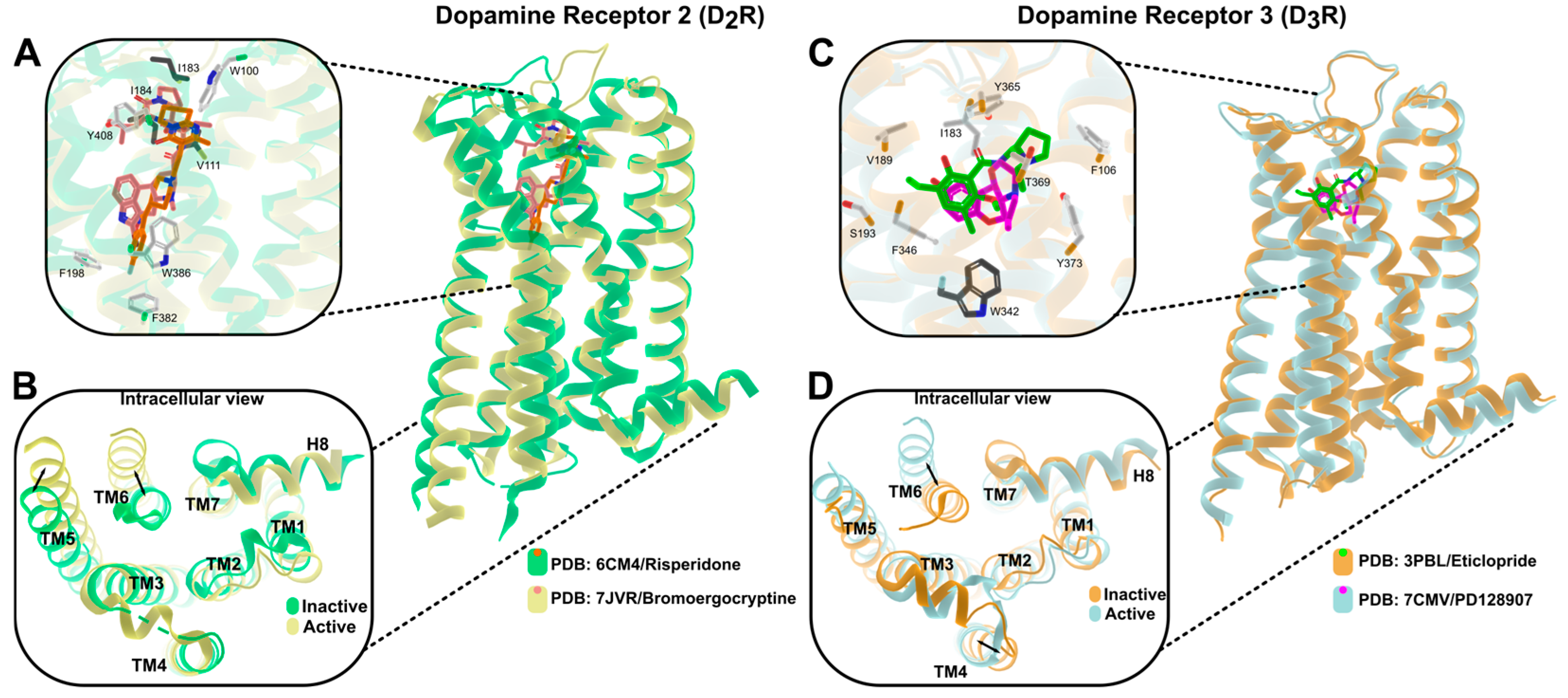
For many years, the absence of high-resolution structural information limited mechanistic insight into the molecular determinants governing D2/D3 selectivity and receptor activation [27,28]. This landscape began to shift in 2010, when the first X-ray crystal structure of the human D3R was solved in an inactive-state conformation stabilized by the antagonist eticlopride [3]. This landmark structure provided a detailed map of the D3R orthosteric pocket and revealed a principle that has since guided rational design: despite broad overall similarity to D2R, discrete residue-level differences and local packing variations within the 7TM bundle can yield disproportionate effects on ligand affinity and subtype preference [3]. Subsequent inactive-state structures of D2R enabled direct receptor-to-receptor comparisons and reinforced the idea that D2/D3 selectivity is not driven by global architectural divergence but rather by microstructural and dynamic differences within and above the orthosteric site that shape ligand entry, pose, and stabilization [6]. These structural principles are illustrated in Figure 1, which compares inactive and active conformations of D2R and D3R and highlights localized differences within the orthosteric binding pocket and intracellular domains.
Although highly informative, inactive-state structures provide only partial insight into PD-relevant pharmacology, particularly when the goal is to understand how chemically distinct agonists can elicit divergent functional outcomes [29,30]. In this regard, the advent of cryo-electron microscopy (cryo-EM) has been transformative by enabling visualization of active-state conformations of D2R and D3R in complex with Gi proteins [6,18,31,32]. These active-state assemblies have clarified conserved hallmarks of GPCR activation, most notably the outward rearrangement of transmembrane helix 6, coordinated movements of TM5 and TM7, and the formation of an intracellular cavity permissive for G-protein engagement [29]. At the same time, they have delineated receptor-specific structural features that may contribute to the high apparent sensitivity of D3R to low dopaminergic tone, providing a structural framework to interpret its functional behavior under hypodopaminergic conditions characteristic of Parkinson’s disease [1,18].
Recent cryo-EM studies have further expanded this structural framework. Structural genomics analyses have resolved active-state conformations of all five human dopamine receptor subtypes bound to the clinically used agonist rotigotine, revealing conserved activation mechanisms together with receptor-specific determinants that shape ligand recognition and G-protein coupling selectivity across the dopaminergic receptor family [31]. In parallel, high-resolution cryo-EM structures of the human D3 receptor in complex with selective bitopic agonists have uncovered an additional selectivity interface involving the TM2–ECL1–TM1 region, which expands the classical secondary binding pocket and provides new opportunities for the rational design of subtype-selective dopaminergic ligands [32]. Together, these advances highlight how subtle extracellular and vestibular structural features can influence ligand binding modes and functional selectivity at D3R.
A central interpretive challenge remains the disentanglement of conformational changes that are intrinsic to receptor activation from those imposed by ligand chemistry, a limitation that has become increasingly evident with the expansion of active-state GPCR structures [29,30]. In practice, the structural differences observed between inactive and active complexes likely reflect a composite of (i) a conserved activation trajectory (for example, the canonical outward movement of TM6 and reorganization of intracellular motifs enabling Gαi coupling) and (ii) ligand-dependent stabilization of distinct microstates within the binding pocket and extracellular vestibule [33,34]. This distinction is particularly relevant for D2R and D3R because active-state structures have been captured with agonists that are chemically heterogeneous (e.g., pramipexole-like and other scaffolds), whereas inactive-state structures often involve antagonists or inverse agonists with fundamentally different pharmacophores [3,6,18]. Thus, apparent “state differences” may conflate activation-linked rearrangements with ligand-specific remodeling of local interaction networks, an overlap that likely underpins the functional diversity observed across D3-directed chemotypes [19,30].
From a therapeutic perspective in PD, these structural advances are more than descriptive. The availability of both inactive and active conformations of the dopamine D3 receptor provides a molecular framework to rationalize how subtype-selective ligands can exploit subtle yet functionally consequential differences between D3R and D2R, despite their high overall structural similarity [3,18]. Notably, these data also support structure-guided strategies aimed at functional selectivity, including the design of ligands that preferentially stabilize signaling-competent conformations associated with favorable intracellular coupling profiles [30,34]. Moreover, they provide a foundation for the development of bitopic ligands that engage the orthosteric site while extending into secondary recognition regions, such as the extracellular vestibule, thereby improving subtype discrimination and enabling finer control over receptor microstates [32,35]. Collectively, advances in structural biology have repositioned D3R from a difficult-to-separate receptor subtype into a tractable target for precision pharmacology, with direct implications for the rational design of therapies that maximize benefit, particularly within non-motor domains, while mitigating liabilities associated with broader D2R engagement [36,37].

3. D3 Receptor Signaling: Gi/o Pathways, β-Arrestin Modulation and Biased Agonism

The D3R predominantly couples to Gαi/o heterotrimeric G proteins, engaging a canonical signaling cascade characterized by inhibition of adenylyl cyclase (AC) and a consequent reduction in intracellular cAMP levels. This pathway underlies the classical modulatory role of D3R in neuronal excitability and dopaminergic tone [1,38]. However, recent studies indicate that D3R exhibits a more restricted coupling profile within the Gi/o/z family than previously appreciated [39,40,41]. In contrast to the D2 receptor (D2R), which shows robust coupling to multiple Gi/o subtypes, D3R displays limited functional interaction with the Gαi1, Gαi2, and Gαi3 proteins and preferentially engages the Gαo and Gαz isoforms. Structural and mutational analyses suggest that this selectivity arises from specific molecular determinants located at the interface between the receptor intracellular loop 2 (ICL2) and the α5 helix of the G protein [39]. In particular, steric constraints and electrostatic interactions involving residues such as Asp350 in Gαi and Thr142 in the D3R ICL2 region may hinder productive coupling with certain Gi proteins, thereby shaping receptor–G protein selectivity [40,41].
These findings highlight that D3R signaling cannot be fully explained by a uniform Gi/o-mediated mechanism. Instead, D3R exhibits a context- and ligand-dependent engagement of intracellular pathways, a feature that contributes to its functional divergence from the closely related D2 receptor and provides a mechanistic basis for differential dopaminergic signaling outcomes [19,30]. This selective coupling profile may also contribute to the signaling bias observed for several D3R ligands, further supporting the concept of functionally selective dopaminergic pharmacology.
Compared with D2R, activation of D3R is associated with a more selective and nuanced modulation of intracellular signaling cascades, including extracellular signal-regulated kinase (ERK) and protein kinase B (Akt) pathways. Importantly, activation of these effectors appears to depend less on robust, receptor-wide signaling and more on cellular context, receptor microenvironment, and ligand-specific conformational states [20,34]. These characteristics suggest that D3R operates as a finely tunable signaling node rather than a binary on-off switch, a property that may be particularly relevant in neurodegenerative conditions such as PD [37].
A distinctive functional hallmark of D3R is its relatively low propensity for desensitization and internalization following prolonged stimulation, especially when compared with D2R [19,42]. This behavior implies a reduced reliance on β-arrestin-dependent regulatory mechanisms and results in more sustained signaling under chronic activation conditions [19]. Such signaling stability is especially relevant in the context of long-term dopaminergic therapies, where receptor desensitization, tolerance, and loss of efficacy contribute substantially to adverse clinical outcomes [43,44].
Within this framework, the concept of biased signaling has emerged as a central paradigm for understanding D3R pharmacology. Biased agonism refers to the ability of a ligand to preferentially activate specific intracellular signaling pathways through a single receptor, rather than uniformly engaging all available effectors [30,38]. In the case of D3R, several ligands have been shown to stabilize receptor conformations that favor Gαi/o-mediated signaling while minimally recruiting β-arrestins. This property provides a mechanistic basis to dissociate therapeutically desirable effects from signaling processes associated with receptor desensitization, tolerance, and adverse outcomes [19,20].
From a mechanistic perspective, Gαi/o-biased D3R signaling involves the stabilization of active receptor conformations that promote efficient G-protein coupling while constraining β-arrestin engagement [19,45]. Activation of Gαi/o leads to suppression of adenylyl cyclase (AC) activity, reduced intracellular cAMP accumulation, and downstream attenuation of protein kinase A (PKA) signaling. This signaling axis intersects with the differential engagement of MAPK/ERK and PI3K–Akt pathways, which have been implicated in neuronal survival, cellular stress resilience, and the maintenance of synaptic plasticity [9,14,46]. In parallel, D3R-mediated Gαi/o signaling modulates neuronal excitability through inhibition of voltage-gated calcium channels (N- and P/Q-type) and activation of G protein-gated inwardly rectifying potassium (GIRK) channels, thereby exerting fine control over neuronal firing patterns and mesolimbic dopaminergic tone [9,14].
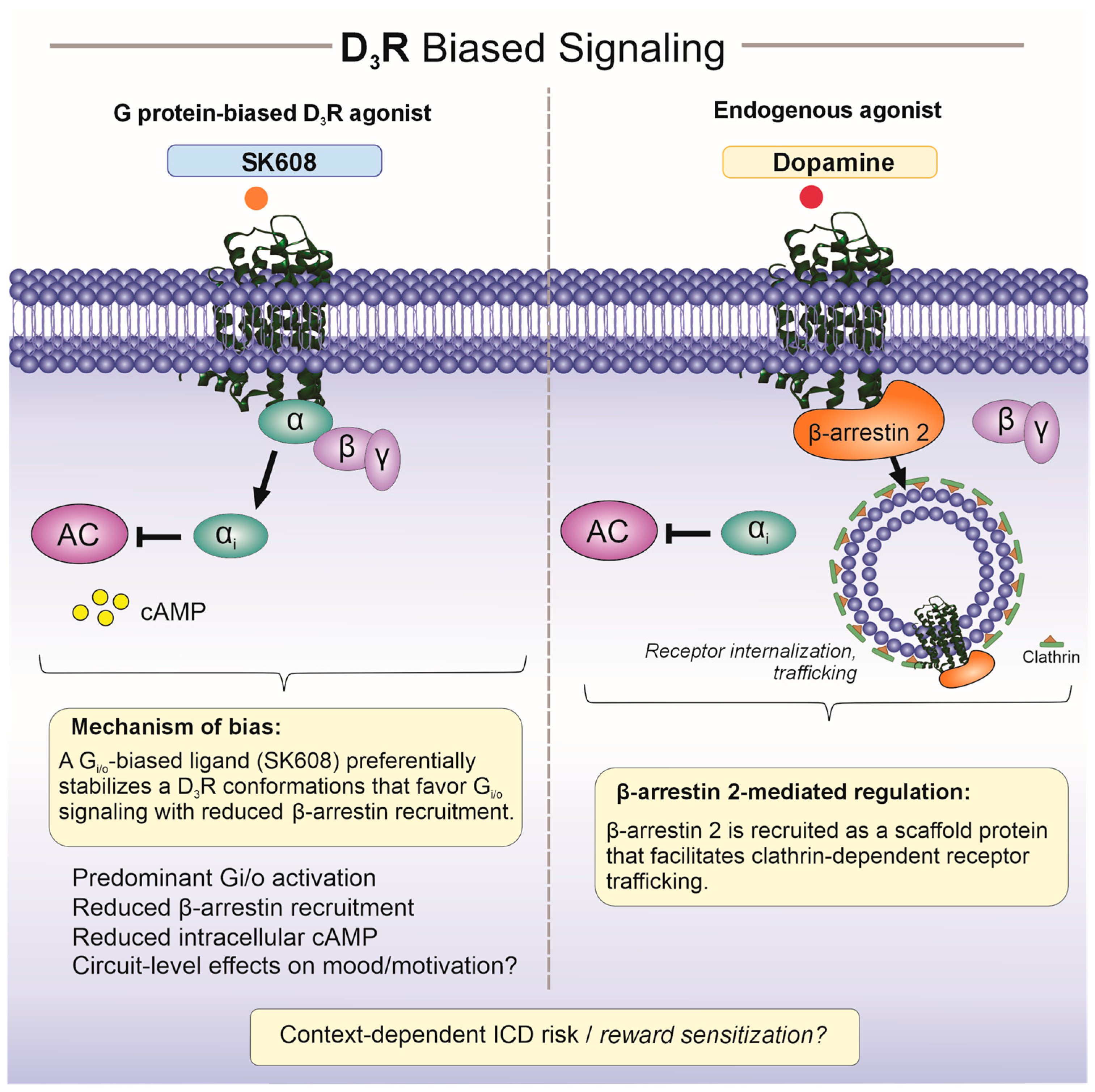
Conversely, limited recruitment of β-arrestins attenuates receptor internalization and downstream regulatory processes, dampens pro-inflammatory signaling, and reduces maladaptive compensatory responses, collectively favoring a more stable and sustained functional output during chronic stimulation. In this context, functionally biased agonists [45], including bitopic ligands and experimental compounds such as SK608, have demonstrated pharmacological profiles consistent with preferential Gαi/o signaling, reduced tolerance liability, and engagement of pathways associated with neuroprotection and synaptic maintenance [19]. These ligand-dependent differences in signaling quality are schematically illustrated in Figure 2.
In Parkinson’s disease, these signaling properties acquire direct translational relevance. Biased activation of D3R may contribute to the modulation of non-motor symptoms, including affective and motivational disturbances, while potentially enabling a more refined influence on motor circuits with a reduced risk of desensitization and off-target effects associated with broad dopaminergic stimulation [18,32]. In aggregate, biased D3R signaling emerges as a promising pharmacological framework to enhance therapeutic efficacy while improving tolerability, supporting its consideration as a component of next-generation dopaminergic strategies in the comprehensive management of PD.

4. Functional Role of D3 Receptor in Parkinson’s Disease

PD is classically defined by nigrostriatal dopaminergic degeneration and the ensuing disruption of basal ganglia circuitry. Yet, the clinical phenotype extends well beyond motor impairment, encompassing prominent non-motor symptoms, including depression, anxiety, apathy, anhedonia and motivational deficits, that frequently precede, accompany, or outlast fluctuations in motor control [47,48,49]. Within this broader clinical framework, the D3R occupies a distinctive functional niche. Its preferential enrichment in mesolimbic and associative circuits, together with its high affinity for dopamine, positions D3R as a key modulator of affective–motivational processing under conditions of reduced dopaminergic tone [10,14,50].
In contrast to the D2R, which is widely expressed across the dorsal striatum and tightly coupled to motor output, D3R expression is biased toward ventral striatal and limbic nodes, including the nucleus accumbens, ventral pallidum, amygdala and related mesolimbic structures [14,27,50]. These regions are critically involved in reward valuation, reinforcement learning, goal-directed behavior, and emotional regulation, functional domains that are disproportionately affected in PD, particularly within its non-motor symptom spectrum [47,48]. From a mechanistic standpoint, progressive dopaminergic depletion is expected to differentially reshape signaling dynamics within these circuits. Because D3R displays high agonist affinity and remains responsive at low extracellular dopamine concentrations, it may function as a sensitive gain controller of mesolimbic dopaminergic tone, thereby influencing behavioral outputs even under hypodopaminergic conditions [1,51]. This framework provides a biologically plausible substrate for the selective vulnerability and, in some cases, the early emergence of non-motor neuropsychiatric features in PD.
Consistent with this circuit logic, experimental evidence across genetic, pharmacological, and lesion-based models supports an involvement of D3R in affective and motivational phenotypes. Reduced D3R function, achieved through receptor inhibition or loss-of-function paradigms, has been associated with anxiety-like and depressive-like behaviors, whereas preferential D3R activation can produce antidepressant- and anxiolytic-like effects in selected experimental settings [52,53]. Although such findings cannot be directly equated to clinical efficacy, they reinforce the concept that D3R participates in the regulatory architecture of mood and motivation under hypodopaminergic conditions.
A defining aspect of PD management is the requirement for long-term dopaminergic replacement and/or dopaminomimetic therapy, which is frequently complicated over time by wearing-off phenomena, motor fluctuations, and variability in treatment response [54,55]. Within this therapeutic landscape, D3R is functionally relevant in two complementary ways. First, D3R shows a comparatively lower propensity for agonist-driven internalization and desensitization than D2R, suggesting that D3R-mediated signaling may remain more stable during chronic stimulation, an attractive property given the time-dependent waning of therapeutic response observed with long-term dopaminergic treatment [56,57]. Second, D3R is closely linked to mesolimbic reward circuitry, and dopaminergic agonists with high D2/D3 affinity, particularly those displaying relative D3 preference, are consistently implicated in reinforcement-related behavioral plasticity and impulse control disorders (ICDs) in PD. This association supports the view that pharmacological engagement of D3R-enriched circuits may influence not only symptom control but also reinforcement learning and impulse regulation [58,59,60].
This dual role becomes particularly evident when considering ICDs and related behavioral syndromes, which represent well-recognized complications of dopamine agonist therapy in PD and are thought to reflect, at least in part, altered signaling within mesolimbic reward circuits [60,61,62]. Given the preferential expression of the dopamine D3 receptor (D3R) in ventral striatal and limbic regions, its pharmacological engagement has been proposed as a mechanistic link between the intended therapeutic modulation of motivational deficits and the unintended amplification of maladaptive reward-driven behaviors [14,58]. Accordingly, the functional role of D3R in Parkinson’s disease is best interpreted within a precision pharmacology framework, in which the same receptor node that may support improvements in affective-motivational domains can also contribute to vulnerability for behavioral adverse effects, depending on ligand pharmacological properties, dosing regimens, and patient-specific circuit states [59,62].
The emergence of biased signaling further refines how D3R function can be conceptualized in PD. Rather than treating D3R activation as a single pharmacological output, ligand-dependent stabilization of distinct receptor conformations implies that D3R engagement may be tuned toward Gαi/o-dominant signaling with limited β-arrestin recruitment. Such bias may reduce receptor regulatory processes that contribute to tolerance and variability under chronic stimulation [30]. Functionally, this signaling profile could support a more predictable modulation of limbic dopaminergic tone while limiting pathways associated with desensitization and broader network adaptation [19,63]. Importantly, this framework does not imply guaranteed clinical benefit; instead, it provides a mechanistic rationale for why next-generation D3R ligands, particularly those engineered for functional selectivity, may better align therapeutic intent with tolerability [14,37].

5. Therapeutic Potential: From Agonists to Biased Ligands and Neuroprotective Concepts

Advances in the structural and functional characterization of the D3R have enabled a more precise appraisal of pharmacological strategies that are both plausible and mechanistically justified for therapeutic exploration in PD. This progress has been driven in part by the integration of structural biology with modern signaling frameworks, which increasingly support structure-guided and pathway-aware ligand design [10,37]. Rather than positioning D3R as a primary target for motor symptom control, converging evidence indicates that its principal therapeutic value may lie in the selective modulation of dopaminergic circuits involved in non-motor symptomatology, including affective and motivational domains linked to mesolimbic function and dopamine agonist exposure [14,64]. In parallel, contemporary D3R-focused syntheses and PD-specific reviews discuss emerging preclinical evidence that D3R signaling may engage cellular programs relevant to neuronal resilience and plasticity under hypodopaminergic conditions, framing these concepts as mechanistic opportunities rather than established disease-modifying effects [10,43,65].
D3-preferring dopamine agonists represent the most established pharmacological approach to engage D3R in PD. Compounds such as pramipexole, ropinirole, and rotigotine, while not strictly subtype-selective, display a higher relative affinity for D3R compared with D2R and are widely used in clinical practice [66,67]. Beyond their effects on motor symptoms, these agents have demonstrated efficacy in alleviating non-motor manifestations, including depression, apathy, and motivational disturbances, which are increasingly recognized as core components of the PD phenotype [47,68]. In particular, pramipexole has been consistently shown to exert antidepressant and anxiolytic effects in PD patients, effects that cannot be fully explained by improvements in motor function alone and are also recapitulated in preclinical models of depression and dopaminergic dysfunction [69,70]. These observations are mechanistically congruent with the preferential mesolimbic distribution of D3R and with its high agonist affinity, which enables sustained receptor engagement under conditions of diminished dopaminergic tone, a defining feature of PD progression [1,50].
Beyond clinically established D2/D3-preferring agonists, several experimental D3R-preferring or partial agonists have also been evaluated in preclinical models of Parkinson’s disease. Among these, the D3R-preferring agonist PD-128907 and the more recently developed D3R partial agonist PD13R have provided important proof-of-concept evidence for D3R-directed pharmacology. In 6-hydroxydopamine (6-OHDA) lesions, PD-128907 was reported to reverse motivational deficits and improve affective-like behavioral alterations, suggesting a role for D3R activation in domains related to motivation and reward processing [16]. However, studies in levodopa-primed MPTP nonhuman primates indicated that although PD-128907 produced antiparkinsonian motor effects comparable to apomorphine, it did not significantly attenuate levodopa-induced dyskinesia. These observations highlight that D3R-preferring agonism alone does not necessarily translate into improved control of motor complications [71]. More recently, the D3R partial agonist PD13R has expanded this experimental framework. In a nonhuman primate model of Parkinson’s disease, PD13R suppressed the expression of levodopa-induced dyskinesia while maintaining antiparkinsonian efficacy, suggesting that more selective or functionally differentiated D3R ligands may offer advantages over earlier D3R-preferring compounds [72]. Collectively, these experimental studies broaden the therapeutic landscape of D3R-targeted pharmacology and illustrate that the translational potential of these ligands depends not only on receptor preference but also on ligand efficacy profile, model context, and the symptom domain being evaluated.
Despite these benefits, the long-term clinical use of D2/D3 agonists is accompanied by important limitations. Chronic dopaminergic stimulation may lead to fluctuating therapeutic responses and behavioral complications, including impulse control disorders and reward-related behavioral dysregulation. These effects are thought to arise, at least in part, from excessive stimulation of mesolimbic dopaminergic circuits enriched in D3 receptors. Consequently, while currently available D2/D3 agonists demonstrate that dopaminergic modulation of D3R-containing circuits can provide meaningful symptomatic benefit, their clinical profile also highlights the need for more selective pharmacological strategies capable of preserving therapeutic effects while minimizing adverse behavioral outcomes.
These considerations raise an important translational question: whether highly selective D3R-targeted ligands or broader D2/D3 agonists are better suited to address the combined motor and non-motor symptom burden of Parkinson’s disease. Selective modulation of D3R, particularly through biased agonists, may offer greater precision for affective and motivational domains, consistent with the preferential enrichment of D3R within mesolimbic circuits involved in reward processing and motivational behavior [14,50]. However, pharmacological agents with combined D2/D3 activity may retain a practical advantage when both motor and non-motor manifestations need to be controlled simultaneously, because they engage limbic circuits enriched in D3R while also maintaining D2-mediated signaling within striatal pathways that are more directly linked to motor regulation. This broader receptor engagement may help explain the clinical profile of widely used dopamine agonists such as pramipexole, ropinirole, and rotigotine, which improve motor symptoms while also alleviating selected non-motor manifestations of Parkinson’s disease [66,67].
Within this framework, strategies centered on D3R selectivity are better viewed not as universal replacements for conventional D2/D3 agonists, but rather as candidates for more targeted therapeutic applications, particularly when modulation of limbic dopaminergic signaling and stabilization of non-motor symptoms are prioritized. For comparison, pharmacological approaches targeting D1-like receptors have also regained interest in Parkinson’s disease. Agonists and partial agonists acting on D1/D5 receptors can provide robust motor benefit through facilitation of the striatonigral pathway and restoration of direct pathway signaling [64]. However, current clinical development of D1-directed agents has focused primarily on motor outcomes, and their potential relevance for affective or motivational symptoms remains less clearly defined than that proposed for D3R- or D2/D3-directed strategies.
In this context, the recognition that D3R signaling can be functionally biased has catalyzed interest in ligands engineered to preferentially engage Gαi/o-mediated pathways while minimizing β-arrestin recruitment. From a mechanistic standpoint, this strategy aims to preserve signaling axes associated with therapeutic benefit, such as modulation of affective and motivational circuits and stabilization of dopaminergic tone, while attenuating receptor desensitization, tolerance development, and signaling pathways linked to adverse outcomes [14,63].
Although biased D3R agonists remain largely in preclinical or early experimental stages, compounds such as SK608, as well as emerging bitopic ligands, have provided proof-of-concept that functional selectivity at D3R is achievable. Experimental pharmacological studies demonstrate that these ligands can stabilize receptor conformations preferentially coupled to Gαi/o signaling, with reduced engagement of regulatory pathways associated with receptor desensitization. This profile offers a rational framework to improve the predictability and durability of dopaminergic signaling [5,14,19]. Importantly, biased agonism does not imply enhanced efficacy per se, but rather greater control over signaling quality, an attribute that may be particularly valuable in the context of long-term PD management [46].
Beyond symptomatic modulation, a body of preclinical evidence suggests that D3R activation may influence neuronal survival, plasticity, and repair-related processes. Seminal studies led by Joyce and colleagues demonstrated that D3R-preferring agonists attenuate dopaminergic neuron loss, promote axonal sprouting, and improve functional recovery in toxin-based models of PD, including 6-OHDA paradigms [65,73]. Subsequent experimental work has implicated D3R signaling in the regulation of oxidative stress responses, neurotrophic factor pathways, and synaptic remodeling, further supporting a role for this receptor in adaptive responses to dopaminergic injury [10,43,74,75].
Crucially, these findings should be interpreted with appropriate caution. At present, neuroprotective effects associated with D3R activation remain mechanistic and experimental, and there is insufficient evidence to support disease-modifying claims in clinical PD. Nevertheless, the consistency of these observations across model systems supports the view that D3R may participate in adaptive responses to dopaminergic injury, warranting continued investigation within carefully designed translational frameworks.

6. Future Directions and Outstanding Questions

Despite substantial progress in elucidating the structural, signaling, and functional properties of the D3R, several key questions remain that will determine its translational relevance in PD. Addressing these gaps will require coordinated advances across structural biology, pharmacology, and clinical neuroscience.
A central challenge is the development of ligands with improved subtype selectivity and functional precision. Although structural data have clarified determinants of D3/D2 discrimination, translating these insights into compounds with robust receptor selectivity, favorable pharmacokinetic properties, and adequate central nervous system penetration remains a non-trivial task. In parallel, the field would benefit from standardized experimental frameworks to quantify biased signaling at D3R, enabling meaningful comparisons across ligands, cellular contexts, and experimental systems.
Another unresolved issue concerns the circuit-specific roles of D3R in PD. While its preferential enrichment within mesolimbic regions supports involvement in non-motor symptom domains, the extent to which D3R signaling contributes to interactions between limbic and motor networks, particularly under conditions of chronic dopaminergic therapy, remains incompletely understood. Integrating receptor-level insights with circuit-resolved approaches, including advanced neuroimaging, electrophysiology, and human-derived cellular models, will be essential to clarify these dynamics.
From a translational perspective, there is a critical need for biomarkers capable of reporting D3R engagement in vivo. Further refinement of D3-selective positron emission tomography (PET) ligands and complementary pharmacodynamic readouts would facilitate target validation, dose optimization, and patient stratification in early-stage clinical studies. Such tools are especially important given the heterogeneity of PD phenotypes and the variable expression and progression of non-motor symptoms.
Finally, the putative neuroprotective and neurorestorative roles of D3R warrant cautious but systematic evaluation. Although preclinical findings are intriguing, future work must rigorously define the conditions under which D3R signaling influences neuronal resilience, distinguish direct receptor-mediated effects from secondary network-level adaptations, and establish whether these mechanisms can be meaningfully harnessed in human disease.

7. Conclusions

Overall, recent advances in structural biology and receptor pharmacology have refined the understanding of the D3R as a functionally distinct component of the dopaminergic system in PD. Rather than acting as a primary mediator of motor control, D3R emerges as a receptor strategically positioned to modulate mesolimbic circuits, non-motor symptom domains, and signaling dynamics under conditions of dopaminergic depletion. High-resolution structural insights, together with growing recognition of ligand-dependent signaling bias, have provided a mechanistic basis to reconsider how D3R may be pharmacologically engaged with greater precision.
While currently available clinical agents only partially exploit these properties, concepts such as functional selectivity and biased agonism offer a rational framework to refine dopaminergic interventions and improve long-term signaling stability. Importantly, evidence supporting neuroprotective or disease-modifying roles of D3R remains preliminary and largely confined to preclinical settings. At present, the most compelling value of D3R lies in its potential to support selective and modulable dopaminergic signaling, particularly in relation to non-motor features of PD.

Author Contributions

Conceptualization, F.P., A.P.-M. and I.D.L.; literature search and analysis, F.P., E.M.D. and A.V.-M.; interpretation of structural and receptor–ligand interaction data, E.M.D. and J.M.P.-A.; interpretation of pharmacological and translational aspects, F.P., A.P.-M. and I.D.L.; writing—original draft preparation, F.P., E.M.D. and A.V.-M.; writing—review and editing, F.P., E.M.D., A.P.-M., A.V.-M., J.M.P.-A. and I.D.L.; visualization, F.P., E.M.D. and J.M.P.-A.; supervision, F.P. and I.D.L.; project administration, F.P. All authors have read and agreed to the published version of the manuscript and agree to be accountable for all aspects of the work.

Funding

This research received no external funding.

Data Availability Statement

No new data were created or analyzed in this study. Data sharing is not applicable to this article.

Acknowledgments

During the preparation of this manuscript, the authors used ChatGPT (OpenAI, GPT-5.3) for language refinement and structuring of scientific content. The authors critically reviewed and edited all outputs and take full responsibility for the content of this publication.

Conflicts of Interest

The authors declare no conflicts of interest.

References

  1. Beaulieu, J.M.; Gainetdinov, R.R. The physiology, signaling, and pharmacology of dopamine receptors. Pharmacol. Rev. 2011, 63, 182–217. [Google Scholar] [CrossRef] [Scilit] [PubMed]
  2. Sokoloff, P.; Giros, B.; Martres, M.P.; Bouthenet, M.L.; Schwartz, J.C. Molecular cloning and characterization of a novel dopamine receptor (D3) as a target for neuroleptics. Nature 1990, 347, 146–151. [Google Scholar] [CrossRef] [Scilit] [PubMed]
  3. Chien, E.Y.T.; Liu, W.; Zhao, Q.; Katritch, V.; Han, G.W.; Hanson, M.A.; Shi, L.; Newman, A.H.; Javitch, J.A.; Cherezov, V.; et al. Structure of the human dopamine D3 receptor in complex with a D2/D3 selective antagonist. Science 2010, 330, 1091–1095. [Google Scholar] [CrossRef] [Scilit] [PubMed]
  4. Eryilmaz, I.E.; Erer, S.; Zarifoglu, M.; Egeli, U.; Karakus, E.; Yurdacan, B.; Cecener, G.; Tunca, B.; Colakoglu, B.; Bora Tokcaer, A.; et al. Contribution of functional dopamine D2 and D3 receptor variants to motor and non-motor symptoms of early onset Parkinson’s disease. Clin. Neurol. Neurosurg. 2020, 199, 106257. [Google Scholar] [CrossRef] [Scilit] [PubMed]
  5. Yang, P.; Perlmutter, J.S.; Benzinger, T.L.S.; Morris, J.C.; Xu, J. Dopamine D3 receptor: A neglected participant in Parkinson Disease pathogenesis and treatment? Ageing Res. Rev. 2020, 57, 100994. [Google Scholar] [CrossRef] [Scilit]
  6. Wang, S.; Che, T.; Levit, A.; Shoichet, B.K.; Wacker, D.; Roth, B.L. Structure of the D2 dopamine receptor bound to the atypical antipsychotic drug risperidone. Nature 2018, 555, 269–273. [Google Scholar] [CrossRef] [Scilit] [PubMed]
  7. Sokoloff, P.; Le Foll, B. The dopamine D3 receptor, a quarter century later. Eur. J. Neurosci. 2017, 45, 2–19. [Google Scholar] [CrossRef] [Scilit] [PubMed]
  8. Liu, X.Y.; Mao, L.M.; Zhang, G.C.; Papasian, C.J.; Fibuch, E.E.; Lan, H.X.; Zhou, H.F.; Xu, M.; Wang, J.Q. Activity-dependent modulation of limbic dopamine D3 receptors by CaMKII. Neuron 2009, 61, 425–438. [Google Scholar] [CrossRef] [Scilit] [PubMed]
  9. Neve, K.A.; Seamans, J.K.; Trantham-Davidson, H. Dopamine receptor signaling. J. Recept. Signal Transduct. Res. 2004, 24, 165–205. [Google Scholar] [CrossRef] [PubMed]
  10. Xu, J. Dopamine D3 Receptor in Parkinson Disease: A Prognosis Biomarker and an Intervention Target. Curr. Top. Behav. Neurosci. 2023, 60, 89–107. [Google Scholar] [CrossRef] [Scilit] [PubMed]
  11. Chagraoui, A.; Di Giovanni, G.; De Deurwaerdère, P. Neurobiological and Pharmacological Perspectives of D3 Receptors in Parkinson’s Disease. Biomolecules 2022, 12, 243. [Google Scholar] [CrossRef] [Scilit] [PubMed]
  12. Ryoo, H.L.; Pierrotti, D.; Joyce, J.N. Dopamine D3 receptor is decreased and D2 receptor is elevated in the striatum of Parkinson’s disease. Mov. Disord. 1998, 13, 788–797. [Google Scholar] [CrossRef] [Scilit] [PubMed]
  13. Anzalone, A.; Lizardi-Ortiz, J.E.; Ramos, M.; De Mei, C.; Hopf, F.W.; Iaccarino, C.; Halbout, B.; Jacobsen, J.; Kinoshita, C.; Welter, M.; et al. Dual control of dopamine synthesis and release by presynaptic and postsynaptic dopamine D2 receptors. J. Neurosci. 2012, 32, 9023–9034. [Google Scholar] [CrossRef] [Scilit] [PubMed]
  14. Kiss, B.; Laszlovszky, I.; Krámos, B.; Visegrády, A.; Bobok, A.; Lévay, G.; Lendvai, B.; Román, V. Neuronal Dopamine D3 Receptors: Translational Implications for Preclinical Research and CNS Disorders. Biomolecules 2021, 11, 104. [Google Scholar] [CrossRef] [Scilit] [PubMed]
  15. Favier, M.; Carcenac, C.; Drui, G.; Vachez, Y.; Boulet, S.; Savasta, M.; Carnicella, S. Implication of dorsostriatal D3 receptors in motivational processes: A potential target for neuropsychiatric symptoms in Parkinson’s disease. Sci. Rep. 2017, 7, 41589. [Google Scholar] [CrossRef] [Scilit] [PubMed]
  16. Carnicella, S.; Drui, G.; Boulet, S.; Carcenac, C.; Favier, M.; Duran, T.; Savasta, M. Implication of dopamine D3 receptor activation in the reversion of Parkinson’s disease-related motivational deficits. Transl. Psychiatry 2014, 4, e401. [Google Scholar] [CrossRef] [Scilit] [PubMed]
  17. Tritsch, N.X. Motivating interest in D3 dopamine receptors. Nat. Neurosci. 2025, 28, 6–7. [Google Scholar] [CrossRef] [Scilit] [PubMed]
  18. Xu, P.; Huang, S.; Mao, C.; Krumm, B.E.; Zhou, X.E.; Tan, Y.; Huang, X.P.; Liu, Y.; Shen, D.D.; Jiang, Y.; et al. Structures of the human dopamine D3 receptor-Gi complexes. Mol. Cell 2021, 81, 1147–1159.e1144. [Google Scholar] [CrossRef] [Scilit] [PubMed]
  19. Xu, W.; Reith, M.E.A.; Liu-Chen, L.Y.; Kortagere, S. Biased signaling agonist of dopamine D3 receptor induces receptor internalization independent of β-arrestin recruitment. Pharmacol. Res. 2019, 143, 48–57. [Google Scholar] [CrossRef] [Scilit] [PubMed]
  20. Xu, W.; Bearoff, F.; Kortagere, S. G-protein biased signaling agonists of Dopamine D3 receptor promote distinct activation patterns of ERK1/2. Pharmacol. Res. 2022, 179, 106223. [Google Scholar] [CrossRef] [Scilit] [PubMed]
  21. Bono, F.; Mutti, V.; Fiorentini, C.; Missale, C. Dopamine D3 Receptor Heteromerization: Implications for Neuroplasticity and Neuroprotection. Biomolecules 2020, 10, 1016. [Google Scholar] [CrossRef] [Scilit] [PubMed]
  22. Tropea, M.R.; Melone, M.; Li Puma, D.D.; Vacanti, V.; Aceto, G.; Bandiera, B.; Trovato, R.C.; Torrisi, S.A.; Leggio, G.M.; Palmeri, A.; et al. Blockade of dopamine D3 receptors improves hippocampal synaptic function and rescues age-related cognitive phenotype. Aging Cell 2024, 23, e14291. [Google Scholar] [CrossRef] [Scilit] [PubMed]
  23. Hayatshahi, H.S.; Luedtke, R.R.; Taylor, M.; Chen, P.J.; Blass, B.E.; Liu, J. Factors Governing Selectivity of Dopamine Receptor Binding Compounds for D2R and D3R Subtypes. J. Chem. Inf. Model. 2021, 61, 2829–2843. [Google Scholar] [CrossRef] [Scilit] [PubMed]
  24. Battiti, F.O.; Cemaj, S.L.; Guerrero, A.M.; Shaik, A.B.; Lam, J.; Rais, R.; Slusher, B.S.; Deschamps, J.R.; Imler, G.H.; Newman, A.H.; et al. The Significance of Chirality in Drug Design and Synthesis of Bitopic Ligands as D3 Receptor (D3R) Selective Agonists. J. Med. Chem. 2019, 62, 6287–6314. [Google Scholar] [CrossRef] [Scilit] [PubMed]
  25. Cortés, A.; Moreno, E.; Rodríguez-Ruiz, M.; Canela, E.I.; Casadó, V. Targeting the dopamine D3 receptor: An overview of drug design strategies. Expert Opin. Drug Discov. 2016, 11, 641–664. [Google Scholar] [CrossRef] [Scilit] [PubMed]
  26. Leggio, G.M.; Bucolo, C.; Platania, C.B.M.; Salomone, S.; Drago, F. Current drug treatments targeting dopamine D3 receptor. Pharmacol. Ther. 2016, 165, 164–177. [Google Scholar] [CrossRef] [Scilit] [PubMed]
  27. Missale, C.; Nash, S.R.; Robinson, S.W.; Jaber, M.; Caron, M.G. Dopamine receptors: From structure to function. Physiol. Rev. 1998, 78, 189–225. [Google Scholar] [CrossRef] [Scilit] [PubMed]
  28. Orrù, M.; Strathman, H.J.; Floris, G.; Scheggi, S.; Levant, B.; Bortolato, M. The adverse effects of pramipexole on probability discounting are not reversed by acute D2 or D3 receptor antagonism. Eur. Neuropsychopharmacol. 2020, 32, 104–119. [Google Scholar] [CrossRef] [Scilit] [PubMed]
  29. Weis, W.I.; Kobilka, B.K. The Molecular Basis of G Protein-Coupled Receptor Activation. Annu. Rev. Biochem. 2018, 87, 897–919. [Google Scholar] [CrossRef] [Scilit] [PubMed]
  30. Wacker, D.; Stevens, R.C.; Roth, B.L. How Ligands Illuminate GPCR Molecular Pharmacology. Cell 2017, 170, 414–427. [Google Scholar] [CrossRef] [Scilit] [PubMed]
  31. Xu, P.; Huang, S.; Krumm, B.E.; Zhuang, Y.; Mao, C.; Zhang, Y.; Wang, Y.; Huang, X.P.; Liu, Y.F.; He, X.; et al. Structural genomics of the human dopamine receptor system. Cell Res. 2023, 33, 604–616. [Google Scholar] [CrossRef] [Scilit] [PubMed]
  32. Arroyo-Urea, S.; Nazarova, A.L.; Carrión-Antolí, Á.; Bonifazi, A.; Battiti, F.O.; Lam, J.H.; Newman, A.H.; Katritch, V.; García-Nafría, J. A bitopic agonist bound to the dopamine 3 receptor reveals a selectivity site. Nat. Commun. 2024, 15, 7759. [Google Scholar] [CrossRef] [Scilit] [PubMed]
  33. Manglik, A.; Kim, T.H.; Masureel, M.; Altenbach, C.; Yang, Z.; Hilger, D.; Lerch, M.T.; Kobilka, T.S.; Thian, F.S.; Hubbell, W.L.; et al. Structural Insights into the Dynamic Process of β2-Adrenergic Receptor Signaling. Cell 2015, 161, 1101–1111. [Google Scholar] [CrossRef] [Scilit] [PubMed]
  34. Latorraca, N.R.; Venkatakrishnan, A.J.; Dror, R.O. GPCR Dynamics: Structures in Motion. Chem. Rev. 2017, 117, 139–155. [Google Scholar] [CrossRef] [Scilit]
  35. Lane, J.R.; May, L.T.; Parton, R.G.; Sexton, P.M.; Christopoulos, A. A kinetic view of GPCR allostery and biased agonism. Nat. Chem. Biol. 2017, 13, 929–937. [Google Scholar] [CrossRef] [Scilit] [PubMed]
  36. Hauser, A.S.; Attwood, M.M.; Rask-Andersen, M.; Schiöth, H.B.; Gloriam, D.E. Trends in GPCR drug discovery: New agents, targets and indications. Nat. Rev. Drug Discov. 2017, 16, 829–842. [Google Scholar] [CrossRef] [Scilit] [PubMed]
  37. Congreve, M.; de Graaf, C.; Swain, N.A.; Tate, C.G. Impact of GPCR Structures on Drug Discovery. Cell 2020, 181, 81–91. [Google Scholar] [CrossRef] [Scilit] [PubMed]
  38. Sibley, D.R.; Nilson, A.N.; Moritz, A.E.; Shi, L. Dopamine receptor divergence revealed using a common ligand. Trends Pharmacol. Sci. 2023, 44, 637–639. [Google Scholar] [CrossRef] [Scilit] [PubMed]
  39. Zanetti, L.; Franchini, L.; Saha, S.; Liao, Y.; Muntean, B.S.; Orlandi, C.G. Go/z-biased coupling profile of the dopamine D3 receptor. bioRxiv 2025. [Google Scholar] [CrossRef] [Scilit] [PubMed]
  40. Sánchez-Soto, M.; Bonifazi, A.; Cai, N.S.; Ellenberger, M.P.; Newman, A.H.; Ferré, S.; Yano, H. Evidence for Noncanonical Neurotransmitter Activation: Norepinephrine as a Dopamine D2-Like Receptor Agonist. Mol. Pharmacol. 2016, 89, 457–466. [Google Scholar] [CrossRef] [Scilit] [PubMed]
  41. Mönnich, D.; Humphrys, L.J.; Höring, C.; Hoare, B.L.; Forster, L.; Pockes, S. Activation of Multiple G Protein Pathways to Characterize the Five Dopamine Receptor Subtypes Using Bioluminescence Technology. ACS Pharmacol. Transl. Sci. 2024, 7, 834–854. [Google Scholar] [CrossRef] [Scilit] [PubMed]
  42. Peterson, S.M.; Pack, T.F.; Wilkins, A.D.; Urs, N.M.; Urban, D.J.; Bass, C.E.; Lichtarge, O.; Caron, M.G. Elucidation of G-protein and β-arrestin functional selectivity at the dopamine D2 receptor. Proc. Natl. Acad. Sci. USA 2015, 112, 7097–7102. [Google Scholar] [CrossRef] [Scilit] [PubMed]
  43. Lanza, K.; Bishop, C. Dopamine D3 Receptor Plasticity in Parkinson’s Disease and L-DOPA-Induced Dyskinesia. Biomedicines 2021, 9, 314. [Google Scholar] [CrossRef] [Scilit] [PubMed]
  44. Luis-Ravelo, D.; Fumagallo-Reading, F.; Castro-Hernandez, J.; Barroso-Chinea, P.; Afonso-Oramas, D.; Febles-Casquero, A.; Cruz-Muros, I.; Salas-Hernandez, J.; Mesa-Infante, V.; Rodriguez-Nuñez, J.; et al. Prolonged dopamine D3 receptor stimulation promotes dopamine transporter ubiquitination and degradation through a PKC-dependent mechanism. Pharmacol. Res. 2021, 165, 105434. [Google Scholar] [CrossRef] [Scilit] [PubMed]
  45. Xu, W.; Wang, X.; Tocker, A.M.; Huang, P.; Reith, M.E.A.; Liu-Chen, L.Y.; Smith, A.B.; Kortagere, S. Functional Characterization of a Novel Series of Biased Signaling Dopamine D3 Receptor Agonists. ACS Chem. Neurosci. 2017, 8, 486–500. [Google Scholar] [CrossRef] [Scilit] [PubMed]
  46. Nepal, B.; Barnett, J.; Bearoff, F.; Kortagere, S. Biased Signaling Agonists Promote Distinct Phosphorylation and Conformational States of the Dopamine D3 Receptor. Int. J. Mol. Sci. 2024, 25, 470. [Google Scholar] [CrossRef] [Scilit] [PubMed]
  47. Chaudhuri, K.R.; Schapira, A.H. Non-motor symptoms of Parkinson’s disease: Dopaminergic pathophysiology and treatment. Lancet Neurol. 2009, 8, 464–474. [Google Scholar] [CrossRef] [Scilit] [PubMed]
  48. Zis, P.; Erro, R.; Walton, C.C.; Sauerbier, A.; Chaudhuri, K.R. The range and nature of non-motor symptoms in drug-naive Parkinson’s disease patients: A state-of-the-art systematic review. npj Park. Dis. 2015, 1, 15013. [Google Scholar] [CrossRef] [Scilit] [PubMed]
  49. Borgemeester, R.W.; Drent, M.; van Laar, T. Motor and non-motor outcomes of continuous apomorphine infusion in 125 Parkinson’s disease patients. Park. Relat. Disord. 2016, 23, 17–22. [Google Scholar] [CrossRef] [Scilit] [PubMed]
  50. Gurevich, E.V.; Joyce, J.N. Distribution of dopamine D3 receptor expressing neurons in the human forebrain: Comparison with D2 receptor expressing neurons. Neuropsychopharmacology 1999, 20, 60–80. [Google Scholar] [CrossRef] [Scilit] [PubMed]
  51. Wang, P.; Bi, Y.; Li, M.; Chen, J.; Wang, Z.; Wen, H.; Zhou, M.; Luo, M.; Zhang, W. Cortico-striatal gamma oscillations are modulated by dopamine D3 receptors in dyskinetic rats. Neural Regen. Res. 2025, 20, 1164–1177. [Google Scholar] [CrossRef] [Scilit] [PubMed]
  52. Moraga-Amaro, R.; Gonzalez, H.; Pacheco, R.; Stehberg, J. Dopamine receptor D3 deficiency results in chronic depression and anxiety. Behav. Brain Res. 2014, 274, 186–193. [Google Scholar] [CrossRef] [Scilit] [PubMed]
  53. Leggio, G.M.; Micale, V.; Le Foll, B.; Mazzola, C.; Nobrega, J.N.; Drago, F. Dopamine D3 receptor knock-out mice exhibit increased behavioral sensitivity to the anxiolytic drug diazepam. Eur. Neuropsychopharmacol. 2011, 21, 325–332. [Google Scholar] [CrossRef] [Scilit] [PubMed]
  54. Demailly, A.; Moreau, C.; Devos, D. Effectiveness of Continuous Dopaminergic Therapies in Parkinson’s Disease: A Review of L-DOPA Pharmacokinetics/Pharmacodynamics. J. Park. Dis. 2024, 14, 925–939. [Google Scholar] [CrossRef] [Scilit] [PubMed]
  55. Freitas, M.E.; Hess, C.W.; Fox, S.H. Motor Complications of Dopaminergic Medications in Parkinson’s Disease. Semin. Neurol. 2017, 37, 147–157. [Google Scholar] [CrossRef] [Scilit] [PubMed]
  56. Kim, K.M. Unveiling the Differences in Signaling and Regulatory Mechanisms between Dopamine D2 and D3 Receptors and Their Impact on Behavioral Sensitization. Int. J. Mol. Sci. 2023, 24, 6742. [Google Scholar] [CrossRef] [Scilit] [PubMed]
  57. Kim, K.M.; Valenzano, K.J.; Robinson, S.R.; Yao, W.D.; Barak, L.S.; Caron, M.G. Differential regulation of the dopamine D2 and D3 receptors by G protein-coupled receptor kinases and beta-arrestins. J. Biol. Chem. 2001, 276, 37409–37414. [Google Scholar] [CrossRef] [Scilit] [PubMed]
  58. Ahlskog, J.E. Pathological behaviors provoked by dopamine agonist therapy of Parkinson’s disease. Physiol. Behav. 2011, 104, 168–172. [Google Scholar] [CrossRef] [Scilit] [PubMed]
  59. Zhang, J.F.; Wang, X.X.; Feng, Y.; Fekete, R.; Jankovic, J.; Wu, Y.C. Impulse Control Disorders in Parkinson’s Disease: Epidemiology, Pathogenesis and Therapeutic Strategies. Front. Psychiatry 2021, 12, 635494. [Google Scholar] [CrossRef] [Scilit] [PubMed]
  60. Carbone, F.; Djamshidian, A. Impulse Control Disorders in Parkinson’s Disease: An Overview of Risk Factors, Pathogenesis and Pharmacological Management. CNS Drugs 2024, 38, 443–457. [Google Scholar] [CrossRef] [Scilit] [PubMed]
  61. Voon, V.; Pessiglione, M.; Brezing, C.; Gallea, C.; Fernandez, H.H.; Dolan, R.J.; Hallett, M. Mechanisms underlying dopamine-mediated reward bias in compulsive behaviors. Neuron 2010, 65, 135–142. [Google Scholar] [CrossRef] [Scilit] [PubMed]
  62. Weintraub, D.; Claassen, D.O. Impulse Control and Related Disorders in Parkinson’s Disease. Int. Rev. Neurobiol. 2017, 133, 679–717. [Google Scholar] [CrossRef] [Scilit] [PubMed]
  63. Urs, N.M.; Snyder, J.C.; Jacobsen, J.P.R.; Peterson, S.M.; Caron, M.G. Deletion of GSK3β in D2R-expressing neurons reveals distinct roles for β-arrestin signaling in antipsychotic and lithium action. Proc. Natl. Acad. Sci. USA 2012, 109, 20732–20737. [Google Scholar] [CrossRef] [Scilit] [PubMed]
  64. Isaacson, S.H.; Hauser, R.A.; Pahwa, R.; Gray, D.; Duvvuri, S. Dopamine agonists in Parkinson’s disease: Impact of D1-like or D2-like dopamine receptor subtype selectivity and avenues for future treatment. Clin. Park. Relat. Disord. 2023, 9, 100212. [Google Scholar] [CrossRef] [Scilit] [PubMed]
  65. Joyce, J.N.; Millan, M.J. Dopamine D3 receptor agonists for protection and repair in Parkinson’s disease. Curr. Opin. Pharmacol. 2007, 7, 100–105. [Google Scholar] [CrossRef] [Scilit] [PubMed]
  66. Contin, M.; Lopane, G.; Mohamed, S.; Calandra-Buonaura, G.; Capellari, S.; De Massis, P.; Nassetti, S.; Perrone, A.; Riva, R.; Sambati, L.; et al. Clinical pharmacokinetics of pramipexole, ropinirole and rotigotine in patients with Parkinson’s disease. Park. Relat. Disord. 2019, 61, 111–117. [Google Scholar] [CrossRef] [Scilit] [PubMed]
  67. Jing, X.Z.; Yang, H.J.; Taximaimaiti, R.; Wang, X.P. Advances in the Therapeutic Use of Non-Ergot Dopamine Agonists in the Treatment of Motor and Non-Motor Symptoms of Parkinson’s Disease. Curr. Neuropharmacol. 2023, 21, 1224–1240. [Google Scholar] [CrossRef] [Scilit] [PubMed]
  68. Rektorová, I.; Rektor, I.; Bares, M.; Dostál, V.; Ehler, E.; Fanfrdlová, Z.; Fiedler, J.; Klajblová, H.; Kulist’ák, P.; Ressner, P.; et al. Pramipexole and pergolide in the treatment of depression in Parkinson’s disease: A national multicentre prospective randomized study. Eur. J. Neurol. 2003, 10, 399–406. [Google Scholar] [CrossRef] [Scilit] [PubMed]
  69. Barone, P. Neurotransmission in Parkinson’s disease: Beyond dopamine. Eur. J. Neurol. 2010, 17, 364–376. [Google Scholar] [CrossRef] [Scilit] [PubMed]
  70. Tundo, A.; Betro’, S.; de Filippis, R.; Marchetti, F.; Nacca, D.; Necci, R.; Iommi, M. Pramipexole Augmentation for Treatment-Resistant Unipolar and Bipolar Depression in the Real World: A Systematic Review and Meta-Analysis. Life 2023, 13, 1043. [Google Scholar] [CrossRef] [Scilit] [PubMed]
  71. Blanchet, P.J.; Konitsiotis, S.; Chase, T.N. Motor response to a dopamine D3 receptor preferring agonist compared to apomorphine in levodopa-primed 1-methyl-4-phenyl-1,2,3,6-tetrahydropyridine monkeys. J. Pharmacol. Exp. Ther. 1997, 283, 794–799. [Google Scholar] [CrossRef] [Scilit]
  72. Oh, T.; Daadi, E.S.; Kim, J.; Daadi, E.W.; Chen, P.J.; Roy-Choudhury, G.; Bohmann, J.; Blass, B.E.; Daadi, M.M. Dopamine D3 receptor ligand suppresses the expression of levodopa-induced dyskinesia in nonhuman primate model of parkinson’s disease. Exp. Neurol. 2022, 347, 113920. [Google Scholar] [CrossRef] [Scilit] [PubMed]
  73. Joyce, J.N.; Millan, M.J. Dopamine D3 receptor antagonists as therapeutic agents. Drug Discov. Today 2005, 10, 917–925. [Google Scholar] [CrossRef] [Scilit] [PubMed]
  74. Romero-Sánchez, H.A.; Mendieta, L.; Austrich-Olivares, A.M.; Garza-Mouriño, G.; Benitez-Diaz Mirón, M.; Coen, A.; Godínez-Chaparro, B. Unilateral lesion of the nigroestriatal pathway with 6-OHDA induced allodynia and hyperalgesia reverted by pramipexol in rats. Eur. J. Pharmacol. 2020, 869, 172814. [Google Scholar] [CrossRef] [Scilit] [PubMed]
  75. Patricio, F.; Parra, I.; Martínez, I.; Pérez-Severiano, F.; Montes, S.; Aguilera, J.; Limón, I.D.; Tizabi, Y.; Mendieta, L. Effectiveness of Fragment C Domain of Tetanus Toxin and Pramipexole in an Animal Model of Parkinson’s Disease. Neurotox. Res. 2019, 35, 699–710. [Google Scholar] [CrossRef] [Scilit] [PubMed]
Figure 1. Structural comparison of dopamine D2 and D3 receptors highlighting ligand recognition and conformational states. Active and inactive receptor conformations are shown in distinct colors, whereas ligands and key interacting residues are represented with contrasting colors to facilitate visualization of the orthosteric binding pockets and conformational differences. (A) Structures of the dopamine D2 receptor (D2R) showing a zoomed view of the orthosteric ligand-binding pocket. Key amino acid residues forming the binding cavity are depicted to illustrate ligand orientation within a highly conserved transmembrane environment. (B) Intracellular view of D2R comparing inactive and active conformations, highlighting activation-related rearrangements of TM5, TM6, TM7, and helix 8 (H8) that enable G-protein coupling. (C) Structures of the dopamine D3 receptor (D3R) with a detailed view of the orthosteric binding pocket. While overall pocket architecture is conserved relative to D2R, subtle differences in local residue composition and geometry contribute to subtype-specific ligand recognition. (D) Intracellular view of D3R in inactive and active conformations, illustrating characteristic movements of TM5–TM7 and H8 associated with receptor activation. While global receptor architecture is largely preserved between D2R and D3R, localized conformational differences support distinct signaling properties. Overall, these panels illustrate that functional divergence between D2R and D3R arises from subtle, spatially restricted structural features rather than large-scale architectural rearrangements. Notably, regions corresponding to the extracellular vestibule and secondary binding pockets, recently implicated in bitopic ligand selectivity, are highlighted as structurally divergent despite overall receptor homology, providing a structural framework for subtype discrimination and ligand-dependent signaling outcomes.
Receptors 05 00021 g001
Figure 2. Conceptual framework of biased signaling at the dopamine D3 receptor (D3R). Schematic representation comparing G protein–biased D3R activation by the experimental ligand SK608 (left) with signaling elicited by the endogenous agonist dopamine (right). A Gαi/o-biased ligand preferentially stabilizes D3R conformations that favor Gαi/o protein coupling, leading to inhibition of adenylyl cyclase (AC) activity and reduced intracellular cyclic adenosine monophosphate (cAMP) levels, while limiting β-arrestin 2 recruitment and associated receptor regulatory processes. In contrast, dopamine engagement of D3R promotes both Gαi/o-mediated signaling and β-arrestin 2–dependent pathways, including clathrin-mediated receptor internalization. The schematic illustrates how ligand-dependent signaling bias can differentially shape intracellular signaling quality and downstream circuit-level effects. Of note, signaling outputs are context-dependent, as indicated in the diagram, and may vary according to cell type, receptor reserve, and stimulation kinetics, as well as ligand-specific pharmacological properties. Pathway engagement is depicted conceptually and does not imply uniform or invariant responses across biological systems. AC, adenylyl cyclase; cAMP, cyclic adenosine monophosphate; ICDs, impulse control disorders.
Receptors 05 00021 g002
Disclaimer/Publisher’s Note: The statements, opinions and data contained in all publications are solely those of the individual author(s) and contributor(s) and not of MDPI and/or the editor(s). MDPI and/or the editor(s) disclaim responsibility for any injury to people or property resulting from any ideas, methods, instructions or products referred to in the content.