Review

Ectopic Olfactory Receptors: Expression and Functions Outside of the Nasal Cavity


Department of Environmental and Public Health Sciences, University of Cincinnati, Cincinnati, OH 45267, USA
Receptors 2026, 5(2), 20; https://doi.org/10.3390/receptors5020020
Submission received: 6 January 2026 / Revised: 21 May 2026 / Accepted: 27 May 2026 / Published: 8 June 2026

Abstract

Olfactory (or odorant) receptors (ORs) were initially characterized in 1991 by Drs. Richard Axel and Linda Buck, and subsequent additional efforts have contributed to our understanding of their canonical function in odorant identification in the nasal cavity, including ligands for many of the ORs and the signaling pathways involved. More recently, OR transcripts and proteins have been identified in cells and organs outside of the nasal cavity, ranging from skin to sperm to tumors, suggesting that they have biological roles in ectopic locations other than their canonical function of odorant molecule detection in the nose. This mini narrative review discusses ectopic human ORs and their potential ligand-activated functions in the skin, lung, and sperm, as well as in diseases such as nonalcoholic steatohepatitis (NASH), melanoma and prostate cancer.

1. Introduction

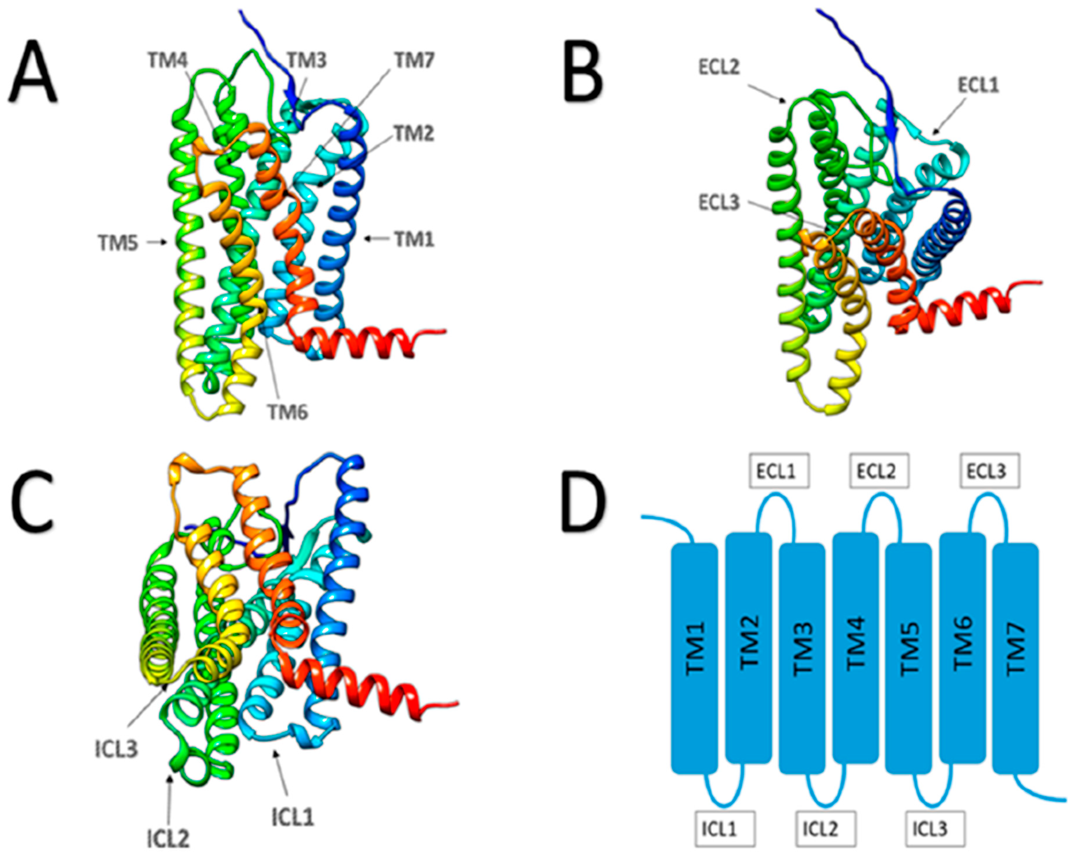
Drs. Richard Axel and Linda Buck were awarded the Nobel Prize in Physiology or Medicine in 2004 for their findings on the cellular and molecular organization of the olfactory system by genetic and molecular analyses of a novel superfamily of G-protein-coupled receptors with seven transmembrane domains (Figure 1) as putative olfactory receptors (ORs) in the nasal olfactory epithelium [1,2]. In 2004, the Buck laboratory reported 339 intact human OR genes and 297 OR pseudogenes, with the OR genes and pseudogenes distributed unevenly distributed among 51 different loci on 21 human chromosomes (with none found on human chromosome 21 or the Y-chromosome), and a heavy concentration of OR loci on human chromosome 11 [3]. Many human odorant receptors were long considered “orphans”, in that their ligands/agonists were unknown. However, the process of deorphanizing ORs has progressed considerably since 2007, with the deorphanization of OR7D4 [4], and a comprehensive annotation for human ORs and their known ligands/agonists has been gradually accumulated and recently reviewed [5].
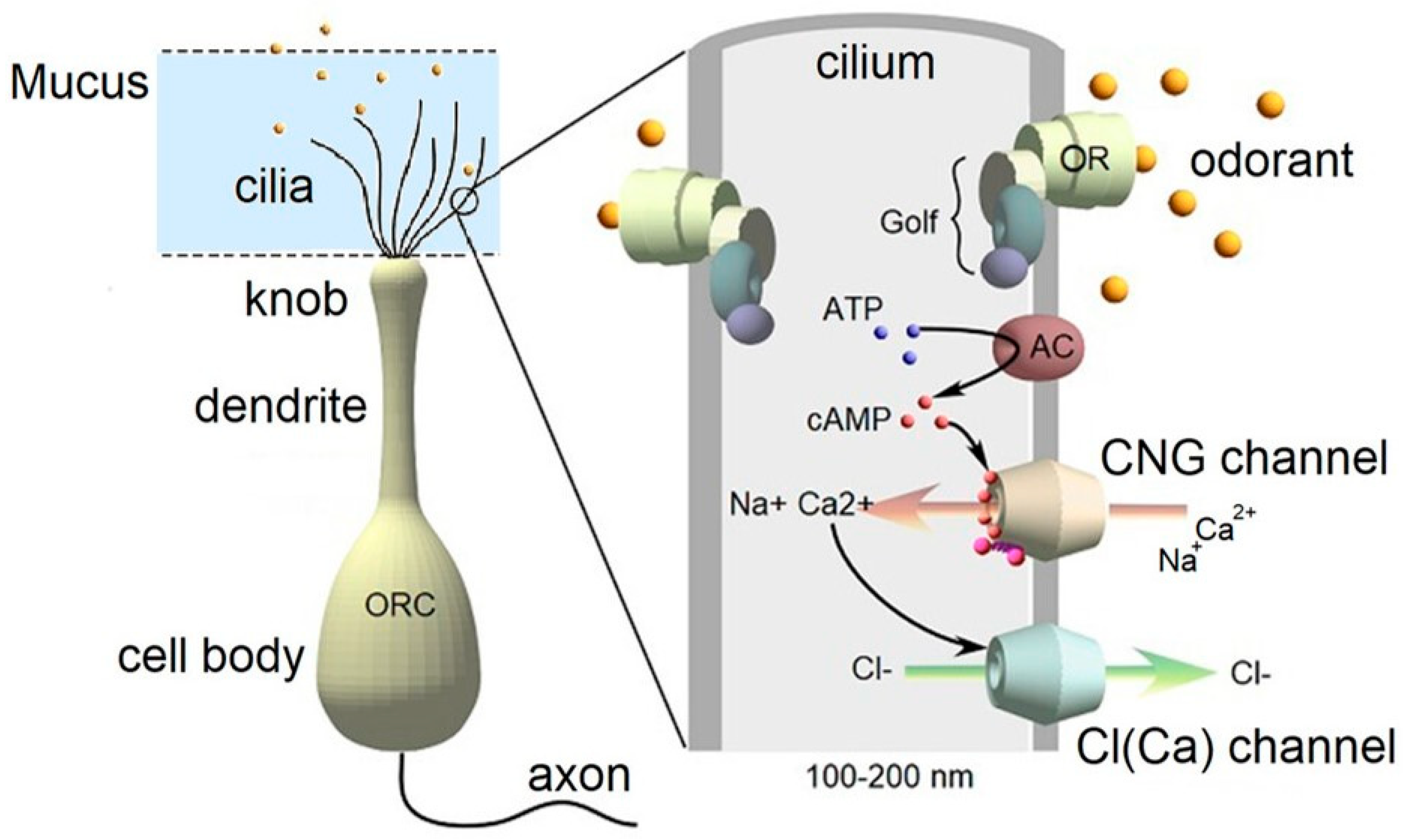
ORs, which are expressed on the ciliary membranes of the apical dendrites of olfactory receptor neurons, interact with odorant molecules in inhaled air in the nasal cavity. Upon odorant molecules specifically binding to the receptor site of an OR, an induced conformational change in the OR triggers the signaling cascade resulting in transduction of the olfactory stimulus via the olfactory neuronal axon (cranial nerve I) to glomeruli in the olfactory bulb of the brain. Extracellular loop 2 (ECL2; Figure 1) appears to be critical in odorant recognition [6]. The odorant signal transduction pathway involves a conformational change in the receptor to engage an intracellular G-protein (Golf) upon ligand binding, cleavage of ATP by adenyl cyclase to generate cAMP, and activation of a cyclic nucleotide-gated cation channel by cAMP (refs. [7,8,9,10]; Figure 2). The resulting influx of cations (primarily calcium and sodium) causes depolarization of the neuron. Increased intracellular Ca2+ and/or cAMP concentrations have been used as a readout for OR activation (e.g., ref. [8]). It is widely held that each individual olfactory neuron is genetically regulated to express multiple copies of a single OR [11,12] and that the axons of neurons that express the same OR are genetically and physiologically controlled to project to the same glomerulus in a target region of the olfactory bulb [13]. This organization has been dubbed the “one neuron–one receptor” and the “one glomerulus–one receptor” paradigm [14]. Subtle differences in odors (e.g., the difference in the aroma between a lime vs. a lemon) are thought to result from activation of different combinations of ORs by different but similar odorant stimuli [15,16].
As the genomics revolution began with the widespread use of technologies such as microarrays and gene-chips, investigators in many disciplines found, often to their surprise, that odorant receptor transcripts were found in multiple tissues in multiple species, and in many cultured cell lines and tumors. These observations spawned studies to confirm expression of the encoded proteins in non-nasal tissues and to understand potential non-canonical functions of odorant receptors in “ectopic” locations (i.e., outside of the nasal cavity). The purpose of this mini narrative review is to describe key studies on ectopically expressed human ORs, including their putative roles outside of the nasal cavity, with a particular focus on ligand-activated effects in cells and tissues, and disease states such as prostate, skin and intestinal cancers, asthma, infertility, and obesity.

2. Ectopic Expression of Olfactory (Odorant) Receptors (ORs)

2.1. ORs and the Skin

Inhalation of odorant molecules has long been known to have physiological effects on tissues such as the skin, which was an early clue that ORs might exist and function in the skin. For example, mice exposed to citralva by inhalation recovered from skin barrier insults more quickly than mice not treated with the odorant therapy; there was some specificity to this response, as another odorant, lyral, did not have the same beneficial effect [17]. Skin barrier disruption induced by chronic restraint stress in rats was reduced by inhalation exposure of the rats to a “green odor” (equal parts trans-2-hexenal and cis-3-hexenol) [18]. In humans, “rose essential oil” (isolated from Rosa alba), impregnated in a patch that was worn around the neck, suppressed stress-induced transepithelial water loss (TEWL) in students at a stressful time (during examinations) [19]. Similarly, Denda et al. [20] demonstrated that inhalation of a component of Bulgarian rose oil, dimethoxy-methylbenzene, accelerated repair of tape-stripping-induced barrier damage in both mice and humans.
Evidence of an OR basis for the beneficial effects of a rose extract on skin has more recently been described. Duroux et al. [21] demonstrated the gene and protein expression of three novel ORs in skin, namely OR10A6, OR2AG2, and OR11H4, in human epidermal keratinocytes and human skin explants. A rose extract containing 56.7% phenylethyl alcohol (plus ~30 other volatile molecules) was found to potently activate these ORs. Eighteen individual molecules were screened as possible ligands for these receptors, based on the likelihood of finding these molecules with a high percentage in flower extracts (Table 1). To elucidate potential functional consequences of these ORs in the presence of ligands, skin explants were stressed with epinephrine and evaluated for stress markers (G6PD, loricrin, and γH2AX), as well as expression of the ORs themselves. The rose extract prevented the increased expression of the stress markers in human skin explants exposed to epinephrine. To determine a potential cosmetic benefit of the rose extract in humans, a split face study was conducted on female volunteers with under-eye darkness who applied a cream containing the rose extract (5 × 10⁻³%) twice daily for 28 days. A decrease in the appearance of dark under-eye circles, as measured by Bio blue-light 3D scanning analysis, was demonstrated with application of the rose extract.
OR2AT4 has been shown to have multiple roles in skin physiology. Using a customized microarray and RT-PCR analysis, Busse et al. [22] confirmed transcripts for five ORs in primary human keratinocytes (OR6M1, OR11A1, OR2AT4, OR5V1, and OR6V1). Immunocytochemical staining confirmed the expression of OR2AT4 protein in HaCaT cells and primary human keratinocytes. The group tested the hypothesis that one of these ORs would be activated by sandalore, based on previous data that showed that stimulation of keratinocytes with synthetic sandalwood odorants (often found in perfumes and other personal care products) caused an increase in intracellular calcium. Using calcium imaging, sandalore and a related molecule, brahmanol, were determined to be agonists of OR2AT4. Moreover, sandalore treatment stimulated cell migration in an in vitro wound-healing assay. The specificity of the receptor–ligand interaction in keratinocyte migration was demonstrated, as other structurally similar molecules such as ebanol and sandranol were inactive in the migration assay, and others, such as oxyphenylon, displayed antagonistic activity [22]. The expression of OR2AT4 was independently confirmed in normal human keratinocytes [21].
To explore a potential role of OR2AT4 in hair growth, Mataix et al. [23] identified a highly specific DNA aptamer targeting OR2AT4. The aptamer, Ap.OR2AT4.17, effectively prolonged the anagen phase of the hair growth cycle in a preclinical human organ culture model. In addition, Ap.OR2AT4.17 was shown to be functional, leading to enhanced OR2AT4 expression in hair follicles, increased proliferation of hair matrix keratinocytes, and significant hair shaft elongation in organ-cultured human hair follicles. The OR2AT2 ligand sandalore has also been shown to prolong human hair growth ex vivo by decreasing apoptosis and increasing production of the anagen-prolonging growth factor IGF-1. In support of this observation, co-administration of the specific OR2AT4 antagonist Phenirat, as well as silencing of OR2AT4, inhibited hair growth in this model system [24]. These findings, taken together, suggest modulation of OR2AT may yield a novel, targeted therapy for hair loss disorders. Interestingly, the aptamer treatment also upregulated IL-37 and human β defensin-3 antimicrobial peptide expression, highlighting its role as a microbiota-modulating compound.
In related work, Edelkamp et al. [25], using in situ hybridization, demonstrated OR2A4/7 mRNA expression in the hair follicle (HF) epithelium (outer root sheath [ORS], hair matrix [HM]) of freshly embedded scalp skin from healthy donors; OR2A4/7 protein expression was restricted largely to the infundibulum. During HF organ culture, which induces tissue distress, OR2A4/7 protein expression was upregulated also in the ORS and HM. Subsequently, OR2A4/7 activation by the odorant molecule cyclohexyl salicylate promoted both human HF growth and stem cell progeny expansion.
In 2017, Tsai and collaborators [26] revealed an additional OR in the skin, namely OR51B5, and further expanded on the role of OR2A4/7 in skin. Cyclohexyl salicylate was confirmed as an agonist of OR2A4/7, and isononyl alcohol was identified as an agonist of OR51B5, and studies in human keratinocytes in vitro revealed that both odorants induced strong Ca2+ signals that were mediated by the receptors, as demonstrated by knockdown experiments. OR2A4/7 was localized to suprabasal keratinocytes and basal melanocytes of the epidermis and was found to influence cytokinesis, cell proliferation, phosphorylation of AKT and Chk-2 and secretion of IL-1. In contrast, OR51B5 was exclusively localized to suprabasal keratinocytes, and found to support cell migration and regeneration of keratinocyte IL-6 secretion.

2.2. ORs and Lung Function

Li et al. [27] reported expression of eight different olfactory receptors (ORs 1014, 657, 622, 568, 446, 352, 272, and 65) on mouse pulmonary macrophages, with upregulation of OR expression by combined treatment with TNF-γ and lipopolysaccharide to mimic lung inflammation. Stimulation of alveolar macrophages with the odorant octanal (ligand for human ORs 1A1, 1A2, and 6A2 [5]) increased monocyte chemotactic protein-1 production and macrophage motility, suggesting that certain odorant molecules may be capable of modulating immune function in the lung [27]. More recently, human OR2AT4 and OR1A2 were described on the plasma membrane of human alveolar macrophages and were hypothesized to be therapeutic targets in human pulmonary diseases. Stimulation of alveolar macrophages with sandalore, a ligand of OR2AT4, and citronellal, a ligand of OR1A2, triggered a transient increase in intracellular calcium and cAMP, which was associated with decreased phagocytic activity and cytokine release [28] (discussed further below).

2.3. ORs and Sperm

Ectopic OR expression also has been evaluated for a role in chemotaxis. Over a dozen ORs have been found on human sperm, and bourgeonal (fragrance of the lily of the valley flower and a ligand of OR1D2) appears to have a role in sperm chemotaxis and fertilization [29]. Flegel et al. described the expression and localization of multiple ORs on human sperm [ref. [30]; Table 1]. Of note, expression of OR51E2 was observed on the extracellular membrane of the flagella of human sperm [30]. Teveroni et al. subsequently demonstrated that various short-chain fatty acids produced by vaginal microbiota can promote sperm migration via OR51E2 [31]. Polymorphisms in human ability to detect various odorants have been documented (e.g., ref. [4]), and human olfactory insensitivity to the odor of bourgeonal has been associated with idiopathic infertility in human males [32].

2.4. ORs and Obesity with NASH

Torres-Reyes et al. [33] used whole-exome sequencing of DNA isolated from peripheral blood samples to identify potential causal variants and biological processes associated with obesity with progression to nonalcoholic steatohepatitis (NASH) in a small group of Mexican subjects (BMIs ranging from 32 to 56). A total of 1359 variants with a probable pathogenic effect were identified in obese patients with NASH diagnosis. Further analysis showed that the variants were associated with processes such as “detection of stimulus in sensory perception” (GO:0050906, GO:0050907) and “sensory perception of smell” (GO:0050911, GO:0007608). Interestingly, rs8105737 (OR1I1) and rs998544 (OR5R1) variants were present in 56% and 44% of obese/NASH subjects, respectively. Neither of these ORs had been deorphanized with regard to their ligands as of 2025 [5], and according to The Human Protein Atlas (https://www.proteinatlas.org/), the tissue localization of the encoded proteins is poorly characterized. Identification of the tissue localization and ligands for these ORs might lead to novel approaches to treating or slowing the progression of obesity to NASH.

3. Ectopic ORs and Disease States

3.1. ORs and Lung Disease

The human olfactory receptors OR2AT4 and OR1A2 were investigated as potential therapeutic targets in alveolar macrophages in human pulmonary diseases. Ligands of these receptors (sandalore and citronella, respectively) decreased phagocytic activity and cytokine release in human alveolar macrophages [28]. Given that there are limited therapeutic options for treating steroid-resistant non-type 2 inflammation in obstructive lung diseases, the results were interpreted as promising preliminary evidence for the potential to use ORs as therapeutic targets in treating steroid-resistant lung diseases with non-type 2 inflammation.
In related work [34], A549 cells and primary human bronchiolar epithelial cells were evaluated for responses to a panel of 100 potential OR ligands. Brahmanol (a ligand for OR2AT4) and cinnamaldehyde (ligand for OR2J3) increased intracellular calcium concentrations in A549 cells. As previously demonstrated by Busse et al. [22] in the skin, brahmanol similarly promoted cell migration in an in vitro scratch (healing) assay in A549 cells without affecting cell proliferation or cAMP levels. In contrast, treatment of A549 cells with cinnamaldehyde increased intracellular cAMP and reduced IL-8 release. Mitigation of IL-8 release may represent an early intervention strategy in asthma, as neutrophils and IL-8 distinguished mild asthma from moderate/severe asthma in a study of control bronchoalveolar lavage fluids from control subjects, compared to moderate and severe asthmatics [35]. These results similarly suggested that ORs might be promising drug target candidates for lung diseases with non-type 2 inflammation [34].
In other work on ORs and lung disorders, OR2AG2 mRNA was reported to be significantly lower in the lung tissues of patients with asthma than in those of healthy subjects [36].

3.2. OR Expression in Diseased Skin

Tham et al. [37] examined OR mRNA expression using whole-transcriptome sequencing of skin tape strip samples collected from patients with atopic dermatitis (AD) and healthy control (HC) subjects. A total of 381 OR gene transcripts were detected in the skin samples, with the greatest OR expression detected in tape strip samples corresponding to the upper granular layer of the skin. In a study focusing on OR10G7, expression was significantly increased in skin biopsy specimens from patients with AD, compared to those from HC subjects, and inversely correlated with filaggrin (FLG-1) expression. OR10G7 expression was greatest in undifferentiated keratinocytes from patients with AD and was downregulated with progressive differentiation, but remained higher in AD subjects vs. HCs. Differing responses to ligands of OR10G7, namely eugenol and acetophenone, were observed. While primary human keratinocytes transfected with OR10G7 siRNA produced ATP in response to both ligands, cells treated with eugenol responded with a significant upregulation of IL-1β, a proinflammatory cytokine; the acetophenone effect was abolished in OR10G7-siRNA-transfected keratinocytes. This observation is of potential therapeutic importance, as acetophenone is a S. aureus degradation product of the amino acid phenylalanine [38], and S. aureus colonization is a common feature of AD. The authors concluded that ORs may serve as potential future therapeutic targets for the inhibition of pathological pain or itch pathways in allergic inflammatory skin disorders such as AD [37].
In response to ultraviolet (UV) light or inflammation, which are well-recognized stimuli for skin barrier dysfunction, the HaCaT human keratinocyte cell line was evaluated for OR expression and ligand responsiveness [39]. Expression of twelve ectopic ORs was detected in HaCaT cells, and the expression levels of ORs 1F1, 2A4, 2H2, 5C1, 7D2, 10H1, 52I1, and 52W1 were upregulated in response to UV light [39]. Similarly, all 12 receptors (1F1, 2A4, 2AE1, 2W3, 2H2, 5C1, 7D2, 10A2, 10H1, 52B2, 52I1, and 52W1) were upregulated in response to a cocktail of TNF-a and IFN1-g, intended to mimic an inflammatory response. Notably, OR2AT4 was not reported to be expressed in the HaCaT cell line, in contrast to the observations of others [22,23] in primary human keratinocytes. In response to the ligands cyclohexyl salicylate (ligand for OR2A4) and sandacanol (ligand for OR10H1), expression of the skin barrier genes keratin 1, keratin 10, filaggrin, and loricrin was downregulated after dose-dependent treatment (1–100 μM) with both ligands in HaCaT cells [39]. The results suggest that a subset of human ORs are responsive to both perturbation to barrier function and stimulation with ligands, and with further validation, ORs may serve as a marker for skin barrier deficits.

3.3. Ectopic ORs in Cancer

3.3.1. ORs in Prostate Cancer Cells and Cell Lines

Human prostate epithelial cells and the prostate cancer cell line LNCaP (derived from a metastatic tumor) express OR51E2, which is also known as prostate-specific G-protein-coupled receptor (PSGR). Efforts to deorphanize OR51E2 identified several steroid molecules and the odorant β-ionone as agonists of the receptor. LNCaP cells responded to β-ionone with increased intracellular calcium, an effect that was reduced by the PSGR inhibitor α-ionone. Cell proliferation in both primary prostate epithelial cells and the LNCaP cell line was inhibited by β-ionone, and the proliferative effect of DHT treatment was also suppressed by β-ionone. These effects were not noted in the PC-3 prostate cancer cell line, which lacks PSGR/OR51E2 [40].
In an in vivo study, castrated male Nod Scid Gamma mice were inoculated subcutaneously with LNCaP cells and treated topically (“brushed”) with nothing, with mineral oil, or with β-ionone in mineral oil over a period of six weeks. The mice were then evaluated by X-ray examinations for metastases and invasive tumors [41]. While the most widespread metastatic and invasive findings were in the LNCaP-inoculated, β-ionone-treated mice, metastases were also prevalent in the mice exposed only to mineral oil, calling into question whether PSGR activation alone is sufficient to promote metastases and invasion [41]. Taken together, these two studies suggest that PSGR activation suppresses cell proliferation in vitro and promotes metastases in vivo with both effects involving ligand-activated PSGR/OR51E2.
In another study evaluating the role of OR51E2 in prostate cancer, Pronin and Slepak [42] used an inducible expression system in LNCaP cell lines to investigate the impact of ligand stimulation of OR51E2, as well as OR51E1, on cell proliferation and cytotoxicity. OR51E1 increased intracellular adenylyl cyclase in response to treatment by short-chain to medium-chain organic acids (C3-C9), whereas OR51E2 responded to acetate and propionate but not to the longer-chain organic acids. Stimulation of LNCaP cells with butyrate inhibited their growth, and the knockdown of the endogenous OR51E1 negated the cytostatic effect of butyrate exposure. In the same study, overexpression of OR51E1 or OR51E2 suppressed LNCaP cell proliferation. Overexpression of OR51E1 caused an upregulation of cytostatic and cell death markers, including p27, p21, and p53. These results further suggest that ectopic ORs may be useful therapeutic targets in prostate cancer.
Like observations in human sperm, expression of OR1D2 mRNA was demonstrated in LNCaP cells [43]. The use of a fluorescently labeled conjugate of the OR1D2 ligand bourgeonal was proposed as a novel method to detect prostate cancer and metastasized lymph nodes [43].

3.3.2. ORs in Melanoma

Gelis et al. [44] reported OR51E2 expression at both the gene and protein levels in human melanoma tissue sections. qPCR analysis revealed that the receptor is upregulated in melanoma cells compared to normal melanocytes, indicating that OR51E2 may play a role in early melanoma development and progression. Activation of endogenous OR51E2 by its ligand β-ionone in cultured cells derived from metastatic and vertical-growth phase (VGP) cells resulted in increased intracellular Ca2+ concentration. OR51E2 activation inhibited the growth of VGP melanoma cells by inducing apoptosis, and cell motility assays showed decreased migration of VGP melanoma cells treated with β-ionone. These observations suggest that OR51E2 may serve as a novel target for melanoma therapy.

3.3.3. ORs in Liver Carcinoma Cells

The human hepatocellular carcinoma cell line Huh7 was found to express OR1A2, in addition to PSGR/OR51E2, which has been demonstrated to be a marker for prostate cancer. Exposure to the terpene (−)-citronellal, a ligand for OR1A2, resulted in enhanced phosphorylation of p38 MAPK and decreased Huh7 cell proliferation, suggesting that OR1A2 might be a therapeutic target in some cancers [45]. Similarly, HepG2 cells, which are derived from a human liver tumor, were found to express OR1A1 [46]. Treatment with the OR1A1 ligand (−)-carvone resulted in activation of the cyclic adenosine monophosphate (cAMP)–protein kinase A (PKA)–cAMP response element-binding protein (CREB) pathway, with subsequent upregulation of the CREB-responsive gene hairy and enhancer of split (HES)-1. The observed activation of the PKA-CREB-HES-1 signaling axis resulted in decreased intracellular triglyceride concentrations and decreased intracellular lipid accumulation [46].

3.3.4. OR10H1 and Urinary Bladder Cancer

Weber et al. [47] described the expression of OR10H1 in the human urinary bladder and noted higher expression (both in mRNA and protein) in bladder cancer tissues. In addition, significantly elevated levels of OR10H1 transcripts were detectable in the urine of bladder cancer patients. Using OR10H1-transfected Hana3A-cells, sandranol was identified as an agonist of OR10H1 (evidenced by increased intracellular cAMP in the presence of sandranol). Receptor activation resulted in morphological alterations, accompanied by significantly diminished cell viability, cell proliferation and migration, and a limited degree of apoptosis. These observations suggest that OR10H1 may be both a biomarker and a therapeutic target for bladder cancer.

3.3.5. OR51E1 and Small Intestine Neuroendocrine Cancer

Overexpression of OR51E1, whose ligands include gut microbiota-derived molecules such as isovaleric acid and butyrate [5], was investigated as a possible tissue biomarker for small intestine neuroendocrine cancer (SI-NEC) by qRT-PCR and immunohistochemistry [48]. SI-NEC are rare tumors that are frequently diagnosed at a late stage and arise from enterochromaffin (EC) cells. In this study, wild-type OR51E1 expression was found to be higher in microdissected SI-NEC cells than in adjacent non-tumor cells. In tissue biopsy specimens, both cytoplasmic and membranous OR51E1 immunostaining was observed in primary SI-NECs and metastases. The authors propose that the abundant expression of OR51E1 in SI-NEC cases makes it an attractive candidate target in future diagnostics and/or therapeutics. Further, given that the OR51E1 ligand butyrate is generated in the lower gastrointestinal tract from fermentable dietary fiber, and is also proven to have the ability to selectively kill cancer cells and inhibit colorectal cancer cell migration [49], dietary interventions to promote butyrate formation may be a complementary treatment option in SI-NECs and other gastrointestinal cancers.
Table 1. Summary of ectopic ORs and ligand responses in human cells and tissues.
Ectopic ORTissue(s)/CellsTranscript (T) or Protein (P)Ligand(s)Functional ObservationsReference(s)
OR1A1Human HepG2 cellsT, P(−)-Carvone,
(S)-(−)-citronellal, helional,
heptanal,
octanal,
nonanal,
hydroxy-citronellal,
citral,
4-decenal, octanol,
(S)-(−)-citronellol
Carvone exposure decreased intracellular triglyceride concentrations and intracellular lipid accumulation[5,46]
OR1A2Human alveolar macrophagesT, PCitronellal,
helional,
heptanal,
octanal,
nonanal,
hydroxy-citronellal,
citral,
4-decenal, octanol
Citronellal reduced phagocytic capacity and release of proinflammatory cytokines[5,28]
Human hepatocellular carcinoma cell line Huh7T, P(−)-Citronellal exposure enhanced phosphorylation of p38 MAPK, decreased cell proliferation[5,45]
OR1D2Human spermN.R.Bourgeonal, cyclamalDose-dependent chemotaxis, hyperactive flagellar beating in sperm in presence of an ascending bourgeonal gradient; effects abolished by co-application of undecanal[29]
OR2A4/7Human scalp skin, hair follicle cultures, human keratinocytes,
HaCaT cells
T, PCyclohexyl salicylateHair follicle growth, increased cell proliferation of HaCaT cells and primary keratinocytes[25,26]
OR2AG2Normal human epidermal keratinocytes, skin explants (suprabasal layer)T, PCitronellol,
nerol, cis-3-hexenol,
linalool,
geraniol,
α-cinnamyl alcohol, phenyl ethyl alcohol,
phenyl propyl alcohol,
benzyl acetone, rose extract
Increased cAMP in response to ligands[21]
Human asthma patient lung tissueTLigand effects N.R.Decreased expression in asthma patients[36]
OR2AT4HaCaT cells, primary human keratinocytesT, PSandalore, brahmanolPromotion of epithelial cell migration, hair growth[21,22,24]
Human alveolar macrophagesT, PSandalore exposure reduced phagocytosis and release of proinflammatory cytokines[28]
Primary human airway epithelial cells, A549 cellsT, PBrahmanol promoted cell migration in A549 cells[34]
OR2J3Primary human airway epithelial cells, A549 cellsT, PCinnamaldehydeIncreased intracellular cAMP and reduced IL-8 release in A549 cells[34]
OR10A6Normal human epidermal keratinocytes, skin explants (throughout epidermis)T, PCyclamen aldehyde, lyral, nonadecane, α-ionone,
3-phenyl propyl propionate,
citronellol,
nerol,
linalool,
geraniol,
α-cinnamyl alcohol,
phenyl ethyl alcohol,
phenyl propyl alcohol,
benzyl acetone,
cyclemone a, rose extract
Increased cAMP in response to 13 ligands tested[5,21]
OR10G7Human tape stripping samples, human skin biopsies (atopic dermatitis [AD] vs. healthy controls [HC])T, PEugenol, acetophenoneExpression significantly higher in biopsies from AD patients vs. HC; inversely correlated with filaggrin expression; OR10G7-transfected cells treated with eugenol showed upregulation of IL-1β[37]
OR11H4Normal human epidermal keratinocytes, skin explants (basal
membrane of the epidermis)
T, PPhenylethyl alcohol, phenyl propyl alcohol, rose extractIncreased cAMP in response to ligands[21]
OR51B5HaCaT cells, human keratinocytes, human skinT, PIsononyl alcoholAccelerated regeneration rate of HaCaT cells and keratinocyte (“wound closure” assay)[26]
OR51E1LNCaP cellsT, PButyrate,
isovaleric acid
Decreased proliferation[5,42]
Small intestine neuroendocrine tumorsPLigand effects N.R.Expression higher in microdissected tumor cells than in adjacent non-tumor cells[48]
OR51E2Human spermT, Pβ-ionone propionate,
acetate
Enhanced sperm migration[5,30]
T, PShort-chain fatty acids[31]
Human prostate epithelial cells, LNCaP cellsT, P Cell proliferation in both primary prostate epithelial cells and LNCaP cells was inhibited by β-ionone[40]
Human melanoma tissue sections, cultured cells derived from metastasesP β-ionone activation inhibited growth of vertical-growth phase melanoma cells, induced apoptosis, and decreased migration[44]
Multiple:
OR2W3, OR2H1, OR10J1
Human spermT, PNerol, methional, dimetol, respectivelyIncreased Ca2+ influx[30]
Multiple:
OR1F1, OR2A4, OR7D2, OR2AE1, OR2W3, OR2H2, OR5C1, OR10A2, OR10H1, OR52B2, OR52I1, OR52W1
HaCaT cellsTCyclohexyl salicylate (OR2A4); sandacanol (OR10H1)All 12 ORs upregulated in response to UV light; decreased expression of skin barrier genes in response to ligands[39]
N.R.: Not reported.

4. Conclusions and Future Directions

What began as curious observations—namely detection of olfactory/odorant receptor gene transcripts in tissues outside of the nasal cavity—has given biomedical researchers new tools and potential drug targets to understand and manipulate the biology of multiple cell types and organs. The authors of many of the studies summarized herein recognize that use of agonists and antagonists of ectopically expressed ORs may provide new tools for treating conditions ranging from cancer to infertility. The standard-of-care treatments for conditions such as prostate cancer and melanoma often have some initial benefits, but ultimately show relatively high rates of tumor relapse, treatment resistance, and, hence, failure. Therefore, identification of ectopic ORs in pathological conditions, and manipulating them with agonists and/or antagonists [examples are provided in Table 1], potentially presents novel treatment options. Additionally, given that many OR ligands are naturally occurring (non-synthetic) molecules, adoption of these ligands, or simple modifications thereof, may accelerate the development of lower-cost treatments for challenging diseases worldwide.

Funding

This research received no external funding.

Institutional Review Board Statement

Not applicable.

Informed Consent Statement

Not applicable.

Data Availability Statement

No new data were created or analyzed in this study. Data sharing is not applicable to this article.

Acknowledgments

The author is grateful to the colleagues who reviewed and critiqued this manuscript.

Conflicts of Interest

The author declares no conflicts of interest.

Abbreviations

The following abbreviations are used in this manuscript:
cAMPCyclic adenosine monophosphate
ATPAdenosine triphosphate
GolfOlfactory G-protein
OROlfactory (Odorant) receptor

References

  1. Buck, L.; Axel, R. A novel multigene family may encode odorant receptors: A molecular basis for odor recognition. Cell 1991, 65, 175–187. [Google Scholar] [CrossRef]
  2. Abbott, A. Science of smell wins medicine Nobel. Nature 2004, 431, 616. [Google Scholar] [CrossRef][Green Version]
  3. Malnic, B.; Godfrey, P.A.; Buck, L.B. The human olfactory receptor gene family. Proc. Natl. Acad. Sci. USA 2004, 101, 2584–2589, Erratum in: Proc. Natl. Acad. Sci. USA 2004, 101, 7205. [Google Scholar] [CrossRef]
  4. Keller, A.; Zhuang, H.; Chi, Q.; Vosshall, L.B.; Matsunami, H. Genetic variation in a human odorant receptor alters odour perception. Nature 2007, 449, 468–472. [Google Scholar] [CrossRef]
  5. Wang, J.; Zhang, Q.; Fan, W.; Shi, Q.; Mao, J.; Xie, J.; Chai, G.; Zhang, C. Deciphering olfactory receptor binding mechanisms: A structural and dynamic perspective on olfactory receptors. Front. Mol. Biosci. 2025, 11, 1498796. [Google Scholar] [CrossRef] [PubMed]
  6. Yu, Y.; Ma, Z.; Pacalon, J.; Xu, L.; Li, W.; Belloir, C.; Topin, J.; Briand, L.; Golebiowski, J.; Cong, X. Extracellular loop 2 of G protein-coupled olfactory receptors is critical for odorant recognition. J. Biol. Chem. 2022, 298, 102331. [Google Scholar] [CrossRef] [PubMed]
  7. Brunet, L.J.; Gold, G.H.; Ngai, J. General anosmia caused by a targeted disruption of the mouse olfactory cyclic nucleotide-gated cation channel. Neuron 1996, 17, 681–693. [Google Scholar] [CrossRef]
  8. Shirokova, E.; Schmiedeberg, K.; Bedner, P.; Niessen, H.; Willecke, K.; Raguse, J.D.; Meyerhof, W.; Krautwurst, D. Identification of specific ligands for orphan olfactory receptors. G protein-dependent agonism and antagonism of odorants. J. Biol. Chem. 2005, 280, 11807–11815. [Google Scholar] [CrossRef]
  9. Takeuchi, H. Olfactory cilia, regulation and control of olfaction. Physiol. Rep. 2024, 12, e70057. [Google Scholar] [CrossRef] [PubMed]
  10. Billesbølle, C.B.; de March, C.A.; van der Velden, W.J.C.; Ma, N.; Tewari, J.; Del Torrent, C.L.; Li, L.; Faust, B.; Vaidehi, N.; Matsunami, H.; et al. Structural basis of odorant recognition by a human odorant receptor. Nature 2023, 615, 742–749. [Google Scholar] [CrossRef]
  11. Khan, M.; Vaes, E.; Mombaerts, P. Regulation of the probability of mouse odorant receptor gene choice. Cell 2011, 147, 907–921. [Google Scholar] [CrossRef] [PubMed]
  12. Chess, A.; Simon, I.; Cedar, H.; Axel, R. Allelic inactivation regulates olfactory receptor gene expression. Cell 1994, 78, 823. [Google Scholar] [CrossRef]
  13. Mombaerts, P.; Wang, F.; Dulac, C.; Chao, S.K.; Nemes, A.; Mendelsohn, M.; Edmondson, J.; Axel, R. Visualizing an olfactory sensory map. Cell 1996, 87, 675–686. [Google Scholar] [CrossRef]
  14. Takeuchi, H.; Sakano, H. Neural map formation in the mouse olfactory system. Cell Mol. Life Sci. 2014, 71, 3049–3057. [Google Scholar] [CrossRef] [PubMed]
  15. Luu, P.; Acher, F.; Bertrand, H.O.; Ngai, J. Odorant receptor specificities and receptor combinatorials: Implications for olfactory coding. Chem. Senses 2005, 30, i97–i98. [Google Scholar] [CrossRef]
  16. Malnic, B.; Hirono, J.; Sato, T.; Buck, L.B. Combinatorial receptor codes for odors. Cell 1999, 96, 713–723. [Google Scholar] [CrossRef]
  17. Hosoi, J.; Tsuchiya, T. Regulation of cutaneous allergic reaction by odorant inhalation. J. Investig. Dermatol. 2000, 114, 541–544. [Google Scholar] [CrossRef]
  18. Fukada, M.; Kaidoh, T.; Ito, A.; Yano, T.; Hayashibara, C.; Watanabe, T. “Green odor” inhalation reduces the skin-barrier disruption induced by chronic restraint stress in rats: Physiological and histological examinations. Chem. Senses 2007, 32, 633–639. [Google Scholar] [CrossRef]
  19. Fukada, M.; Kano, E.; Miyoshi, M.; Komaki, R.; Watanabe, T. Effect of “rose essential oil” inhalation on stress-induced skin-barrier disruption in rats and humans. Chem. Senses 2012, 37, 347–356. [Google Scholar] [CrossRef]
  20. Denda, M.; Tsuchiya, T.; Shoji, K.; Tanida, M. Odorant inhalation affects skin barrier homeostasis in mice and humans. Br. J. Dermatol. 2000, 142, 1007–1010. [Google Scholar] [CrossRef] [PubMed]
  21. Duroux, R.; Mandeau, A.; Guiraudie-Capraz, G.; Quesnel, Y.; Loing, E. A rose extract protects the skin against stress mediators: A Potential role of olfactory receptors. Molecules 2020, 25, 4743. [Google Scholar] [CrossRef]
  22. Busse, D.; Kudella, P.; Grüning, N.M.; Gisselmann, G.; Ständer, S.; Luger, T.; Jacobsen, F.; Steinsträßer, L.; Paus, R.; Gkogkolou, P.; et al. A synthetic sandalwood odorant induces wound-healing processes in human keratinocytes via the olfactory receptor OR2AT4. J. Investig. Dermatol. 2014, 134, 2823–2832. [Google Scholar] [CrossRef] [PubMed]
  23. Mataix, M.; Illera, N.; Hidalgo, I.; Arriba, M.D.C.; Martín, E.; Fernández, G.; González, C.; Larcher, F.; González, V.M.; Rio, M.D.; et al. Targeting olfactory receptor OR2AT4: An innovative aptamer-based treatment for hair growth promotion. Mol. Ther. Nucleic Acids 2025, 36, 102608. [Google Scholar] [CrossRef]
  24. Chéret, J.; Bertolini, M.; Ponce, L.; Lehmann, J.; Tsai, T.; Alam, M.; Hatt, H.; Paus, R. Olfactory receptor OR2AT4 regulates human hair growth. Nat. Commun. 2018, 9, 3624. [Google Scholar] [CrossRef]
  25. Edelkamp, D.; Pinto, H.; Erdmann, T.; Purba, F.; Jiménez, R.; Paus, M.; Bertolini, M. Olfactory receptor 2A4/7 activation by the fragrance, cyclohexyl salicylate, promotes human hair follicle growth and stem cell progeny expansion. J. Investig. Dermatol. 2022, 142, S279. [Google Scholar] [CrossRef]
  26. Tsai, T.; Veitinger, S.; Peek, I.; Busse, D.; Eckardt, J.; Vladimirova, D.; Jovancevic, N.; Wojcik, S.; Gisselmann, G.; Altmüller, J.; et al. Two olfactory receptors-OR2A4/7 and OR51B5-differentially affect epidermal proliferation and differentiation. Exp. Dermatol. 2017, 26, 58–65. [Google Scholar] [CrossRef]
  27. Li, J.J.; Tay, H.L.; Plank, M.; Essilfie, A.T.; Hansbro, P.M.; Foster, P.S.; Yang, M. Activation of olfactory receptors on mouse pulmonary macrophages promotes monocyte chemotactic protein-1 production. PLoS ONE 2013, 8, e80148. [Google Scholar] [CrossRef]
  28. Weidinger, D.; Jamal Jameel, K.; Alisch, D.; Jacobsen, J.; Bürger, P.; Ruhe, M.; Yusuf, F.; Rohde, S.; Störtkuhl, K.; Kaufmann, P.; et al. OR2AT4 and OR1A2 counterregulate molecular pathophysiological processes of steroid-resistant inflammatory lung diseases in human alveolar macrophages. Mol. Med. 2022, 28, 150. [Google Scholar] [CrossRef] [PubMed]
  29. Spehr, M.; Schwane, K.; Riffel, J.A.; Zimmer, R.K.; Hatt, H. Odorant receptors and olfactory-like signaling mechanisms in mammalian sperm. Mol. Cell Endocrinol. 2006, 250, 128–136. [Google Scholar] [CrossRef] [PubMed]
  30. Flegel, C.; Vogel, F.; Hofreuter, A.; Schreiner, B.S.P.; Osthold, S.; Veitinger, S.; Becker, C.; Brockmeyer, N.H.; Muschol, M.; Wennemuth, G.; et al. Characterization of the olfactory receptors expressed in human spermatozoa. Front. Mol. Biosci. 2016, 2, 73. [Google Scholar] [CrossRef]
  31. Teveroni, E.; Di Nicuolo, F.; Vergani, E.; Bruno, C.; Maulucci, G.; Bianchetti, G.; Astorri, A.L.; Grande, G.; Gervasoni, J.; Santucci, L.; et al. Short-Chain Fatty Acids Modulate Sperm Migration through Olfactory Receptor 51E2 Activity. Int. J. Mol. Sci. 2022, 23, 12726. [Google Scholar] [CrossRef] [PubMed]
  32. Ottaviano, G.; Zuccarello, D.; Menegazzo, M.; Perilli, L.; Marioni, G.; Frigo, A.C.; Staffieri, A.; Foresta, C. Human olfactory sensitivity for bourgeonal and male infertility: A preliminary investigation. Eur. Arch. Otorhinolaryngol. 2013, 270, 3079–3086. [Google Scholar] [CrossRef]
  33. Torres-Reyes, L.A.; Gonzalez-Aldaco, K.; Panduro, A.; Jose-Abrego, A.; Roman, S. Whole-Exome Sequencing identified Olfactory Receptor genes as a key contributor to extreme obesity with progression to nonalcoholic steatohepatitis in Mexican patients: Olfactory receptor genes in obese NASH patients. Ann. Hepatol. 2022, 27, 100767. [Google Scholar] [CrossRef]
  34. Weidinger, D.; Jacobsen, J.; Alisch, D.; Uebner, H.; Heinen, N.; Greune, L.; Westhoven, S.; Jamal Jameel, K.; Kronsbein, J.; Pfaender, S.; et al. Olfactory receptors impact pathophysiological processes of lung diseases in bronchial epithelial cells. Eur. J. Cell Biol. 2024, 103, 151408. [Google Scholar] [CrossRef]
  35. Sur, S.; Ying, S.; Corrigan, C.; Kurosky, A.; Boldogh, I.; Qi, H. IL-8 and neutrophils in bronchoalveolar fluids distinguish mild asthma from moderate to severe aasthma. J. Allergy Clin. Immunol. 2012, 129, AB52. [Google Scholar] [CrossRef]
  36. Chakraborty, S.; Dakle, P.; Sinha, A.; Vishweswaraiah, S.; Nagori, A.; Salimath, S.; Prakash, Y.; Lodha, R.; Kabra, S.; Ghosh, B. Genetic variations in olfactory receptor gene OR2AG2 in a large multigenerational family with asthma. Sci. Rep. 2019, 9, 19029. [Google Scholar] [CrossRef]
  37. Tham, E.H.; Dyjack, N.; Kim, B.E.; Rios, C.; Seibold, M.A.; Leung, D.Y.M.; Goleva, E. Expression and function of the ectopic olfactory receptor OR10G7 in patients with atopic dermatitis. J. Allergy Clin. Immunol. 2019, 143, 1838–1848.e4. [Google Scholar] [CrossRef]
  38. Filipiak, W.; Sponring, A.; Baur, M.M.; Filipiak, A.; Ager, C.; Wiesenhofer, H.; Nagl, M.; Troppmair, J.; Amann, A. Molecular analysis of volatile metabolites released specifically by Staphylococcus aureus and Pseudomonas aeruginosa. BMC Microbiol. 2012, 12, 113. [Google Scholar] [CrossRef]
  39. Kang, W.; Son, B.; Park, S.; Choi, D.; Park, T. UV-Irradiation- and inflammation-induced skin barrier dysfunction is associated with the expression of olfactory receptor genes in human keratinocytes. Int. J. Mol. Sci. 2021, 22, 2799. [Google Scholar] [CrossRef]
  40. Neuhaus, E.M.; Zhang, W.; Gelis, L.; Deng, Y.; Noldus, J.; Hatt, H. Activation of an olfactory receptor inhibits proliferation of prostate cancer cells. J. Biol. Chem. 2009, 284, 16218–16225. [Google Scholar] [CrossRef] [PubMed]
  41. Sanz, G.; Leray, I.; Dewaele, A.; Sobilo, J.; Lerondel, S.; Bouet, S.; Grébert, D.; Monnerie, R.; Pajot-Augy, E.; Mir, L.M. Promotion of cancer cell invasiveness and metastasis emergence caused by olfactory receptor stimulation. PLoS ONE 2014, 9, e85110. [Google Scholar] [CrossRef]
  42. Pronin, A.; Slepak, V. Ectopically expressed olfactory receptors OR51E1 and OR51E2 suppress proliferation and promote cell death in a prostate cancer cell line. J. Biol. Chem. 2021, 296, 100475. [Google Scholar] [CrossRef] [PubMed]
  43. Sturzu, A.; Sheikh, S.; Echner, H.; Nägele, T.; Deeg, M.; Schwentner, C.; Horger, M.; Ernemann, U.; Heckl, S. Novel bourgeonal fragrance conjugates for the detection of prostate cancer. Investig. New Drugs 2013, 31, 1151–1157. [Google Scholar] [CrossRef]
  44. Gelis, L.; Jovancevic, N.; Bechara, F.G.; Neuhaus, E.M.; Hatt, H. Functional expression of olfactory receptors in human primary melanoma and melanoma metastasis. Exp. Dermatol. 2017, 26, 569–576. [Google Scholar] [CrossRef] [PubMed]
  45. Maßberg, D.; Simon, A.; Häussinger, D.; Keitel, V.; Gisselmann, G.; Conrad, H.; Hatt, H. Monoterpene (−)-citronellal affects hepatocarcinoma cell signaling via an olfactory receptor. Arch. Biochem. Biophys. 2015, 566, 100–109. [Google Scholar] [CrossRef]
  46. Wu, C.; Jia, Y.; Lee, J.H.; Kim, Y.; Sekharan, S.; Batista, V.S.; Lee, S.J. Activation of OR1A1 suppresses PPAR-γ expression by inducing HES-1 in cultured hepatocytes. Int. J. Biochem. Cell Biol. 2015, 64, 75–80. [Google Scholar] [CrossRef] [PubMed]
  47. Weber, L.; Schulz, W.A.; Philippou, S.; Eckardt, J.; Ubrig, B.; Hoffmann, M.J.; Tannapfel, A.; Kalbe, B.; Gisselmann, G.; Hatt, H. Characterization of the olfactory receptor OR10H1 in human urinary bladder cancer. Front. Physiol. 2018, 9, 456. [Google Scholar] [CrossRef]
  48. Cui, T.; Tsolakis, A.V.; Li, S.C.; Cunningham, J.L.; Lind, T.; Öberg, K.; Giandomenico, V. Olfactory receptor 51E1 protein as a potential novel tissue biomarker for small intestine neuroendocrine carcinomas. Eur. J. Endocrinol. 2013, 168, 253–261. [Google Scholar] [CrossRef]
  49. Wang, W.; Fang, D.; Zhang, H.; Xue, J.; Wangchuk, D.; Du, J.; Jiang, L. Sodium Butyrate Selectively Kills Cancer Cells and Inhibits Migration in Colorectal Cancer by Targeting Thioredoxin-1. OncoTargets Ther. 2020, 13, 4691–4704. [Google Scholar] [CrossRef]
Figure 1. (A) Depiction of the 7 transmembrane (TM) regions of olfactory receptors. (B) Depiction of extracellular loops (ECLs) 1–3. (C) Depiction of intracellular loops (ICLs) 1–3. (D) Two-dimensional diagram of the 7 TM regions, as well as the three ECLs and ICLs. Reprinted from Wang et al., 2025 [5].
Receptors 05 00020 g001
Figure 2. Olfactory signal transduction in response to odorant molecules binding to ORs in the nasal olfactory mucosa. Odorant molecules (yellow dots) bind to odorant receptors which are located on the ciliary membranes of individual odorant receptor cells (ORCs). Upon binding of an odorant molecule to an OR, the olfactory G-protein Golf is activated, which results in generation of cyclic AMP (cAMP) by Gα-mediated activation of adenylyl cyclase. cAMP, in turn, opens a cation-selective channel on the ciliary membrane, which permits the influx of Na+ and Ca2+ into the cilia, resulting in depolarization of the olfactory neuron. Following the influx of cations, cytoplasmic Ca2+ opens Cl(Ca) channels expressed on the cilia, which enables Cl to flow out of the cell. This further increases the inward current, resulting in signal amplification. Reprinted from Takeuchi, 2024 [9].
Receptors 05 00020 g002
Disclaimer/Publisher’s Note: The statements, opinions and data contained in all publications are solely those of the individual author(s) and contributor(s) and not of MDPI and/or the editor(s). MDPI and/or the editor(s) disclaim responsibility for any injury to people or property resulting from any ideas, methods, instructions or products referred to in the content.