Perspective

Role of Glucocorticoid Receptor in Triple-Negative Breast Cancer

Department of Pharmaceutical and Biomedical Sciences, Touro College of Pharmacy, Touro University, New York, NY 10036, USA
Receptors 2025, 4(2), 8; https://doi.org/10.3390/receptors4020008
Submission received: 4 October 2024 / Revised: 15 December 2024 / Accepted: 10 March 2025 / Published: 1 April 2025

Abstract

:
Triple-negative breast cancer (TNBC) is an aggressive tumor among breast cancer subtypes with much lower overall survival at metastasis compared to other subtypes and with limited treatment options due to a lack of targeted therapies. This has led to the investigation of molecular targets to advance the development of novel therapeutic agents aimed at treating TNBC patients. Recent studies have led us to believe that glucocorticoid receptor (GR) expression may be predictive of decreased survival and increased risk of metastasis in TNBC tumors. Thus, a detailed understanding of GR signaling in TNBC may help understand the role of GR in TNBC proliferation as well as its role as a potential biomarker and therapeutic target. Recent research findings indicate that GR-induced gene regulations may provide an important platform for the development of GR-based therapeutic targets in TNBC. Emerging data from laboratories indicate that targeting GR has the potential to inhibit cancer cell proliferation and reduce tumor growth in TNBC. Therefore, future research focused on underlying molecular mechanisms of GR action in TNBC could lead to a new effective treatment option for TNBC patients, which is urgently needed.

1. Introduction

Based on the presence or absence of molecular markers, estrogen (ER) or progesterone (PR) receptors, and human epidermal growth factor 2 (HER2), breast cancer is categorized into three major subtypes [1]. Triple-negative breast cancer (TNBC) is an aggressive tumor characterized by highly proliferative cancer cells, an early pattern of metastasis, and poor prognosis among breast cancer subtypes [1,2,3,4,5]. TNBC is frequently diagnosed in younger women and is highly prevalent in African American women [1,2,3,4,5]. The median overall survival of patients with metastatic TNBC is much lower than the other two subtypes [1]. Finding effective treatment targets for TNBC has proven difficult so far. Due to the absence of any known targeted therapy for TNBC, several laboratories have been fervently investigating molecular targets to advance the development of novel therapeutic agents to treat this clinically aggressive phenotype. TNBCs are reported to respond to neoadjuvant chemotherapy, but overall, survival in patients with such tumors is poor [6,7,8,9,10,11,12,13,14].
Available options for TNBC treatment are confined to surgery and chemotherapy, either individually or in combination [3,4]. Ideal conditions for the therapeutic assessment and management of TNBC patients remain to be validated in prospective investigations. Several studies have shown that TNBC cells proliferate at a higher rate, exhibit central necrosis with a pushing border, and are highly prone to metastasis and recurrence [15,16,17]. Five-year survival rates also tend to be lower for TNBCs [1,2,3,4]. Finding effective treatment targets for TNBC has been difficult so far. TNBCs are reported to respond to neoadjuvant chemotherapy [7,8,9]. However, overall, survival in patients with such tumors remains quite poor [7,8,9]. Various investigations suggest that TNBC patients are more likely to benefit from chemotherapeutic agents in development that use a strategy of pursuing new treatments and/or drug combinations [7,8,9].
TNBCs lack the expression of ER and PR; therefore, patients with TNBCs are at a disadvantage from currently available ER/PR-targeted systemic therapies. The glucocorticoid receptor (GR) has diverse cell-type-specific effects, promoting apoptosis in cells of lymphoid origin and conversely promoting survival in cells of epithelial origin [18,19]. In solid tumors, GR is emerging as a mediator of cell survival and resistance to chemotherapy-induced cell death, and GR expression is predictive of decreased survival and increased risk of metastasis in TNBC tumors [20,21,22]. The GR appears to be a mediator of pro-survival in TNBC cells [23]. Breast cancer patients typically receive high-dose glucocorticoid treatment prior to chemotherapy to alleviate adverse side effects [6,7]. Thus, a detailed understanding of GR signaling in breast cancer biology and its impact on TNBC is immensely important. Recent studies have shown that GR-mediated anti-apoptotic activity in early-stage TNBC significantly correlates with chemotherapy resistance and increased recurrence [9,10]. In this review article, we discuss up-to-date knowledge of the role of GR in TNBC cell proliferation as well as its potential as a biomarker of disease and therapeutic target for TNBC.

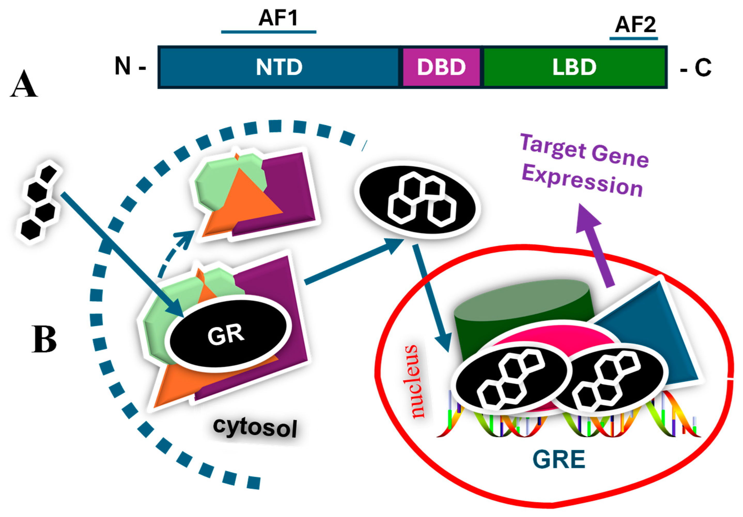
2. Glucocorticoids and Glucocorticoid Receptor

Transcription factors activate or repress the expression of specific genes by binding to specific sequences of DNA to regulate development, physiology, and homeostasis in eukaryotes [24,25]. One of the well-characterized metazoan transcription factors is the superfamily of nuclear receptors. The GR belongs to this superfamily of intracellular transcription factors and is expressed in nearly all vertebrate cells [24,25,26,27]. The GR mediates the biological effects of glucocorticoids, which are involved in several physiological processes, including metabolism, water and electrolyte balance, immune response, growth, cardiovascular function, mood and cognitive functions, reproduction, and development [24,25,26,27,28,29,30,31,32,33,34,35,36,37]. Like other members, GR directly up- and/or downregulates many genes in a cell- and promoter-specific manner to regulate various aspects of development, metabolism, stress response, inflammation, and immunosuppressive effects, among others [25,26,27].
Most of the biological effects of glucocorticoids occur at the level of regulation of gene transcription mediated via binding to the GR [38,39,40,41,42]. The GR resides in the cytosol as a monomer in a multiprotein complex consisting of various chaperone proteins, such as HSP90, HSP70, immunophilins, FKBPs, CyP-40, P23, and possibly others [24,42,43,44]. Once ligand-bound, GR dissociates itself from this complex, and the receptor undergoes conformational rearrangements that facilitate its translocation to the nucleus, where it binds to site-specific DNA as a dimer and various coregulatory proteins, including those from the basal transcription machinery complex, leading to the expression of target specific genes [43] (Figure 1). The transcriptional activation of GR target genes involving the regulated assembly of coregulatory protein complexes acts in a ligand-, cell type-, and promoter-specific manner [24,25,26,41,42].
The molecular mechanisms that explain the phenomena of the transcriptional activation of GR target genes involving the regulated assembly of coregulatory protein complexes have not been fully understood. However, studies over the years from various laboratories have led to the belief that cell-specific effects of GR-mediated gene expressions are largely dependent on how specific sets of coregulatory proteins are constantly excluded or included in the complex of site-specific DNA on the genome; yet, the precise dynamics of the receptor-coregulatory proteins in the complex are not completely understood [41,42,43] (Figure 2). It has been hypothesized that this process is strongly regulated by structural dynamics within the complex involving both receptor and coregulatory proteins controlled by specific hormones/steroids acting as agonists or antagonists [41,42,43]. In this context, the role of protein-interacting surfaces, specifically the AF1 and AF2 regions involved in the process, is of immense importance [41,42,43]. This phenomenon has extensively been manipulated for designing selective receptor modulators for the treatment of endocrine cancers, including estrogen/progesterone receptor-positive breast cancers [41,42,43]. A typical example of this is Tamoxifen, a selective estrogen receptor modulator widely used for the treatment of breast cancers [41,42]. However, due to a lack of steroid receptor targets, this does not work in the case of estrogen/progesterone receptor-negative breast cancers, including TNBC.

3. Type of Breast Cancers and Targeted Therapies

Breast cancer is the most frequent malignancy in women worldwide and the second most common cause of death from cancer among women worldwide [45]. Despite significant progress being made in the last few decades, which led to the curability of many patients with early-stage, non-metastatic disease, advanced breast cancer with distant organ metastases remains incurable with currently available therapies [45,46]. On the molecular level, breast cancer is a heterogeneous disease, and treatment strategies differ according to the molecular subtype [45,47,48]. The pathogenesis, treatment, and prognosis are closely associated with the molecular subtypes of breast cancer, which include subtypes such as Luminal A (ER/PR]-positive, HER2-negative), Luminal B (ER/PR-positive, HER2-positive), Basal-like subtypes (ER/PR/HER2-negative), and HER-enriched subtypes (HER-2-positive, ER/PR-negative) [45,47,48]. Among these subtypes, ER/PR-positive tumors tend to be less aggressive, with improved survival rates [45,46], whereas HER-2-enriched and ER/PR/HER2-negative tumors are more aggressive, with a poor prognosis without targeted therapy and poor survival rates [45,47,48].
Therefore, depending on the subtype, breast cancer treatment and management approaches are divided into early breast cancer, locally advanced breast cancer, and metastatic breast cancer treatment [48]. Targeted therapy such as (Trastuzumab, a monoclonal antibody directly targeting the HER2 protein) is indicated in HER2-positive breast cancers and reduces the risk of recurrence and death when combined with chemotherapy in early stages [49,50]. Poly adenosine diphosphate–ribose polymerase (PARP) inhibitors such as olaparib and talazoparib, which prevent the activation of PARP, are indicated in the adjuvant setting in individuals with BRCA mutations and HER2-negative breast cancer [51]. CDK4/6 inhibitors such as palbociclib are indicated in metastatic HR-positive and HER2-negative tumors and selected patients with early ER/PR-positive tumors [52]. Immune checkpoint inhibitors such as pembrolizumab can be used in combination with chemotherapy as a neoadjuvant treatment for patients with high-risk and early-stage TNBC [53]. Selective estrogen receptor modulators (SERMs) such as tamoxifen are indicated in ER/PR-positive breast cancers.
Among the several genetic mutations reported to be highly associated with an increased risk of breast cancer include BRCA1 (located on chromosome 17) and BRCA2 (located on chromosome 13) [54]. On the other hand, a longer duration of the breastfeeding period reduces the risk of both ER/PR-positive and -negative cancers [55,56]. Age and reproductive history are also associated with risk factors [57]. Starting menstrual periods before age 12 or going through menopause after age 55 can increase the risk of breast cancer [57]. Irrespective, and in general, the risk of breast cancer increases with age, and most breast cancers are diagnosed in women over 50 [57]. Being overweight or obese, sedentary lifestyle, and/or hormone replacement therapy (HRT) can also increase the risk of breast cancer [58,59]. Based on race and ethnicity, in comparison to Black, Hispanic, or Asian individuals, White women are slightly more likely to develop breast cancer, whereas Black women are more likely to develop more aggressive and more advanced-stage breast cancer, including TNBC, diagnosed at a young age [55,56,57].
In normal breast tissue, GR is expressed in the epithelial cells and is involved in the development of the mammary gland [60]. Several studies have been carried out to determine the biological effects of GR expression in breast cancer cell survival and progression [60]. Based on these studies, GR expression has been found to be a useful prognostic marker depending on the breast cancer subtypes [60,61,62,63,64,65]. Further studies revealed that the transcriptional regulation of genes via GR activation initiates signaling pathways in breast epithelial cells, which may be involved in apoptotic pathways [23,63,64].

4. Triple-Negative Breast Cancer and Glucocorticoid Receptor

Exogenous glucocorticoids are widely used as an adjuvant therapy in breast cancer treatment to prevent hypersensitivity reactions, and studies have shown that glucocorticoids generally inhibit estrogen-induced cell proliferation in receptor-positive breast cancer cells. On the contrary, glucocorticoid treatment in TNBCs induces metastasis [59,60,61]. GR expression has been suggested to be predictive of an increased risk of metastasis in TNBC tumors and a decreased rate of patient survival [4,5,6]. Furthermore, GR has been reported as a mediator of pro-survival genes in TNBC cells [4,5,6]. An association between high GR expression and chemotherapy resistance, and increased mortality in TNBC has been reported [59,60,61]. However, in steroid receptor-positive breast cancer, elevated GR expression is associated with improved overall survival and a better prognosis [59]. It is important that in receptor-positive breast cancer cells, GR activation often shows anti-apoptotic effects and generally exhibits a protective response, whereas, in TNBC cells, high GR expression is often associated with poor prognosis, and increased resistance to therapy due to its pro-survival signaling pathways [59,60]. Thus, the GR-mediated apoptotic pathway appears to be one of the key mechanisms responsible for the differential effects of GR activation in breast cancer cells.
It has been suggested that in MDA-MB-231, TNBC cell lines, GR can induce the expression of multiple genes, including Serum and glucocorticoid-inducible protein kinase-1 (SGK-1) and mitogen-activated protein kinase phosphatase-1 (MKP-1) that promote cell survival and thereby decrease the efficacy of chemotherapy-induced cell death [60]. The inhibition of GR activity has also been suggested to be an important determinant of an effective strategy for the treatment of TNBC [62]. These observations are well supported by studies using TNBC cells (MDA-MB-231) and xenograft mice models in which glucocorticoid administration led to the reduced efficacy of chemotherapy and increased tumor size due, mainly to the overexpression of the anti-apoptotic gene MKP-1 and reduced the expression of some apoptotic proteins [63]. Therefore, blocking/inhibiting GR activity and its downstream targets, including MPK-1 and SGK1, appears to be an attractive approach for treating chemotherapy-resistant TNBC [64].
Other studies have reported that in TNBC cells, GR activates oncogenes, inhibits apoptosis, and blocks tumor suppressor genes, and the overexpression of GR is associated with increased mortality in TNBC patients and chemoresistance and increased recurrence [16,63]. Consistent with these observations, GR antagonism has been demonstrated to help in sensitizing TNBC in chemotherapy-induced cytotoxicity [64]. Thus, GR could be used as a biomarker in TNBC to improve diagnostics, prognostics, and therapy and, by extension, to identify individuals who could benefit from therapies based on GR antagonism [65,66]. Together, these studies support the notion that GR may serve as an important biomarker both for steroid receptor-positive cancer as well as TNBC, wherein high GR expression can be correlated with poor prognosis, shorter survival, and an earlier relapse in the early stages of TNBC, and good prognosis in receptor-positive breast cancer. [65,66].
The current trends clearly indicate that the glucocorticoid/GR-induced upregulation or downregulation of resistance genes and/or modulatory genes to chemotherapy may provide an important platform for the development of GR-based therapeutic targets in TNBC [16,67,68]. Notably, recent studies have implicated the phosphorylation status of the dexamethasone/GR-mediated upregulation of genes related to TNBC cell motility and dysregulated metabolism [69,70]. Furthermore, it has been reported that the ligand-independent but p38 MAPK-induced site-specific phosphorylation of GR plays a critical role in its deleterious actions of TNBC migration, invasion, and tumor formation [71]. There are also reports suggesting that ganetespib, a new-generation small-molecule Hsp90 inhibitor, may result in GR degradation and decreased GR-mediated gene expression in TNBC cells, resulting in the sensitization of TNBC cells to paclitaxel-induced cell death both in vitro and in vivo [72]. These observations suggest that GR-regulated anti-apoptotic and/or pro-proliferative signaling networks in TNBC may be disrupted by Hsp90 inhibitors, and thereby, TNBC patients expressing high levels of GR may benefit from adding an Hsp90 inhibitor to paclitaxel-based chemotherapy.

5. Summary and Future Perspectives

Among different breast cancer subtypes, TNBC is considered one of the most aggressive, with poor survival rates with limited treatment options compared to other subtypes. The high heterogeneity of TNBC further complicates conventional chemotherapy-based interventions, leading to drug resistance and intolerable side effects. The survival of patients with metastatic or recurrent TNBC remains poor, which is a major concern for these patients. Given the lack of effective biological targets in this subtype of breast cancer, there is an urgent need to develop new and novel therapeutic forms of management for these patients that require more aggressive intervention. As a result, the scientific community has been exploring various target-specific therapeutic tools for TNBC. Targeting GR has shown some potential in inhibiting cancer cell proliferation and reducing tumor growth in in vitro and in vivo models, as well as a potential biomarker for determining TNBC patient prognosis and sensitivity to related treatments. Clinical trials targeting GR are showing promising outcomes in patients. Unfortunately, several targeted therapies directed against selective biomarkers have not resulted in significant improvement and positive outcomes in TNBC patients. Therefore, the emphasis must be placed on scientific discoveries aimed at finding effective drug targets for the treatment of TNBC, which could be translated into clinical uses. Thus, further research regarding the underlying molecular mechanisms of GR action in TNBC could lead to a new effective treatment option for TNBC patients in the future.

Funding

This research received no external funding.

Institutional Review Board Statement

Not applicable.

Informed Consent Statement

Not applicable.

Data Availability Statement

No new data were created or analyzed in this study. Data sharing is not applicable to this article.

Conflicts of Interest

The author declares no conflicts of interest.

References

  1. Waks, A.G.; Winer, E.P. Breast Cancer Treatment: A Review. JAMA 2019, 321, 288–300. [Google Scholar]
  2. Geyer, F.C.; Pareja, F.; Weigelt, B.; Rakha, E.; Ellis, I.O.; Schnitt, S.J.; Reis-Filho, J.S. The Spectrum of Triple-Negative Breast Disease: High- and Low-Grade Lesions. Am. J. Pathol. 2017, 187, 2139–2151. [Google Scholar] [CrossRef]
  3. Bianchini, G.; De Angelis, C.; Licata, L.; Gianni, L. Treatment landscape of triple-negative breast cancer–expanded options, evolving needs. Nat. Rev. Clin. Oncol. 2022, 19, 91–113. [Google Scholar]
  4. Aysola, K.; Desai, A.; Welch, C.; Xu, J.; Qin, Y.; Reddy, V.; Matthews, R.; Owens, C.; Okoli, J.; Beech, D.J.; et al. Triple Negative Breast Cancer—An Overview. Hereditary Genet. 2013, 2013 (Suppl. S2), 001. [Google Scholar]
  5. Buschmann, D.; Gonzalez, R.; Kirchner, B.; Mazzone, C.; Pfaffl, M.W.; Schelling, G.; Steinlein, O.; Reithmair, M. Glucocorticoid receptor overexpression slightly shifts microRNA expression patterns in triple-negative breast cancer. Int. J. Oncol. 2018, 52, 1765–1776. [Google Scholar] [CrossRef]
  6. Mitre-Aguilar, I.B.; Moreno-Mitre, D.; Melendez-Zajgla, J.; Maldonado, V.; Jacobo-Herrera, N.J.; Ramirez-Gonzalez, V.; Mendoza-Almanza, G. The Role of Glucocorticoids in Breast Cancer Therapy. Curr. Oncol. 2022, 30, 298–314. [Google Scholar] [CrossRef]
  7. Dowsett, M.; Smith, I.E.; Ebbs, S.R.; Dixon, J.M.; Skene, A.; Griffith, C.; Boeddinghaus, I.; Salter, J.; Detre, S.; Hills, M.; et al. Proliferation and apoptosis as markers of benefit in neoadjuvant endocrine therapy of breast cancer. Clin. Cancer Res. 2006, 12, 1024s–1030s. [Google Scholar]
  8. Lehmann, B.D.; Bauer, J.A.; Chen, X.; Sanders, M.E.; Chakravarthy, A.B.; Shyr, Y.; Pietenpol, J.A. Identification of human triple-negative breast cancer subtypes and preclinical models for selection of targeted therapies. J. Clin. Investig. 2011, 121, 2750–2767. [Google Scholar]
  9. Cleator, S.; Heller, W.; Coombes, R.C. Triple-negative breast cancer: Therapeutic options. Lancet Oncol. 2007, 8, 235–244. [Google Scholar]
  10. Carey, L.A.; Dees, E.C.; Sawyer, L.; Gatti, L.; Moore, D.T.; Collichio, F.; Ollila, D.W.; Sartor, C.I.; Graham, M.L.; Perou, C.M. The triple negative paradox: Primary tumor chemosensitivity of breast cancer subtypes. Clin. Cancer Res. 2007, 13, 2329–2334. [Google Scholar]
  11. Liedtke, C.; Rody, A. Neoadjuvant therapy for patients with triple negative breast cancer (TNBC). Rev. Recent Clin. Trials. 2017, 12, 73–80. [Google Scholar]
  12. Bhattacharya, R.; Banerjee, K.; Mukherjee, N.; Sen, M.; Mukhopadhyay, A. From molecular insight to therapeutic strategy: The holistic approach for treating triple negative breast cancer. Pathol. Res. Pract. 2017, 213, 177–182. [Google Scholar] [PubMed]
  13. Székely, B.; Silber, A.L.; Pusztai, L. New Therapeutic Strategies for Triple-Negative Breast Cancer. Oncology 2017, 31, 130–137. [Google Scholar]
  14. Rouzier, R.; Perou, C.M.; Symmans, W.F.; Ibrahim, N.; Cristofanilli, M.; Anderson, K.; Hess, K.R.; Stec, J.; Ayers, M.; Wagner, P.; et al. Breast cancer molecular subtypes respond differently to preoperative chemotherapy. Clin. Cancer Res. 2005, 11, 5678–5685. [Google Scholar]
  15. Li, Z.; Dong, J.; Zou, T.; Du, C.; Li, S.; Chen, C.; Liu, R.; Wang, K. Dexamethasone induces docetaxel and cisplatin resistance partially through up-regulating Krüppel-like factor 5 in triple-negative breast cancer. Oncotarget 2017, 8, 11555–11565. [Google Scholar]
  16. Regan Anderson, T.M.; Ma, S.H.; Raj, G.V.; Cidlowski, J.A.; Helle, T.M.; Knutson, T.P.; Krutilina, R.I.; Seagroves, T.N.; Lange, C.A. Breast Tumor Kinase (Brk/PTK6) Is Induced by HIF, Glucocorticoid Receptor, and PELP1-Mediated Stress Signaling in Triple-Negative Breast Cancer. Cancer Res. 2016, 76, 1653–1663. [Google Scholar] [CrossRef]
  17. Chen, Z.; Lan, X.; Wu, D.; Sunkel, B.; Ye, Z.; Huang, J.; Liu, Z.; Clinton, S.K.; Jin, V.X.; Wang, Q. Ligand-dependent genomic function of glucocorticoid receptor in triple-negative breast cancer. Nat. Commun. 2015, 6, 8323. [Google Scholar] [CrossRef]
  18. Frei, E., 3rd; Karon, M.; Levin, R.H.; Freireich, E.J.; Taylor, R.J.; Hananian, J.; Selawry, O.; Holland, J.F.; Hoogstraten, B.; Wolman, I.J.; et al. The effectiveness of combinations of ntileukemic agents in inducing and maintaining remission in children with acute leukemia. Blood 1965, 26, 642–656. [Google Scholar]
  19. Schmidt, S.; Rainer, J.; Ploner, C.; Presul, E.; Riml, S.; Kofler, R. Glucocorticoid-induced apoptosis and glucocorticoid resistance: Molecular mechanisms and clinical relevance. Cell Death Differ. 2004, 11 (Suppl. S1), S45–S55. [Google Scholar]
  20. Zhang, C.; Wenger, T.; Mattern, J.; Ilea, S.; Frey, C.; Gutwein, P.; Altevogt, P.; Bodenmüller, W.; Gassler, N.; Schnabel, P.A.; et al. Clinical and mechanistic aspects of glucocorticoid-induced chemotherapy resistance in the majority of solid tumors. Cancer Biol. Ther. 2007, 6, 278–287. [Google Scholar] [CrossRef]
  21. Rutz, H.P. Effects of corticosteroid use on treatment of solid tumours. Lancet 2002, 360, 1969–1970. [Google Scholar]
  22. Pan, D.; Kocherginsky, M.; Conzen, S.D. Activation of the glucocorticoid receptor is associated with poor prognosis in estrogen receptor negative breast cancer. Cancer Res. 2011, 71, 6360–6370. [Google Scholar] [PubMed]
  23. Pang, D.; Kocherginsky, M.; Krausz, T.; Kim, S.Y.; Conzen, S.D. Dexamethasone decreases xenograft response to Paclitaxel through inhibition of tumor cell apoptosis. Cancer Biol. Ther. 2006, 5, 933–940. [Google Scholar] [PubMed]
  24. McEwan, I.J.; Kumar, R. Historical overview of nuclear hormone receptor structure. In Nuclear Receptors: From Structure to the Clinic; Springer Publishers: New York, NY, USA, 2015; Chapter 1; pp. 1–14. [Google Scholar]
  25. Kumar, R.; Johnson, B.H.; Thompson, E.B. Overview of the structural basis for transcription regulation by nuclear hormone receptors. Essay Biochem. 2004, 40, 27–39. [Google Scholar]
  26. Vegiopoulos, A.; Herzig, S. Glucocorticoids, metabolism and metabolic diseases. Mol. Cell. Endocrinol. 2007, 275, 43–61. [Google Scholar]
  27. Hawkins, U.A.; Gomez-Sanchez, E.P.; Gomez-Sanchez, C.M.; Gomez-Sanchez, C.E. The ubiquitous mineralocorticoid receptor: Clinical implications. Curr. Hypertens. Rep. 2012, 14, 573–580. [Google Scholar]
  28. Cruz-Topete, D.; Cidlowski, J.A. One hormone, two actions: Anti- and pro-inflammatory effects of glucocorticoids. Neuroimmunomodulation 2014, 22, 20–32. [Google Scholar]
  29. Bosscher, K.; Haegeman, G. Minireview: Latest perspectives on antiinflammatory actions of glucocorticoids. Mol. Endocrinol. 2008, 23, 281–291. [Google Scholar]
  30. Donatti, T.; Koch, V.; Takayama, L.; Pereira, R. Effects of glucocorticoids on growth and bone mineralization. J. Pediatria. 2011, 87, 4–12. [Google Scholar]
  31. Nussinovitch, U.; de Carvalho, J.F.; Pereira, R.M.; Shoenfeld, Y. Glucocorticoids and the cardiovascular system: State of the art. Curr. Pharm. Des. 2010, 16, 3574–3585. [Google Scholar]
  32. Cruz-Topete, D.; Myers, P.H.; Foley, J.F.; Willis, M.S.; Cidlowski, J.A. Corticosteroids are essential for maintaining cardiovascular function in male mice. Endocrinology 2016, 157, 2759–2771. [Google Scholar] [PubMed]
  33. Farrell, C.; O’Keane, V. Epigenetics and the glucocorticoid receptor: A review of the implications in depression. Psychiatry Res. 2016, 242, 349–356. [Google Scholar]
  34. Joëls, M. Impact of glucocorticoids on brain function: Relevance for mood disorders. Psychoneuroendocrinology 2011, 36, 406–414. [Google Scholar]
  35. Tatomir, A.; Micu, C.; Crivii, C. The impact of stress and glucocorticoids on memory. Clujul Med. 2014, 87, 3–6. [Google Scholar]
  36. Whirledge, S.; Cidlowski, J.A. Glucocorticoids and reproduction: Traffic control on the road to reproduction. Trends Endocrinol. Metab. 2017, 28, 399–415. [Google Scholar] [PubMed]
  37. Fowden, A.L.; Forehead, A.J. Glucocorticoids as regulatory signals during intrauterine development. Exp. Physiol. 2015, 100, 1477–1487. [Google Scholar] [PubMed]
  38. Timmermans, S.; Souffriau, J.; Libert, C. A General Introduction to Glucocorticoid Biology. Front. Immunol. 2019, 10, 1545. [Google Scholar]
  39. Kumar, R.; Thompson, E.B. Gene regulation by the glucocorticoid receptor: Structure and functions relationship. J. Steroid Biochem. Mol. Biol. 2005, 94, 383–394. [Google Scholar]
  40. Khan, S.H.; Ling, J.; Kumar, R. TBP binding-induced folding of the glucocorticoid receptor AF1 domain facilitates its interaction with steroid receptor coactivator-1. PLoS ONE 2011, 6, e21939. [Google Scholar]
  41. Simons, S.S.; Kumar, R. Variable responses by steroid receptors: Intrinsically disordered AF1 is the key. Mol. Cell. Endo. 2013, 376, 81–84. [Google Scholar]
  42. Simons, S.S.; Edwards, D.P.; Kumar, R. Dynamic Structures of Nuclear Hormone Receptors: New Promises and Challenges. Mol. Endocrinol. 2014, 28, 173–182. [Google Scholar] [PubMed]
  43. Kumar, R.; McEwan, I.J. Allosteric modulators of steroid hormone receptors: Structural dynamics and gene regulation. Endocr. Rev. 2012, 33, 271–299. [Google Scholar] [PubMed]
  44. Vandevyver, S.; Dejager, L.; Libert, C. On the trail of the glucocorticoid receptor: Into the nucleus and back. Traffic 2011, 13, 364–374. [Google Scholar] [PubMed]
  45. Menon, G.; Alkabban, F.M.; Ferguson, T. Breast Cancer. [Updated 2024 Feb 25]. In StatPearls [Internet]; StatPearls Publishing: Treasure Island, FL, USA, 2024. [Google Scholar]
  46. Roulot, A.; Héquet, D.; Guinebretière, J.M.; Vincent-Salomon, A.; Lerebours, F.; Dubot, C.; Rouzier, R. Tumoral heterogeneity of breast cancer. Ann. Biol. Clin. 2016, 74, 653–660. [Google Scholar]
  47. Perou, C.M.; Sørlie, T.; Eisen, M.B.; van de Rijn, M.; Jeffrey, S.S.; Rees, C.A.; Pollack, J.R.; Ross, D.T.; Johnsen, H.; Akslen, L.A.; et al. Molecular portraits of human breast tumours. Nature 2000, 406, 747–752. [Google Scholar]
  48. Gradishar, W.J.; Moran, M.S.; Abraham, J.; Aft, R.; Agnese, D.; Allison, K.H.; Anderson, B.; Burstein, H.J.; Chew, H.; Dang, C.; et al. Breast Cancer, Version 3.2022, NCCN Clinical Practice Guidelines in Oncology. J. Natl. Compr. Canc. Netw. 2022, 20, 691–722. [Google Scholar]
  49. Wu, Y.T.; Xu, Z.; Zhang, K.; Wu, J.S.; Li, X.; Arshad, B.; Li, Y.C.; Wang, Z.L.; Li, H.Y.; Wu, K.N.; et al. Efficacy and cardiac safety of the concurrent use of trastuzumab and anthracycline-based neoadjuvant chemotherapy for HER2-positive breast cancer: A systematic review and meta-analysis. Ther. Clin. Risk Manag. 2018, 14, 1789–1797. [Google Scholar]
  50. Dieci, M.V.; Vernaci, G.; Guarneri, V. Escalation and de-escalation in HER2 positive early breast cancer. Curr. Opin. Oncol. 2019, 31, 35–42. [Google Scholar]
  51. Ye, F.; Dewanjee, S.; Li, Y.; Jha, N.K.; Chen, Z.S.; Kumar, A.; Vishakha, B.T.; Jha, S.K.; Tang, H. Advancements in clinical aspects of targeted therapy and immunotherapy in breast cancer. Mol. Cancer 2023, 22, 105. [Google Scholar]
  52. Husinka, L.; Koerner, P.H.; Miller, R.T.; Trombatt, W. Review of cyclin-dependent kinase 4/6 inhibitors in the treatment of advanced or metastatic breast cancer. J. Drug Assess. 2020, 10, 27–34. [Google Scholar]
  53. Debien, V.; De Caluwé, A.; Wang, X.; Piccart-Gebhart, M.; Tuohy, V.K.; Romano, E.; Buisseret, L. Immunotherapy in breast cancer: An overview of current strategies and perspectives. NPJ Breast Cancer. 2023, 9, 7. [Google Scholar] [PubMed]
  54. Shiovitz, S.; Korde, L.A. Genetics of breast cancer: A topic in evolution. Ann. Oncol. 2015, 26, 1291–1299. [Google Scholar] [PubMed]
  55. Ursin, G.; Bernstein, L.; Lord, S.J.; Karim, R.; Deapen, D.; Press, M.F.; Daling, J.R.; Norman, S.A.; Liff, J.M.; Marchbanks, P.A.; et al. Reproductive factors and subtypes of breast cancer defined by hormone receptor and histology. Br. J. Cancer. 2005, 93, 364–371. [Google Scholar]
  56. Hirko, K.A.; Rocque, G.; Reasor, E.; Taye, A.; Daly, A.; Cutress, R.I.; Copson, E.R.; Lee, D.W.; Lee, K.H.; Im, S.A.; et al. The impact of race and ethnicity in breast cancer-disparities and implications for precision oncology. BMC Med. 2022, 20, 72. [Google Scholar]
  57. Hurson, A.M.; Ahearn, T.U.; Koka, H.; Jenkins, B.D.; Harris, A.R.; Roberts, S.; Fan, S.; Franklin, J.; Butera, G.; Keeman, R.; et al. Risk factors for breast cancer subtypes by race and ethnicity: A scoping review. JNCI J. Natl. Cancer Inst. 2024, 116, 1992–2002. [Google Scholar]
  58. Fillon, M. The association between menopausal hormone therapy and breast cancer remains unsettled. CA Cancer J. Clin. 2024, 74, 210–212. [Google Scholar]
  59. Leehy, K.A.; Regan Anderson, T.M.; Daniel, A.R.; Lange, C.A.; Ostrander, J.H. Modifications to glucocorticoid and progesterone receptors alter cell fate in breast cancer. J. Mol. Endocrinol. 2016, 56, R99–R114. [Google Scholar]
  60. Wu, W.; Chaudhuri, S.; Brickley, D.R.; Pang, D.; Karrison, T.; Conzen, S.D. Microarray analysis reveals glucocorticoid-regulated survival genes that are associated with inhibition of apoptosis in breast epithelial cells. Cancer Res. 2004, 64, 1757–1764. [Google Scholar]
  61. Zagami, P.; Carey, L.A. Triple negative breast cancer: Pitfalls and progress. NPJ Breast Cancer 2022, 8, 95. [Google Scholar]
  62. Conway, M.E.; McDaniel, J.M.; Graham, J.M.; Guillen, K.P.; Oliver, P.G.; Parker, S.L.; Yue, P.; Turkson, J.; Buchsbaum, D.J.; Welm, B.E.; et al. STAT3 and GR Cooperate to Drive Gene Expression and Growth of Basal-Like Triple-Negative Breast Cancer. Cancer Res. 2020, 80, 4355–4370. [Google Scholar]
  63. Noureddine, L.M.; Trédan, O.; Hussein, N.; Badran, B.; Le Romancer, M.; Poulard, C. Glucocorticoid Receptor: A Multifaceted Actor in Breast Cancer. Int. J. Mol. Sci. 2021, 22, 4446. [Google Scholar] [CrossRef]
  64. Skor, M.N.; Wonder, E.L.; Kocherginsky, M.; Goyal, A.; Hall, B.A.; Cai, Y.; Conzen, S.D. Glucocorticoid receptor antagonism as a novel therapy for triple-negative breast cancer. Clin. Cancer Res. 2013, 19, 6163–6172. [Google Scholar] [CrossRef] [PubMed]
  65. Buoso, E.; Ronfani, M.; Galasso, M.; Ventura, D.; Corsini, E.; Racchi, M. Cortisol-induced SRSF3 expression promotes GR splicing, RACK1 expression and breast cancer cells migration. Pharmacol. Res. 2019, 143, 17–26. [Google Scholar] [CrossRef] [PubMed]
  66. Buoso, E.; Galasso, M.; Ronfani, M.; Serafini, M.M.; Lanni, C.; Corsini, E.; Racchi, M. Role of spliceosome proteins in the regulation of glucocorticoid receptor isoforms by cortisol and dehydroepiandrosterone. Pharmacol. Res. 2017, 120, 180–187. [Google Scholar] [CrossRef] [PubMed]
  67. Chu, Y.H.; Huang, Y.C.; Chiu, P.Y.; Kuo, W.H.; Pan, Y.R.; Kuo, Y.T.; Wang, R.H.; Kao, Y.C.; Wang, Y.H.; Lin, Y.F.; et al. Combating breast cancer progression through combination therapy with hypomethylating agent and glucocorticoid. iScience. 2023, 26, 106597. [Google Scholar] [CrossRef]
  68. Diab, T.; AlKafaas, S.S.; Shalaby, T.I.; Hessien, M. Dexamethasone simulates the anticancer effect of nano-formulated paclitaxel in breast cancer cells. Bioorg. Chem. 2020, 99, 103792. [Google Scholar] [CrossRef]
  69. Dwyer, A.R.; Perez Kerkvliet, C.; Truong, T.H.; Hagen, K.M.; Krutilina, R.I.; Parke, D.N.; Oakley, R.H.; Liddle, C.; Cidlowski, J.A.; Seagroves, T.N.; et al. Glucocorticoid Receptors Drive Breast Cancer Cell Migration and Metabolic Reprogramming via PDK4. Endocrinology 2023, 164, bqad083. [Google Scholar] [CrossRef]
  70. Posani, S.H.; Gillis, N.E.; Lange, C.A. Glucocorticoid receptors orchestrate a convergence of host and cellular stress signals in triple negative breast cancer. J. Steroid Biochem. Mol. Biol. 2024, 243, 106575. [Google Scholar] [CrossRef]
  71. Perez Kerkvliet, C.; Dwyer, A.R.; Diep, C.H.; Oakley, R.H.; Liddle, C.; Cidlowski, J.A.; Lange, C.A. Glucocorticoid receptors are required effectors of TGFβ1-induced p38 MAPK signaling to advanced cancer phenotypes in triple-negative breast cancer. Breast Cancer Res. 2020, 22, 39. [Google Scholar] [CrossRef]
  72. Agyeman, A.S.; Jun, W.J.; Proia, D.A.; Kim, C.R.; Skor, M.N.; Kocherginsky, M.; Conzen, S.D. Hsp90 Inhibition Results in Glucocorticoid Receptor Degradation in Association with Increased Sensitivity to Paclitaxel in Triple-Negative Breast Cancer. Horm. Cancer 2016, 7, 114–126. [Google Scholar] [CrossRef]
Figure 1. (A) A schematic diagram of the GR showing its major functional domains. (B) Model for the control of cell- and gene-selective transactivation with glucocorticoid (shown by four-fused aromatic rings in black) and GR. Ligand-free GR associated with chaperone proteins (colored geometric shapes) resides in the cytosol. The binding of the ligand modifies GR conformation, dissociating it from chaperones and allowing ligand-bound GR to translocate to the nucleus (shown by red circle), where it binds to the glucocorticoid response element (GRE) as a dimer. Coregulatory proteins (colored geometric shapes) then bind receptors either simultaneously or separately. The resultant GR/coregulatory protein complexes can then uniquely interact with other basal transcriptional machinery components, resulting in GR-mediated target gene expression. This is based on work from several investigators over the years. AF = activation function; NTD = N-terminal domain; DBD = DNA-binding domain; and LBD = ligand-binding domain.
Receptors 04 00008 g001
Figure 2. A potential mechanism of the regulation of transcription of target genes by the ligand (shown by four-fused aromatic rings)-bound GR involving coregulatory protein complexes. The glucocorticoid response element (GRE)-bound GR interacts with several coregulatory proteins (shown by different shapes and colors), remodels chromatin’s structure (shown by the dark red color in DNA sequences), and various mediators (cofactors) that allow cross-talk between the GR and the basal transcription machinery (shown by arrows) including the TATA-box-binding protein and RNAPol II complexes leading to the transcription of GR target genes. This is based on work from several investigators over the years.
Receptors 04 00008 g002
Disclaimer/Publisher’s Note: The statements, opinions and data contained in all publications are solely those of the individual author(s) and contributor(s) and not of MDPI and/or the editor(s). MDPI and/or the editor(s) disclaim responsibility for any injury to people or property resulting from any ideas, methods, instructions or products referred to in the content.

