Review

Vascular Complications in Transcatheter Aortic Valve Implantation (TAVI): Incidence, Predictors, Prevention, and Management


1
2nd Cardiology Department, Ioannina University Hospital, 45500 Ioannina, Greece
2
Athens Medical Center, 11526 Athens, Greece
3
Physiology Department, Ioannina Medical School, 45110 Ioannina, Greece
4
Vascular Surgery Department, Ioannina University Hospital, 45500 Ioannina, Greece
*
Author to whom correspondence should be addressed.
J. Vasc. Dis. 2026, 5(2), 19; https://doi.org/10.3390/jvd5020019
Submission received: 10 February 2026 / Revised: 29 March 2026 / Accepted: 2 April 2026 / Published: 21 April 2026
(This article belongs to the Section Peripheral Vascular Diseases)

Abstract

Transcatheter aortic valve implantation (TAVI) has become the standard of care for patients with severe aortic stenosis. Despite significant procedural refinement, vascular complications (VCs) remain among the most frequent and clinically relevant adverse events associated with TAVI. These complications are closely associated with adverse clinical outcomes and continue to represent one of the most significant limiting factors for the broader expansion of TAVI indications to larger patient populations. Over the past decade, their incidence has declined substantially, largely due to device evolution, improved closure techniques, and the widespread adoption of meticulous pre-procedural imaging and planning. This narrative review provides a comprehensive overview of VCs in TAVI, focusing on contemporary incidence rates, underlying mechanisms, and patient as well as procedural-related risk factors. Additionally, the role of alternative access routes is discussed, alongside emerging technologies and future perspectives aimed at further reducing complication rates.

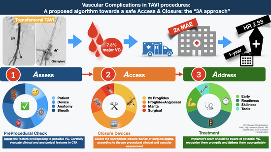
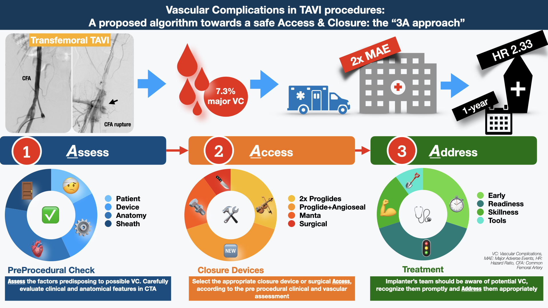
Graphical Abstract

1. Introduction

Transcatheter aortic valve implantation (TAVI) has evolved significantly in the last decade to emerge as the preferred treatment modality for severe aortic stenosis (AS) across a broad range of patient profiles and phenotypes [1,2]. With few remaining exceptions—such as selected patients with bicuspid aortic valve anatomy, younger individuals, or those with unfavorable anatomical characteristics—TAVI has been demonstrated to be at least non-inferior, and in many settings superior, to surgical aortic valve replacement (SAVR) for the treatment of degenerative AS [3,4].
The minimally invasive nature of TAVI represents one of the most important reasons for its widespread adoption and clinical success. Modern percutaneous access techniques, the evolution of the access and closure devices, as well as meticulous pre-procedural planning, have gradually transformed the TAVI vascular access approach [5]. While early TAVI procedures frequently relied on surgical cut-down for vascular access, contemporary practice increasingly favors fully percutaneous access and closure strategies [6,7,8]. This transition has been facilitated by meticulous pre-procedural evaluation of patient-specific vascular anatomy using advanced imaging modalities—most notably multidetector computed tomography (CT) angiography—which allows for precise access planning and optimization of procedural safety [9]. As a result, modern TAVI has evolved into a predominantly fully percutaneous procedure, significantly improving patient comfort and reducing procedural invasiveness.
Nevertheless, a subset of patients—particularly those with severe peripheral artery disease—remain unsuitable for standard percutaneous transfemoral access, necessitating alternative access routes or surgical approaches. Importantly, despite substantial technical progress, vascular complications (VCs) continue to occur at a clinically appraisable rate with significant impact in short- and long-term adverse outcomes [10,11]. Major vascular complications during TAVI are consistently linked to increased mortality, largely mediated through bleeding events, transfusion requirements, and acute kidney injury.
In this manuscript, we provide a comprehensive overview of the incidence, underlying causes, predictive factors, and pathophysiological mechanisms of VCs associated with TAVI. The present document has been prepared as a narrative review of the contemporary literature on vascular complications associated with TAVI procedures. An extensive literature search was conducted, using Pubmed/Scopus/Google Scholar to identify relevant studies, large randomized trials, meta-analyses, registries and guideline documents published from 2000 onward. In addition, we review contemporary percutaneous large-bore access and closure techniques, with particular emphasis on the role of advanced imaging and meticulous pre-procedural planning in optimizing procedural safety, improving patient outcomes, and enhancing overall patient comfort.

2. Definitions and Classifications

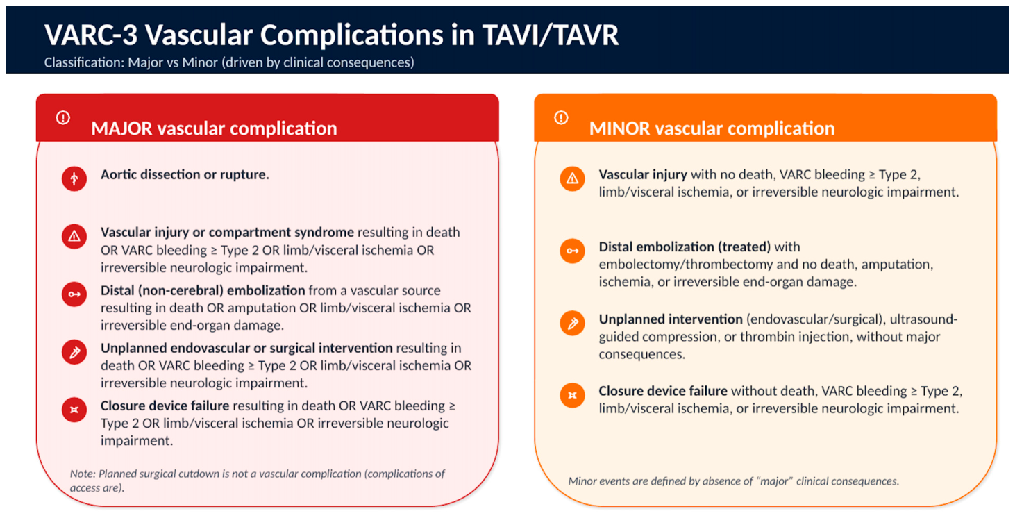
Vascular and bleeding complications, along with stroke, conduction disturbances, paravalvular leak and acute kidney injury, represent one of the “Big 5” adverse events of TAVI and significantly impact patient mortality and morbidity [12]. Before the introduction of standardized definitions for relevant clinical end points and complications, the incidence of major VCs in the same patient cohort ranged from ~5% to 16%, based on each center’s own categorization. The need to accurately determine the safety and efficacy of TAVI procedures, as well as the composite clinical endpoints for randomized clinical trials (RCTs) and research, led to the formation of the 1st Valve Academic Research Consortium (VARC-1) in January of 2011. This consortium had the goal to standardize the clinical endpoint definitions and reporting in TAVI trials [13]. However, as clinical experience with TAVI technology has expanded, the VARC-1 definitions have become inadequate, leading to the introduction of VARC-2 definitions in October 2012. The most notable change in the VARC-2 was the inclusion of major bleeding events in the definition of major VCs, leading to an almost three-times higher overall rate (2.1 vs. 6.9%, p < 0.001) of such events [14]. In 2021, the VARC-3 definitions were further developed, in order to include refined endpoints providing more accurate and consistent assessment of the clinical trial events (Figure 1). The updated consortium definitions differ from the previous ones in two key aspects: first, the number of bleeding categories has been expanded from three (minor, major, and life-threatening bleeding) to four (types 1–4); and second, in VARC-3, all procedural blood loss were regarded as overt bleeding, even without an identifiable source. The adoption of the new VARC-3 bleeding criteria led to a substantial increase in bleeding events and 30-day mortality. However, it should be noted that the VARC-2 bleeding classification remains clinically meaningful, as it also correlates with progressive mortality risk in proportion to the extent of bleeding. Importantly, the VARC-3 criteria have less prognostic relevance when analyzing the transfemoral cohort only, whereas in this respect VARC-2 criteria do not lose their prognostic significance. However, the above findings require further validation with similar or even larger patient cohorts [15].
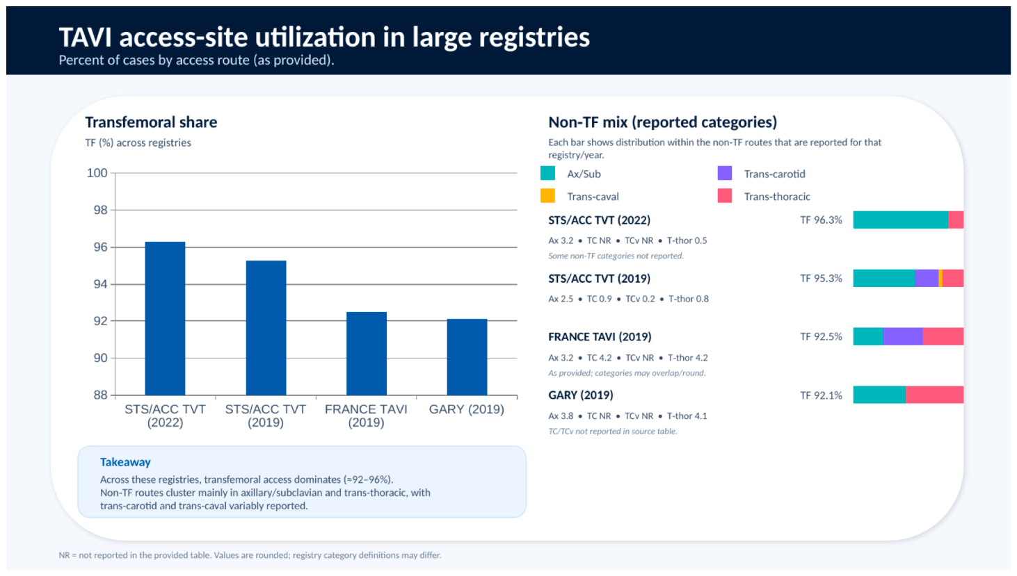
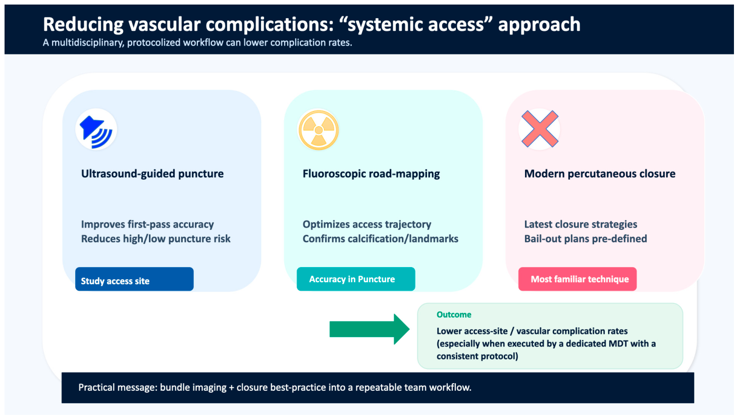
The incidence of vascular and bleeding complications has decreased over time, though there is still significant variation between centers. In the early clinical trials with 1st-generation devices and 22–18 Fr sheath delivery systems, VCs were reported in nearly 15% of patients [16]. Based on STS-ACC TVT Registry (Society of Thoracic Surgeons–American College of Cardiology Transcatheter Valve Therapy Registry), which represents the largest illustration of trends in the incidence of VCs and bleeding complications from the onset of the commercial TAVI era, in 2019, the 30-day major VCs occurred at 1.5%, 1.1% and 0,7% in the high/extreme-, intermediate- and low-risk cohorts, respectively. In addition, in the same period, the rates of life-threatening/disabling bleeding events during index hospitalization declined from 6.3% in the early TAVI period to 1.8% in 2019, and blood transfusions declined from 18.2% during the early phase to 5.8% [16]. In 2019, the corresponding events were 1.45% and 1.2% for the intermediate- and low-risk cohorts, respectively. Nowadays, based on the STS/ACC TVT Registry, the in-hospital rates of major and minor VCs are 1.3% and 2.4%, respectively, as defined by VARC-2 criteria [17]. Notably, it was shown that a multidisciplinary team systemic approach using ultrasound-guided puncture, fluoroscopic road mapping and the latest percutaneous closure strategies may result in even lower complication rates [18].
The recently published European Society of Cardiology (ESC) 2025 Valvular Heart Disease (VHD) guidelines emphasize the advantages of TAVI in patients managed via the transfemoral (TF) approach. TAVI via a non-TF access approach is an alternative supported by observational data in patients considered unsuitable for SAVR with an indication IIA in the recent guidelines. At present, the choice of alternative access route largely depends on the experience of each TAVI team [19]. Based on data from the TVT Registry, in 2013 there was a transient decrease in the proportion of TF patients due to the expansion of the FDA indications to include alternative access. Transthoracic access, once common (up to 30%), is now rarely performed (<0.5%), as it requires intubation and general anesthesia. In 2019, the axillary–subclavian approach was the most commonly used alternative access, with an incidence of 2.5% [11]. Subsequently, based on recent STS/ACC TVT data, carotid access has gained traction, representing 2.1% of all TAVI cases [20]. Transcarotid access was associated with higher mortality rates at 1 month compared to TF access (3.7% vs. 2.6%, p = 0.02) but had lower rates of VCs (1.5% vs. 3.4%, p = 0.04) without increasing the risk for stroke [21]. In April 2019, the transcaval approach was introduced as an alternative, albeit with limited adoption at present. Currently, based on real-world data, approaches other than transfemoral access are required in a small proportion of patients (~2%) [17] (Figure 2). Nevertheless, given the necessity for access in those patients who have declined after surgery, it is imperative that every team possesses expertise in alternative access routes. This ensures that the TAVI therapy can be administered even to this limited number of patients.

3. Pathophysiology

Transcatheter aortic valve implantation (TAVI) is a minimal invasive technique with significant challenges concerning vascular access. All these bulky devices need to pass through the peripheral vessels, with the potential to create vascular trauma of any kind: dissection, kinking, spasm or even rupture. The pathophysiological mechanisms underlying these complications are related to the magnitude of mechanical trauma, the extent of calcification, and the presence of tortuosity [22,23].
Delivery of the valve devices through the peripheral arteries, most commonly the femoral, either inside a delivery sheath or not, will damage the artery wall due to the relative rigidity and size of the delivery systems. All stages of arterial access (via femoral or alternative peripheral), from initial needle puncture to final sheath insertion, can produce endothelial and medial injury, predisposing to access-site dissection or even arterial rupture [24,25]. If unrecognized or inadequately managed, these complications may progress to extensive vessel trauma associated with access-related dissection, thrombosis or even occlusion, which may result in clinically significant limb ischemia or major hemorrhage [26].
Calcification of the access vessels poses another significant risk for VCs. Patients undergoing this minimally invasive procedure are often elderly, with increased vascular calcification and stiff and frail vessel walls [27]. The aforementioned features significantly elevate the risk of vessel wall fracture, particularly in the inner and medial layers, due to the stress exerted by the devices’ passage. These fractures can exacerbate pre-existing vascular trauma, potentially leading to the complete fracture of all three arterial wall layers, leading to complete vessel rupture with profound and potentially fatal bleeding. This is especially likely at sites of extensive calcification [26,27]. Furthermore, calcified plaques can dislodge during the procedure due to the increased stiffness and reduced flexibility of the arteries, which can result in embolic events, a key component of VCs [24].
The tortuosity of the access artery, characterized by twisted or complex artery anatomy, is a well-recognized cause of access site complications. This anatomical complexity increases the technical difficulty of guiding the catheter to the aortic valve, often requiring excessive manipulation that increases vascular injuries such as endothelial tearing and acute vessel occlusion [28]. In severe cases, vessel tortuosity can impede the advancement of the catheter, necessitating alternative access sites or selective procedures, which can cause significant procedural risks.
Post-procedural bleeding is driven primarily by vascular access site–related mechanisms, with 94% of total bleeding events attributable to vascular access [26,29]. Limb ischemia is often categorized as a major VC and affects approximately 3.0–22.9% of patients, with rates varying with access route and operator experience, but in most studies is not reported separately [28,29]. Advancements in imaging technology and device design can mitigate some of these risks, although individual patient anatomy and the presence of vascular conditions will always demand tailored approaches.

4. Clinical Outcomes

As TAVI expands to a broader population, including younger and lower-risk patients, the imperative need to minimize vascular and bleeding complications is becoming more critical. Vascular complications at the time of TAVI, combined with the associated bleeding, trigger a “cascade of adverse events”, such as acute kidney disease, a need for blood transfusions, infections, immunomodulatory effects, and additional percutaneous or surgical interventions, prolonging hospital stay and affecting short- and long-term survival. Many studies have consistently shown that patients who experience a major VC have 2- to 4-fold increased risk of death, with a mortality rate of 10–15% within first 30 days compared to those without. In the SOURCE (SAPIEN Aortic Bioprosthesis European Outcome) registry, a numerically higher rate for 30-day mortality was observed in patients with major VCs compared to those with no major VCs, (12.2% vs. 5.6%, p  =  0.108) [30]. Based on data from the TF subgroup of PARTNER trial for high-risk or inoperable patients at 30 days, major VCs were associated with increased major bleeding (60.9% vs. 6.8%; p < 0.001), blood transfusions (40.7% vs. 5.4%; p < 0.001), and renal failure (8.1% vs. 1.7%; p = 0.003). Furthermore, on multivariable analysis, major VCs were an independent predictor of 1-year mortality (HR 2.31; 95% CI 1.20–4.43; p = 0.012) [31]. Similarly, in the recent observational prospective multicenter OBSERVANT II study, the minor VC group showed a survival that is comparable to the no-VC group at 1-year follow-up, while major-VC patients had significantly worse results [32]. Interestingly, a recent single-center retrospective study including 2160 patients based on VARC-3 classification revealed that even minor VCs may lead to serious consequences impacting short- and long-term health outcomes [33]. Notably, the female sex is one of the strongest independent predictors of major VCs within 30 days (HR 2.31; 95% CI 1.08–4.98; p = 0.03) [32]. In a recent retrospective multicenter European study, the TRITAVI, females present a lower risk of mortality than men at 1-year follow-up, but not at 30 days, likely due to higher rates of vascular/bleeding complications and blood transfusions [34]. Furthermore, in the RHEIA trial, which currently represents the only prospective, randomized study enrolling women using the balloon-expandable platform (Sapien-3 or Sapien-3 Ultra, Edwards), although TAVI was superior to SAVR in terms of death, stroke or rehospitalization at 1 year, the major VCs based on VARC-2 definitions were higher, though not statistically relevant, in the percutaneous approach (3.3% vs. 0.5%, p = 0.07) [35].
In the first clinical validation of the updated VARC-3 bleeding criteria in a contemporary TAVI cohort from Germany, VARC-3 bleeding was 2.5 times more prevalent than VARC-2 (13.9% vs. 34.4%) [15]. No patient with Type 1 bleeding died within 30 days of TAVI, and the impact of Type 2 bleeding on 30-day mortality was not significant (HR, 1.00 [95% CI, 0.49–2.03], p = 0.998). However, Type 3 bleeding was strongly associated with mortality (HR, 2.89 [95% CI, 1.35–6.19], p = 0.006), and Type 4 bleeding led to death per the definition. When applying VARC-2 criteria, minor bleeding was not associated with 30-day mortality. Nevertheless, both major (HR, 2.74 [95% CI, 1.26–5.95], p = 0.011) and life-threatening bleeding (HR, 29.60 [95% CI, 17.42–50.30], p < 0.001) were strongly associated with increased mortality risk [15]. Notably, it appears that major bleeding complications are not associated with age. In an observational retrospective study, when elderly patients (70–80 yrs) were compared with very-elderly ones (>80 yrs), no significant difference was found in bleeding complications (10.8% vs. 18.2%, p = 0.706) [36]. The observational and retrospective nature of the study does not allow definite conclusions, but it emphasizes that age itself must not be considered as an adverse factor for bleeding, and the frailty status of the patient should probably be taken more into account instead.
At present, studies reporting long-term data are limited. A real-world multicenter registry, with a mean follow-up of 751 days, revealed that major but not minor VCs are related to worse long-term outcomes. The freedom from major adverse cardiac and cerebral events (MACCE) at 2 years, even after adjusting for confounding factors, was 83% for patients with VCs and 86.7% for those without (log-rank p  =  0.143). It was also found that, along with the severity of VCs, the presence of diabetes mellitus was an independent predictor of 2-year outcomes (HR 2.41, 95% CI 1.14–5.08; p  =  0.021). Patients with VCs treated surgically presented numerically worse, albeit not statistically significant, clinical outcomes compared with those treated percutaneously. Only major VCs, even when adjusted for confounding factors, were associated with lower MACCE-free survival at 2 years, with double the risk of MACCE (adjusted HR 2.07) compared to those with minor VCs [11].

5. Risk Factors and Predictors (Figure 3)
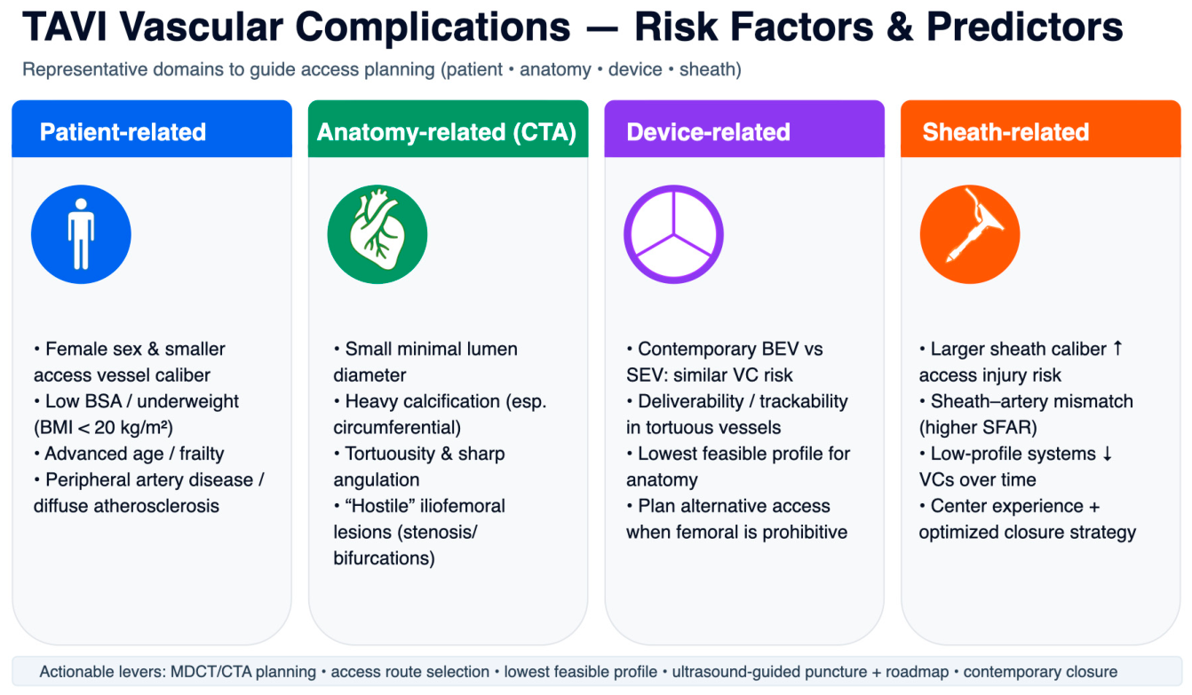
Vascular complications in TAVI are multifactorial in nature and arise from the interaction between patient-related characteristics and procedural factors. Patient body characteristics, such as body habitus and stature, as well as the presence of complex vascular anatomy—including vessel tortuosity and calcification—may play an important role in predicting access-related complications. In addition, procedural choices, particularly the selection of the transcatheter valve and delivery system, can significantly influence vascular risk. Devices with improved trackability through tortuous vessels or those requiring smaller sheath calibers may be preferred in anatomically challenging cases in order to minimize vascular injury. In this section, we review the principal risk factors and predictors of VCs in TAVI, with particular emphasis on factors that may be modified through meticulous pre-procedural planning and optimized procedural strategy.
Figure 3. Categorization of risk factors and predictors for vascular complications in TAVI patients.
Jvd 05 00019 g003

5.1. Patient-Related Factors

Female sex has consistently been associated with a higher risk of VCs across a wide range of percutaneous cardiovascular interventions, including TAVI. This association is largely attributed to anatomical and procedural factors rather than sex-specific biological differences. Generally, women undergoing TAVI have smaller-caliber access vessels, a higher prevalence of vascular tortuosity, and, frequently, are older at the time of intervention, all of which may contribute to an increased possibility of access-site injury. In the randomized controlled PARTNER (Placement of AoRTic TraNscathetER Valve) trial, patients with major VCs were more frequently female and had a smaller body surface area [31]. Importantly, female sex was identified as the only independent predictor of major VCs in this trial (hazard ratio [HR]: 2.31; 95% confidence interval [CI]: 1.08 to 4.98). In another prospective observational study, which included a total of 260 patients, females (n = 131) presented with less peripheral artery disease compared to males, and although there was no significant difference in the rate of major VCs between sexes (11.5% vs. 9.3%, p = 0.570), iliac complications were more frequent in women compared to men (9.0% vs. 2.5%, p = 0.030) [27].
However, more recent data have shown that female sex is not the sole factor for VCs. In this observational analysis of a prospectively designed cohort (546 consecutive patients), even though female patients had experienced higher absolute numbers of 30-day VCs comparing to male ones, the difference did not reach statistical significance (5.6% vs. 2.6%, p = 0.118) [37]. A possible explanation would be that in this series of patients, women presented with less incidence of peripheral artery disease (45.6% vs. 61.2%, p = 0.052). The authors note that the relatively low number of vascular events (4.2% overall) may not provide the proper statistical power to reveal any important differences between the sexes. Further to this, the center’s experience in percutaneous access and expanded utilization of closure devices may have been confounding factors.
In contrast to sex, obesity is not associated with increased risk of VCs in patients undergoing TAVI procedures. In fact, in a large metanalysis, underweight patients (body mass index (BMI) < 20 kg/m2) experienced significantly higher rates of major VCs (2566 patients, odds ratio 1.86, 95% CI 1.16 to 2.98, p = 0.01). Interestingly, overweight and obese patients (BMI ≥ 30 kg/m2) experienced comparable rates of VCs and demonstrated more favorable overall cardiovascular outcomes when compared with normal-weight patients (BMI 25–30 kg/m2) [38]. The notion of this “obesity paradox” is supported by a large cohort of patients, in which obese patients (BMI > 25 kg/m2) were found to experience better outcome, less hospitalization costs and decreased rates of atrial fibrillation, acute kidney injury and acute heart failure compared to the normal-weight ones (BMI 20–25 kg/m2) [39,40].
Generally, female sex has traditionally been associated with higher vascular complication rates in TAVI, primarily reflecting anatomical factors such as smaller access vessel caliber and increased tortuosity rather than sex-specific biological differences. In contemporary practice, this association appears attenuated with improved access techniques and closure devices. In contrast, obesity has not been linked to increased vascular risk, whereas underweight patients exhibit a higher incidence of VCs, supporting the presence of an obesity paradox in the TAVI population.

5.2. Anatomy-Related Factors

It is evident that vascular anatomy significantly influences the suitability of TAVI candidates. Nearly all TAVI devices necessitate a minimum caliber for all peripheral arteries, either femoral or other access sites (subclavian, carotid), to accommodate the valve catheter and facilitate successful delivery of the valve. Given the paramount importance of access sites, VCs are undoubtedly associated with the vascular anatomy. Iliofemoral vascular anatomy—particularly vessel diameter, calcification, and tortuosity—are all identified as major determinants of VCs during transfemoral TAVI.
The introduction of computed tomography angiography (CTA) as a routine component of pre-procedural assessment represented a major milestone in TAVI planning. Meticulous analysis of pre-procedural CTA is essential for the identification of key anatomical characteristics that determine vascular access suitability and enables safe and successful delivery of the transcatheter valve system. Early studies highlighted the relationship between iliofemoral artery dimensions and delivery sheath size as a critical determinant of VCs. In a cohort of 225 patients undergoing TAVI, CTA-based measurements demonstrated that both the sheath-to-femoral artery diameter ratio (SFAR) and the sheath-to-femoral artery area ratio (SFAAR) were significant predictors of VCs. More specifically, an SFAR threshold of 1.45 and an SFAAR threshold of 1.35 were associated with the occurrence of VCs, with reported sensitivities of 64.2% and 78.6%, respectively [41]. Recent data confirmed the association between iliofemoral artery caliber and VCs. A recent meta-analysis, which included 28 studies and 8697 patients, of whom 94.8% had contrast-enhanced MD-CTA, once again identified SFAR (>1.03–1.45) and SFAAR (>1.35) as strong predictors of complications [42].
Apart from artery diameter, additional anatomical features like tortuosity and calcification have been established as significant predictive factors for VCs in TAVI patients. In a study by Vavouranakis et al., 84 patients underwent transfemoral TAVI with percutaneous access and closure using the Prostar® XL device (Chicago, Illinois, US). Among those experiencing major VCs (17/84 [20.2%]) compared to those without, both arterial calcification (11 [64.7%] vs. 8 [11.9%]; p < 0.01) and iliofemoral artery tortuosity (30.2 ± 11.25 vs. 22.06 ± 8.64; p < 0.01) emerged as significant independent predictors [43]. In larger prospective registries, femoral calcification (HR: 3.44, 95% CI: 1.16 to 10.17) was shown to be a factor strongly associated with complications [27].
In contemporary TAVI practice, iliofemoral arterial tortuosity remains a relevant determinant of access-site complications, even in the era of fully percutaneous closure using plug-based vascular closure devices. In a detailed analysis of 109 patients undergoing transfemoral TAVI with closure using the MANTA® vascular closure device (Teleflex Inc., Wayne, PA, USA), Lux et al. demonstrated that increased iliofemoral tortuosity was significantly associated with vascular and bleeding complications. Specifically, a tortuosity index of 22.8 and an iliofemoral angulation greater than 49.5° emerged as important and synergistic predictors of adverse access-site outcomes [44]. These findings are consistent with the results of a larger observational analysis from the Vienna Cardio Thoracic Aortic Valve Registry (VICTORY), which included 240 patients, of whom 28.8% received balloon-expandable valves [45]. In this cohort, a simple iliofemoral tortuosity (IFT) score was utilized, which is defined as [((true vessel length/ideal vessel length) − 1)*100]. It was proven that a higher incidence of access and bleeding complications was found in patients with higher IFT scores (56 [36.8%] vs. 17 [19.3%]; p = 0.003), while in the multivariate logistic regression analysis, only the IFT score was a significant predictor of the primary endpoint (OR: 2.11; 95% CI: 1.09–4.05; p = 0.026).
Recently, all the predictive factors for VCs were gathered in a single score that incorporates the iliofemoral atherosclerosis and anatomy, the “Hostile Score”. Data from a large prospective registry that included 2023 patients who underwent transfemoral TAVI using contemporary devices were used to identify factors for puncture and non-puncture VCs [46]. The extent of significant stenosis, the presence of lesions bifurcations, the lesion length, the extent of calcification and the minimal lumen diameter (<5 mm) were all taken into account to calculate the “Hostile Score”. Interestingly, along with female sex (OR: 2.69; 95% CI: 1.12–6.42), the Hostile Score was proven to be useful in predicting only non-puncture VCs (OR: 1.91; 95% CI: 1.55–2.35). On the other hand, a higher body mass index (OR: 1.23; 95% CI: 1.04–1.50) and the use of a Prostar® (OR: 6.03; 95% CI: 2.23–16.30) or MANTA® device (Morrisville, North Caroline, US) (OR: 6.18; 95% CI: 2.67–14.27), compared with a ProGlide™ device (Chicago, IL, USA), were independent predictors of puncture-site VCs.
Overall, the vascular anatomy represents the dominant determinant of access-site safety in transfemoral TAVI, with vessel caliber, calcification, and tortuosity consistently associated with vascular and bleeding complications across a wide range of studies and contemporary devices. The routine use of multidetector CT angiography has enabled comprehensive, anatomy-driven risk assessment, allowing these factors to be integrated into quantitative metrics and composite scores that refine patient selection and access strategy. Such an individualized, anatomy-based approach is essential to minimize VCs and optimize procedural outcomes in modern TAVI practice.

5.3. Device-Related Factors

Although differences in vascular complication rates were earlier reported between balloon-expandable and self-expanding transcatheter valves, these observations were largely confounded by differences in delivery system profiles and access strategies. Contrary to the earlier notion that device type may play a role, it was shown that the use of contemporary TAVI heart valves did not influence the VC rate. In the CHOICE randomized study, 240 patients were randomized to receive either a balloon- (n = 120) or self-expandable (n = 120) transcatheter heart valve [47]. Vascular complications occurred in 17/121 (14.0%) of patients receiving the balloon-expandable valve and in 15/117 (12.0%) of patients receiving the self-expandable valve (p = 0.78). These results have also been confirmed via a large meta-analysis including four [4] randomized controlled trials and 14 propensity-score trials, with the aim to evaluate differences in outcome between different transcatheter heart valve platforms [48]. While self-expandable TAVI valves had a larger effective orifice area and lower mean transvalvular gradients compared to balloon-expandable ones, no significant difference was observed across the three different platforms [(Sapien 3/Ultra Balloon Expandable Valve (BEV) 6.7% vs. Evolut R/Pro 4.3% (p = 0.45) and vs. Acurate Neo 7.2% (p = 0.96))]. It appears that in contemporary TAVI practice, modern transcatheter valve platforms—both balloon-expandable and self-expandable—do not influence the risk of VCs.

5.4. Sheath-Related Factors

Sheath caliber has long been recognized as a key determinant of vascular complication rates in transfemoral TAVI. Across multiple studies, delivery sheath size—particularly in relation to access vessel dimensions—has consistently emerged as one of the strongest predictors of access-site injury and major vascular events.
Technological evolution toward lower-profile delivery systems has substantially contributed to the reduction in VCs over time. Early studies showed that the sheath sizes of 19–24 Fr were associated with significantly higher rates of VCs compared to sheaths of smaller sizes (14–18 Fr) (10.5% vs. 0.5%, p < 0.001) and identified that sheaths > 19 Fr were independently associated with an increased risk of complications and bleeding events (adjusted odds ratio [OR]: 3.06, 95% confidence interval [CI]: 1.20–7.83; p = 0.019) [49]. In a recent meta-analysis including 24 studies and 14,308 patients, the use of early-generation, high-profile devices was associated with significantly higher rates of VCs compared with contemporary low-profile systems (8.46% vs. 5.51%, p = 0.0015) [50]. This association was observed for both balloon-expandable and self-expanding valve platforms. Notably, progressive device improvements within the balloon-expandable platform were accompanied by a stepwise reduction in vascular complication rates, with first-generation Edwards SAPIEN valves demonstrating higher complication rates compared with second-generation SAPIEN XT devices (15.1% vs. 8.5%, p < 0.00001) and further reductions observed with third-generation SAPIEN 3 devices compared with SAPIEN XT devices (4.5% vs. 8.5%, p = 0.005). These findings underscore the pivotal role of the delivery system profile—rather than the valve expansion mechanism—in determining vascular risk in contemporary TAVI practice.
The outer sheath caliber relative to the internal artery caliber (sheath-to-femoral artery ratio (SFAR)) has been considered as one of the strongest predictors of VCs. Research suggests that the SFAR is often linked to higher vascular complication risks in TAVI, with thresholds such as ≥1.05 indicating potential issues, though newer devices may reduce this impact in some cases. The SFAR was first prominently evaluated in early TAVI trials to quantify the mismatch between device size and vessel anatomy, which can lead to shearing forces, dissections, perforations, or bleeding. Hayashida et al. calculated the SFAR for 130 elderly patients (mean age 83.3 ± 5.9 years) undergoing transfemoral TAVI with Edwards or CoreValve systems [27]. The mean SFAR was 0.99 ± 0.16, with VCs occurring in 27.6% of cases (major: 17.3%, minor: 10.2%). Major complications were linked to a 30-day mortality rate of 22.7%, versus 7.6% in patients without major complications (p = 0.049). Multivariate analysis identified the SFAR as a strong predictor (hazard ratio [HR]: 186.20, 95% CI: 4.41–7855.11), alongside center experience (HR: 3.66, 95% CI: 1.17–11.49) and femoral calcification (HR: 3.44, 95% CI: 1.16–10.17). An SFAR threshold of ≥1.05 (area under the curve = 0.727) predicted major complications at 30.9%, versus 6.9% below the threshold (p = 0.001), as well as higher mortality (18.2% vs. 4.2%, p = 0.016).
Modern transcatheter delivery systems have been progressively improved to facilitate safe insertion through challenging vascular anatomies. Nevertheless, even with the use of specifically designed systems, such as balloon-expandable sheaths, no significant reduction in vascular complication rates has been demonstrated [51]. Importantly, the SFAR remains strongly associated with VCs, indicating that relative sheath–artery mismatch continues to be a significant risk factor for vascular events, even in complex anatomies treated with advanced sheath designs. Therefore, preoperative multidetector computed tomography (MDCT) is essential for SFAR calculation, enabling risk stratification and alternative access if the ratio is ≥1.05 [14,41]. A detailed preprocedural analysis of the anatomy, complexity and size of the iliofemoral arteries is essential and has been shown to accurately identify patients at high risk for VCs [42,52].
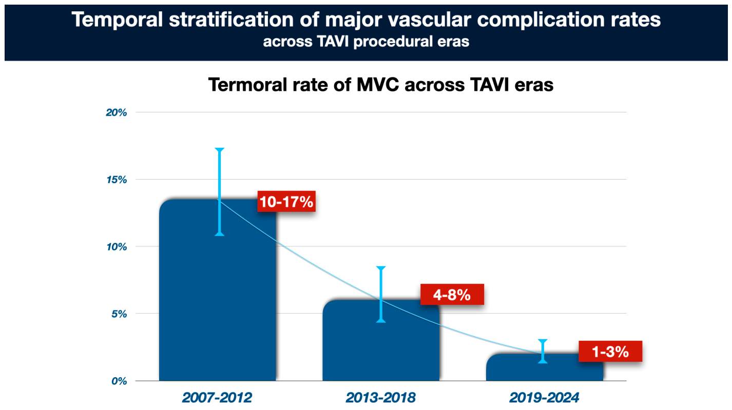
In general, it appears that continuous evolution of TAVI devices has led to a substantial reduction in the rates of major VCs. As the transferal site remains the predominant access site, most studies have identified a significant reduction in vascular complications when comparing older and newer devices. While early TAVI studies reported vascular complication rates of approximately 10–15%, contemporary registries using current-generation devices and lower-profile delivery systems report major vascular complication rates that are generally below 3% (Figure 4) [20,31,48].

6. Contemporary Techniques and Devices for Percutaneous Access (Figure 5)—(Table 1)

Fully percutaneous access, when feasible, is regarded as the ultimate objective for contemporary TAVI procedures, as it reduces the length of hospital stay and enhances patient comfort. Recent data suggest that, compared to surgical cut-down approaches, fully percutaneous transfemoral TAVI is associated with shorter lengths of hospital stay, likely due to reduced recovery time and less requirement for intensive care [53,54]. In propensity-matched patients who underwent transfemoral TAVI procedures using either a surgical or percutaneous approach (a total of 774 patients, 323 matched for each group), patients in the percutaneous group experienced a higher number of minor vascular complications but had shorter hospital stays [54]. A meta-analysis including thirteen [13] trials and 5859 patients confirmed these findings, demonstrating that the percutaneous approach resulted in significantly reduced lengths of hospital stay, albeit accompanied by an increase in the number of minor VCs. Notably, the incidence of major VCs remained unchanged [53]. In general, the comprehensive refinement of the procedure, encompassing a number of techniques, including a fully percutaneous approach, has resulted in a substantial reduction in hospital costs, a decrease in the duration of length of hospital stay, and an increase in patient comfort [55,56].
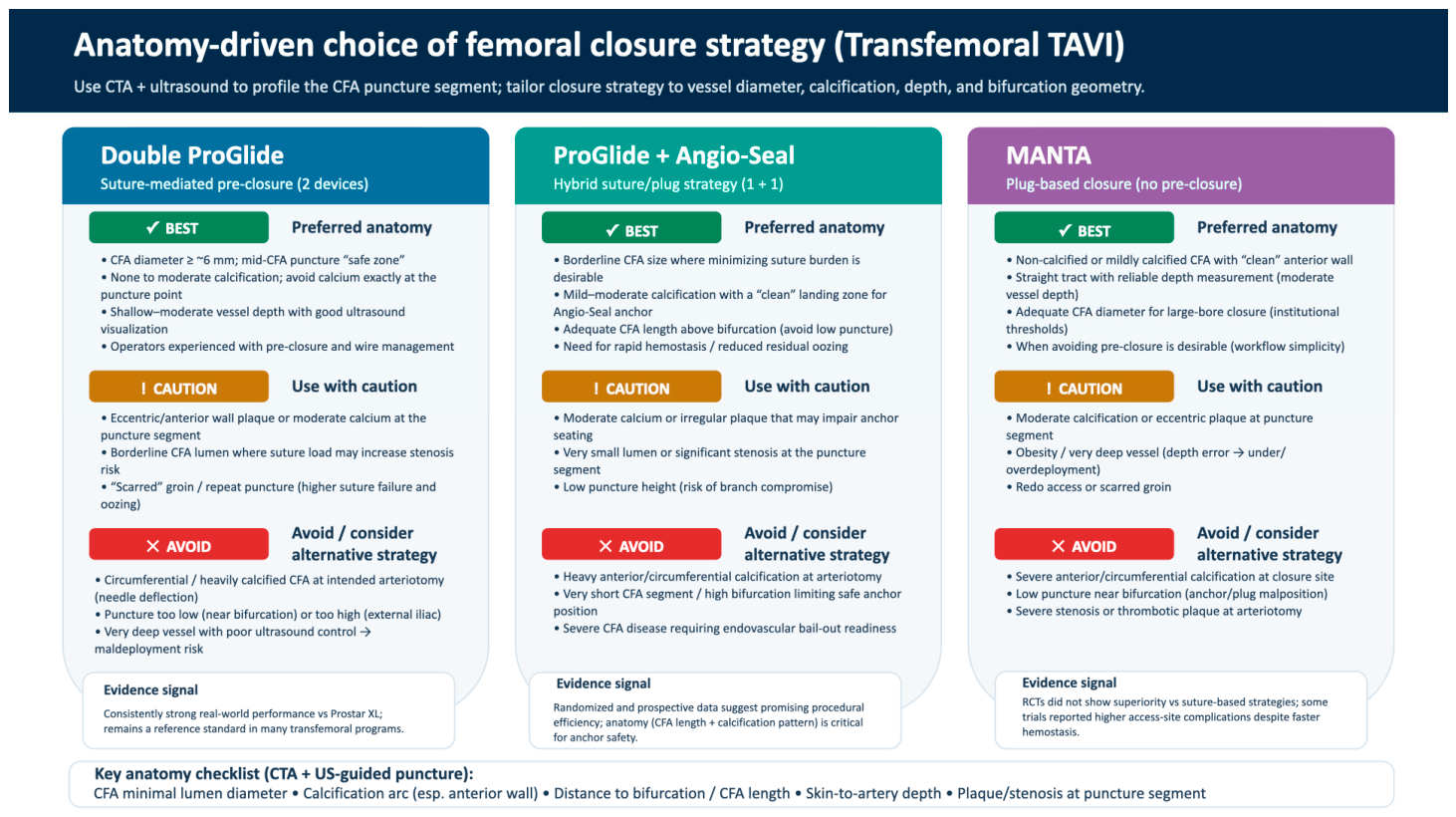
Figure 5. Selection of appropriate closure device according to patient anatomical and clinical characteristics.
Jvd 05 00019 g005
Table 1. Summary of the causes, preventive measures and management of vascular complications in TAVI procedures.
ComplicationApproximate IncidenceMain Causes/Predisposing FactorsPreventive MeasuresUsual ManagementRisk-Reduction Strategies/Future Directions
Overall vascular complicationsEarly-era TAVI: ~10– 20% overall; contemporary major VC usually ~1–3%Large-bore sheath use, PAD, small femoral diameter, high sheath-to-femoral artery ratio, circumferential calcification, tortuosity, female sex, obesity, operator inexperienceSystematic CTA-based access planning, ultrasound-guided CFA puncture, low-profile systems, contralateral angiographic control, experienced heart teamEarly recognition, endovascular bailout readiness, vascular surgery backup when neededStandardized access pathways, micropuncture, fluoroscopic road-mapping, structured training and quality-improvement programs
Iliofemoral dissectionHistorical reports:
1.6–21.4% with percutaneous TF access; contemporary practice is markedly lower
Sheath passage/withdrawal in calcified, tortuous, or borderline-caliber vessels; suboptimal puncture siteCTA assessment of vessel diameter, calcification and tortuosity; ultrasound-guided true CFA puncture; avoid unsuitable transfemoral accessConservative follow-up if small and non-flow-limiting; prolonged balloon angioplasty; uncovered/covered stent or surgery if flow-limiting/extensiveLower-profile or sheathless systems, CT-derived complexity scores, selective IVL-assisted access facilitation in hostile anatomy
Iliofemoral perforation/ruptureReported around 0.7–9.3%, lower in modern seriesSmall calcified vessel, posterior-wall injury, high-risk sheath withdrawal, arterial avulsion, hostile iliofemoral anatomyAccurate anterior-wall puncture, appropriate sheath/vessel matching, crossover protection strategy, alternative access or adjunctive vessel preparation when anatomy is hostileImmediate anticoagulation reversal; balloon tamponade; covered stent–graft; surgical patch/interposition graft when neededImproved access algorithms, routine bailout planning, smaller-profile devices, selective vessel preparation techniques
Access-site hematoma/retroperitoneal bleedingGroin hematoma 2.2–12.5%;
retroperitoneal hematoma up to 2.2%
Inadequate hemostasis, anticoagulation, high puncture into EIA/inferior epigastric region, closure failureUltrasound/fluoroscopy-guided puncture at mid-CFA, careful closure, final angiographic check, prompt anticoagulation reversal when appropriateConservative treatment/compression for minor cases; transfusion if needed; balloon occlusion, covered stent, coil embolization or surgery if ongoing bleedingStandardized post-closure angiography and bleeding-response algorithms
PseudoaneurysmApproximately 2–6%High or low puncture, large sheaths, concomitant artery and vein puncture, severe calcification, failed compression, anticoagulationPrecise CFA puncture, ultrasound guidance, meticulous closure, low threshold for duplex imaging if groin findings are suspiciousObservation if small/stable (<3–3.5 cm); ultrasound- guided thrombin injection if larger or persistent; endovascular or surgical repair when unsuitable for thrombin or if expanding/infectedWider routine use of ultrasound-guided puncture and improved closure-device selection
Arteriovenous fistulaRare; around 1–2% in contemporary reports, 1.49% in one recent cohortLow puncture below femoral bifurcation, simultaneous arterial and venous puncture, transfixion of posterior wallUltrasound-guided puncture, avoidance of low stick and artery–vein overlapObservation/compression if small and asymptomatic; endovascular covered stent or surgical ligation if persistent, symptomatic, or high flowRoutine ultrasound and micropuncture techniques should further reduce this complication
Closure-device failure/access-site stenosis or occlusionClosure failure reported at around 4.4–8.7% in older series; 1.94% in one recent cohort; access-site stenosis/occlusion 0.37% in that cohortHeavy anterior calcification, deep CFA, obesity, large sheath size, unfavorable puncture site, suboptimal device deployment, device-specific limitationsPre-close planning, anatomy-based device selection, avoidance of percutaneous closure in clearly unsuitable anatomy, routine completion angiographyManual compression if simple oozing; balloon angioplasty, stent/stent–graft, thrombectomy, or surgery if bleeding or limb ischemia occursNewer closure devices, hybrid suture–plug strategies, anatomy-tailored closure selection, operator standardization
Acute limb ischemia/
thrombosis/arterial occlusion
Uncommon; often reported within major/minor VC definitions rather than as a separate endpoint; rare contemporary stenosis/occlusion rates are <1% in some cohortsDissection flap, thrombus formation, closure-induced stenosis/occlusion, embolized debris, prolonged low-flow stateMaintain wire access until hemostasis confirmed, completion angiography, careful pulse assessment, early duplex/CTA when ischemia suspectedThrombectomy/embolectomy, balloon angioplasty, stenting, and surgical revascularization when requiredRoutine completion angiography and rapid ischemia-response protocols
The evolution of large-bore vascular closure devices has been instrumental in the shift from routine surgical femoral cut-down to fully percutaneous transfemoral TAVI. In particular, refinements in suture-mediated closure systems and the widespread adoption of standardized pre-closure techniques have improved access-site hemostasis and contributed to lower access-related morbidity.
Early comparative data consistently showed that a ProGlide-based “double-ProGlide” pre-closure strategy (sequential deployment of two Perclose ProGlide™ devices) provides higher procedural success and a more favorable safety profile than the earlier-generation Prostar™ XL system. In the multicenter CONTROL study (3138 consecutive percutaneous transfemoral TAVI procedures), outcomes differed significantly by closure strategy: despite a lower incidence of femoral stenosis in the Prostar group, the ProGlide-based approach was associated with shorter hospitalization duration and lower rates of vascular and bleeding complications [57]. These observations were subsequently supported by additional real-world studies and pooled analyses, which, overall, reported less bleeding and fewer adverse access-site events with ProGlide-based strategies compared with Prostar XL, contributing to a progressive decline in Prostar XL use and the establishment of the double-ProGlide technique as standard practice in many centers [58,59].
More recently, the most commonly used closure device, ProGlide,™ Abbott, has now been equipped with stronger and longer needles made from custom-made high-tensile stainless steel that are able to successfully penetrate harder and more calcified tissues, while the device has a larger wire entrance to facilitate easier wire reinsertion for faster and safer femoral closure. The design updates within suture-mediated closure systems have aimed to facilitate more reliable suture delivery and smoother wire management during pre-closure, thereby supporting procedural efficiency and access-site safety in contemporary transfemoral TAVI programs.
Additionally, novel plug-based closure devices have also been developed. The MANTA® Vascular Closure device, Teleflex, has been specifically designed for large-bore femoral arterial closure. It is a plug-based device that requires no pre-closure and utilizes the coagulation properties of collagen. The MANTA® device has emerged as an easy-to-use and effective closure device that simplifies TAVI procedures. In a large study including 500 patients who underwent several types of procedures requiring large-bore access (TAVI, endovascular aneurysm repair, balloon valvuloplasty), the MANTA® device appeared to be safe and effective, with low rates of major complications (4.0%) [60]. In more recent data including 1000 consecutive TAVI-only patients, the MANTA® device demonstrated its safe and effective access management by again being associated with low rates of major complications (4.2%), making the MANTA device an attractive closure device due to its simplicity and the lack of need for pre-closure [61].
Several studies have examined whether the MANTA® device offers any clear advantage over the standard double-ProGlide pre-closure technique. In early retrospective analyses—often using propensity-matched cohorts—the overall vascular complication rates were generally similar between MANTA and double-ProGlide, while some studies reported lower bleeding rates with MANTA [60,62]. In the randomized controlled trial MASH (MANTA vs. Suture-based vascular closure after transcatHeter aortic valve replacement), the plug-based device was not superior to the suture-based closure one, but the ProGlide technique required additional closure devices more often [63]. The CHOICE-CLOSURE trial (Randomized Comparison of Catheter-based Strategies for Interventional Access Site Closure during Transfemoral TAVI) was the largest randomized, multicenter study to compare the two techniques [64]. In this study, which included 516 patients, the MANTA® device was associated with higher rates of complications and bleeding events but a shorter time to hemostasis.
Additional techniques combining suture-based (ProGlide/Prostyle™) and plug-based (Angio-Seal®, Terumo, Somerset, NJ, USA) techniques have recently been developed, aiming to simplify the procedure, increase hemostasis time and maintain safety. Initially, Angio-Seal® was successfully used as a bail-out device in case of double-ProGlide™ failure [65]. The rationale behind this combined access approach is to use only one ProGlide™ device, applied before large-bore insertion (“pre-closure”), along with one Angio-Seal® positioned after ProGlide™ closure to secure hemostasis. Recent studies have already examined the efficacy and safety of these techniques compared to the traditional double-ProGlide™ ones. Randomized data demonstrated that the simpler approach with one ProGlide™ and an Angio-Seal device provided at least equal or even better VC rates, with less femoral artery restriction and lower rates of bleeding [66,67]. The ACCESS-TAVI (Comparison of Strategies for Vascular ACCESS Closure after TAVI) study is a prospective, multicenter trial including 454 patients and comparing the two strategies [64]. The study’s authors concluded that the combined technique using one ProGlide™ and one Angio-Seal® device resulted in lower VC rates compared to the two-ProGlide/Prostyle™ pre-closure technique (27% vs. 54%, p < 0.001). Time to hemostasis was significantly shorter in the suture/plug group compared with the suture-only group (108 ± 208 s vs. 206 ± 171 s; p < 0.001), and late bleeding events at 30 days occurred less often in the suture/plug group compared with the suture-only group. Even though ultrasound-guided puncture was performed in only 59% of cases and the closure techniques were not fully standardized—factors that may limit generalizability to routine practice—the available evidence still suggests that a combined suture/plug closure strategy can offer procedural efficiency and safety, with fewer access-site complications than suture-only approaches.
Overall, the transition toward fully percutaneous transfemoral TAVI has been enabled by advances in access techniques and vascular closure devices, resulting in shorter hospital stays, lower procedural costs, and improved patient comfort without increasing the number of major VCs. While suture-mediated closure—particularly the double-ProGlide technique—has long represented the standard of care, contemporary data highlight that closure strategy, rather than device type alone, is central to optimizing outcomes, with hybrid suture/plug approaches showing promising signs for procedural efficiency and access-site safety. However, the lack of fully standardized application techniques across studies, together with the wide variability and complexity of iliofemoral vascular anatomy, limits the universal adoption of any single closure strategy for all patients. Accordingly, operators should be familiar with multiple closure techniques and prepared to tailor the approach based on individual patient characteristics and access anatomy, ensuring readiness to escalate or combine strategies when needed.

7. Management of Vascular Complications (Figure 6 and Figure 7)

Transcatheter aortic valve implantation (TAVI) has been widely adopted as a primary treatment for patients with symptomatic aortic valve stenosis who are older than 75 years or who are considered at high surgical risk for or ineligible for open surgical repair [68]. Despite these technological advances, VCs remain clinically relevant, with the most recent publication reporting a complication rate of 12.4% [69].
Currently, the Valve Academic Research Consortium (VARC-3) classification is used to identify and categorize the broad spectrum of VCs associated with TAVI. This ranges from minor complications such as vascular (arterial or venous) injury (including rupture, dissection, stenosis, ischemia, and arterial or venous thrombosis, including pulmonary embolism, arteriovenous fistula, pseudoaneurysm, hematoma, retroperitoneal hematoma, or infection) not resulting in death, VARC type ≥2 bleeding, limb or visceral ischemia, or irreversible neurologic impairment typically limited to the iliofemoral axis, to major VCs. Major complications are limited to the aorta, including aortic dissection, perforation, rupture, retroperitoneal hematoma, and abdominal compartment syndrome resulting in death [70]. The most common complications are reported below.
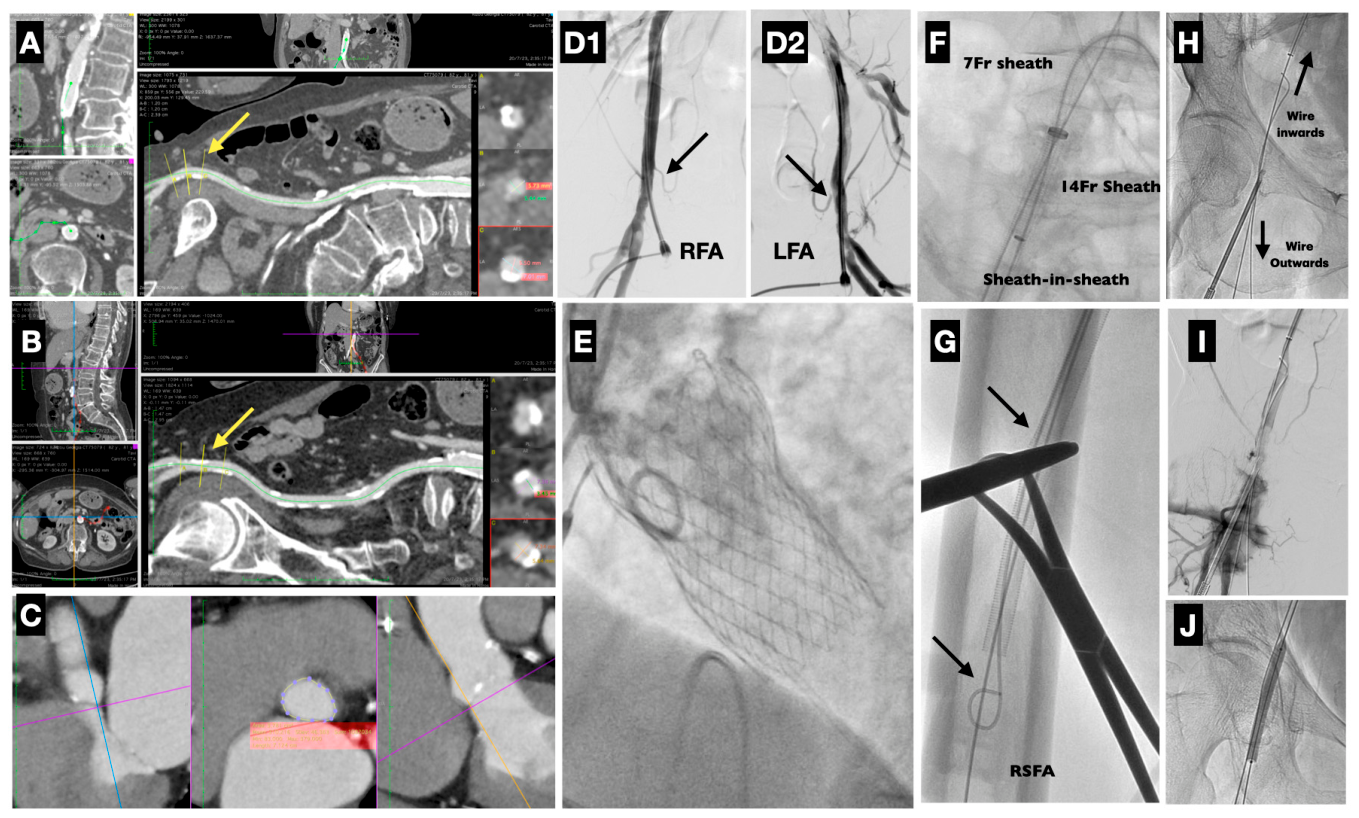
Figure 6. Case example of a TAVI patient experiencing a severe vascular complication: pre-procedural evaluation and planning, complication and how it was managed. Case presentation (left-to-right sequence of images): An 82-year-old female patient underwent TAVI procedure. (A) Pre-procedural CTA evaluation of the right femoral anatomy showed an acceptable primary access site, using the “double-Proglide” technique. Note the relatively small caliber and the calcification in the posterior–lateral wall, risk factors for complications. (B) Inappropriate access site due to severe calcification of the anterior wall. (C) CTA evaluation of the aortic valve anatomy suitable for an EvolutPro+ Medtronic THV. (D1,D2) Verification of puncture heights for both the right and left CFA. (E) Uncomplicated valve implantation. (F) Due to the access-site calcification and the anticipated complications, a proactive approach with wire exteriorization used in order to maintain both ante- and retro-grade wire access. A 7 Fr contralateral sheath was introduced inside the 14 Fr sheath (“sheath-in-sheath” technique) in order to deliver balloons or catheters, if necessary. (G,H) A 0.035” wire was clamped inside the outside part of the 14 Fr sheath and exteriorized (“wire-outwards), leaving the other in place (“wire-inwards”). (I) After the removal of the sheath and the application of the Proglides, severe bleeding was noticed. (J) A peripheral balloon 6.0 × 40 mm was delivered to stop the bleeding.
Jvd 05 00019 g006
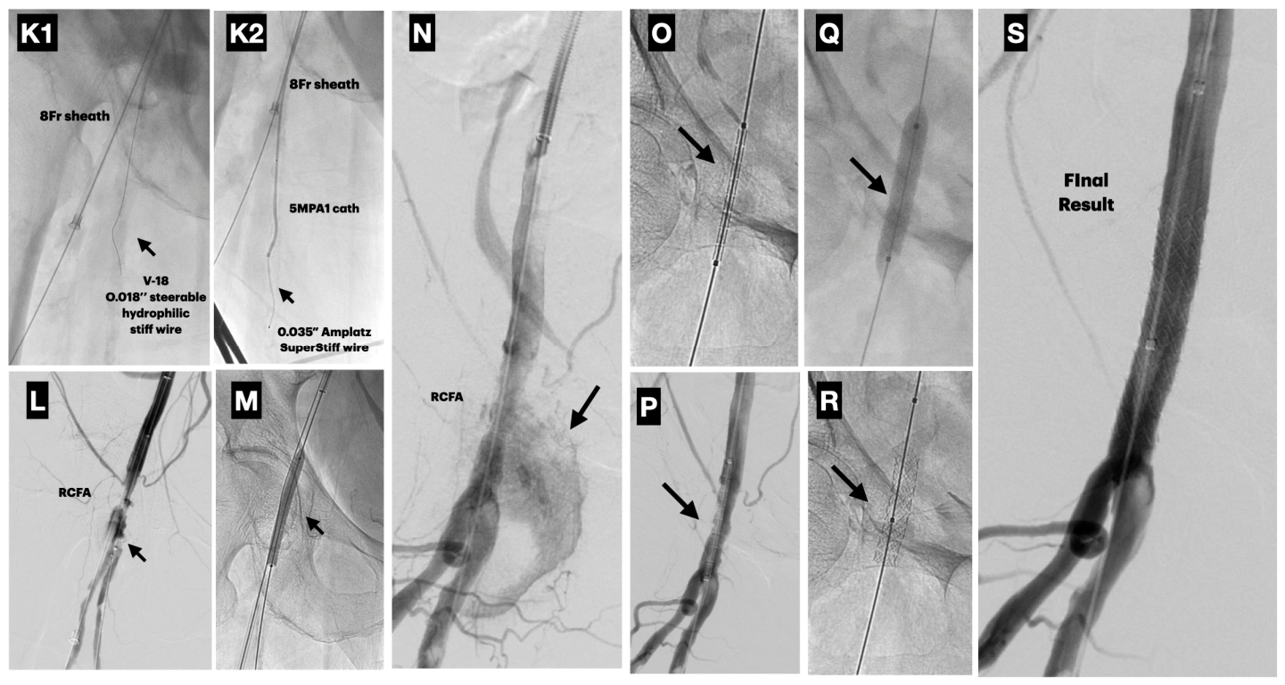
Figure 7. Case example (continued from Figure 5). Case presentation (continued from Figure 6), (left-to-right sequence of images). (K1,K2) An 8 Fr sheath was re-introduced to apply additional Proglides and, via a diagnostic 5JR4.0 catheter, a 0.018 “V-18 wire (Boston Scientific, Marlborough, MA, USA) was placed into the RSFA and exchanged with a 0.035”Amplatz SuperStiff wire (Boston Scientific). (L) An additional two (2) Proglide closure devices were applied, which resulted in successful bleeding control but destroyed and occluded the artery. (M) Prolonged balloon inflation with a 7.0 × 60 mm balloon resulted in (N) restoration of artery’s patency, but bleeding occurred again (arrow). (O) A graft-stent 8.0 × 40 mm was positioned to repair the artery. (P) Graft-stent positioning. (Q) Graft-stent inflation (@ 8 Atm). (R) Graft-stent deployment and expansion. (S) Final result with complete repair of the femoral artery with no bleeding.
Jvd 05 00019 g007

8. Iliofemoral Rupture

Rupture of the iliofemoral axis may occur in cases of small and heavily calcified iliac and/or femoral arteries after the induction of the delivery sheath. Retroperitoneal bleeding is a serious complication after perforation and can be unrecognized. In these cases, angiography from the ipsilateral or contralateral axis should be performed during or after the retrieval of the large sheath. Once the arterial perforation has been detected, timely bleeding control can be achieved by placing an occlusion balloon (e.g., Reliant Stent Graft Balloon Catheter, Medtronic, Minneapolis, Minnesota; or Coda Occlusion Balloon Catheter, Cook Medical Inc., Bloomington, Indiana) proximal to the vascular lesion or a large sheath over the ruptured segment. Through the sheath, a covered stent–graft can be sealed covering the perforation site, avoiding the risks of an upcoming open procedure. Although endovascular repair is effective in most cases, surgical revision should be performed if bleeding persists [71].

9. Iliofemoral Dissection

Iliofemoral segment dissection most commonly results from traumatic advancement of a wire or sheath through a fragile or diseased arterial vessel. The need for treatment depends on the extent of the dissection and its hemodynamic relevance. Retrograde, non-occlusive dissections can usually be managed conservatively, as antegrade flow typically preserves arterial patency by apposing the dissection flap against the vessel wall. Physicians should be aware that larger arterial dissection is usually associated with vascular occlusion due to arterial thrombosis leading to acute limb ischemia. This condition requires immediate intervention to restore arterial flow, typically using a self-expandable or balloon-expandable stent. Successful treatment can be achieved using a crossover technique via the contralateral arterial axis or through access from the left upper extremity vessels. After completion of the procedure, it is advisable to insert the dilatator inside the sheath to avoid the traumatic effect of the tip of the introducer damaging the arterial walls, especially in sharp arterial curves.

10. Pseudoaneurysm

A pseudoaneurysm is defined as a pulsatile hematoma that communicates with the surrounding structures through a defect in the arterial wall. At the end of the procedure, conventional or digital subtraction angiography of the iliofemoral arteries may demonstrate active contrast extravasation, representing either an early arterial leak or an established pseudoaneurysm, depending on the timing of evaluation. If an angiographic diagnosis has not been made at the end of the procedure, close clinical monitoring may reveal an increase in new tingling or murmur, pulsatile hematoma, or marked pain or tenderness, and the pseudoaneurysm may be confirmed by ultrasound. Symptoms such as the development of a new bruit, a pulsatile groin mass, localized pain or tenderness, or sensory disturbances should raise suspicion, with duplex ultrasonography serving as the confirmatory diagnostic modality [72,73]. Potential complications include rupture, distal embolization, infection, nerve compression, and localized skin ischemia. Although lower limb perfusion is typically preserved, treatment is required to prevent progression. Ultrasound-guided compression represents the first-line treatment; it is a safe and cost-effective technique to induce thrombosis of the pseudoaneurysm. However, it is associated with prolonged procedure times, patient discomfort, and relatively high recurrence rates, particularly in patients receiving anticoagulation therapy. In cases of failed compression or in patients receiving anticoagulants, alternative therapeutic strategies include ultrasound-guided thrombin injection, which offers high efficacy and improved patient tolerance; coil embolization; stent–graft placement; or surgical repair. Surgical repair may be performed in patients with pseudoaneurysms who have unfavorable anatomical characteristics, such as a short and wide necks [74].

11. Artery Avulsion

This is an uncommon complication caused by adhesion of the sheath to the arterial endothelium. The operator feels resistance during the withdrawal of the sheath at the end of the procedure and sudden bleeding with subsequent hemodynamic collapse may occur. The use of latest-generation low-profile sheaths has reduced the risk of this adverse event; however, if it occurs, an occlusion balloon should be promptly deployed and the patient prepared for possible surgical repair [24].

12. Failed Percutaneous Closure

Failure of puncture-site closure using suture-mediated closure devices often results in significant bleeding, necessitating surgical intervention [75]. In cases of minor bleeding, prolonged manual compression is usually sufficient, whereas major bleeding events require surgical intervention. Occasionally, when minor bleeding persists, adjunctive hemostatic measures may be required, including deployment of an additional suture- or collagen-based closure device. As an alternative, prolonged low-pressure inflation of an appropriately sized occlusion balloon at the arteriotomy site can achieve hemostasis and may reduce the need for prolonged manual compression.

13. Aortic Rupture

Rupture of the descending aorta or the annulus has been reported up to 2.0% in patients undergoing TAVI. Annular rupture may occur after balloon valvuloplasty or after valve implantation leading to rupture, cardiac tamponade and death [76]. Preoperative meticulous measurement of the annulus valve is recommended using computed tomography angiography and transesophageal echocardiography, reducing the risk of extreme oversizing. A heavily calcified aortic valve may increase the risk of valve perforation, whereas valve undersizing may result in paravalvular regurgitation or valve embolization [24].

14. Arteriovenous Fistula—Hematoma—Stenosis/Occlusion

Apart from the above complications, which significantly affect patient outcomes, other complications like arteriovenous fistula, local hematomas and stenosis or occlusion of the primary access artery can also occur. These complications are often less catastrophic than major rupture or dissection, but they may still prolong hospitalization and occasionally require transfusion, endovascular repair, or surgery [25,77].
Arteriovenous fistulas can be managed conservatively, mainly with prolonged manual ultrasound-guided pressure [78]. If unsuccessful, surgical ligation of the fistula may be required. In cases of appropriate anatomy, placement of a stent–graft may successfully occlude the communication channel between the femoral artery and vein [79].
Minor local hematomas are common in TAVI cases and predominantly include the primary (valve-device insertion) access site and do not require further interventions or blood transfusion. Major hematomas may occur at lower rates compared to minor ones and usually require blood transfusion and/or local surgical intervention to complete arterial access repair [24,25]. In some of the cases, especially in those where arterial blood extravasation is ongoing, a stent–graft placement at the bleeding site may provide successful sealing of the bleeding site. In cases where inadvertent puncture of nearby arterial branches occurs, local hematomas may be challenging to control. In such cases, selective catheterization and coil embolization of the culprit arterial branch may be required.
Stenosis or occlusion of the primary access is a known complication that mainly involves the primary arterial access, usually due to the presence of extensive dissection or arterial wall damage during insertion or removal of the TAVI device. Occasionally, this may be associated with the suture-mediated closure-device process. In such cases, contralateral access to the stenosed/occluded artery is needed in order to perform appropriate balloon dilatations and stenting, if necessary [25,80]. In cases of unsuccessful wire passage through the stenosed/occluded segment of the artery or the failure of ballooning or stenting, surgical intervention may be appropriate for limb salvage or relief of leg ischemia.

15. Pre-Procedural Preventive Strategies (Figure 8)

Optimal pre-procedural planning plays a pivotal role in minimizing VCs during TAVI. As vascular injury remains one of the most frequent and clinically significant adverse events, careful imaging assessment and access planning are fundamental components of procedural safety and success.
Figure 8. Representation of “systemic access” approach to reduce vascular complications.
Jvd 05 00019 g008

15.1. Pre-Procedural Computed Tomography Angiography (CTA)

Multidetector CTA has become the gold-standard for pre-procedural assessment in TAVI. CTA provides high-resolution, three-dimensional visualization of the iliofemoral, subclavian, and aortic anatomies, enabling precise evaluation of vessel diameter, tortuosity, calcification and plaque morphology. These parameters are crucial in identifying patients at high risk for access-related complications. Accurate measurements can only occur if CTA imaging protocols for TAVI planning include ECG-synchronized acquisition, full iliofemoral coverage, and assessment of the femoral puncture zone [81]. In 2024, Androshchuk et al. found that independent predictors of access-site complications included common femoral artery depth (>54 mm), sheath-to-artery diameter ratio (>0.91–1.19), significant vessel tortuosity and calcification [42]. By enabling accurate measurement of vessel lumen, calcification burden and anatomical angles, CTA can guide both device selection (sheath size) and access route determination.

15.2. US-Guided Puncture

Ultrasound (US) guidance at the time of femoral arterial puncture enhances procedural accuracy by enabling real-time visualization of the common femoral artery, identification of bifurcation level, and avoidance of heavily calcified or diseased segments. Compared to fluoroscopic or anatomical landmark-based techniques, US-guided puncture is associated with higher first-pass success, fewer inadvertent venipunctures and a lower risk of bleeding and pseudoaneurysm formation. During transfemoral TAVI, US guidance significantly reduces vascular access and bleeding complications [82]. The results are even better when US-guided femoral access is used in conjunction with closure devices [83]. While large-bore access for TAVI differs somewhat from coronary access, the principle of improved puncture precision applies and is increasingly adopted in TAVI centers.

15.3. Determining Access According to the CTA

Integrating CTA findings into access planning allows for truly individualized decision making. The transfemoral route remains preferred when the minimal luminal diameter of the iliofemoral pathway exceeds the outer sheath size, with an adequate safety margin, and when calcification and tortuosity are minimal. When severe stenosis, excessive tortuosity, or circumferential calcium are identified on CTA, alternative access routes (e.g., trans-subclavian, trans-carotid, transcaval) should be considered. In 2019, Van den Wulp et al. showed that CTA measurements played a key role in identifying predictors of VCs and guiding alternative access selection in transaxillary TAVI patients [84]. Multidisciplinary review of CTA datasets (interventional cardiologist, imaging specialist, vascular surgeon) facilitates optimal access selection and device choice, aligning procedural strategy with anatomical feasibility and safety.

15.4. Predictive Imaging Signs for Complications

Several CTA-derived imaging markers reliably predict VCs and permit pre-procedural risk stratification. These include minimal luminal diameter below the critical sheath-to-artery ratio thresholds, severe circumferential calcification, increased vessel tortuosity, sharp angulations in the aorto-iliac pathway, and a deep common femoral artery location (>54 mm beneath skin surface) [42]. The “iliofemoral tortuosity score”, for instance, was associated with 2- to 5-fold higher vascular risk in transfemoral TAVI cohorts [45]. The adoption of such quantitative scoring systems enables operators to anticipate technical challenges, choose smaller sheath systems if possible, and consider adjunctive techniques (such as the pre-dilation of iliac vessels and use of peripheral lithotripsy) or alternative access when the risk is high.

16. Alternative Access Routes

Transfemoral (TF) access remains the gold standard, but approximately 10–15% of patients require alternative access. According to the 2025 SCAI Expert Consensus, extrathoracic routes (transcarotid, transcaval) are preferred over intrathoracic routes due to their superior safety profiles [85].
Access via the subclavian (or axillary) artery has historically been the preferred method and is regarded as a retrograde approach performed through surgical cut-down or percutaneous puncture of the axillary/subclavian artery. It is mainly considered in patients with unsuitable iliofemoral anatomy, provided the vessel caliber, calcification burden, tortuosity, and side-branch anatomy are acceptable. Particular caution is needed in coronary artery bypass patients with an ipsilateral internal mammary graft. However, recent propensity-matched analyses suggest that it carries a higher risk of stroke compared to transcarotid access [86]. In the study by Allen K et al., which included 576 patients who were propensity-score matched with 1142 patients receiving transaxillary access. All patients received self-expandable valves. At 30 days, patients with transcarotid access had similar mortality (Kaplan–Meier estimates 3.7% vs. 4.3%, p = 0.57) but significantly lower stroke (3.1% vs. 5.9%; p = 0.017) and mortality or stroke (6.0% vs. 8.9%; p = 0.033) rates compared with patients receiving transaxillary access. These results have been confirmed at one (1)-year follow-up. Furthermore, it has been associated with brachial plexus injury and is generally contraindicated in patients with patent internal mammary artery grafts on the ipsilateral side. Transcarotid access is the favored route for its direct coaxial trajectory and stability. The 2025 SCAI Consensus identifies this as a preferred alternative to transaxillary access [85]. It is usually obtained via surgical exposure of the common carotid artery and provides a short, relatively straight path to the aortic valve. It may be particularly useful in patients with severely calcified or tortuous iliofemoral arteries or with descending aortic pathology. Importantly, careful selection is required in the presence of heavily diseased carotid vessels or significant contralateral carotid stenosis/occlusion in order to avoid mechanical damage of the artery or contralateral brain ischemia. Complications include stroke, recurrent laryngeal and vagus nerve injury [87]. Direct transthoracic access (transapical and direct aortic) requires a mini-thoracotomy. It is now considered a “last-resort” option due to its invasiveness, bleeding risks, and mortality.
Inferior vena cava (transcaval) access is a promising technique that involves an electrosurgical puncture of the IVC to the aorta. To initiate the crossing, a snare loop is positioned within the aorta to serve as a target, while a catheter is simultaneously placed in the adjacent IVC. An electrified guidewire burns a precise opening through the IVC wall and into the aorta. Once across, the waiting snare captures the wire and pulls it upward toward the heart to establish a secure rail. Deployment of the closure device needed in the aortic side (Amplatzer Duct Occluder) is successful in approximately 99% of cases [88]. It is mainly considered when transfemoral arterial access is precluded by small-caliber or severely diseased and calcified iliofemoral arteries, and its use depends heavily on pre-procedural CT planning and operator experience. The main complications include bleeding from the aorto-caval tract, retroperitoneal hemorrhage, the need for covered-stent or occluder-based bailout, and, less commonly, persistent fistula or vascular injury, making meticulous CT planning and closure technique essential [89].
Finally, the hybrid approach (iliofemoral conduit) remains a viable option for unpassable vessels. However, novel devices that utilize the intravascular lithotripsy (IVL) modality have emerged as a major validator of “facilitated TF access,” allowing operators to avoid surgical conduits and fracture calcified plaques [90].

17. Conclusions

Vascular complications in patients undergoing TAVI procedures pose a significant challenge to both the patients’ acute and long-term health outcomes. Technological advancements, material innovations, and procedural refinements have led to a substantial reduction in VCs over the past few years. However, as the number of elderly patients undergoing TAVI continues to rise, as well as the number of younger patients at high risk for surgery, the absolute number of VCs is expected to increase. Consequently, it is crucial for operators to develop a comprehensive understanding of promptly recognizing and treating these complications. Furthermore, it is imperative for operators to identify the patient and anatomical characteristics that may predispose individuals to higher risk. Meticulous pre-procedural evaluation of both the patient and the access route anatomy is essential to mitigate the risk of vascular complications.

Author Contributions

Conceptualization: D.N. and A.H.; Methodology: D.N., A.H., L.L., K.K.N. and L.M.; Formal analysis: D.N., V.A., D.C., M.P., K.K.N. and L.M.; Investigation: D.N., G.K., P.K., C.F., V.B. and X.S.; Writing—original draft preparation: D.N., A.H., L.L., P.K., C.F., X.S., V.A., G.K. and M.P.; Critical review and revision of the manuscript: D.N., X.S., K.K.N. and L.M. All authors have read and agreed to the published version of the manuscript.

Funding

This research received no external funding.

Institutional Review Board Statement

Not applicable.

Informed Consent Statement

Not applicable.

Data Availability Statement

No new data were created or analyzed in this study.

Conflicts of Interest

The authors declare no conflicts of interest.

References

  1. Tarantini, G.; Nai Fovino, L.; Gersh, B.J. Transcatheter aortic valve implantation in lower-risk patients: What is the perspective? Eur. Heart J. 2018, 39, 658–666. [Google Scholar] [CrossRef] [PubMed]
  2. Jørgensen, T.H.; Thyregod, H.G.H.; Savontaus, M.; Willemen, Y.; Bleie, Ø.; Tang, M.; Niemela, M.; Angerås, O.; Gudmundsdóttir, I.J.; Sartipy, U.; et al. Transcatheter aortic valve implantation in low-risk tricuspid or bicuspid aortic stenosis: The NOTION-2 trial. Eur. Heart J. 2024, 45, 3804–3814. [Google Scholar] [CrossRef] [PubMed]
  3. Dowling, C.; Kondapally Seshasai, S.R.; Firoozi, S.; Brecker, S.J. Transcatheter aortic valve replacement versus surgery for symptomatic severe aortic stenosis: A reconstructed individual patient data meta-analysis. Catheter. Cardiovasc. Interv. 2020, 96, 158–166. [Google Scholar] [CrossRef]
  4. Tchetche, D.; Willemen, Y.; Cesario, V.; De Backer, O. Transcatheter treatment of bicuspid aortic valve stenosis. EuroIntervention 2025, 21, 1302–1315. [Google Scholar] [CrossRef]
  5. Van Mieghem, N.M.; Tchetche, D.; Chieffo, A.; Dumonteil, N.; Messika-Zeitoun, D.; van der Boon, R.M.; Vahdat, O.; Buchanan, G.L.; Marcheix, B.; Himbert, D.; et al. Incidence, predictors, and implications of access site complications with transfemoral transcatheter aortic valve implantation. Am. J. Cardiol. 2012, 110, 1361–1367. [Google Scholar] [CrossRef]
  6. Griese, D.P.; Reents, W.; Diegeler, A.; Kerber, S.; Babin-Ebell, J. Simple, effective and safe vascular access site closure with the double-ProGlide preclose technique in 162 patients receiving transfemoral transcatheter aortic valve implantation. Catheter. Cardiovasc. Interv. 2013, 82, E734–E741. [Google Scholar] [CrossRef]
  7. Spitzer, S.G.; Wilbring, M.; Alexiou, K.; Stumpf, J.; Kappert, U.; Matschke, K. Surgical cut-down or percutaneous access-which is best for less vascular access complications in transfemoral TAVI? Catheter. Cardiovasc. Interv. 2016, 88, E52–E58. [Google Scholar]
  8. Vora, A.N.; Rao, S.V. Percutaneous or surgical access for transfemoral transcatheter aortic valve implantation. J. Thorac. Dis. 2018, 10, S3595–S3598. [Google Scholar] [CrossRef]
  9. Wiegerinck, E.M.A.; Marquering, H.A.; Oldenburger, N.Y.; Elattar, M.A.; Planken, R.N.; De Mol, B.A.J.M.; Piek, J.J.; Baan, J. Imaging for approach selection of TAVI: Assessment of the aorto-iliac tract diameter by computed tomography-angiography versus projection angiography. Int. J. Cardiovasc. Imaging 2014, 30, 399–405. [Google Scholar] [CrossRef]
  10. Sinclair, N.; Mordhorst, A.; Yang, G.K.; MacDonald, P.S.; Sidhu, R.; Reid, J.D.S. Vascular Access Complications and Clinical Outcomes of Vascular Surgical Repairs Following Transcatheter Aortic Valve Implantation (TAVI). Ann. Vasc. Surg. 2021, 74, 258–263. [Google Scholar] [CrossRef] [PubMed]
  11. Lunardi, M.; Pighi, M.; Banning, A.; Reimers, B.; Castriota, F.; Tomai, F.; Venturi, G.; Pesarini, G.; Scarsini, R.; Kotronias, R.; et al. Vascular complications after transcatheter aortic valve implantation: Treatment modalities and long-term clinical impact. Eur. J. Cardiothorac. Surg. 2022, 61, 934–941. [Google Scholar] [CrossRef] [PubMed]
  12. Grube, E.; Sinning, J.M. The “Big Five” Complications After Transcatheter Aortic Valve Replacement: Do We Still Have to Be Afraid of Them? JACC Cardiovasc. Interv. 2019, 12, 370–372. [Google Scholar] [CrossRef] [PubMed]
  13. Leon, M.B.; Piazza, N.; Nikolsky, E.; Blackstone, E.H. Standardized endpoint definitions for transcatheter aortic valve implantation clinical trials: A consensus report from the Valve Academic Research Consortium. J. Am. Coll. Cardiol. 2011, 57, 253–269. [Google Scholar] [CrossRef]
  14. Okuyama, K.; Jilaihawi, H.; Abramowitz, Y.; Kashif, M.; Patel, J.; Pokhrel, H.; Chakravarty, T.; Nakamura, M.; Cheng, W.; Makkar, R. The clinical impact of vascular complications as defined by VARC-1 vs. VARC-2 in patients following transcatheter aortic valve implantation. EuroIntervention 2016, 12, e636–e642. [Google Scholar] [CrossRef]
  15. Nienaber, S.; Himken, J.; Meertens, M.; Mauri, V.; Lee, S.; Curio, J.; Kuhn, E.; Nies, R.; Eghbalzadeh, K.; Dohr, J.; et al. Comparison of Valve Academic Research Consortium (VARC)-2 and VARC-3 Criteria for Bleeding Complications After Transcatheter Aortic Valve Replacement. J. Am. Heart Assoc. 2025, 14, e039342. [Google Scholar] [CrossRef]
  16. Sherwood, M.W.; Xiang, K.; Matsouaka, R.; Li, Z.; Vemulapalli, S.; Vora, A.N.; Fanaroff, A.; Harrison, J.K.; Thourani, V.H.; Holmes, D.; et al. Incidence, Temporal Trends, and Associated Outcomes of Vascular and Bleeding Complications in Patients Undergoing Transfemoral Transcatheter Aortic Valve Replacement: Insights From the Society of Thoracic Surgeons/American College of Cardiology Transcatheter Valve Therapies Registry. Circ. Cardiovasc. Interv. 2020, 13, e008227. [Google Scholar]
  17. Vekstein, A.M.; Wegermann, Z.K.; Manandhar, P.; Mack, M.J.; Cohen, D.J.; Hughes, G.C.; Harrison, J.K.; Kaneko, T.; Kapadia, S.R.; Stathogiannis, K.; et al. Outcomes of Transcatheter Aortic Valve Replacement in Low-Risk Patients in the United States: A Report From the STS/ACC TVT Registry. Circulation 2025, 151, 1134–1146. [Google Scholar] [CrossRef]
  18. Theriot, M.L.; Bergen, E.C.; Lugo, J.G.; Graham, W.M. Reducing TAVR Vascular Complications Through Multidisciplinary Process Optimization. JACC Case Rep. 2025, 30, 105614. [Google Scholar] [CrossRef] [PubMed]
  19. Praz, F.; Borger, M.A.; Lanz, J.; Marin-Cuartas, M.; Abreu, A.; Adamo, M.; Marsan, N.A.; Barili, F.; Bonaros, N.; Cosyns, B.; et al. 2025 ESC/EACTS Guidelines for the management of valvular heart disease. Eur. Heart J. 2025, 46, 4635–4736. [Google Scholar] [CrossRef]
  20. Arnold, S.V.; Manandhar, P.; Vemulapalli, S.; Kosinski, A.S.; Batchelor, W.B.; Thourani, V.H.; Mack, M.J.; Cohen, D.J. Trends in Transcatheter Aortic Valve Replacement Outcomes: Insights From the STS/ACC TVT Registry. JAMA Cardiol. 2024, 9, 1115–1123. [Google Scholar] [CrossRef]
  21. Abraham, B.; Sous, M.; Sedhom, R.; Megaly, M.; Roman, S.; Sweeney, J.; Alkhouli, M.; Pollak, P.; El Sabbagh, A.; Garcia, S.; et al. Meta-Analysis on Transcarotid Versus Transfemoral and Other Alternate Accesses for Transcatheter Aortic Valve Implantation. Am. J. Cardiol. 2023, 192, 196–205. [Google Scholar] [CrossRef]
  22. Webb, J.G.; Pasupati, S.; Humphries, K.; Thompson, C.; Altwegg, L.; Moss, R.; Sinhal, A.; Carere, R.G.; Munt, B.; Ricci, D.; et al. Percutaneous transarterial aortic valve replacement in selected high-risk patients with aortic stenosis. Circulation 2007, 116, 755–763. [Google Scholar] [CrossRef] [PubMed]
  23. Smith, C.R.; Leon, M.B.; Mack, M.J.; Miller, D.C.; Moses, J.W.; Svensson, L.G.; Tuzcu, E.M.; Webb, J.G.; Fontana, G.P.; Makkar, R.R.; et al. Transcatheter versus surgical aortic-valve replacement in high-risk patients. N. Engl. J. Med. 2011, 364, 2187–2198. [Google Scholar] [CrossRef]
  24. Toggweiler, S.; Leipsic, J.; Binder, R.K.; Freeman, M.; Barbanti, M.; Heijmen, R.H.; Wood, D.A.; Webb, J.G. Management of Vascular Access in Transcatheter Aortic Valve Replacement. JACC Cardiovasc. Interv. 2013, 6, 767–776. [Google Scholar] [CrossRef]
  25. Mach, M.; Okutucu, S.; Kerbel, T.; Arjomand, A.; Fatihoglu, S.G.; Werner, P.; Simon, P.; Andreas, M. Vascular Complications in TAVR: Incidence, Clinical Impact, and Management. J. Clin. Med. 2021, 10, 5046. [Google Scholar] [CrossRef]
  26. Stępińska, J.; Czerwińska, K.; Witkowski, A.; Dąbrowski, M.; Chmielak, Z.; Kuśmierski, K.; Hryniewiecki, T.; Demkow, M. Risk factors for bleeding complications in patients undergoing transcatheter aortic valve implantation (TAVI). Cardiol. J. 2013, 20, 125–133. [Google Scholar] [CrossRef]
  27. Hayashida, K.; Lefèvre, T.; Chevalier, B.; Hovasse, T.; Romano, M.; Garot, P.; Mylotte, D.; Uribe, J.; Farge, A.; Donzeau-Gouge, P.; et al. Transfemoral aortic valve implantation: New criteria to predict vascular complications. JACC Cardiovasc. Interv. 2011, 4, 851–858. [Google Scholar] [CrossRef]
  28. Amrane, H.; Porta, F.; van Boven, A.V.; Kappetein, A.P.; Head, S.J. A meta-analysis on clinical outcomes after transaortic transcatheter aortic valve implantation by the Heart Team. EuroIntervention 2017, 13, e168–e176. [Google Scholar] [CrossRef]
  29. Useini, D.; Haldenwang, P.; Schlömicher, M.; Christ, H.; Naraghi, H.; Moustafine, V.; Strauch, J. Mid-Term Outcomes after Transapical and Transfemoral Transcatheter Aortic Valve Implantation for Aortic Stenosis and Porcelain Aorta with a Systematic Review of Transfemoral versus Transapical Approach. Thorac. Cardiovasc. Surg. 2020, 68, 623–632. [Google Scholar] [CrossRef] [PubMed]
  30. Wendler, O.; Schymik, G.; Treede, H.; Baumgartner, H.; Dumonteil, N.; Ihlberg, L.; Neumann, F.-J.; Tarantini, G.; Zamarano, J.L.; Vahanian, A. SOURCE 3 Registry: Design and 30-Day Results of the European Postapproval Registry of the Latest Generation of the SAPIEN 3 Transcatheter Heart Valve. Circulation 2017, 135, 1123–1132. [Google Scholar] [CrossRef] [PubMed]
  31. Généreux, P.; Webb, J.G.; Svensson, L.G.; Kodali, S.K.; Satler, L.F.; Fearon, W.F.; Davidson, C.J.; Eisenhauer, A.C.; Makkar, R.R.; Bergman, G.W.; et al. Vascular complications after transcatheter aortic valve replacement: Insights from the PARTNER (Placement of AoRTic TraNscathetER Valve) trial. J. Am. Coll. Cardiol. 2012, 60, 1043–1052. [Google Scholar] [CrossRef]
  32. Seccareccia, F.; D’ERrigo, P.; Tarantini, G.; Barbanti, M.; Tamburino, C.; Musumeci, G.; Bedogni, F.; Berti, S.; Rosato, S.; Santoro, G.; et al. First results from the OBSERVANT II study: Clinical characteristics of patients with aortic stenosis undergoing transcatheter treatment with new generation devices. G. Ital. Cardiol. 2020, 21, 469–477. [Google Scholar]
  33. Piperata, A.; Eynde, J.V.D.; Pernot, M.; Avesani, M.; Seguy, B.; Bonnet, G.; Ben Ali, W.; Leroux, L.; Labrousse, L.; Modine, T. Impact of Valve Academic Research Consortium 3 (VARC-3) minor access site vascular complications in patients undergoing percutaneous transfemoral transcatheter aortic valve implantation. Eur. J. Cardiothorac. Surg. 2023, 64, ezad255. [Google Scholar] [CrossRef] [PubMed]
  34. Rodriguez-Gabella, T.; Zimarino, M.; Barbanti, M.; Testa, L.; Capodanno, D.; Stefanini, G.G.; Radico, F.; Fabbiocchi, F.; Piva, T.; Saia, F.; et al. Sex based analysis of the impact of red blood cell transfusion and vascular or bleeding complications related to TAVI—The TRITAVI-Women Study. Int. J. Cardiol. 2021, 333, 69–76. [Google Scholar] [CrossRef]
  35. Tchetche, D.; Pibarot, P.; Bax, J.J.; Bonaros, N.; Windecker, S.; Dumonteil, N.; Nietlispach, F.; Messika-Zeitoun, D.; Pocock, S.J.; Berthoumieu, P.; et al. Transcatheter vs. surgical aortic valve replacement in women: The RHEIA trial. Eur. Heart J. 2025, 46, 2079–2088. [Google Scholar] [CrossRef] [PubMed]
  36. Jiritano, F.; Serraino, G.F.; Sorrentino, S.; Napolitano, D.; Costa, D.; Ielapi, N.; Bracale, U.M.; Mastroroberto, P.; Andreucci, M.; Serra, R. Risk of Bleeding in Elderly Patients Undergoing Transcatheter Aortic Valve Implantation or Surgical Aortic Valve Replacement. Prosthesis 2024, 6, 175–185. [Google Scholar] [CrossRef]
  37. Yousif, N.; Obeid, S.; Binder, R.; Denegri, A.; Shahin, M.; Templin, C.; Lüscher, T.F. Impact of gender on outcomes after transcatheter aortic valve implantation. J. Geriatr. Cardiol. 2018, 15, 394–400. [Google Scholar]
  38. Sannino, A.; Schiattarella, G.G.; Toscano, E.; Gargiulo, G.; Giugliano, G.; Galderisi, M.; Losi, M.-A.; Stabile, E.; Cirillo, P.; Imbriaco, M.; et al. Meta-Analysis of Effect of Body Mass Index on Outcomes After Transcatheter Aortic Valve Implantation. Am. J. Cardiol. 2017, 119, 308–316. [Google Scholar] [CrossRef]
  39. Alharbi, A.; Shah, M.; Mhanna, M.; Rejent, K.; Safi, M.; Alsughayer, A.; Patel, N.; Assaly, R. Body Mass Index and Outcomes of Transcatheter Aortic Valve Replacement: A Retrospective Observational Study. Curr. Probl. Cardiol. 2023, 48, 101879. [Google Scholar] [CrossRef]
  40. Reichl, J.J.; Poljo, A.; Stolte, T.; Schmitt, R.; Boeddinghaus, J.; Wagener, M.; Leibundgut, G.; Kaiser, C.; Westermann, D.; Hartikainen, T.; et al. Association of Body Mass Index With Procedural Success and Outcomes in Patients undergoing Transcatheter Aortic Valve Implantation. Am. J. Cardiol. 2025, 248, 67–72. [Google Scholar] [CrossRef] [PubMed]
  41. Krishnaswamy, A.; Parashar, A.; Agarwal, S.; Modi, D.K.; Poddar, K.L.; Svensson, L.G.; Roselli, E.E.; Schoenhagen, P.; Tuzcu, E.M.; Kapadia, S.R. Predicting vascular complications during transfemoral transcatheter aortic valve replacement using computed tomography: A novel area-based index. Catheter. Cardiovasc. Interv. 2014, 84, 844–851. [Google Scholar] [CrossRef]
  42. Androshchuk, V.; Chehab, O.; Prendergast, B.; Rajani, R.; Patterson, T.; Redwood, S. Computed tomography derived anatomical predictors of vascular access complications following transfemoral transcatheter aortic valve implantation: A systematic review. Catheter. Cardiovasc. Interv. 2024, 103, 169–185. [Google Scholar] [CrossRef]
  43. Vavuranakis, M.; Kariori, M.; Voudris, V.; Kalogeras, K.; Vrachatis, D.; Aznaouridis, C.; Moldovan, C.; Masoura, C.; Thomopoulou, S.; Lazaros, G.; et al. Predictive factors of vascular complications after transcatheter aortic valve implantation in patients treated with a default percutaneous strategy. Cardiovasc. Ther. 2013, 31, e46–e54. [Google Scholar] [CrossRef]
  44. Lux, A.; Müllenberg, L.; Veenstra, L.F.; Dohmen, W.; Kats, S.; Maesen, B.; Hof, A.W.V. Iliofemoral Tortuosity Increases the Risk of Access-Site-Related Complications After Aortic Valve Implantation and Plug-Based Access-Site Closure. CJC Open 2022, 4, 609–616. [Google Scholar] [CrossRef] [PubMed]
  45. Mach, M.; Poschner, T.; Hasan, W.; Szalkiewicz, P.; Andreas, M.; Winkler, B.; Geisler, S.; Geisler, D.; Rudziński, P.N.; Watzal, V.; et al. The Iliofemoral tortuosity score predicts access and bleeding complications during transfemoral transcatheter aortic valve replacement: Data from the VIenna Cardio Thoracic aOrtic valve registrY (VICTORY). Eur. J. Clin. Investig. 2021, 51, e13491. [Google Scholar] [CrossRef] [PubMed]
  46. Nakase, M.; Tomii, D.; Samim, D.; Gräni, C.; Praz, F.; Lanz, J.; Stortecky, S.; Reineke, D.; Windecker, S.; Pilgrim, T. Impact of Severity and Extent of Iliofemoral Atherosclerosis on Clinical Outcomes in Patients Undergoing TAVR. JACC Cardiovasc. Interv. 2024, 17, 2353–2363. [Google Scholar] [CrossRef]
  47. Abdel-Wahab, M.; Mehilli, J.; Frerker, C.; Neumann, F.-J.; Kurz, T.; Tölg, R.; Zachow, D.; Guerra, E.; Massberg, S.; Schäfer, U.; et al. Comparison of balloon-expandable vs self-expandable valves in patients undergoing transcatheter aortic valve replacement: The CHOICE randomized clinical trial. JAMA 2014, 311, 1503–1514. [Google Scholar] [CrossRef]
  48. Wang, B.; Mei, Z.; Ge, X.; Li, Y.; Zhou, Q.; Meng, X.; An, G. Comparison of outcomes of self-expanding versus balloon-expandable valves for transcatheter aortic valve replacement: A meta-analysis of randomized and propensity-matched studies. BMC Cardiovasc. Disord. 2023, 23, 382. [Google Scholar] [CrossRef]
  49. Barbanti, M.; Binder, R.K.; Freeman, M.; Wood, D.A.; Leipsic, J.; Cheung, A.; Ye, J.; Tan, J.; Toggweiler, S.; Yang, T.-H.; et al. Impact of low-profile sheaths on vascular complications during transfemoral transcatheter aortic valve replacement. EuroIntervention 2013, 9, 929–935. [Google Scholar] [CrossRef] [PubMed]
  50. Rahhab, Z.; Misier, K.R.; El Faquir, N.; Kroon, H.; Ziviello, F.; Kardys, I.; Daemen, J.; De Jaegere, P.; Reardon, M.J.; Popma, J.; et al. Vascular Complications after Transfemoral Transcatheter Aortic Valve Implantation: A Systematic Review and Meta-Analysis. Struct. Heart 2020, 4, 62–71. [Google Scholar] [CrossRef]
  51. Sedaghat, A.; von Dobbeler, C.; Sontag, B.; Sinning, J.-M.; Fimmers, R.; Mellert, F.; Schiller, W.; Welz, A.; Grube, E.; Nickenig, G.; et al. Use of a balloon-expandable transfemoral sheath in a TAVI cohort with complex access site—A propensity score matched analysis. EuroIntervention 2015, 11, 698–704. [Google Scholar] [CrossRef]
  52. Leipsic, J.; Yang, T.H.; Min, J.K. Computed tomographic imaging of transcatheter aortic valve replacement for prediction and prevention of procedural complications. Circ. Cardiovasc. Imaging 2013, 6, 597–605. [Google Scholar] [CrossRef] [PubMed]
  53. Abdelaziz, H.K.; Megaly, M.; Debski, M.; Rahbi, H.; Kamal, D.; Saad, M.; Wiper, A.; More, R.; Roberts, D.H. Meta-Analysis Comparing Percutaneous to Surgical Access in Trans-Femoral Transcatheter Aortic Valve Implantation. Am. J. Cardiol. 2020, 125, 1239–1248. [Google Scholar] [CrossRef]
  54. Cammardella, A.G.; Russo, M.; Di Mauro, M.; Romagnoni, C.; Ceresa, F.; Patanè, F.; Gelpi, G.; Pollari, F.; Barili, F.; Parolari, A.; et al. SURgical vs. PERcutaneous ACCESS in Transfemoral Transcatheter Aortic Valve Implantation (SU-PER-ACCESS Study). J. Clin. Med. 2024, 13, 4471. [Google Scholar] [CrossRef]
  55. Sangaraju, S.; Cox, I.; Dalrymple-Hay, M.; Lloyd, C.; Suresh, V.; Riches, T.; Melhuish, S.; Asopa, S.; Newcombe, S.; Deutsch, C.; et al. Effect of procedural refinement of transfemoral transcatheter aortic valve implantation on outcomes and costs: A single-centre retrospective study. Open Heart 2019, 6, e001064. [Google Scholar] [CrossRef]
  56. Tagliari, A.P.; Taramasso, M. New Practices in Transcatheter Aortic Valve Implantation: How I Do It in 2023. J. Clin. Med. 2023, 12, 1342. [Google Scholar] [CrossRef]
  57. Barbash, I.M.; Barbanti, M.; Webb, J.; De Nicolas, J.M.-M.; Abramowitz, Y.; Latib, A.; Nguyen, C.; Deuschl, F.; Segev, A.; Sideris, K.; et al. Comparison of vascular closure devices for access site closure after transfemoral aortic valve implantation. Eur. Heart J. 2015, 36, 3370–3379. [Google Scholar] [CrossRef]
  58. Maniotis, C.; Andreou, C.; Karalis, I.; Koutouzi, G.; Agelaki, M.; Koutouzis, M. A systematic review on the safety of Prostar XL versus ProGlide after TAVR and EVAR. Cardiovasc. Revasc. Med. 2017, 18, 145–150. [Google Scholar] [CrossRef] [PubMed]
  59. Berti, S.; Bedogni, F.; Giordano, A.; Petronio, A.S.; Iadanza, A.; Bartorelli, A.L.; Reimers, B.; Spaccarotella, C.; Trani, C.; Attisano, T.; et al. Efficacy and Safety of ProGlide Versus Prostar XL Vascular Closure Devices in Transcatheter Aortic Valve Replacement: The RISPEVA Registry. J. Am. Heart Assoc. 2020, 9, e018042. [Google Scholar] [CrossRef] [PubMed]
  60. Kroon, H.G.; Tonino, P.A.; Savontaus, M.; Amoroso, G.; Laine, M.; Christiansen, E.H.; Toggweiler, S.; Berg, J.T.; Sathananthan, J.; Daemen, J.; et al. Dedicated plug based closure for large bore access—The MARVEL prospective registry. Catheter. Cardiovasc. Interv. 2021, 97, 1270–1278. [Google Scholar]
  61. Kastengren, M.; Settergren, M.; Rück, A.; Feldt, K.; Saleh, N.; Linder, R.; Verouhis, D.; Meduri, C.U.; Bager, J.; Dalén, M. Percutaneous plug-based vascular closure device in 1000 consecutive transfemoral transcatheter aortic valve implantations. Int. J. Cardiol. 2022, 359, 7–13. [Google Scholar] [CrossRef]
  62. Biancari, F.; Romppanen, H.; Savontaus, M.; Siljander, A.; Mäkikallio, T.; Piira, O.-P.; Piuhola, J.; Vilkki, V.; Ylitalo, A.; Vasankari, T.; et al. MANTA versus ProGlide vascular closure devices in transfemoral transcatheter aortic valve implantation. Int. J. Cardiol. 2018, 263, 29–31. [Google Scholar] [CrossRef]
  63. van Wiechen, M.P.; Tchétché, D.; Ooms, J.F.; Hokken, T.W.; Kroon, H.; Ziviello, F.; Ghattas, A.; Siddiqui, S.; Laperche, C.; Spitzer, E.; et al. Suture- or Plug-Based Large-Bore Arteriotomy Closure: A Pilot Randomized Controlled Trial. JACC Cardiovasc. Interv. 2021, 14, 149–157. [Google Scholar] [CrossRef]
  64. Rheude, T.; Ruge, H.; Altaner, N.; Pellegrini, C.; Covarrubias, H.A.; Mayr, P.; Cassese, S.; Kufner, S.; Taniguchi, Y.; Thilo, C.; et al. Comparison of strategies for vascular ACCESS closure after Transcatheter Aortic Valve Implantation: The ACCESS-TAVI randomized trial. Eur. Heart J. 2025, 46, 635–645. [Google Scholar] [CrossRef]
  65. Kiramijyan, S.; Magalhaes, M.A.; Ben-Dor, I.; Koifman, E.; Escarcega, R.O.; Baker, N.C.; Torguson, R.; Okubagzi, P.; Bernardo, N.L.; Satler, L.F.; et al. The adjunctive use of Angio-Seal in femoral vascular closure following percutaneous transcatheter aortic valve replacement. EuroIntervention 2016, 12, 88–93. [Google Scholar] [CrossRef]
  66. Ko, T.-Y.; Kao, H.-L.; Liu, Y.-J.; Yeh, C.-F.; Huang, C.-C.; Chen, Y.-H.; Hung, C.-S.; Chan, C.-Y.; Lin, L.-C.; Chen, Y.-S.; et al. Intentional combination of ProGlide and Angio-Seal for femoral access haemostasis in transcatheter aortic valve replacement. Int. J. Cardiol. 2019, 293, 76–79. [Google Scholar] [CrossRef]
  67. Yeh, C.-F.; Kao, H.-L.; Ko, T.-Y.; Chen, C.-K.; Tsai, C.-H.; Huang, C.-C.; Chen, Y.-H.; Chan, C.-Y.; Lin, M.-S. Dual ProGlide vs. ProGlide and Angio-Seal for Femoral Access Hemostasis After Transcatheter Aortic Valve Replacement: A Randomised Comparative Trial. Can. J. Cardiol. 2025, 41, 12–20. [Google Scholar] [CrossRef] [PubMed]
  68. Vahanian, A.; Beyersdorf, F.; Praz, F.; Milojevic, M.; Baldus, S.; Bauersachs, J.; Capodanno, D.; Conradi, L.; De Bonis, M.; De Paulis, R.; et al. 2021 ESC/EACTS Guidelines for the management of valvular heart disease: Developed by the Task Force for the management of valvular heart disease of the European Society of Cardiology (ESC) and the European Association for Cardio-Thoracic Surgery (EACTS). Eur. Heart J. 2022, 43, 561–632. [Google Scholar] [CrossRef] [PubMed]
  69. Radhakrishnan, S.L.; Ho, K.K. Transradial vs transfemoral secondary access outcomes in transcatheter aortic valve implantation: A systematic review and meta-analysis. World J. Cardiol. 2020, 12, 571–583. [Google Scholar] [CrossRef] [PubMed]
  70. VARC-3 Writing Committee; Généreux, P.; Piazza, N.; Alu, M.C.; Nazif, T.; Hahn, R.T.; Pibarot, P.; Bax, J.J.; Leipsic, J.A.; Blanke, P.; et al. Valve Academic Research Consortium 3: Updated endpoint definitions for aortic valve clinical research. Eur. Heart J. 2021, 42, 1825–1857. [Google Scholar] [CrossRef]
  71. Masson, J.B.; Al Bugami, S.; Webb, J.G. Endovascular balloon occlusion for catheter-induced large artery perforation in the catheterization laboratory. Catheter. Cardiovasc. Interv. 2009, 73, 514–518. [Google Scholar] [CrossRef]
  72. Friedman, S.G.; Pellerito, J.S.; Scher, L.; Faust, G.; Burke, B.; Safa, T. Ultrasound-guided thrombin injection is the treatment of choice for femoral pseudoaneurysms. Arch. Surg. 2002, 137, 462–464. [Google Scholar] [CrossRef]
  73. Kurzawski, J.; Janion-Sadowska, A.; Zandecki, L.; Sadowski, M. Comparison of the Efficacy and Safety of Two Dosing Protocols for Ultrasound Guided Thrombin Injection in Patients with Iatrogenic Femoral Pseudoaneurysms. Eur. J. Vasc. Endovasc. Surg. 2020, 59, 1019–1025. [Google Scholar] [CrossRef] [PubMed]
  74. Wu, H.; Zhang, L.; Zhang, C.; Xie, B.; Lou, C.; Liu, Y.; Bai, H. Non-Surgical treatment Versus Surgery for Iatrogenic Femoral Artery Pseudoaneurysms: Systematic Review and Meta-Analysis. Front. Surg. 2022, 9, 905701. [Google Scholar] [CrossRef]
  75. Karaolanis, G.; Kostakis, I.D.; Moris, D.; Palla, V.V.; Moulakakis, K.G. Fascia Suture Technique and Suture-mediated Closure Devices: Systematic Review. Int. J. Angiol. 2018, 27, 13–22. [Google Scholar] [CrossRef]
  76. Lange, R.; Bleiziffer, S.; Piazza, N.; Mazzitelli, D.; Hutter, A.; Tassani-Prell, P. Incidence and treatment of procedural cardiovascular complications associated with trans-arterial and trans-apical interventional aortic valve implantation in 412 consecutive patients. Eur. J. Cardiothorac. Surg. 2011, 40, 1105–1113. [Google Scholar] [CrossRef]
  77. Vasileios, C.; Ilias, K.; George, F.; Eutyxia, S.; Euaggelos, L.; Antonios, M.; Athanasios, P.; Ioannis, M.; Athanasios, S.; Anna, K.; et al. Vascular complications in TAVI procedures: Assessment, management, and outcomes-a retrospective study. Hell. J. Cardiol. 2024; in press. [CrossRef]
  78. Massie, M.T.; Rohrer, M.J.; Cardullo, P.A.; Cutler, B.S. Ultrasound-Guided Compression for Treatment of latrogenic Arteriovenous Fistulae. Vasc. Surg. 1998, 32, 263–268. [Google Scholar] [CrossRef]
  79. Kelm, M.; Perings, S.M.; Jax, T.; Lauer, T.; Schoebel, F.C.; Heintzen, M.P.; Perings, C.; Strauer, B.E. Incidence and clinical outcome of iatrogenic femoral arteriovenous fistulas: Implications for risk stratification and treatment. J. Am. Coll. Cardiol. 2002, 40, 291–297. [Google Scholar] [CrossRef] [PubMed]
  80. Sumii, Y.; Morisaki, A.; Okai, T.; Taniuchi, S.; Shintani, A.; Kawase, T.; Nishiya, K.; Sakon, Y.; Fujii, H.; Shibata, T.; et al. Vascular access site complications after transfemoral transcatheter aortic valve implantation: A comparison of open and percutaneous puncture approaches. J. Thorac. Dis. 2023, 15, 5901–5912. [Google Scholar] [CrossRef] [PubMed]
  81. Blanke, P.; Weir-McCall, J.R.; Achenbach, S.; Delgado, V.; Hausleiter, J.; Jilaihawi, H.; Marwan, M.; Norgaard, B.L.; Piazza, N.; Schoenhagen, P.; et al. Computed Tomography Imaging in the Context of Transcatheter Aortic Valve Implantation (TAVI)/Transcatheter Aortic Valve Replacement (TAVR): An Expert Consensus Document of the Society of Cardiovascular Computed Tomography. JACC Cardiovasc. Imaging 2019, 12, 1–24. [Google Scholar] [CrossRef]
  82. Vincent, F.; Spillemaeker, H.; Kyheng, M.; Belin-Vincent, C.; Delhaye, C.; Piérache, A.; Denimal, T.; Verdier, B.; Debry, N.; Moussa, M.; et al. Ultrasound Guidance to Reduce Vascular and Bleeding Complications of Percutaneous Transfemoral Transcatheter Aortic Valve Replacement: A Propensity Score-Matched Comparison. J. Am. Heart Assoc. 2020, 9, e014916. [Google Scholar] [CrossRef]
  83. D’ENtremont, M.-A.; Alrashidi, S.; Alansari, O.; Brochu, B.; Heenan, L.; Skuriat, E.; Tyrwhitt, J.; Raco, M.; Tsang, M.; Valettas, N.; et al. Ultrasound-guided femoral access in patients with vascular closure devices: A prespecified analysis of the randomised UNIVERSAL trial. EuroIntervention 2023, 19, 73–79. [Google Scholar] [CrossRef] [PubMed]
  84. van der Wulp, K.; Thijs, I.; van Wely, M.; Loverbos, A.; Gehlmann, H.; Verkroost, M.; Van Garsse, L.; Kievit, P.; Vart, P.; El Messaoudi, S.; et al. Incidence and predictors of vascular complications in transaxillary TAVI. EuroIntervention 2020, 15, e1325–e1331. [Google Scholar] [CrossRef]
  85. Sherwood, M.; Allen, K.B.; Dahle, T.G.; Devireddy, C.M.; Gaca, J.; Garcia, S.; Grubb, K.J.; Kaneko, T.; Kliger, C.A.; Lederman, R.J.; et al. SCAI Expert Consensus Statement on Alternative Access for Transcatheter Aortic Valve Replacement. J. Soc. Cardiovasc. Angiogr. Interv. 2025, 4, 102514. [Google Scholar] [CrossRef] [PubMed]
  86. Allen, K.B.; Watson, D.; Vora, A.N.; Mahoney, P.; Chhatriwalla, A.K.; Schwartz, J.G.; Keller, A.; Sodhi, N.; Haugan, D.; Caskey, M. Transcarotid versus transaxillary access for transcatheter aortic valve replacement with a self-expanding valve: A propensity-matched analysis. JTCVS Tech. 2023, 21, 45–55. [Google Scholar] [CrossRef]
  87. Panagides, V.; Mohammadi, S.; Rodes-Cabau, J. Cranial nerve injury: A word of caution for transcarotid transcatheter aortic valve replacement. Int. J. Cardiol. 2022, 358, 26. [Google Scholar] [CrossRef]
  88. Greenbaum, A.B.; Babaliaros, V.C.; Chen, M.Y.; Stine, A.M.; Rogers, T.; O’neill, W.W.; Paone, G.; Thourani, V.H.; Muhammad, K.I.; Leonardi, R.A.; et al. Transcaval Access and Closure for Transcatheter Aortic Valve Replacement: A Prospective Investigation. J. Am. Coll. Cardiol. 2017, 69, 511–521. [Google Scholar] [CrossRef] [PubMed]
  89. Doyle, C.J.; McGrath, B.; Margey, R.; Sharp, A.; Mitchell, A.J.; Beirne, E.; Casserly, I.P. Transcaval Transcatheter Aortic Valve Replacement: Clinical Outcomes From a TAVI Program Without Prior Transcaval Experience. Catheter. Cardiovasc. Interv. 2025, 106, 2592–2600. [Google Scholar] [CrossRef]
  90. Del Sole, P.A.; Lunardi, M.; Andreaggi, S.; Fezzi, S.; Pesarini, G.; Scarsini, R.; Ribichini, F. Intravascular lithotripsy-assisted transfemoral transcatheter aortic valve implantation after failed balloon angioplasty in patients with severe calcified peripheral artery disease. J. Invasive Cardiol. 2024, 36. [Google Scholar] [CrossRef]
Figure 1. Illustrative representation of VARC-3 vascular complications classification.
Jvd 05 00019 g001
Figure 2. Graphical representation of access-site utilization in large registries for TAVI patients.
Jvd 05 00019 g002
Figure 4. Temporal rates of major vascular complications (MVCs) across three different TAVI eras (5 years for each). Rates are crudely representative of the rates reported in the literature for each of the eras. Data for the 2007–2012 era, References [5,11,27]. Data for the 2013–2018 era, References [5,8,30,37,48]. Data for the 2007–2012 era, References [18,20].
Jvd 05 00019 g004
Disclaimer/Publisher’s Note: The statements, opinions and data contained in all publications are solely those of the individual author(s) and contributor(s) and not of MDPI and/or the editor(s). MDPI and/or the editor(s) disclaim responsibility for any injury to people or property resulting from any ideas, methods, instructions or products referred to in the content.