
Multimodal Imaging of Choroidal Tumors: What Ultrasonography Still Uniquely Provides



Abstract
1. Introduction
1.1. Choroidal Tumors and the Evolution of Multimodal Imaging
1.2. Why No Single Modality Is Sufficient
1.3. Purpose of the Review
1.4. Literature Search and Review Approach
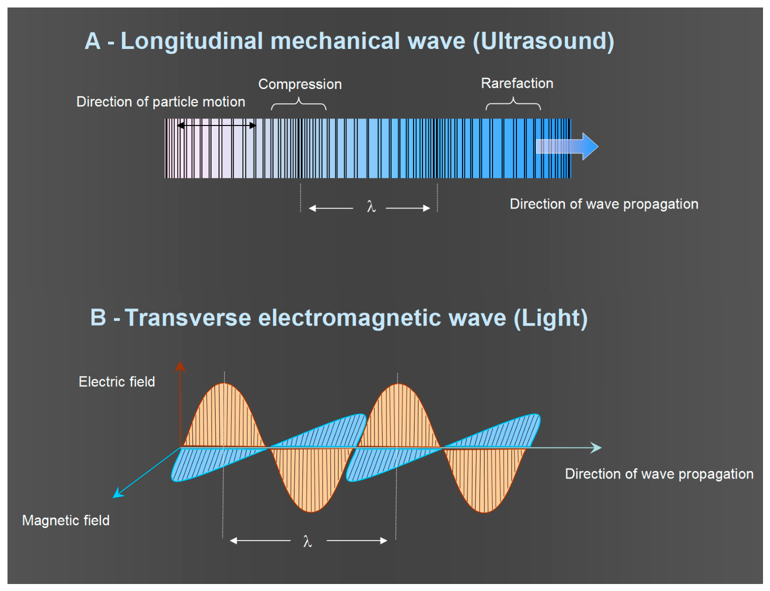
2. Light and Ultrasound: Different Physical Principles
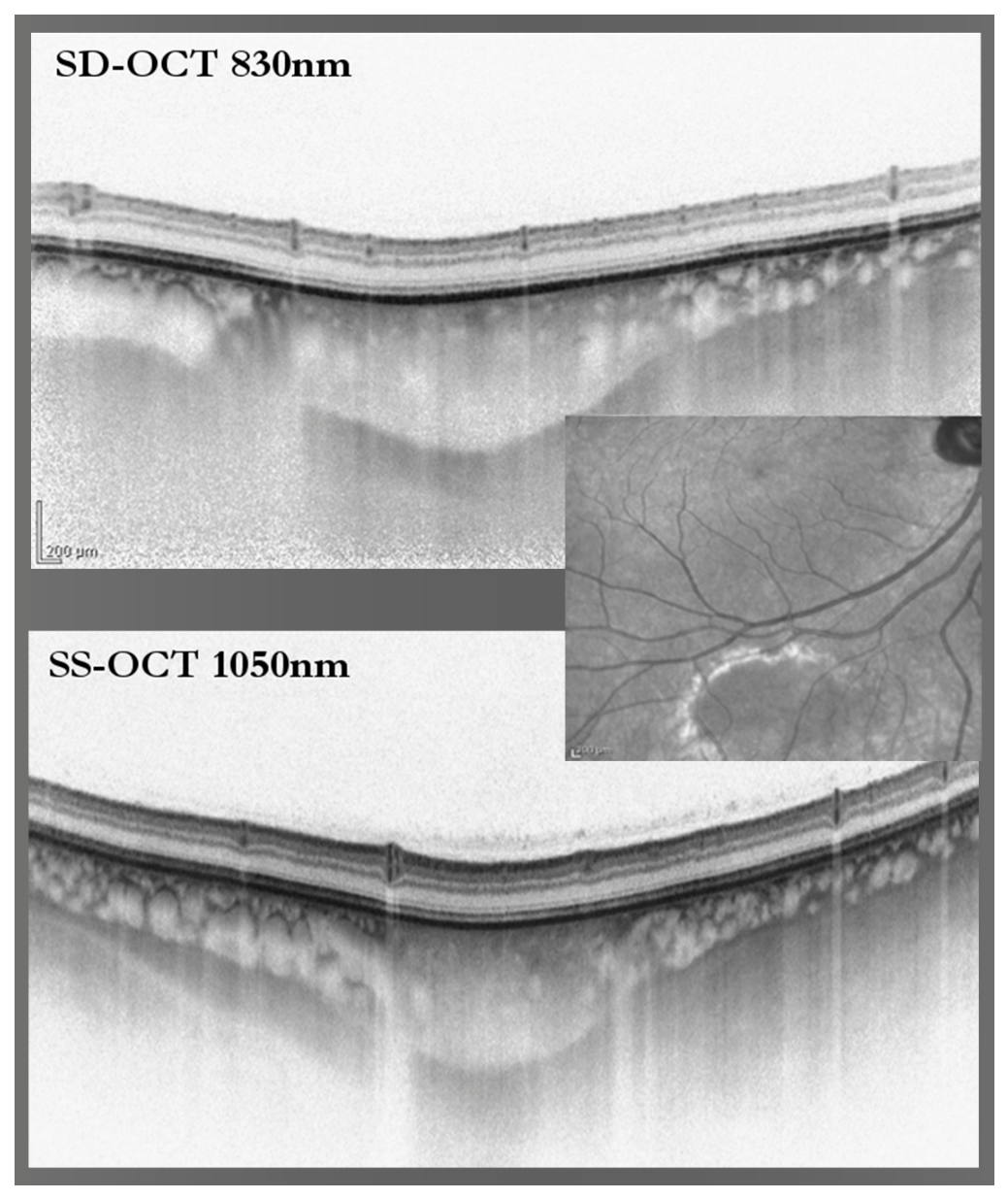
2.1. Optical Imaging and Tissue Reflectance
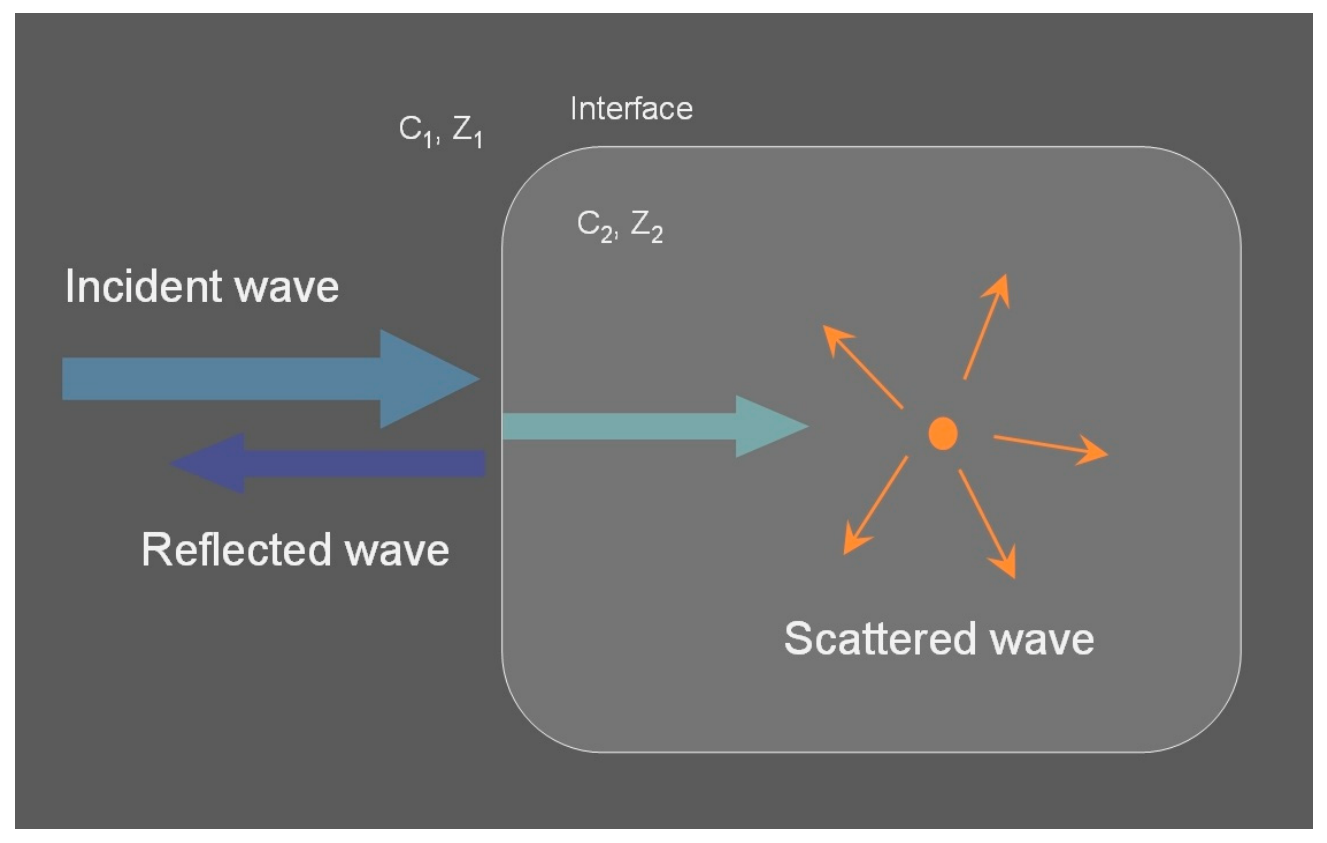
2.2. Ultrasonography and Acoustic Reflectivity
2.3. Why These Physical Differences Matter Clinically
3. Multimodal Imaging of Tumor-Associated Tissue Responses
3.1. Retinal Pigment Epithelium Distress and Retinal Response
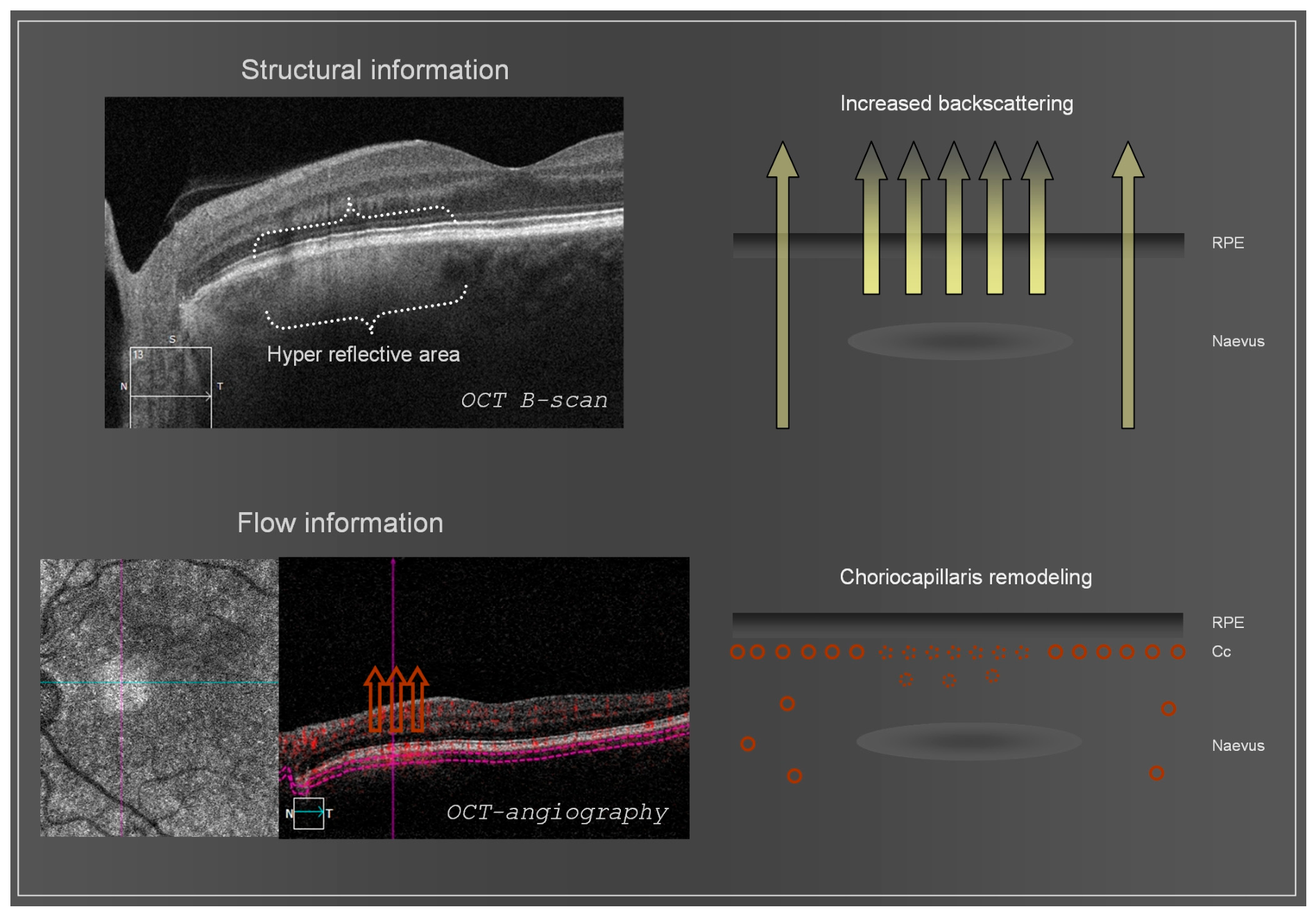
3.2. Choroidal Vascular and Tissue Changes Associated with Choroidal Tumors
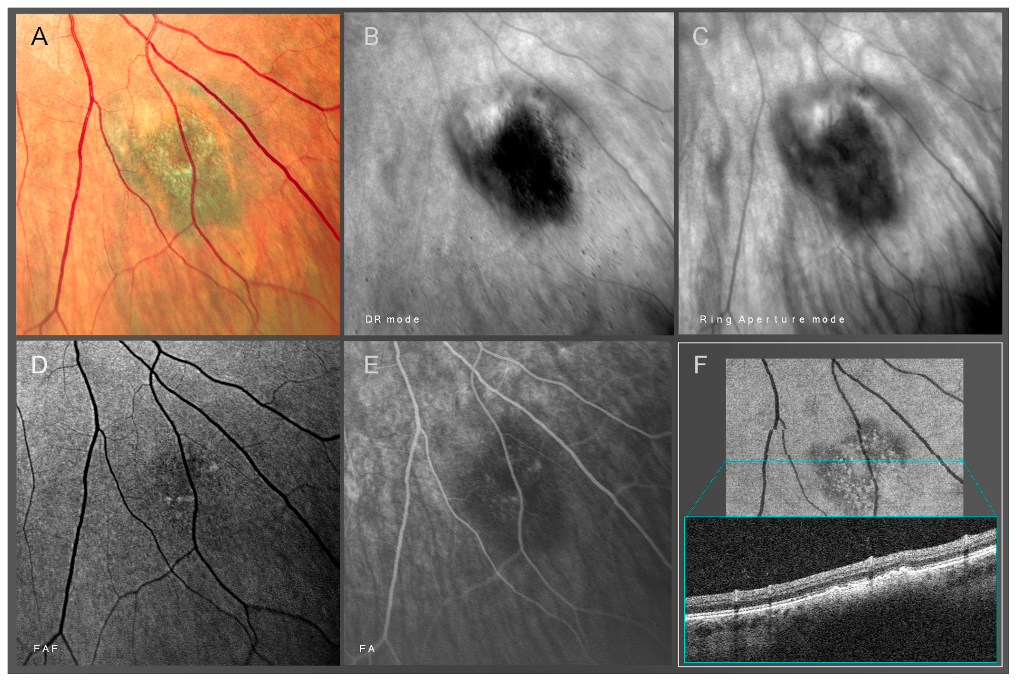
3.3. Emerging Role of Retro Mode Imaging
4. Choroidal Melanoma
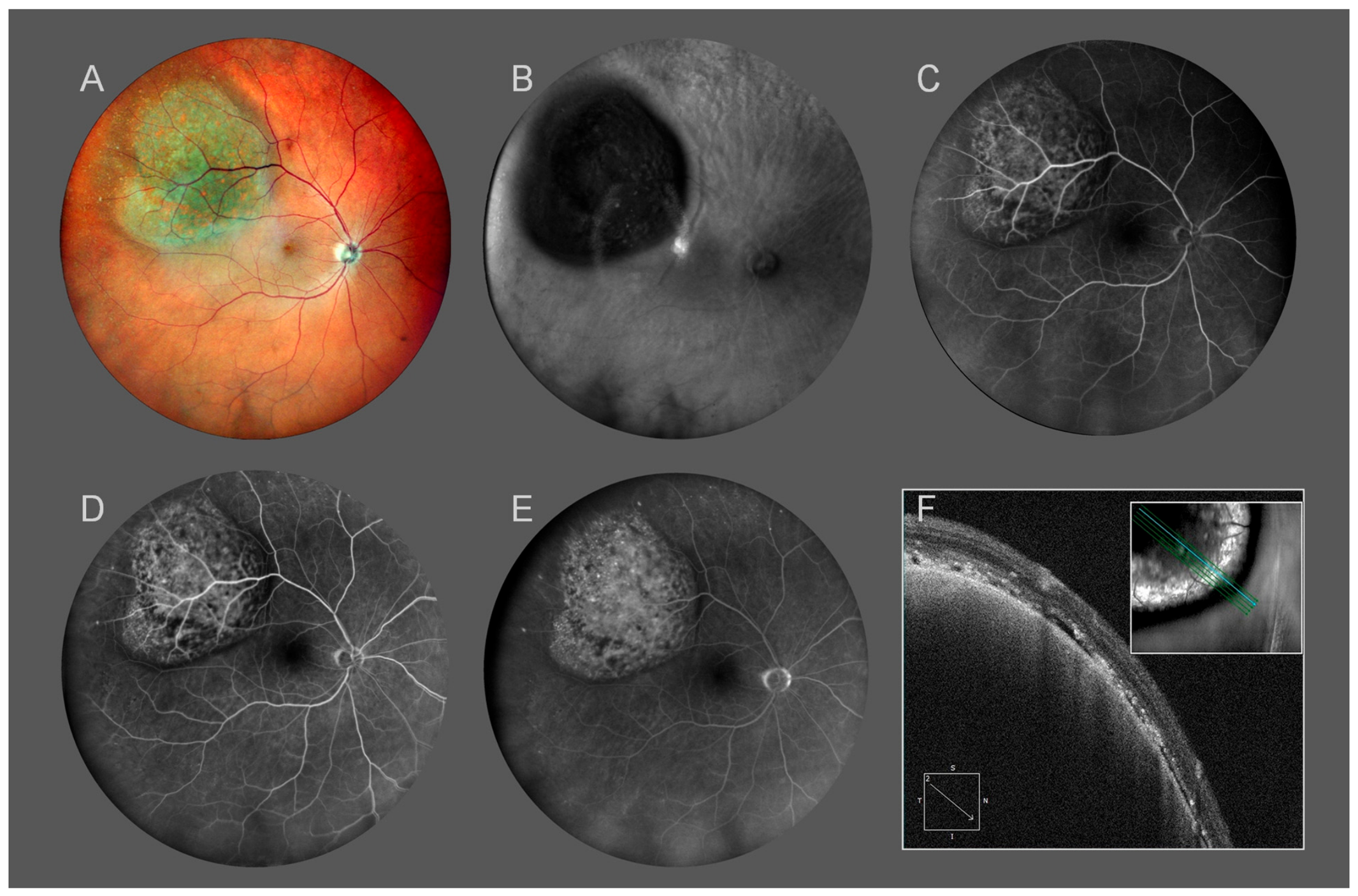
4.1. Clinical and Multimodal Characteristics
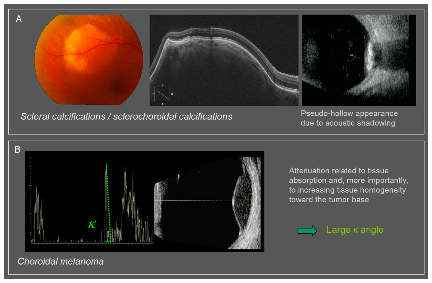
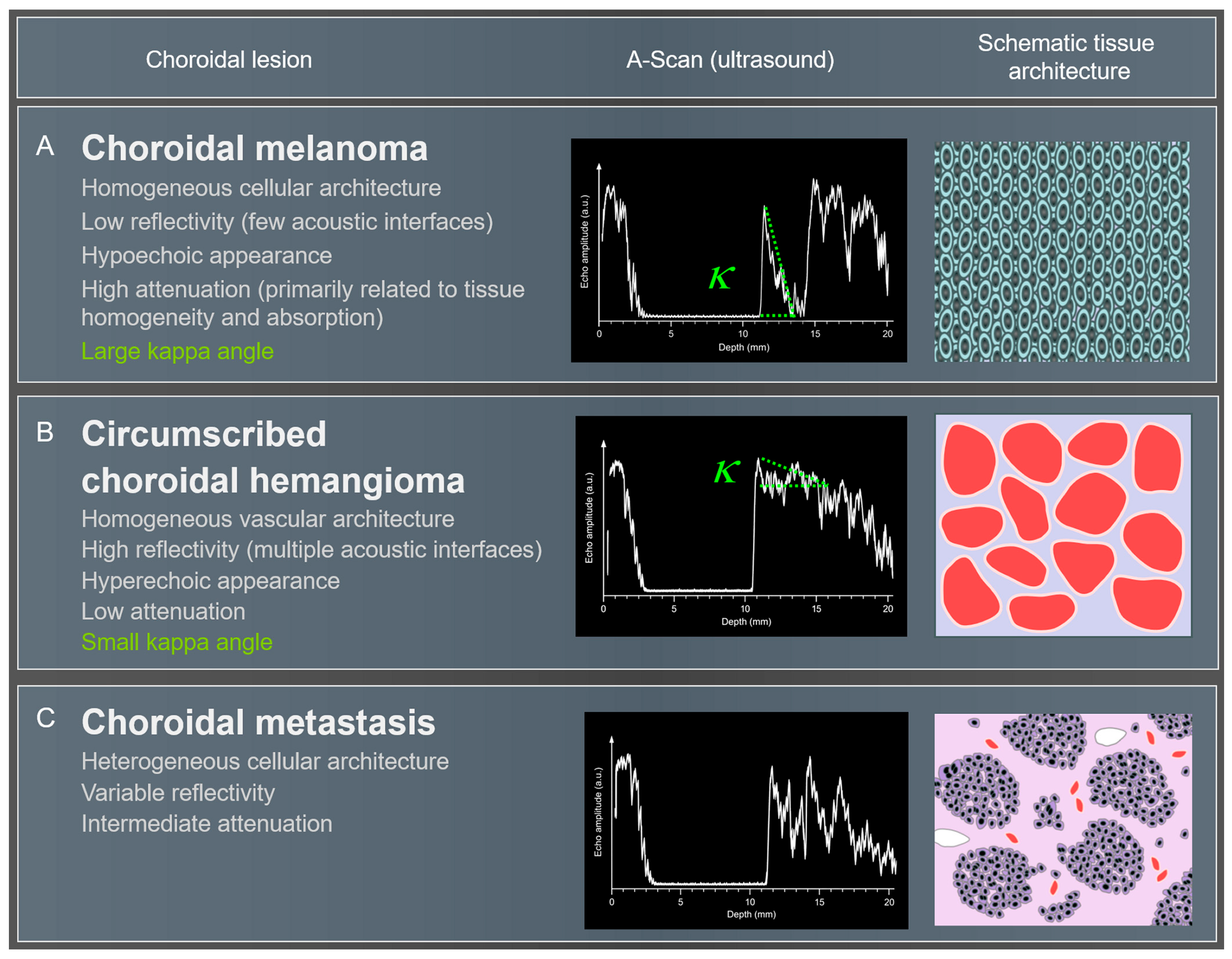
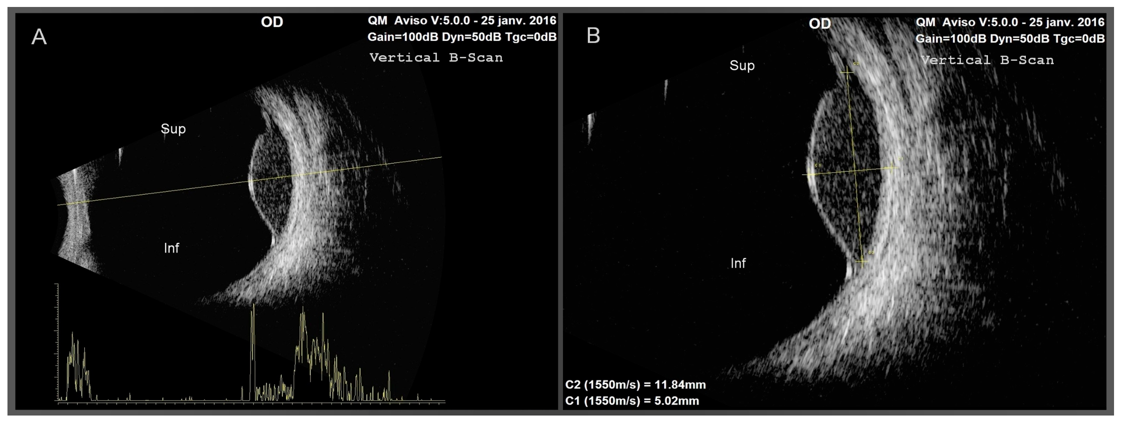
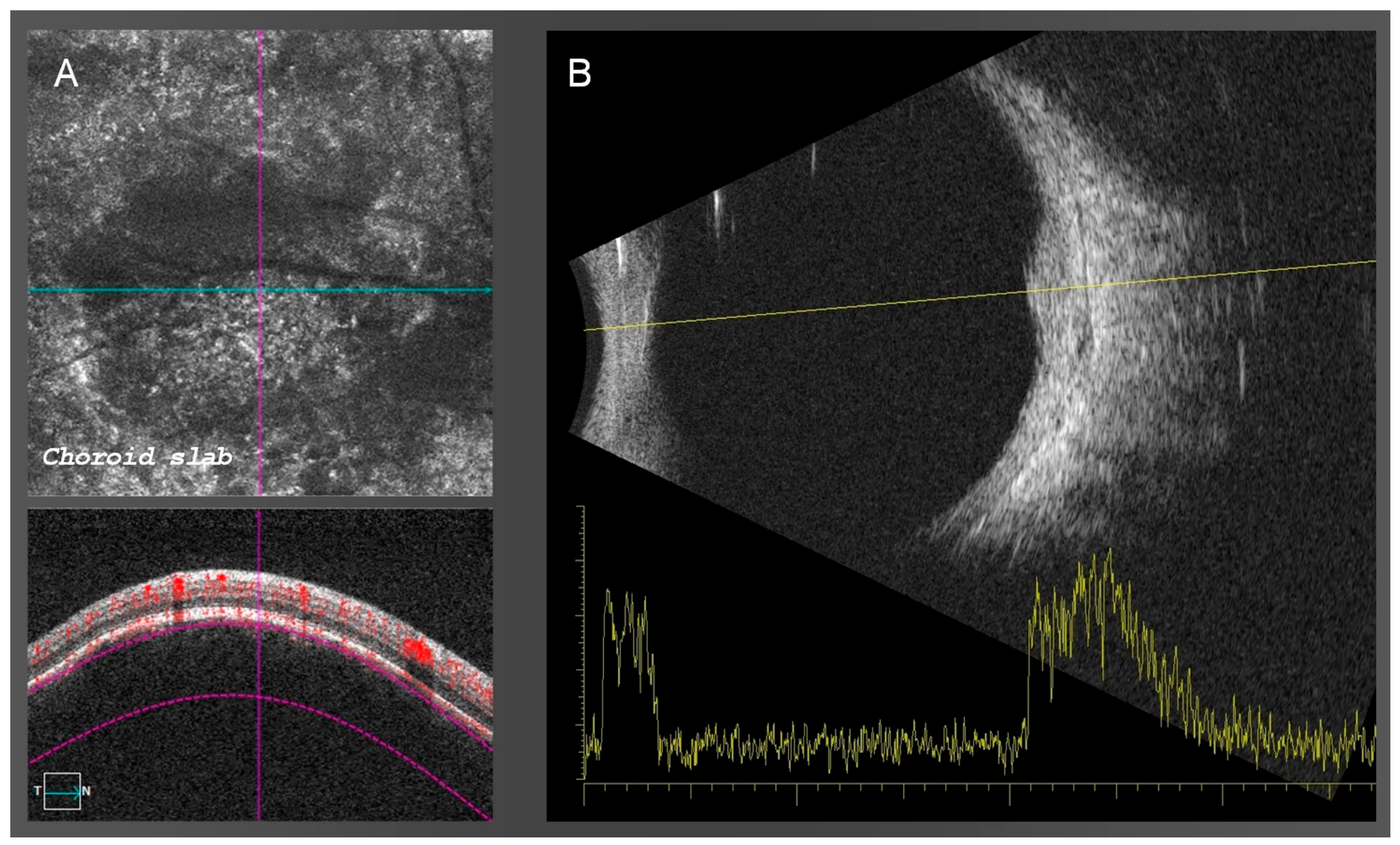
4.2. Ultrasonographic Characteristics
4.3. Longitudinal Evolution and Risk Assessment
5. Circumscribed Choroidal Hemangioma
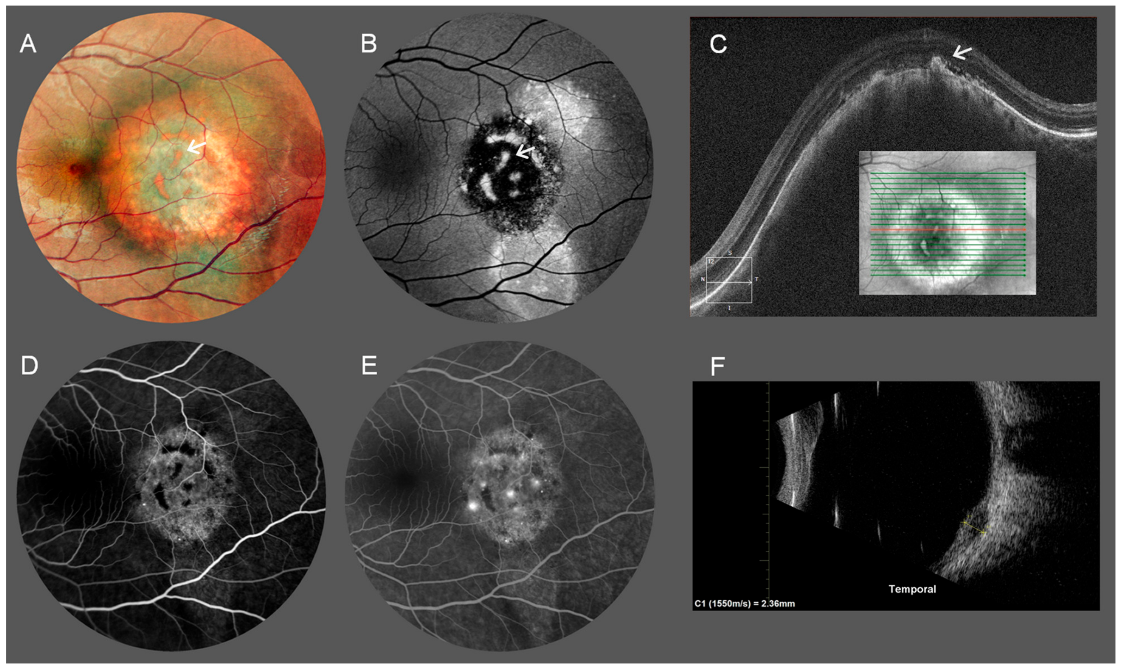
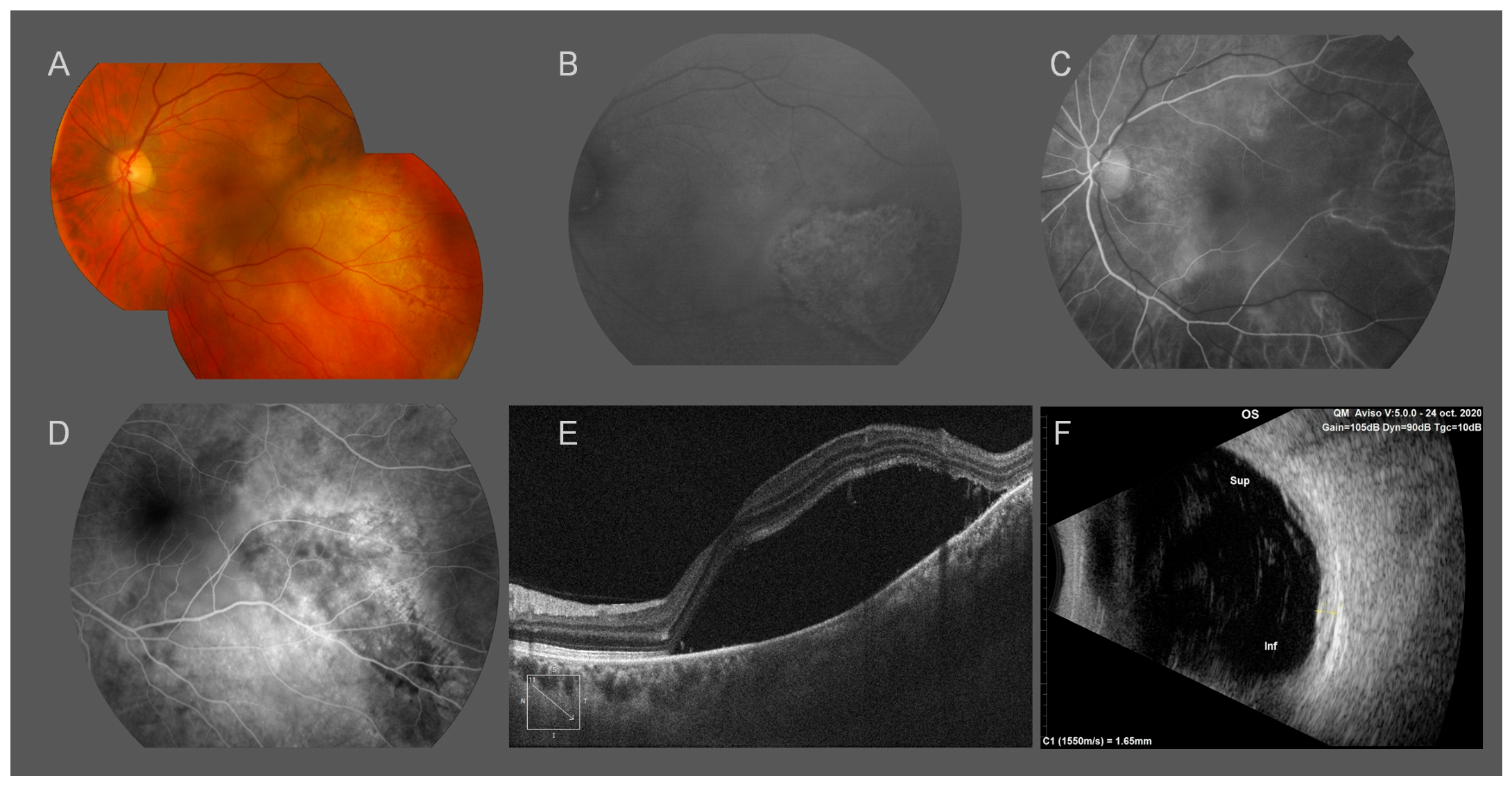
5.1. Clinical Appearance and Retinal Consequences
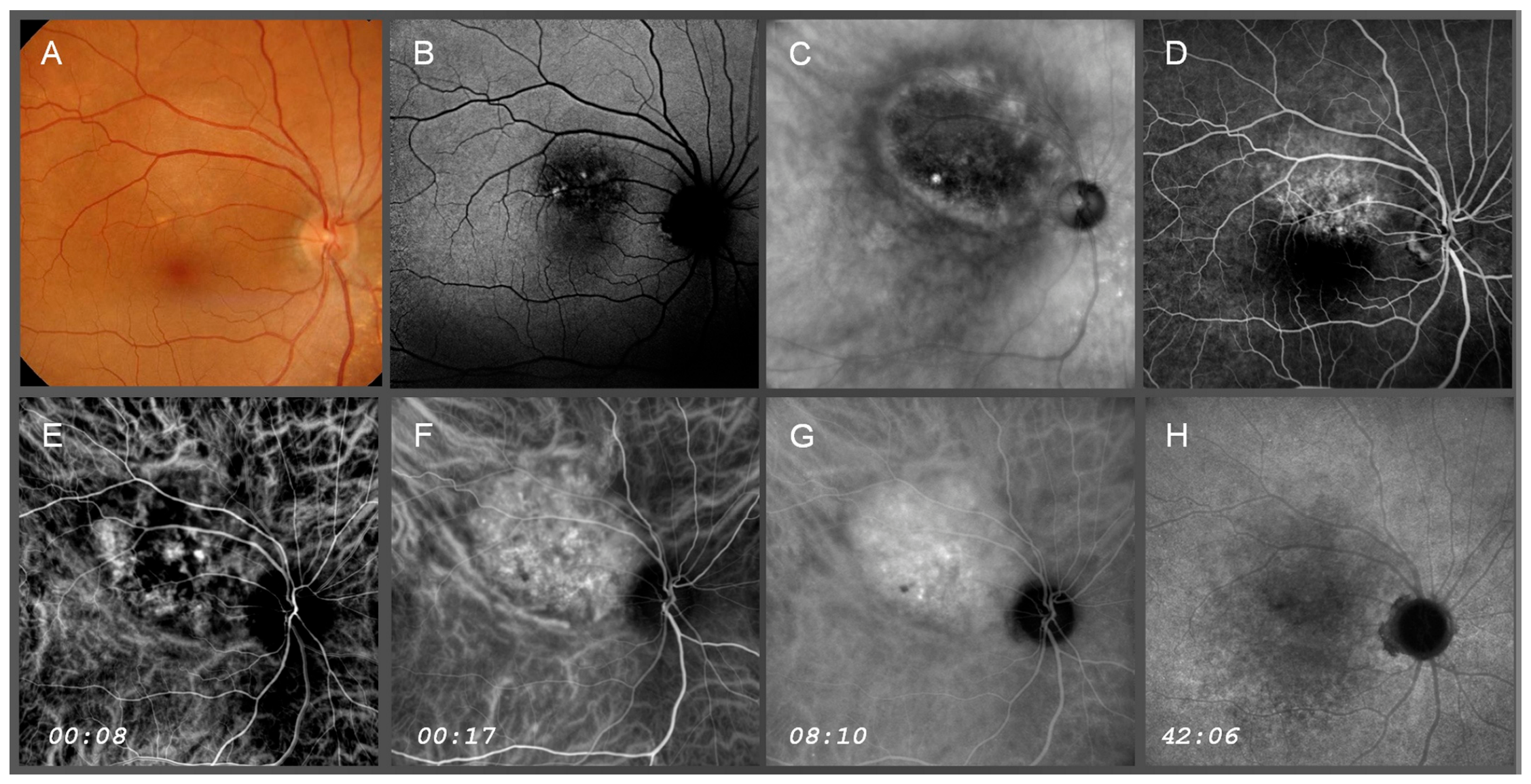
5.2. Angiographic Dynamics
5.3. OCT and OCT-A Findings
5.4. Ultrasonographic Characteristics
6. Choroidal Metastases and Other Differential Diagnoses
6.1. Choroidal Metastases
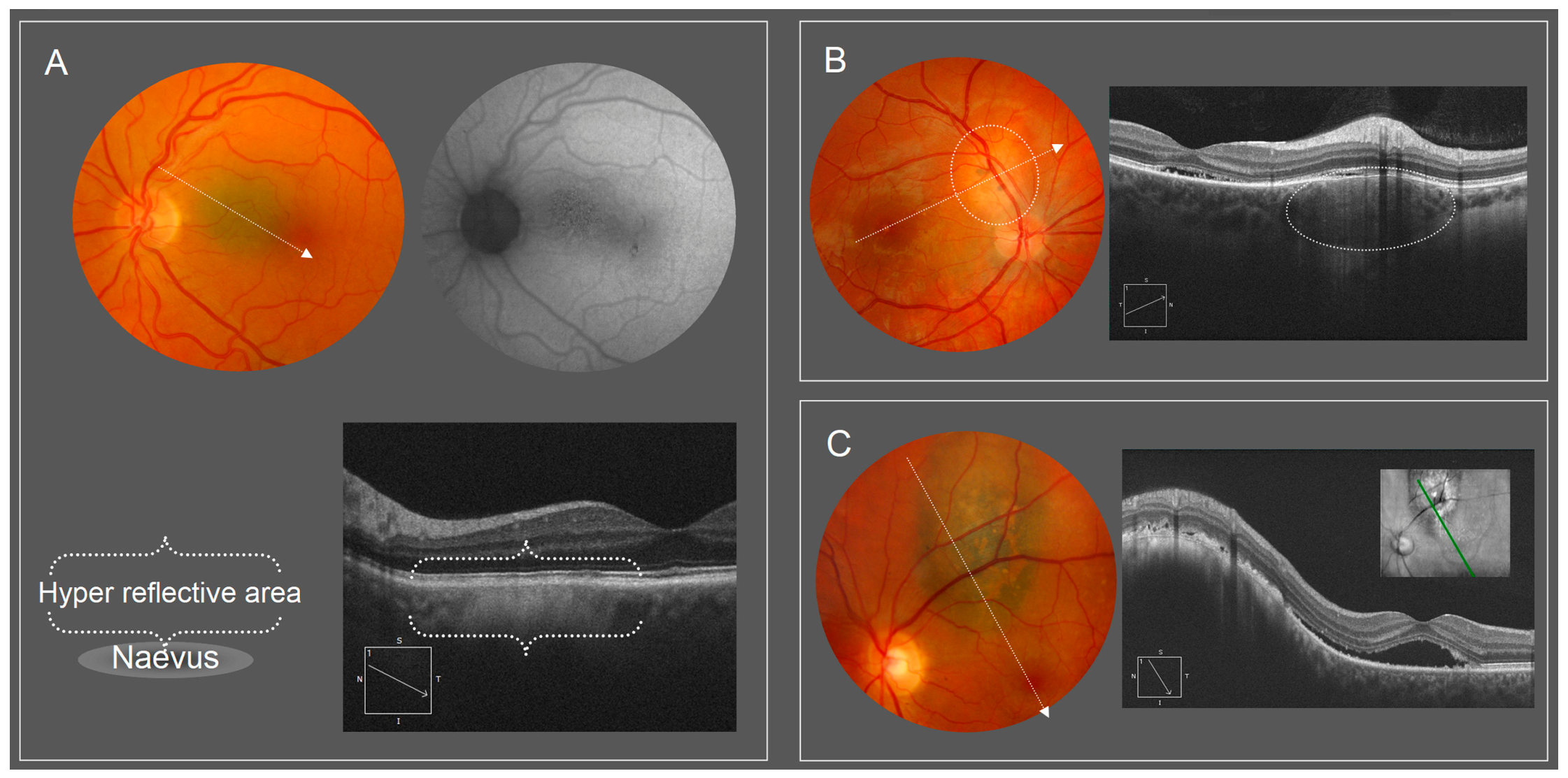
6.2. Choroidal Nevus
6.3. Choroidal and Pseudotumoral Mimickers
7. Longitudinal Multimodal Imaging
7.1. Temporal Evolution of Choroidal Lesions
7.2. Complementarity of Imaging Modalities
7.3. Limits and Future Directions
7.4. Clinical Implications of Multimodal Imaging
8. What Ultrasonography Still Uniquely Provides
8.1. Intrinsic Acoustic Tissue Characterization
8.2. Independence from Optical Transparency
8.3. Persistent Role and Limitations of Ultrasonography
9. Conclusions
Author Contributions
Funding
Institutional Review Board Statement
Informed Consent Statement
Data Availability Statement
Acknowledgments
Conflicts of Interest
References
- DeSimone, J.D.; Dockery, P.W.; Kreinces, J.B.; Soares, R.R.; Shields, C.L. Survey of ophthalmic imaging use to assess risk of progression of choroidal nevus to melanoma. Eye 2023, 37, 953–958. [Google Scholar] [CrossRef] [Scilit] [PubMed]
- Shields, C.L.; Dalvin, L.A.; Ancona-Lezama, D.; Yu, M.D.; Di Nicola, M.; Williams, B.K., Jr.; Lucio-Alvarez, J.A.; Ang, S.M.; Maloney, S.; Welch, R.J.; et al. Choroidal nevus imaging features in 3,806 cases and risk factors for transformation into melanoma in 2,355 CASES: The 2020 Taylor R. Smith and Victor T. Curtin Lecture. Retina 2019, 39, 1840–1851. [Google Scholar] [CrossRef] [Scilit] [PubMed]
- Dalvin, L.A.; Shields, C.L.; Ancona-Lezama, D.A.; Yu, M.D.; Di Nicola, M.; Williams, B.K., Jr.; Lucio-Alvarez, J.A.; Ang, S.M.; Maloney, S.M.; Welch, R.J.; et al. Combination of multimodal imaging features predictive of choroidal nevus transformation into melanoma. Br. J. Ophthalmol. 2019, 103, 1441–1447. [Google Scholar] [CrossRef] [Scilit] [PubMed]
- Geiger, F.; Said, S.; Bajka, A.; Toro, M.D.; Wiest, M.R.J.; Stahel, M.; Barthelmes, D.; Zweifel, S.A. Assessing Choroidal Nevi, Melanomas and Indeterminate Melanocytic Lesions Using Multimodal Imaging-A Retrospective Chart Review. Curr. Oncol. 2022, 29, 1018–1028. [Google Scholar] [CrossRef] [Scilit] [PubMed]
- Bayasi, F.M.; Negretti, G.S.; Shields, C.L. Choroidal Nevus with Growth into Melanoma over 10 Years. Ophthalmol. Retin. 2022, 6, 921. [Google Scholar] [CrossRef] [Scilit] [PubMed]
- Shields, C.L.; Furuta, M.; Berman, E.L.; Zahler, J.D.; Hoberman, D.M.; Dinh, D.H.; Mashayekhi, A.; Shields, J.A. Choroidal nevus transformation into melanoma: Analysis of 2514 consecutive cases. Arch. Ophthalmol. 2009, 127, 981–987. [Google Scholar] [CrossRef] [Scilit] [PubMed]
- Costa, R.A.; Skaf, M.; Melo, L.A., Jr.; Calucci, D.; Cardillo, J.A.; Castro, J.C.; Huang, D.; Wojtkowski, M. Retinal assessment using optical coherence tomography. Prog. Retin. Eye Res. 2006, 25, 325–353. [Google Scholar] [CrossRef] [Scilit] [PubMed]
- Cohen, S.Y.; Weber, M.; Oubraham, H.; Gaudric, A.; Quentel, G.; Tadayoni, R. Imaging in retina units: Changes observed during the last 12 years. Eur. J. Ophthalmol. 2014, 24, 216–220. [Google Scholar] [CrossRef] [Scilit] [PubMed]
- Rosenfeld, P.J. Optical Coherence Tomography and the Development of Antiangiogenic Therapies in Neovascular Age-Related Macular Degeneration. Investig. Ophthalmol. Vis. Sci. 2016, 57, OCT14–OCT26. [Google Scholar] [CrossRef] [Scilit] [PubMed]
- Feo, A.; Ramtohul, P.; Govetto, A.; Borrelli, E.; Sacconi, R.; Corradetti, G.; Querques, G.; Romano, M.R.; Rosenfeld, P.J.; Spaide, R.F.; et al. En face OCT: Breakthroughs in understanding the pathoanatomy of retinal disease and clinical applications. Prog. Retin. Eye Res. 2025, 106, 101351. [Google Scholar] [CrossRef] [Scilit] [PubMed]
- Mainster, M.A.; Timberlake, G.T.; Webb, R.H.; Hughes, G.W. Scanning laser ophthalmoscopy. Clinical applications. Ophthalmology 1982, 89, 852–857. [Google Scholar] [CrossRef] [Scilit] [PubMed]
- Kreminger, J.E.; Told, R.; Zafeiri, E.; Reumueller, A.; Dunavoelgyi, R.; Sacu, S. Volumes of Choroidal Nevi on Widefield Optical Coherence Tomography Compared to Calculations Based on Ultrasound Measurements. Ophthalmol. Ther. 2026, 15, 351–359. [Google Scholar] [CrossRef] [Scilit] [PubMed]
- Troisi, M.; Vitiello, L.; Lixi, F.; Timofte Zorila, M.M.; Abbinante, G.; Pellegrino, A.; Namazbayeva, A.; Adamo, G.G.; Coco, G.; Cuccu, A.; et al. Clinical Applications of Optical Coherence Tomography and Optical Coherence Tomography Angiography in Uveal Melanoma: A Narrative Review. Diagnostics 2025, 15, 2421. [Google Scholar] [CrossRef] [Scilit] [PubMed]
- Zhang, R.; Wu, H.; Li, Y.; Zhou, W.; Shi, X.; Yu, C.; Yang, Y.; Zhao, H.; Li, H.; Wang, S.; et al. Swept-Source OCT Angiography Features of Melanotic Choroidal Tumors: An Analysis of 102 Consecutive Cases. Ophthalmol. Sci. 2026, 6, 100932. [Google Scholar] [CrossRef] [Scilit] [PubMed]
- Laugier, P. Principes physiques de la propagation ultrasonore et de la formation de l’image échographique. In Echographie de l’Oeil et de l’Orbite; Bergès, O., Ed.; Sauramps Medical: Montpellier, France, 2022; pp. 17–26. [Google Scholar]
- de Korte, C.L.; van der Steen, A.F.; Thijssen, J.M. Acoustic velocity and attenuation of eye tissues at 20 MHz. Ultrasound Med. Biol. 1994, 20, 471–480. [Google Scholar] [CrossRef] [Scilit] [PubMed]
- Coleman, D.; Lizzi, F. Ultrasonography of the Eye and Orbit; Lippincott: Waltham, MA, USA, 2006. [Google Scholar]
- Ossoinig, K.C. Standardized echography: Basic principles, clinical applications, and results. Int. Ophthalmol. Clin. 1979, 19, 127–210. [Google Scholar] [PubMed]
- Shields, J.A.; Canny, C.L. Ultrasonography and 32P test in the diagnosis of choroidal melanoma with massive vitreous hemorrhage. Can. J. Ophthalmol. 1977, 12, 230–233. [Google Scholar] [PubMed]
- Ferreira, T.A.; Jaarsma-Coes, M.G.; Marinkovic, M.; Verbist, B.; Verdijk, R.M.; Jager, M.J.; Luyten, G.P.M.; Beenakker, J.M. MR imaging characteristics of uveal melanoma with histopathological validation. Neuroradiology 2022, 64, 171–184. [Google Scholar] [CrossRef] [Scilit] [PubMed]
- Jacobsen, B.H.; Ricks, C.; Harrie, R.P. Ocular ultrasound versus MRI in the detection of extrascleral extension in a patient with choroidal melanoma. BMC Ophthalmol. 2018, 18, 320. [Google Scholar] [CrossRef] [Scilit] [PubMed]
- Jaarsma-Coes, M.G.; Ferreira, T.A.; Luyten, G.P.M.; Beenakker, J.W.M. Reaction on “Ocular ultrasound versus MRI in the detection of extrascleral extension in a patient with choroidal melanoma”. BMC Ophthalmol. 2019, 19, 193. [Google Scholar] [CrossRef] [Scilit] [PubMed]
- Azzolini, C.; Di Nicola, M.; Pozzo Giuffrida, F.; Cappelli, F.; Bellina, C.; Viola, F.; Chelazzi, P. Retromode Scanning Laser Ophthalmoscopy for Choroidal Nevi: A Preliminary Study. Life 2023, 13, 1253. [Google Scholar] [CrossRef] [Scilit] [PubMed]
- Mainster, M.A.; Desmettre, T.; Querques, G.; Turner, P.L.; Ledesma-Gil, G. Scanning laser ophthalmoscopy retroillumination: Applications and illusions. Int. J. Retin. Vitr. 2022, 8, 71. [Google Scholar] [CrossRef] [Scilit] [PubMed]
- Monash University. Available online: https://www.monash.edu/student-academic-success/physics/wave-particle-duality/light-as-a-wave (accessed on 17 August 2026).
- Jacques, S.L. Optical properties of biological tissues: A review. Phys. Med. Biol. 2013, 58, R37–R61. [Google Scholar] [CrossRef] [Scilit] [PubMed]
- Huang, D.; Swanson, E.A.; Lin, C.P.; Schuman, J.S.; Stinson, W.G.; Chang, W.; Hee, M.R.; Flotte, T.; Gregory, K.; Puliafito, C.A.; et al. Optical coherence tomography. Science 1991, 254, 1178–1181. [Google Scholar] [CrossRef] [Scilit] [PubMed]
- Michalewska, Z.; Michalewski, J.; Nawrocki, J. Swept Source optical coherence tomography of choroidal nevi. Can. J. Ophthalmol. 2016, 51, 271–276. [Google Scholar] [CrossRef] [Scilit] [PubMed]
- Szabo, T. Diagnostic Ultrasound Imaging: Inside Out, 2nd ed.; Elsevier (Academic Press): Amsterdam, The Netherlands, 2013; p. 832. [Google Scholar]
- Byrne, S.; Green, R. Ultrasound of the Eye and Orbit; Jaypee Publishers: New Delhi, India, 2010. [Google Scholar]
- Callaway, N.F.; Mruthyunjaya, P. Widefield imaging of retinal and choroidal tumors. Int. J. Retin. Vitr. 2019, 5, 49. [Google Scholar] [CrossRef] [Scilit] [PubMed]
- Maloca, P.; Gyger, C.; Schoetzau, A.; Hasler, P.W. Inter-device size variation of small choroidal nevi measured using stereographic projection ultra-widefield imaging and optical coherence tomography. Graefe’s Arch. Clin. Exp. Ophthalmol. 2016, 254, 797–808. [Google Scholar] [CrossRef] [Scilit] [PubMed]
- Kulikov, A.N.; Maltsev, D.S.; Burnasheva, M.A.; Chhablani, J. Characterization of Choroidal Nevi with Dark-Field Infrared Scanning Laser Ophthalmoscopy. Ophthalmol. Retin. 2019, 3, 703–708. [Google Scholar] [CrossRef] [Scilit] [PubMed]
- Verbeek, S.; Dalvin, L.A. Advances in multimodal imaging for diagnosis of pigmented ocular fundus lesions. Can. J. Ophthalmol. 2024, 59, 218–233. [Google Scholar] [CrossRef] [Scilit] [PubMed]
- Bindewald-Wittich, A.; Holz, F.G.; Ach, T.; Fiorentzis, M.; Bechrakis, N.E.; Willerding, G.D. Fundus Autofluorescence Imaging in Patients with Choroidal Melanoma. Cancers 2022, 14, 1809. [Google Scholar] [CrossRef] [Scilit] [PubMed]
- Konana, V.K.; Shanmugam, P.M.; Ramanjulu, R.; Mishra, K.C.D.; Sagar, P. Optical coherence tomography angiography features of choroidal hemangioma. Indian J. Ophthalmol. 2018, 66, 581–583. [Google Scholar] [CrossRef] [Scilit] [PubMed]
- Pellegrini, M.; Corvi, F.; Invernizzi, A.; Ravera, V.; Cereda, M.G.; Staurenghi, G. Swept-source optical coherence tomography angiography in choroidal melanoma: An analysis of 22 consecutive cases. Retina 2019, 39, 1510–1519. [Google Scholar] [CrossRef] [Scilit] [PubMed]
- Kurdiani, T.; Fiorentzis, M.; Bechrakis, N.E.; Kiefer, T. OCT Angiography-Based Quantitative Choroidal Vasculature Analysis in Choroidal Melanomas and Nevi. Ophthalmol. Sci. 2026, 6, 100979. [Google Scholar] [CrossRef] [Scilit] [PubMed]
- Chadwick, W.F.; Ganesh, S.; Dadzie, A.K.; Lokesh, Y.; Alahmadi, R.; Yao, X.; Heiferman, M.J. A Framework for Standardized Manual Segmentation and Measurement of Uveal Melanoma on Ultrasound. Ophthalmol. Sci. 2026, 6, 101229. [Google Scholar] [CrossRef] [Scilit] [PubMed]
- Shields, C.L.; Bianciotto, C.; Pirondini, C.; Materin, M.A.; Harmon, S.A.; Shields, J.A. Autofluorescence of orange pigment overlying small choroidal melanoma. Retina 2007, 27, 1107–1111. [Google Scholar] [CrossRef] [Scilit] [PubMed]
- Maloca, P.; Gyger, C.; Hasler, P.W. A pilot study to compartmentalize small melanocytic choroidal tumors and choroidal vessels with speckle-noise free 1050 nm swept source optical coherence tomography (OCT choroidal “tumoropsy”). Graefe’s Arch. Clin. Exp. Ophthalmol. 2016, 254, 1211–1219. [Google Scholar] [CrossRef] [Scilit] [PubMed]
- Maloca, P.M.; Tufail, A.; Hasler, P.W.; Rothenbuehler, S.; Egan, C.; Ramos de Carvalho, J.E.; Spaide, R.F. 3D printing of the choroidal vessels and tumours based on optical coherence tomography. Acta Ophthalmol. 2019, 97, e313–e316. [Google Scholar] [CrossRef] [Scilit] [PubMed]
- Ito, H.; Takahashi, A.; Ishiko, S.; Nagaoka, T.; Yoshida, A. Retinal Cavernous Hemangioma Documented by Spectral Domain Optical Coherence Tomography and Confocal Scanning Laser Ophthalmoscope Retro-Mode Imaging. Retin. Cases Brief. Rep. 2016, 10, 373–376. [Google Scholar] [CrossRef] [Scilit] [PubMed]
- Ramtohul, P.; Chehaibou, I.; Couturier, A. Retromode imaging for choroidal osteoma. Can. J. Ophthalmol. 2022, 57, e200. [Google Scholar] [CrossRef] [Scilit] [PubMed]
- Lopez, J.M.; Rabinovich, M.; Colantuono, D.; Souied, E.H. Multimodal and retro-mode imaging in sclerochoroidal calcification: A case report. Arch. Soc. Esp. Oftalmol. 2024, 99, 213–217. [Google Scholar] [CrossRef] [Scilit] [PubMed]
- Shields, C.L.; Honavar, S.G.; Shields, J.A.; Cater, J.; Demirci, H. Circumscribed choroidal hemangioma: Clinical manifestations and factors predictive of visual outcome in 200 consecutive cases. Ophthalmology 2001, 108, 2237–2248. [Google Scholar] [CrossRef] [Scilit] [PubMed]
- Arepalli, S.; Shields, C.L.; Kaliki, S.; Emrich, J.; Komarnicky, L.; Shields, J.A. Diffuse choroidal hemangioma management with plaque radiotherapy in 5 cases. Ophthalmology 2013, 120, 2358–2359.e2. [Google Scholar] [CrossRef] [Scilit] [PubMed]
- Spaide, R. Autofluorescence from the outer retina and subretinal space: Hypothesis and review. Retina 2008, 28, 5–35. [Google Scholar] [CrossRef] [Scilit] [PubMed]
- Arevalo, J.F.; Shields, C.L.; Shields, J.A.; Hykin, P.G.; De Potter, P. Circumscribed choroidal hemangioma: Characteristic features with indocyanine green videoangiography. Ophthalmology 2000, 107, 344–350. [Google Scholar] [CrossRef] [Scilit] [PubMed]
- Desmettre, T.; Devoisselle, J.M.; Mordon, S. Fluorescence properties and metabolic features of indocyanine green (ICG) as related to angiography. Surv. Ophthalmol. 2000, 45, 15–27. [Google Scholar] [CrossRef] [Scilit] [PubMed]
- Di Khor, H.; Lott, P.W.P.; Lim, Y.W.; Devan, D.; Iqbal, T. The role of multimodal ophthalmic imaging in diagnosing choroidal metastasis. Int. Ophthalmol. 2025, 45, 339. [Google Scholar] [CrossRef] [Scilit] [PubMed]
- Shields, C.L.; Shields, J.A.; Gross, N.E.; Schwartz, G.P.; Lally, S.E. Survey of 520 eyes with uveal metastases. Ophthalmology 1997, 104, 1265–1276. [Google Scholar] [CrossRef] [Scilit] [PubMed]
- Barbieri, L.; Preziosa, C.; Staurenghi, G.; Pellegrini, M. Multifocal Circumscribed Choroidal Hemangiomas Mimicking Metastases: A Diagnostic Challenge. Retin. Cases Brief. Rep. 2025. Online ahead of print. [Google Scholar] [CrossRef] [Scilit] [PubMed]
- Murati Calderon, R.A.; Nieves-Rios, C.; Nieves Garrastegui, L.F.; Flores Caban, L.D.; Oliver, A. Bilateral Choroidal Metastases Mimicking Central Serous Chorioretinopathy: A Diagnostic Challenge. Cureus 2025, 17, e91842. [Google Scholar] [CrossRef] [Scilit] [PubMed]
- Seong, H.J.; Kim, C.; Chang, J.; Cha, J.; Lee, C.S. A deep-learning model for detecting choroidal metastases and predicting primary tumors from ultra-widefield fundus imaging. Graefe’s Arch. Clin. Exp. Ophthalmol. 2026, 264, 569–577. [Google Scholar] [CrossRef] [Scilit] [PubMed]
- Rissotto, F.; Matteo, C.; Cicinelli, M.V.; Bandello, F.; Miserocchi, E. Multimodal Imaging of Choroidal Lymphoma: Diagnostic Features, Differential Diagnosis, and Clinical Monitoring. Ocul. Immunol. Inflamm. 2026, 1–10. [Google Scholar] [CrossRef] [Scilit] [PubMed]
- Xu, L.T.; Huang, Y.; Liao, A.; Anthony, C.L.; Voloschin, A.; Yeh, S. Multimodal diagnostic imaging in primary vitreoretinal lymphoma. Int. J. Retin. Vitr. 2022, 8, 58. [Google Scholar] [CrossRef] [Scilit] [PubMed]
- Hasanreisoglu, M.; Saktanasate, J.; Shields, P.W.; Shields, C.L. Classification of Sclerochoroidal Calcification Based on Enhanced Depth Imaging Optical Coherence Tomography “Mountain-Like” Features. Retina 2015, 35, 1407–1414. [Google Scholar] [CrossRef] [Scilit] [PubMed]
- Aubakirova, A.; Sarsembekova, K.; Jumatayeva, Z.; Utelbayeva, Z.; Serikova, Z.; Berlibek, D. Choroidal Osteoma: Case Series. Case Rep. Med. 2026, 2026, 8005092. [Google Scholar] [CrossRef] [Scilit] [PubMed]
- Celik Dulger, S.; Sogut, F.E.; Teke, M.Y.; Citirik, M. Optical Coherence Tomography Features and Multimodal Imaging Findings in Choroidal Osteoma: A Case Series. Ocul. Oncol. Pathol. 2026. Online ahead of print. [Google Scholar] [CrossRef] [PubMed]
- Ally, N.; Makgotloe, A. Nodular Posterior Scleritis Masquerading as a Subretinal Mass. Middle East Afr. J. Ophthalmol. 2020, 27, 231–234. [Google Scholar] [CrossRef] [Scilit] [PubMed]
- Babu, N.; Kumar, K.; Upadhayay, A.; Kohli, P. Nodular posterior scleritis—The great masquerader. Taiwan J. Ophthalmol. 2021, 11, 408–412. [Google Scholar] [CrossRef] [Scilit] [PubMed]
- Agarwal, A.; Aggarwal, K.; Pichi, F.; Meng, T.; Munk, M.R.; Bazgain, K.; Bansal, R.; Agrawal, R.; Gupta, V. Clinical and Multimodal Imaging Clues in Differentiating Between Tuberculomas and Sarcoid Choroidal Granulomas. Am. J. Ophthalmol. 2021, 226, 42–55. [Google Scholar] [CrossRef] [Scilit] [PubMed]
- Desai, A.; Sahoo, N.; Tyagi, M.; Raval, V. Clinical and Multimodal Imaging Study to Differentiate Amelanotic Choroidal Lesions. Ocul. Oncol. Pathol. 2024, 10, 206–218. [Google Scholar] [CrossRef] [Scilit] [PubMed]
- Badawi, A.H.; Semidey, V.A.; Magliyah, M.; Al-Dhibi, H. Updated Systematic Review and Clinical Spectrum of Peripheral Exudative Hemorrhagic Chorioretinopathy. Middle East Afr. J. Ophthalmol. 2020, 27, 4–9. [Google Scholar] [CrossRef] [Scilit] [PubMed]
- Mishra, K.; Mruthyunjaya, P. The Small Choroidal Melanoma: Diagnosis, Treatment Outcomes, and Unending Controversy. Semin. Ophthalmol. 2025, 40, 624–633. [Google Scholar] [CrossRef] [Scilit] [PubMed]
- Piperno-Neumann, S.; Angi, M.; Ascierto, P.A.; Baurain, J.F.; Burgmans, M.C.; Cassoux, N.; Coupland, S.E.; Dendale, R.; Fortuna, A.; Gastaud, L.; et al. Uveal melanoma: ESMO-EURACAN Clinical Practice Guideline for diagnosis, treatment and follow-up. ESMO Open 2026, 11, 106888. [Google Scholar] [CrossRef] [Scilit] [PubMed]



| Imaging Modality | What is Measured or Detected | Main Limitations, Artifacts, and Reproducibility Issues | Principal Contribution to Clinical Assessment |
|---|---|---|---|
| Color fundus photography/ultra-widefield imaging | Reflected visible-light signal documenting lesion color, pigmentation, surface features, margins, and associated retinal changes. | Two-dimensional representation; color and contrast depend on illumination, acquisition system, media clarity, and pigmentation. Peripheral distortion and differences between imaging systems may affect apparent lesion dimensions and longitudinal comparisons. | Baseline documentation of lesion appearance and margins; assessment of pigmentation, orange pigment, hemorrhage, and associated retinal changes; photographic documentation of growth during follow-up [31,32]. |
| Near-infrared reflectance/multicolor imaging | Reflected and scattered light at near-infrared or multiple wavelengths, providing contrast related to retinal, RPE, and choroidal structures. | Signal depends on pigmentation, tissue depth, wavelength, and device-specific acquisition and processing. Reflectivity differences are not specific for tissue composition. | May improve visualization of lesion boundaries and subtle reflectivity differences and complements color photography and OCT for lesion localization and multimodal documentation [33,34]. |
| Fundus autofluorescence (FAF) | Endogenous fluorophore-derived signal, predominantly related to lipofuscin and melanolipofuscin within the RPE, and influenced by absorbing structures. | Provides indirect rather than direct information on tissue biology. Signal is influenced by fluorophore composition, masking, pigmentation, media opacity, excitation wavelength, and acquisition system. | Documents tumor-associated RPE alterations and orange pigment; provides indirect information regarding RPE status and contributes to multimodal risk assessment of melanocytic lesions [3,35]. |
| Fluorescein angiography (FA) | Time-dependent fluorescence of intravenously administered fluorescein, demonstrating vascular filling, leakage, staining, pooling, and blockage. | Invasive and time-dependent acquisition; late leakage may obscure lesion boundaries. Limited penetration through pigment, hemorrhage, and RPE reduces visualization of deeper choroidal structures. | Characterizes leakage and secondary retinal vascular or exudative changes; contributes to differential diagnosis and assessment of lesion-associated retinal consequences [34]. |
| Indocyanine green angiography (ICGA) | Near-infrared fluorescence of intravenously administered indocyanine green, providing information on choroidal vascular filling and lesion-associated vascular patterns. | Invasive and time-dependent acquisition; interpretation depends on acquisition phase and lesion characteristics. Provides vascular rather than direct histologic information. | Characterizes intrinsic choroidal vascular patterns and may substantially narrow the differential diagnosis, particularly for vascular lesions such as circumscribed choroidal hemangioma [34,36]. |
| Structural OCT/EDI-OCT/swept-source OCT | Optical backscatter providing high-resolution cross-sectional and en face structural information from the retina, RPE, choroid, and optically accessible portions of choroidal lesions. | Signal attenuation and shadowing from pigmentation or lesion thickness; segmentation artifacts; incomplete penetration through large or highly attenuating lesions. Measurements depend on scan orientation and visibility of lesion boundaries. | High-resolution assessment of lesion contour and secondary retinal/RPE changes; sensitive detection of shallow subretinal fluid, photoreceptor alterations, and hyperreflective deposits; useful for characterization and longitudinal monitoring of small lesions [8,12]. |
| OCT angiography (OCT-A) | Motion-contrast signal generated predominantly by moving erythrocytes within predefined segmentation slabs, providing flow-related vascular information without intravenous dye. | Segmentation and projection artifacts, shadowing, motion artifacts, slab selection, and flow-detection thresholds may alter apparent vascular patterns. Quantitative measurements are device-, segmentation-, and processing-dependent, and absence of detectable signal does not necessarily indicate absence of vessels. | Provides noninvasive assessment of flow-related vascular organization and may complement structural OCT in lesion characterization. Quantitative differences have been reported among lesion types, but validated tumor-specific diagnostic thresholds remain limited [37,38]. |
| Retro mode imaging | Indirectly scattered near-infrared light collected through laterally displaced (DL/DR) or annular (RA) apertures, generating shadow-based topographic contrast. | Platform-specific and predominantly qualitative. Apparent pseudo-relief depends on aperture geometry and illumination direction and does not represent true three-dimensional topography. Tumor-specific evidence remains limited and validated diagnostic thresholds are lacking. | May enhance visualization of lesion borders, surface contour, and associated topographic or pigmentary alterations. At present, it should be considered a complementary rather than decision-defining modality in ocular oncology [23,24]. |
| B-scan ultrasonography | Spatial distribution and amplitude of returning ultrasound echoes, providing information on lesion configuration, dimensions, internal echogenicity, and acoustic attenuation within the acquired scanning plane. | Operator- and orientation-dependent. Oblique insonation, boundary selection, inclusion of retina, sclera, or adjacent subretinal fluid, acoustic shadowing, and limited resolution for small lesions may affect measurements. Conventional two-dimensional examinations sample selected planes rather than the entire lesion volume. | Measures lesion thickness and configuration; evaluates lesions when optical visualization is limited by opaque media; assesses peripheral or larger lesions and possible extrascleral extension; contributes to differential diagnosis, treatment planning, and longitudinal follow-up [17,39]. |
| Standardized A-scan ultrasonography | Echo amplitude as a function of tissue depth under standardized acquisition conditions, providing an internal acoustic reflectivity profile. | Requires standardized acquisition and calibration and remains dependent on probe alignment and insonation angle. Interpretation may be influenced by lesion size and internal heterogeneity. | Provides acoustic tissue characterization complementary to B-scan morphology. Internal reflectivity patterns may contribute to differentiation among melanoma, hemangioma, metastasis, and pseudotumoral lesions when interpreted within the complete multimodal examination [18,30]. |
| Lesion | Ultrasonography (B-Scan/A-Scan) | OCT | FAF/Angiography | Key Differential Clues and Pitfalls | Principal Contribution of Multimodal Imaging |
|---|---|---|---|---|---|
| Choroidal nevus | Flat/slightly elevated; small lesions often poorly characterized; no typical melanoma acoustic pattern | Shallow choroidal thickening; drusen/RPE and outer retinal changes; ±SRF | FAF: RPE changes, drusen, lipofuscin; angiography usually nonspecific | Stability, drusen, chronic RPE changes favor nevus; growth, orange pigment, SRF, increasing thickness raise suspicion | Longitudinal surveillance for malignant transformation |
| Choroidal melanoma | Dome- or mushroom-shaped; acoustic hollowness; low-to-medium internal reflectivity; progressive decline in A-scan echo amplitude with large κ angle; thickness measurement | Choroidal elevation; RPE/outer retinal disruption; SRF; lipofuscin-associated changes | FAF: increased autofluorescence frequently associated with clinically visible orange pigment; FA/ICGA: intrinsic vascularity, leakage and variable masking | Growth, orange pigment, SRF and increasing thickness favor melanoma; amelanotic lesions may mimic metastasis, hemangioma or inflammatory lesions | Intrinsic acoustic characterization and thickness assessment, integrated with optical markers of tumor activity |
| Circumscribed choroidal hemangioma | Smooth dome-shaped mass; high, relatively homogeneous internal echoes; high A-scan amplitudes with limited decline and small κ angle | Smooth choroidal elevation; compression of choroidal structures; ±SRF and secondary outer retinal/RPE changes | FAF: variable with chronic RPE changes/SRF; FA: progressive staining; ICGA: early filling with characteristic late washout | Orange-red appearance and high internal reflectivity favor hemangioma; may mimic amelanotic melanoma or metastasis | Acoustic differentiation from other amelanotic masses, combined with vascular characterization and assessment of retinal consequences |
| Choroidal metastasis | Usually shallow/dome-shaped; moderate-to-high, often heterogeneous internal reflectivity; variable A-scan pattern | Irregular “lumpy-bumpy” choroidal surface; prominent SRF; RPE and outer retinal alterations | FAF: variable RPE-related changes; FA: heterogeneous staining with late leakage; ICGA often hypofluorescent | Creamy-yellow appearance; multifocality/bilaterality; lumpy-bumpy OCT surface; may mimic amelanotic melanoma or lymphoma | Recognition of an infiltrative/exudative pattern and differentiation from melanoma, integrating morphology, vascular behavior, and acoustic heterogeneity |
| Choroidal lymphoma | Diffuse or nodular choroidal thickening; variable internal reflectivity; may demonstrate extrascleral extension | Diffuse/placoid hyporeflective choroidal infiltration; loss of normal vascular architecture; “placid”, “rippled” or “seasick” surface; ±SRF/RPE changes | FAF: variable RPE alterations; ICGA: multifocal hypofluorescent areas, often beyond clinically visible involvement | Creamy-yellow, multifocal/bilateral infiltration; distinguish from metastasis, inflammatory disease and PVRL | Define the extent and predominantly choroidal pattern of infiltration, integrating OCT/ICGA with ultrasonographic assessment |
| Sclerochoroidal calcification | Highly reflective calcified lesion with marked posterior acoustic shadowing | Scleral origin; compression/thinning of overlying choroid; “mountain-like” configurations | FAF: variable; angiography usually nonspecific; Retro mode enhances surface elevation/topography | Typically yellow-white and superotemporal; may mimic osteoma or amelanotic tumor; scleral rather than choroidal origin is a key clue | Establish calcific nature and scleral origin, integrating ultrasound with OCT topography |
| Choroidal osteoma | Highly reflective calcified lesion with posterior acoustic shadowing | Choroidal origin; hyperreflective lamellar/spongiform intralesional structures; outer retinal/RPE changes with decalcification; ±SRF/CNV | FAF varies with calcification/RPE status; FA/ICGA may delineate vascular and decalcified areas | Typically young patient; juxtapapillary/macular location; distinguish from sclerochoroidal calcification and other amelanotic masses | Confirm calcific nature and define choroidal architecture, while OCT monitors decalcification and secondary retinal/CNV changes |
| Nodular posterior scleritis | Highly echogenic mass; posterior scleral thickening; ±sub-Tenon fluid/T-sign; absence of choroidal excavation | Focal sclerochoroidal elevation; choroidal/retinal folds; ±prominent SRF | FAF: variable; FA: inflammatory leakage; ICGA may show hypofluorescence | May closely mimic amelanotic melanoma; pain/inflammation may be absent; rapid response to anti-inflammatory therapy is a useful clue | Identify scleral/inflammatory origin and distinguish a pseudotumoral process from a solid choroidal mass |
| Granulomatous choroidal lesions | Focal choroidal mass/thickening; variable internal reflectivity; A-scan findings nonspecific | Elevated choroidal lesion; variable internal reflectivity; compression of choriocapillaris; ±SRF and outer retinal/RPE changes | FAF: variable; FA/ICGA: inflammatory staining/leakage and hypofluorescent areas | Tuberculosis/sarcoidosis may mimic amelanotic melanoma or metastasis; systemic/inflammatory context and evolution are critical | Differentiate inflammatory infiltration from neoplasia through multimodal and longitudinal convergence |
| PEHCR | Peripheral mass-like elevation; variable internal reflectivity; absence of choroidal excavation; no specific A-scan pattern | Peripheral subretinal/sub-RPE hemorrhagic elevation; ±SRF/exudation; OCT often limited by peripheral location/hemorrhage | FAF: variable; FA/ICGA may show blockage by hemorrhage and peripheral vascular abnormalities | Major pseudomelanoma in elderly patients; hemorrhage may conceal underlying structures; characteristic peripheral hemorrhagic/exudative distribution | Exclude an underlying solid choroidal mass when optical visualization is limited by hemorrhage |
| Clinical Scenario | Key Multimodal Imaging Findings | Specific Contribution of Ultrasonography | When Additional Evaluation May Be Required | Clinical Implication/Next Diagnostic Step |
|---|---|---|---|---|
| Small melanocytic lesion: nevus vs. melanoma | Documented growth and multimodal risk features increase concern for melanoma. TFSOM-DIM identifies factors associated with subsequent growth: thickness > 2 mm (US), subretinal fluid (OCT), symptomatic visual loss, orange pigment (FAF), acoustic hollowness (US), and diameter > 5 mm (fundus photography). These are risk factors for growth/transformation and should not be regarded individually as diagnostic criteria for melanoma [3,6]. | Measures lesion thickness and basal extent within acquired planes and assesses internal acoustic characteristics. Serial examinations may document growth, but small differences require consistent probe orientation and boundary selection. | Indeterminate lesions may require closer multimodal surveillance or referral to an ocular oncology center. Biopsy may be considered when the clinical diagnosis remains uncertain and histopathologic confirmation is expected to influence management [66,67]. | Integrate longitudinal behavior with multimodal risk features rather than relying on any single imaging sign. Increasing risk features or documented growth increases the need for specialist evaluation. |
| Amelanotic melanoma vs. choroidal metastasis or inflammatory granuloma | Color, OCT, FAF, FA/ICGA, and clinical context may narrow the differential diagnosis. Metastases often show an irregular or “lumpy-bumpy” anterior contour and prominent SRF; granulomas may show inflammatory-associated features; amelanotic melanoma may retain melanoma-associated acoustic and structural characteristics. No individual imaging sign is fully specific [51,63,64]. | Confirms the presence and dimensions of a solid lesion and provides internal reflectivity and echo-pattern information that may help distinguish competing diagnoses [17]. | Systemic oncologic evaluation is required when metastasis is suspected, particularly without a known primary malignancy. Infectious/inflammatory investigation is appropriate when granuloma is suspected. Biopsy may remain necessary when imaging and systemic evaluation do not establish a sufficiently confident diagnosis [67]. | Multimodal imaging substantially narrows the differential diagnosis but does not always establish tissue diagnosis; clinical history and targeted systemic evaluation remain essential. |
| Circumscribed choroidal hemangioma vs. amelanotic melanoma | Hemangioma typically demonstrates a smooth dome-shaped lesion, characteristic vascular behavior on ICGA, and relatively homogeneous structural features. OCT defines associated SRF and retinal effects. Multimodal concordance is more informative than any isolated sign [36]. | Hemangioma generally shows high internal reflectivity and relatively homogeneous internal echoes, contrasting with the lower reflectivity and acoustic hollowness often associated with melanoma; overlap remains possible [17,30]. | Additional angiographic characterization, particularly ICGA, may be useful when clinical and ultrasonographic findings are equivocal [17]. | Concordant optical, angiographic, and acoustic findings can substantially increase diagnostic confidence and reduce the risk of misclassifying a benign vascular tumor as an amelanotic melanoma. |
| Hemorrhagic pseudotumor/PEHCR vs. choroidal melanoma | Peripheral hemorrhage, exudation, pigment epithelial detachment, and subretinal or sub-RPE blood may obscure the underlying anatomy and mimic a solid choroidal mass. Evolution on serial imaging may be diagnostically informative [65]. | Particularly important when hemorrhage limits optical visualization; helps determine whether a persistent solid mass is present beneath or adjacent to the hemorrhage [65]. | Follow-up after partial resolution of hemorrhage may clarify lesion anatomy. Persistent or atypical mass-like findings require further investigation, and biopsy may occasionally be necessary when melanoma cannot otherwise be excluded. | Avoid both premature tumor treatment and premature reassurance: hemorrhagic appearance alone does not establish a benign pseudotumoral diagnosis. |
| Choroidal lesion obscured by opaque media | Fundus photography, FAF, angiography, and OCT may be unavailable or nondiagnostic because of cataract, vitreous hemorrhage, or other media opacity. | Becomes the principal ocular imaging technique for detecting a mass, defining its location, measuring thickness and extent within acquired planes, and assessing acoustic characteristics [19]. | MRI may complement ultrasonography when local extension cannot be adequately characterized or when ciliary body, extrascleral, or extraocular extension is suspected [67]. | Ultrasonography may determine whether a true intraocular mass is present and whether further oncologic evaluation is required despite absence of direct optical visualization. |
| Suspected melanoma requiring assessment of local extent or unresolved tissue diagnosis | Multimodal ocular imaging establishes lesion morphology and associated retinal/choroidal changes but may not fully define extraocular extension or determine histologic identity in atypical lesions. | Tumor dimensions influence staging and treatment planning; ultrasonography/UBM is particularly important for thickness and anterior/ciliary body involvement [67]. | Orbital MRI is indicated when ciliary body, extrascleral, or extraocular extension requires further assessment. Biopsy is not mandatory for a clinically typical uveal melanoma but is recommended when the clinical diagnosis remains uncertain; biopsy may also provide prognostic molecular information in selected confirmed melanomas [67]. | Recognize the limits of imaging: additional cross-sectional imaging or tissue sampling should be considered when the unanswered diagnostic or staging question could alter clinical management. |
Disclaimer/Publisher’s Note: The statements, opinions and data contained in all publications are solely those of the individual author(s) and contributor(s) and not of MDPI and/or the editor(s). MDPI and/or the editor(s) disclaim responsibility for any injury to people or property resulting from any ideas, methods, instructions or products referred to in the content. |
© 2026 by the authors. Licensee MDPI, Basel, Switzerland. This article is an open access article distributed under the terms and conditions of the Creative Commons Attribution (CC BY) license.
Share and Cite
Desmettre, T.; Lumbroso-Le Rouic, L. Multimodal Imaging of Choroidal Tumors: What Ultrasonography Still Uniquely Provides. J. Clin. Transl. Ophthalmol. 2026, 4, 23. https://doi.org/10.3390/jcto4030023
Desmettre T, Lumbroso-Le Rouic L. Multimodal Imaging of Choroidal Tumors: What Ultrasonography Still Uniquely Provides. Journal of Clinical & Translational Ophthalmology. 2026; 4(3):23. https://doi.org/10.3390/jcto4030023
Chicago/Turabian StyleDesmettre, Thomas, and Livia Lumbroso-Le Rouic. 2026. "Multimodal Imaging of Choroidal Tumors: What Ultrasonography Still Uniquely Provides" Journal of Clinical & Translational Ophthalmology 4, no. 3: 23. https://doi.org/10.3390/jcto4030023
APA StyleDesmettre, T., & Lumbroso-Le Rouic, L. (2026). Multimodal Imaging of Choroidal Tumors: What Ultrasonography Still Uniquely Provides. Journal of Clinical & Translational Ophthalmology, 4(3), 23. https://doi.org/10.3390/jcto4030023