Article

An Improved Approach to Protoplast Regeneration and Transfection in Banana (Musa acuminata AAA cv. Williams)

Centre for Agriculture and the Bioeconomy, Queensland University of Technology, Brisbane, QLD 4000, Australia
*
Author to whom correspondence should be addressed.
Appl. Biosci. 2025, 4(3), 42; https://doi.org/10.3390/applbiosci4030042
Submission received: 11 July 2025 / Revised: 15 August 2025 / Accepted: 25 August 2025 / Published: 1 September 2025

Abstract

Protoplasts offer a promising alternative to embryogenic cell suspensions (ECS) for gene editing in banana, potentially overcoming several limitations associated with ECS-based transformation systems. This study aimed to optimize protoplast isolation and regeneration in Cavendish banana (cv. Williams) and to assess their suitability for transient gene expression. Enzymatic digestion of ECS using cellulase and macerozyme consistently yielded approximately 3 × 10⁶ protoplasts per milliliter of settled cell volume. Protoplast yield was further enhanced, by approximately threefold, through the addition of an antioxidant mixture (ascorbic acid, citric acid and L-cysteine) combined with 0.01% bovine serum albumin. Polyethylene glycol-mediated transfection with a green fluorescent protein reporter gene yielded transient expression in approximately 0.75% of protoplasts five days post-transfection. While phenotypically normal plants were regenerated from untransfected protoplasts after 12 weeks in agarose bead culture with conditioned liquid medium, no regeneration was observed from transfected cells. These findings establish a reproducible protocol for protoplast isolation and plant regeneration in Cavendish banana and provide insight into the barriers limiting successful regeneration following transfection.

1. Introduction

Bananas (Musa spp.) are a major fruit crop worldwide and are cultivated in more than 135 countries across the tropics and subtropics. They serve not only as a food staple for millions of people in low socio-economic communities but also as an important income source for smallholder farmers. Global banana production exceeds more than 120 million tons per annum, with about 80% consumed domestically and the remaining 20% exported [1], mainly from Latin America. Although a diverse range of banana varieties are grown worldwide, the Cavendish banana cultivar dominates the export market, with around 50 million tons of fruit produced each year, of which over 20 million tons are exported with an estimated value of 25 billion USD [1].
Despite its importance as a food security crop and an export commodity, banana production is currently facing increasing threats from diseases and climate change [2,3]. The use of conventional breeding approaches to improve bananas such as Cavendish is hampered by their low level of fertility [4]. A more viable alternative to banana improvement is through genetic modification (GM); recent successful examples include banana plants with improved disease resistance [5,6,7] and an enhanced fruit pro-vitamin A carotenoid content [8]. However, a negative public perception of GM crops and the time and costs associated with their regulatory approval have largely prohibited the widespread adoption of this technology. Gene editing methods such as CRISPR/Cas9 have recently emerged as a promising solution to address the challenges facing modern agriculture by providing a convenient means of engineering crops without foreign DNA integration. Bananas developed using these new breeding technologies are likely to be considered as non-GM in some jurisdictions and thus have greater consumer acceptance and a simplified pathway to release [6,9].
There are already many examples of bananas with improved traits developed using CRISPR/Cas9 gene editing [9,10,11,12,13,14,15]. In all these cases, Agrobacterium tumefaciens was used to deliver the sequences encoding the guide RNAs (gRNAs) and the Cas9 protein into the target cells, and plants were regenerated using an antibiotic selection agent. Consequently, the resulting events all contained integrated T-DNA and, therefore, would be regulated as genetically modified organisms (GMOs). Gene edited plants free of exogenous DNA can be obtained using a multitude of techniques, including transient editing with or without an antibiotic selection window [16,17], ribonucleoproteins (RNPs) [18,19] or by delivery of editing components via nanomaterials [20,21] directly into embryogenic callus or embryogenic cell suspensions (ECS). One of the main drawbacks of using multicellular aggregates such as calli or ECS for this purpose is the potential for regenerating chimeric plants containing a mixture of edited and non-edited cells [22].
One approach to address the potential challenge of chimerism is to use individual cells for transfection, from which whole plants can be regenerated. Protoplasts could be an ideal candidate for such purposes as these cell types can be individualized and have been isolated from various tissues, including leaves, roots, pseudostem, corm, callus and ECS of a range of banana cultivars and genotypes [22,23,24,25,26]. Successful plant regeneration from protoplasts has been achieved in a range of dessert and cooking banana cultivars, including cv. Bluggoe (ABB) [22], Cavendish cv. Grande Naine (AAA) [23] and Gros Michel (AAA), as well as plantains such as Currare Enano (AAB), Dominico (AAB) and various AA diploids [24]. Most successful protocols involve the isolation of protoplasts from ECS via enzyme digestion of the cell wall, followed by culturing on media supplemented with a ‘feeder layer’ or ‘nurse cell’ culture [23,24,25,27]. In these protocols, ECS are embedded into the culture media and the isolated protoplasts grown on filters, with the nurse cells supplementing the growth requirements of the protoplasts. An alternative to this is ‘conditioned media’, also known as the ‘cell secretome’, which is the spent media in which the cells have been cultured and which contains growth factors released by the cultured cells, which can serve as a substitute for nurse cells [28]. While this strategy has been successful in other plant systems for somatic embryogenesis [29], there is only one report on the use of conditioned media for banana protoplasts [27]. Embedding protoplasts in agarose or calcium alginate beads has also been explored in other plant species [30,31,32]. Assani et al. [23] reported embedding banana protoplasts in calcium alginate beads but were unable to regenerate plants. Therefore, this system has yet to be utilized as a means of successfully culturing banana protoplasts through to whole plants.
To date, there have been few reports of transient reporter gene expression in banana protoplasts. Sagi et al. [33] transfected ECS-derived banana protoplasts (cv. Bluggoe) with the β-glucuronidase reporter gene using electroporation. Unfortunately, a significant number of protoplasts died following transfection, and no plants were regenerated. More recently, Wu et al. [19] and Zhao et al. [34] reported the transfection of protoplasts isolated from ECS (Cavendish cv. Baxi) and inner sheaths of banana (Cavendish cv. Williams) suckers, respectively, with the green fluorescent protein (gfp) reporter gene using polyethylene glycol (PEG). Similarly, Awasthi et al. [16] reported transient editing in banana cv. Rasthali (AAB) protoplasts using electroporation. However, in these studies no reported attempt was made to regenerate plants following transfection.
Despite reports of transient gene expression and gene editing in banana protoplasts, there remain obvious challenges in preserving protoplast viability following transfection and regenerating whole plants from these cell types. These limitations underscore the need for further research to improve the efficiency of the transfection process, increase cell viability and optimize the regeneration process. In this study, a novel protocol for high-density protoplast isolation from Cavendish banana ECS, plant regeneration and PEG-mediated transfection is described. The methodology opens a new pathway for the development of engineered Cavendish banana varieties with enhanced traits.

2. Materials and Methods

2.1. Buffers and Media

All buffers and media used in protoplast isolation, purification, transfection, culture and plant regeneration are summarized in Table 1 [35,36,37,38,39,40,41,42]. All components of the media were purchased from Sigma, Australia. SeaPlaqueTM agarose and the enzyme powders were purchased from Adelab Scientific, South Australia.

2.2. Protoplast Isolation and Purification

ECS of Cavendish cv. Williams were established in liquid culture medium from fresh embryogenic calli, as described by Becker and Dale [40], and maintained by weekly subculture. Protoplast isolation was performed as per published protocols [22,23,24,25] and modified accordingly. General observation of protoplast cultures was performed using an Aunet ANIB-100-LED inverted biological microscope.
Briefly, ECS were sieved through 250 µm mesh one day prior to transfection. The cells were allowed to sediment in 50 mL Falcon tubes by gravity, and the settled cell volume (SCV) was determined in milliliters. The cells were then plasmolyzed for 1 h in 40 mL of protoplast isolation buffer (PIB). After plasmolysis, the cells were allowed to sediment by gravity and the supernatant removed by pipetting using a 10 mL serological pipette. PCM-8G medium (15 mL) was then added, and the cells were heat-shocked at 45 °C for 5 min, then cooled on ice for 1 min, followed by incubation at room temperature (RT) for 30 min. Heat-shocked medium was removed, and 3 mL of fresh PCM-8G was added to the cells. Then, 30 mL of filter sterilized enzyme solution consisting of different combinations and concentrations of cellulase “Onozuka” R-10, macerozyme R-10, driselase and pectinase (Table 2) dissolved in protoplast isolation buffer (PIB) was added to 1 mL of SCV, and gently mixed, before 10 mL aliquots were dispensed into 90 × 25 mm Petri dishes. The plates were sealed with micropore tape and incubated in the dark at 23 ± 2 °C for 16–20 h without agitation.
Following incubation, the enzyme reaction was stopped by adding 5 mL of protoplast wash buffer (PWB). Protoplasts were isolated from cell debris by sieving serially through 100, 70 and 25 µm screens and collected by centrifugation at 60× g for 6 min. Cells were purified from the enzymes by repeated washing and centrifugation thrice with PWB.
After the final rinse, the pellet was resuspended in 10 mL of PWB and incubated in the dark at room temperature for 1 h. An aliquot was diluted 5–10 times in PWB, the density determined using a hemocytometer and viability assessed by the microscopic observation of cytoplasmic streaming [43].
Once the optimal combination and concentration of enzymes for protoplast isolation were determined, the effect of antioxidants, NaHSO3 and the antioxidant mix (AOM), which consisted of ascorbic acid, citric acid and cysteine, and BSA, on protoplast yield and viability was assessed. The duration of exposure of cells to each antioxidant for either 1 or 72 h before enzyme digestion was also tested. Protoplasts for all regeneration and transfection experiments were subsequently isolated using the optimized parameters.

2.3. Preparation of Conditioned Media

Spent medium was collected from 3-day-old ECS following subculture, centrifuged at 120× g for 10 min and filter sterilized. IAA and NAA were added at 0.5 mg/L, tryptophan (Trp) and proline (Pro) at 10 µM and AOM at full-strength. Hormones (e.g., 2,4-D and zeatin) were excluded since trace amounts of these were already deemed to be present in the spent media.

2.4. Protoplast Culture and Plant Regeneration

Protoplast suspensions were centrifuged at 50× g for 3 min and the supernatant removed. The pellet was resuspended in 500 µL of PCM-8G containing AOM, IAA (0.5 mg/L), NAA (0.5 mg/L), zeatin (1 mg/L), Trp (10 µM) and Pro (10 µM) and mixed with an equal volume of 1.2% molten agarose. The contents were mixed well by gentle pipetting, and then approximately 100 µL aliquots were dispensed as beads into 60 × 15 mm Petri dishes. After the beads were solidified for 30–40 min, 3 mL of PCM-8G containing AOM, IAA (0.5 mg/L), NAA (0.5 mg/L), 2,4-D (0.5 mg/L), zeatin (1 mg/L), Trp (10 µM), Pro (10 µM) and 25% conditioned medium was added to each plate, which were then sealed with Parafilm and cultured in the dark at 25 °C with slow shaking (30 rpm).
Protoplasts were maintained in culture by removing 1 mL of old medium and replacing it with the same volume of PCM-2G containing IAA (0.5 mg/L), NAA (0.5 mg/L), 2,4-D (0.5 mg/L), zeatin (1 mg/L) and NaHSO3 (0.1 mM) weekly, for 4–6 weeks until micro-colonies formed. Once micro-colonies had formed, medium was replaced with PCM-E containing AOM, IAA (0.5 mg/L), NAA (0.5 mg/L), 2,4-D (0.1 mg/L), zeatin (1 mg/L) and NaHSO3 (0.1 mM). Subculturing was performed weekly for another 4–6 weeks by removing 3 mL of old medium and replacing it with the same volume of fresh medium until macro-colonies became visible to the naked eye.
The macro-colonies were transferred onto sterile filter paper overlaid on agarose-solidified regeneration medium-I (RM-I) containing myo-inositol (100 mg/L), IAA (0.1 mg/L), BAP (0.5 mg/L) and NaHSO3 (0.1 mM), and cultured under a low light intensity with a photon flux density (PFD) of 20 µmol m−2 s−1 for 4–6 weeks until callus developed and started to form globular embryos.
Calli with developing embryos were then transferred to fresh regeneration medium-II (RM-II) containing myo-inositol (100 mg/L), BAP (0.05 mg/L) and NaHSO3 (0.1 mM) for embryo maturation and germination. The germinated embryos rooted and formed miniature plantlets on the same medium. Single isolated plantlets were transferred to hormone-free MS medium for plant development. Once the plants were well established, they were acclimatized in 15 cm pots containing Premium Potting Mix (Searles®, Kilcoy, Australia).

2.5. Protoplast Transfection

PEG-mediated transfection of protoplasts was performed using the plasmid pNXT-GFP, a binary vector based on the pOPT-NXT backbone [44]. In this vector, the T-DNA was modified to contain a nptII selection gene under the transcriptional control of the nopaline synthase (nos) promoter and terminator for selection of cells with the antibiotic kanamycin. Also located on the T-DNA was a gfp reporter gene under the transcriptional control of the CaMV 35S promoter and nos terminator for visualization of transformed cells using blue light. In addition to the optimized protocol described below, a number of variables were assessed during development. These include the protoplast and DNA incubation time, PEG average molecular weight, PEG-3350 concentration and incubation time, PEG transfection buffer, PEG transfection buffer pH and dilution regime, transfection culture vessel, ECS subculture regime, protoplast recovery, temperature shock and protoplast culture post-transfection.
For the optimized method, protoplast suspensions were centrifuged at 50× g for 5 min, the supernatant removed and the pellet resuspended in an appropriate volume of MaMg buffer to obtain a density of 1 × 10⁴ protoplasts/µL, then incubated in the dark at RT for a further 20 min. A 100 µL aliquot (1 × 10⁶ protoplasts) was pipetted into a 5 mL Eppendorf tube, and 10 µg of plasmid DNA diluted in 50 µL nuclease-free water was added and mixed well. After 5 min, 150 µL of 20% PEG-3350 dissolved in MaMg (PEG-MaMg) was added and the contents mixed well by tapping the sides of the tube, before it was incubated for a further 5 min. Stop buffer (600 µL) was then added dropwise and mixed by inverting the tubes gently. Henceforth, 1 mL of stop buffer was added dropwise every 5 min until a total volume of approximately 5 mL was reached. The tubes were then incubated at RT for 45 min followed by centrifugation at 50× g for 3 min. The supernatant was removed, and the pellet resuspended in 5 mL of stop buffer and incubated again for a further 45 min at RT. After incubation, the tubes were centrifuged as before, the supernatant removed and the pellet resuspended in 500 µL of MaMg:PCM-8G (100:1 v/v) and incubated between 4 and 10 °C for 3 days to allow the cells to recover from the transfection process and osmotic stress induced by PEG treatment.

2.6. Post Transfection and Culture Maintenance

Following recovery, protoplasts were kept at RT for 1 h and then centrifuged at 50× g for 2 min. The supernatant was removed completely, and the pellet resuspended in 500 µL of PCM-8G containing AOM, IAA (0.5 mg/L), NAA (0.5 mg/L), zeatin (1 mg/L), Trp (10 µM) and Pro (10 µM) and mixed with an equal volume of 1.2% molten agarose, before 100 µL beads were prepared as described previously (in Section 2.4 above). After the beads solidified, 3 mL of PCM-8G containing AOM, IAA (0.5 mg/L), NAA (0.5 mg/L), 2,4-D (0.5 mg/L), zeatin (1 mg/L), Trp (10 µM), Pro (10 µM) and 25% conditioned medium was added. The plates were sealed with Parafilm and cultured in the dark at 25 °C with slow shaking (30 rpm). The success of transfection was determined by an assessment of transient GFP expression at 24, 48, 72, 96 and 120 h post-culture using a MZ 12 stereomicroscope (Leica Microsystems, Wetzlar, Germany) fitted with a GFP Plus fluorescence module.
Protoplasts were maintained in a culture by removing 1 mL of old media and replacing it with the same volume of PCM-2G containing IAA (0.5 mg/L), NAA (0.5 mg/L), 2,4-D (0.5 mg/L), zeatin (1 mg/L) and NaHSO3 (0.1 mM) weekly, for 4–6 weeks until micro-colonies formed.
The medium was subsequently replaced completely with PCM-E containing AOM, IAA (0.5 mg/L), NAA (0.5 mg/L), 2,4-D (0.1 mg/L), zeatin (1 mg/L) and NaHSO3 (0.1 mM). Subculturing was performed weekly for 4–6 weeks by removing 3 mL of old media and replacing it with the same volume of fresh medium until macro-colonies became visible to the naked eye.

2.7. Statistical Analysis

Data were analyzed by analysis of variance (ANOVA) using a 95% confidence interval. Significant differences between individual treatment means were determined using Fisher’s Least Significant Difference (LSD) test. All data were analyzed by SPSS for Windows, version 11.

3. Results

3.1. Protoplast Isolation

To determine the optimal conditions to isolate protoplasts, banana ECS were incubated with various enzyme cocktails consisting of different concentrations of cellulase, macerozyme, driselase and pectinase for 16–20 h (Table 2). Using only cellulase and macerozyme (Figure 1A, mixtures E1 and E2), protoplast yields of approximately 3 × 10⁶ per mL of SCV were obtained, with no significant yield differences between the two concentrations of macerozyme used. The addition of driselase to mixture E2 (Figure 1A, mixture E3) did not result in a significant increase in the yields of protoplasts. The inclusion of pectinase to enzyme mixture E2 and E3 (Figure 1A, mixtures E4 and E5, respectively) appeared to be detrimental since no protoplasts were observed. The enzyme mixture E2 containing cellulase (1%) and macerozyme (1%) was subsequently used to prepare protoplasts for viability studies.
In preliminary experiments all protoplasts died within 2 weeks of culturing in agarose beads following isolation. Viability was assessed by following cytoplasmic streaming (Supplementary Video S1), which ceased in dead protoplasts. In an attempt to extend the viability of protoplasts and thus maximize their potential to regenerate into plants, the effect of antioxidants (NaHSO3 or AOM; Table 1) and BSA on both yield and viability was assessed. While the addition of NaHSO3 did not have any effect on protoplast yield (Figure 1B), the inclusion of AOM for 72 h during the isolation and purification steps significantly increased the yield of protoplasts around four-fold compared to either no AOM or a shorter 1 h exposure prior to the isolation and purification steps (Figure 1B,C). The addition of 0.01% BSA in the digestion buffer also improved protoplast yield (Figure 1D) and viability considerably, with cytoplasmic streaming continuing for over four months in approximately 80–90% of protoplasts. Henceforth, both AOM and BSA (0.01%) were included in all stages of isolation and purification.

3.2. Protoplast Culture and Plant Regeneration

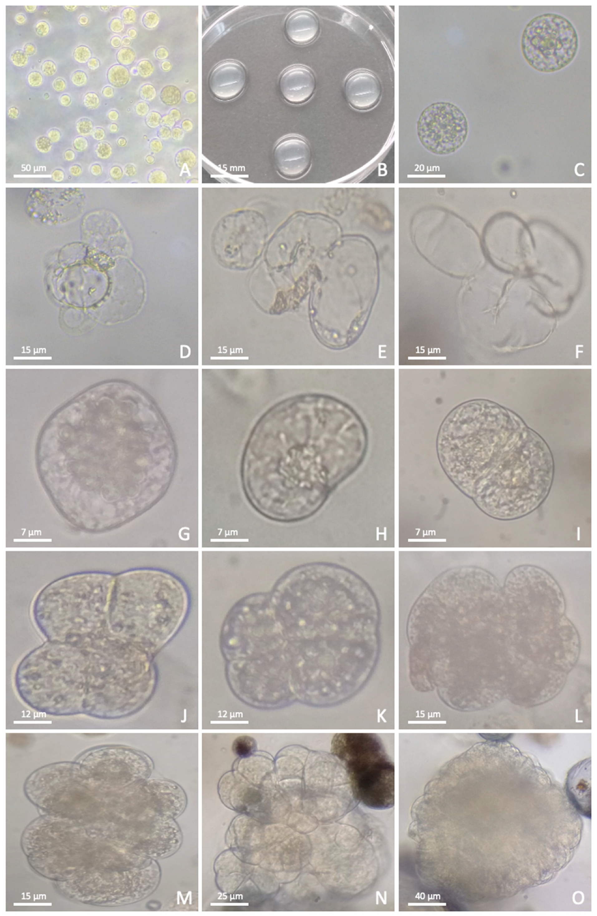
With the optimized protocol, large numbers of viable protoplasts could be isolated from the ECS (Figure 2A). Regeneration of plants from some of these protoplasts was achieved by embedding them into agarose beads immersed in a thin layer of liquid medium (PCM-8G) (Figure 2B,C), which was refreshed weekly by removing 1 mL of the spent medium and replacing it with the same volume of PCM-2G until the protoplasts developed new cell walls and divided to form cell aggregates. At this stage of development, different medium (PCM-E) was used to promote callus formation and eventually embryos and plants.
In general, the protoplast population was heterogenous and consisted of two types, designated Type I and II. Type I protoplasts had less cytoplasmic contents than Type II and divided rapidly to form colonies with large cells but did not form regenerable calli (Figure 2D–F). These types did not proceed to the micro-colony stage. Type II protoplasts contained dense cytoplasm and divided slowly to form dense colonies with smaller cells (Figure 2G–O). Some of these colonies formed embryogenic calli, which subsequently formed embryos on RM. In all protoplast preparations, the percentage of Type II protoplasts was significantly lower than Type I.
Cell wall formation and division were nonsynchronous in Type II protoplasts. Three days after initial culture, many protoplasts became irregular in shape, indicating that cell wall formation had started, and this continued for at least two weeks (Figure 2G). Between the second and third weeks, protoplasts were at the first and second stages of cell division (Figure 2H,I), progressing from two cells to tetrads (Figure 2J,K) and rapidly multiplying to clumps of 10–20 cells (Figure 2M). By four weeks, micro-colonies were observed (Figure 2N), the growth of which expedited to macro-colonies (Figure 2O), which were visible to the naked eye after 4 weeks on PCM-E containing IAA (0.5 mg/L), NAA (0.5 mg/L), 2,4-D (0.1 mg/L), zeatin (1 mg/L), Trp (10 µM), Pro (10 µM) and NaHSO3 (0.1 mM). By the end of the fourth week in PCM-E, many macro-colonies were visible (Figure 2O).
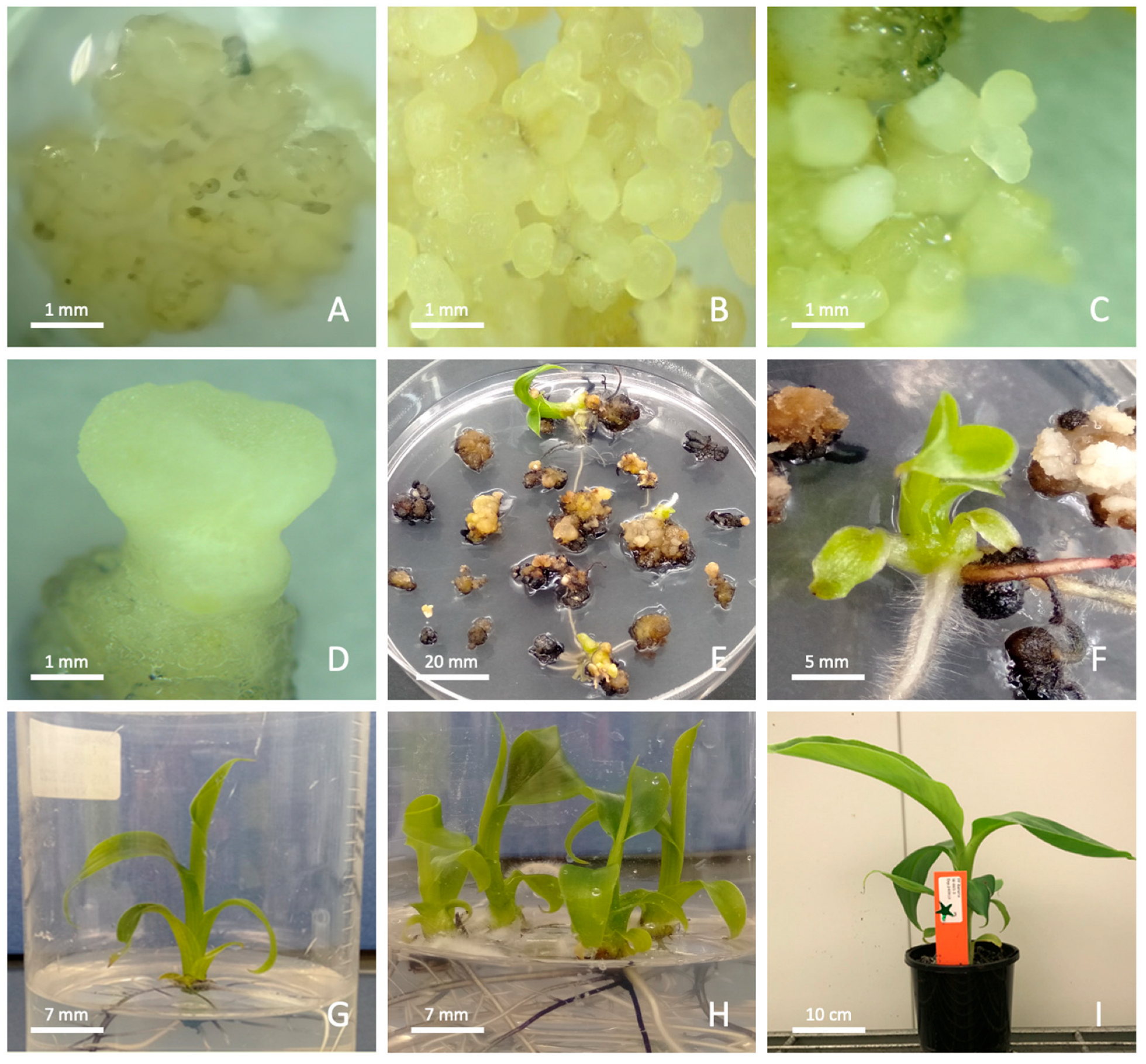
For embryo formation, germination and plant regeneration, the macro-colonies were removed from the agarose beads using forceps and transferred onto Whatman filter papers overlaid on agarose-solidified RM-I, then cultured for 4–6 weeks for callus and embryo formation and maturation at 25 °C under light. The colonies subsequently developed into mature calli, and the color changed from translucent to a cream color typical of embryogenic callus (Figure 3A). Globular embryos were observed to form on this medium within 3–4 weeks and were considered mature by 6 weeks on RM-I (Figure 3B–D).
To determine whether these embryos could regenerate into plantlets, 60 matured embryos were transferred onto RM-II in 90 × 25 mm Petri dishes. After 3 weeks, all embryos started to germinate by simultaneously forming roots and shoots (Figure 3E,F). Twenty fully germinated embryos (Figure 3F) were transferred into larger culture vessels containing hormone-free MS medium for plant development (Figure 3G,H). Fully rooted plantlets were regenerated after seven months, and plants were acclimatized in soil. The plants were grown in a growth cabinet for seven weeks and all appeared to be phenotypically normal (Figure 3I).

3.3. Transfection

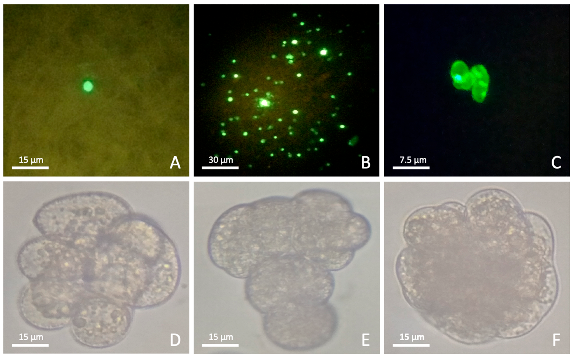
Following the establishment of the plant regeneration protocol, the competency of the freshly isolated protoplasts for PEG-mediated transfection was assessed using the gfp reporter gene. Between 24 and 120 h post-transfection, the number of protoplasts expressing GFP per 1 × 10⁶ transfected cells was as low as 1 to 5 (Figure 4A). Therefore, several variables were tested to optimize the transfection efficiency.
Initially, reducing the incubation time from 5 min to 1 min following addition of the plasmid DNA (pDNA) to the protoplasts (prior to the addition of PEG-3350) improved the frequency of transfection based on an increase in the number of green fluorescing protoplasts (Table 3). Both the average molecular weight and concentration of PEG also had an observable effect on transfection efficiency. Low-molecular-weight PEG-3350 gave higher transfection efficiencies than PEG-4000; therefore, two concentrations of PEG-3350 were subsequently tested with 10% (w/v) providing the best outcome. The frequency of transfection decreased when the protoplast incubation time with PEG was increased from 5 to 10 min. Transfection efficiency was also higher with PEG dissolved in MaMg buffer when compared with CaCl2 buffer. Similarly, MaMg buffer prepared at pH 5.6 enhanced transfection when compared with pH 7.0. Following transfection, PEG was diluted out by rinsing the protoplasts with PWB (Table 1). A dilution factor of 40× improved the recovery of transfected protoplasts as well as the frequency of transfection when compared with a 20× dilution. Transfection experiments carried out in 5 mL Eppendorf tubes resulted in a higher frequency of transfected protoplasts compared with transfection experiments carried out using 15 mL Falcon tubes. As with plant regeneration, reducing the subculture cycle of the ECS from 10 days to 7 days again improved the outcome. Following digestion, a 60 min recovery period at RT was more beneficial to the efficiency of transfection than a 30 min incubation on ice. The influence of a temperature shock just prior to transfection was then tested on RT protoplasts, with 5 min exposure to a cold shock (ice) having the larger positive influence on transfection efficiency than a 5 min 45 °C heat shock.
Finally, no observable difference in transfection efficiency was observed when protoplasts were cultured on either liquid or solid medium. The optimized parameters (Table 3) were combined in subsequent transfections, which resulted in a transfection efficiency of 0.75% based on observed green fluorescing foci 120 h post transfection (Figure 4B).

3.4. Culture of Transfected Protoplasts

Initially, protoplasts were cultured on the same day following transfection, but this resulted in a very low transfection efficiency. Consequently, transfected protoplasts were allowed to recover for 1, 2 and 3 days at two different temperatures, namely 10 °C and 22–23 °C (temperature used during cell wall digestion). After incubation at different temperatures, transfected protoplasts were embedded into agarose and cultured in small beads as described above. The highest frequency of transfection (Figure 4B) and micro-colony formation was achieved when the transfected protoplasts were allowed to recover for 3 days at 10 °C (Figure 4C–F). However, none of the micro-colonies advanced to the calli stage; hence, no plants could be regenerated from the transfected protoplasts.

4. Discussion

Gene editing using CRISPR/Cas9 technology offers a transformative approach to enhancing agronomic traits in popular vegetatively propagated food crops such as bananas. Since integrated DNA cannot be bred out in triploid bananas following editing because of sterility, the use of transient gene editing is a promising alternative. This may enable the generation of plants that are devoid of any foreign DNA, which in some jurisdictions circumvents the regulatory impost associated with genetically modified plants. Protoplasts, being single cells capable of regenerating into uniform, non-chimeric plants, represent an ideal target for such gene editing endeavors. This research aimed to establish a reliable protocol for the generation and transfection of Cavendish banana protoplasts, a cultivar of paramount commercial significance globally. Through careful optimization of the conditions necessary for the growth and transfection of Cavendish protoplasts, this study resulted in a robust protocol to regenerate wild-type Cavendish plants from individual protoplasts. This advance paves the way for applying transient gene editing to Cavendish bananas, facilitating targeted genetic enhancements within this crucial cultivar group.

4.1. Protoplast Isolation

Trialing published protoplast isolation methods [22,23,24,25] in this study resulted in low protoplast yields and cell viability. To increase protoplast populations and improve survival, a range of parameters were examined and optimized. Both the yield and viability were impacted by modifying the combination and concentration of enzymes used. Cellulase (1%) and macerozyme (1%) were identified as the best enzyme combination for effectively degrading cell walls and generating protoplasts. Although pectinase has been reported to give high protoplast yields in other plant species [45,46], our results are consistent with previous work in bananas [34], where pectinase was ineffective for the isolation of protoplasts. Although the addition of driselase did not have a negative impact on protoplast yield (Figure 1A), this enzyme mixture contains xylanase, which has been shown to have detrimental effects on rice protoplasts [47] and therefore was not used for subsequent protoplast preparations to prevent its possible negative impact on banana protoplast regeneration.
The use of AOM to combat the effect of phenolics has been reported previously [42,48]. Antioxidants including ascorbic acid, citric acid, cysteine and sodium bisulfite have all been used to facilitate cell wall digestibility and increase protoplast yield and viability [42,48]. Here, the addition of AOM in the culture media significantly increased both the number and viability of protoplasts. Modification of the preculture stage (prior to plasmolysis) to include AOM for the entire three days following subculture (as opposed to a 1 h treatment prior to cell wall digestion) also significantly enhanced the yield, likely resulting from the inhibition of phenolics.
As a protective measure to prevent the digestion of protoplast membranes, BSA can be added to the enzyme digestion mixture [49]. In the current study, the effect of BSA at three different concentrations in the enzyme mixture was assessed on the protoplast yield, with 0.01% of BSA shown to be the most effective. A reduction in endogenous auxins can cause cells to lose their ability to develop into embryos [50]. By supplementing the culture media with exogenous IAA, NAA and 2,4-D as well as Trp, a precursor of IAA [51], a high level of endogenous IAA can be maintained in the protoplasts [52]. The use of these chemicals in the present study also improved the yield and viability of banana protoplasts. Polyphenol oxidase (PPO) activity in response to wounding of cell walls is a known plant defense strategy [48]. As a protective measure to block the activity of PPOs in protoplasts, AOM was added at each stage of preculture, protoplast isolation and protoplast culture.
The protoplast density was determined using a hemocytometer and viability assessed by the microscopic observation of cytoplasmic streaming [43]. This method was preferred over flow cytometry and chemical staining because it allows the real-time monitoring of living protoplasts without introducing chemical agents that could alter their metabolic state. Additionally, it avoids potential false negatives that may arise from FDA leakage through partially compromised membranes [53].

4.2. Protoplast Regeneration

Several published protocols for banana protoplast culture were initially evaluated [22,23,24,25,26]. However, when Cavendish banana protoplasts were cultured on nitrocellulose membranes placed over nurse cell-enriched medium, no growth was observed, and persistent contamination with endophytes was a recurring issue. Embedding the nurse cells in agarose did not resolve the problem, as endophytes continued to colonize the medium and ultimately overran the protoplast cultures. Therefore, culturing of protoplasts in agarose beads was trialed. The use of bead culture reduced aggregation of the cells and minimized shaking-associated damage in liquid cultures. Further, removing nurse cells and incorporating sterile conditioned medium negated endogenous contamination.
Using the protocol developed in the present study, two clear types of protoplasts were observed to develop. Type I protoplasts had reduced cytoplasmic components and failed to divide and regenerate, whereas Type II protoplasts began to divide after two weeks of growth and regenerated successfully into whole plants. Of 60 germinated embryos, 20 were selected and all regenerated into plants, all of which appeared to be phenotypically normal. Although the overall frequency of protoplasts regenerating was very low (less than 1%), this study demonstrates the successful regeneration of Cavendish banana protoplasts into whole plants using agarose bead cultivation in conditioned growth media.

4.3. Protoplast Transfection

Approaches using either electroporation or PEG-mediated transfection of banana protoplasts have previously been reported, with electroporation known to significantly reduce cell viability [16,19,33,34]. For this reason, the present study focused on PEG-mediated transfection and optimization of critical parameters which influence transfection success. PEG-mediated protoplast transfection has been reported in several plant species, typically using a PEG concentration between 20 and 40% [34,41]. In this study, the effective concentration of PEG-3350 that produced the highest frequency of transfection was 10%. The ideal PEG incubation period was also established to be 5 min. Transfection efficiency drastically reduced as the incubation time was increased. Although PEG incubation for 15 min has previously resulted in a very high transfection frequency in Cavendish cv. Williams [34], this was not the case with the Williams protoplasts used in our study. These differences could be due to the source material used to prepare the protoplasts. ECS was used in this study, whereas the previous researchers used in vitro grown suckers. PEG is an osmoticum which induces osmotic stress in cells [54], and when introduced to protoplasts, it results in plasmolysis and aggregation, which are exacerbated by prolonged incubation. This may damage the cell membrane and protoplast viability. Different plant species clearly have varying degrees of tolerance to the concentration or exposure time used in transfection with PEG. Dilution of PEG after transfection was also found to be crucial. Inadequate dilution prevented the protoplasts from becoming fully turgid, which also decreased the transfection efficiency. Other important factors optimized for the Cavendish protoplasts were the level of plasmid DNA and the recovery stage following transfection. Our results show that three days of recovery at 4–10 °C produced the highest frequency of GFP expressing protoplasts.
Protoplast transfection has been reported in a number of important crops such as Brassica napus [55], chickpea [56], maize [57] and banana [19,33,34]; however, plant regeneration from transfected protoplasts in these crops has not yet been reported. In this study, transfected protoplasts divided and formed micro-colonies, which has not been achieved previously. However, additional research is still required to establish a protocol for the regeneration of whole plants from this stage of growth after the transfection of Cavendish banana protoplasts.

5. Conclusions

Protoplasts are an ideal target for gene editing as whole plants can be regenerated from single cells. Here, we report on the development of an efficient plant regeneration system from protoplasts for Cavendish banana, which comprises approximately half of world production. A range of variables affecting PEG-mediated transient transfection of Cavendish protoplasts were assessed, and an optimized method was demonstrated using GFP, with 0.75% of transfected protoplasts showing expression at five days post-transfection. This optimized protocol will facilitate future efforts to develop a transient transfection and regeneration protocol for developing Cavendish bananas with improved traits.

Supplementary Materials

The following supporting information can be downloaded at https://www.mdpi.com/article/10.3390/applbiosci4030042/s1, Video S1: Cytoplasmic streaming in a healthy viable banana protoplast.

Author Contributions

All authors contributed to the study conception and design. All experiments, data collection and analysis were performed by P.C.D. The initial draft of the manuscript was written by P.C.D., and all authors contributed to revision of the manuscript. All authors have read and agreed to the published version of the manuscript.

Funding

This work was funded by Horticulture Innovation Australia Limited.

Acknowledgments

The authors would like to acknowledge Jen Kleidon and Anthony Brinin for providing ECS and Kirsten Kenny, Georgie Stephan, Amba Phillips and Ben Dugdale for preparing plasmid DNA for transfections.

Conflicts of Interest

The authors declare no conflicts of interest.

References

  1. Voora, V.; Larrea, C.; Bermudez, S. Global Market Report: Banana Prices and Sustainability. 2023. Available online: https://www.iisd.org/system/files/2023-03/2023-global-market-report-banana.pdf (accessed on 6 February 2024).
  2. Varma, V.; Bebber, D.P. Climate Change Impacts on Banana Yields around the World. Nat. Clim. Change 2019, 9, 752–757. [Google Scholar] [CrossRef] [PubMed]
  3. Drenth, A.; Kema, G. The Vulnerability of Bananas to Globally Emerging Disease Threats. Phytopathology 2021, 111, 2146–2161. [Google Scholar] [CrossRef]
  4. Khanna, H.K.; Deo, P.C. Novel Gene Transfer Technologies. In Banana: Genomics and Transgenic Approaches for Genetic Improvement; Mohandas, S., Ravishankar, K.V., Eds.; Springer Singapore: Singapore, 2016; pp. 127–140. [Google Scholar] [CrossRef]
  5. Tripathi, L.; Tripathi, J.N.; Kiggundu, A.; Korie, S.; Shotkoski, F.; Tushemereirwe, W.K. Field Trial of Xanthomonas Wilt Disease-Resistant Bananas in East Africa. Nat. Biotechnol. 2014, 32, 868–870. [Google Scholar] [CrossRef]
  6. Tripathi, L.; Ntui, V.O.; Tripathi, J.N. Control of Bacterial Diseases of Banana Using CRISPR/Cas-Based Gene Editing. Int. J. Mol. Sci. 2022, 23, 3619. [Google Scholar] [CrossRef]
  7. Dale, J.; James, A.; Paul, J.-Y.; Khanna, H.; Smith, M.; Peraza-Echeverria, S.; Garcia-Bastidas, F.; Kema, G.; Waterhouse, P.; Mengersen, K.; et al. Transgenic Cavendish Bananas with Resistance to Fusarium Wilt Tropical Race 4. Nat. Commun. 2017, 8, 1496. [Google Scholar] [CrossRef]
  8. Paul, J.; Khanna, H.; Kleidon, J.; Hoang, P.; Geijskes, J.; Daniells, J.; Zaplin, E.; Rosenberg, Y.; James, A.; Mlalazi, B.; et al. Golden Bananas in the Field: Elevated Fruit Pro-vitamin A from the Expression of a Single Banana Transgene. Plant Biotechnol. J. 2017, 15, 520–532. [Google Scholar] [CrossRef] [PubMed]
  9. Kaur, N.; Alok, A.; Shivani; Kaur, N.; Pandey, P.; Awasthi, P.; Tiwari, S. CRISPR/Cas9-Mediated Efficient Editing in Phytoene Desaturase (PDS) Demonstrates Precise Manipulation in Banana cv. Rasthali Genome. Funct. Integr. Genom. 2018, 18, 89–99. [Google Scholar] [CrossRef] [PubMed]
  10. Kaur, N.; Alok, A.; Shivani; Kumar, P.; Kaur, N.; Awasthi, P.; Chaturvedi, S.; Pandey, P.; Pandey, A.; Pandey, A.K.; et al. CRISPR/Cas9 Directed Editing of Lycopene Epsilon-Cyclase Modulates Metabolic Flux for β-Carotene Biosynthesis in Banana Fruit. Metab. Eng. 2020, 59, 76–86. [Google Scholar] [CrossRef]
  11. Tripathi, L.; Ntui, V.O.; Tripathi, J.N. CRISPR/Cas9-Based Genome Editing of Banana for Disease Resistance. Curr. Opin. Plant Biol. 2020, 56, 118–126. [Google Scholar] [CrossRef]
  12. Tripathi, J.N.; Ntui, V.O.; Shah, T.; Tripathi, L. CRISPR/Cas9-mediated Editing of DMR6 Orthologue in Banana (Musa spp.) Confers Enhanced Resistance to Bacterial Disease. Plant Biotechnol. J. 2021, 19, 1291–1293. [Google Scholar] [CrossRef] [PubMed]
  13. Shao, X.; Wu, S.; Dou, T.; Zhu, H.; Hu, C.; Huo, H.; He, W.; Deng, G.; Sheng, O.; Bi, F.; et al. Using CRISPR/Cas9 Genome Editing System to Create MaGA20ox2 Gene-modified Semi-dwarf Banana. Plant Biotechnol. J. 2020, 18, 17–19. [Google Scholar] [CrossRef]
  14. Hu, C.; Sheng, O.; Deng, G.; He, W.; Dong, T.; Yang, Q.; Dou, T.; Li, C.; Gao, H.; Liu, S.; et al. CRISPR/Cas9-mediated Genome Editing of MaACO1 (Aminocyclopropane-1-carboxylate Oxidase 1) Promotes the Shelf Life of Banana Fruit. Plant Biotechnol. J. 2021, 19, 654–656. [Google Scholar] [CrossRef]
  15. Ntui, V.O.; Tripathi, J.N.; Shah, T.; Tripathi, L. Targeted Knockout of Early Nodulin-like 3 (MusaENODL3) Gene in Banana Reveals Its Function in Resistance to Xanthomonas Wilt Disease. Plant Biotechnol. J. 2024, 22, 1101–1112. [Google Scholar] [CrossRef]
  16. Awasthi, P.; Khan, S.; Lakhani, H.; Chaturvedi, S.; Shivani; Kaur, N.; Singh, J.; Kesarwani, A.K.; Tiwari, S. Transgene-Free Genome Editing Supports CCD4 Role as a Negative Regulator of β-Carotene in Banana. J. Exp. Bot. 2022, 73, 3401–3416. [Google Scholar] [CrossRef]
  17. Yue, J.-J.; Yuan, J.-L.; Wu, F.-H.; Yuan, Y.-H.; Cheng, Q.-W.; Hsu, C.-T.; Lin, C.-S. Protoplasts: From Isolation to CRISPR/Cas Genome Editing Application. Front. Genome Ed. 2021, 3, 717017. [Google Scholar] [CrossRef]
  18. Liang, Z.; Chen, K.; Li, T.; Zhang, Y.; Wang, Y.; Zhao, Q.; Liu, J.; Zhang, H.; Liu, C.; Ran, Y.; et al. Efficient DNA-Free Genome Editing of Bread Wheat Using CRISPR/Cas9 Ribonucleoprotein Complexes. Nat. Commun. 2017, 8, 14261. [Google Scholar] [CrossRef] [PubMed]
  19. Wu, S.; Zhu, H.; Liu, J.; Yang, Q.; Shao, X.; Bi, F.; Hu, C.; Huo, H.; Chen, K.; Yi, G. Establishment of a PEG-Mediated Protoplast Transformation System Based on DNA and CRISPR/Cas9 Ribonucleoprotein Complexes for Banana. BMC Plant Biol. 2020, 20, 425. [Google Scholar] [CrossRef] [PubMed]
  20. Demirer, G.S.; Zhang, H.; Goh, N.S.; González-Grandío, E.; Landry, M.P. Carbon Nanotube–Mediated DNA Delivery without Transgene Integration in Intact Plants. Nat. Protoc. 2019, 14, 2954–2971. [Google Scholar] [CrossRef] [PubMed]
  21. Demirer, G.S.; Zhang, H.; Matos, J.L.; Goh, N.S.; Cunningham, F.J.; Sung, Y.; Chang, R.; Aditham, A.J.; Chio, L.; Cho, M.-J.; et al. High Aspect Ratio Nanomaterials Enable Delivery of Functional Genetic Material without DNA Integration in Mature Plants. Nat. Nanotechnol. 2019, 14, 456–464. [Google Scholar] [CrossRef]
  22. Panis, B.; Van Wauwe, A.; Swennen, R. Plant Regeneration through Direct Somatic Embryogenesis from Protoplasts of Banana (Musa spp.). Plant Cell Rep. 1993, 12, 403–407. [Google Scholar] [CrossRef]
  23. Assani, A.; Haicour, R.; Wenzel, G.; Côte, F.; Bakry, F.; Foroughi-Wehr, B.; Ducreux, G.; Aguillar, M.-E.; Grapin, A. Plant Regeneration from Protoplasts of Dessert Banana cv. Grande Naine (Musa spp., Cavendish Sub-Group AAA) via Somatic Embryogenesis. Plant Cell Rep. 2001, 20, 482–488. [Google Scholar] [CrossRef]
  24. Assani, A.; Haïcour, R.; Wenzel, G.; Foroughi-Wehr, B.; Bakry, F.; Côte, F.-X.; Ducreux, G.; Ambroise, A.; Grapin, A. Influence of Donor Material and Genotype on Protoplast Regeneration in Banana and Plantain Cultivars (Musa spp.). Plant Sci. 2002, 162, 355–362. [Google Scholar] [CrossRef]
  25. Haïcour, R.; Assani, A.; BuiTrang, V.; Guedira, A. Protoplast Isolation and Culture for Banana Regeneration via Somatic Embryogenesis. Fruits 2009, 64, 261–269. [Google Scholar] [CrossRef]
  26. Dai, X.-M.; Xiao, W.; Huang, X.; Zhao, J.-T.; Chen, Y.-F.; Huang, X.-L. Plant Regeneration from Embryogenic Cell Suspensions and Protoplasts of Dessert Banana cv. ‘Da Jiao’ (Musa paradisiaca ABB Linn.) via Somatic Embryogenesis. Vitr. Cell. Dev. Biol. Plant 2010, 46, 403–410. [Google Scholar] [CrossRef]
  27. Panis, B.; Sagi, L.; Swennen, R. Regeneration of Plants from Protoplasts of Musa Species (Banana). In Plant Protoplasts and Genetic Engineering V; Bajaj, Y.P.S., Ed.; Springer: Berlin/Heidelberg, Germany, 1994; Volume 29, pp. 102–114. [Google Scholar] [CrossRef]
  28. Dowling, P.; Clynes, M. Conditioned Media from Cell Lines: A Complementary Model to Clinical Specimens for the Discovery of Disease-specific Biomarkers. Proteomics 2011, 11, 794–804. [Google Scholar] [CrossRef]
  29. Couillerot, J.-P.; Windels, D.; Vazquez, F.; Michalski, J.-C.; Hilbert, J.-L.; Blervacq, A.-S. Pretreatments, Conditioned Medium and Co-Culture Increase the Incidence of Somatic Embryogenesis of Different Cichorium Species. Plant Signal. Behav. 2012, 7, 121–131. [Google Scholar] [CrossRef]
  30. Deryckere, D.; Eeckhaut, T.; Van Huylenbroeck, J.; Van Bockstaele, E. Low Melting Point Agarose Beads as a Standard Method for Plantlet Regeneration from Protoplasts within the Cichorium Genus. Plant Cell Rep. 2012, 31, 2261–2269. [Google Scholar] [CrossRef]
  31. Kiełkowska, A.; Adamus, A. An Alginate-Layer Technique for Culture of Brassica oleracea L. Protoplasts. Vitr. Cell. Dev. Biol.-Plant 2012, 48, 265–273. [Google Scholar] [CrossRef] [PubMed]
  32. Maćkowska, K.; Jarosz, A.; Grzebelus, E. Plant Regeneration from Leaf-Derived Protoplasts within the Daucus Genus: Effect of Different Conditions in Alginate Embedding and Phytosulfokine Application. Plant Cell Tiss. Organ. Cult. 2014, 117, 241–252. [Google Scholar] [CrossRef]
  33. Sagi, L.; Remy, S.; Panis, B.; Swennen, R.; Volckaert, G. Transient Gene Expression in Electroporated Banana (Musa spp., cv. Bluggoe, ABB Group) Protoplasts Isolated from Regenerable Embryogenetic Cell Suspensions. Plant Cell Rep. 1994, 13, 262–266. [Google Scholar] [CrossRef]
  34. Zhao, C.; Li, S.; Du, C.; Gao, H.; Yang, D.; Fu, G.; Cui, H. Establishment of a Protoplasts-Based Transient Expression System in Banana (Musa spp.). Agronomy 2022, 12, 2648. [Google Scholar] [CrossRef]
  35. Morel, G.; Wetmore, R.H. Fern Callus Tissue Culture. Am. J. Bot. 1951, 38, 141–143. [Google Scholar] [CrossRef]
  36. Murashige, T.; Skoog, F. A Revised Medium for Rapid Growth and Bio Assays with Tobacco Tissue Cultures. Physiol. Plant. 1962, 15, 473–497. [Google Scholar] [CrossRef]
  37. Schenk, R.U.; Hildebrandt, A.C. Medium and Techniques for Induction and Growth of Monocotyledonous and Dicotyledonous Plant Cell Cultures. Can. J. Bot. 1972, 50, 199–204. [Google Scholar] [CrossRef]
  38. Chu, C.C.; Wang, C.C.; Sun, C.S.; Hsu, C.; Yin, K.C.; Chu, C.Y.; Bi, F.Y. Establishment of an Efficient Medium for Anther Culture of Rice Through Comparative Experiments on the Nitrogen Sources. Sci. Sin. 1975, 18, 659–669. [Google Scholar]
  39. Kao, K.N.; Michayluk, M.R. Nutritional Requirements for Growth of Vicia Hajastana Cells and Protoplasts at a Very Low Population Density in Liquid Media. Planta 1975, 126, 105–110. [Google Scholar] [CrossRef]
  40. Becker, D.K.; Dale, J.L. Transformation of Banana Using Microprojectile Bombardment. In Transgenic Crops of the World; Curtis, I.S., Ed.; Springer: Dordrecht, The Netherlands, 2004; pp. 131–143. [Google Scholar] [CrossRef]
  41. Yoo, S.-D.; Cho, Y.-H.; Sheen, J. Arabidopsis Mesophyll Protoplasts: A Versatile Cell System for Transient Gene Expression Analysis. Nat. Protoc. 2007, 2, 1565–1572. [Google Scholar] [CrossRef]
  42. Tricoli, D.M. Interim Progress Report for CDFA Agreement Number # 18-0397 Grape Protoplast Isolation and Regeneration of Plants for Use in Gene Editing Technology Project. Available online: https://www.semanticscholar.org/paper/6ee0998387de581fea1820aab88aae247051d7fb (accessed on 8 February 2024).
  43. Verchot-Lubicz, J.; Goldstein, R.E. Cytoplasmic Streaming Enables the Distribution of Molecules and Vesicles in Large Plant Cells. Protoplasma 2010, 240, 99–107. [Google Scholar] [CrossRef]
  44. Kidanemariam, D.B.; Sukal, A.C.; Crew, K.; Jackson, G.V.H.; Abraham, A.D.; Dale, J.L.; Harding, R.M.; James, A.P. Characterization of an Australian Isolate of Taro Bacilliform Virus and Development of an Infectious Clone. Arch. Virol. 2018, 163, 1677–1681. [Google Scholar] [CrossRef] [PubMed]
  45. Chamani, E.; Tahami, S.K.; Zare, N.; Asghari-Zakaria, R.; Mohebodini, M.; Joyce, D. Effect of Different Cellulase and Pectinase Enzyme Treatments on Protoplast Isolation and Viability in Lilium ledebeourii Bioss. Not. Bot. Hort. Agrobot. Cluj. 2012, 40, 123. [Google Scholar] [CrossRef]
  46. Wang, P.; Pu, Y.; Abid, M.A.; Kang, L.; Ye, Y.; Zhang, M.; Liang, C.; Wei, Y.; Zhang, R.; Meng, Z. A Rapid and Efficient Method for Isolation and Transformation of Cotton Callus Protoplast. Int. J. Mol. Sci. 2022, 23, 8368. [Google Scholar] [CrossRef]
  47. Ishii, S. Factors Influencing Protoplast Viability of Suspension-Cultured Rice Cells during Isolation Process. Plant Physiol. 1988, 88, 26–29. [Google Scholar] [CrossRef]
  48. Jones, A.M.P.; Shukla, M.R.; Biswas, G.C.G.; Saxena, P.K. Protoplast-to-Plant Regeneration of American Elm (Ulmus Americana). Protoplasma 2015, 252, 925–931. [Google Scholar] [CrossRef]
  49. Taylor, R.J.; Ruby, C.L.; Secor, G.A. Effects of Enzyme Dialysis, Cellulase Concentration, Bovine Serum Albumin and Origin of Source Leaflet Tissue on Yield and Plating Efficiency of Potato Mesophyll Protoplasts. Biochem. Und Physiol. Der Pflanz. 1989, 185, 227–232. [Google Scholar] [CrossRef]
  50. Ribnicky, D.M.; Ilic, N.; Cohen, J.D.; Cooke, T.J. The Effects of Exogenous Auxins on Endogenous Indole-3-Acetic Acid Metabolism (The Implications for Carrot Somatic Embryogenesis). Plant Physiol. 1996, 112, 549–558. [Google Scholar] [CrossRef]
  51. Cao, X.; Yang, H.; Shang, C.; Ma, S.; Liu, L.; Cheng, J. The Roles of Auxin Biosynthesis YUCCA Gene Family in Plants. Int. J. Mol. Sci. 2019, 20, 6343. [Google Scholar] [CrossRef] [PubMed]
  52. Sakamoto, Y.; Kawamura, A.; Suzuki, T.; Segami, S.; Maeshima, M.; Polyn, S.; De Veylder, L.; Sugimoto, K. Transcriptional Activation of Auxin Biosynthesis Drives Developmental Reprogramming of Differentiated Cells. Plant Cell 2022, 34, 4348–4365. [Google Scholar] [CrossRef]
  53. Papadakis, A.K.; Siminis, C.I.; Roubelakis-Angelakis, K.A. Reduced Activity of Antioxidant Machinery Is Correlated with Suppression of Totipotency in Plant Protoplasts. Plant Physiol. 2001, 126, 434–444. [Google Scholar] [CrossRef] [PubMed]
  54. Fan, S.; Blake, T.J. Comparison of Polyethylene Glycol 3350 Induced Osmotic Stress and Soil Drying for Drought Simulation in Three Woody Species. Trees 1997, 11, 342. [Google Scholar] [CrossRef]
  55. Li, X.; Sandgrind, S.; Moss, O.; Guan, R.; Ivarson, E.; Wang, E.S.; Kanagarajan, S.; Zhu, L.-H. Efficient Protoplast Regeneration Protocol and CRISPR/Cas9-Mediated Editing of Glucosinolate Transporter (GTR) Genes in Rapeseed (Brassica napus L.). Front. Plant Sci. 2021, 12, 680859. [Google Scholar] [CrossRef] [PubMed]
  56. Cheng, N.; Nakata, P.A. Development of a Rapid and Efficient Protoplast Isolation and Transfection Method for Chickpea (Cicer arietinum). MethodsX 2020, 7, 101025. [Google Scholar] [CrossRef] [PubMed]
  57. Hu, Y.; Song, D.; Gao, L.; Ajayo, B.S.; Wang, Y.; Huang, H.; Zhang, J.; Liu, H.; Liu, Y.; Yu, G.; et al. Optimization of Isolation and Transfection Conditions of Maize Endosperm Protoplasts. Plant Methods 2020, 16, 96. [Google Scholar] [CrossRef] [PubMed]
Figure 1. Optimization of protoplast isolation from Cavendish banana (cv. Williams) ECS. (A) effect of enzyme mixtures on the yield of protoplasts derived from ECS. Embryogenic cells were mixed with different combinations and concentrations of enzymes (E1 to E5) and digested for 16–20 h in the dark at 23 °C. Protoplasts were purified by sieving and centrifugation and quantified using a hemocytometer. (BD) Parameters affecting the yield of banana (cv. Williams) ECS-derived protoplasts. (B) Influence of antioxidant mixtures on protoplast yield, where ECS was pre-treated with either AOM or NaHSO3 for 72 h. (C) AOM was included in the ECS preculture media for 1 h or 72 h during plasmolysis and preculture, respectively. (D) Influence of BSA concentration in the enzyme digestion mixture on protoplast yield. Protoplasts were purified from ECS using enzyme mixture E2 and quantified using a hemocytometer. NA, no antioxidants. Values are expressed as the mean ± SEM from three independent experiments. Means followed by the same letter were not significantly different (p < 0.05).
Applbiosci 04 00042 g001
Figure 2. Culture and growth of banana (cv. Williams) ECS-derived Type I and II protoplasts in agarose beads. Freshly isolated protoplasts from ECS (A) were embedded and cultured in 1.2% agarose beads (B) from which individual protoplasts were isolated (C). Type I protoplasts (DF) started to divide with enlarged cells but failed to form viable and regenerable colonies over time. In contrast, type II protoplasts started to form a cell wall (G) and became irregular in shape. Upon completion of cell wall development, the first stage of cell division could be observed (H,I), followed by the second stage, where the cells formed tetrads (J,K), and finally, multicellular aggregates (L). These later formed micro-colonies (M), followed by macro-colonies (N,O), which were visible to the naked eye.
Applbiosci 04 00042 g002
Figure 3. Whole-plant regeneration from protoplast-derived calli. Macro-calli developed into calli, which turned creamy on RM-I (A). After 3–4 weeks of culture, the calli started to form globular somatic embryos (B), which later matured and turned translucent (C,D). Upon transfer to RM-II, the matured embryos started to germinate, forming roots and shoots simultaneously (E,F), and when they reached almost 2 cm in height, they were transferred on MS in 250 mL culture vessels for plant development (G,H). Well-developed plants were acclimatized in soil, monitored for 3 months and all appeared phenotypically normal (I). The time taken from protoplast culture to fully developed plants in soil was 9 months.
Applbiosci 04 00042 g003
Figure 4. PEG-mediated transfection and regeneration in banana (cv. Williams) ECS-derived protoplasts. Protoplasts (1 × 10⁶) were mixed with 10 µg of plasmid DNA containing a 35S-gfp expression cassette and 20% PEG-3350 in MaMg buffer and incubated for 5 min, then diluted with wash buffer. After dilution of PEG, protoplasts were cultured at 4–10 °C for 3 days and then embedded in agarose beads for regeneration. GFP expression was seen at 120 h post-transfection with very low expression initially (A) and very high expression following optimization (B). A very small number of transfected protoplasts continued to divide and express GFP after 3 weeks (C). Most of the transfected protoplasts divided and formed micro-colonies (DF) but failed to form macro-colonies and calli.
Applbiosci 04 00042 g004
Table 1. List of media used for Cavendish banana (cv. Williams) protoplast isolation, purification, transfection, culture and plant regeneration.
Buffers and MediaComponents *
Banana Cell Culture Media (BL)MS macro and micronutrients (1×), MS-Fe-EDTA (1×), sucrose (20 g/L), Bluggoe vitamins (1×), ascorbic acid (50 mg/L), 2,4-D (1.1 mg/L), zeatin (0.25 mg/L), pH 5.7
Protoplast Isolation Buffer (PIB)CaCl2.2H2O (7 mM), NaH2PO4.2H2O (0.7 mM), mannitol (0.55 M), MES (3 mM), pH 5.7
Protoplast Wash Buffer (PWB)KCl (0.4 M), CaCl2.2H2O (0.034 M), mannitol (0.55 M), MES (3.5 mM), pH 5.7
Antioxidant Mix (AOM)Ascorbic acid (50 mg/L), citric acid (5 mg/L), L-cysteine (50 mg/L)
Protoplast Culture Media-8G (PCM-8G)N6 salts (1×), KM vitamins (1×), KM sugars (1×), KM organic acids (1×), KH2PO4 (0.25 g/L), glucose (80 g/L), MES (0.5 mM), pH 5.7
Protoplast Culture Media-2G (PCM-2G)N6 salts (1×), KM vitamins (1×), KM sugars (1×), KM organic acids (1×), KH2PO4 (0.25 g/L), glucose (20 g/L), sucrose (1 g/L), MES (0.5 mM), pH 5.7
Protoplast Culture Medium for Embryogenesis (PCM-E)SH macro and micro salts (½×), MS micronutrients (½×), KM vitamins (½×), sucrose (20 g/L), glucose (2 g/L), MES (0.5 mM), pH 5.7
Regeneration Medium-I (RM-I)MS macro and micronutrients (1×), MS-Fe-EDTA (1×), MW vitamins (1×), sucrose (30 g/L), IAA (0.1 mg/L), BAP (0.5 mg/L), MI (100 mg/L), pH 5.7
Regeneration Medium-II (RM-II)MS macro and micronutrients (1×), MS-Fe-EDTA (1×), MW vitamins (1×), sucrose (30 g/L), BAP (0.05 mg/L), MI (100 mg/L), pH 5.7
MSMS macro and micronutrients (1×), MS-Fe-EDTA (1×), MS vitamins (1×), sucrose (30 g/L), pH 5.7
MaMgMgCl2.6H2O (15 mM), mannitol (0.5 M), MES (4 mM), pH 5.7
PEG-MaMgMgCl2.6H2O (15 mM), mannitol (0.55 M), MES (5.12 mM), pH 5.7, PEG-3350 (20%)
Stop BufferMannitol (0.5 M), KCl (5 mM), MES (4 mM), pH 5.7
* Abbreviations/References: BAP = 6-benzylaminopurine; IAA = indole-3-acetic acid; MES = 2-(N-morpholino)-ethane sulfonic acid; MI = myoinositol; NAA = naphthaleneacetic acid; 1×: full-strength; ½×: half-strength; Bluggoe vitamins [40]; KM = Kao and Michayluk [39]; MS = Murashige and Skoog [36]; MW = Morel and Wetmore [35]; N6 salts [38]; SH = Schenk and Hildebrandt [37].
Table 2. Enzyme mixtures assessed for protoplast isolation from banana ECS.
Enzyme
Combinations
Enzymes (%)
CellulaseMacerozymeDriselasePectinase
E110.500
E21100
E3110.50
E41100.5
E5110.50.5
Table 3. Variables tested to optimize PEG-mediated transfection of banana protoplasts.
Tested VariableComparative Results *
Protoplast and DNA incubation (min)1+++
5-
PEG size (ave. molecular weight)3350+++
4000-
PEG-3350 concentration (%)10+++
20+
PEG-3350 incubation time (min)5+++
10-
PEG-3350 transfection bufferMaMg a+++
CaCl2 b-
PEG-3350 transfection buffer pH5.6+++
7-
PEG-3350 dilution regime20×-
40×+++
Transfection culture vesselFalcon tubes-
Microfuge tubes+++
ECS subculture regime (days)7+++
10+
Protoplast recovery Ice (30 min)-
RT (60 min)+++
Temperature shock (5 min)Ice+++
45 °C+
Protoplast culture post-transfectionLiquid medium+++
Solid medium+++
* (+++), (+) and (-) transfection with more than 5, between 1 and 5 and no GFP-expressing protoplasts, respectively. Data based on observations from 5 independent transfections. a See Table 1; b [41].
Disclaimer/Publisher’s Note: The statements, opinions and data contained in all publications are solely those of the individual author(s) and contributor(s) and not of MDPI and/or the editor(s). MDPI and/or the editor(s) disclaim responsibility for any injury to people or property resulting from any ideas, methods, instructions or products referred to in the content.