Article

Direct Expression of CPT1a Enables a High Throughput Platform for the Discovery of CPT1a Modulators

by
Jason Chen
1,
Tuyen Tran
1,2,
Anthony Wong
2,
Luofei Wang
2,
Pranavi Annaluru
2,
Vibha Sreekanth
2,
Samika Murthy
2,
Laasya Munjeti
2,
Tanya Park
1,
Utkarsh Bhat
2,
Glynnis Leong
2,
Yumeng Li
1,
Simeng Chen
2,
Natalie Kong
1,
Rushika Raval
1,
Yining Xie
1,
Shreya Somani
1,
Aditi Manohar Bhambhani
1,
Zoey Zhu
1,
Landen Chu
1,
Kimai Dosch
1,
Edward Njoo
1,* and
Zhan Chen
1,2,*
add Show full author list remove Hide full author list
1
Department of Chemistry, Biochemistry and Physics, Aspiring Scholars Directed Research Program, Fremont, CA 94539, USA
2
Department of Biological, Human and Life Sciences, Aspiring Scholars Directed Research Program, Fremont, CA 94539, USA
*
Authors to whom correspondence should be addressed.
Appl. Biosci. 2025, 4(2), 25; https://doi.org/10.3390/applbiosci4020025
Submission received: 16 December 2024 / Revised: 15 March 2025 / Accepted: 17 April 2025 / Published: 12 May 2025

Abstract

:
Carnitine palmitoyltransferase 1 (CPT1), which catalyzes the rate-limiting step of fatty acid oxidation, has been implicated in therapeutic approaches to several human diseases characterized by aberrant lipid metabolism. The isoform-specific quantification of CPT1 activity is essential in the characterization of small molecule inhibitors of CPT1, but several existing means to quantify enzymatic activity, including the use of radioisotope-labeled carnitine, are not amenable to scalable, high throughput screening. Here, we demonstrate that mitochondrial extracts from Expi293 cells transfected with a CPT1a plasmid are a reliable and robust source of catalytically active human CPT1. Moreover, with a source of catalytically active enzyme in hand, we modified a previously reported colorimetric method of coenzyme A (CoA) easily scalable to a 96-well format for the screening of CPT1a inhibitors. This assay platform was validated by two previously reported inhibitors of CPT1a: R-etomoxir and perhexiline. To further demonstrate the applicability of this method in small molecule screening, we prepared and screened a library of 87 known small molecule APIs, validating the inhibitory effect of chlorpromazine on CPT1.

Graphical Abstract

1. Introduction

Carnitine palmitoyltransferase 1 (CPT1) catalyzes the rate-limiting step of fatty acid oxidation by shuttling long-chain fatty acids across the mitochondrial membrane [1,2]. This enzyme has been demonstrated to be a druggable target for the treatment of type 2 diabetes [3,4,5], cancer [6,7,8,9], obesity [10,11], and inflammation [12,13,14]. Carnitine acyltransferases catalyze the reversible transfer of acyl groups from acyl-coenzyme A (acyl-CoA) esters to L-carnitine, forming acylcarnitine esters [15,16]. The CPT system is made up of two separate proteins: CPT1, located in the outer mitochondrial membrane, is predominantly responsible for the forward acyl transfer to acylcarnitine, and CPT2, located in the inner mitochondrial membrane, catalyzes the hydrolysis of acylcarnitine back to acyl-CoA esters into the mitochondrial lumen [17,18,19].
To date, three distinct and yet closely related isoforms of CPT1 have been identified that are dissimilarly expressed in different tissues. CPT1a is known as the liver isoform and is primarily expressed in the brain, intestine, kidney, lung, ovary, pancreas, and spleen tissues [18,20]. CPT1a is endogenously inhibited by malonyl-CoA, the first intermediate in fatty acid synthesis [21,22]. CPT1b is predominantly found in high abundance in skeletal muscle, the heart, and brown adipose tissue (BAT) [23]. Unlike CPT2, which has a well-established structural basis from X-ray crystallography, no crystal structures of CPT1 exist to date, which further obfuscates efforts to understand the structural basis for small molecule ligands [24,25]. The third isoform, CPT1c, is predominantly expressed in the brain [26,27,28].
Given the clinical significance of fatty acid metabolism regulation in several indications [29,30,31,32], the CPT1 system has been subject to several small molecule inhibitor discovery campaigns [33]. Notably, etomoxir [34,35,36], a lipid-like α-epoxy carboxylate small molecule, has been previously identified as a covalent inhibitor of the active site of CPT1 for the treatment of type 2 diabetes mellitus [37] and chronic heart failure [38], but was withdrawn from late-stage clinical studies due to severe off-target toxicity [39]. Based on such a background, a more selective CPT1 inhibitor named ST1326 (Teglicar) [40,41] was developed as a competitive, reversible, and isoform-selective CPT1 inhibitor with an improved toxicity and PK profile. Teglicar was in phase 2 studies [42] for the treatment of type 2 diabetes and has also shown great potential in the treatment of leukemias [43,44]. Other small molecule inhibitors, including perhexiline [45,46,47,48], dexamethasone [49,50], amiodarone [42,51,52], metoprolol [53,54], trimetazidine [55,56,57], and oxfenicine [58,59,60], have also been described to modulate the activity of the CPT1 system.
The success of such small molecule campaigns in identifying lead compounds requires a sensitive and specific CPT1 activity assay. Traditionally, the catalytic activity of CPT1 has been measured through tritium-labeled L-[3H]carnitine [61,62,63,64,65,66]. While exceptionally sensitive, such tritium-based biochemical assays have inherent limitations in operational safety and scalability given the hazards of tritium waste and limited access to the equipment necessary to measure radiolabeled samples. Additionally, while several immunohistochemical approaches to quantify CPT1a concentration exist, specific immunohistochemical assays to measure the enzyme’s activity are not commercially available. Furthermore, they have limited sensitivity to low quantities of target proteins from cell lysates and thus lack precision in quantifying protein activity and inhibition in the context of limited quantities such as mitochondrial isolation. Immunohistochemical assays are further susceptible to high background interference during detection and protein analysis, diluting the quantification of protein activity [67]. On the other hand, our method offers high specificity to quantify protein activity and the inhibition of isolated mitochondrial fractions to offer an effective method of analyzing inhibitive activity in possible compound libraries. Moreover, given the necessity of being in a mitochondrial-membrane environment for proper CPT1a structure and function, recombinant, commercially available sources of CPT1a are typically catalytically inactive, rendering them useless for inhibitor screening. Radiolabeled assays, another alternative method of quantifying CPT1a inhibition, require extensive control of environmental factors and high insurance of radioactive material distribution. Thus, these assays are not a feasible method of screening new compound libraries for possible inhibitors due to an excessively high background. Radiolabeled assays further demand considerable exposure to radiolabeled material and long periods of fluorogram exposure in order to obtain sufficient signal for measurement, operating over a significantly longer time duration in comparison to our proposed assay that quantifies inhibition solely within the time period of the assay’s execution [68].
Rapid and sensitive spectrophotometric and fluorogenic assays for the detection of carnitine palmitoyltransferase activity by the measurement of released CoA-SH have been previously described [69,70,71,72]. Bieber et al. [69] reported that the amount of reduced CoA liberated from palmitoyl-CoA by CPT is quantitated using 5,5′-dithiobis-(2-nitrobenzoic acid) (DTNB). This assay measures the initial rates of CoA-SH formation by CPT1-catalyzed deacylation of palmitoyl-CoA. Importantly, Bieber et al. demonstrate that this assay method does not require the purification of recombinant human CPT1, but rather crude extracts from Sprague Dawley rat liver could be directly employed as a reliable and standardizable source of human CPT1 [69,73]. While this approach circumvented the isolation instability of CPT1 as is typical of membrane-bound proteins, the in vivo source of mitochondrial extracts described therein is not easily sourceable and also potentially contains mixtures of CPT isoforms among the other enzymes present in fresh liver tissue [74].
To address the limited availability of high throughput screening platforms for the evaluation of CPT1a activity and its inhibitors, we directly expressed CPT1a as a single isoform through the transformation of Expi293 cells and showed that the mitochondrial extracts isolated from in vitro cell culture are a scalable and reliable source of catalytically active CPT1a. Furthermore, we modified a direct, colorimetric CoA detection method for CPT enzyme activity inspired by previous reports from Bieber et al. [69] employing thiol-disulfide exchange from free CoA and DTNB, which leads to the production of 2-thio-5-nitrobenzoic acid (TNB) and an optical readout at 412 nm. We applied this system to the detection of free thiols from the release of CoA from palmitoyl-CoA. This assay detects the concentration of CoA liberated from CPT1 catalysis without the need to purify recombinant human CPT1 and was validated with a series of previously established CPT1 inhibitors. Further, to demonstrate the generalizability of our approach in high throughput small molecule screening campaigns, we applied this platform to screen for potential inhibitors of CPT1 in a sample of 87 small molecules. In accordance with previous reports, chlorpromazine was identified as a part of this high throughput screen to exert inhibitory effects on CPT1 [52,75,76]. The inhibitory effects we viewed in our high throughput screen demonstrate the validity of the modified assay, even when performed in large quantities.

2. Materials and Methods

2.1. Cell Culture

Mammalian Expi293F cells (Gibco, WA, USA, Cat. #A14527) were stably transfected with a human CPT1a expression plasmid with an ExpiFectamine 293 Kit (Gibco, WA, USA, Cat. #A14524) and maintained in Expi293 Expression Medium (Gibco, WA, USA, Cat. #12338018). All the cells were cultured in 250 mL bottles in a humidified incubator at 37 °C in 5% CO2.

2.2. Human CPT1a Enzyme Expression

To construct a human CPT1a expression plasmid, two expression constructions, CPT1A(NM_001876.3) ORF, which contains the whole human CPT1a sequence, and sp_CPT1A(NM_001876.3) ORF, which contains an N-terminal signal peptide for extracellular release, were each cloned into the vector pcDNA3.1+/C-(K)-DYK backbone and purchased from GenScript USA Inc. (Piscataway, NJ, USA). The plasmid was transformed into TOP10 bacteria and amplified. After plasmid extraction and purification, the inserted CPT1a gene was sequenced. After sequencing confirmation, they were transfected into mammalian Expi293 cell lines. After growing the Expi293 culture, the supernatant and cell pellet were lysed with radioimmunoprecipitation assay (RIPA) buffer [25 mM Tris•HCl pH 7.6, 150 mM NaCl, 1% NP-40, 1% sodium deoxycholate, 0.1% SDS] (Thermofisher, Bothell, WA, USA, Cat. #88901). The protein was harvested through differential, two-step centrifugation (modification of Yang et al.) with the remaining mitochondrial pellet suspended in the extraction buffer [77]. Protein concentration was quantified with a Bradford protein assay kit.

2.3. Immunoassay to Quantify CPT1a

ELISA was used to detect the expression level of human CPT1a in the supernatant and pellet and validate the successful transgenic expression of human CPT1a. Briefly, the CPT1a-transfected cell pellet and supernatant, which contained 100 μg of total protein, was coated in a coating buffer (NaHCO3-Na2CO3 buffer, pH = 6.4) that was freshly made and aliquoted into 100 μL per well, and was left at 4 °C for 18 h. Following three washes with PBST (Phosphate-buffered saline with Tween), 200 μL of blocking buffer was added to each well (2.5% w/v non-fat dry milk powder in PBST, 0.1% Tween-20, VWR Life Science, Radnor, PA, USA, Cat. #0777-1L) for 1 h at 18 °C. Following aspiration, mouse anti-human CPT1a monoclonal antibody (Abcam, Fremont, CA, USA, Cat. #8F6AE9) was added in a 1:1000 dilution (100 μL per well) and shaken for 2 h at 18 °C. Following a second wash, the goat-anti-mouse HRP secondary antibody (Sino Biological, Wayne, NE, USA, Cat. #SSA007) was added with the blocking buffer. Subsequently, we added 100 μL of the diluted secondary antibody to each well after washing three times with PBST for 1 h at 18 °C while shaking. Then, 50 μL TMB (3,3′,5,5′-Tetramethylbenzidine) substrate (0.4 g/L, Tribioscience, Sunnyvale, CA, USA, Cat. #TBS5021) was added to each well after washing three times with PBST at 18 °C. A total of 100 μL of stop solution (1 M H2SO4) was then added to each well. We measured absorbance at 450 nm after 10 s of shaking. The Expi293 cell pellet protein was used as an endogenous control.

2.4. CPT Enzyme Activity Assay and Inhibitor Sensitivity Assay

CPT spectrophotometric assay using DTNB was modified from the methods originally reported by Bieber et al. [69]. The reaction master mix was prepared with 35 μM Palmitoyl-CoA (Sigma Aldrich, St. Louis, MO, USA, Cat. #P9716), 1.5 mM EDTA (Acros, Geel, Belgium, Cat. #AC118432500), 40 mM HEPES (ThermoScientific, Waltham, MA, USA, Cat. #15630080), 1.25 mM L-carnitine (AK Scientific, Union City, NJ, USA, Cat. #J94077), and a stock aliquot of the CPT1-containing mitochondrial extract. A total of 100 mM Tris buffer (pH = 7.5) was used to make the final volume 90 μL. The samples were incubated at 30 °C on a Compact Digital Dry Bath/Block Heater (ThermoFisher, Waltham, MA, USA) for 15 min, at which point 10 μL of 1 mg/mL DTNB (AK Scientific, Union City, NJ, USA, Cat. #J92390) in 100 mM Tris buffer was added to each well. The plate was mixed on a plate shaker for 10 min and absorbances were recorded at 412 nm (SpectraMAX, Molecular Devices, San Jose, CA, USA). To correlate the appearance of yellow with the absolute concentration of CoA and to determine the dynamic range of the assay, we prepared a standard curve. To account for a high background and normalize the assay, we subtracted the value of the denatured enzyme from the produced value.
Two previously reported CPT1 inhibitors, etomoxir sodium salt (Cayman Chemical, Ann Arbor, MI, USA, Cat. #11969) and perhexiline (AK Scientific, Union City, NJ, USA, Cat. #X4611), were used to validate the potential of this assay as a platform to screen for novel CPT1 inhibitors. IC50 calculations were performed on the GraphPad Prism 10 software.

2.5. High-Throughput Screening of a Small Molecule Library

To demonstrate the validity of this approach in scalable, high-throughput screening campaigns, 87 small molecule compounds were screened using this platform, with etomoxir as a positive control. The compounds screened were selected from a library of natural products, anti-virals, immunomodulators, and other bioactive compounds. Compounds were diluted to 10 mM in dimethyl sulfoxide (DMSO) (Stellar Chemical, Rahway, NJ, USA, 99.99%) and tested at a final concentration of 0.556 mM. Compounds that exhibited inhibitory effects were run in a serial concentration to create an inhibitory effect curve and determine the IC50.

2.6. Data Analysis

All the data and statistics (mean, SEM, p-values) were analyzed with GraphPad Prism 10 and Excel. The acquired data are presented as mean ± SEM. p-values of 0.05 were considered statistically significant.

3. Results

3.1. Human CPT1a Enzyme Expression and Protein Isolation

Two human CPT1a gene expression constructions, pcDNA3.1+/C-(K)DYK with CPT1A(NM_001876.3) ORF Clone and sp_CPT1A(NM_001876.3) ORF Clone, were sequenced and aligned to Genbank. The sequences are identical to previously deposited sequences on Genbank. The supernatant and cell pellet proteins were harvested separately after transfection into mammalian cell Expi293 as shown in Figure 1. The normal Expi293 cell pellet proteins were harvested as a control.

3.2. CPT1a Immunogenicity Assay

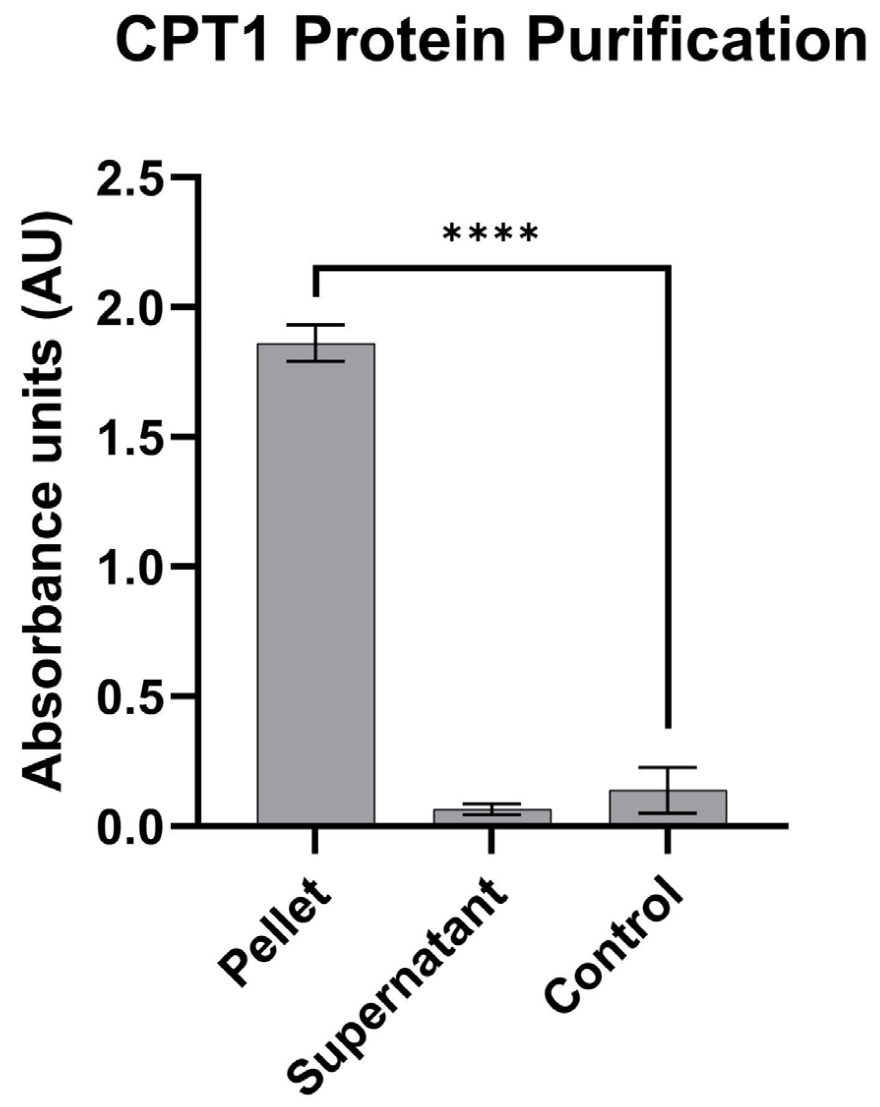
ELISA was used to check the immunity and protein expression level of CPT1a in transfected Expi293 mitochondria. The CPT1 expression level in the CPT1a transgenic cell pellet had a 13-fold increase in the control cell pellet, while minimal CPT1 was detected in the supernatant, as shown in Figure 2. The supernatant concentration of CPT1a in the cells transfected with a signal peptide fusion protein did not exhibit higher extracellular content of CPT1a, suggesting that CPT1a cannot be secreted out of the cell into the supernatant, potentially because it is very tightly bound to the mitochondrial membrane. An anti-human CPT1a monoclonal antibody was used as a mitochondrial marker to ensure localization and quantify the CPT1a protein level within the lysate.

3.3. CPT1 Enzyme Activity Assay Validation

A CoA standard curve with authentic CoA (Cayman Chemical, Ann Arbor, MI, USA, Cat. #16147) was used to quantify the concentration of CoA being released during the catalysis. The enzyme activity calculation formula: Units/mL enzyme = (ΔA412 nm/min Test − ΔA412 nm/min Blank)(3)(df)(0.0136)(0.05), where 3 = Total volume (in milliliters) of assay, df = Dilution Factor, 0.0136 = Micromolar extinction coefficient for TNB at 412 nm, 0.05 = Volume (in milliliters) of enzyme used. As a background, the CPT1a cell pellet has a value of 5.02 ± 0.2 μM.
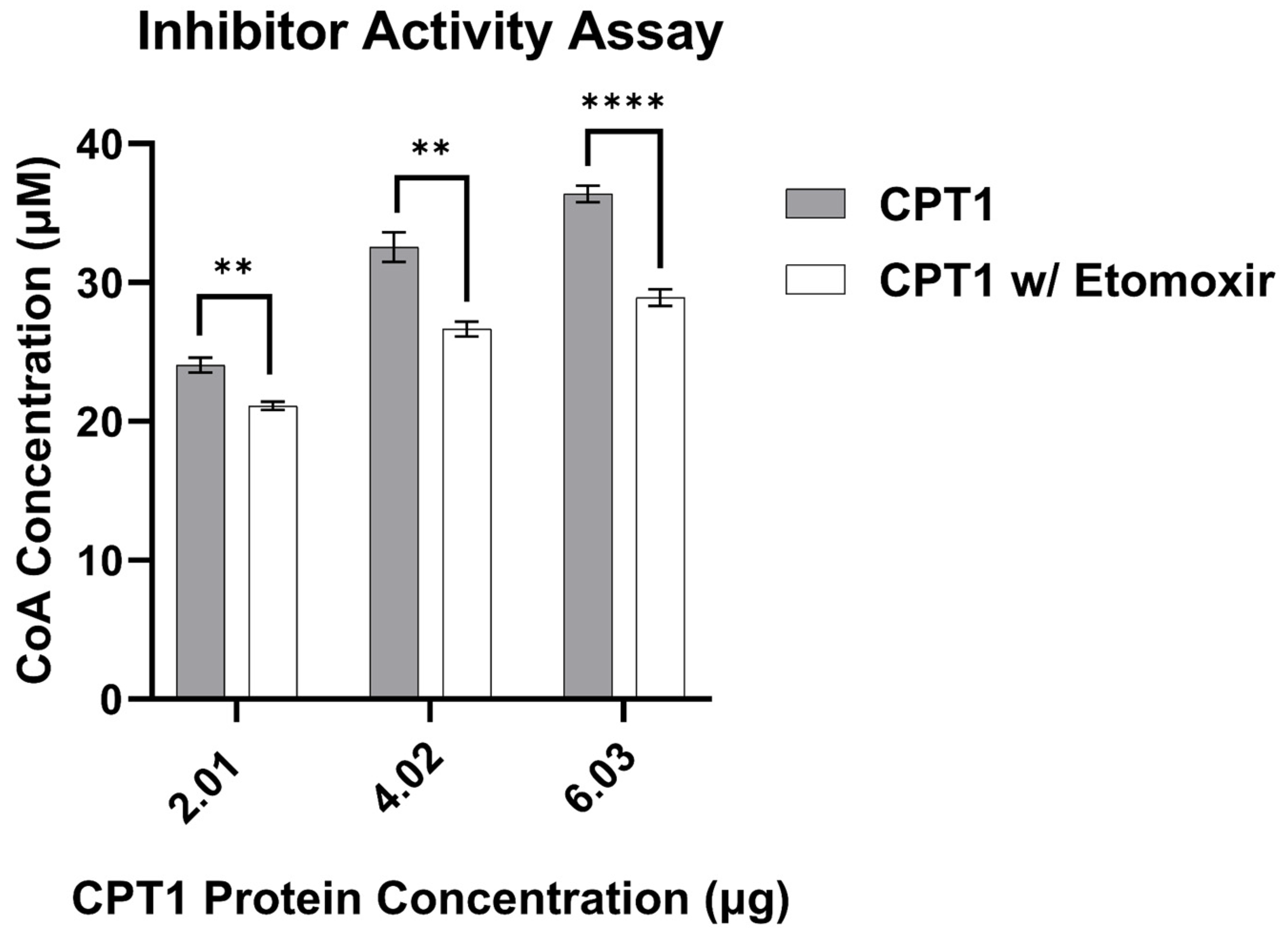
The value of CPT1 enzyme activity is proportional to the concentration of CPT1 enzyme as shown in Figure 3. The inhibition efficacy of etomoxir was calculated by running a serial dilution of etomoxir concentrations in the enzyme activity assay. Replicates of this assay demonstrated nearly identical etomoxir-dependent activity profiles, indicating a high degree of reliability.

3.4. CPT1 Enzyme Inhibitor Sensitivity Assay

We used the CPT1 enzyme inhibition assay to validate the new protocols that were shown in Figure 3 and Figure 4. The inhibition rate IC50 of etomoxir was 123.9 nM (Figure 5), which falls within the known IC50 range of 10–700 nM [78]. Two additional inhibitors, perhexiline and malonyl-CoA, were additionally used to validate this platform.
The inhibition curves of etomoxir, perhexiline, and malonyl-CoA demonstrate that the new CPT1 enzyme activity assay method effectively quantifies the different effects of various inhibitors.

3.5. Application and Performance Qualification to Screen Small Molecule Compounds

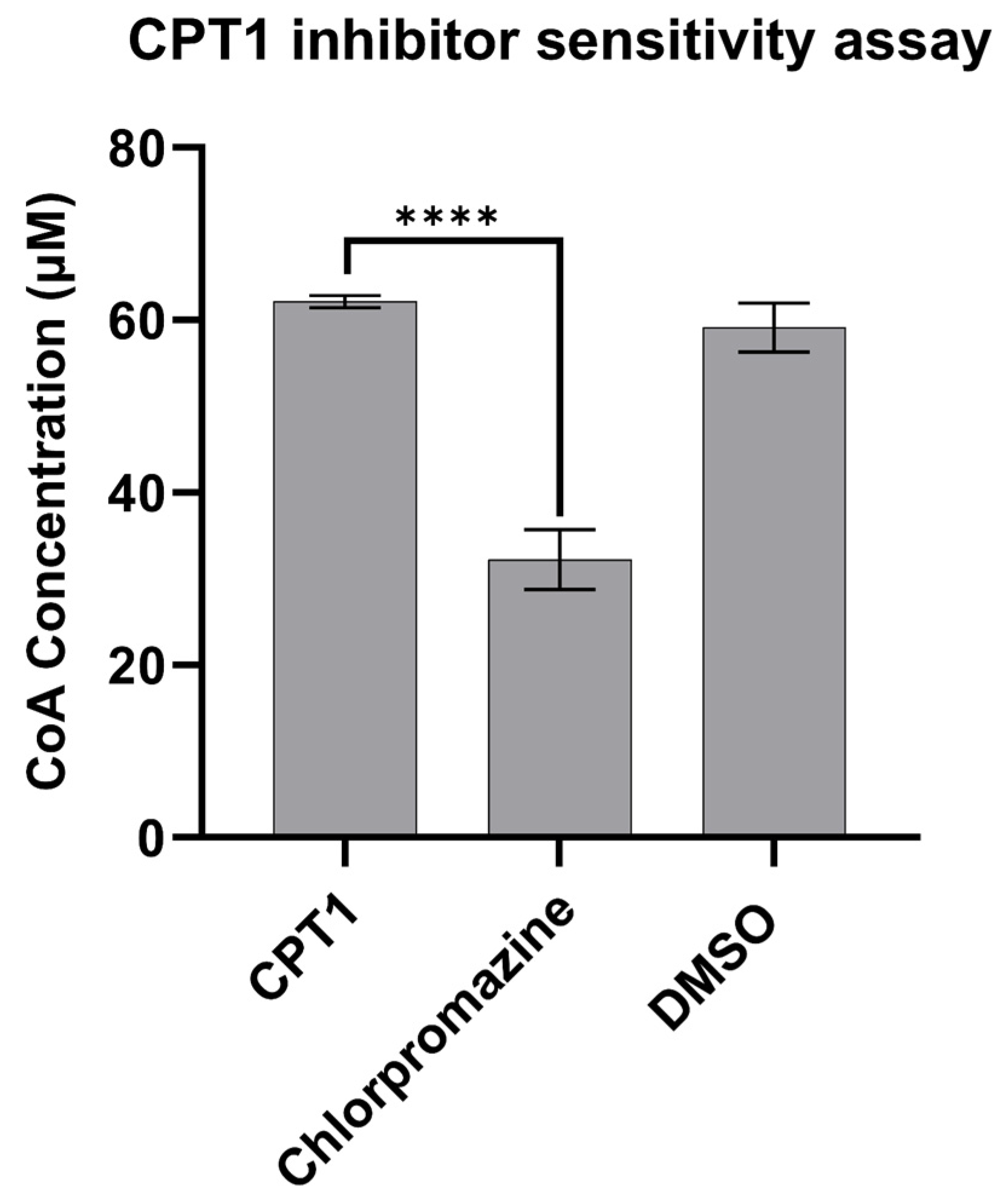
In order to demonstrate the applicability of this system in the high-throughput screening of new CPT1 inhibitors and activators, we prepared and screened a library of 87 small molecule compounds using our new approaches, including several FDA-approved small molecules for different indications, as well as natural products and previously reported preclinical candidates (full panel disclosed in online Supporting Information). From this screen, we validated that chlorpromazine, a previously clinically studied antipsychotic small molecule, exhibits inhibitory activity against CPT1a, as shown in Figure 4 and Figure 5. Its IC50 for CPT1a inhibition is 0.1793 mM.

4. Discussion

CPT1 facilitates the transfer of long-chain fatty acids into the mitochondria for beta-oxidation, and a lack of regulation promotes the development of various human diseases. Therefore, a highly reproducible, cost-effective method of identifying CPT1 inhibitors paves the way for substantial medical breakthroughs in developing potential treatments for type 2 diabetes and cancer. While highly sensitive and accurate radioisotope-based quantification methods exist for the direct measurement of CPT1a activity, safety limitations and limited access to scintillation equipment pose major drawbacks to the applicability and scalability of 3H-carnitine-based assays. Additionally, while commercially available ELISA kits exist for the detection of CPT1 through immunogenicity with poly- or monoclonal antibodies, such methods cannot detect the enzyme activity of CPT1.
In summary, we modified and optimized a highly robust assay for the quantification of CPT1 activity. This method allows for the use of the direct cell lysis of human CPT1-transformed Expi293 cells without the need for purification of recombinant proteins. Further, we optimized the spectrophotometric detection of CoA liberated from CPT1 catalysis using a well-established DTNB-based detection of free thiols, which is a proxy for enzyme activity based on the direct relationship between CPT1 catalytic activity and increasing CoA concentration. We then validated this approach by using previously reported CPT1 inhibitors, etomoxir, perhexiline, and chlorpromazine, and found that this assay method provided highly reproducible and dose-responsive quantification for CPT1 activity.
Our approach overcomes the limitations of isotope-based screening methods and is, thus, suitable for high-throughput applications. Finally, we showed that this assay is easily adaptable to a 96-well format, suggesting the feasibility of performing screenings on large sets of samples, including the potential for automation. The IC50 of chlorpromazine we reported using this assay is consistent with the IC50 trends published in the first report of chlorpromazine as a CPT1 inhibitor, which used the isotope forward exchange method, further demonstrating the validity of this assay even in high throughput formats [75]. Additionally, the IC50 values of etomoxir we reported are consistent with the reported IC50 trends of etomoxir’s nanomolar range, demonstrating the reproducibility of this assay. However, competitive inhibitors such as malonyl-CoA are more challenging with the method reported herein given the high enzyme concentrations utilized. To further assess the consistency of this high-throughput assay, we evaluated both intra- and inter-assay variability. Intra-assay variability was determined by performing three technical replicates within the same plate each time we ran the assay, which demonstrated low standard deviation and high consistency across the wells. Inter-assay variability was assessed across multiple independent experiments by an array of researchers throughout various months, confirming the reproducibility of the assay across biological replicates. It should be noted that while convenient, this assay requires regular baseline validation given the high background signal from basal esterases and acyl-CoA thioesterases.
Since the reaction of DTNB with the sulfhydryl group on CoA results in an increase in A412nm due to the yellow TNB anion, compounds that absorb in a similar range will interfere with the assay and the data acquisition, and thus are not suitable for screening by this method. This strategy has the potential to be further applied to the study of small molecule ligands to other CPT1 isoforms.

Supplementary Materials

The following supporting information can be downloaded at https://www.mdpi.com/article/10.3390/applbiosci4020025/s1. The following supporting information is available at the electronic supporting information upload: sequence of novel cloned CPT1 plasmid construct, a standard curve for DTNB, and high throughput compound library.

Author Contributions

J.C.: data curation and investigation. T.T.: investigation and resources. A.W.: data curation, investigation, validation, and writing—review and editing. L.W. (Luofei Wang): data curation, investigation, validation, and writing—review and editing. P.A.: data curation, investigation, methodology, validation, and writing—review and editing. V.S.: data curation, investigation, methodology, validation, and writing—review and editing. S.M.: data curation, investigation, methodology, validation, and writing—review and editing. L.M. (Laasya Munjeti): data curation, investigation, validation, and writing—review and editing. T.P.: data curation, investigation, validation, and writing—review and editing. U.B.: data curation, investigation, methodology, validation, and writing—review and editing. G.L.: data curation, investigation, validation, and writing—review and editing. Y.L.: data curation, investigation, methodology, validation, and writing—review and editing. S.C.: data curation, investigation, and methodology. N.K.: formal analysis, resources, visualization, writing—original draft, and writing—review and editing. R.R.: formal analysis, resources, visualization, writing—original draft, and writing—review and editing. Y.X.: formal analysis, resources, visualization, writing—original draft, and writing—review and editing. S.S.: resources. A.M.B.: data curation, investigation, validation, and writing—review and editing. Z.Z.: data curation, investigation, validation, and writing—review and editing. L.C.: data curation, investigation, validation, and writing—review and editing. K.D.: resources. E.N.: conceptualization, funding acquisition, project administration, resources, supervision, writing—original draft, and writing—review and editing. Z.C.: conceptualization, funding acquisition, project administration, supervision, writing—original draft, and writing—review and editing. All authors have read and agreed to the published version of the manuscript.

Funding

This research was funded by the Olive Children Foundation.

Institutional Review Board Statement

Not applicable.

Informed Consent Statement

Not applicable.

Data Availability Statement

The raw data supporting the conclusions of this article will be made available by the authors upon request.

Acknowledgments

The authors acknowledge the support of the Aspiring Scholars Directed Research Program (ASDRP).

Conflicts of Interest

The authors declare no conflicts of interest.

References

  1. Ma, Y.; Temkin, S.M.; Hawkridge, A.M.; Guo, C.; Wang, W.; Wang, X.-Y.; Fang, X. Fatty Acid Oxidation: An Emerging Facet of Metabolic Transformation in Cancer. Cancer Lett. 2018, 435, 92–100. [Google Scholar] [CrossRef] [PubMed]
  2. Wang, J.; Xiang, H.; Lu, Y.; Wu, T.; Ji, G. The Role and Therapeutic Implication of CPTs in Fatty Acid Oxidation and Cancers Progression. Am. J. Cancer Res. 2021, 11, 2477–2494. [Google Scholar] [PubMed]
  3. Roden, M.; Bernroider, E. Hepatic Glucose Metabolism in Humans—Its Role in Health and Disease. Best. Pract. Res. Clin. Endocrinol. Metab. 2003, 17, 365–383. [Google Scholar] [CrossRef]
  4. Park, E.A.; Mynatt, R.L.; Cook, G.A.; Kashfi, K. Insulin Regulates Enzyme Activity, Malonyl-CoA Sensitivity and MRNA Abundance of Hepatic Carnitine Palmitoyltransferase-I. Biochem. J. 1995, 310, 853–858. [Google Scholar] [CrossRef] [PubMed]
  5. Ren, Q.; Guo, M.; Yang, F.; Han, T.; Du, W.; Zhao, F.; Li, J.; Li, W.; Feng, Y.; Wang, S.; et al. Association of CPT1A Gene Polymorphism with the Risk of Gestational Diabetes Mellitus: A Case-Control Study. J. Assist. Reprod. Genet. 2021, 38, 1861–1869. [Google Scholar] [CrossRef]
  6. Duan, Y.; Liu, J.; Li, A.; Liu, C.; Shu, G.; Yin, G. The Role of the CPT Family in Cancer: Searching for New Therapeutic Strategies. Biol. 2024, 13, 892. [Google Scholar] [CrossRef]
  7. Sadeghi, R.N.; Karami-Tehrani, F.; Salami, S. Targeting Prostate Cancer Cell Metabolism: Impact of Hexokinase and CPT-1 Enzymes. Tumor Biol. 2015, 36, 2893–2905. [Google Scholar] [CrossRef]
  8. Thupari, J.N.; Pinn, M.L.; Kuhajda, F.P. Fatty Acid Synthase Inhibition in Human Breast Cancer Cells Leads to Malonyl-CoA-Induced Inhibition of Fatty Acid Oxidation and Cytotoxicity. Biochem. Biophys. Res. Commun. 2001, 285, 217–223. [Google Scholar] [CrossRef]
  9. Joshi, M.; Kim, J.; D’Alessandro, A.; Monk, E.; Bruce, K.; Elajaili, H.; Nozik-Grayck, E.; Goodspeed, A.; Costello, J.C.; Schlaepfer, I.R. CPT1A Over-Expression Increases Reactive Oxygen Species in the Mitochondria and Promotes Antioxidant Defenses in Prostate Cancer. Cancers 2020, 12, 3431. [Google Scholar] [CrossRef]
  10. Soler-Vázquez, M.C.; del Mar Romero, M.; Todorcevic, M.; Delgado, K.; Calatayud, C.; Benitez-Amaro, A.; La Chica Lhoëst, M.T.; Mera, P.; Zagmutt, S.; Bastías-Pérez, M.; et al. Implantation of CPT1AM-Expressing Adipocytes Reduces Obesity and Glucose Intolerance in Mice. Metab. Eng. 2023, 77, 256–272. [Google Scholar] [CrossRef]
  11. Orellana-Gavaldà, J.M.; Herrero, L.; Malandrino, M.I.; Pañeda, A.; Sol Rodríguez-Peña, M.; Petry, H.; Asins, G.; Van Deventer, S.; Hegardt, F.G.; Serra, D. Molecular Therapy for Obesity and Diabetes Based on a Long-Term Increase in Hepatic Fatty-Acid Oxidation §Δ. Hepatology 2011, 53, 821–832. [Google Scholar] [CrossRef] [PubMed]
  12. Calle, P.; Muñoz, A.; Sola, A.; Hotter, G. CPT1a Gene Expression Reverses the Inflammatory and Anti-Phagocytic Effect of 7-Ketocholesterol in RAW264.7 Macrophages. Lipids Health Dis. 2019, 18, 215. [Google Scholar] [CrossRef] [PubMed]
  13. Malandrino, M.I.; Fucho, R.; Weber, M.; Calderon-Dominguez, M.; Mir, J.F.; Valcarcel, L.; Escoté, X.; Gómez-Serrano, M.; Peral, B.; Salvadó, L.; et al. Enhanced Fatty Acid Oxidation in Adipocytes and Macrophages Reduces Lipid-Induced Triglyceride Accumulation and Inflammation. Am. J. Physiol.-Endocrinol. Metab. 2015, 308, E756–E769. [Google Scholar] [CrossRef] [PubMed]
  14. Wang, M.; Wang, K.; Liao, X.; Hu, H.; Chen, L.; Meng, L.; Gao, W.; Li, Q. Carnitine Palmitoyltransferase System: A New Target for Anti-Inflammatory and Anticancer Therapy? Front. Pharmacol. 2021, 12, 760581. [Google Scholar] [CrossRef]
  15. Jogl, G.; Hsiao, Y.; Tong, L. Structure and Function of Carnitine Acyltransferases. Ann. N. Y. Acad. Sci. 2004, 1033, 17–29. [Google Scholar] [CrossRef]
  16. Ramsay, R.R.; Gandour, R.D.; van der Leij, F.R. Molecular Enzymology of Carnitine Transfer and Transport. Biochim. Biophys. Acta (BBA)—Protein Struct. Mol. Enzymol. 2001, 1546, 21–43. [Google Scholar] [CrossRef]
  17. Kim, C.S.; Hoppel, C.L. Carnitine Palmitoyltransferase Activity in the Rabbit Choroid Plexus: Its Possible Function in Fatty Acid Metabolism and Transport. Neurosci. Lett. 1992, 140, 13–15. [Google Scholar] [CrossRef]
  18. Liang, K. Mitochondrial CPT1A: Insights into Structure, Function, and Basis for Drug Development. Front. Pharmacol. 2023, 14, 1160440. [Google Scholar] [CrossRef]
  19. Bonnefont, J. Carnitine Palmitoyltransferases 1 and 2: Biochemical, Molecular and Medical Aspects. Mol. Asp. Med. 2004, 25, 495–520. [Google Scholar] [CrossRef]
  20. Schlaepfer, I.R.; Joshi, M. CPT1A-Mediated Fat Oxidation, Mechanisms, and Therapeutic Potential. Endocrinology 2020, 161, bqz046. [Google Scholar] [CrossRef]
  21. Foster, D.W. Malonyl-CoA: The Regulator of Fatty Acid Synthesis and Oxidation. J. Clin. Investig. 2012, 122, 1958–1959. [Google Scholar] [CrossRef] [PubMed]
  22. Cook, G.A. Differences in the Sensitivity of Carnitine Palmitoyltransferase to Inhibition by Malonyl-CoA Are Due to Differences in Ki Values. J. Biol. Chem. 1984, 259, 12030–12033. [Google Scholar] [CrossRef] [PubMed]
  23. Warfel, J.D.; Vandanmagsar, B.; Dubuisson, O.S.; Hodgeson, S.M.; Elks, C.M.; Ravussin, E.; Mynatt, R.L. Examination of Carnitine Palmitoyltransferase 1 Abundance in White Adipose Tissue: Implications in Obesity Research. Am. J. Physiol. Regul. Integr. Comp. Physiol. 2017, 312, R816–R820. [Google Scholar] [CrossRef]
  24. Rufer, A.C.; Thoma, R.; Benz, J.; Stihle, M.; Gsell, B.; De Roo, E.; Banner, D.W.; Mueller, F.; Chomienne, O.; Hennig, M. The Crystal Structure of Carnitine Palmitoyltransferase 2 and Implications for Diabetes Treatment. Structure 2006, 14, 713–723. [Google Scholar] [CrossRef]
  25. Hsiao, Y.-S.; Jogl, G.; Esser, V.; Tong, L. Crystal Structure of Rat Carnitine Palmitoyltransferase II (CPT-II). Biochem. Biophys. Res. Commun. 2006, 346, 974–980. [Google Scholar] [CrossRef]
  26. Rodríguez-Rodríguez, R.; Fosch, A.; Garcia-Chica, J.; Zagmutt, S.; Casals, N. Targeting Carnitine Palmitoyltransferase 1 Isoforms in the Hypothalamus: A Promising Strategy to Regulate Energy Balance. J. Neuroendocrinol. 2023, 35, e13234. [Google Scholar] [CrossRef]
  27. Brown, N.F.; Weis, B.C.; Husti, J.E.; Foster, D.W.; McGarry, J.D. Mitochondrial Carnitine Palmitoyltransferase I Isoform Switching in the Developing Rat Heart. J. Biol. Chem. 1995, 270, 8952–8957. [Google Scholar] [CrossRef]
  28. Price, N.T.; van der Leij, F.R.; Jackson, V.N.; Corstorphine, C.G.; Thomson, R.; Sorensen, A.; Zammit, V.A. A Novel Brain-Expressed Protein Related to Carnitine Palmitoyltransferase I. Genomics 2002, 80, 433–442. [Google Scholar] [CrossRef]
  29. Shetty, S.S.; Kumari, S. Fatty Acids and Their Role in Type-2 Diabetes (Review). Exp. Ther. Med. 2021, 22, 706. [Google Scholar] [CrossRef]
  30. Chen, M.; Huang, J. The Expanded Role of Fatty Acid Metabolism in Cancer: New Aspects and Targets. Precis. Clin. Med. 2019, 2, 183–191. [Google Scholar] [CrossRef]
  31. Koundouros, N.; Poulogiannis, G. Reprogramming of Fatty Acid Metabolism in Cancer. Br. J. Cancer 2020, 122, 4–22. [Google Scholar] [CrossRef]
  32. Wakil, S.J.; Abu-Elheiga, L.A. Fatty Acid Metabolism: Target for Metabolic Syndrome. J. Lipid Res. 2009, 50, S138–S143. [Google Scholar] [CrossRef]
  33. Massart, J.; Begriche, K.; Buron, N.; Porceddu, M.; Borgne-Sanchez, A.; Fromenty, B. Drug-Induced Inhibition of Mitochondrial Fatty Acid Oxidation and Steatosis. Curr. Pathobiol. Rep. 2013, 1, 147–157. [Google Scholar] [CrossRef]
  34. Mørkholt, A.S.; Oklinski, M.K.; Larsen, A.; Bockermann, R.; Issazadeh-Navikas, S.; Nieland, J.G.K.; Kwon, T.-H.; Corthals, A.; Nielsen, S.; Nieland, J.D.V. Pharmacological Inhibition of Carnitine Palmitoyl Transferase 1 Inhibits and Reverses Experimental Autoimmune Encephalitis in Rodents. PLoS ONE 2020, 15, e0234493. [Google Scholar] [CrossRef] [PubMed]
  35. Hu, A.; Wang, H.; Xu, Q.; Pan, Y.; Jiang, Z.; Li, S.; Qu, Y.; Hu, Y.; Wu, H.; Wang, X. A Novel CPT1A Covalent Inhibitor Modulates Fatty Acid Oxidation and CPT1A-VDAC1 Axis with Therapeutic Potential for Colorectal Cancer. Redox Biol. 2023, 68, 102959. [Google Scholar] [CrossRef] [PubMed]
  36. McGuffee, R.M.; McCommis, K.S.; Ford, D.A. Etomoxir: An Old Dog with New Tricks. J. Lipid Res. 2024, 65, 100604. [Google Scholar] [CrossRef]
  37. Ratheiser, K.; Schneeweif, B.; Waldhausl, W.; Nowotny, P. Inhibition by Etomoxir of Carnitine Palmitoyltransferase I Reduces Hepatic Glucose Production and Plasma Lipids in Non-Insulin-Dependent Diabetes Mellitus. Metabolism 1991, 40, 1185–1190. [Google Scholar] [CrossRef]
  38. Bristow, M. Etomoxir: A New Approach to Treatment of Chronic Heart Failure. Lancet 2000, 356, 1621–1622. [Google Scholar] [CrossRef]
  39. Holubarsch, C.J.F.; Rohrbach, M.; Karrasch, M.; Boehm, E.; Polonski, L.; Ponikowski, P.; Rhein, S. A Double-Blind Randomized Multicentre Clinical Trial to Evaluate the Efficacy and Safety of Two Doses of Etomoxir in Comparison with Placebo in Patients with Moderate Congestive Heart Failure: The ERGO (Etomoxir for the Recovery of Glucose Oxidation) Study. Clin. Sci. 2007, 113, 205–212. [Google Scholar] [CrossRef]
  40. Conti, R.; Mannucci, E.; Pessotto, P.; Tassoni, E.; Carminati, P.; Giannessi, F.; Arduini, A. Selective Reversible Inhibition of Liver Carnitine Palmitoyl-Transferase 1 by Teglicar Reduces Gluconeogenesis and Improves Glucose Homeostasis. Diabetes 2011, 60, 644–651. [Google Scholar] [CrossRef]
  41. Giannessi, F.; Pessotto, P.; Tassoni, E.; Chiodi, P.; Conti, R.; De Angelis, F.; Dell’Uomo, N.; Catini, R.; Deias, R.; Tinti, M.O.; et al. Discovery of a Long-Chain Carbamoyl Aminocarnitine Derivative, a Reversible Carnitine Palmitoyltransferase Inhibitor with Antiketotic and Antidiabetic Activity. J. Med. Chem. 2003, 46, 303–309. [Google Scholar] [CrossRef] [PubMed]
  42. Liang, K.; Dai, J.-Y. Progress of Potential Drugs Targeted in Lipid Metabolism Research. Front. Pharmacol. 2022, 13, 1067652. [Google Scholar] [CrossRef] [PubMed]
  43. Gugiatti, E.; Tenca, C.; Ravera, S.; Fabbi, M.; Ghiotto, F.; Mazzarello, A.N.; Bagnara, D.; Reverberi, D.; Zarcone, D.; Cutrona, G.; et al. A Reversible Carnitine Palmitoyltransferase (CPT1) Inhibitor Offsets the Proliferation of Chronic Lymphocytic Leukemia Cells. Haematologica 2018, 103, e531–e536. [Google Scholar] [CrossRef] [PubMed]
  44. Ricciardi, M.R.; Mirabilii, S.; Allegretti, M.; Licchetta, R.; Calarco, A.; Torrisi, M.R.; Foà, R.; Nicolai, R.; Peluso, G.; Tafuri, A. Targeting the Leukemia Cell Metabolism by the CPT1a Inhibition: Functional Preclinical Effects in Leukemias. Blood 2015, 126, 1925–1929. [Google Scholar] [CrossRef]
  45. Ashrafian, H.; Horowitz, J.D.; Frenneaux, M.P. Perhexiline. Cardiovasc. Drug Rev. 2007, 25, 76–97. [Google Scholar] [CrossRef]
  46. Dhakal, B.; Tomita, Y.; Drew, P.; Price, T.; Maddern, G.; Smith, E.; Fenix, K. Perhexiline: Old Drug, New Tricks? A Summary of Its Anti-Cancer Effects. Molecules 2023, 28, 3624. [Google Scholar] [CrossRef]
  47. Kennedy, J.A.; Unger, S.A.; Horowitz, J.D. Inhibition of Carnitine Palmitoyltransferase-1 in Rat Heart and Liver by Perhexiline and Amiodarone. Biochem. Pharmacol. 1996, 52, 273–280. [Google Scholar] [CrossRef]
  48. Kargman, S.; Wong, E.; Greig, G.M.; Falgueyret, J.-P.; Cromlish, W.; Ethier, D.; Yergey, J.A.; Riendeau, D.; Evans, J.F.; Kennedy, B.; et al. Mechanism of Selective Inhibition of Human Prostaglandin G/H Synthase-1 and -2 in Intact Cells. Biochem. Pharmacol. 1996, 52, 1113–1125. [Google Scholar] [CrossRef]
  49. Wang, X.J.; Song, Z.G.; Jiao, H.C.; Lin, H. Dexamethasone Facilitates Lipid Accumulation in Chicken Skeletal Muscle. Stress 2012, 15, 443–456. [Google Scholar] [CrossRef]
  50. Xu, L.; Xia, H.; Ni, D.; Hu, Y.; Liu, J.; Qin, Y.; Zhou, Q.; Yi, Q.; Xie, Y. High-Dose Dexamethasone Manipulates the Tumor Microenvironment and Internal Metabolic Pathways in Anti-Tumor Progression. Int. J. Mol. Sci. 2020, 21, 1846. [Google Scholar] [CrossRef]
  51. Ceccarelli, S.M.; Chomienne, O.; Gubler, M.; Arduini, A. Carnitine Palmitoyltransferase (CPT) Modulators: A Medicinal Chemistry Perspective on 35 Years of Research. J. Med. Chem. 2011, 54, 3109–3152. [Google Scholar] [CrossRef] [PubMed]
  52. Hu, X.; Zhu, X.; Yu, W.; Zhang, Y.; Yang, K.; Liu, Z.; Qiao, X.; Song, Y. A Mini Review of Small-Molecule Inhibitors Targeting Palmitoyltransferases. Eur. J. Med. Chem. Rep. 2022, 5, 100041. [Google Scholar] [CrossRef]
  53. Sabbah, H.N. Biologic Rationale for the Use of Beta-Blockers in the Treatment of Heart Failure. Heart Fail. Rev. 2004, 9, 91–97. [Google Scholar] [CrossRef] [PubMed]
  54. Sharma, V.; Dhillon, P.; Wambolt, R.; Parsons, H.; Brownsey, R.; Allard, M.F.; McNeill, J.H. Metoprolol Improves Cardiac Function and Modulates Cardiac Metabolism in the Streptozotocin-Diabetic Rat. Am. J. Physiol.-Heart Circ. Physiol. 2008, 294, H1609–H1620. [Google Scholar] [CrossRef]
  55. Hamdan, M.; Urien, S.; Le Louet, H.; Tillement, J.-P.; Morin, D. Inhibition of Mitochondrial Carnitine Palmitoyltransferase-1 by a Trimetazidine Derivative, S-15176. Pharmacol. Res. 2001, 44, 99–104. [Google Scholar] [CrossRef]
  56. Zhang, X.; Liu, C.; Liu, C.; Wang, Y.; Zhang, W.; Xing, Y. Trimetazidine and L-carnitine Prevent Heart Aging and Cardiac Metabolic Impairment in Rats via Regulating Cardiac Metabolic Substrates. Exp. Gerontol. 2019, 119, 120–127. [Google Scholar] [CrossRef]
  57. Kennedy, J.A.; Horowitz, J.D. Effect of Trimetazidine on Carnitine Palmitoyltransferase-1 in the Rat Heart. Cardiovasc. Drugs Ther. 1998, 12, 359–363. [Google Scholar] [CrossRef]
  58. Stephens, T.W.; Higgins, A.J.; Cook, G.A.; Harris, R.A. Two Mechanisms Produce Tissue-Specific Inhibition of Fatty Acid Oxidation by Oxfenicine. Biochem. J. 1985, 227, 651–660. [Google Scholar] [CrossRef]
  59. Maarman, G.; Marais, E.; Lochner, A.; du Toit, E.F. Effect of Chronic CPT-1 Inhibition on Myocardial Ischemia-Reperfusion Injury (I/R) in a Model of Diet-Induced Obesity. Cardiovasc. Drugs Ther. 2012, 26, 205–216. [Google Scholar] [CrossRef]
  60. Bielefeld, D.; Vary, T.; Neely, J. Inhibition of Carnitine Palmitoyl-CoA Transferase Activity and Fatty Acid Oxidation by Lactate and Oxfenicine in Cardiac Muscle. J. Mol. Cell Cardiol. 1985, 17, 619–625. [Google Scholar] [CrossRef]
  61. Cambridge, G.; Stern, C.M. The Uptake of Tritium-Labelled Carnitine by Monolayer Cultures of Human Fetal Muscle and Its Potential as a Label in Cytotoxicity Studies. Clin. Exp. Immunol. 1981, 44, 211–219. [Google Scholar] [PubMed]
  62. Guzmán, M.; Geelen, M.J.H. Activity of Carnitine Palmitoyltransferase in Mitochondrial Outer Membranes and Peroxisomes in Digitonin-Permeabilized Hepatocytes. Selective Modulation of Mitochondrial Enzyme Activity by Okadaic Acid. Biochem. J. 1992, 287, 487–492. [Google Scholar] [CrossRef] [PubMed]
  63. McGarry, J.D.; Leatherman, G.F.; Foster, D.W. Carnitine Palmitoyltransferase I. The Site of Inhibition of Hepatic Fatty Acid Oxidation by Malonyl-CoA. J. Biol. Chem. 1978, 253, 4128–4136. [Google Scholar] [CrossRef]
  64. Bruce, C.R.; Brolin, C.; Turner, N.; Cleasby, M.E.; van der Leij, F.R.; Cooney, G.J.; Kraegen, E.W. Overexpression of Carnitine Palmitoyltransferase I in Skeletal Muscle in Vivo Increases Fatty Acid Oxidation and Reduces Triacylglycerol Esterification. Am. J. Physiol.-Endocrinol. Metab. 2007, 292, E1231–E1237. [Google Scholar] [CrossRef]
  65. McGarry, J.D.; Mills, S.E.; Long, C.S.; Foster, D.W. Observations on the Affinity for Carnitine, and Malonyl-CoA Sensitivity, of Carnitine Palmitoyltransferase I in Animal and Human Tissues. Demonstration of the Presence of Malonyl-CoA in Non-Hepatic Tissues of the Rat. Biochem. J. 1983, 214, 21–28. [Google Scholar] [CrossRef]
  66. Bruce, C.R.; Hoy, A.J.; Turner, N.; Watt, M.J.; Allen, T.L.; Carpenter, K.; Cooney, G.J.; Febbraio, M.A.; Kraegen, E.W. Overexpression of Carnitine Palmitoyltransferase-1 in Skeletal Muscle Is Sufficient to Enhance Fatty Acid Oxidation and Improve High-Fat Diet-Induced Insulin Resistance. Diabetes 2009, 58, 550–558. [Google Scholar] [CrossRef]
  67. Kim, S.-W.; Roh, J.; Park, C.-S. Immunohistochemistry for Pathologists: Protocols, Pitfalls, and Tips. J. Pathol. Transl. Med. 2016, 50, 411–418. [Google Scholar] [CrossRef]
  68. Draper, J.M.; Smith, C.D. Palmitoyl Acyltransferase Assays and Inhibitors (Review). Mol. Membr. Biol. 2009, 26, 5–13. [Google Scholar] [CrossRef]
  69. Bieber, L.L.; Abraham, T.; Helmrath, T. A Rapid Spectrophotometric Assay for Carnitine Palmitoyltransferase. Anal. Biochem. 1972, 50, 509–518. [Google Scholar] [CrossRef]
  70. Hassett, R.P.; Crockett, E.L. Endpoint Fluorometric Assays for Determining Activities of Carnitine Palmitoyltransferase and Citrate Synthase. Anal. Biochem. 2000, 287, 176–179. [Google Scholar] [CrossRef]
  71. Tang, M.; Dong, X.; Xiao, L.; Tan, Z.; Luo, X.; Yang, L.; Li, W.; Shi, F.; Li, Y.; Zhao, L.; et al. CPT1A-Mediated Fatty Acid Oxidation Promotes Cell Proliferation via Nucleoside Metabolism in Nasopharyngeal Carcinoma. Cell Death Dis. 2022, 13, 331. [Google Scholar] [CrossRef] [PubMed]
  72. Tan, Z.; Xiao, L.; Tang, M.; Bai, F.; Li, J.; Li, L.; Shi, F.; Li, N.; Li, Y.; Du, Q.; et al. Targeting CPT1A-Mediated Fatty Acid Oxidation Sensitizes Nasopharyngeal Carcinoma to Radiation Therapy. Theranostics 2018, 8, 2329–2347. [Google Scholar] [CrossRef] [PubMed]
  73. Faye, A.; Borthwick, K.; Esnous, C.; Price, N.T.; Gobin, S.; Jackson, V.N.; Zammit, V.A.; Girard, J.; Prip-Buus, C. Demonstration of N- and C-Terminal Domain Intramolecular Interactions in Rat Liver Carnitine Palmitoyltransferase 1 That Determine Its Degree of Malonyl-CoA Sensitivity. Biochem. J. 2005, 387 Pt 1, 67–76. [Google Scholar] [CrossRef] [PubMed]
  74. Pucci, S.; Zonetti, M.J.; Fisco, T.; Polidoro, C.; Bocchinfuso, G.; Palleschi, A.; Novelli, G.; Spagnoli, L.G.; Mazzarelli, P. Carnitine Palmitoyl Transferase-1A (CPT1A): A New Tumor Specific Target in Human Breast Cancer. Oncotarget 2016, 7, 19982–19996. [Google Scholar] [CrossRef] [PubMed]
  75. Zierz, S.; Neumann-Schmidt, S. Inhibition of Carnitine Palmitoyltransferase (CPT) by Chlorpromazine in Muscle of Patients with CPT Deficiency. J. Neurol. 1989, 236, 251–252. [Google Scholar] [CrossRef]
  76. Chen, Z.; Cui, F.; Meng, L.; Chen, G.; Li, Z.; Ma, Z.; Woldegiorgis, G. Carnitine Palmitoyltransferase Inhibitor in Diabetes. J. Mol. Genet. Med. 2016, 10, 1747-0862. [Google Scholar] [CrossRef]
  77. Liao, P.-C.; Bergamini, C.; Fato, R.; Pon, L.A.; Pallotti, F. Isolation of Mitochondria from Cells and Tissues. Methods Cell Biol. 2020, 155, 3–31. [Google Scholar] [CrossRef]
  78. Raud, B.; Roy, D.G.; Divakaruni, A.S.; Tarasenko, T.N.; Franke, R.; Ma, E.H.; Samborska, B.; Hsieh, W.Y.; Wong, A.H.; Stüve, P.; et al. Etomoxir Actions on Regulatory and Memory T Cells Are Independent of Cpt1a-Mediated Fatty Acid Oxidation. Cell Metab. 2018, 28, 504–515. [Google Scholar] [CrossRef]
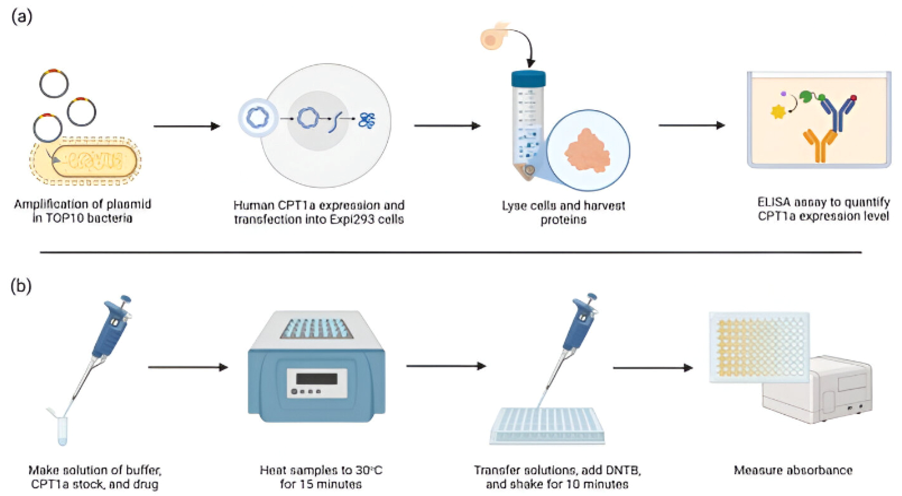
Figure 1. Workflow of CPT1 enzyme expression, protein purification, and activity assay using DTNB. (a) The workflow of the purification and quantification of human CPT1a enzyme. First, the plasmid was constructed and transformed into TOP10 bacteria for amplification. The plasmids were extracted and purified before being transfected into Expi293 cells, where they were cultured. The cells were then lysed, and the supernatant and pellet were collected for protein quantification by ELISA. (b) The CPT1 enzyme activity assay workflow. First, a solution of the buffer reagents, diluted CPT1 stock, and drug was created. The samples were mixed well and warmed to 30 °C with a block plate heater for 15 min for the reaction to occur. The samples were then transferred to 96-well plates and DTNB was added. The plate was shaken on a cell shaker for 10 min before the absorbance was read in a plate reader at 412 nm.
Applbiosci 04 00025 g001
Figure 2. ELISA of CPT1a transgenic cell line. CPT1a immunogenicity assay using ELISA. The pellet is the human CPT1a transgenic cell line pellet. The supernatant is the human CPT1a transgenic cell line supernatant. The control is the wildtype Expi293 cell pellet without CPT1a gene transfection. The data are represented as mean  ±  SEM (n = 4), and significance was calculated using a Welch’s t-test (**** p < 0.0001).
Applbiosci 04 00025 g002
Figure 3. CPT activity assay and inhibitor sensitivity assay with varying enzyme concentrations. CPT activity assay and etomoxir inhibitor sensitivity assay. We used 3 μL, 6 μL, and 9 μL of the same cell lysis pellet solution with a protein concentration of 0.67 μg/μL. A total of 2 μL of a 15.58 mM stock solution of etomoxir was added to each group to make a final concentration of 0.306 mM. Data are represented as mean  ±  SEM (n  =  3), and significance was calculated using a Welch’s t-test (** p  <  0.01 and **** p < 0.0001).
Applbiosci 04 00025 g003
Figure 4. CPT1 inhibitor sensitivity assay for chlorpromazine shows compound-induced CPT1 inhibition. CPT1 inhibitor sensitivity assay using chlorpromazine. We used the same amount of human CPT1a pellet solution at the protein concentration of 0.67 μg/μL, 2 μL each group. A total of 2 μL of 10 mM chlorpromazine as a solution in DMSO was used. A total of 2 μL of DMSO (Fisher Scientific, 99.7%) was used as a negative control. Data are represented as means  ±  SEM (n  =  3), and significance was calculated using a Welch’s t-test (**** p < 0.0001).
Applbiosci 04 00025 g004
Figure 5. IC50 of etomoxir, perhexiline, chlorpromazine, and malonyl-CoA. IC50 of (a) etomoxir, (b) perhexiline, (c) chlorpromazine, and (d) malonyl-CoA. A series of concentrations of each compound was used accordingly. We used the same amount of human CPT1a pellet solution with a protein concentration of 0.67 μg/μL, adding 2 μL of protein in each group.
Applbiosci 04 00025 g005
Disclaimer/Publisher’s Note: The statements, opinions and data contained in all publications are solely those of the individual author(s) and contributor(s) and not of MDPI and/or the editor(s). MDPI and/or the editor(s) disclaim responsibility for any injury to people or property resulting from any ideas, methods, instructions or products referred to in the content.