Communication

Bioactive Factors Isolated and Purified from Bovine Colostrum Can Restore Extracellular Matrix Under Degradation by Metalloproteinases

Department of Biological, Geological and Environmental Sciences, University of Catania, 95124 Catania, Italy
*
Author to whom correspondence should be addressed.
Appl. Biosci. 2025, 4(2), 21; https://doi.org/10.3390/applbiosci4020021
Submission received: 1 September 2024 / Revised: 6 January 2025 / Accepted: 20 February 2025 / Published: 8 April 2025
(This article belongs to the Special Issue Anatomy and Regenerative Medicine: From Methods to Applications)

Abstract

:
The ECM is composed of a considerable number of biochemically and structurally diverse constituents. ECM is a highly dynamic system that constantly receives and sends biological, chemical and mechanical signals. Several studies suggest that mechanical signals derived from the extracellular microenvironment regulate skin regeneration and wound healing. Tests measuring collagen contraction showed a significant difference in contraction activation in samples treated with the 2% colostrum derivative mixture compared to the control. The analysis of the supernatant showed an inhibition of metalloproteinase-2 expression and an increase in collagen secretion by fibroblasts in treatment samples. Our hypothesis is that the molecules extracted and purified from bovine colostrum can restore the ECM environment qualitative and quantitative characteristics, thus permitting, through a mechanical action, the restoration of the wound due to the transduction of the signal activated by the integrins.

Graphical Abstract

1. Introduction

The cells within tissues are inserted in a highly structured microenvironment and are very sensitive to the geometric and mechanical constraints of the latter [1]. The cellular microenvironment, formed by the extracellular matrix (ECM) and neighboring cells, influences not only cellular architecture and mechanics, but also the polarity and functions of the cell [1,2]. Mechanical properties of the ECM depend on a complex protein network that forms a fibrous 3D scaffold whose structural components are collagen fibers, proteoglycans, and glycosaminoglycans [3]. Cells adhere to this network and act as a reservoir for nonstructural components such as growth factors, cytokines, and proteolytic enzymes [4]. ECM proteins, by moving under the influence of forces, can act as mechanotransducers by exposing specific sites and growth factors [5]. In physiological conditions, the balance between processes of destruction and regeneration of the constituents of the extracellular matrix is regulated by specific tissue inhibitors of metalloproteinases (MMPs) [6,7]. An alteration of this balance, as in the case of wounds, is related to a catalytic hyperactivity of MMPs that play a critical role in wound healing. Their main function is degradation with removal of the damaged ECM during the inflammatory phase. The presence of these enzymes is necessary for effective wound healing, but they can play a harmful role at high concentrations, causing excessive tissue degradation and slow wound healing [7].
Knowledge of the cellular response to mechanical stimuli coming from the cellular microenvironment can be fundamental for applications in regenerative medicine to de-sign new and more effective scaffolds or biomaterials [8].
Scaffolds should mimic the properties of the target tissue [9], ensure the restoring of anisotropy (one of the fundamental characteristics of most tissues) [10,11,12], and through mechanotransduction activate integrins and induce the cascade of growth factors (TGF-beta 1, CTGF, IGF-1, etc.) necessary, among other things, to produce new collagen by the fibroblast [13]. The response of integrins to force involves three mechanochemical steps. Integrins bind to ECM molecules transmitting forces into the cell that are converted into biochemical signals (mechanotransduction). Finally, integrins connect to the cytoskeleton to transmit forces throughout the cell and strengthen adhesions to resist forces. Mechanical signals that are transmitted through structural components of the cytoskeleton play a key role in events that regulate cell migration, polarity, and proliferation [14].
The aim of our research is to demonstrate that bioactive factors purified from bovine colostrum are like molecules normally present in the extracellular matrix [15,16] and that if supplied to stressed tissues (such as in chronic wounds), they can provide the scaffolding necessary to activate the signal transduction mechanisms that activate the response of integrins. Bovine colostrum is a nutrient milk secretion containing bioactive compounds that support calf nutrition and immune development. Colostrum is profuse in bioactive compounds like immunoglobulins, growth factors, lysozymes, lactoferrin, and lactoperoxidase, but also possesses elevated levels of fats, proteins, minerals, and vitamins. Bovine colostrum is also rich in extracellular nanovesicles, such as exosomes, which protect bioactive components from degradation [16,17]. These bioactive molecules are also components of the ECM, and their mechanical role is fundamental in its remodeling [16,18,19]. In order to demonstrate our hypothesis, we also carried out a test to evaluate collagen contraction and measured the expression of collagen production and metalloproteinases-2, crucial elements for wound healing [7,20,21].

2. Materials and Methods

2.1. Colostrum Derivative Mixture Preparation

The colostrum derivative mixture (CDM) preparation was processed according to the procedure described by [22]. Bovine colostrum was collected from Holstein cows from 1 up to 6 h after parturition. This methodology, through micro and nano filtration, allows the elimination of casein, fats, and other non-functional macromolecules including bovine immunoglobulins from the colostrum, and essentially isolates growth factors and cytokines identified with ELISA tests [22].

2.2. Propagation and Maintenance of Cells

The Human Dermal Fibroblasts (HDFs) (106-05A, Merck, Darmstadt, Germany) were grown in Dulbecco’s Modified Eagle Medium (DMEM). The medium was supplemented with 10% fetal bovine serum, streptomycin and penicillin (0.3 mg mL−1 and 50 IU mL−1, respectively), and GlutaMAX (1 mM). Cells were maintained in a humidified environment (37 °C and 5% CO2) and split every 2 days depending on cell confluence (80%). All experiments were performed in triplicate wells for each condition and repeated at least twice.

2.3. Scratch-Wound Assay

Cells were grown in DMEM containing 10% FBS or 2% of colostrum mixture in 6-well plates until they reached confluence. After incubation, a scratch was made manually on the cell monolayer. After 24 h incubation, cell migration was determined based on the images obtained with an inverted optical microscope (Leica DIM IRB). The wound closure was calculated using the equation:
Wound Closure % = [A0h − A24h/A0h] × 100
where A0h is the area (pixels) of the wound calculated after scratching (t = 0 h) and A24h is the area (pixels) of the unhealed wound (which is not covered by the cells) that remained 24 h after the scratching.

2.4. Collagen Contraction

To assess matrix contraction, free-floating collagen lattice models were used according to [20]. We used a Cell Contraction Assay Kit (Cell Biolabs, Inc., CBA-201, San Diego, CA, USA). After preparation of collagen gel solution, it was stored on ice. We collected the cells and resuspended them in FBS or CDM (3 × 10⁶ cells/mL). We mixed 2 parts of cell suspension and 8 parts of cold collagen gel solution. We included negative control wells that contain no cells in the matrix. After adding 0.5 mL of the cell and collagen mixture per well in a 24-well plate, we incubated them for 1 h at 37 °C to allow collagen polymerization. Finally, 1 mL of FBS or CDM was added on top of each collagen gel lattice. The reduction in the lattice area due to contraction was evaluated at 24 h intervals for up to 96 h. Experiments were performed in duplicate.

2.5. Collagen Production

Matrix synthesis was determined over 7 days in the presence or absence of colostrum derivative mixture 2%. Secreted C-terminal propeptide of collagen type I (CICP) was measured in culture supernatant using an enzyme immunoassay kit (Metra Biosystem, Quidel Corporation, San Diego, CA, USA), according to the manufacturer’s instructions. The reaction was stopped, and the collagen synthesis was measured at 405 nm with Epoch Microplate Spectrophotometer (Bio Tek Instruments, Inc., Winooski, VT, USA). This assay allows us to quantify the release of CICP and provides a stoichiometric representation of the production of collagen in a cell culture environment. Experiments were performed in duplicate.

2.6. Determination of MMP-2 Concentration

MMP-2 expression, constitutively secreted from fibroblasts, was determined with a commercially available quantitative ELISA test (Amersham Pharmacia Biotech, Piscataway, NJ, USA). All procedures were performed according to the instructions of the manufacturer. Experiments were performed in duplicate.

2.7. Statistical Analysis

Data were analyzed using the GraphPad Prism 5.0 program (GraphPad Software, La Jolla, CA, USA), using the analysis of variance (ANOVA) test and Tukey’s test. A p value ≤ 0.05 was used to identify statistically significant differences. Parametric correlation was calculated using Pearson correlation coefficient.

3. Results and Discussion

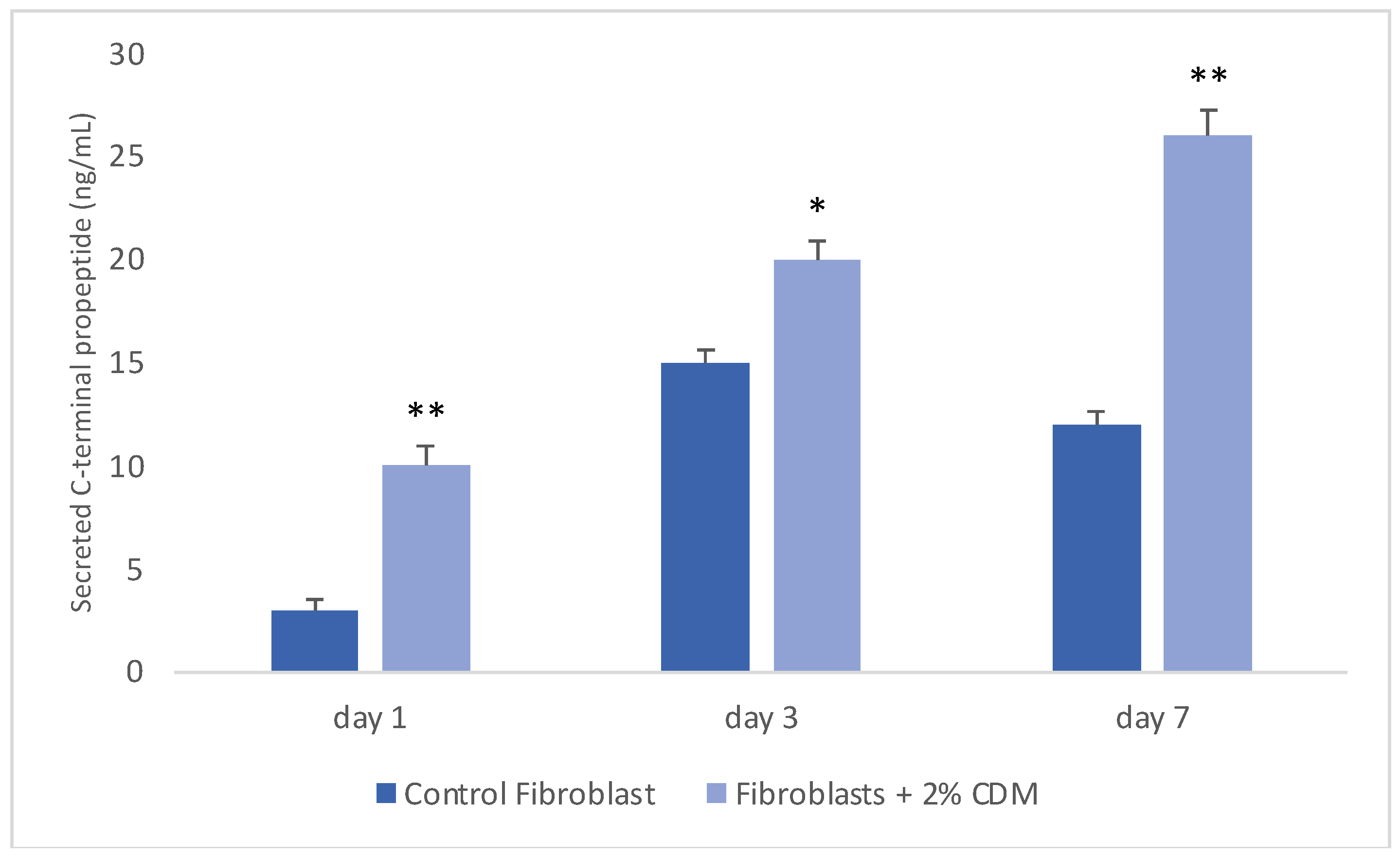
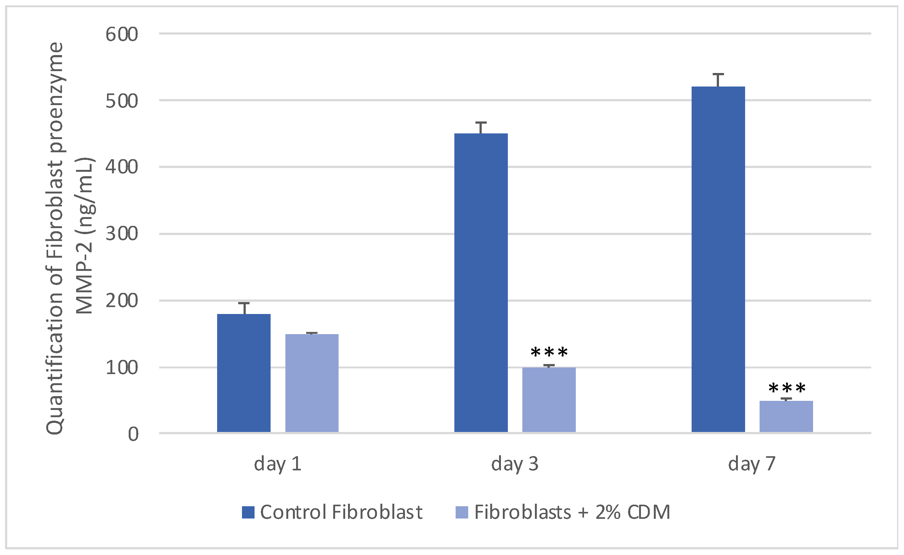
Tissues are not only made up of cells, but a significant part of their volume is formed by the extracellular space, occupied by an intricate network of macromolecules, whose three-dimensional organization is represented by the extracellular matrix. Biochemical analysis of the ECM reveals that it is composed of several proteins and polysaccharides, which aggregate in a compactly organized network connected to the surface of the cells that produced it and the surrounding ones [6,16,23]. The ECM forms the bio-scaffold of our body and adapts to the environmental variables. The biomechanical properties of the ECM are negatively affected by aging, but also during wound healing or restoring skin in burn injuries [24]. Many computational models and experimental studies have revealed the important effects of cell-generated mechanical forces, forces acting upon cells, and mechanical characteristics of the extracellular matrix on cell morphology and function [24]. The micro-environment surrounding cells has a large role in directing cell behavior. Cells can actively sense the mechanical properties of their surroundings by exerting contractile force, which can transmit between cell and matrix or cell and cell. Cells produce and modify the composition and organization of the ECM, thus varying its mechanical properties [24]. In fact, mechanical forces exerted from the ECM can act on cells, such as in wounds [25,26,27]. Our hypothesis is that the molecules extracted from colostrum, identified with ELISA test according to [22], can restore the chemical/physical ECM environment, thus permitting, through a mechanical action, the restoration of the wound due to the transduction of the signal activated by the integrins. Our hypothesis is supported by the results obtained with a collagen contraction test that demonstrated that exposure of fibroblast-populated collagen lattices to concentrations of colostrum derivative mixture significantly activated contraction by day 7, compared with the control (Figure 1). These results are of great interest, especially if correlated to the simultaneous results of the ELISA test performed during collagen contraction. Tests demonstrated that fibroblasts have actively secreted CICP during lattice contraction up to 7 days (Figure 2) and inhibited MMP-2 expression (Figure 3) in the samples treated with the 2% colostrum derivative mixture.
Statistical analysis using Pearson correlation coefficient confirmed a significant positive linear correlation between collagen contraction and collagen production during collagen contraction from fibroblasts (r = 0.98, p < 0.001) and a significant negative linear correlation between collagen contraction and proenzyme MMP-2 production during collagen contraction (r = 0.99, p < 0.001) (Figure 4).
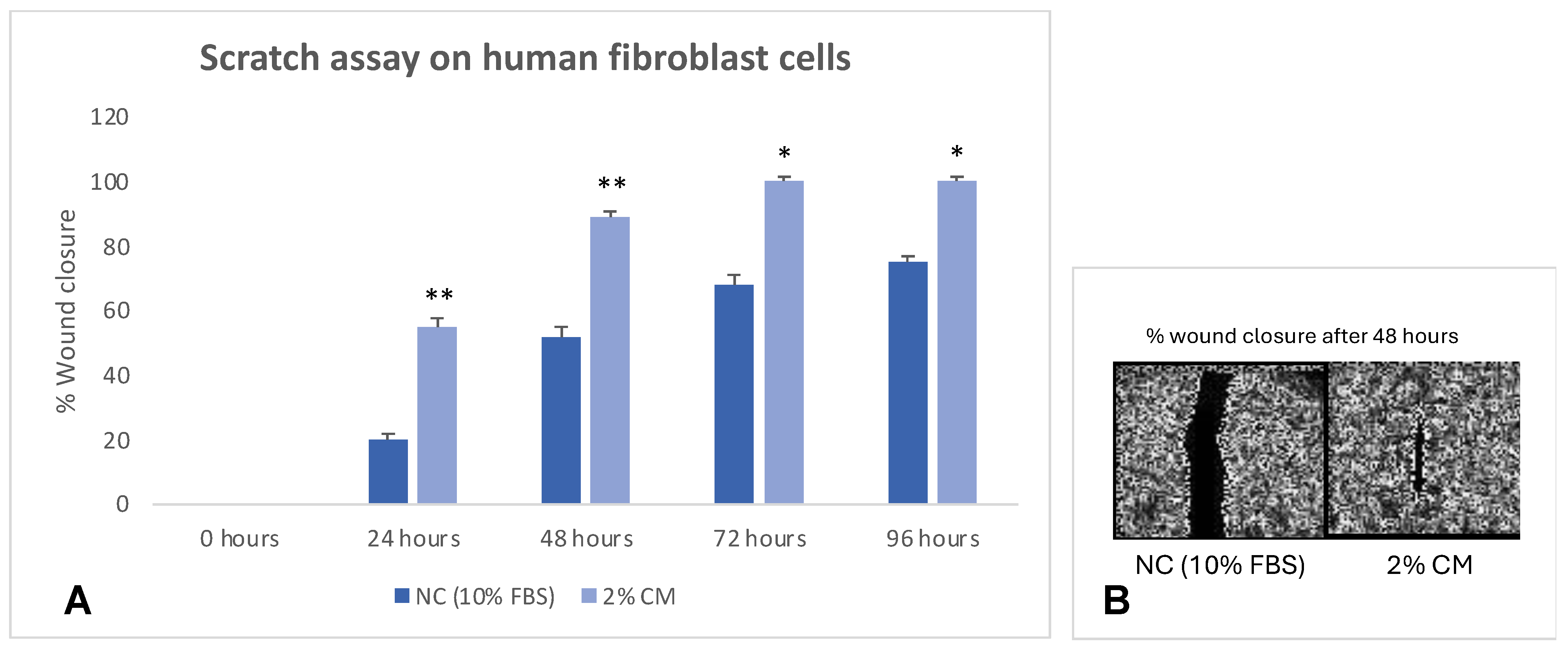
Contraction and remodeling of the extracellular matrix are essential processes during wound healing [28,29]. At the center of these two phenomena are fibroblasts, which not only produce and secrete extracellular matrix proteins but can also reorganize them through mechanical interactions [30,31]. Collagen is the predominant structural protein in the ECM not only providing tensile strength but also playing a role in cell adhesion and migration [32]. Matrix metalloproteinases (MMPs) are a family of proteolytic enzymes capable of degrading various structural components of the matrix [33,34,35,36,37,38]. They therefore play a fundamental role in various physiological and pathological processes including wound healing [18,29]. It is now known that the biomechanical properties of the ECM contribute to the physiological development of cells and tissues [25]. A scratch assay on fibroblast cells proved in fact that the colostrum mixture was able to significantly increase cell proliferation of fibroblasts compared to untreated cells (control) (Figure 5). Interestingly, 48 h treatment with 2% colostrum mixture produced an inductive effect higher at 80% (Figure 5).
The elasticity of the ECM allows us to perceive external forces and, therefore, provides an important environmental input signal that determines the cell’s response. Indeed, the adhesion complex, which consists of integrins and a multicomplex of adapters and signaling proteins, can be considered as a mechanosensor that connects the actomyosin cytoskeleton with the ECM [39]. Many of the adhesion components undergo conformational changes that determine functional consequences in the response to applied force. The cytoskeleton, together with nuclear matrices, nuclear envelope, and chromatin, constitutes a mechano-sensing system that determines how cells react to forces transmitted by the ECM [39,40,41,42,43]. Many of the adhesion factors undergo conformational changes and, together with the cytoskeleton and nuclear matrices, nuclear envelope, and chromatin, they constitute a sophisticated mechano-sensing machinery that determines how cells react to forces transmitted by the ECM, regulating various essential cellular behaviors, including cell fate determination and differentiation [39,40,41,42,43]. The different properties of the ECM are not independent of each other but influence each other. Therefore, when the ECM increases in stiffness, as, for example, in pathological conditions, its biomechanical properties change, and cells respond by exerting markedly different types of force. Furthermore, the stiffening of the matrix also determines a change in the other physical properties of the ECM and, consequently, directly modifies the cellular ability to migrate. The ECM, constantly undergoing restructuring in different tissues, is highly dynamic [43]. ECM dynamics can arise from changes in the amount or composition of the ECM, due to altered synthesis or degradation of one or more components. Alternatively, ECM dynamics may show no changes in the composition of its components, but instead involve only the transformation of its components into spatially organized structures, thanks to covalent and non-covalent bonds [6,23].
Finally, one of the most important features of cell–ECM interactions is reciprocity [44]. Cells constantly make, degrade, or rearrange components of the ECM to modify one or more properties; but since the ECM regulates the behavior of cells, any change in it, as a result of cellular activities, will in turn influence adjacent cells and modify their behaviors. This feedback regulatory mechanism between cells and the ECM allows cells and tissues to rapidly adapt to the changes in environment [6].

4. Conclusions

Wounds heal through the coordinated action of fibroblast-mediated extracellular matrix (ECM) deposition, ECM remodeling, and wound contraction. For these processes, the mechanical signals between the ECM and cells are fundamental. In this paper, we demonstrate that extracted and purified biomolecules of bovine colostrum can restore chemical/physical ECM environment, reestablish the mechanical properties of the ECM, and the anisotropy characteristics of the damaged tissue. This process activates the transduction of the signal by the integrins and reactivates the ability of the fibroblast to synthesize new collagen, inducing the autologous mechanisms of repair and remodeling of damaged connective tissue. Fibroblasts, in fact, are able to generate tensile forces as well as receive them; these contraction forces of the fibroblasts are indispensable for wound resolution processes.

Author Contributions

Conceptualization, M.V.B.; methodology, M.V.B.; software, G.F.; formal analysis, F.C., G.G., G.I. and S.P.; investigation, F.C., G.G., G.I. and S.P.; data curation, M.V.B.; writing—original draft preparation, M.V.B.; writing—review and editing, G.F. and M.V.B.; supervision, M.V.B.; funding acquisition, M.V.B. All authors have read and agreed to the published version of the manuscript.

Funding

This research received no external funding.

Institutional Review Board Statement

Not applicable.

Informed Consent Statement

Not applicable.

Data Availability Statement

The raw data supporting the conclusions of this article will be made available by the authors on request.

Acknowledgments

G.F. thanks the Ph.D. program FSE Notice 1/2021. F.C., G.G., G.I. and S.P. thank the Ph.D. program PNRR-D.M. 117/2023.

Conflicts of Interest

The authors declare no conflicts of interest.

References

  1. Mierke, C.T. Bidirectional Mechanical Response Between Cells and Their Microenvironment. Front. Phys. 2021, 9, 749830. [Google Scholar]
  2. Xie, N.; Xiao, C.; Shu, Q.; Cheng, B.; Wang, Z.; Xue, R.; Wen, Z.; Wang, J.; Shi, H.; Fan, D.; et al. Cell response to mechanical microenvironment cues via Rho signaling: From mechanobiology to mechanomedicine. Acta Biomater. 2023, 159, 1–20. [Google Scholar] [PubMed]
  3. Jansen, K.A.; Donato, D.M.; Balcioglu, H.E.; Schmidt, T.; Danen, E.H.; Koenderink, G.H. A guide to mechanobiology: Where biology and physics meet. Biochim. Biophys. Acta 2015, 1853, 3043–3052. [Google Scholar] [PubMed]
  4. Boyd, D.F.; Thomas, P.G. Towards integrating extracellular matrix and immunological pathways. Cytokine 2017, 98, 79–86. [Google Scholar]
  5. Di, X.; Gao, X.; Peng, L.; Ai, J.; Jin, X.; Qi, S.; Li, H.; Wang, K.; Luo, D. Cellular mechanotransduction in health and diseases: From molecular mechanism to therapeutic targets. Sig Transduct. Target. Ther. 2023, 8, 282. [Google Scholar]
  6. Lu, P.; Takai, K.; Weaver, V.M.; Werb, Z. Extracellular matrix degradation and remodeling in development and disease. Cold Spring Harb. Perspect. Biol. 2011, 3, a005058. [Google Scholar]
  7. Caley, M.P.; Martins, V.L.; O’Toole, E.A. Metalloproteinases and Wound Healing. Adv. Wound Care 2015, 4, 225–234. [Google Scholar]
  8. Zhao, X.; Li, Q.; Guo, Z.; Li, Z. Constructing a cell microenvironment with biomaterial scaffolds for stem cell therapy. Stem Cell Res. Ther. 2021, 12, 583. [Google Scholar]
  9. Kozaniti, F.K.; Manara, A.E.; Kostopoulos, V.; Mallis, P.; Michalopoulos, E.; Polyzos, D.; Deligianni, D.D.; Portan, D.V. Computational and Experimental Investigation of the Combined Effect of Various 3D Scaffolds and Bioreactor Stimulation on Human Cells’ Feedback. Appl. Biosci. 2023, 2, 249–277. [Google Scholar] [CrossRef]
  10. Caliari, S.R.; Harley, B.A. The effect of anisotropic collagen-GAG scaffolds and growth factor supplementation on tendon cell recruitment, alignment, and metabolic activity. Biomaterials 2011, 32, 5330–5340. [Google Scholar]
  11. Harmansa, S.; Erlich, A.; Eloy, C.; Zurlo, G.; Lecuit, T. Growth anisotropy of the extracellular matrix shapes a developing organ. Nat. Commun. 2023, 14, 1220. [Google Scholar] [PubMed]
  12. Datta, P.; Vyas, V.; Dhara, S.; Chowdhury, A.; Barui, A. Anisotropy Properties of Tissues: A Basis for Fabrication of Biomimetic Anisotropic Scaffolds for Tissue Engineering. J. Bionic Eng. 2019, 16, 842–868. [Google Scholar] [CrossRef]
  13. Reid, J.A.; Dwyer, K.D.; Schmitt, P.R.; Soepriatna, A.H.; Coulombe, K.L.; Callanan, A. Architected fibrous scaffolds for engineering anisotropic tissues. Biofabrication 2021, 13, 045007. [Google Scholar] [CrossRef]
  14. Ross, T.D.; Coon, B.G.; Yun, S.; Baeyens, N.; Tanaka, K.; Ouyang, M.; Schwartz, M.A. Integrins in mechanotransduction. Curr. Opin. Cell Biol. 2013, 25, 613–618. [Google Scholar] [PubMed]
  15. Yalçıntaş, Y.M.; Duman, H.; López, J.M.M.; Portocarrero, A.C.M.; Lombardo, M.; Khallouki, F.; Koch, W.; Bordiga, M.; El-Seedi, H.; Raposo, A.; et al. Revealing the Potency of Growth Factors in Bovine Colostrum. Nutrients 2024, 16, 2359. [Google Scholar] [CrossRef]
  16. Yue, B. Biology of the extracellular matrix: An overview. J. Glaucoma 2014, 23 (Suppl. S1), S20–S23. [Google Scholar] [CrossRef]
  17. Ross, M.; Atalla, H.; Karrow, N.; Mallard, B.A. The bioactivity of colostrum and milk exosomes of high, average, and low immune responder cows on human intestinal epithelial cells. J. Dairy. Sci. 2021, 104, 2499–2510. [Google Scholar]
  18. Diller, R.B.; Tabor, A.J. The Role of the Extracellular Matrix (ECM) in Wound Healing: A Review. Biomimetics 2022, 7, 87. [Google Scholar] [CrossRef]
  19. Kuehlmann, B.; Bonham, C.A.; Zucal, I.; Prantl, L.; Gurtner, G.C. Mechanotransduction in Wound Healing and Fibrosis. J. Clin. Med. 2020, 9, 1423. [Google Scholar] [CrossRef]
  20. Daniels, J.T.; Cambrey, A.D.; Occleston, N.L.; Garrett, Q.; Tarnuzzer, R.W.; Schultz, G.S.; Khaw, P.T. Matrix metalloproteinase inhibition modulates fibroblast-mediated matrix contraction and collagen production in vitro. Invest. Ophthalmol. Vis. Sci. 2003, 44, 1104–1110. [Google Scholar] [CrossRef]
  21. Singh, D.; Rai, V.; Agrawal, D.K. Regulation of Collagen I and Collagen III in Tissue Injury and Regeneration. Cardiol. Cardiovasc. Med. 2023, 7, 5–16. [Google Scholar] [CrossRef] [PubMed]
  22. Sacerdote, P.; Mussano, F.; Franchi, S.; Panerai, A.E.; Bussolati, G.; Carossa, S.; Bartorelli, A.; Bussolati, B. Biological components in a standardized derivative of bovine colostrum. J. Dairy Sci. 2013, 96, 1745–1754. [Google Scholar] [PubMed]
  23. Hu, M.; Ling, Z.; Ren, X. Extracellular matrix dynamics: Tracking in biological systems and their implications. J. Biol. Eng. 2022, 16, 13. [Google Scholar]
  24. Franchi, M.; Piperigkou, Z.; Mastronikolis, N.S.; Karamanos, N. Extracellular matrix biomechanical roles and adaptation in health and disease. FEBS J. 2024, 291, 430–440. [Google Scholar]
  25. Holle, A.W.; Young, J.L.; Van Vliet, K.J.; Kamm, R.D.; Discher, D.; Janmey, P.; Spatz, J.P.; Saif, T. Cell-Extracellular Matrix Mechanobiology: Forceful Tools and Emerging Needs for Basic and Translational Research. Nano Lett. 2018, 18, 1–8. [Google Scholar]
  26. Arnold, M.; Cavalcanti-Adam, E.A.; Glass, R.; Blümmel, J.; Eck, W.; Kantlehner, M.; Kessler, H.; Spatz, J.P. Activation of integrin function by nanopatterned adhesive interfaces. Chemphyschem 2004, 5, 383–388. [Google Scholar]
  27. Xu, R.; Boudreau, A.; Bissell, M.J. Tissue architecture and function: Dynamic reciprocity via extra- and intra-cellular matrices. Cancer Metastasis Rev. 2009, 28, 167–176. [Google Scholar]
  28. Olczyk, P.; Mencner, Ł.; Komosinska-Vassev, K. The role of the extracellular matrix components in cutaneous wound healing. Biomed. Res. Int. 2014, 2014, 747584. [Google Scholar] [CrossRef]
  29. Potekaev, N.N.; Borzykh, O.B.; Medvedev, G.V.; Pushkin, D.V.; Petrova, M.M.; Petrov, A.V.; Dmitrenko, D.V.; Karpova, E.I.; Demina, O.M.; Shnayder, N.A. The Role of Extracellular Matrix in Skin Wound Healing. J. Clin. Med. 2021, 10, 5947. [Google Scholar] [CrossRef]
  30. Tracy, L.E.; Minasian, R.A.; Caterson, E.J. Extracellular Matrix and Dermal Fibroblast Function in the Healing Wound. Adv. Wound Care 2016, 5, 119–136. [Google Scholar]
  31. Kular, J.K.; Basu, S.; Sharma, R.I. The extracellular matrix: Structure, composition, age-related differences, tools for analysis and applications for tissue engineering. J. Tissue Eng. 2014, 5, 2041731414557112. [Google Scholar] [PubMed]
  32. Sun, B. The mechanics of fibrillar collagen extracellular matrix. Cell Rep. Phys. Sci. 2021, 2, 100515. [Google Scholar] [PubMed]
  33. Gill, S.E.; Parks, W.C. Metalloproteinases and their inhibitors: Regulators of wound healing. Int. J. Biochem. Cell Biol. 2008, 40, 1334–1347. [Google Scholar]
  34. Lukes, A.; Mun-Bryce, S.; Lukes, M.; Rosenberg, G.A. Extracellular matrix degradation by metalloproteinases and central nervous system diseases. Mol. Neurobiol. 1999, 19, 267–284. [Google Scholar]
  35. Cabral-Pacheco, G.A.; Garza-Veloz, I.; Castruita-De la Rosa, C.; Ramirez-Acuña, J.M.; Perez-Romero, B.A.; Guerrero-Rodriguez, J.F.; Martinez-Avila, N.; Martinez-Fierro, M.L. The Roles of Matrix Metalloproteinases and Their Inhibitors in Human Diseases. Int. J. Mol. Sci. 2020, 21, 9739. [Google Scholar] [CrossRef]
  36. Burbridge, M.F.; Cogé, F.; Galizzi, J.P.; Boutin, J.A.; West, D.C.; Tucker, G.C. The role of the matrix metalloproteinases during in vitro vessel formation. Angiogenesis 2002, 5, 215–226. [Google Scholar]
  37. Lombard, C.; Saulnier, J.; Wallach, J. Assays of matrix metalloproteinases (MMPs) activities: A review. Biochimie 2005, 87, 265–272. [Google Scholar]
  38. Agren, M.S.; Mirastschijski, U.; Karlsmark, T.; Saarialho-Kere, U.K. Topical synthetic inhibitor of matrix metalloproteinases delays epidermal regeneration of human wounds. Exp. Dermatol. 2001, 10, 337–348. [Google Scholar]
  39. Wong, V.W.; Akaishi, S.; Longaker, M.T.; Gurtner, G.C. Pushing back: Wound mechanotransduction in repair and regeneration. J. Investig. Dermatol. 2011, 131, 2186–2196. [Google Scholar]
  40. DuFort, C.C.; Paszek, M.J.; Weaver, V.M. Balancing forces: Architectural control of mechanotransduction. Nat. Rev. Mol. Cell Biol. 2011, 12, 308–319. [Google Scholar]
  41. Ricca, B.L.; Venugopalan, G.; Fletcher, D.A. To pull or be pulled: Parsing the multiple modes of mechanotransduction. Curr. Opin. Cell Biol. 2013, 25, 558–564. [Google Scholar] [PubMed]
  42. Saraswathibhatla, A.; Indana, D.; Chaudhuri, O. Cell-extracellular matrix mechanotransduction in 3D. Nat. Rev. Mol. Cell Biol. 2023, 24, 495–516. [Google Scholar]
  43. Xie, W.; Wei, X.; Kang, H.; Jiang, H.; Chu, Z.; Lin, Y.; Hou, Y.; Wei, Q. Static and Dynamic: Evolving Biomaterial Mechanical Properties to Control Cellular Mechanotransduction. Adv. Sci. 2023, 10, e2204594. [Google Scholar]
  44. Urciuolo, F.; Imparato, G.; Netti, P.A. In vitro strategies for mimicking dynamic cell–ECM reciprocity in 3D culture models. Front. Bioeng. Biotechnol. 2023, 11, 1197075. [Google Scholar]
Figure 1. Graphical representation of collagen contraction. (A) Collagen gel contraction over 96 h at 24 h time intervals. (B) Fold change in collagen gel contraction at 96 h. Data are plotted as mean ± SEM. Differences between groups were tested for statistical significance using one-way ANOVA followed by Dunnett’s multiple comparison post-test (n = 3). Standard deviations were represented by vertical bars (B). Asterisks denote the degree of significance between results and control: * p < 0.01.
Applbiosci 04 00021 g001
Figure 2. Graphical representation of collagen production during collagen contraction. Fibroblasts actively secreted CICP during lattice contraction up to 7 days. In control samples, secretion of CICP increased up to 3 days and after was reduced. Data are plotted as mean ± SEM. Differences between groups were tested for statistical significance using one-way ANOVA followed by Dunnett’s multiple comparison post-test (n = 3). Standard deviations were represented by vertical bars. Asterisks denote the degree of significance between results: * p < 0.01, ** p < 0.001.
Applbiosci 04 00021 g002
Figure 3. Graphical representation of proenzyme MMP-2 production during collagen contraction. Data are plotted as mean ± SEM. Differences between groups were tested for statistical significance using one-way ANOVA followed by Dunnett’s multiple comparison post-test (n = 3). Standard deviations were represented by vertical bars. Asterisks denote the degree of significance between results: *** p < 0.0001.
Applbiosci 04 00021 g003
Figure 4. Scatter plot: (A) strong positive linear correlation between collagen contraction and collagen production from fibroblasts; (B) strong negative linear correlation between collagen contraction and proenzyme MMP-2 production from fibroblasts. Pearson coefficient values (r).
Applbiosci 04 00021 g004
Figure 5. Scratch assay on fibroblast cells. (A) The results represent the size of the wound in percentage (%), with 0% indicating the original wound size and 100% representing the complete absence of the wound. Asterisks denote the degree of significance between results: ** p < 0.001, * p < 0.01. (B) Images of % wound closure after 48 h. The experiment was repeated 3 times.
Applbiosci 04 00021 g005
Disclaimer/Publisher’s Note: The statements, opinions and data contained in all publications are solely those of the individual author(s) and contributor(s) and not of MDPI and/or the editor(s). MDPI and/or the editor(s) disclaim responsibility for any injury to people or property resulting from any ideas, methods, instructions or products referred to in the content.