Article

Deciphering the Role of the Nucleus Accumbens Shell Area on Spatial Memory Deficits Induced by Neuropathic Pain in Rats

by
Mariana Cerqueira-Nunes
1,2,3,4,
Clara Monteiro
1,2,3,
Vasco Galhardo
1,2,3 and
Helder Cardoso-Cruz
1,2,3,*
1
i3S—Instituto de Investigação e Inovação em Saúde, Pain Neurobiology Research Group, Universidade do Porto, Rua Alfredo Allen 208, 4200-135 Porto, Portugal
2
IBMC—Instituto de Biologia Molecular e Celular, Universidade do Porto, Rua Alfredo Allen 208, 4200-135 Porto, Portugal
3
FMUP—Faculdade de Medicina, Departamento de Biomedicina—Unidade de Biologia Experimental, Universidade do Porto, Rua Doutor Plácido da Costa, 4200-450 Porto, Portugal
4
Mestrado em Neurobiologia da FMUP, Universidade do Porto, Rua Doutor Plácido da Costa, 4200-450 Porto, Portugal
*
Author to whom correspondence should be addressed.
Appl. Biosci. 2024, 3(2), 283-295; https://doi.org/10.3390/applbiosci3020019
Submission received: 8 April 2024 / Revised: 20 May 2024 / Accepted: 7 June 2024 / Published: 12 June 2024

Abstract

:
The nucleus accumbens shell (NAcSh) is a major structure associated with distinct aspects of reward and mnemonic information encoding, relying on spatial data to define optimal behavioral strategies. Chronic pain-derived striatal plasticity is considered one underpinning cause of working memory (WM) impairments. However, it is unclear how the NAcSh is involved in these spatial deficits. To address this, we evaluated the impact of unilateral local NAcSh electrical lesions during the execution of a food-reinforced eight-shaped spatial alternation WM task. Behavioral performance was assessed in rats after the onset of the neuropathic pain model—spared nerve injury (SNI). Our findings indicate that the induction of SNI and/or NAcSh lesions did not significantly impact the animals’ performance accuracy or motor activity during the execution of the behavioral task, but altered their response latency patterns. In addition, these manipulations did not induce significant antinociceptive effects. Collectively, these results suggest that the NAcSh may participate in specific aspects of spatial information integration and processing under neuropathic pain conditions.

1. Introduction

The nucleus accumbens shell area (NAcSh) plays a pivotal role in reinforcement learning, emotional processing, and nociceptive information encoding [1,2,3,4,5,6]. Due to its strategic position, the NAcSh acts as a hub connecting limbic, prefrontal, and motor regions, shaping reward-directed behaviors through spatial information [7,8,9,10]. Despite several conceptualizations, we still lack a comprehensive understanding of how the disruption of the NAcSh area by pain leads to spatial working memory (WM) deficits.
Imaging studies have revealed distinct connectivity clusters in the nucleus accumbens (NAc) during nociceptive information processing [11,12]. Preclinical research has also indicated that suppression of local activity via lidocaine infusion [13] and concurrent suppression of dopamine (DA) receptor 2-expressing neurons and activation of NAc DA receptor 1-expressing neurons [14,15] reduce allodynia and neuropathic behaviors. The observed alterations in local NAcSh excitability during neuropathic pain provide evidence of this region as a hallmark in both ascending and descending pathways, establishing a robust correlation between NAcSh activity and pain-related behaviors [16].
Persistent pain conditions, including neuropathic pain, are characterized by a hypoDAergic state and compromised NAc integrity [17,18]. This condition not only compromises the role of the NAcSh in supporting cortical networks but also impedes the efficient encoding of mnemonic information [3,19]. The DAergic drive from mesolimbic areas directly regulates NAc activity, influencing the maintenance of spatial WM information to plan forthcoming goal-directed actions [20]. NAcSh manipulation using glutamatergic NMDA and non-NMDA receptor antagonists has resulted in impairments of spatial information acquisition and a reduction in exploratory behavior [21]. Additionally, abnormal increases in local NAcSh activity are correlated with alterations in spatial WM and pain information processing [19]. FMRI imaging uncovered distinct chronic pain-induced connectivity changes between the NAc and prefrontal cortical areas during cognitive demand, which are considered one underlying factor regarding WM impairments [11,13,19,22].
Despite significant strides in understanding the neurophysiological aspects of the NAcSh, especially in response to noxious stimuli, a comprehensive insight into how neuropathic pain specifically impacts this accumbal subregion and its associated cognitive functions remains elusive. This study aims to bridge this gap by investigating the role of the NAcSh in spatial information encoding and how its functionality influences the manifestation of pain-related WM impairments. To investigate this hypothesis, we induced unilateral lesions in the NAcSh area to evaluate their impact on behavioral accuracy while rats perform a classical food-reinforced eight-shaped spatial alternation task.

2. Materials and Methods

2.1. Animal Model

Eighteen adult male CD rats weighing between 250 and 300 g (strain 001, Charles River Laboratories, Saint-Germain-Nuelles, France) were used for the behavioral procedures. The rats were housed in individually ventilated cages (four animals per cage) under a 12 h light/dark cycle (lights on at 8:00 a.m.), with temperature (22 ± 2 °C) and humidity (55 ± 5%) controlled using a double decker IVC (Tecniplast, Varese, Italy). Prior to any experimental procedures, the rats underwent habituation to the experimenter and handling protocol. Training and behavioral sessions occurred approximately at the same time during the light period. Throughout the experiments, rats were maintained on a food deprivation protocol (90–95% of their ad libitum feeding body weight) with free access to water. Their weight was monitored every 2 days and compared with the standard growth curve for CD rats (Charles River Laboratories, Saint-Germain-Nuelles, France). All efforts were made to minimize animal suffering and reduce the number of animals used in this study. The sample size was pre-estimated based on previously conducted experiments in the laboratory and published research.

2.2. Surgical Procedures

2.2.1. Electrical Lesion of the NAcSh

The NAcSh lesion was performed using a custom bipolar electrode consisting of two stainless steel Teflon-coated filaments (Cat. no. 791600, A-M Systems Inc., Sequim, WA, USA) intertwined, with a total outer diameter of 100–130 μm. Briefly, rats were anesthetized with a ketamine/medetomidine mixture (75 and 0.5 mg/kg, respectively, I.P.). The depth of anesthesia and muscular paralysis were assessed through regular testing of corneal blink, hindpaw withdrawal, and tail-pinch reflexes. Rats were secured in a stereotaxic frame using ear bars, and the skull was exposed and cleaned using hydrogen peroxide. A hole was bored in the skull to allow for the insertion of the bipolar electrode. The electrode was mounted in the holder of a hydraulic micropositioner (FHC Inc., St. Bowdoin, ME, USA) and gradually driven (250 μm/min) into the NAcSh region. The following stereotaxic coordinates relative to the bregma point were used to target the electrode: anterior–posterior (AP): +[1.6–1.8] mm, medial–lateral (ML): ±[0.4–0.9] mm, and dorsal–ventral (DV): −[6.7–7.0] mm [23]. After reaching the final location, the electrode was held immobile for 10 min before initiating the stimulation protocol, which consisted of electrical pulses of 20 μA at 0.5 Hz for 20 s. Following the lesion, the electrode was kept immobile for an additional period of 5 min before withdrawal. The NAcSh lesions were performed unilaterally and counterbalanced between brain hemispheres across rats. The control rats underwent an identical protocol, excluding the electrical lesion. After surgery, analgesic ketoprofen (5–10 mg/kg) and antibiotic enrofloxacin (5 mg/kg) diluted in a 1/10 ratio with NaCl 9% (w/v) solution were administered subcutaneously every 24 h for 3–5 days. Rats were allowed to recover for a minimum of 7–10 days before behavioral testing sessions began, and their overall health status was monitored daily.

2.2.2. Neuropathic Pain Model

The spared nerve injury method (SNI) [24] was performed immediately after the surgical protocol for the NAcSh lesion. Briefly, the tibial and common peroneal nerve branches of the sciatic nerve plexus were ligated and a portion of 2–4 mm was axotomized, while the sural nerve branch remained intact. Rats were assigned to the SNI group (n = 10) or the control Sham group (n = 8). The Sham procedure replicated the same skin incision and muscle dissection but without nerve lesion. The peripheral nerve lesions were performed counterbalanced between the left and right hindpaws, contralateral to the NAcSh lesion.

2.3. Experimental Design and Behavioral Procedures

2.3.1. Spatial Alternation Task

Rats were tested in an eight-shaped arena composed of two reward locations, based on a previously described study [25]. The arena comprised a 90 × 60 cm total area, with 15 cm wide corridors and 30 cm high opaque walls. During training, rats initiated their journey in the center and navigated alternately (left or right) to each of the two reward locations to obtain a sucrose pellet (Figure 1a). To successfully earn a pellet, the rats were required to choose one of the reward locations, follow a trajectory opposite to their approach, return to the center, and then proceed to the other reward location without retracing corridors (termed correct alternation). Failure to cross the central corridor or consecutive returns to the same reward location resulted in no sucrose pellet dispensation (considered an error). The OpenControl software (version 1.0) [26] was adopted to control the sucrose pellet dispensers based on the navigation pattern recorded. Throughout this study, three different navigation zones were considered: the “choice zone”, corresponding to the end of the delay zone and the zone preceding the reward locations; the “reward zone”, the zone where the animal receives the reward pellet for a correct alternation; and the “delay zone”, corresponding to the navigation trajectory that mediates the navigation between the previous and prospective response. Additionally, for further behavioral analysis, a “decision point” was considered to be defined as the transition limit between the delay and choice zones. The learning phase spanned 10 days with daily sessions lasting 12 min each. Only rats with a rate of 80% or higher of correct trials in the last three sessions were selected to undergo surgical procedures. The animals were assessed during 2 daily probe sessions on day 14, 21, and 28 post-surgery. The mechanical noxious threshold was evaluated 1 h after the second daily probe session (see details below). A schematic timeline on the behavioral experiments and experimental design is illustrated in Figure 1b.

2.3.2. Evaluation of Peripheral Mechanical Pain Responses

The sensory threshold for noxious mechanical stimulation was evaluated 1 h after the end of every behavioral testing day using von Frey filaments (Somedic, Sösdala, Sweden). The traditional Dixon up–down method with filaments of logarithmically incremental stiffness (8, 11, 12, 14, 18, 23, 38, 49, and 53 g/mm2) was used to measure mechanical hypersensitivity, as previously described [27]. Individual rats were placed in a Plexiglas chamber with a mesh floor and allowed to acclimate for at least 10 min. The filaments were applied to the somatotopic region of the sural nerve of both paws. The calculation of fifty percent withdrawal thresholds followed established procedures, as previously described [28].

2.3.3. Anatomical and Histological Validation

After the end of the experiments, the rats were anesthetized using sodium pentobarbital (150 mg/kg, I.P.) and transcardially perfused with 0.1 M phosphate-buffered saline (PBS) followed by paraformaldehyde (PFA) 4% (w/v) to evaluate the tissue extension of the electric lesions in the NAcSh. The brains were collected, then fixed in PFA 4% for 4 h, and stored in a sucrose 30% (w/v) solution mixed with sodium azide 1% (w/v) to prevent fungal growth. The brains were sectioned into coronal 60 µm slices and further counterstained with thionine to help identify the extension and location of the electrical NAcSh lesion. Only rats with accurately identified NAcSh lesion extension and location were included in subsequent analyses.

2.4. Data Analysis, Representations, and Statistics

Custom MatLab scripts were employed in this study to categorize trial outcomes (correct, incorrect, or omissions) and determine laterality, zone navigation time patterns, total distance traveled, as well as the mean and instantaneous velocity (MatLab 2023a; MathWorks Inc., Natick, MA, USA). On day 14, 21, and 28 post-surgery, the results from the two daily probe testing sessions were averaged to derive the final outcome for each animal. The percentage of time spent in each navigation area and the intra-maze representation of the normalized instantaneous velocity vector were calculated using the navigation vector extracted using the OpenControl software (version 1.0) log report (bin resolution = 50 msec/frame). Correct trials were counted when the animal traveled from the reward location along the path opposite to the approach, returned to the central corridor, and selected the opposite reward zone. In contrast, incorrect trials were counted when the animal followed the same trajectory until reaching the choice zone but chose the same reward location. Omissions included navigation routes where animals (1) moved from the first reward location to the other reward location using the direct trajectory, (2) circumvented the maze (after visiting the reward location, the animals continued on the path without crossing the central corridor), and (3) traveled on the intended path in reverse (from the choice zone and through the central corridor, selecting one of the corridors and reaching the reward location). To evaluate the influence of NAcSh lesion and peripheral nerve lesion on spatial retrospective memory preservation, we computed the response latency. This metric corresponds to the time elapsed between entrance into the choice zone (hereafter referred to as the decision point) and activation of the feeder dispenser. Feeder dispenser activation timestamps were obtained from the video recordings of the behavioral sessions and/or accessed from the log report in the OpenControl software (version 1.0). The operant area was divided into a 12 × 16 grid for the calculation of navigation vector allocation. Each square was colored between dark red (indicating higher time allocation) and dark blue (indicating lower time allocation). Instantaneous velocity throughout operant area navigation was represented with a color from dark red (maximum velocity) to dark blue (minimum velocity).
The sample size was pre-estimated based on previously published research, laboratory pilot experiments, and in-house expertise. The Kolmogorov–Smirnov (ks) test (with the Dallal–Wilkinson–Lillie test for p-value correction) was used to determine whether the experimental datasets were normally distributed (Prism 8.0.1, GraphPad). For single comparisons, we used a non-parametric Wilcoxon matched-pairs signed rank (W) test. For multiple comparisons and under normality assumptions, we performed either an ordinary one-way Analysis of Variance (ANOVA), followed by a post hoc Bonferroni test, or a non-parametric Kruskal–Wallis (KW) rank test, followed by a post hoc Dunn’s multiple comparison test where appropriate. During the learning phase, left and right correct trial distributions were compared using a two-sample Kolmogorov–Smirnov test (MatLab native function kstest2). All independence tests were two-tailed. The level of significance was set as 5%. The results are presented as the mean ± standard deviation (S. D.).

3. Results

3.1. Spatial Alternation Task Learning Phase

In this study, we used an eight-shaped spatial alternation WM task to assess the impact of neuropathic pain and NAcSh dysfunction on behavioral accuracy during cognitive demand (Figure 1). Figure 1c illustrates an example of a rat’s navigation path in early and later stages of the learning protocol. As illustrated, in early sessions (day 2), rats exhibited more erroneous trajectories, characterized by direct trajectories between reward locations. In contrast, later sessions have cleaner and more accurate trajectories (day 9). All rats underwent a ten-day training period in the eight-shaped spatial alternation task, achieving an average of 80% correct trajectory choices before undergoing surgical procedures (Figure 1d, left panel). This was reflected in the global increase in the percentage of correct trials performed (Figure 1d, central panel) from the early to late phase of learning, and a significant decrease in omissions performed (Figure 1d, right panel). To investigate potential discrepancies in reward distribution between left and right feeders, we compared the number of responses for each training session. We found no significant differences in reward side preference during the learning phase (Figure 1e). As expected, rats spent more time navigating in the reward locations and in the delay area (Figure 1f). Statistical analysis revealed that the animals decreased their time spent navigating across the delay and choice areas (Figure 1f, third and fourth panel), while they increased their time spent in the reward location from the early to late phase of learning (Figure 1f, second panel).

3.2. Impact of NAcSh Lesion on Spatial Memory and Nociception

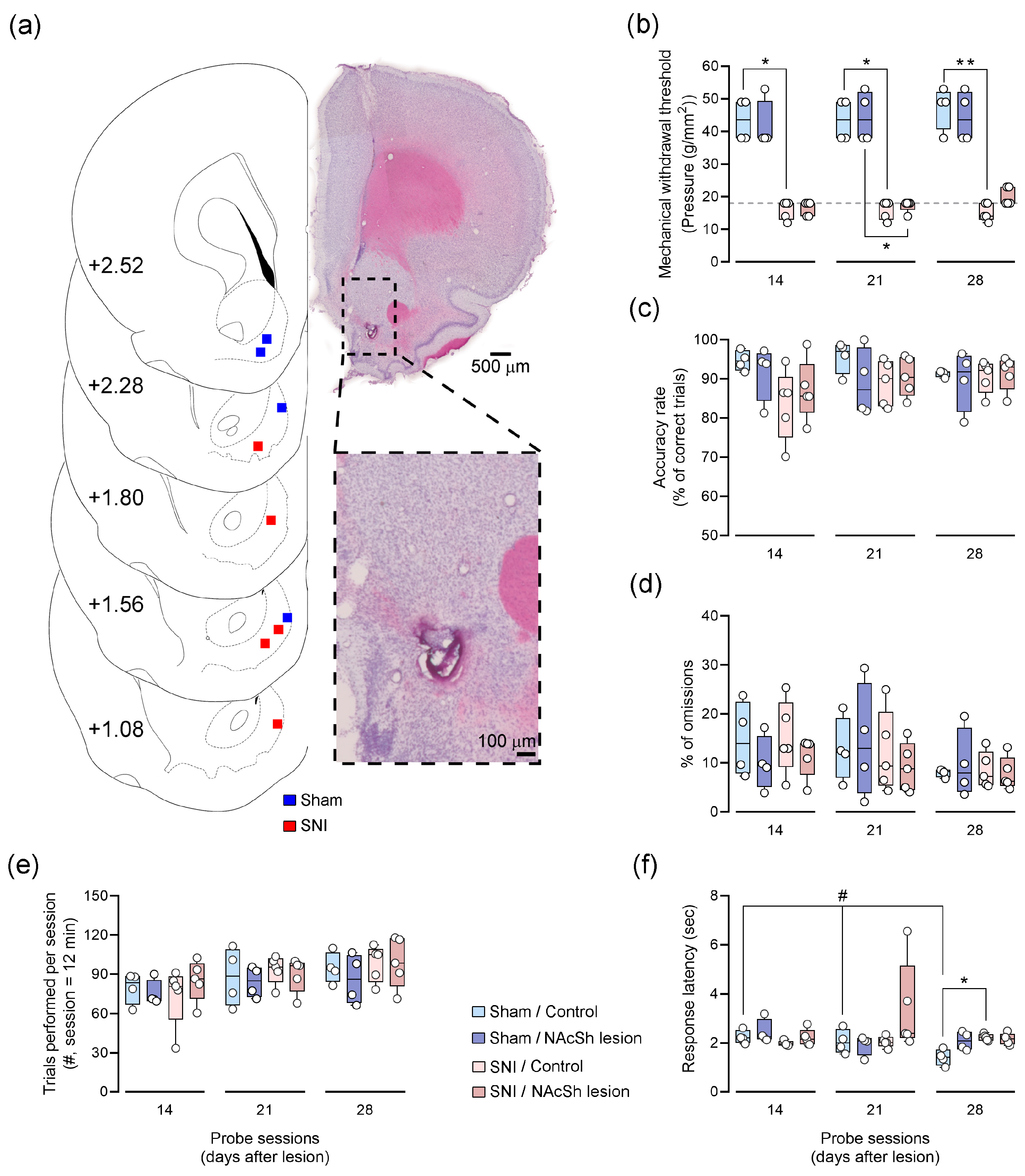
Following the surgical procedures, the rats were assigned to one of the four distinct experimental groups: Sham/Control (n = 4), Sham/NAcSh lesion (n = 4), SNI/Control (n = 5), and SNI/NAcSh lesion (n = 5). To validate the results obtained during the probe sessions, verification of the anatomic location and lesion extension was conducted (Figure 2a). Histological assessment revealed that the electrical lesions in the 18 rats included in this study were localized to the NAcSh area.
The behavioral responses were evaluated on days 14, 21, and 28 post-lesion (Figure 2). To investigate the role of the NAcSh in nocifensive responses, we assessed mechanical withdrawal thresholds using the von Frey filaments test one hour after the second daily probe session. All SNI-treated rats developed mechanical allodynia, evidenced by a significant decrease in the mechanical noxious threshold for all probe sessions comparing the Sham/Control and SNI/Control experimental groups (Figure 2b). Furthermore, it is important to note that SNI/NAcSh-lesioned animals presented a significantly lower mechanical noxious threshold than the Sham/NAcSh-lesioned animals on day 21, but not on day 14 and 28 post-injury. Together, these results suggest that a unilateral NAcSh lesion has no significant impact on peripheral nocifensive responses.
To evaluate the role of NAcSh dependency and peripheral nerve lesions on behavioral accuracy, the rats were tested during the performance of an eight-shaped spatial alternation task at different time points. The rats consistently exhibited performances above 70% throughout all probe sessions, but statistical analysis revealed no significant differences between experimental groups and testing time points (Figure 2c). These findings were also supported by the absence of significant effects related to the number of omissions performed (Figure 2d) and the total number of trials performed (Figure 2e). The lack of differences in these behavioral parameters partially explains the performance similarities and suggests that the animals did not differ in their WM capacity to adequately perform the task.
Apart from performance values, ponderation at the decision point provides insight into the spatial working memory maintenance of the previously navigated path. To test this, we calculated the averaged response latency, representing the time spent between the decision point and the feeder location. Our data revealed that Sham/Control rats significantly decreased their response latency across testing sessions (Figure 2f), suggesting that repeated exposure to behavioral contingencies may impact the behavioral accuracy of these animals. On day 28 post-injury, we also found that SNI-treated rats revealed a higher response latency when compared with Sham-treated rats.

3.3. Impact of NAcSh Lesion on Motor Activity

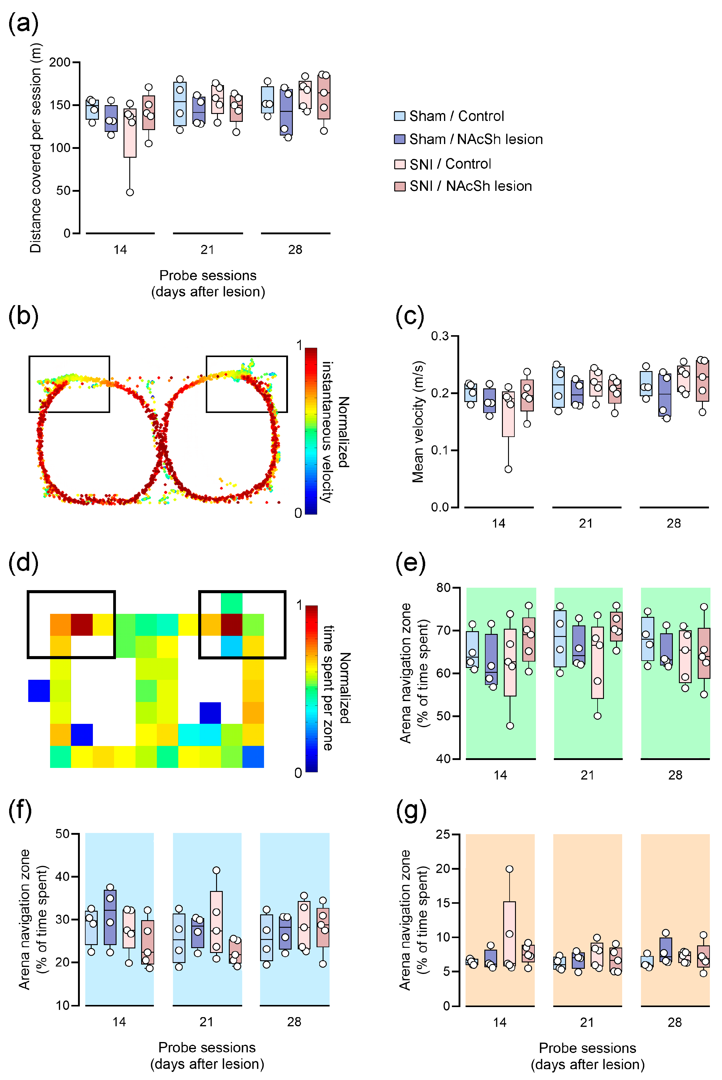
The intrinsic locomotion component linked to the spatial alternation task facilitates the evaluation of motor performance in the intervened animals. An essential factor to consider in this evaluation is the total distance covered during the testing session. Our results show no statistically significant differences in the total distance covered among all experimental groups over time (Figure 3a). This suggests that the SNI-treated groups do not demonstrate motor impairments in executing the task, as the distance covered values closely resemble those of the Sham-treated groups.
To explore potential oscillations in navigation velocity vectors throughout each session, we assessed the instantaneous velocity acquired by each animal in every probe session. An illustration of a typical distribution of instantaneous velocity tracks is depicted in Figure 3b. Consistent navigation patterns were observed across experimental groups in each probe session: the animals exhibited an increase in velocity immediately after departing from the reward area and began to decrease upon reaching the decision point. Next, we evaluated the mean velocity to determine whether neuropathic pain and/or NAcSh lesion induced overall pacing alternations throughout the arena (Figure 3c). In this context, no significant changes were observed in the mean velocity traces of all experimental groups across testing sessions. The absence of a statistical effect suggests that the neuropathic pain model did not influence the animals’ ability to execute motor actions in the spatial alternation task.
To assess alterations in time allocation, we partitioned the behavioral arena into a grid of activity. The colored grid image in Figure 3d illustrates a quantitative approach to the time-spent distribution per navigation zone. Each square is color-coded from dark red (indicating the most time allocated) to dark blue (indicating less time allocated). Across all experimental groups, we observed similar patterns of time allocations throughout the maze, with the majority of time spent in the reward zone and reduced allocation to the delay and choice areas. Our results do not reveal statistical differences between the experimental groups in the reward (Figure 3e), delay (Figure 3f), or choice (Figure 3g) zones across testing sessions. This suggests that animals exhibited similar navigation patterns while performing the spatial alternation task. Overall, these findings indicate that the neuropathic pain model did not disrupt motor capacity in this behavioral task.

4. Discussion

The present study aimed to elucidate the impact of neuropathic pain on the NAcSh and its subsequent effect on spatial WM encoding. The major findings of this study indicate that neither the neuropathic pain model nor NAcSh lesions resulted in changes in the animal’s performance levels during the spatial alternation task, but led to changes in latency responses across probe sessions. Interestingly, unilateral NAcSh lesions showed no significant impact on peripheral nocifensive responses. Finally, we demonstrated that SNI-treated rats did not exhibit motor impairments in the spatial WM task. These outcomes complement our previous findings concerning long-term alterations in NAc local information processing and its transmission to other brain regions in the context of neuropathic pain [18]. The NAc is involved in reward-based learning, spatial memory processes, and nociceptive information processing [13,14,15,29]. Disruptions in NAc activity, often observed in chronic pain conditions including neuropathy, can lead to impairments in WM and pain encoding [21,30,31]. Here, to investigate the involvement of the NAcSh in neuropathic pain-related WM performance, we impaired NAcSh functioning by performing local electrical lesions.
The NAc receives significant projections from both the medial prefrontal cortex and the hippocampus, contributing to the processing of spatial temporary information [10,32]. It has been shown that local infusions of an NMDA receptor antagonist into the NAcSh did not disrupt specific spatial sequences learned by the animals, but NAcSh lesions produced impairments in performance during the task of the eight-arm radial maze [31,33]. This indicates that NAcSh disruption may impair selective spatial WM processes, yet in this study, no spatial impairments were observed. In the current study, neither SNI nor NAcSh lesions reduced the number of trials performed by the animals nor did they impair their accuracy in the eight-shaped alternation task. In addition, SNI-treated rats exhibited a similar number of omissions to Sham-treated rats. While some studies have reported WM performance deficits in SNI-injured animals [34,35,36], others have reported unchanged or improved performance values after dynamic training periods [37]. Previous research using the same task observed similar performance values between Sham and SNI-treated rats from day 5 to day 21 post-surgery [25]. These findings suggest that the pathophysiology of chronic pain may have differential central effects on different brain structures, which may change depending on the temporal evaluation of cognitive alterations. Furthermore, NAcSh-lesioned animals did not display performance differences compared to non-lesioned animals, suggesting an intact capacity to retain temporary spatial information. This contrasts with previous research, where selective excitotoxic, electrical lesions, or AMPA antagonist infusion in the NAcSh led to spatial mnemonic impairments [21,30,31]. However, the observed behavioral phenotype could arise from procedural differences between our study and others, including differences in behavioral contingencies, chosen timepoints of evaluation, and lesion extent. Alterations in these factors could potentiate behavioral differences between experimental groups.
Although the performance values were similar between experimental groups, we found evidence that their response latency was altered differently according to the peripheral lesion. Our data showed that Sham-treated rats decreased their response latency over time, while SNI-treated rats exhibited an increased response latency compared to controls in later testing sessions. This indicates that neuropathic pain interfered with temporary information processing velocity, but this disturbance did not contribute to alterations in WM maintenance. In contrast to previous research on spatial learning that described slower choice responses in animals with electric lesions in the NAcSh [38], our own data revealed that a unilateral NAcSh lesion has no significant impact on the animals’ response. In terms of motor performance, chronic pain patients may exhibit varying degrees of impairments in walking coordination and kinematics [39]. Limiting movement to reduce painful episodes is a common protective response following injury, which can result in a reduction in muscle strength and gait control [40]. Animal studies employing different pain models have observed similar outcomes [18,41]. In the current study, there were no differences observed in total distance traveled, instantaneous velocity, and mean velocity between the experimental groups, suggesting that peripheral injury did not induce motor impairments. This finding aligns with previous studies that reported comparable distances traveled by control and SNI-treated animals [18,37], where animals with peripheral nerve lesions altered their navigation posture to circumvent the painful impact of the injury. In this study, no changes were observed in the allocation time per each designated maze zone during the probe sessions between the experimental groups. This finding is consistent with previous research conducted by our laboratory, where the time spent in the delay and choice zone was comparable between experimental groups [25,42]. In contrast, different results were observed in another study involving an inflammatory pain model [43], which suggests that the lack of differences between groups derived exclusively from the pain model applied. Furthermore, there were no differences between NAcSh-lesioned and non-lesioned animals. Since we previously observed behavioral differences with an inflammatory pain model, deficits derived from the interaction of NAcSh lesioning and distinct pain models could produce distinct results regarding performance in this task.
Several reports support the notion that chronic pain can trigger a substantial reorganization of the ventral striatum [13,44]. In the literature, there are several documented attempts to restore balance within the ventral striatum to alleviate pain responses. Recently, it has been reported that the activation of prefrontal cortex projections to the NAc alleviates both the sensory and affective symptoms associated with neuropathic pain [45,46]. However, it is noteworthy that the NAc plays a more prominent role in pain affective behaviors than in the sensory dimension of pain [47]. As expected, our data showed that SNI-treated rats presented significantly reduced mechanical noxious thresholds after lesions. Furthermore, SNI/NAcSh-lesioned animals showed lower mechanical thresholds compared to Sham/NAcSh-lesioned animals on day 21 post-surgery, though not on days 14 and 28. Although not significant, the SNI/NAcSh lesion experimental group exhibited a slight increase in their mechanical threshold of response compared to the SNI/Control group, which may explain the lack of differences between the two NAcSh-lesioned groups during the 4-week testing period. In neuropathic pain cases, the indirect pathway of the NAcSh becomes hyperexcited, amplifying neuropathic pain [16]. Conversely, blocking DAergic receptors in the NAcSh has been shown to produce a similar effect [48], akin to the slight increase in the mechanical noxious threshold observed in SNI/NAcSh-lesioned animals. Finally, it is important to note that our study had an important limitation associated with the gender-specific effects of neuropathic pain. Pain also demonstrates notable sex dimorphism in NAc-dependent structural functions. Recent data have uncovered sex-specific differences in the NAc area [49,50]. Whether these differences in NAc function are relevant to the neuropathic pain phenotype remains an intriguing area for future research in both pre-clinical and clinical settings. Adding to this, the absence of a significant impact on WM performance and peripheral pain responses could be related to a compensatory plasticity effect induced by the opposing non-lesioned NAcSh and/or other brain areas, which could shadow any role that the NAcSh may have in WM acquisition. Since NAc is regulated by lateral processing [36], one possible explanation may relate to the bilateral cognitive processing of WM in the NAcSh and, as such, could pose as another limitation to our study. Finally, while current therapeutic efforts focus mostly on the sensory dimension of pain to promote pain relief, managing the coupling affective and cognitive aspects of pain should be reinforced in pain treatments. A robust understanding of the therapeutical effects of targeting the NAcSh area to potentially mitigate chronic pain-related symptomatology is therefore necessary.

5. Conclusions

In summary, the present study provides new insights into the role of the NAcSh area in maintaining and processing ongoing spatial information crucial to sustain cognitive demands during prolonged neuropathic pain conditions. Together, our findings suggest that alterations in NAcSh functioning may contribute to precise disruptions in spatial memory encoding, as observed in latency response alterations, shedding light on the contribution of this brain region in navigating cognitive challenges amidst chronic pain states. Future investigations into the specific involvement of this area in WM preserving capacity mechanisms and pain modulation, specifically in the context of chronic pain, will contribute to a deeper comprehension of NAcSh function in normal and pathological conditions.

Author Contributions

All authors had full access to all of the data in the study and take responsibility for the integrity of the data. Conceptualization, C.M., H.C.-C. and V.G.; investigation, H.C.-C. and M.C.-N.; data curation, H.C.-C. and M.C.-N.; writing—original draft, M.C.-N.; writing—review and editing, C.M., H.C.-C., M.C.-N. and V.G.; supervision, C.M., H.C.-C. and V.G.; funding acquisition, H.C.-C. and V.G. All authors have read and agreed to the published version of the manuscript.

Funding

This study was supported by FEDER funds through the Operational Competitiveness (POCI) Program—COMPETE2020, National Funds through Fundação para a Ciência e Tecnologia—FCT (Project POCI-01-0145-FEDER-028181/2017, Grant PTDC/MED-NEU/28181/2017 (H.C.-C.), Project POCI-01-0145-FEDER-028498/2017, Grant PTDC/MED-NEU/28498/2017 (V.G.)), FCT Project 2022.05193.PTDC–DOI: 10.54499/2022.05193.PTDC (V.G.), FCT Grants 2022.00128.CEECind—DOI: 10.54499/2022.00128.CEECIND/CP1735/CT0019 and DL 57/2016/CP1355/CT0015—DOI: 10.54499/DL57/2016/CP1355/CT0015 (H.C.-C.) supported by National Funds, a PhD grant PRT/BD/154966/2023 attributed to M. C.-N. through the EUGLOH/University of Porto Program supported by the European Social Fund (FSE), and Operational Programs—PORTUGAL 2020 and NORTE 2020.

Institutional Review Board Statement

All experiments were performed in accordance with European Union (2010/63/CE) guidelines and the Research and Ethical Issues of the International Association for the Study of Pain [51]. The experimental protocols were also approved by the local ethical committee of the Faculty of Medicine of University of Porto (Project 82/2019; ORBEA, Porto, Portugal) and by the National Direcção Geral de Alimentação e Veterinária Board (Project Ref. 8335/2019; Lisbon, Portugal). At least one category C FELASA-certified experimenter was present when animals were manipulated. All efforts were made to minimize animal suffering and reduce the number of animals used.

Informed Consent Statement

Not applicable.

Data Availability Statement

The data presented in this study are available upon request from the corresponding author.

Conflicts of Interest

The authors declare no conflicts of interest. The funders had no role in the design of the study; in the collection, analyses, or interpretation of the data; in the writing of the manuscript; or in the decision to publish the results.

References

  1. West, E.A.; Moschak, T.M.; Carelli, R.M. Distinct Functional Microcircuits in the Nucleus Accumbens Underlying Goal-Directed Decision-Making. In Goal-Directed Decision Making; Academic Press: Cambridge, MA, USA, 2018; pp. 199–219. [Google Scholar]
  2. Salgado, S.; Kaplitt, M.G. The nucleus accumbens: A comprehensive review. Ster. Funct. Neurosurg. 2015, 93, 75–93. [Google Scholar] [CrossRef] [PubMed]
  3. Makary, M.M.; Polosecki, P.; Cecchi, G.A.; DeAraujo, I.E.; Barron, D.S.; Constable, T.R.; Whang, P.G.; Thomas, D.A.; Mowafi, H.; Small, D.M.; et al. Loss of nucleus accumbens low-frequency fluctuations is a signature of chronic pain. Proc. Natl. Acad. Sci. USA 2020, 117, 10015–10023. [Google Scholar] [CrossRef] [PubMed]
  4. Becerra, L.; Borsook, D. Signal valence in the nucleus accumbens to pain onset and offset. Eur. J. Pain 2008, 12, 866–869. [Google Scholar] [CrossRef] [PubMed]
  5. Becerra, L.; Breiter, H.C.; Wise, R.; Gonzalez, R.; Borsook, D. Reward circuitry activation by noxious thermal stimuli. Neuron 2001, 32, 927–946. [Google Scholar] [CrossRef] [PubMed]
  6. Beyeler, A.; Eckhardt, C.A.; Tye, K.M. Deciphering memory function with optogenetics. Prog. Mol. Biol. Transl. Sci. 2014, 122, 341–390. [Google Scholar] [PubMed]
  7. Ito, R.; Robbins, T.W.; Pennartz, C.M.; Everitt, B.J. Functional Interaction between the hippocampus and nucleus accumbens shell is necessary for the acquisition of appetitive spatial context conditioning. J. Neurosci. 2008, 28, 6950–6959. [Google Scholar] [CrossRef] [PubMed]
  8. Ito, R.; Hayen, A. Opposing roles of nucleus accumbens core and shell dopamine in the modulation of limbic information processing. J. Neurosci. 2011, 31, 6001–6007. [Google Scholar] [CrossRef] [PubMed]
  9. Dutta, S.; Beaver, J.; Halcomb, C.J.; Jasnow, A.M. Dissociable roles of the nucleus accumbens core and shell subregions in the expression and extinction of conditioned fear. Neurobiol. Stress 2021, 15, 100365. [Google Scholar] [CrossRef] [PubMed]
  10. Del Arco, A.; Mora, F. Prefrontal cortex–nucleus accumbens interaction: In vivo modulation by dopamine and glutamate in the prefrontal cortex. Pharmacol. Biochem. Behav. 2008, 90, 226–235. [Google Scholar] [CrossRef]
  11. Baliki, M.N.; Petre, B.; Torbey, S.; Herrmann, K.M.; Huang, L.; Schnitzer, T.J.; Fields, H.L.; Apkarian, A.V. Corticostriatal functional connectivity predicts transition to chronic back pain. Nat. Neurosci. 2012, 15, 1117–1119. [Google Scholar] [CrossRef]
  12. Baliki, M.N.; Mansour, A.R.; Baria, A.T.; Apkarian, A.V. Functional reorganization of the default mode network across chronic pain conditions. PLoS ONE 2014, 9, e106133. [Google Scholar] [CrossRef] [PubMed]
  13. Chang, P.-C.; Pollema-Mays, S.L.; Centeno, M.V.; Procissi, D.; Contini, M.; Baria, A.T.; Martina, M.; Apkarian, A.V. Role of nucleus accumbens in neuropathic pain: Linked multi-scale evidence in the rat transitioning to neuropathic pain. Pain 2014, 155, 1128–1139. [Google Scholar] [CrossRef] [PubMed]
  14. Taylor, A.M.; Becker, S.; Schweinhardt, P.; Cahill, C. Mesolimbic dopamine signaling in acute and chronic pain: Implications for motivation, analgesia, and addiction. Pain 2016, 157, 1194. [Google Scholar] [CrossRef] [PubMed]
  15. Sato, D.; Narita, M.; Hamada, Y.; Mori, T.; Tanaka, K.; Tamura, H.; Yamanaka, A.; Matsui, R.; Watanabe, D.; Suda, Y.; et al. Relief of neuropathic pain by cell-specific manipulation of nucleus accumbens dopamine D1- and D2-receptor-expressing neurons. Mol. Brain 2022, 15, 10. [Google Scholar] [CrossRef] [PubMed]
  16. Ren, W.; Centeno, M.V.; Berger, S.; Wu, Y.; Na, X.; Liu, X.; Kondapalli, J.; Apkarian, A.V.; Martina, M.; Surmeier, D.J. The indirect pathway of the nucleus accumbens shell amplifies neuropathic pain. Nat. Neurosci. 2016, 19, 220–222. [Google Scholar] [CrossRef]
  17. Jarcho, J.M.; Mayer, E.A.; Jiang, Z.K.; Feier, N.A.; London, E.D. Pain, affective symptoms, and cognitive deficits in patients with cerebral dopamine dysfunction. Pain 2012, 153, 744–754. [Google Scholar] [CrossRef]
  18. Cardoso-Cruz, H.; Laranjeira, I.; Monteiro, C.; Galhardo, V. Altered prefrontal-striatal theta-band oscillatory dynamics underlie working memory deficits in neuropathic pain rats. Eur. J. Pain 2022, 26, 1546–1568. [Google Scholar] [CrossRef] [PubMed]
  19. Yang, S.; Boudier-Revéret, M.; Choo, Y.J.; Chang, M.C. Association between chronic pain and alterations in the mesolimbic dopaminergic system. Brain Sci. 2020, 10, 701. [Google Scholar] [CrossRef] [PubMed]
  20. Ploeger, G.E.; BSpruijt, M.; Cools, A.R. Spatial localization in the Morris water maze in rats: Acquisition is affected by intra-accumbens injections of the dopaminergic antagonist haloperidol. Behav. Neurosci. 1994, 108, 927. [Google Scholar] [CrossRef]
  21. Klein, S.; Hadamitzky, M.; Koch, M.; Schwabe, K. Role of glutamate receptors in nucleus accumbens core and shell in spatial behaviour of rats. Neuroscience 2004, 128, 229–238. [Google Scholar] [CrossRef]
  22. Baliki, M.N.; Chang, P.C.; Baria, A.T.; Centeno, M.V.; Apkarian, A.V. Resting-state functional reorganization of the rat limbic system following neuropathic injury. Sci. Rep. 2014, 4, srep06186. [Google Scholar] [CrossRef]
  23. Paxinos, G.; Watson, C. The Rat Brain in Stereotaxic Coordinates: Hard Cover Edition; Elsevier: Amsterdam, The Netherlands, 2006. [Google Scholar]
  24. Decosterd, I.; Woolf, C.J. Spared nerve injury: An animal model of persistent peripheral neuropathic pain. Pain 2000, 87, 149–158. [Google Scholar] [CrossRef] [PubMed]
  25. Cardoso-Cruz, H.; Lima, D.; Galhardo, V. Impaired spatial memory performance in a rat model of neuropathic pain is associated with reduced hippocampus–prefrontal cortex connectivity. J. Neurosci. 2013, 33, 2465–2480. [Google Scholar] [CrossRef]
  26. Aguiar, P.; Mendonça, L.; Galhardo, V. OpenControl: A free opensource software for video tracking and automated control of behavioral mazes. J. Neurosci. Methods 2007, 166, 66–72. [Google Scholar] [CrossRef] [PubMed]
  27. Chaplan, S.R.; Bach, F.W.; Pogrel, J.W.; Chung, J.M.; Yaksh, T.L. Quantitative assessment of tactile allodynia in the rat paw. J. Neurosci. Methods 1994, 53, 55–63. [Google Scholar] [CrossRef]
  28. Cardoso-Cruz, H.; Lima, D.; Galhardo, V. Instability of spatial encoding by CA1 hippocampal place cells after peripheral nerve injury. Eur. J. Neurosci. 2011, 33, 2255–2264. [Google Scholar] [CrossRef]
  29. Haber, S.N. Corticostriatal circuitry. Dialogues Clin. Neurosci. 2016, 18, 7–21. [Google Scholar] [CrossRef]
  30. Jongen-Rêlo, A.L.; Kaufmann, S.; Feldon, J. A differential involvement of the shell and core subterritories of the nucleus accumbens of the rats in memory processes. Behav. Neurosci. 2003, 117, 150. [Google Scholar] [CrossRef] [PubMed]
  31. Gal, G.; Joel, D.; Gusak, O.; Feldon, J.; Weiner, I. The effects of electrolytic lesion to the shell subterritory of the nucleus accumbens on delayed non-matching-to-sample and four-arm baited eight-arm radial-maze tasks. Behav. Neurosci. 1997, 111, 92–103. [Google Scholar] [CrossRef]
  32. Kerfoot, E.C.; Williams, C.L. Contributions of the Nucleus Accumbens Shell in Mediating the Enhancement in Memory Following Noradrenergic Activation of Either the Amygdala or Hippocampus. Front. Pharmacol. 2018, 9, 47. [Google Scholar] [CrossRef]
  33. Maldonado-Irizarry, C.S.; Kelley, A.E. Excitatory amino acid receptors within nucleus accumbens subregions differentially mediate spatial learning in the rat. Behav. Pharmacol. 1995, 6, 527–539. [Google Scholar] [CrossRef] [PubMed]
  34. Ren, W.-J.; Liu, Y.; Zhou, L.-J.; Li, W.; Zhong, Y.; Pang, R.-P.; Xin, W.-J.; Wei, X.-H.; Wang, J.; Zhu, H.-Q.; et al. Peripheral nerve injury leads to working memory deficits and dysfunction of the hippocampus by upregulation of tnf-α in rodents. Neuropsychopharmacology 2011, 36, 979–992. [Google Scholar] [CrossRef] [PubMed]
  35. Hu, Y.; Yang, J.; Hu, Y.; Wang, Y.; Li, W. Amitriptyline rather than lornoxicam ameliorates neuropathic pain-induced deficits in abilities of spatial learning and memory. Eur. J. Anaesthesiol. 2010, 27, 162–168. [Google Scholar] [CrossRef] [PubMed]
  36. Leite-Almeida, H.; Cerqueira, J.J.; Wei, H.; Ribeiro-Costa, N.; Anjos-Martins, H.; Sousa, N.; Pertovaara, A.; Almeida, A. Differential effects of left/right neuropathy on rats’ anxiety and cognitive behavior. Pain 2012, 153, 2218–2225. [Google Scholar] [CrossRef] [PubMed]
  37. Guida, F.; De Gregorio, D.; Palazzo, E.; Ricciardi, F.; Boccella, S.; Belardo, C.; Iannotta, M.; Infantino, R.; Formato, F.; Marabese, I.; et al. Behavioral, Biochemical and Electrophysiological Changes in Spared Nerve Injury Model of Neuropathic Pain. Int. J. Mol. Sci. 2020, 21, 3396. [Google Scholar] [CrossRef] [PubMed]
  38. Thifault, S.; Krémarik, P.; Lalonde, R. Effects of bilateral electrolytic lesions of the medial nucleus accumbens on exploration and spatial learning. Arch. Physiol. Biochem. 1998, 106, 297–307. [Google Scholar] [CrossRef] [PubMed]
  39. Lamoth, C.J.; Stins, J.F.; Pont, M.; Kerckhoff, F.; Beek, P.J. Effects of attention on the control of locomotion in individuals with chronic low back pain. J. Neuroeng. Rehabil. 2008, 5, 13. [Google Scholar] [CrossRef] [PubMed]
  40. Borsook, D. Pain and motor system plasticity. Pain 2007, 132, 8–9. [Google Scholar] [CrossRef] [PubMed]
  41. Dourado, M.; Cardoso-Cruz, H.; Monteiro, C.; Galhardo, V. Effect of Motor Impairment on Analgesic Efficacy of Dopamine D2/3 Receptors in a Rat Model of Neuropathy. J. Exp. Neurosci. 2016, 10, 51–57. [Google Scholar] [CrossRef]
  42. Cardoso-Cruz, H.; Dourado, M.; Monteiro, C.; Matos, M.R.; Galhardo, V. Activation of dopaminergic D2/D3 receptors modulates dorsoventral connectivity in the hippocampus and reverses the impairment of working memory after nerve injury. J. Neurosci. 2014, 34, 5861–5873. [Google Scholar] [CrossRef]
  43. Cardoso-Cruz, H.; Sousa, M.; Vieira, J.B.; Lima, D.; Galhardo, V. Prefrontal cortex and mediodorsal thalamus reduced connectivity is associated with spatial working memory impairment in rats with inflammatory pain. Pain 2013, 154, 2397–2406. [Google Scholar] [CrossRef] [PubMed]
  44. Apkarian, A.V.; Baliki, M.N.; Farmer, M.A. Predicting transition to chronic pain. Curr. Opin. Neurol. 2013, 26, 360–367. [Google Scholar] [CrossRef] [PubMed]
  45. Lee, M.; Manders, T.R.; Eberle, S.E.; Su, C.; D’Amour, J.; Yang, R.; Lin, H.Y.; Deisseroth, K.; Froemke, R.C.; Wang, J. Activation of corticostriatal circuitry relieves chronic neuropathic pain. J. Neurosci. 2015, 35, 5247–5259. [Google Scholar] [CrossRef] [PubMed]
  46. Martinez, E.; Lin, H.H.; Zhou, H.; Dale, J.; Liu, K.; Wang, J. Corticostriatal Regulation of Acute Pain. Front. Cell. Neurosci. 2017, 11, 146. [Google Scholar] [CrossRef] [PubMed]
  47. Navratilova, E.; Xie, J.Y.; Meske, D.; Qu, C.; Morimura, K.; Okun, A.; Arakawa, N.; Ossipov, M.; Fields, H.L.; Porreca, F. Endogenous opioid activity in the anterior cingulate cortex is required for relief of pain. J. Neurosci. 2015, 35, 7264–7271. [Google Scholar] [CrossRef] [PubMed]
  48. Fields, H.L.; Margolis, E.B. Understanding opioid reward. Trends Neurosci. 2015, 38, 217–225. [Google Scholar] [CrossRef] [PubMed]
  49. Kumar, M.; Gusain, C.; Bhatt, B.; Lal, R.; Bishnoi, M. Sex-specific effects of sucrose withdrawal on anxiety-like behavior and neuroimmune response. Neuropharmacology 2024, 249, 109868. [Google Scholar] [CrossRef] [PubMed]
  50. Douton, J.E.; Carelli, R.M. Unraveling Sex Differences in Affect Processing: Unique Oscillatory Signaling Dynamics in the Infralimbic Cortex and Nucleus Accumbens Shell. Biol. Psychiatry Glob. Open Sci. 2023, 4, 354–362. [Google Scholar] [CrossRef]
  51. Zimmermann, M. Ethical guidelines for investigations of experimental pain in conscious animals. Pain 1983, 16, 109–110. [Google Scholar] [CrossRef]
Figure 1. The learning curve of the eight-shaped spatial alternation working memory task. (a) A schematic illustration of the correct trajectory of travel in the eight-shaped maze to obtain a reward at the reward location. (b) A timeline of the experimental protocol. Each rat underwent a NAcSh lesion procedure or sole electrode insertion (control) and was subjected to a Sham treatment or spared nerve injury (SNI). Rats performed three behavioral probe sessions on days 14, 21, and 28 post-lesion. (c) A comparison of the navigation traces of a rat during days 2 and 9 of the training protocol. The rat navigates more efficiently to obtain reward pellets at the end of the learning phase. (d) The percentage of correct trials and omissions per daily testing session. The shaded regions denote the days for which early- (days 2–4, light gray) and late-phase (days 8–10, light gray) learning curve analyses were performed. The percentage of correct trials for all rats increased significantly from the early phase to the late phase in learning (central panel; W = 171, p < 0.001), and the percentage of omissions decreased (left panel; W = −169, p < 0.001). Dashed red and gray lines indicate 50% and 80% performance thresholds, respectively, which animals needed to surpass to qualify for the surgical interventions. (e) Reward side preference of correct trials performed. Rats did not exhibit significant preference for the left or right feeder location during learning (ks2 = 0.20, p = 0.975 (n.s.)). (f) The percentage of time spent in each navigation area during the performance of the eight-shaped operant arena. Rats significantly decreased the time spent in the delay and choice areas (third and fourth panels; W = −117, p < 0.001; W = −169, p = 0.009, respectively) and increased the time spent in the reward location area (second panel; W = 167, p < 0.001). Comparisons between learning phases were conducted using a non-parametric Wilcoxon matched-pairs signed rank test (two-tailed). The comparison of reward side preference distributions was based on the two-sample Kolmogorov–Smirnov test (ks2) test. The analysis involved 18 rats, and the values are presented as mean ± S.D. Significance levels are denoted as ** p < 0.01 and *** p < 0.001.
Applbiosci 03 00019 g001
Figure 2. Peripheral nerve injury had no impact on task performance accuracy, but did affect response latency over time. (a) Histological NAcSh electric lesion validation. Lesion locations are illustrated in blue (Sham) and red (SNI) dots on schematic representations of NAcSh. (b) Mechanical noxious threshold evaluated using von Frey filaments test. SNI experimental groups presented mechanical allodynia over time (day 14: KW = 13.50, p < 0.0001; day 21: KW = 13.79, p < 0.0001; day 28: KW = 14.40, p < 0.0001). (c) Percentage of correct trials performed. Rats presented similar accuracy rates between experimental groups across testing sessions. (d) Percentage of omissions performed. No significant differences were revealed regarding probe session variability between experimental groups. (e) Total number of trials performed in each testing session did not present significant differences between groups and did not change over time for each group. (f) Response latency was significantly different between experimental groups on day 28 (KW = 8.447, p = 0.0234); more specifically, SNI/Control animals presented higher response latencies compared to Sham/Control animals (p < 0.05 (*)). Furthermore, Sham animals significantly improved latency responses over time (KW = 6.962, p = 0.0194 (#)), while other groups did not. Comparisons between experimental groups and probe sessions are based on ordinary one-way ANOVA, followed by post hoc Bonferroni test and Kruskal–Wallis test, and followed by post hoc Dunn’s multiple comparison test. Sham/Control, n = 4; Sham/NAcSh lesion, n = 4; SNI/Control, n = 5; SNI/NAcSh lesion, n = 5. Values are presented as mean ± S.D. Significance levels are denoted as */# p < 0.05, and ** p < 0.01.
Applbiosci 03 00019 g002
Figure 3. Peripheral nerve lesion did not affect motor activity during spatial alternation task. Throughout testing sessions, rats exhibited consistent navigation patterns with no differences in (a) total distance traveled, (b) instantaneous velocity, (c) mean velocity, and (d) time distribution across arena. Furthermore, percentage of time spent in (e) reward, (f) delay, and (g) choice navigation zones did not differ between experimental groups across testing sessions. Black square boxes represent the reward zones. Comparisons between experimental groups and probe sessions were conducted using Kruskal–Wallis test, followed by post hoc Dunn’s multiple comparison test. Sham/Control, n = 4; Sham/NAcSh lesion, n = 4; SNI/Control, n = 5; SNI/NAcSh lesion, n = 5. Values are presented as mean ± S.D.
Applbiosci 03 00019 g003
Disclaimer/Publisher’s Note: The statements, opinions and data contained in all publications are solely those of the individual author(s) and contributor(s) and not of MDPI and/or the editor(s). MDPI and/or the editor(s) disclaim responsibility for any injury to people or property resulting from any ideas, methods, instructions or products referred to in the content.