Article

Exploring the Cellular Interactions of Flavonoids with Similar Structures in Cells Overexpressing the 70 kDa Human Heat Shock Protein

by
Garyfallia Papa
1,
Yannis V. Simos
1,*,
Antrea-Maria Athinodorou
1,
Konstantinos I. Tsamis
1,
Dimitrios Peschos
1,
Charalampos Angelidis
2,
Periklis Pappas
3 and
Patra Vezyraki
1
1
Laboratory of Physiology, Department of Medicine, School of Health Sciences, University of Ioannina, 45110 Ioannina, Greece
2
Laboratory of Biology, Department of Medicine, School of Health Sciences, University of Ioannina, 45110 Ioannina, Greece
3
Laboratory of Pharmacology, Department of Medicine, School of Health Sciences, University of Ioannina, 45110 Ioannina, Greece
*
Author to whom correspondence should be addressed.
Appl. Biosci. 2024, 3(1), 137-150; https://doi.org/10.3390/applbiosci3010009
Submission received: 16 November 2023 / Revised: 30 January 2024 / Accepted: 1 March 2024 / Published: 6 March 2024
(This article belongs to the Special Issue Plant Natural Compounds: From Discovery to Application)

Abstract

:
Flavonoids share a common structural framework that serves as a hallmark indicative of their biological activity. In this study, we investigated the effects of two structurally similar flavonoids, fisetin and morin, through independent and combined in vitro assessments on embryonic mouse cells overexpressing the human 70 kDa heat shock protein (Hsp70) (Tg/Tg) and normal mouse fibroblast cell line (NIH/3T3). The primary objectives were to evaluate the biocompatibility and potential cytotoxicity of these flavonoids, along with assessing the cytoprotective role of Hsp70 in these cellular environments. To address these objectives, we conducted dose- and time-dependent cell survival tests. Additionally, we utilized flow cytometry to detect intracellular reactive oxygen species (ROS) production and to analyze apoptosis and the cell cycle. Throughout the experimental procedures, a notable observation was made: NIH/3T3 normal cells exhibited greater susceptibility compared to Tg/Tg cells when exposed to fisetin and morin. This difference in susceptibility is likely attributed to the robust cytoprotective effect of Hsp70 in Tg/Tg cells. Importantly, both cell lines demonstrated increased sensitivity to fisetin toxicity in comparison to morin, leading to significantly lower cell survival rates. These findings shed light on the differential responses of cell lines to flavonoid exposure, emphasizing the influence of Hsp70 and the distinct impact of fisetin and morin on cell viability.

1. Introduction

Flavonoids are the low-molecular-weight polyphenolic secondary metabolic compounds, present in plant-based foods and beverages, responsible for the color, fragrance, and flavor of plants [1]. Over 8000 individual flavonoids have been identified [2], which are classified into different subgroups such as flavones, flavonols, flavanones, flavanols, isoflavones, anthocyanins, and chalcones [3]. Flavonoids regulate many processes in plants, such as cell growth, pollinator attraction that helps plant germination, and protection against biotic and abiotic stresses, acting as unique UV radiation filters, detoxifying, and antimicrobial agents, as well as imparting heat resistance against frost or drought conditions, supporting the thermal acclimatization of plants [4,5]. Flavonoids are also known for their beneficial biological activities in bacteria, animals, and human health, and are now considered an essential ingredient in the nutraceutical and pharmaceutical sectors [5]. The multifaceted advantages of flavonoids, such as their antioxidant, anti-inflammatory, anti-mutagenic, anti-cancer, anti-angiogenic, anti-microbial, anti-viral, anti-thrombotic, analgesic, neuroprotective [6,7], hepatoprotective [8], and cardioprotective properties [9], contribute to this recognition. While the precise mechanisms of action of flavonoids remain incompletely understood, recent trends in flavonoid research and development focus on the isolation, identification, characterization, and exploration of their functions, particularly in the context of their applications for health benefits [10].
Flavonoids share a common structure consisting of fifteen carbon atoms in their basic skeleton. This structure is characterized by two six-membered phenyl rings (A and B) and a three-carbon unit linking them as C6-C3-C6. Typically, the three-carbon unit connecting the phenyl groups cyclizes with oxygen, forming a third heterocyclic pyran ring (C) [11,12]. The antioxidant capacity of flavonoids is influenced by the quantity and positioning of hydroxy groups on the catechol group of the B-ring, along with the position of the catechol B-ring on the pyran C-ring. The functional hydroxy groups of flavonoids can stabilize free radicals and contribute to antioxidant protection by donating electrons through resonance [13]. Therefore, the biological antioxidant activity of each flavonoid is directly linked to its chemical structure.
Fisetin (3,7,3′,4′-tetrahydroxyflavone) is a bioactive flavonol derived from plants [14]. Structurally, fisetin adopts the diphenylpropane form, consisting of two aromatic rings connected through an oxygenated three-carbon heterocyclic ring. It is complemented by four hydroxyl group substitutions and one carbonyl group (Figure 1A) [15]. Fisetin’s biological activity is attributed to the presence of four hydroxyl groups in the positions (C-3, C-3′, C-4′, C-7) of the three rings and the carbonyl group in the position (C-4) of the heterocyclic ring [16]. The hydroxyl groups of fisetin can bind free radicals, providing protection against ROS [17]. Morin (3,5,7,2′,4′-pentahydroxyflavone) is isolated as a yellow pigment from plants and fruits [18]. Morin’s structure adheres to the basic flavonoid skeletal structure (C6-C3-C6), containing two aromatic benzene rings (A and B), linked by a heterocyclic pyrene ring (C) containing oxygen (Figure 1B) [19]. The chemical properties of morin arise from the presence of five polar hydroxyl groups attached to the positions (C-2′, C-4′, C-3, C-5, and C-7) of the three rings [20].
Hence, structurally, fisetin and morin exhibit minimal differences. The primary distinction between these two flavonoids lies in the presence of an extra hydroxyl group in the morin skeleton. In our study, the similar structure of fisetin and morin prompted us to investigate the possible synergistic or antagonistic action of these two substances. We conducted in vitro studies independently and in combination to explore their effects, examining both normal mice fibroblast cells (NIH/3T3) and embryonic mice cells that overexpress the human 70 kDa heat shock protein Hsp70 (Tg/Tg). The Hsp70 protein reported in this paper is the same as the HSPA1A described in the human Hsp70 nomenclature paper [21]. Hsp70 is normally expressed in cells and its role is the quality control of newly synthesized proteins [22], control of apoptotic mechanisms in an inductive or inhibitory manner [23,24], protein transportation [25], cytoprotection, and ensuring proteοstasis in normal and abnormal conditions. In silico studies have shown that flavonoids such as luteolin, tangeretin, quercetin, kaempferol, myricetin, and taxifolin can bind to the ATPase domain of human Hsp70 [26]. Furthermore, distinct roles have been identified for different flavonoids. Notably, quercetin has been found to inhibit the activity of heat shock factor (HSF) [7], which is essential for the stimulation of Hsp70. Conversely, myricetin has been observed to elevate the levels of intracellular HSF-1 [27]. These observations suggest that investigating the therapeutic potential of a variety of flavonoids across different classes could prove valuable in the development of agents to combat dengue and cancer. Our emphasis was on examining the biocompatibility and potential cytotoxic effects of these two flavonoids. Additionally, we aimed to determine the cytoprotective impact of Hsp70 on cells.

2. Materials and Methods

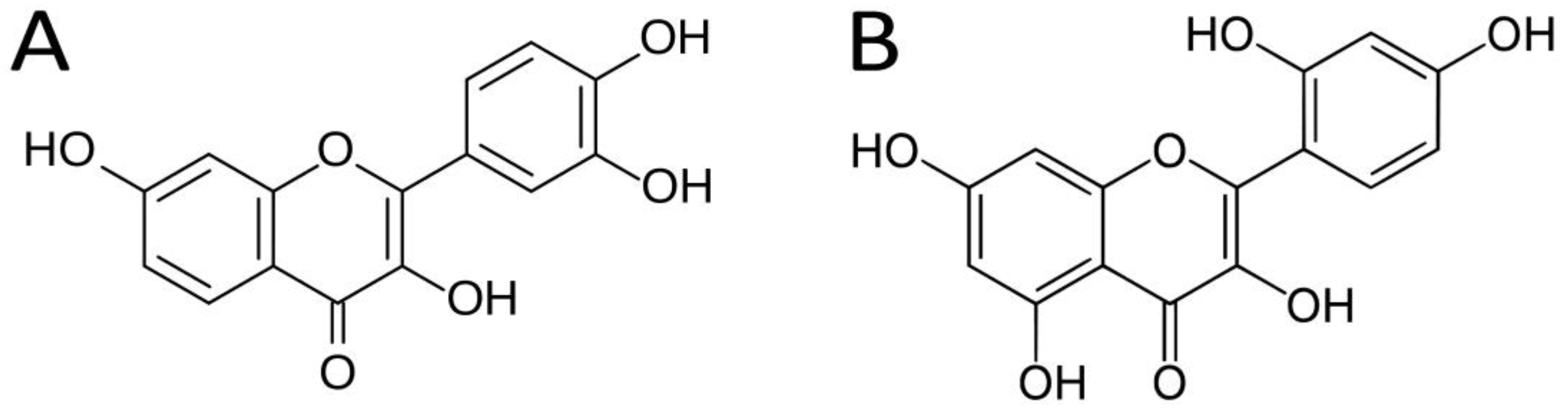
2.1. Chemicals and Reagents

High-glucose Dulbecco’s Modified Eagle’s Medium (DMEM), Phosphate-Buffered Saline (PBS), crystal violet, Propidium Iodide Solution (PI), Dichlorodihydrofluorescein Diacetate (DCFDA), and 3-(4,5-dimethylthiazol-2-yl)—2,5-diphenyltetrazolium bromide solution (MTT) were purchased from Sigma-Aldrich Chemical Co. (St. Louis, MO, USA). Trypsin-EDTA, penicillin–streptomycin, and L-glutamine were obtained from Biowest (Riverside, CA, USA). Fetal Bovine Serum (FBS) was obtained from PAN BIOTECH (Aidenbach, Germany). Hanks’ Balanced Salt Solution (HBSS) was obtained from Biosera (Nuaille, France). Glutaraldehyde 25% and Dimethyl sulfoxide (DMSO) were purchased from Thermo Fisher Scientific Pharmaceutics Inc. (Waltham, MA, USA). Fisetin (3,7,3′,4′-Tetrahydroxyflavone—T0121, purity > 96%-HPLC) and morin (2′,3,4′,5,7-Pentahydroxyflavone Hydrate—P0041, purity > 90%-HPLC) were obtained from Tokyo Chemical Industry Co. (Portland, OR, USA) in a lyophilized solid powder form.

2.2. Cell Lines

In this study, two cell lines were utilized: embryo fibroblasts from Swiss Albino Mice (NIH/3T3, ATCC CRL-1658) and an immortalized cell line derived from transgenic mice that overexpress Hsp70 (Tg/Tg) [28]. Cell growth was conducted in sterile dishes with a diameter of 10 cm. The medium employed for culturing both cell lines consisted of high-glucose DMEM supplemented with 1% (v/v) antibiotics (penicillin–streptomycin), 1% (v/v) L-glutamine, and 10% (v/v) FBS. Both cell lines were nurtured in a humidified incubator at 37 °C with a composition of 95% air and 5% CO2. Subculturing of the cells was carried out approximately two or three times per week.

2.3. Cell Viability Assay

Stock solutions of fisetin and morin were prepared in DMSO at a molar concentration of 50 µM and then stored in the dark at 4 °C. For the evaluation of cell viability, 96-well microplates were employed. In each well, 5000 cells (either Tg/Tg or NIH/3T3 cells) were seeded with 100 μL of the medium and allowed to incubate for 24 h, as previously described [29,30]. After 24 h, fisetin and morin were added in increasing concentrations (5, 10, 25, 50, 100, 200, and 250 μΜ) both individually and in combination. Subsequently, the microwells were supplemented with extra medium, reaching a final volume of 200 µL. Due to DMSO toxicity, its concentration remained below 1% (v/v) at the maximum concentrations. The incubation with the substances lasted 24 or 48 h. After these periods, 40 μL of the reagent MTT was added and the cells were re-incubated under the same conditions for 3 h. Following that, the supernatant was carefully extracted, leaving the formazan crystals undisturbed at the bottom of the microwells. The crystals were then solubilized with 100 µL DMSO. Finally, the optical density of the living cells was measured at 570 nm and 690 nm, with a microplate spectrophotometer (Infinite 200 Pro, Tecan, Switzerland). The experiments were replicated three times for each set of conditions.

2.4. Clonogenic Assay

NIH/3T3 and Τg/Tg cells (at a concentration of 500 cells/mL) were placed in 6-well plates, reaching a final volume of 2 mL per well, and were then incubated for 24 h. Increased concentrations of fisetin and morin were added to the cells, in various combinations, and re-incubation was continued for an additional 24 h. After the incubation period with the two compounds, the supernatant was removed, and the medium was renewed. Following the initial incubation of the cells, they were cultured for 8 days, with a medium renewal performed on day 4. On the 8th day, the medium was removed, and the cells were washed with PBS and stained with 1 mL of dye mixture containing crystal violet (0.5% w/v) and glutaraldehyde (6% v/v), for 30 min. The removal of the dye was accomplished by rinsing the plates, followed by air-drying at room temperature (25 °C) [31]. The quantification of visibly stained colonies was conducted using the Open CFU open-source software (version 3.9.0) [32], and the surviving fraction (SF%) of the cells was then determined. All experiments were repeated three times.

2.5. Determination of Reactive Oxygen Species (ROS) Formation

In 6-well plates, NIH/3T3 and Tg/Tg cells were cultured at a concentration of 75 × 103 cells/mL to a final volume of 2 mL per well. After 6 h, the cells were attached to the plates, and fisetin and morin were added, in two concentrations (5 μM and 10 μM) both individually and in combination, for 24 h. Following the treatment, the supernatant medium was aspirated, and cells were rinsed with PBS, detached using trypsin, washed again with PBS, and then centrifuged at 500× g for 5 min. The cell pellet, obtained through centrifugation, was re-suspended in 2 mL of HBSS containing 2.5 µM DCFDA and incubated for 30 min at 37 °C in the absence of light. Following the incubation, the samples were stained with PI, chilled on ice, and directly analyzed using a fluorescence-activated cell sorting flow cytometer (Partec ML, Partec GmbH, Leipzig, Germany). For every sample, 10,000 events were measured, and all experiments were repeated three times.

2.6. Detection of Apoptosis

NIH/3T3 and Tg/Tg cells were placed in 48-well plates at a density of 5 × 10⁴ cells per well. These plates were then incubated in a humidified environment for 24 h to support cell growth. Following this, the cell medium was replaced with fresh medium containing flavonoids, and the cells were further incubated for an additional 24 h. On the day of processing, cells were detached from the plates using trypsin, and the cell count in each well was determined using a Neubauer hemocytometer. For subsequent analysis, 1 × 10⁵ cells from each well were transferred to a clean Eppendorf tube and centrifuged (500× g), and the cell pellet was re-suspended in 100 µL of Annexin V Binding buffer. The cells were then stained with FITC Annexin V and PI, with a subsequent 15 min incubation at room temperature in the dark. Following incubation, 400 µL of Annexin V binding buffer was added to the samples, and they were examined using a flow cytometer (Partec ML, Partec GmbH, Leipzig, Germany). Annexin V-FITC was excited at 488 nm, yielding a green emission collected at 530 nm by the FL1 detector. In parallel, PI was excited at 561 nm, and its emission was detected at 620 nm (FL3 detector). All experiments were replicated in triplicate.

2.7. Cell Cycle Analysis

NIH/3T3 and Τg/Tg cells (75 × 103 cells/mL to a final volume of 2 mL per well) were seeded in 6-well plates for 24 h. The next day, fisetin and morin were added in two concentrations (5 μM and 10 μM) for another 24 h of incubation. After a 24 h period, the supernatant medium was removed, and the cells underwent two consecutive washes with PBS. Subsequently, they were detached using trypsin, gathered with PBS, and centrifuged at 500× g for 5 min. Following centrifugation, the supernatant PBS was discarded, and the cell pellet was washed once with 1 mL of ice-cold PBS before being re-centrifuged at 500× g for 5 min. The pellet was re-suspended in 0.5 mL of ice-cold PBS, followed by the gradual addition of 0.5 mL of absolute ethanol. In this stage, the samples were stored at −20 °C for a minimum of 1 week. Afterward, the samples underwent centrifugation to eliminate the absolute ethanol, and the cell pellet was re-suspended in 1 mL of fresh ice-cold PBS. PI and RNAse were introduced, and the samples were incubated at 37 °C for 30 min in the absence of light. Measurements were conducted using a fluorescence-activated flow cytometer (Partec ML, Partec GmbH, Jettingen-Scheppach, Germany). Each sample involved the measurement of 10,000 events, and all experiments were replicated three times.

2.8. Statistical Analysis

The data are presented as mean values with standard deviation (+/−). Statistical analysis to determine the significance of differences between means was performed using Student’s t-test. A p-value < 0.05 indicated a statistically significant difference (SPSS version 20.0, Statistical Package for the Social Sciences software, SPSS, Chicago, IL, USA). GraphPad Prism 8 software was utilized for generating all figures.

3. Results and Discussion

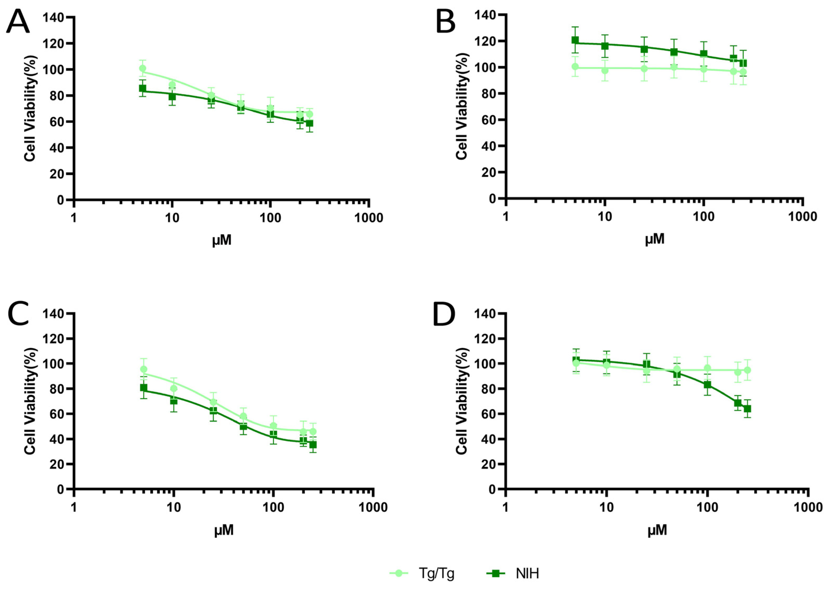
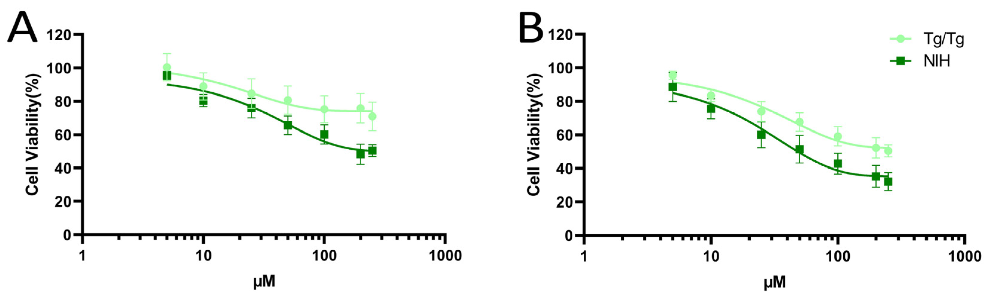
3.1. Cytotoxicity of Fisetin and Morin against NIH/3T3 and Tg/Tg Cells

Fisetin treatment affected cells in a way that depended on both the amount and duration, showing a dose- and time-dependent impact. After 24 h of incubation with fisetin (Figure 2A), there was dose-dependent toxicity, where at the highest concentrations (100–250 µM), the cell viability was reduced by 30–40% in both cell lines. In contrast, morin did not induce any toxic effects on Tg/Tg and NIH/3T3 cells, maintaining their viability at over 90% (Figure 2B). Following 48 h of fisetin treatment, a notable reduction in cell viability was observed in both cell lines. The reduction in cell viability reached levels lower than 40%, indicating a stronger time-dependent toxic effect (Figure 2C). On the contrary, exposure to morin for 48 h induced mild toxicity in a dose- and time-dependent manner, specifically in NIH/3T3 cells. At the highest concentrations (200 µM and 250 µM), only 60% of the cells survived (Figure 2D). When comparing the effects of fisetin, we observed a similar cytotoxic effect in both NIH/3T3 and Tg/Tg cells, and this effect increased over time. In contrast, morin appeared to be non-toxic for Tg/Tg cells at all tested doses.
Fisetin acts antagonistically against Hsp70, inhibiting the heat shock-induced binding of HSF1 to the Hsp70 gene promoter, blocking transcription, and down-regulating protein expression. Therefore, fisetin acts as a potent inhibitor of HSF1, interfering with cancer cell proliferation, and causing apoptosis [33]. Fisetin also showed cytotoxic effects on normal NIH/3T3 cells by reducing their viability, in a study that investigated the anti-angiogenic and anti-cancer effects of this natural flavonoid [34]. Fisetin has previously demonstrated cytotoxicity against various human cancer cell lines, such as breast cancer cells (MCF7) [35], human leukemia cells (HL60) [36], gastric cancer cells (AGS, SNU-1, and FHs74int) [37], and colon cancer cells (HT29) [38]. Morin has also been proven to have cytotoxic effects on many human cancer cell lines such as human melanoma cells (G361, SK-MEL-2) [39], ovarian cancer cells (OVCAR3, SKOV3) [40], bladder cancer cells (EJ) [41] and lung cancer cells (A549) [42].
When both cell lines were simultaneously exposed to both substances at equal concentrations, dose- and time-dependent toxicity was observed. The decline in cell viability followed a pattern similar to that observed after exposure to fisetin (Figure 2). The simultaneous incubation of cells with fisetin and morin did not result in either enhanced cytotoxicity favoring fisetin or decreased cytotoxicity favoring morin. Furthermore, it was demonstrated that NIH/3T3 cells were more susceptible to the toxic effects of both studied substances, with lower rates of cell viability compared to Tg/Tg cells. The observed contrast is likely due to the presence of Hsp70 in Tg/Tg cells, which has a cytoprotective effect, reducing the pronounced toxicity of fisetin and preventing the toxicity of morin (Figure 3).
The IC50 values for fisetin, morin, and the 1:1 mixture of fisetin and morin were calculated where applicable and are presented in Table 1. As mentioned earlier, morin did not impact cell viability; hence, the IC50 values were likely higher than the maximum tested dose. Conversely, fisetin affected cell proliferation. After a 48 h exposure, the IC50 value was lower in NIH/3T3 cells than in Tg/Tg cells (55.6 ± 6 μM vs. 82.5 ± 5 μM). Interestingly, concurrent treatment with equal concentrations of fisetin and morin did not alter the cytotoxic effect of fisetin on NIH/3T3 cells (48.7 ± 7 μM vs. 55.6 μM). However, the presence of morin appeared to weaken fisetin’s toxicity in Tg/Tg cells, where the IC50 value shifted from 82.5 μM to over 250 μM.

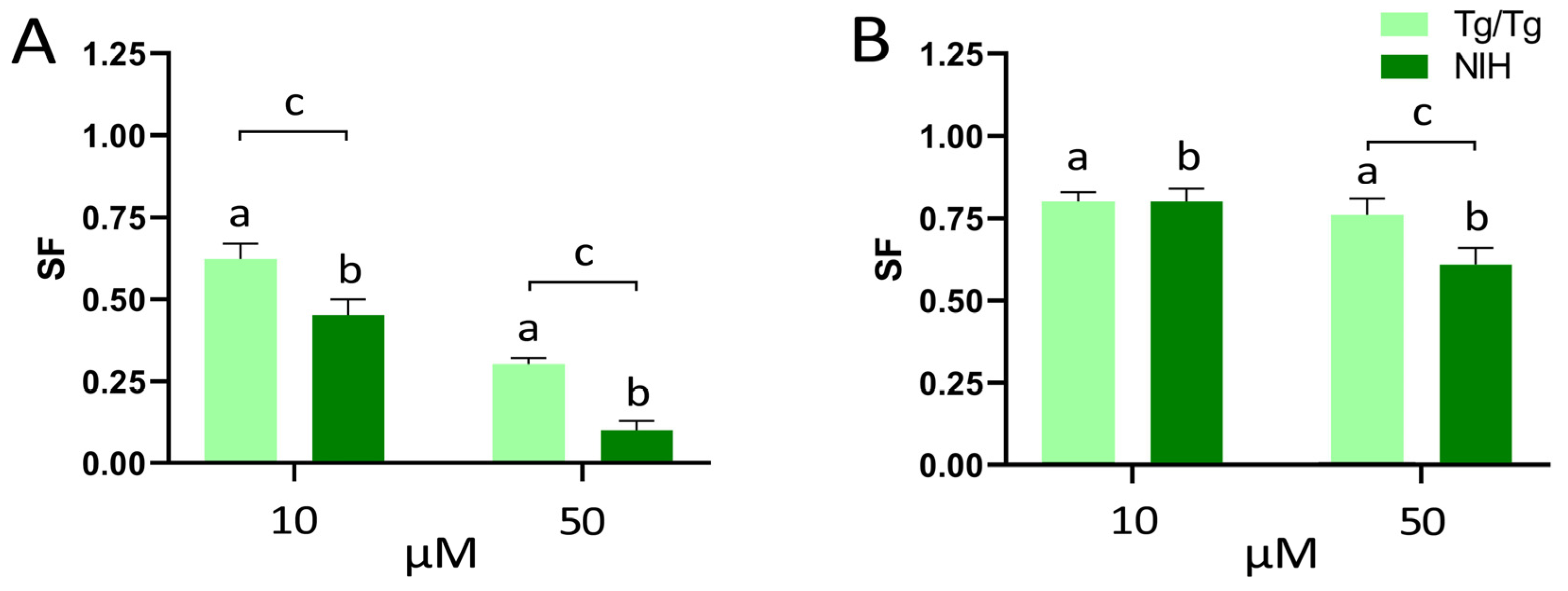
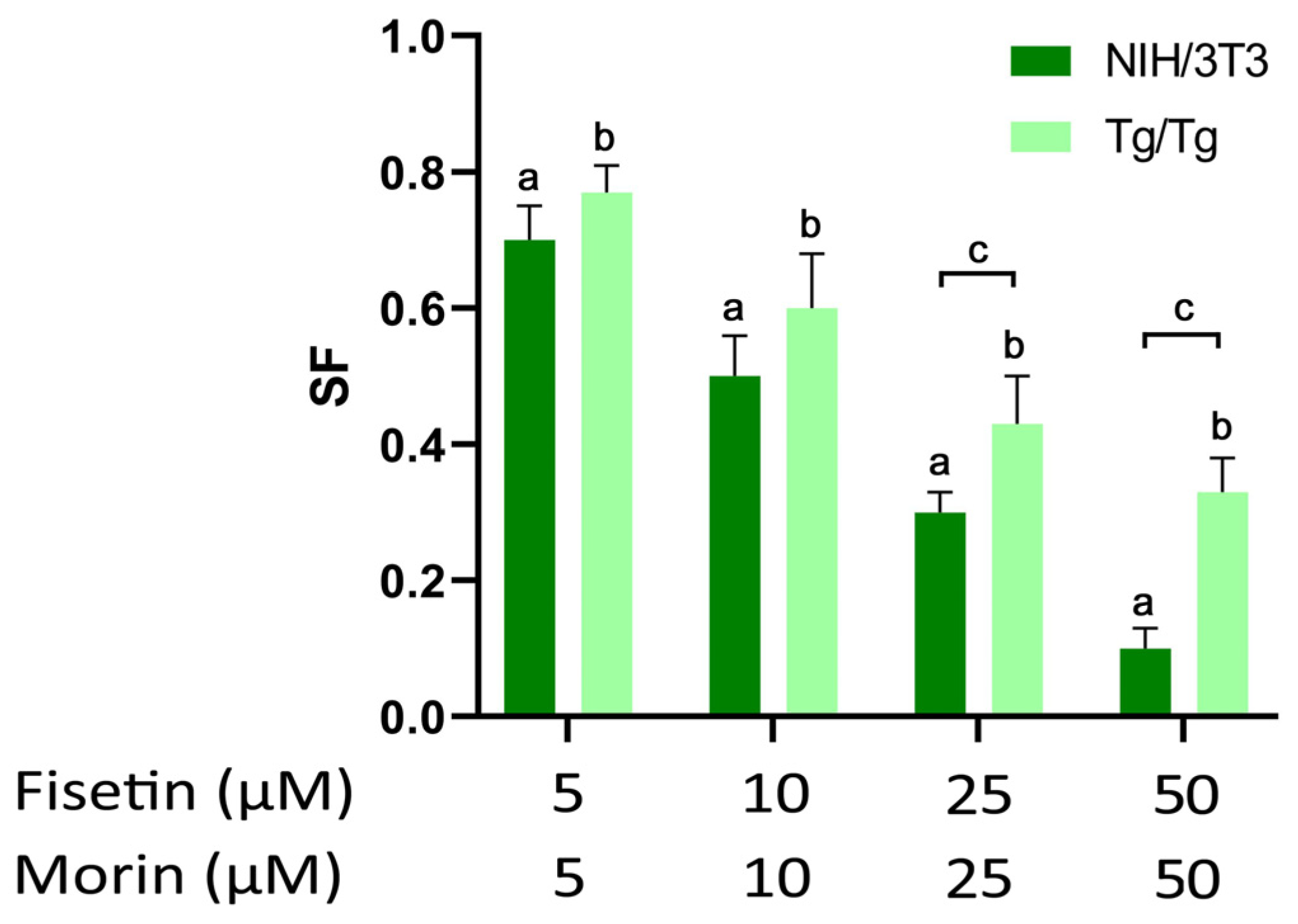
3.2. Long-Term Survival of NIH/3T3 and Tg/Tg Cells after Exposure to Fisetin and Morin

The colony formation assay has been widely employed in numerous cytotoxicity studies because it enables the assessment of compound toxicity over time. Several studies involving both normal and cancer cell lines have demonstrated that the proliferation and clonogenicity of cells were reduced by the actions of various compounds, including flavones (such as calycopterin and luteolin), flavonols (like quercetin), anthocyanins (including cyanidin and delphinidin), and chalcones (e.g., achyrobichalcone) [43,44,45,46].
In NIH/3T3 cells, treatment with fisetin for 24 h (Figure 4A) impaired the cells’ ability to form colonies. At the low dose of 10 µM, the surviving fraction was limited to 0.45, and at the high concentration of 50 µM, a significant decrease to 0.10 was observed. Incubating cells with 10 µM of morin for 24 h (Figure 4B) had a milder effect on the clonogenic ability of cells, resulting in a 0.20 reduction. However, this reduction doubled to 0.40 after treatment with 50 µM.
In Tg/Tg cells, the survival fraction after treatment with fisetin for 24 h (Figure 4A) was higher than that observed in NIH/3T3 cells, with survival rates of 0.62 versus 0.45 and 0.30 versus 0.10, respectively. Conversely, incubation with morin for 24 h (Figure 4B) had a mild effect on the cells, regardless of the exposure dose. The clonogenic ability of the cells remained nearly constant, with survival rates ranging from 0.75 to 0.80.
Exposing both cell lines to a combination of the two flavonoids at equal concentrations for 24 h resulted in a dose-dependent reduction in colony formation when compared to the control. Tg/Tg cells formed more colonies than NIH/3T3 cells at every dose (Figure 5). The survival fraction at 10 μM and 50 μM doses matched that of fisetin alone, confirming our earlier short-term toxicity findings.
In summary, fisetin dose-dependently inhibited the reproductive integrity of both cell lines. NIH/3T3 cells exhibited greater sensitivity to fisetin, resulting in a lower survival fraction compared to Tg/Tg cells. Notably, the inhibition of colony formation in both cell lines was solely due to fisetin’s cytotoxic effect, with no long-term synergistic effect with morin.

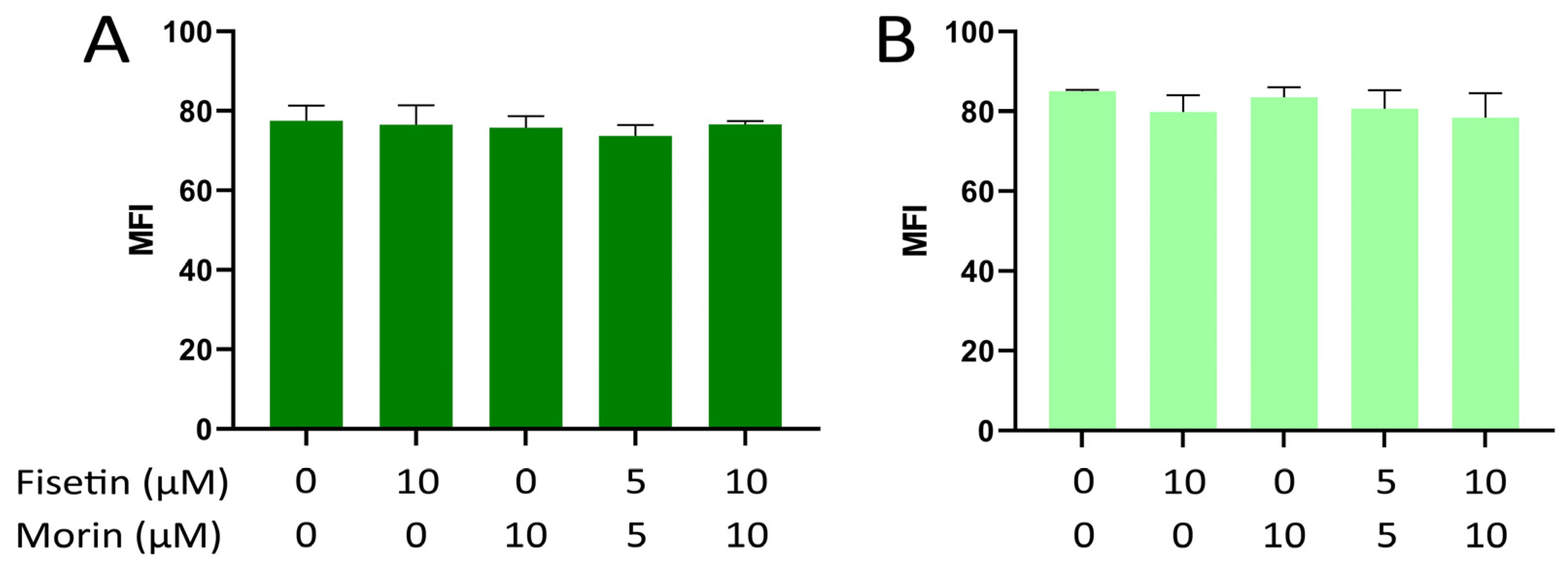
3.3. Intracellular ROS Formation in NIH/3T3 and Tg/Tg Cells

ROS are free radicals that normally function as intracellular messengers, but their overproduction in response to oxidative stress causes damage to the DNA, RNA, and proteins of the cell, resulting in the onset of various diseases in humans, including cancer [47]. Generally, cancer cells have higher ROS levels than normal cells [48]. Flavonoids exhibit anti-cancer effects either by functioning as antioxidant molecules to reduce ROS or by inducing oxidative stress (thereby elevating ROS levels), thus leading to the death of malignant cells [47,49]. However, low doses of some flavonoids have been shown to slightly stimulate cancer cell growth by reducing ROS content [49,50]. Therefore, the evaluation of ROS production is important in the cytotoxic assessment of a flavonoid in both normal and cancer cell lines.
Both fisetin and morin did not trigger the formation of intracellular ROS in NIH/3T3 cells across all tested doses, as the mean fluorescence values (MFI) remained unchanged compared to the control (Figure 6A). Similar findings were noted in Tg/Tg cells, confirming the non-cytotoxicity of these two flavonoids regarding ROS generation (Figure 6B).
Our findings are consistent with the literature, which suggests that fisetin and morin do not act as prooxidants but primarily function as antioxidants after cells are exposed to oxidative stress. Fisetin reduces ROS production triggered by cellular stress stimuli in a dose-dependent manner, observed in both cardiomyocytes (H9C2) [51] and hepatocytes (L02, AML12) [52]. Morin shows similar results in limiting the intracellular generation of ROS in neuroblasts (N2A) and in lung fibroblast cells (V79-4) [53,54]. In neural cells (N2A), the overproduction of ROS induced by high glucose was significantly reduced after treatment with morin, indicating its inhibitory effect on ROS formation [53].

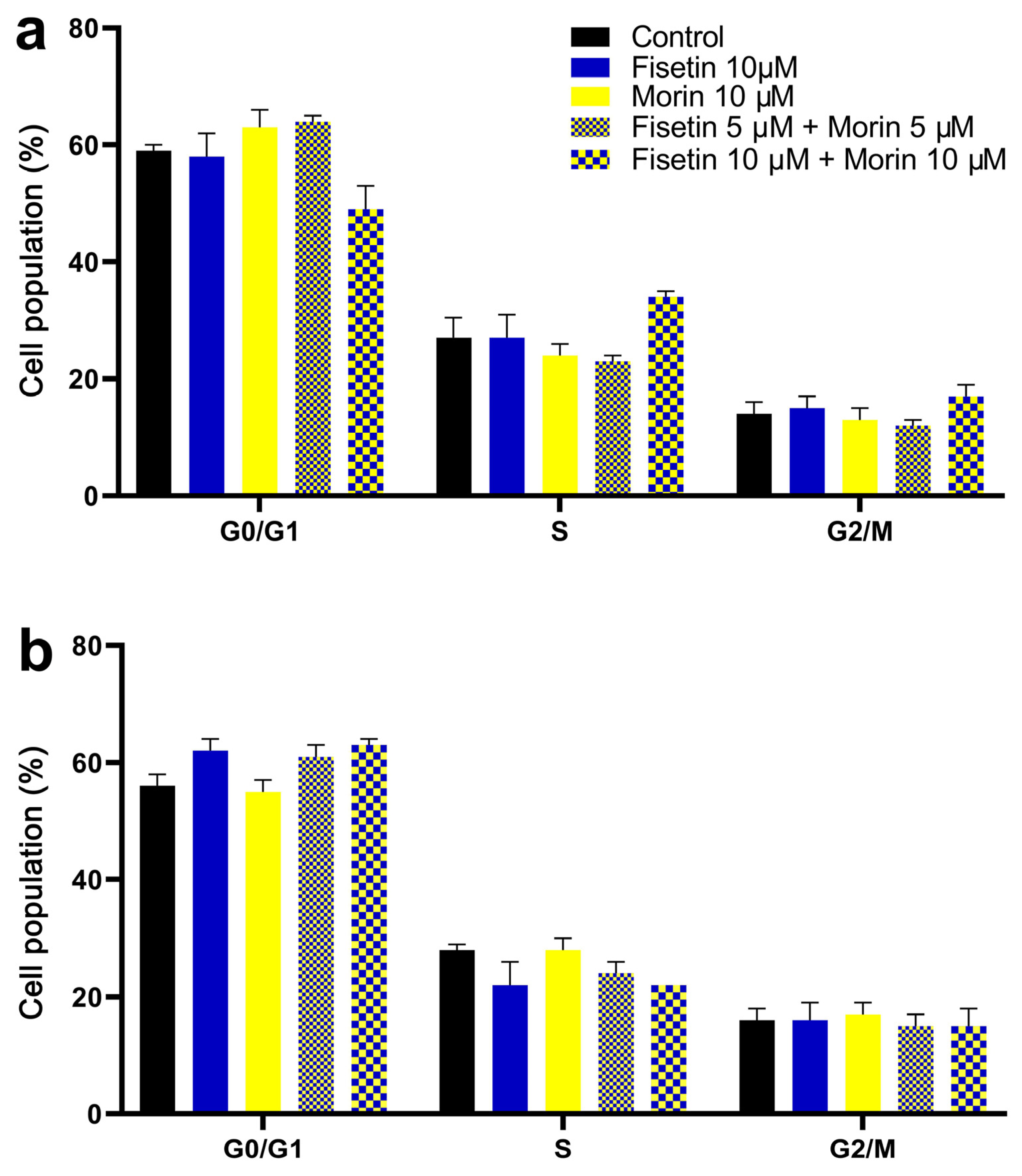
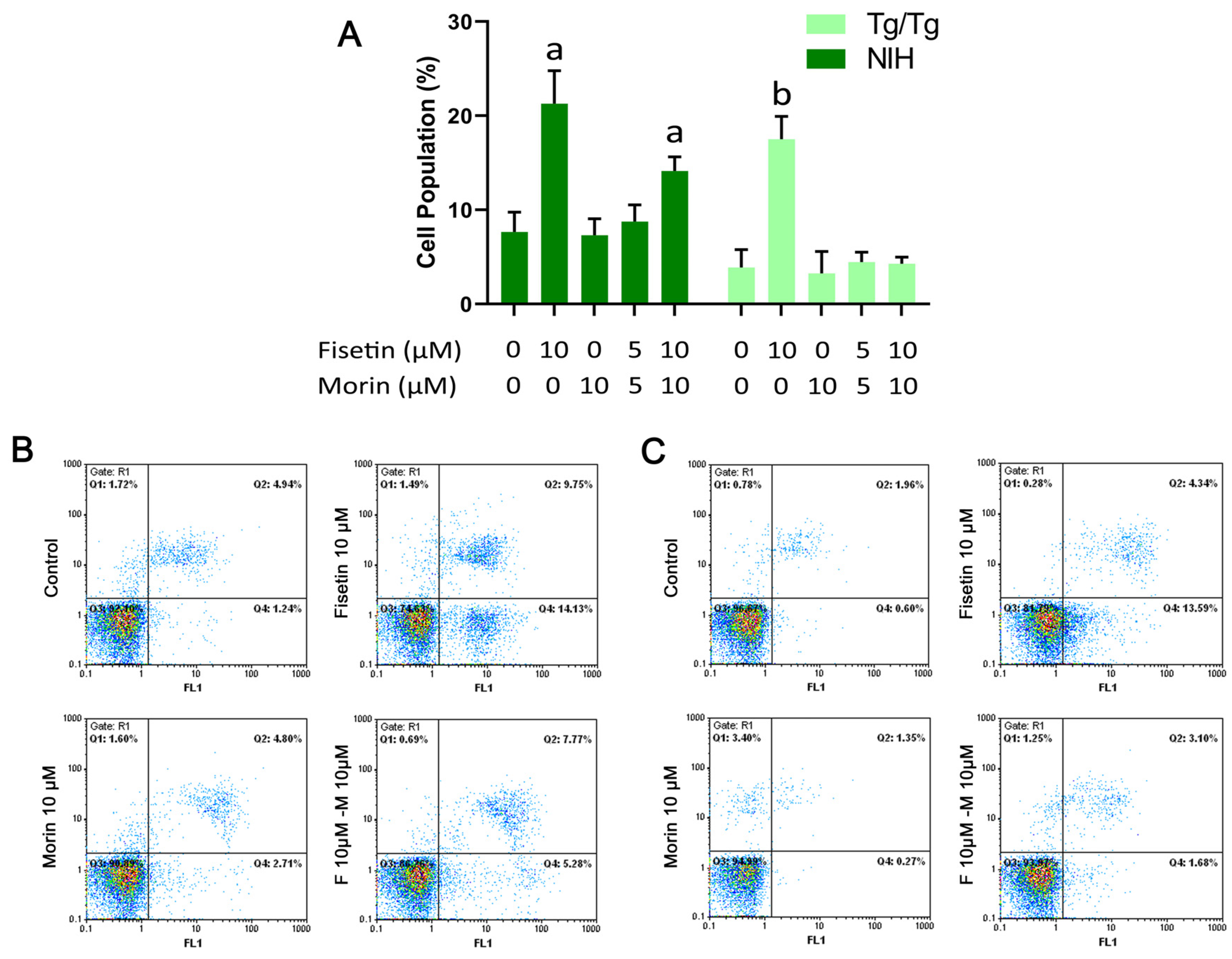
3.4. Cell Cycle Analysis and Induction of Apoptosis

The cell cycle was not significantly affected by the two flavonoids, either alone or in combination, at the investigated concentrations. In NIH/3T3 cells, the concurrent treatment with 10 μΜ of fisetin and morin resulted in a noteworthy, although not significant, rise in the S-phase compared to the control (34 ± 2.2% vs. 27 ± 5.4%) (Figure 7A). Neither of the flavonoids, when administered alone at a dose of 10 μΜ, disrupted the cell cycle progression in NIH/3T3 cells. In Tg/Tg cells, fisetin alone or in combination with morin showed a trend of increasing the cell population by 5–7% in the G0/G1 phase, but again, these changes were not significant (Figure 7B).
In NIH/3T3 cells, exposure to either 10 μM fisetin alone or a combination of fisetin with morin for 24 h led to a 13% and 6% increase in the apoptotic population, respectively (Figure 8). Conversely, in Tg/Tg cells, only fisetin alone triggered a 14% rise in the apoptotic population. However, the presence of morin in Tg/Tg cells nullified the apoptotic potential of fisetin (Figure 8). Notably, none of the flavonoids induced necrosis.
The impact of fisetin on the cell cycle and apoptosis differs across various cell lines. In primary astrocyte cultures, fisetin (at concentrations ranging from 12.5 μM to 50 μM) prompts cell cycle arrest in the G1 phase, while exhibiting no impact on apoptosis [55]. In squamous cell carcinoma cells A431 [56], human HL60 acute promyelocytic leukemia cells [57], and adenocarcinoma HeLa cells [58], fisetin inhibited cell growth by inducing G2/M phase arrest, causing apoptosis. Additionally, in the human colorectal adenocarcinoma cell line HCT-116, the inhibitory impact of fisetin on Hsp70 was examined. Fisetin, at concentrations of 50 µM over a 48 h period, effectively decreased the proliferation of cancer cells. This resulted in a slight elevation in the G2/M phase, and a pronounced increase in the subG1 phase (representing more than 60% of the cell population), indicative of apoptosis induction [59]. A similar outcome was observed in HCT-116 cells with morin, which displayed a dose-dependent inhibition of cell proliferation within the concentration range of 50 μM to 400 µM. This was attributed to cell cycle arrest in both G2/M and S phases, accompanied by an increase in the subG1 phase [60]. In cell lines of human melanoma (G361, SK-MEL-2), morin at rising concentrations (up to 300 μΜ for 48 h) showed a time- and dose-dependent apoptotic effect, as the percentage of cells in the G2/M phase was increased [61]. In human leukemia cells (KCL22, K562, THP-1, and HL-60), morin (50 μΜ) functioned as a significant suppressor of cell growth and induced apoptosis [59]. These findings indicate that fisetin and morin can potentially impede cell growth in various cell lines, possibly by predominantly causing cell cycle arrest in the G2/M phase and triggering apoptosis.
Phenolic compounds exhibit diverse biological effects. When present in mixtures, the characteristics of individual components play a role in determining the overall biological impact of an extract. According to the literature, these properties do not consistently demonstrate an additive effect [39]. It is equally plausible for phytochemicals to exhibit a synergistic or antagonistic mutual interaction with each other. Our experiments revealed that morin acts as an anti-apoptotic factor against the apoptotic activity of fisetin in Tg/Tg cells. This protective effect of morin is observed specifically in cells that overexpress Hsp70. Fisetin is known to induce apoptosis in cancer cells by inhibiting HSF1 activity, thereby impeding its binding to the Hsp70 promoter [62]. Could morin intervene in this pathway to prevent apoptosis? While it is a possibility, as of now, there is a lack of available data supporting this hypothesis regarding the interaction between morin, HSF1, and Hsp70.

4. Conclusions

Our study revolved around two structurally similar flavonoids, fisetin and morin. The objective was to conduct independent and combined in vitro investigations into their effects. We examined their impact on both the normal mouse fibroblast cell line (NIH/3T3) and embryonic mouse cells overexpressing the Hsp70 protein (Tg/Tg). Emphasis was on assessing the biocompatibility, potential cytotoxic effects of the flavonoids, and the potential cytoprotective role of Hsp70. Additionally, this study aimed to explore potential synergistic or antagonistic effects between the two substances. Our experiments revealed that normal cells (NIH/3T3) exhibited greater susceptibility compared to embryonic mouse cells overexpressing human Hsp70 (Tg/Tg) when exposed to these flavonoids. However, both cell lines showed a higher sensitivity to fisetin toxicity compared to morin. These two flavonoids did not generate ROS at low doses, nor did they disturb cell cycle progression; however, fisetin induced apoptosis in both cell lines. The presence of morin in the concurrent scheme of treatment, however, appeared to weaken fisetin’s toxicity in Tg/Tg cells. A finding that was verified with flow cytometry showed that morin reverses the apoptotic potential exerted by fisetin in Tg/Tg cells. With regard to the higher ability of Tg/Tg for long-term survival (clonogenic assay), Hsp70 potentially mediated the survival of the apoptotic cells [63]. Further molecular investigations are necessary to confirm this interaction.
In conclusion, despite sharing similar structures, only fisetin demonstrated noteworthy cellular interactions with NIH/3T3 and Tg/Tg cells. Tg/Tg cells exhibited greater resistance to both substances, particularly against fisetin. Morin appears to block the cytotoxic activity of fisetin in Tg/Tg cells, suggesting a possible cell-protective interaction with Hsp70. Additional research and thorough data analysis are crucial to advance our comprehension of the intricate connection between flavonoid structure and its impact on modulating the activation of apoptotic signal transduction pathways mediated by Hsp70.

Author Contributions

Conceptualization, G.P., Y.V.S. and P.V.; methodology, Y.V.S., C.A. and P.V.; validation, D.P., C.A., P.P. and P.V.; formal analysis, G.P., A.-M.A. and Y.V.S.; investigation, G.P. and A.-M.A.; resources, P.V.; writing—original draft preparation, G.P. and Y.V.S.; writing—review and editing, Y.V.S., K.I.T., D.P., C.A., P.P. and P.V.; visualization, K.I.T., C.A. and Y.V.S.; supervision, P.V.; project administration, Y.V.S.; funding acquisition, P.V. All authors have read and agreed to the published version of the manuscript.

Funding

This research received no external funding.

Data Availability Statement

The data presented in this study are available on request from the corresponding author.

Conflicts of Interest

The authors declare no conflicts of interest.

References

  1. Hernández-Rodríguez, P.; Baquero, L.P.; Larrota, H.R. Flavonoids. In Bioactive Compounds; Campos, M.R.S., Ed.; Elsevier: Amsterdam, The Netherlands, 2019; pp. 265–288. [Google Scholar]
  2. Mutha, R.E.; Tatiya, A.U.; Surana, S.J. Flavonoids as natural phenolic compounds and their role in therapeutics: An overview. Futur. J. Pharm. Sci. 2021, 7, 25. [Google Scholar] [CrossRef]
  3. Safe, S.; Jayaraman, A.; Chapkin, R.S.; Howard, M.; Mohankumar, K.; Shrestha, R. Flavonoids: Structure–function and mechanisms of action and opportunities for drug development. Toxicol. Res. 2021, 37, 147–162. [Google Scholar] [CrossRef]
  4. Shomali, A.; Das, S.; Arif, N.; Sarraf, M.; Zahra, N.; Yadav, V.; Aliniaeifard, S.; Chauhan, D.K.; Hasanuzzaman, M. Diverse physiological roles of flavonoids in plant environmental stress responses and tolerance. Plants 2022, 11, 3158. [Google Scholar] [CrossRef]
  5. Panche, A.N.; Diwan, A.D.; Chandra, S.R. Flavonoids: An overview. J. Nutr. Sci. 2016, 5, e47. [Google Scholar] [CrossRef]
  6. Ullah, A.; Munir, S.; Badshah, S.L.; Khan, N.; Ghani, L.; Poulson, B.G.; Emwas, A.-H.; Jaremko, M. Important flavonoids and their role as a therapeutic agent. Molecules 2020, 25, 5243. [Google Scholar] [CrossRef]
  7. Al-Khayri, J.M.; Sahana, G.R.; Nagella, P.; Joseph, B.V.; Alessa, F.M.; Al-Mussallem, M.Q. Flavonoids as potential anti-inflammatory molecules: A review. Molecules 2022, 27, 2901. [Google Scholar] [CrossRef]
  8. Dhiman, A.; Nanda, A.; Ahmad, S. A quest for staunch effects of flavonoids: Utopian protection against hepatic ailments. Arab. J. Chem. 2016, 9, S1813–S1823. [Google Scholar] [CrossRef]
  9. Ciumărnean, L.; Milaciu, M.V.; Runcan, O.; Vesa, Ș.C.; Răchișan, A.L.; Negrean, V.; Perné, M.-G.; Donca, V.I.; Alexescu, T.-G.; Para, I.; et al. The effects of flavonoids in cardiovascular diseases. Molecules 2020, 25, 4320. [Google Scholar] [CrossRef] [PubMed]
  10. Liga, S.; Paul, C.; Péter, F. Flavonoids: Overview of biosynthesis, biological activity, and current extraction techniques. Plants 2023, 12, 2732. [Google Scholar] [CrossRef] [PubMed]
  11. Ramesh, P.; Jagadeesan, R.; Sekaran, S.; Dhanasekaran, A.; Vimalraj, S. Flavonoids: Classification, function, and molecular mechanisms involved in bone remodeling. Front. Endocrinol. 2021, 12, 779638. [Google Scholar] [CrossRef] [PubMed]
  12. Yamagata, K. Metabolic syndrome: Preventive effects of dietary flavonoids. In Studies in Natural Products Chemistry; Rahman, A., Ed.; Elsevier: Amsterdam, The Netherlands, 2019; Volume 60, pp. 1–28. [Google Scholar]
  13. Dias, M.C.; Pinto, D.C.G.A.; Silva, A.M.S. Plant flavonoids: Chemical characteristics and biological activity. Molecules 2021, 26, 5377. [Google Scholar] [CrossRef] [PubMed]
  14. Khan, N.; Syed, D.N.; Ahmad, N.; Mukhtar, H. Fisetin: A dietary antioxidant for health promotion. Antioxid. Redox. Signal. 2013, 19, 151–162. [Google Scholar] [CrossRef] [PubMed]
  15. Kashyap, D.; Sharma, A.; Sak, K.; Tuli, H.S.; Buttar, H.S.; Bishayee, A. Fisetin: A bioactive phytochemical with potential for cancer prevention and pharmacotherapy. Life Sci. 2018, 194, 75–87. [Google Scholar] [CrossRef] [PubMed]
  16. Mehta, P.; Pawar, A.; Mahadik, K.; Bothiraja, C. Emerging novel drug delivery strategies for bioactive flavonol fisetin in biomedicine. Biomed. Pharmacother. 2018, 106, 1282–1291. [Google Scholar] [CrossRef] [PubMed]
  17. Naeimi, A.F.; Alizadeh, M. Antioxidant properties of the flavonoid fisetin: An updated review of in vivo and in vitro studies. Trends Food Sci. Technol. 2017, 70, 34–44. [Google Scholar] [CrossRef]
  18. Solairaja, S.; Andrabi, M.Q.; Dunna, N.R.; Venkatabalasubramanian, S. Overview of Morin and its complementary role as an adjuvant for anticancer agents. Nutr. Cancer 2021, 73, 927–942. [Google Scholar] [CrossRef]
  19. Deriabina, A.; Prutskij, T.; Castillo Trejo, L.; Sanchez Gutierrez, M.P.; Gonzalez Jimenez, E. Experimental and theoretical study of fluorescent properties of Morin. Molecules 2022, 27, 4965. [Google Scholar] [CrossRef]
  20. Rajput, S.A.; Wang, X.-Q.; Yan, H.-C. Morin hydrate: A comprehensive review on novel natural dietary bioactive compound with versatile biological and pharmacological potential. Biomed. Pharmacother. 2021, 138, 111511. [Google Scholar] [CrossRef]
  21. Kampinga, H.H.; Hageman, J.; Vos, M.J.; Kubota, H.; Tanguay, R.M.; Bruford, E.A.; Cheetham, M.E.; Chen, B.; Hightower, L.E. Guidelines for the nomenclature of the human heat shock proteins. Cell Stress Chaperones 2009, 14, 105–111. [Google Scholar] [CrossRef]
  22. Mayer, M.P.; Bukau, B. Hsp70 chaperones: Cellular functions and molecular mechanism. Cell. Mol. Life Sci. 2005, 62, 670–684. [Google Scholar] [CrossRef]
  23. Ran, R.; Lu, A.; Zhang, L.; Tang, Y.; Zhu, H.; Xu, H.; Feng, Y.; Han, C.; Zhou, G.; Rigby, A.C.; et al. Hsp70 promotes TNF-mediated apoptosis by binding IKKγ and impairing NF-κB survival signaling. Genes. Dev. 2004, 18, 1466–1481. [Google Scholar] [CrossRef]
  24. Beere, H.M.; Wolf, B.B.; Cain, K.; Mosser, D.D.; Mahboubi, A.; Kuwana, T.; Tailor, P.; Morimoto, R.I.; Cohen, G.M.; Green, D.R. Heat-shock protein 70 inhibits apoptosis by preventing recruitment of procaspase-9 to the Apaf-1 apoptosome. Nat. Cell Biol. 2000, 2, 469–475. [Google Scholar] [CrossRef]
  25. Craig, E.A. Hsp70 at the membrane: Driving protein translocation. BMC Biol. 2018, 16, 11. [Google Scholar] [CrossRef] [PubMed]
  26. Hutagalung, R.A.; Reinhart, S.E.; Rosmalena, R.; Nurbaya, S.; Eldafira, E.; Prasasty, V.D. Flavonoid derivatives as potential inhibitors for human Hsp70 using in silico approaches. Eur. Chem. Bull. 2023, 12, 1535–1541. [Google Scholar]
  27. Kim, J.Y.; Barua, S.; Huang, M.Y.; Park, J.; Yenari, M.A.; Lee, J.E. Heat Shock Protein 70 (HSP70) Induction: Chaperonotherapy for Neuroprotection after Brain Injury. Cells 2020, 9, 2020. [Google Scholar] [CrossRef] [PubMed]
  28. Naka, K.K.; Vezyraki, P.; Kalaitzakis, A.; Zerikiotis, S.; Michalis, L.; Angelidis, C. Hsp70 regulates doxorubicin-mediated heart failure in Hsp70-transgenic mice. Cell Stress Chaperones 2014, 19, 853–864. [Google Scholar] [CrossRef]
  29. Zygouri, P.; Athinodorou, A.M.; Spyrou, K.; Simos, Y.V.; Subrati, M.; Asimakopoulos, G.; Vasilopoulos, K.C.; Vezyraki, P.; Peschos, D.; Tsamis, K.; et al. Oxidized-Multiwalled Carbon Nanotubes as Non-Toxic Nanocarriers for Hydroxytyrosol Delivery in Cells. Nanomaterials 2023, 13, 714. [Google Scholar] [CrossRef]
  30. Papanikolaou, E.; Simos, Y.V.; Spyrou, K.; Patila, M.; Alatzoglou, C.; Tsamis, K.; Vezyraki, P.; Stamatis, H.; Gournis, D.P.; Peschos, D.; et al. Does Green Exfoliation of Graphene Produce More Biocompatible Structures? Pharmaceutics 2023, 15, 993. [Google Scholar] [CrossRef] [PubMed]
  31. Franken, N.A.P.; Rodermond, H.M.; Stap, J.; Haveman, J.; van Bree, C. Clonogenic assay of cells in vitro. Nat. Protoc. 2006, 1, 2315–2319. [Google Scholar] [CrossRef]
  32. Geissmann, Q. OpenCFU, a new free and open-source software to count cell colonies and other circular objects. PLoS ONE 2013, 8, e54072. [Google Scholar] [CrossRef]
  33. Qaed, E.; Al-Hamyari, B.; Al-Maamari, A.; Qaid, A.; Alademy, H.; Almoiliqy, M.; Munyemana, J.C.; Al-Nusaif, M.; Alafifi, J.; Alyafeai, E.; et al. Fisetin’s promising antitumor effects: Uncovering mechanisms and targeting for future therapies. Glob. Med. Genet. 2023, 10, 205–220. [Google Scholar] [CrossRef] [PubMed]
  34. Touil, Y.S.; Seguin, J.; Scherman, D.; Chabot, G.G. Improved antiangiogenic and antitumour activity of the combination of the natural flavonoid fisetin and cyclophosphamide in Lewis lung carcinoma-bearing mice. Cancer Chemother. Pharmacol. 2011, 68, 445–455. [Google Scholar] [CrossRef] [PubMed]
  35. Fotsis, T.; Pepper, M.S.; Aktas, E.; Breit, S.; Rasku, S.; Adlercreutz, H.; Wähälä, K.; Montesano, R.; Schweigerer, L. Flavonoids, dietary-derived inhibitors of cell proliferation and in vitro angiogenesis. Cancer Res. 1997, 57, 2916–2921. [Google Scholar] [PubMed]
  36. Lee, W.-R.; Shen, S.-C.; Lin, H.-Y.; Hou, W.-C.; Yang, L.-L.; Chen, Y.-C. Wogonin and fisetin induce apoptosis in human promyeloleukemic cells, accompanied by a decrease of reactive oxygen species, and activation of caspase 3 and Ca2+-dependent endonuclease. Biochem. Pharmacol. 2002, 63, 225–236. [Google Scholar] [CrossRef] [PubMed]
  37. Sabarwal, A.; Agarwal, R.; Singh, R.P. Fisetin inhibits cellular proliferation and induces mitochondria-dependent apoptosis in human gastric cancer cells. Mol. Carcinog. 2017, 56, 499–514. [Google Scholar] [CrossRef] [PubMed]
  38. Kuntz, S.; Wenzel, U.; Daniel, H. Comparative analysis of the effects of flavonoids on proliferation, cytotoxicity, and apoptosis in human colon cancer cell lines. Eur. J. Nutr. 1999, 38, 133–142. [Google Scholar] [CrossRef] [PubMed]
  39. Lee, Y.J.; Kim, W.I.; Kim, S.Y.; Cho, S.W.; Nam, H.S.; Lee, S.H.; Cho, M.K. Flavonoid morin inhibits proliferation and induces apoptosis of melanoma cells by regulating reactive oxygen species, Sp1 and Mcl-1. Arch. Pharm. Res. 2019, 42, 531–542. [Google Scholar] [CrossRef] [PubMed]
  40. Xu, M.; Zhang, Y. Morin inhibits ovarian cancer growth through the inhibition of NF-κB signaling pathway. Anti-Cancer Agents Med. Chem. 2020, 19, 2243–2250. [Google Scholar] [CrossRef]
  41. Shin, S.-S.; Won, S.Y.; Noh, D.-H.; Hwang, B.; Kim, W.-J.; Moon, S.-K. Morin inhibits proliferation, migration, and invasion of bladder cancer EJ cells via modulation of signaling pathways, cell cycle regulators, and transcription factor-mediated MMP-9 expression. Drug Dev. Res. 2017, 78, 81–90. [Google Scholar] [CrossRef]
  42. Yao, D.; Cui, H.; Zhou, S.; Guo, L. Morin inhibited lung cancer cells viability, growth, and migration by suppressing miR-135b and inducing its target CCNG2. Tumor Biol. 2017, 39, 101042831771243. [Google Scholar] [CrossRef]
  43. Lotfizadeh, R.; Sepehri, H.; Attari, F.; Delphi, L. Flavonoid calycopterin induces apoptosis in human prostate cancer cells in-vitro. Iran. J. Pharm. Res. 2020, 19, 391–401. [Google Scholar] [PubMed]
  44. Samuel, T.; Fadlalla, K.; Turner, T.; Yehualaeshet, T.E. The flavonoid quercetin transiently inhibits the activity of taxol and nocodazole through interference with the cell cycle. Nutr. Cancer 2010, 62, 1025–1035. [Google Scholar] [CrossRef] [PubMed]
  45. Intuyod, K.; Priprem, A.; Pairojkul, C.; Hahnvajanawong, C.; Vaeteewoottacharn, K.; Pinlaor, P.; Pinlaor, S. Anthocyanin complex exerts anti-cholangiocarcinoma activities and improves the efficacy of drug treatment in a gemcitabine-resistant cell line. Int. J. Oncol. 2018, 52, 1715–1726. [Google Scholar] [CrossRef] [PubMed]
  46. Souza, P.O.; de Bianchi, S.E.; Figueiró, F.; Heimfarth, L.; Moresco, K.S.; Gonçalves, R.M.; Hoppe, J.B.; Klein, C.P.; Salbego, C.G.; Gelain, D.P.; et al. Anticancer activity of flavonoids isolated from Achyrocline satureioides in gliomas cell lines. Toxicol. Vitr. 2018, 51, 23–33. [Google Scholar] [CrossRef] [PubMed]
  47. Slika, H.; Mansour, H.; Wehbe, N.; Nasser, S.A.; Iratni, R.; Nasrallah, G.; Shaito, A.; Ghaddar, T.; Kobeissy, F.; Eid, A.H. Therapeutic potential of flavonoids in cancer: ROS-mediated mechanisms. Biomed. Pharmacother. 2022, 146, 112442. [Google Scholar] [CrossRef] [PubMed]
  48. Dholaria, M. Flavonoids and its anticancer activity for ROS induced breast cancer. In Handbook of Oxidative Stress in Cancer: Therapeutic Aspects; Chakraborti, S., Ed.; Springer: Singapore, 2021; Volume 1, pp. 305–326. [Google Scholar]
  49. Xi, X.; Wang, J.; Qin, Y.; You, Y.; Huang, W.; Zhan, J. The biphasic effect of flavonoids on oxidative stress and cell proliferation in breast cancer cells. Antioxidants 2022, 11, 622. [Google Scholar] [CrossRef]
  50. Kocyigit, A.; Guler, E.M.; Dikilitas, M. Role of antioxidant phytochemicals in prevention, formation and treatment of cancer. In Reactive Oxygen Species (ROS) in Living Cells; Filip, C., Albu, E., Eds.; InTech: Rijeka, Croatia, 2018. [Google Scholar]
  51. Rodius, S.; de Klein, N.; Jeanty, C.; Sánchez-Iranzo, H.; Crespo, I.; Ibberson, M.; Xenarios, I.; Dittmar, G.; Mercader, N.; Niclou, S.P.; et al. Fisetin protects against cardiac cell death through reduction of ROS production and caspases activity. Sci. Rep. 2020, 10, 2896. [Google Scholar] [CrossRef]
  52. Dai, X.; Kuang, Q.; Sun, Y.; Xu, M.; Zhu, L.; Ge, C.; Tan, J.; Wang, B. Fisetin represses oxidative stress and mitochondrial dysfunction in NAFLD through suppressing GRP78-mediated endoplasmic reticulum (ER) stress. J. Funct. Foods 2022, 90, 104954. [Google Scholar] [CrossRef]
  53. Bachewal, P.; Gundu, C.; Yerra, V.G.; Kalvala, A.K.; Areti, A.; Kumar, A. Morin exerts neuroprotection via attenuation of ROS induced oxidative damage and neuroinflammation in experimental diabetic neuropathy. BioFactors 2018, 44, 109–122. [Google Scholar] [CrossRef] [PubMed]
  54. Zhang, R.; Kang, K.A.; Piao, M.J.; Maeng, Y.H.; Lee, K.H.; Chang, W.Y.; You, H.J.; Kim, J.S.; Kang, S.S.; Hyun, J.W. Cellular protection of morin against the oxidative stress induced by hydrogen peroxide. Chem.-Biol. Interact. 2009, 177, 21–27. [Google Scholar] [CrossRef] [PubMed]
  55. Wang, N.; Yao, F.; Li, K.; Zhang, L.; Yin, G.; Du, M.; Wu, B. Fisetin regulates astrocyte migration and proliferation in vitro. Int. J. Mol. Med. 2017, 39, 783–790. [Google Scholar] [CrossRef]
  56. Pal, H.C.; Sharma, S.; Elmets, C.A.; Athar, M.; Afaq, F. Fisetin inhibits growth, induces G2/M arrest and apoptosis of human epidermoid carcinoma A431 cells: Role of mitochondrial membrane potential disruption and consequent caspases activation. Exp. Dermatol. 2013, 22, 470–475. [Google Scholar] [CrossRef]
  57. Adan, A.; Baran, Y. The pleiotropic effects of fisetin and hesperetin on human acute promyelocytic leukemia cells are mediated through apoptosis, cell cycle arrest, and alterations in signaling networks. Tumor Biol. 2015, 36, 8973–8984. [Google Scholar] [CrossRef] [PubMed]
  58. Afroze, N.; Pramodh, S.; Shafarin, J.; Bajbouj, K.; Hamad, M.; Sundaram, M.K.; Haque, S.; Hussain, A. Fisetin deters cell proliferation, induces apoptosis, alleviates oxidative stress and inflammation in human cancer cells, HeLa. Int. J. Mol. Sci. 2022, 23, 1707. [Google Scholar] [CrossRef]
  59. Kim, J.A.; Lee, S.; Kim, D.-E.; Kim, M.; Kwon, B.-M.; Han, D.C. Fisetin, a dietary flavonoid, induces apoptosis of cancer cells by inhibiting HSF1 activity through blocking its binding to the hsp70 promoter. Carcinogenesis 2015, 36, 696–706. [Google Scholar] [CrossRef]
  60. Hyun, H.-B.; Lee, W.S.; Go, S.-I.; Nagappan, A.; Park, C.; Han, M.H.O.; Hong, S.U.H.; Kim, G.; Kim, G.I.Y.; Cheong, J.; et al. The flavonoid morin from Moraceae induces apoptosis by modulation of Bcl-2 family members and Fas receptor in HCT 116 cells. Int. J. Oncol. 2015, 46, 2670–2678. [Google Scholar] [CrossRef] [PubMed]
  61. Malgorzata, O.T.; Wianowska, D. Antioxidant Properties of Selected Flavonoids in Binary Mixtures—Considerations on Myricetin, Kaempferol and Quercetin. Int. J. Mol. Sci. 2023, 24, 10070. [Google Scholar]
  62. Nie, Z.-Y.; Yang, L.; Liu, X.-J.; Yang, Z.; Yang, G.-S.; Zhou, J.; Qin, Y.; Yu, J.; Jiang, L.-L.; Wen, J.-K.; et al. Morin inhibits proliferation and induces apoptosis by modulating the miR-188-5p/PTEN/AKT regulatory pathway in CML cells. Mol. Cancer Ther. 2019, 18, 2296–2307. [Google Scholar] [CrossRef]
  63. Vasaikar, S.V.; Ghosh, S.; Narain, P.; Basu, A.; Gomes, J. HSP70 mediates survival in apoptotic cells—Boolean network prediction and experimental validation. Front. Cell. Neurosci. 2015, 9, 319. [Google Scholar] [CrossRef]
Figure 1. Chemical structure of fisetin (3,7,3′,4′-tetrahydroxyflavone) (C15H10O6) (A) and morin (3,5,7,2′,4′-pentahydroxyflavone) (C15H10O7) (B).
Applbiosci 03 00009 g001
Figure 2. Viability of Tg/Tg and NIH/3T3 cells post treatment with fisetin for 24 h (A) and 48 h (C), and with morin for 24 h (B) and 48 h (D).
Applbiosci 03 00009 g002
Figure 3. Cell viability in Tg/Tg and NIH/3T3 cells was assessed after concurrent treatment with equal concentrations of fisetin and morin for 24 h (A) and 48 h (B).
Applbiosci 03 00009 g003
Figure 4. Clonogenic assay was conducted on Tg/Tg and NIH/3T3 cells following a 24 h incubation with fisetin (A) and morin (24 h) (B). Statistically significant difference from the following: a, Tg/Tg control; b, NIH/3T3 control; and c, between NIH/3T3 and Tg/Tg cells (p < 0.05).
Applbiosci 03 00009 g004
Figure 5. Clonogenic assay in NIH/3T3 and Tg/Tg cells after simultaneous incubation with fisetin and morin, at equal concentrations for 24 h. Statistically significant difference from the following: a, NIH/3T3 control; b, Tg/Tg control; and c, between NIH/3T3 and Tg/Tg cells (p < 0.05).
Applbiosci 03 00009 g005
Figure 6. ROS generation following the treatment of cells with one dose of fisetin and morin individually (10 μΜ) or two doses of combined fisetin and morin (5 μΜ and 10 μΜ), for 24 h in NIH/3T3 (A) and Tg/Tg cells (B). (MFI, Mean Fluorescence Intensity).
Applbiosci 03 00009 g006
Figure 7. Cell cycle analysis after 24 h treatment of cells with one dose of fisetin and morin individually (10 μΜ) or two doses of combined fisetin and morin (5 μΜ and 10 μΜ), in NIH/3T3 (a) and Tg/Tg cells (b).
Applbiosci 03 00009 g007
Figure 8. Apoptotic cell population after 24 h treatment of cells with one dose of fisetin and morin individually (10 μΜ) or two doses of combined fisetin and morin (5 μΜ and 10 μΜ), in NIH/3T3 and Tg/Tg cells (A). Representative flow cytometry images in NIH/3T3 (B) and Tg/Tg cells (C). Statistically significant difference from control (NIH/3T3) (symbol a) and Tg/Tg (symbol b) (p < 0.05).
Applbiosci 03 00009 g008
Table 1. IC50 values of fisetin and morin.
NIH/3T3Tg/Tg
Fisetin24 h>250 μM>250 μM
48 h55 ± 6 μM82 ± 5 μM
Morin24 h>250 μM>250 μM
48 h>250 μM>250 μM
Fisetin/Morin 1:124 h>250 μM>250 μM
48 h48 ± 7 μM>250 μM
Disclaimer/Publisher’s Note: The statements, opinions and data contained in all publications are solely those of the individual author(s) and contributor(s) and not of MDPI and/or the editor(s). MDPI and/or the editor(s) disclaim responsibility for any injury to people or property resulting from any ideas, methods, instructions or products referred to in the content.

