Article

Treadmill Stepping after Epidural Stimulation Cessation in Decerebrated Cats

by
Natalia Merkulyeva
1,*,
Vsevolod Lyakhovetskii
2,
Oleg Gorskii
3,4 and
Pavel Musienko
3,4,5
1
Laboratory Neuromorphology, Pavlov Institute of Physiology, 199034 St. Petersburg, Russia
2
Lab Motion Physiology, Pavlov Institute of Physiology, 199034 St. Petersburg, Russia
3
Lab of Motor and Visceral Functions Neuromodulation, Pavlov Institute of Physiology, 199034 St. Petersburg, Russia
4
Institute of Translational Biomedicine, St. Petersburg State University, 199034 St. Petersburg, Russia
5
Department of Neurobiology, Sirius University, 1 Olympic Ave., Sirius, 353340 Sochi, Russia
*
Author to whom correspondence should be addressed.
Muscles 2022, 1(2), 102-110; https://doi.org/10.3390/muscles1020011
Submission received: 26 May 2022 / Revised: 28 July 2022 / Accepted: 1 August 2022 / Published: 9 August 2022

Abstract

:
Summary: It was repeatedly shown that the locomotion evoked by epidural-induced electrical stimulation can last for a certain amount of time after stimulation cessation in decerebrated and spinal animals. This so-called after-stepping reflects the maintenance level for the activation of locomotor neuronal circuitry, but only scarce information exists about after-stepping peculiarities. We provide a comparative investigation of after-stepping and stepping under epidural stimulation using electromyographic and kinematic signals as well as ground reaction forces in 16 decerebrated cats. Our principal findings are as follows: (1) the ground reaction forces decrease more after epidural stimulation cessation compared to anterior–posterior limb movements; (2) the step cycle duration is longer for after-steps; (3) the electromyographic signal of the extensor gastrocnemius lateralis muscle during after-stepping decreases faster compared to the signal from the flexors iliopsoas and tibialis anterior and to the extensor soleus muscle; and (4) electromyographic stability is reduced after epidural stimulation cessation. We suppose that different levels of the spinal central pattern generator can be differently attenuated after external trigger cessation. These data could be important for the elaboration of locomotor models and for rehabilitation techniques. New Findings: Our new findings come from comparative investigations of the so-called after-stepping (locomotion after electrical stimulation cessation) and locomotion observed during epidural-induced electrical stimulation. Our new findings are as follows: after epidural stimulation cessation, (1) the ground reaction forces decrease faster compared to anterior–posterior limb movements; (2) the electromyographic signal of the extensor gastrocnemius lateralis muscle decreases faster compared to the signals from the flexors iliopsoas and tibialis anterior and to the extensor soleus muscle; and (3) electromyographic stability is reduced.

1. Introduction

Locomotion is defined as body propulsion by means of coordinated limb movements. These movements are evoked and controlled by specific spinal neuronal networks called central pattern generators (CPGs) [1,2]. CPGs produce a basic rhythmic pattern of alternating flexor and extensor muscle activity during locomotion [3,4]. One method for triggering CPGs is via the electrical stimulation of the brainstem or spinal cord in combination with sensory input from a treadmill [5,6]. It has been repeatedly shown that even after electrical stimulation cessation, locomotion can last for several seconds [7,8,9,10]; these steps are called after-steps [9]. The duration of after-stepping is sensory-dependent; moreover, it is increased after spinalization [9]. Because after-stepping reflects the long-lasting recruitability of the locomotor neuronal networks, we believe that after-stepping is not a simple experimental curiosity, but that it can be a marker for the rhythmogenic status of the spinal CPGs. Many questions related to the after-stepping phenomenon are still unresolved, and its peculiarities, such as its stepping cycle structure and duration and the activity of the flexor/extensor muscles, are scarcely described in the literature. In this study, we investigated after-stepping in decerebrated cats after locomotion evoked by electrical epidural stimulation (ES) using kinematic (for step cycle analysis) and electromyographic recording (to analyse the activity of the flexor and extensor muscles to determine the possible dissimilarity in their ES cessation activity; this dissimilarity may be due to different control schemes performed by CPGs on flexor and extensor muscles).

2. Materials and Methods

Subjects: Sixteen adult cats of either sex (weighing 2.4–4.5 kg) were used. All procedures were conducted in accordance with a protocol approved by the Animal Care Committee of the Pavlov Institute of Physiology, St. Petersburg, Russia, and followed the European Community Council Directive (2010/63EU) for the Care and Use of Laboratory Animals. Experimental procedures have been previously described in detail [11,12,13]. Cats were decerebrated at the precollicular–postmammillary level under isoflurane (2–4%) anaesthesia. A laminectomy was performed in area of lumbar segments L6-L7, and an epidural ball electrode (d = 0.4 mm) was positioned on the dorsal surface of this region. The following stimulation parameters were applied: frequency, 5 Hz; pulse duration, 0.2–0.5 ms; current, 100–250 μA (A-M Systems, model 2100). ES duration varied from 10 s to 40 s. Decerebrated animals were fixed in a rigid frame; hindlimbs were positioned on the treadmill, and the distance between the treadmill belt and the fixed pelvis was 21–25 cm. The treadmill belt was moved backward in relation to the cat at a speed of 0.45 m/s. All of the cats used were able to maintain stepping after ES cessation.
Bipolar electromyographic (EMG) electrodes (0.2 mm flexible stainless steel Teflon-insulated wires, AS632, Cooner Wire, Chatsworth, CA, USA) were implanted into the following left hindlimb muscles: m. iliopsoas (IP, n = 16), m. tibialis anterior (TA, n = 12), m. gastrocnemius lateralis (GL, n = 11), and m. soleus (Sol, n = 10). The signals from the EMG electrodes were differentially amplified (A-M Systems, model 1700, bandwidth of 10 Hz to 5 kHz), digitised at 20 kHz with a LTR System (LTR-EU-16, LTR11, L-Card, Moscow, Russia), and processed using computer programs. EMG data were filtered (100–2000 Hz). Previously, the autocorrelation function of the EMG signal was used in [14] to assess the stepping period. Here, the EMG stability of individual muscles was estimated using the amplitude of the second peak of the autocorrelation function (self-similarity coefficient) [15] of EMG smoothed by a moving average with a window of 50 ms (Figure 1B). This coefficient reflects the similarity between EMG activity for the step series: the more similar the steps are, the greater the value obtained. To avoid an EMG artefact being related to the ES (ES-related artefact), additional filtration was used, and the period lasted from 0.0005 s before to 0.03 s after every ES pulse was removed (Figure 1C). An integrated rectified EMG was calculated using bins corresponding to the stance or the swing phase and according to the potentiometric signal of the hindlimbs. The selection of trials used for EMG analysis was carried out using the following criteria: the trial needed to include at least 10 steps for ES stepping or at least 6 steps for AS stepping, and there needed to be no rude artefacts at the EMG channels. In every cat, one trial was used for every condition (ES stepping and AS stepping).
To characterise the step length and period, the anterior–posterior (A-P) limb movements of all of the cats were estimated using a precision single-turn potentiometer (POT) sensor (Figure 1A) that was digitised at 5 kHz synchronously with the EMG signals. Each step cycle was divided into 20 bins, where 10 bins were in the swing phase, and 10 bins were in the stance phase. Two kinematic characteristics of the stepping were analysed: the total duration of the step cycle, and the duration of the swing and stance phases. The ground reaction forces (GRFs) were also analysed in all cats. Two of the main GRF characteristics were analysed: the maximal GRF values during stepping, and the number of the step phases (1…20) corresponding to the maximal GRFs.
All data were calculated separately to determine the ES-evoked steps (ES steps) and the steps after ES cessation (after-steps, AS). As for the AS steps, only the first steps were assessed as having a GRF signal that was more than or equal to ¼ of the maximum GRF value. Most of the data were calculated as a percent ratio of the AS to ES steps.
Statistical methods: Data are presented as mean ± SD. Mean values for kinematic and EMG parameters were obtained after averaging for 10 to 15 ES steps and from 6 to 10 after-steps. Since there was a non-normal data distribution (Shapiro–Wilk test), and since only paired comparisons (ES vs. AS, inter-muscle comparisons, inter-parameter comparisons) were analysed, a non-parametric Wilcoxon matched-pairs signed-rank test (W test) was used (pairing effectiveness was assessed using the Spearman criterion). Bonferroni adjustment was used to compare these values for the different muscles. A Spearman correlation coefficient was assessed to determine the dependence between the duration of burst activity and the total ES duration. A p-value of 0.05 was used as the cut-off for significance. Statistical calculations were performed using Prism 8.0 (GraphPadSoftware, La Jolla, CA, USA).

3. Results

In all of the cats, we were able to successfully use ES to evoke stepping and the subsequent after-stepping after ES cessation. The number of after-steps varied from 7 to 16. An example of after-stepping is illustrated at Figure 2A. After ES cessation, steps were visible on the POT (anterior–posterior movements) and GRF (increasing and decreasing pressure values) curves, and bursts related to the steps were observed on the EMG curves. An example of a decreasing GRF is illustrated in Figure 2B. In general, both the POT and GRF signals were lowered after ES cessation, but this percent decrease was much more pronounced for the GRF signals (p = 0.0001; W test) (Figure 2C). The number of the step cycle phases corresponding to the maximal GRF signal was unchanged by ES cessation (p = 0.7554; W test) and always corresponded to the middle of the stance phase (Figure 2D).
The step cycle duration of the after-steps was either longer (11/16 cats), equal to (3/16 cats), or shorter (2/16 cats) than the ES-evoked steps. On average, the step cycle became longer for the after-steps (p = 0.0092; W test); only the swing (p = 0.0092; W test) phase, but not the stance (p = 0.1297; W test) phase, of the step cycle was enlarged (Figure 2E). No statistically significant differences were obtained for the percent ratios of the swing and stance phases during after-stepping (p = 0.6685; W test).
The EMG signals of the after-steps differed from those of the ES-evoked steps depending on the muscle. The EMG signal of GL only decreased during after-stepping (11/11 cats, p = 0.0020; W test). The EMG signals of the other muscles (IP, TA, and Sol) could be lowered (7/16 cats, 5/12 cats, and 5/10 cats, respectively) or increased (8/16 cats, 7/12 cats, and 5/10 cats, respectively) compared to the ES steps. In one animal, this signal was unchanged for the IP. Differences in the EMGs of the ES-evoked steps and after-steps were assessed as a percent ratio between their integral EMGs. Two muscles (IP and Sol, but not TA), were significantly different from GL in this regard (p = 0.0010, p = 0.0156, and p = 0.0313; W test) (Figure 2F). Interestingly, the absolute EMG values of the GL signal were not statistically different from those for other muscles (IP, TA, and Sol) during ES-evoked stepping (p = 0.2402, p = 0.9375, p = 0.1094; W test). However, during after-stepping, the EMG value became different from that of the IP (p = 0.0059; W test), but not from those of TA and Sol (p = 0.2188 and p = 0.0313; W test).
The stability of the EMG signal (IP, TA, GL, and Sol) during after-stepping was primarily lower (12/16 cats, 10/12 cats, 10/11 cats, 7/10 cats, respectively) compared to the ES-evoked stepping, but rarely, it could be elevated (4/16 cats, 2/12 cats, 1/11 cats, 3/10 cats, respectively). For two muscles (TA and GL), these differences were statistically reliable (p = 0.0042 and p = 0.0024; W test), but not for IP and Sol (p = 0.0290, p = 0.3223; W test) (Figure 2G). No inter-muscle (IP to TA, IP to GL, IP to Sol, TA to GL, TA to Sol, and GL to Sol) differences in the percent change of EMG stability were obtained (p = 0.3804, p = 0.0674, p = 0.3750, p = 0.8125, p = 0.3125, p = 0.1094, respectively; W test).
We also assessed the duration of the bursts of EMG activity corresponding to hindlimb steps during after-stepping for the IP, TA, GL, and Sol muscles. The shortest duration for this activity was documented for GL; it was significantly different from that of the IP and TA muscles (p = 0.0017, p = 0.0015, respectively; W test) but not for Sol (p = 0.0273; W test). The previous ES duration (varied from 10 s to 40 s) could have had an influence on the investigated parameters, but no significant Spearman correlation coefficient was obtained between the ES duration and the duration of the bursts of EMG activity observed during after-stepping (p = 0.795, p = 0.271, p = 0.443, and p = 0.657, for IP, TA, GL, and Sol, respectively).

4. Discussion

Our principal findings are as follows: (1) the GRFs decreased more after ES cessation compared to anterior–posterior limb movements; (2) the step cycle duration was longer in the after-steps; (3) the EMG signal of the extensor GL muscle decreased faster during AS compared to the flexors and compared to the extensor Sol muscle; and (4) EMG stability was reduced after ES cessation.
The first point may be related to more active flexors being responsible for hindlimb elevation. At the same time, the reduced GRF signal should correspond to the reduced EMG signal of the extensors. However, of the two extensors recorded (GL and Sol), the GL signal was more significantly reduced compared to the Sol signal. Both GL and Sol are extensors, but (1) GL, unlike Sol (and IP and TA), is not a one-joint muscle, but a two-joint muscle [16], and compared to one-joint muscles, several peculiarities have been previously obtained for it [17]. Additionally, (2) Sol, unlike the gastrocnemius, is predominantly composed of type I slow-twitch fibres [18]. Several differences in the behaviour of these two ankle extensors during different locomotor tasks and after the disruption of supraspinal input have been previously documented [19]. For example, regardless of walking speed, Sol activity remained approximately the same, unlike the signals of the gastrocnemius, which varied over a three-fold range at higher speeds [20]. Both the normalised peak and mean EMG of the GL and Sol muscles have different dynamics for down- and up-slope walking in intact cats [21]. A significant reduction in the total EMG signal for Sol, but not for GL, was revealed after spinalization, and this was not related to the degree of muscle atrophy [22]. Moreover, within the ankle muscles, Sol is the most active muscle: its daily activity in cats is approximately 14% compared to 2–9% for other muscles and 7–8% for GL [23]. Relative to the gastrocnemius, the single motor units of Sol are more reactive to vibrations, which allows us to speculate that the one-joint Sol muscle is more sensitive to disturbances in the ankle compared to the two-joint gastrocnemius [24]. It is possible that in our case, the sensory input from the treadmill affected these two ankle extensors differently.
The obtained data can be explained by different spinal and supraspinal control mechanisms for these two muscles. Regardless of these differences, the gastrocnemius and Sol muscles are believed to be a part of the same muscle synergy in some locomotor tasks [25,26]. However, it was previously shown in intact cats that “…posture is the only activity where soleus may be active without corresponding activity in the gastrocnemius muscle” [27]. Moreover, data show that the gastrocnemius is primarily responsible for controlling the direction of the external GRFs and body propulsion but that Sol is primarily an anti-gravity muscle [28]. Correspondingly, the higher rate of Sol activity during after-stepping can be related to the increased retention of postural control after ES cessation.
We also revealed that the fast flexors (IP and TA) are less affected by ES cessation compared to the fast extensor (GL) and that the anterior–posterior movements of the hindlimbs (which are mainly determined by the flexors during anterior shift and by the treadmill during posterior shift) are less attenuated compared to the GRFs (which are mainly determined by extensor activity during the stance phase). Data have shown the peculiarities of flexor and extensor EMG activity during locomotor tasks, e.g., downslope walking [29] or locomotion at high speeds [30,31]. Moreover, these muscles are affected by long-lasting muscle immobilization differently [32]. However, we found no data on the peculiarities regarding the EMG activity of the flexors and extensors during after-stepping.
Additionally, it has been shown that the total duration of after-stepping increased after spinalization [9]; this shows that spinalization disrupts some of the inhibitory supraspinal influence on the CPGs’ recruitability level. In a decerebrated animal model, similar to in intact animals, the activity of the CPGs is mainly affected by medullary reticular formation [33,34]. Interestingly, in thalamic cats, reticular neurons show increased spikes during the period in which flexor muscles are active and decreased spikes when extensor EMGs are observed [35]. However, this was not shown in chronic intact cats when the activity of the reticular neurons was mainly coupled with extensor muscle activity [36]; these differences may be related to the preservation of the above-thalamic influence in intact cats. Thus, different duration of the activity of flexor and extensor muscles during after-stepping in cats with disrupted supraspinal input (decerebrated cats) can be expected.
Thus, the after-stepping that is observed after ES cessation is likely closely related to the rhythmogenic potential of the CPGs. In this sense, the longer duration of the step period can be explained by the “deceleration” of CPG activity after the external trigger disappears. It has been proposed that CPG consists of two principal levels: rhythm generation and pattern formation [37]. The idea of a more stable level of anterior–posterior limb movement (which is mainly determined by the flexors) compared to the GRFs (which are mainly determined by the extensors) possibly suggests that the rhythmogenic and pattern-formation levels of the CPG could be affected differently after ES cessation. Thus, an analysis of after-stepping could provide prognostic value for the efficacy of electrical stimulation in both experimental and clinical studies.
Study limitations: The main limitation of our study is its descriptive nature. We expand the knowledge about the after-stepping phenomenon and describe its peculiarities; however, new studies on the elaboration of algorithms for after-stepping analysis are needed because many questions are still unresolved. In particular, what occurs in the muscles, especially in extensors (knee and hip extensors)? Are any muscle synergies saved for longer than others? Is postural control saved for a longer duration than locomotor ability?
Conclusions: We investigated after-stepping (after ES cessation) in 16 decerebrate cats. EMG, GRF, and kinematic data were analysed. Several facts about after-stepping were obtained in this study. First, the GRFs decreased more after ES cessation compared to anterior–posterior limb movements. Second, the EMG signal of the extensor GL was decreased more after ES cessation compared to the flexor muscles and to another extensor, Sol. Additionally, third, GL has the shortest burst activity duration after ES cessation. We suppose that the data obtained here can be related to the peculiarities of spinal and supraspinal control upon flexor and extensor muscle activity and upon the activity of the main postural muscle, the soleus.

Author Contributions

V.L., O.G., P.M. and N.M. conceived the experiments; N.M. was responsible for the interpreting results and for writing the first draft of the manuscript. V.L., O.G., P.M. and N.M. were responsible for editing the manuscript. All authors have read and agreed to the published version of the manuscript.

Funding

This study was supported by the Russian Science Foundation (RSF grant No. 21-15-00235, data analysis, salary for N.M. and V.L.) and project ID: 73025408/93022798 of the St. Petersburg State University, St. Petersburg, Russia (salary for O.G.).

Institutional Review Board Statement

All procedures were approved by the Ethics Commission of the Pavlov Institute of Physiology (#10/17/2020).

Informed Consent Statement

Not applicable.

Data Availability Statement

The study did not report any data.

Conflicts of Interest

The authors declare no conflict of interest.

References

  1. Kiehn, O. Decoding the organization of spinal circuits that control locomotion. Nat. Rev. Neurosci. 2016, 17, 224–238. [Google Scholar] [CrossRef] [PubMed]
  2. Grillner, S.; El Manira, A. Current principles of motor control, with special reference to vertebrate locomotion. Physiol. Rev. 2020, 100, 271–320. [Google Scholar] [CrossRef] [PubMed] [Green Version]
  3. Orlovsky, G.N.; Deliagina, T.G.; Grillner, S. Neuronal Control of Locomotion; Oxford University Press: Oxford, UK, 1999. [Google Scholar]
  4. Rossignol, S. Neuronal control of stereotypic limb movements. In Handbook of Physiology; Rowell, L.B., Sheperd, J.T., Eds.; Oxford UP: New York, NY, USA, 1996; pp. 173–216. [Google Scholar]
  5. Gerasimenko, Y.; Roy, R.R.; Edgerton, V.R. Epidural stimulation: Comparison of the spinal circuits that generate and control locomotion in rats, cats and humans. Exp. Neurol. 2008, 209, 417–425. [Google Scholar] [CrossRef] [PubMed] [Green Version]
  6. Edgerton, V.R.; Harkema, S. Epidural stimulation of the spinal cord in spinal cord injury: Current status and future challenges. Expert Rev. Neurother. 2011, 11, 1351–1353. [Google Scholar] [CrossRef] [PubMed] [Green Version]
  7. Shik, M.L.; Severin, F.V.; Orlovskiĭ, G.N. Control of walking and running by means of electric stimulation of the midbrain. Biofizika 1966, 11, 659–666. [Google Scholar] [PubMed]
  8. Iwahara, T.; Atsuta, Y.; Garcia-Rill, E.; Skinner, R.D. Spinal cord stimulation-induced locomotion in the adult cat. Brain Res. Bull. 1992, 28, 99–105. [Google Scholar] [CrossRef]
  9. Musienko, P.E.; Bogacheva, I.N.; Gerasimenko, Y.P. Significance of peripheral feedback in the generation of stepping movements during epidural stimulation of the spinal cord. Neurosci. Behav. Physiol. 2007, 37, 181–190. [Google Scholar] [CrossRef]
  10. Gerasimenko, Y.; Musienko, P.; Bogacheva, I.; Moshonkina, T.; Savochin, A.; Lavrov, I.; Roy, R.R.; Edgerton, V.R. Propriospinal bypass of the serotonergic system that can facilitate stepping. J. Neurosci. 2009, 29, 5681–5689. [Google Scholar] [CrossRef] [Green Version]
  11. Musienko, P.E.; Zelenin, P.V.; Lyalka, V.F.; Gerasimenko, Y.P.; Orlovsky, G.N.; Deliagina, T.G. Spinal and supraspinal control of the direction of stepping during locomotion. J. Neurosci. 2012, 32, 17442–17453. [Google Scholar] [CrossRef] [Green Version]
  12. Merkulyeva, N.; Veshchitskii, A.; Gorsky, O.; Pavlova, N.; Zelenin, P.V.; Gerasimenko, Y.; Deliagina, T.G.; Musienko, P. Distribution of spinal neuronal networks controlling forward and backward locomotion. J. Neurosci. 2018, 38, 4695–4707. [Google Scholar] [CrossRef]
  13. Merkulyeva, N.; Lyakhovetskii, V.; Veshchitskii, A.; Gorskii, O.; Musienko, P. Rostrocaudal distribution of the C-Fos-immunopositive spinal network defined by muscle activity during locomotion. Brain Sci. 2021, 11, 69. [Google Scholar] [CrossRef] [PubMed]
  14. Bogacheva, I.N.; Kucher, V.I.; Shcherbakova, N.A.; Musienko, P.E.; Gerasimenko, I.P. Mathematical modeling of the mechanisms of locomotory pattern formation under epidural spinal cord stimulation with consideration of peripheral feedback. Biofizika 2005, 50, 1125–1130. (In Russian) [Google Scholar] [PubMed]
  15. Kim, S.A.; Heinze, K.G.; Schwille, P. Fluorescence correlation spectroscopy in living cells. Nat. Methods 2007, 4, 963–973. [Google Scholar] [CrossRef] [PubMed]
  16. Nichols, T.R. The organization of heterogenic reflexes among muscles crossing the ankle joint in the decerebrate cat. J. Physiol. 1989, 410, 463–477. [Google Scholar] [CrossRef]
  17. Engberg, I.; Lundberg, A. An electromyographic analysis of muscular activity in the hindlimb of the cat during unrestrained locomotion. Acta Physiol. Scand. 1969, 75, 614–630. [Google Scholar] [CrossRef]
  18. Burke, R.E.; Levine, D.N.; Salcman, M.; Tsairis, P. Motor units in cat soleus muscle: Physiological, histochemical and morphological characteristics. J. Physiol. 1974, 238, 503–514. [Google Scholar] [CrossRef] [Green Version]
  19. Côté, M.-P.; Murray, L.M.; Knikou, M. Spinal control of locomotion: Individual neurons, their circuits and functions. Front. Physiol. 2018, 9, 784. [Google Scholar] [CrossRef] [Green Version]
  20. Walmsley, B.; Hodgson, J.A.; Burke, R.E. Forces produced by medial gastrocnemius and soleus muscles during locomotion in freely moving cats. J. Neurophysiol. 1978, 41, 1203–1216. [Google Scholar] [CrossRef]
  21. Gregor, R.J.; Smith, D.W.; Prilutsky, B.I. Mechanics of slope walking in the cat: Quantification of muscle load, length change, and ankle extensor EMG patterns. J. Neurophysiol. 2006, 95, 1397–1409. [Google Scholar] [CrossRef] [Green Version]
  22. Alaimo, M.A.; Smith, J.L.; Roy, R.R.; Edgerton, V.R. EMG activity of slow and fast ankle extensors following spinal cord transection. J. Appl. Physiol. Respir. Environ. Exerc. Physiol. 1984, 56, 1608–1613. [Google Scholar] [CrossRef]
  23. Hensbergen, E.; Kernell, D. Daily durations of spontaneous activity in cat’s ankle muscles. Exp. Brain Res. 1997, 115, 325–332. [Google Scholar] [CrossRef] [PubMed]
  24. Mildren, R.L.; Peters, R.M.; Carpenter, M.G.; Blouin, J.-S.; Inglis, J.T. Soleus single motor units show stronger coherence with Achilles tendon vibration across a broad bandwidth relative to medial gastrocnemius units while standing. J. Neurophysiol. 2019, 122, 2119–2129. [Google Scholar] [CrossRef] [PubMed]
  25. Nishida, K.; Hagio, S.; Kibushi, B.; Moritani, T.; Kouzaki, M. Comparison of muscle synergies for running between different foot strike patterns. PLoS ONE 2017, 12, e0171535. [Google Scholar] [CrossRef] [PubMed]
  26. Klishko, A.N.; Akyildiz, A.; Mehta-Desai, R.; Prilutsky, B.I. Common and distinct muscle synergies during level and slope locomotion in the cat. J. Neurophysiol. 2021, 126, 493–515. [Google Scholar] [CrossRef] [PubMed]
  27. Hodgson, J.A. The relationship between soleus and gastrocnemius muscle activity in conscious cats—A model for motor unit recruitment? J. Physiol. 1983, 337, 553–562. [Google Scholar] [CrossRef] [Green Version]
  28. Kaya, M.; Leonard, T.R.; Herzog, W. Control of ground reaction forces by hindlimb muscles during cat locomotion. J. Biomech. 2006, 39, 2752–2766. [Google Scholar] [CrossRef]
  29. Smith, J.L.; Carlson-Kuhta, P.; Trank, T.V. Forms of forward quadrupedal locomotion. III. A comparison of posture, hindlimb kinematics, and motor patterns for downslope and level walking. J. Neurophysiol. 1998, 79, 1702–1716. [Google Scholar] [CrossRef]
  30. Shik, M.L.; Orlovsky, G.N. Neurophysiology of locomotor automatism. Physiol. Rev. 1976, 56, 465–501. [Google Scholar] [CrossRef]
  31. Frigon, A.; Thibaudier, Y.; Hurteau, M.-F. Modulation of forelimb and hindlimb muscle activity during quadrupedal tied-belt and split-belt locomotion in intact cats. Neuroscience 2015, 290, 266–278. [Google Scholar] [CrossRef]
  32. Hník, P.; Vejsada, R.; Goldspink, D.F.; Kasicki, S.; Krekule, I. Quantitative evaluation of electromyogram activity in rat extensor and flexor muscles immobilized at different lengths. Exp. Neurol. 1985, 88, 515–528. [Google Scholar] [CrossRef]
  33. Schomburg, E.D. Spinal sensorimotor systems and their supraspinal control. Neurosci. Res. 1990, 7, 265–340. [Google Scholar] [CrossRef]
  34. Takakusaki, K.; Chiba, R.; Nozu, T.; Okumura, T. Brainstem control of locomotion and muscle tone with special reference to the role of the mesopontine tegmentum and medullary reticulospinal systems. J. Neural Transm. 2016, 123, 695–729. [Google Scholar] [CrossRef] [PubMed] [Green Version]
  35. Shimamura, M.; Kogure, I.; Wada, S. Reticular neuron activities associated with locomotion in thalamic cats. Brain Res. 1982, 231, 51–62. [Google Scholar] [CrossRef]
  36. Drew, T.; Dubuc, R.; Rossignol, S. Discharge patterns of reticulospinal and other reticular neurons in chronic, unrestrained cats walking on a treadmill. J. Neurophysiol. 1986, 55, 375–401. [Google Scholar] [CrossRef]
  37. Rybak, I.A.; Stecina, K.; Shevtsova, N.A.; McCrea, D.A. Modelling spinal circuitry involved in locomotor pattern generation: Insights from the effects of afferent stimulation. J. Physiol. 2006, 577, 641–658. [Google Scholar] [CrossRef] [PubMed]
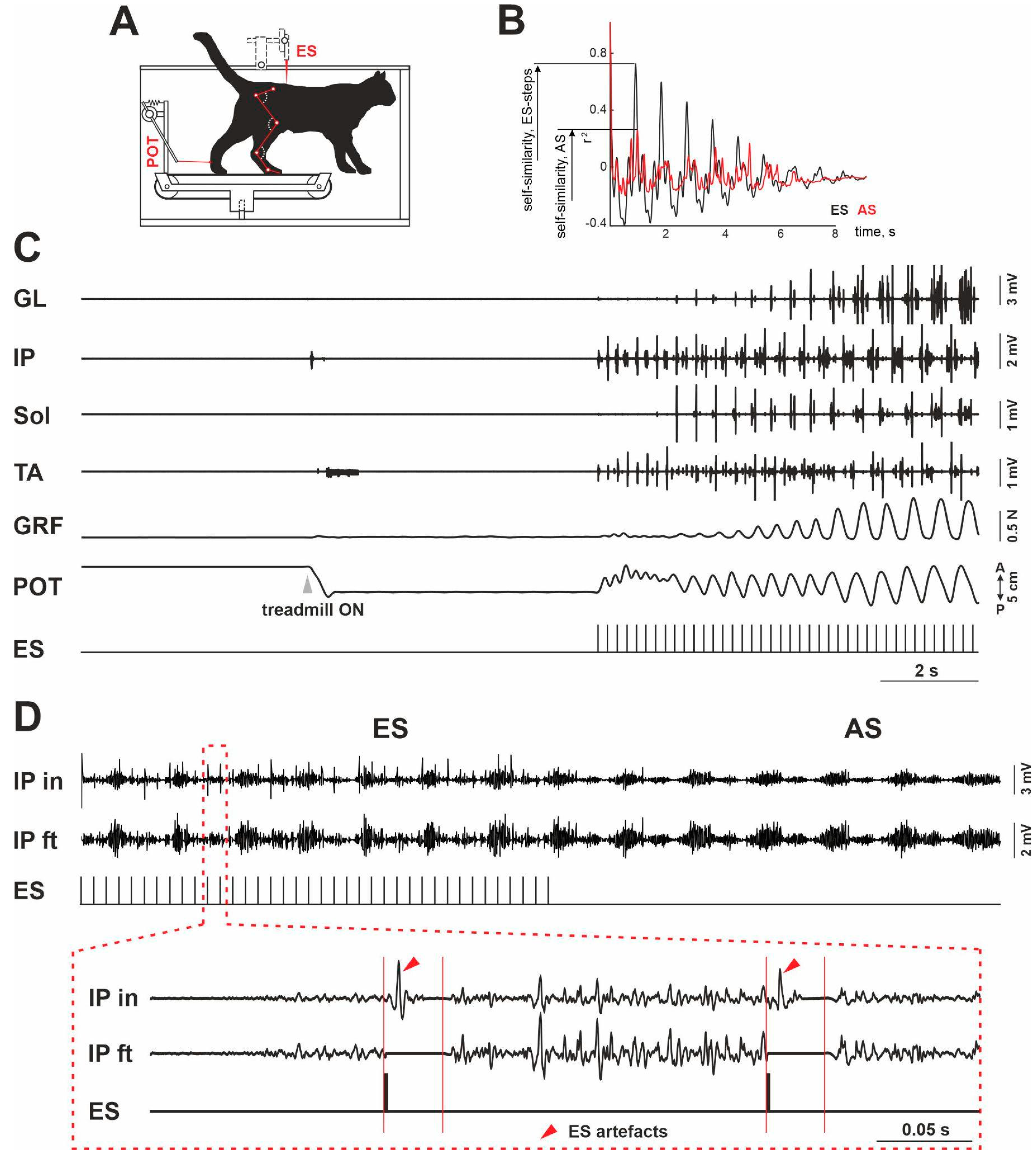
Figure 1. The experimental design (A): an example of the self-similarity assessment for gastrocnemius lateralis (B), an electromyographic (EMG) signal when the treadmill was switched on (C), and the EMG signal filtration method for iliopsoas (D). GL—gastrocnemius lateralis; IP—iliopsoas; TA—tibialis anterior; Sol—soleus muscles; GRF—ground reaction force; POT—potentiometric curve; ES—epidural stimulation; IP in—initial EMG signal of IP; IP ft—filtered EMG signal of IP; AS—after-steps. An example at B for IP, cat # 61. Gray arrow at C marks that the treadmill is switched on; red arrows in D mark ES artifacts.
Muscles 01 00011 g001
Figure 2. Comparative characteristics of the after-stepping (AS) and steps evoked by the electrical epidural stimulation (ES). (A) An example of the original data. GRF—ground reaction forces. POT—potentiometric curve. A—anterior, P—posterior. Numbers 1–9 at GRF and POT curves mark the after-steps (dark grey numbers represent the analysed after-steps). Dotted red lines—maximal GRF signal (max) and 25% of the maximal signal (¼ max). (B) An example of the GRF during ES and AS. Black lines—ES-evoked steps, grey lines—after-steps. Stance and swing—step phases. Red line marks step phase with the maximal GRF value for ES steps. (CG)—kinematic, electromyographic, and GRF data presented as a percent ratio of the AS variables to the ES ones. (C) A percent ratio of the GRF and POT signals during AS. (D) Absolute data for the step phase with the maximal GRF value during ES and AS. (E) Percent ratio of the duration of the swing (SW), stance (ST), and total step cycle (SC) during AS. (F) Percent ratio of the integrated EMG amplitude of iliopsoas (IP), tibialis anterior (TA), gastrocnemius lateralis (GL), and soleus (Sol) muscles during AS. (G) Percent ratio of the stability for the EMG signal of IP, TA, GL, and Sol muscles during AS. *, **, ***, ****—p < 0.05, p < 0.01, p < 0.001, and p < 0.0001 for comparisons between stepping variables; #, ##—p < 0.05 and p < 0.01, for comparisons between AS and ES only; ns—non-significant. Data presented as mean ± SD. Red dotted lines at (C) and (EG) mark 100% level.
Muscles 01 00011 g002
Publisher’s Note: MDPI stays neutral with regard to jurisdictional claims in published maps and institutional affiliations.