Article

Are We Missing Brucella spp. in Portugal? The First Nationwide Systematic Review, Meta-Analysis, and Retrospective Serological Study of Brucella canis (2013–2025)

by
Ricardo Lopes
1,2,3,*,
Hugo Lima de Carvalho
3,
Ana Cristina Ferreira
4,5,6,
Andreia Garcês
7,8,
Cátia Fernandes
9,
Ana Rita Silva
10,
Ana Patrícia Lopes
1,8,
Luís Cardoso
1,8,
Elsa Leclerc Duarte
11,12 and
Ana Cláudia Coelho
1,8
1
Department of Veterinary Sciences, University of Trás-os-Montes e Alto Douro (UTAD), 5000-801 Vila Real, Portugal
2
Department of Veterinary and Animal Sciences, University Institute of Health Sciences (IUCS), CESPU, 4585-116 Gandra, Portugal
3
CEDIVET Veterinary Laboratories, Lionesa Business Hub, R. Lionesa 446 C24, 4465-671 Leça do Balio, Portugal
4
Laboratory of Bacteriology and Mycology, Animal Health Unit, Instituto Nacional de Investigação Agrária e Veterinária (INIAV), I.P., 2780-157 Oeiras, Portugal
5
BioISI-Biosystems and Integrative Sciences Institute, Faculty of Sciences, University of Lisbon, 1749-016 Lisboa, Portugal
6
Investigação em Medicina Veterinária (I-MVET), Faculdade de Medicina Veterinária, Universidade Lusófona, 1749-024 Lisboa, Portugal
7
Wildlife Rehabilitation Centre (CRAS), Veterinary Teaching Hospital, University of Trás-os-Montes e Alto Douro (UTAD), 5000-801 Vila Real, Portugal
8
Animal and Veterinary Research Centre (CECAV), Associate Laboratory for Animal and Veterinary Sciences (AL4AnimalS), University of Trás-os-Montes e Alto Douro (UTAD), 5000-801 Vila Real, Portugal
9
AniCura Santa Marinha Veterinary Hospital, R. Dom Henrique de Cernache 183, 4400-625 Vila Nova de Gaia, Portugal
10
Molecular Diagnostics Laboratory, Lionesa Business Hub, R. Lionesa 446 C24, 4465-671 Leça do Balio, Portugal
11
Department of Veterinary Medicine, School of Science and Technology, University of Évora, Polo da Mitra, Apartado 94, 7002-554 Évora, Portugal
12
Mediterranean Institute for Agriculture, Environment and Development (MED), Global Change and Sustainability Institute (CHANGE), University of Évora, Polo da Mitra, Apartado 94, 7002-554 Évora, Portugal
*
Author to whom correspondence should be addressed.
Zoonotic Dis. 2025, 5(4), 26; https://doi.org/10.3390/zoonoticdis5040026
Submission received: 2 July 2025 / Revised: 15 September 2025 / Accepted: 20 September 2025 / Published: 24 September 2025

Simple Summary

Brucellosis is a major zoonotic disease globally, yet Brucella canis, which primarily affects dogs, remains underrecognised in Portugal. The aim of this study is to present the first systematic review and meta-analysis of Brucella spp. in the country, coupled with a comprehensive 13-year seroepidemiological analysis of B. canis infection in dogs across mainland Portugal and Insular Autonomous Regions (2013–2025). The findings of this study reveal a low but geographically widespread seropositivity of B. canis, with seasonal and regional variations. Observed differences by breed reflect small and uneven subgroup sizes and should not be interpreted as biological susceptibility, sex, and age showed no association. Although often asymptomatic, infected dogs may act as silent reservoirs, increasing the risk of zoonotic transmission, particularly to veterinarians, breeders, and dog owners. The study further reveals a striking discrepancy between the number of dogs tested and the expected exposure levels, strongly suggesting that B. canis infections remain substantially underdiagnosed in Portugal. This underlines the urgent need to strengthen diagnostic capabilities, implement systematic surveillance, and raise awareness across all health domains, including veterinary, medical, and public health sectors. By providing novel epidemiological evidence, this study highlights critical gaps in current monitoring efforts and reinforces the necessity of integrated One Health strategies to enhance early detection, containment, and prevention of B. canis transmission, ultimately safeguarding both animal and human health.

Abstract

Brucella canis is a neglected zoonotic pathogen associated with canine reproductive disorders and emerging public health concerns. This study presents the first nationwide systematic review and meta-analysis of Brucella spp. in Portugal, integrated with a 13-year retrospective seroepidemiological investigation (2013–2025) of B. canis in dogs across mainland Portugal and Insular Autonomous Regions. Among 132 canine serum samples, a seropositivity of 23.48% was observed using an immunochromatographic assay confirmed by indirect immunofluorescence (IFAT). Significant associations were identified with seasonality (p < 0.001) and breed (p = 0.001), while sex and age were not statistically significant. Municipal-level analysis revealed marked heterogeneity, with Trofa showing the highest seropositivity (58.82%) and a pooled odds ratio of 11.28 (95% CI: 2.90–43.94; p < 0.001). In parallel, meta-analyses of published data estimated a pooled seroprevalence of 2.42% in animals (95% CI: 1.79–3.13) and 10.57% in humans (95% CI: 8.80–12.47), underscoring the broader burden of Brucella spp. exposure in Portugal. These findings suggest underdiagnosis of canine brucellosis and highlight the need for enhanced surveillance in high-risk breeds and regions. The study reinforces the importance of integrated One Health strategies to improve early detection, control, and prevention of B. canis infection in both veterinary and public health contexts.

Graphical Abstract

1. Introduction

Brucellosis is one of the most widespread and impactful zoonotic diseases globally, caused by facultative intracellular bacteria of the genus Brucella [1,2,3]. Thirteen species have been identified to date, with B. melitensis, B. abortus, B. suis, and B. canis representing the most relevant zoonotic agents due to their ability to cross species barriers, persist in animal reservoirs, and cause chronic, debilitating infections in humans [4,5,6,7,8,9].
Brucella species are remarkably well adapted to a broad range of hosts, including cattle, sheep, goats, pigs, dogs, and wildlife, which maintain the pathogen in the environment and facilitate zoonotic spillover [5,6,10]. This broad host range contributes to its global spread and persistence of the disease.
Among the zoonotic Brucella species, B. melitensis is widely recognised as the most virulent and most prevalent in human cases, posing substantial public health risks, particularly across the Mediterranean Basin, the Middle East, and other endemic regions [11,12,13]. This predominance is attributed to its remarkable adaptability to multiple hosts and a broad arsenal of virulence-associated genes, despite its striking genetic homogeneity, with over 98–99% DNA similarity across species, reflecting a highly clonal population structure [14,15]. The bacterium primarily infects small ruminants, especially sheep and goats, but is also found in cattle and other livestock, facilitating persistent zoonotic transmission and environmental contamination [16,17,18]. Regionally, B. melitensis accounts for the vast majority of human and animal brucellosis cases in endemic areas such as Greece, Iran, and parts of Asia and Africa, with reported human seroprevalence rates reaching up to 12% in Asia and approximately 17% in Africa [11,12,13,19,20]. The species’ pathogenicity is reinforced by the expression of key virulence genes including virB, omp25, omp31, manA, manB, and ure, which support intracellular survival, immune evasion, and chronic infection in both humans and animals [15,21,22]. Significantly, B. melitensis exhibits notable genetic heterogeneity across different geographic regions, suggesting ongoing microevolutionary adaptation and potentially influencing regional transmission dynamics and diagnostic sensitivity [11,15].
Transmission primarily occurs via direct contact with infected animals or through the consumption of unpasteurised dairy products. Individuals close to livestock, such as farmers, veterinarians, slaughterhouse workers, and butchers, are at increased occupational risk. Working domestic animals like dogs and farm cats are also at risk [6,23,24,25]. Although rare, vector-borne transmission via ticks has also been proposed [26].
Control and prevention of brucellosis are challenged by diagnostic and therapeutic limitations. The disease typically presents non-specific symptoms, making clinical diagnosis difficult, and Brucella’s intracellular survival complicates both detection and treatment [1,2]. No effective human vaccine is available, and while vaccination of livestock against B. melitensis and B. abortus has reduced incidence in some regions, control of B. canis remains problematic due to the absence of vaccines and limited public health focus [4,23,24]. Brucellosis remains a major public health concern, with over 500,000 new human cases reported annually, particularly in areas with insufficient veterinary services and food safety regulations [1,4,27]. The disease has a dynamic geographic distribution, often linked to animal trade, migration, and evolving farming practices [6,23]. Its economic and health impacts are substantial, affecting food security, animal productivity, and human livelihoods [10,24].
Addressing brucellosis effectively requires a One Health approach, which integrates the human, animal, and environmental health sectors. Coordinated surveillance, intersectoral collaboration, and harmonised control strategies are essential to reduce transmission risks and disease burden [1,6,10]. In this context, the neglect of B. canis as an emerging zoonotic agent is concerning, especially given its increasing detection in dogs across Europe and its capacity to cause undiagnosed febrile illness in humans [5,24].
B. canis, traditionally considered a pathogen confined to canine populations, is now increasingly recognised as a significant zoonotic agent with serious public health implications [28,29]. In dogs, infection frequently presents with reproductive disturbances such as infertility, abortions, epididymitis, prostatitis, and neonatal mortality. However, a substantial proportion of infected animals may remain clinically asymptomatic, making detection and control particularly challenging, especially in breeding kennels and urban settings [30,31,32]. Unlike the classical smooth species (B. abortus, B. melitensis, B. suis), B. canis is a rough strain lacking the O-polysaccharide antigen, which means standard serological tests for smooth Brucella often fail to detect B. canis. Additionally, B. canis infections in humans tend to be less virulent than those caused by B. melitensis or B. abortus, although B. canis remains a zoonotic threat if undiagnosed. Human cases, although underreported, can manifest flu-like symptoms, undulant fever, splenomegaly, and lymphadenopathy, and are frequently misdiagnosed due to non-specific presentations and the limited sensitivity of available diagnostic tools [31,32].
In Portugal, brucellosis continues to pose a major concern in both veterinary and public health domains, despite long-standing national eradication programmes and strict regulatory control. Outbreaks remain recurrent, particularly in the northern regions, where they have been frequently attributed to the consumption of unpasteurised dairy products, such as fresh cheese, or direct contact with infected livestock [33]. Concurrently, brucellosis in livestock, particularly in cattle, continues to exert a significant economic burden. This is especially evident in high-incidence regions such as Alentejo, where persistent infection within herds leads to reproductive losses, diminished productivity, and trade limitations [34]. In small ruminants, particularly sheep and goats raised in the Trás-os-Montes region, brucellosis outbreaks remain a concern due to insufficient vaccination coverage, inadequate biosecurity, and frequent interspecies contact [35].
Notably, a high seroprevalence of Brucella antibodies in wild boar populations in northeast Portugal raises concerns regarding potential wildlife reservoirs that may complicate eradication efforts and facilitate cross-species transmission [8]. While test-and-slaughter remains the cornerstone of control in cattle, its limitations in settings with persistent infection are well recognised. More promising outcomes have been observed with integrated approaches combining RB51 vaccination, regular diagnostic testing, and improved on-farm biosecurity [34,35]. Additionally, farmer education and engagement have been shown to significantly impact disease control outcomes, particularly through improved understanding of transmission routes, vaccine benefits, and biosecurity compliance [36].
The aim of this study is to present the first nationwide systematic review and meta-analysis of Brucella spp. in Portugal, coupled with a comprehensive 13-year seroepidemiological assessment of B. canis infection in dogs (2013–2025). By integrating primary diagnostic datasets with a critical appraisal of national scientific evidence, this study aims to estimate the true burden of Brucella infection, elucidate spatial and temporal trends, and identify canine populations at increased risk. To the authors’ knowledge, this is the first systematic with meta-analysis study to comprehensively characterise Brucella spp. epidemiology in Portugal within a One Health framework.

2. Materials and Methods

2.1. Data Collection, Sampling, and Diagnostic Procedures

Serum samples from suspected cases of canine brucellosis were submitted to CEDIVET Veterinary Laboratories (Porto, Portugal). These samples originated from veterinary practices, including clinics and hospitals, across mainland Portugal and the Insular Autonomous Regions. Each submission included a standard laboratory requisition form, which provided clinical data for each dog, specifically detailing breed, sex, age, vaccination and prophylactic status, clinical suspicion or observed clinical signs (e.g., abortion or orchitis), and the analyses requested.
Serological testing was initially performed using an immunochromatographic assay (FASTest® Brucella canis, MEGACOR Diagnostik GmbH, Hörbranz, Austria), designed to detect immunoglobulin G (IgG) antibodies specific to B. canis, in strict accordance with the manufacturer’s instructions.
Samples yielding positive results were subsequently subjected to confirmation by an indirect immunofluorescence antibody test (IFAT), employing a commercial kit for the qualitative detection of anti-B. canis IgGs in canine serum (MegaFLUO Brucella canis, MEGACOR Diagnostik GmbH, Hörbranz, Austria). According to the manufacturer, combining the IgG lateral flow assay with IFAT yields approximately 92% sensitivity and 99% specificity for B. canis serodiagnosis. Sera were tested according to the manufacturer’s protocol, starting at a 1:50 dilution, and the corresponding 50 titre (i.e., the reciprocal of that dilution) was established as the cut-off value for determining seropositivity. If a positive result was detected at this dilution, further serial dilutions were performed until a negative result was observed. In this study, those additional dilutions included 1:100, 1:200, 1:400, and 1:800. Samples were considered positive when green fluorescence comparable in intensity to the positive control was observed. Results were reported as the highest dilution at which characteristic fluorescence was detected.
Interpretation of antibody titres was categorised as follows:
Negative/low titre: results below 50 were classified as negative or indicative of a null or low antibody level, suggesting no exposure to or infection with B. canis.
Borderline/low positive: a titre of 50 was considered borderline or low positive, indicating possible exposure, but not necessarily an active infection.
Moderate positive: titres of 100 and 200 reflected moderate antibody levels, suggesting likely exposure to and infection with B canis.
High positive: titres of 400 or higher were classified as high, indicative of a strong immune response and likely a recent or ongoing infection. These classifications provide a framework for assessing the degree of exposure and immune response to B. canis, aiding in the interpretation of serological results.

2.2. Systematic Review and Meta-Analysis

The systematic review and meta-analysis were conducted following the PRISMA (Preferred Reporting Items for Systematic Reviews and Meta-Analyses) and MOOSE (Meta-analysis of Observational Studies in Epidemiology) guidelines. A comprehensive systematic search was performed to retrieve all available studies reporting quantitative seroprevalence data of Brucella spp. in Portugal, suitable for inclusion in meta-analytical models, including both animal and human populations, due to the zoonotic relevance of the disease. A few reports that used PCR to detect Brucella (e.g., in wildlife) were mentioned in the narrative for context.
The search was conducted in seven databases and institutional repositories: PubMed (n = 44), Scopus (n = 817), SciELO (n = 2), RCAAP (n = 3), INSA—Instituto Nacional de Saúde Doutor Ricardo Jorge (n = 3), DGAV—Direção-Geral de Alimentação e Veterinária (n = 1), and ECDC—European Centre for Disease Prevention and Control (n = 1). The search terms used were: “Brucella” AND “Portugal”. The final search was completed in June 2025.

2.3. Eligibility Criteria

Inclusion criteria comprised all observational studies and official reports reporting Brucella spp. seroprevalence data from Portugal, regardless of host species.
Studies were included if they reported:
  • Total sample size;
  • Number of positive cases (or prevalence with 95% CI);
  • Clear indication of location (Mainland Portugal and the Insular Autonomous Regions (Azores and Madeira);
  • Year or study period (2000 to 2025).
Exclusion criteria included:
  • Case reports, editorials, or opinion articles;
  • Experimental infection studies or vaccine trials;
  • Studies lacking extractable epidemiological data or not clearly related to Brucella spp.;
  • Duplicate data from the same population.

2.4. Data Extraction

All records retrieved from the databases were imported into Mendeley Reference Manager (version 2.98.0, 2023 Mendeley Ltd., London, UK). Two independent reviewers conducted the screening process. Following the removal of duplicate entries, titles and abstracts were systematically assessed to determine eligibility. When the available information was insufficient to assess inclusion criteria, the full texts were retrieved and reviewed to confirm relevance and eligibility.
Data extraction was carried out using a structured Microsoft Excel® (Microsoft Corp., Redmond, WA, USA). The following information was collected from each eligible study:
  • Study characteristics, including the name(s) of the author(s), year of publication, geographical region (e.g., mainland Portugal, Azores), species studied, and the classification of the host (e.g., domestic animals, livestock, wildlife, or humans);
  • Sample size, defined as the total number of individuals tested, and the number of positive cases identified;
  • Diagnostic method employed in the study (e.g., Rapid Slide Agglutination Test [RSAT], Indirect Fluorescent Antibody Test [IFAT], Enzyme-Linked Immunosorbent Assay [ELISA], among others);
  • Prevalence estimates, expressed as a percentage (%), accompanied by the corresponding 95% confidence intervals (95% CI).

2.5. Statistical Analysis

All available data were extracted in digital format from the Sislab® system (Glintt, Global Intelligent Technologies, Lisbon, Portugal) and transferred to Microsoft Excel® (Microsoft Corp., Redmond, WA, USA) for preprocessing. Variables were categorised into geographical region (NUTS 2: Nomenclature of Territorial Units for Statistics), municipality, season, month, breed, sex, age group, and serological antibody titre.
Statistical analysis was performed using JMP® version 14.3 (SAS Institute Inc., Cary, NC, USA), DATAtab® (DATAtab e.U., Graz, Austria), and MedCalc® statistical software version 20.006 (MedCalc Software Ltd., Ostend, Belgium). Descriptive statistics were used to summarise frequencies and proportions. The chi-squared (χ2) test was applied to assess associations between seropositivity and categorical variables, such as sex, age group, breed, geographical location, season, and month of sample collection. Fisher’s exact test was applied when expected cell frequencies were below five.
Seropositivity estimates were reported with exact binomial 95% confidence intervals (CI). For ordinal or non-normally distributed continuous variables (e.g., antibody titres), the Kruskal–Wallis test was employed, followed by Dunn–Bonferroni post hoc tests where appropriate. The Mann–Whitney U test was applied for pairwise comparisons. Univariable logistic regression was conducted to estimate odds ratios (ORs) and corresponding 95% CIs to identify potential risk factors for B. canis seropositivity. Lagos was used as the reference municipality for OR analysis due to its sample size and zero seropositivity, providing a conservative baseline. A p-value ≤ 0.05 was considered statistically significant in all analyses [37].
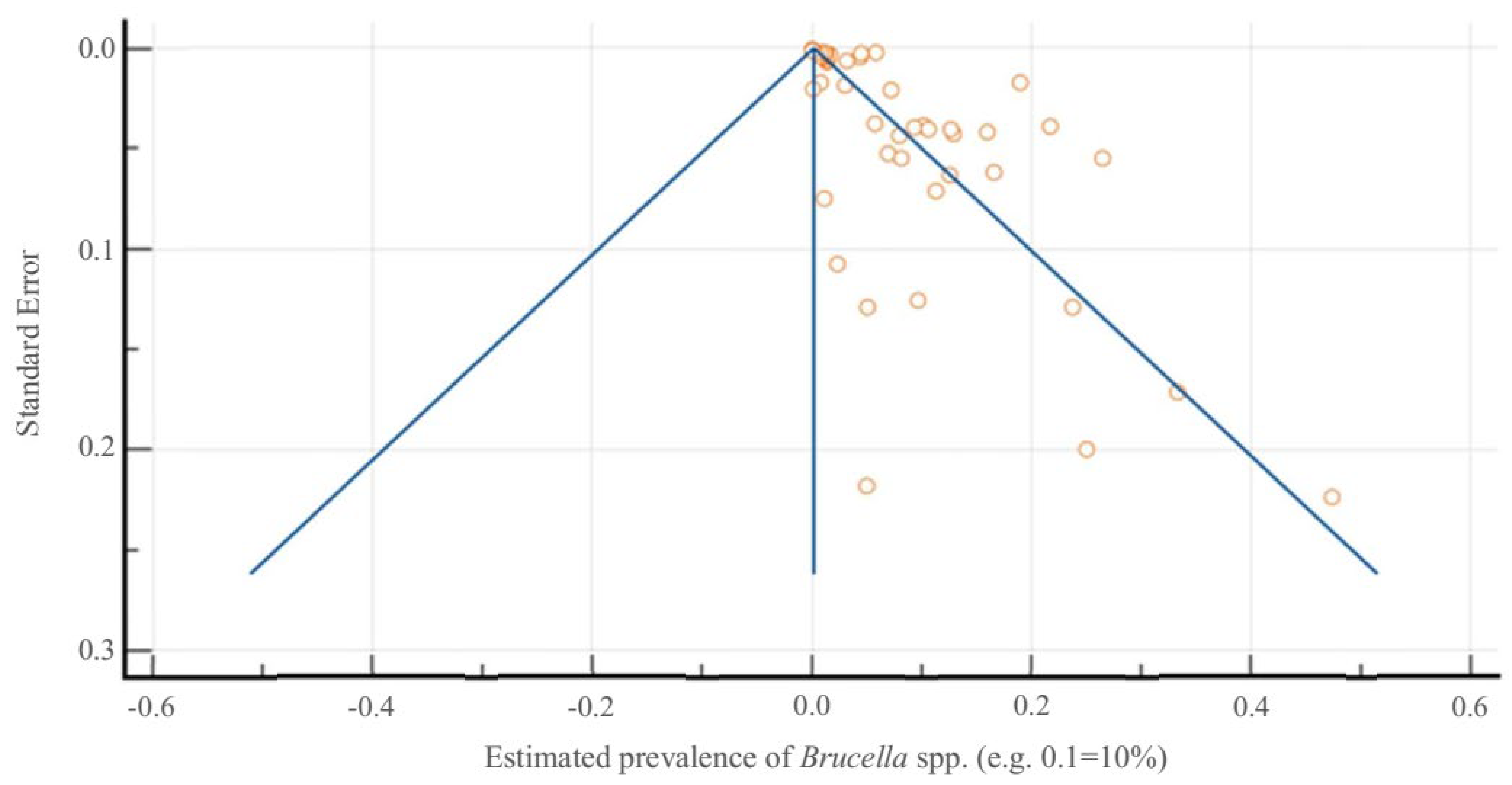
Meta-analysis was performed using MedCalc® statistical software version 20.006 (MedCalc Software Ltd., Ostend, Belgium), applying a random effects model to account for expected heterogeneity. Prevalence estimates were transformed into proportions, and heterogeneity was assessed using Cochran’s Q test and I2 statistic. Publication bias was evaluated through Egger’s test, Begg’s test, and Kendall’s Tau, complemented by a funnel plot.

3. Results

3.1. Seroepidemiological Study of Brucella canis in Portugal Between 2013–2025

3.1.1. General Seropositivity, Geographical Distribution, and Seasons

Of the total of 132 animals included in this study, 31 (23.48%) tested positive, whilst 101 (76.52%) tested negative for B. canis. The distribution of seropositivity across serological titres is detailed in the Table 1.
The majority of samples originated from the districts of Porto (n = 106; 80.3%), Faro (n = 11; 8.33%), Aveiro (n = 5; 3.79%), and Madeira (n = 4; 3.03%). The distribution of results by geographical regions (NUTS 2) of mainland Portugal and Insular Autonomous Regions is presented in Table 2. Notably, Northern Portugal accounted for 82.6% of all samples (109/132), with Porto district alone contributing 106 samples (80.3%) of the total.
Data from the Greater Lisbon, Setúbal Peninsula, West and Tagus Valley, and Autonomous Region of the Azores regions were not available for the present study. The seropositivity of exposure to or infection with B. canis in this study across the different NUTS 2 ranged from 0.00% to 27.52%. The highest seropositivity of B. canis was observed in the North region (27.52%), followed by the Centre (16.67%) regions. However, the chi-squared test indicated no statistical association between region (NUTS 2) and seropositivity of B. canis2 = 6.3, df = 4, p = 0.177).
A more detailed analysis at the municipality level revealed apparent heterogeneity in B. canis seropositivity. The municipality of Trofa exhibited the highest proportion of seropositive cases (20/34; 58.82%), markedly surpassing all other localities, likely reflecting a localised outbreak. In contrast, several municipalities, namely Matosinhos, Ovar, Santo Tirso, and Lagos, recorded no seropositive cases. This heterogeneous pattern likely reflects the non-uniform and passive nature of sampling, rather than a true absence of infection in the areas with zero detections. The chi-squared test demonstrated a statistically significant association between municipality and B. canis seropositivity (χ2 = 48.71, df = 21, p = 0.001). This association appears to be driven largely by the cluster in Trofa, which contributed a disproportionate number of seropositive cases. Given that most municipalities contributed only 1–4 samples, these results should be interpreted as descriptive of that localised outbreak rather than as evidence of widespread regional differences.
The meta-analysis revealed a pooled odds ratio of 11.28 (95% CI: 2.90–43.94, p < 0.001) for B. canis seropositivity, comparing several municipalities to the municipality of Lagos, which was selected as the reference group due to its moderate sample size (n = 9) and complete absence of seropositive cases. The analysis demonstrated no statistically significant heterogeneity across municipalities (Q = 3.29, p = 0.6546; I2 = 0.0%), and no evidence of publication bias (Egger’s test, p = 0.1355; Begg’s test, p = 0.1885). These results suggest that dogs residing in certain municipalities may face increased risk of exposure to B. canis relative to those in seronegative areas. However, given the opportunistic and highly uneven distribution of samples, these findings should be interpreted with caution and regarded as descriptive of a localised cluster rather than as robust evidence of broad regional differences. This highlights the need for geographically targeted epidemiological surveillance and control measures. These data are summarised in Figure 1.
The seropositivity of B. canis in the North region between 2017 and 2025 is summarised in Table 3. Full regional data are available in Supplementary Table S1, although temporal interpretation outside the North is not meaningful due to the limited number of samples.
Notably, in 2018, the North region’s seropositivity rate spiked to 74.2% of samples tested that year.
In addition, Table 4 and Supplementary Table S2 present the seropositivity of B. canis by season and month, respectively.
Statistically significant association was observed between the ordinal seropositivity for B. canis and both seasons of the year (χ2 = 18.1, df = 3, p < 0.001) and the month (χ2 = 39.89, df = 11, p < 0.001), as determined by the Kruskal–Wallis test. Post hoc analysis using the Dunn–Bonferroni method revealed that the median seropositivity score was significantly higher during spring compared to winter (p < 0.001) and summer (p = 0.012). No statistically significant differences were observed between the remaining seasonal categories (p > 0.05).
Regarding monthly distribution, May exhibited significantly higher seropositivity scores compared to most other months. Statistically significant differences were identified between February and May (p < 0.001), March (p = 0.011), April (p = 0.046), and several other months including June, July, August, and September (adjusted p-values ranging from 0.006 to 0.031). These findings suggest a pronounced seasonal pattern, with increased seropositivity to B. canis during the spring months, peaking in May.

3.1.2. Breed

Out of 132 dogs tested, 59 were mixed-breed (44.70%), and the remaining 73 (55.30%) represented 26 distinct pure breeds, including Pointers (n = 11; 8.33%), German Shepherds (n = 9; 6.82%), and Labrador Retrievers (n = 9; 6.82%) being the most represented, alongside others such as Great Danes, Yorkshire Terriers, Pugs, and Border Collies.
As many breeds comprised very few dogs (≤3–11 in several categories), which results in wide confidence intervals and unstable estimates, differences between breeds must be interpreted with caution.
A statistically significant association was observed between breed and seropositivity to B. canis, as revealed by the chi-squared test (χ2 = 54.91, df = 26, p = 0.001). This test reflects heterogeneity across numerous breeds and does not by itself demonstrate biological susceptibility. The highest apparent seropositivity was observed in Pugs and Setters (3/3 and 2/2, respectively; 100% each), although only a few individuals of these breeds were tested. Pointers also showed a high positivity rate (8/11; 72.7%), followed by German Shepherds (5/9; 55.6%). In contrast, no seropositive cases were detected among Labrador Retrievers, Boxers, Yorkshire Terriers, or Beagles.
Mixed-breed dogs, which accounted for the largest subgroup (n = 59, 44.70%), demonstrated a seropositivity rate of 18.64% (11/59).
The Labrador Retriever was used as the reference category due to the absence of seropositive cases in this breed (0/9; 0.00%). Compared to this baseline, Pugs (3 seropositive; 100%) exhibited the highest odds of seropositivity (OR = 133.0; 95% CI: 2.2–8082.5), followed by Setters (2 seropositive; 100%; OR = 95.0; 95% CI: 1.5–6088.1) and Schnauzers (1 seropositive; 100%; OR = 57.0; 95% CI: 0.8–4124.2). Pointers (8 seropositive; 72.72%) and German Shepherds (5 seropositive; 55.56%) also showed elevated odds of seropositivity with OR values of 46.1 (95% CI: 2.1–1028.8) and 23.2 (95% CI: 1.0–518.0), respectively. The ORs of the other breeds were not statistically significantly different, showing that the observed variability in seropositivity was apparently restricted to a few breeds.

3.1.3. Sex

Although males exhibited a higher proportion of positive cases (26/98; 26.53%) compared to females (5/34; 14.71%), this difference was not statistically significant (χ2 = 1.96, df = 1, p = 0.161). Fisher’s exact test also confirmed the absence of a significant association (p = 0.240).

3.1.4. Age

Of the 132 dogs included in the study, age data were available for 96 individuals (72.72%). The remaining 36 dogs (27.27%) were excluded from age-related analyses due to missing age information. The distribution of ages ranged from 6 months (≤1 year) to 14 years (≥11 years), with a median age of 5 years (interquartile range: 2 to 8.25 years).
Statistical analysis revealed no significant association between age and B. canis seropositivity, whether age was assessed as a continuous variable (Mann–Whitney U = 394, p = 0.221) or categorised into ordinal age groups (χ2 = 3.1, df = 4, p = 0.541), indicating no consistent age-related trend.

3.2. Systematic Review and Meta-Analysis on Brucella spp. in Portugal

A total of 62 studies were included in the final meta-analysis, selected and reported following the PRISMA 2020 guidelines (Figure 2).
These studies included 6,137,223 animals—bovines, dogs, dolphins, goats, red deer, sheep and wild boars—and 8724 humans screened for Brucella spp. (including B. abortus, B. canis, B. ceti and B. melitensis) across mainland Portugal, the Lisbon and Tagus Valley Coast, and the Azorean islands of São Miguel, Terceira, and São Jorge. A wide range of diagnostic techniques was employed, including RSAT, IFAT, ELISA, and qPCR.
The pooled prevalence in animals was estimated at 2.42% (95% CI: 1.79–3.13), based on a random-effects model (Figure 3). This estimate was derived from a total of 6,137,223 animals tested, of which 30,480 (0.50%) were positive for Brucella spp. using a wide range of diagnostic techniques, including RSAT, IFAT, ELISA, and qPCR.
Heterogeneity across animal studies was significant (Q = 76,024.62, df = 30, p < 0.0001; I2 = 99.96%, 95% CI: 99.96–99.96), suggesting substantial variation likely due to differences in species, sampling strategies, regional foci, and diagnostic methods.
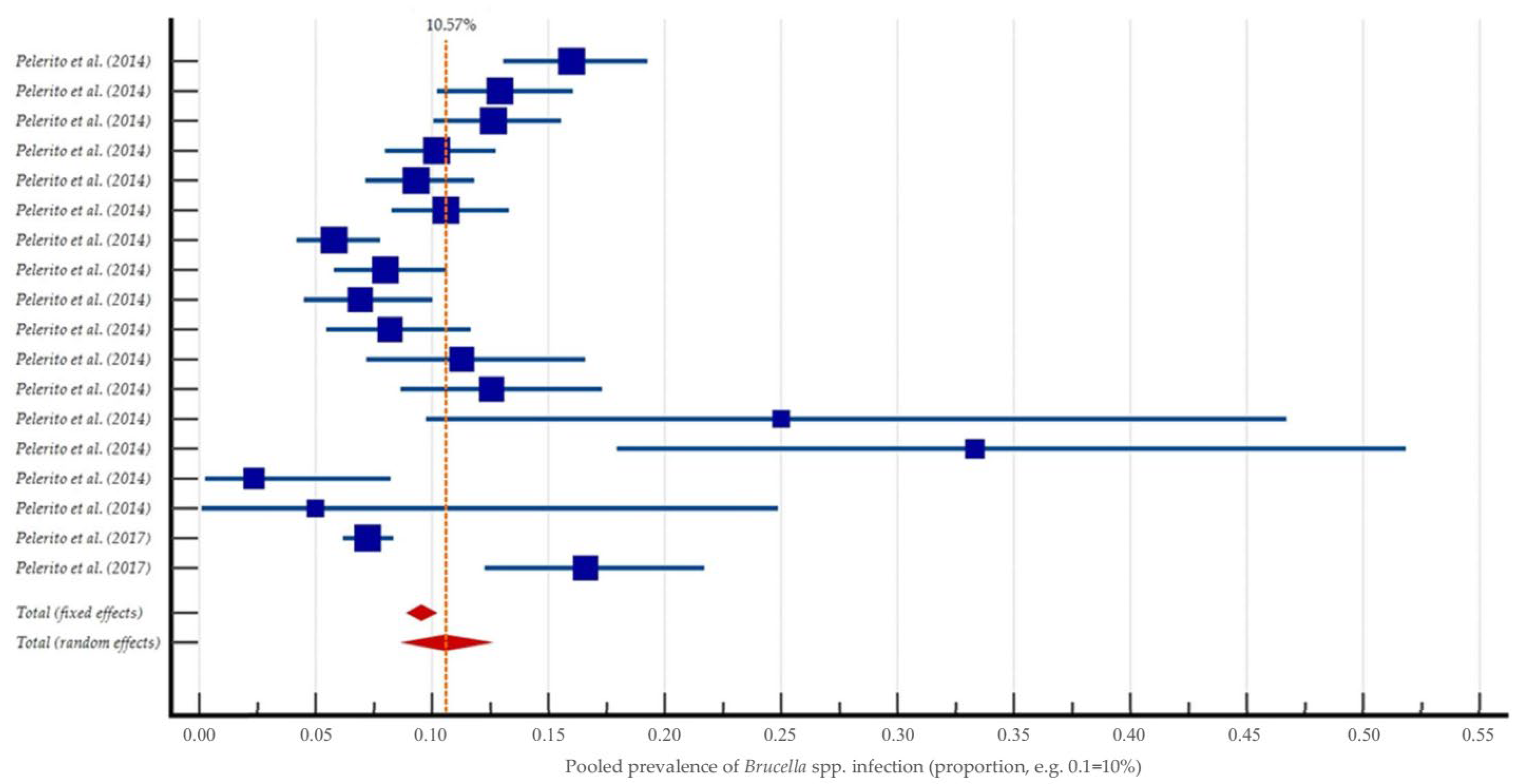
In contrast, the pooled prevalence in humans was 10.57% (95% CI: 8.80–12.47), also estimated using a random-effects model (Figure 4). A total of 8724 humans were tested, of whom 847 (9.71%) were positive for Brucella spp. based on serological or molecular detection methods.
Heterogeneity was high (Q = 110.90, df = 17, p < 0.0001; I2 = 84.67%, 95% CI: 77.09–89.74), although lower than in animal studies.
To estimate the overall pooled prevalence across all populations (animals and humans), the data were combined into a unified meta-analysis using a random-effects model. The resulting prevalence was 4.49% (95% CI: 3.77–5.27), confirming a significant burden of Brucella infection across species and regions in Portugal (Table 5).
The heterogeneity remained extremely high (Q = 78,680.89, df = 61, p < 0.0001; I2 = 99.94%, 95% CI: 99.94–99.94).
The combined analysis demonstrated clear funnel plot asymmetry (Figure 5), with Egger’s regression test showing statistical significance (Intercept = 23.86, 95% CI: 11.23–36.51, p = 0.0004), supporting the presence of small-study effects. Begg’s test (Kendall’s Tau = −0.2629, p = 0.0077) and Kendall’s Tau test confirmed the likelihood of bias.

4. Discussion

4.1. Contextualisation of Canine Brucellosis and Zoonotic Relevance

Brucella canis is a Gram-negative intracellular coccobacillus that causes canine brucellosis, a contagious disease primarily affecting the reproductive system of dogs [44]. Beyond the reproductive tract, B. canis can produce nonspecific clinical signs such as lethargy, lymphadenopathy, lameness, or discospondylitis (infection of intervertebral discs) [28]. This pathogen has long been endemic in regions of the Americas and Asia, but in Europe it was historically rare; prior to the 2000s, most canine brucellosis cases in Europe were caused by other Brucella species (e.g., B. melitensis, B. abortus or B. suis from livestock) or were traced to imported dogs [28]. However, B. canis is now recognised as an emerging cause of canine brucellosis in Europe, having been detected in dogs in nearly all European countries in recent years [28].
Crucially, B. canis is a zoonotic pathogen and can infect humans who are exposed to infected dogs. In people, it generally causes illness similar to classical brucellosis (undulant fever) with non-specific symptoms such as fever, fatigue, headaches, lymph node enlargement and splenomegaly [28]. If untreated, serious complications like endocarditis, arthritis or neurological involvement can occur in rare cases. Fortunately, no human fatalities due to B. canis have been reported [45]. Nonetheless, the public health importance of canine brucellosis is likely underestimated, since human infection with B. canis is probably underdiagnosed and underreported worldwide [28,46]. Standard serology for brucellosis (geared toward smooth Brucella like B. abortus) often fails to detect B. canis due to its lack of the O-polysaccharide antigen, leading to false-negative results in human patients [45,47]. This, combined with physicians’ low index of suspicion for B. canis, means many cases may go unrecognised [46]. Despite these challenges, B. canis should be considered a public health concern in the context of close human-dog relationships. Children, immunosuppressed individuals and pregnant women in contact with infected dogs could be at particular risk, and the bacterium represents an occupational hazard for veterinarians, kennel workers, and laboratory staff handling canine samples [28].
In Portugal specifically, canine brucellosis due to B. canis has not been part of routine surveillance and was historically not believed to be endemic. The high dog ownership in the country and frequent close contact between pets and owners underscore the importance of heightened awareness and a One Health approach to this neglected zoonosis.

4.2. Critical Analysis of Study Findings

This 13-year seroepidemiological study provides the first comprehensive seroepidemiological data on B. canis exposure in dogs from mainland Portugal and Insular Autonomous Regions. An overall seropositivity of 23.48% was found, with 31 out of 132 dogs testing positive by serology. This indicates that nearly one in four dogs suspected of brucellosis had been exposed to B. canis, a strikingly high proportion that suggests the infection is more widespread than previously assumed [44]. Our findings reveal a substantially higher exposure rate in Portugal, aligning with the upper range of seroprevalences observed in endemic regions globally (serological surveys in parts of Latin America, Asia, and Africa have reported 6–35% positivity) [48]. This underscores that canine brucellosis may be an under-recognised problem in Portugal and potentially in other regions where systematic surveillance has been lacking.
Geographically, seropositive dogs were identified in two NUTS 2 regions (North and Centre). Although the North had the highest proportion (27.52%), differences between regions were not statistically significant (p = 0.177). Given the uneven and opportunistic nature of sampling and the fact that several regions were either unsampled or sparsely sampled, these findings suggest that B. canis exposure is not confined to a single area of Portugal. Indeed, positive dogs were identified across multiple regions, reflecting a nationwide distribution of exposure rather than a localised anomaly. This pattern is consistent with evidence from broader European data, where seropositive cases have been detected in dogs from numerous countries across the continent [44]. In other words, wherever susceptible dogs and risk factors (such as contact with infected animals) are present, B. canis can establish itself. The infection can likely circulate at low levels undetected in various areas until active testing is undertaken.
However, analysis at the finer municipal level revealed stark localised clustering of infection. The municipality of Trofa stood out with the highest infection rate: 20 of 34 dogs from Trofa (58.82%) were seropositive. This was dramatically higher than other municipalities, for example, Lagos had 0.00% positivity, and dogs in Trofa had 11.3 times higher odds of infection compared to those in Lagos. These findings point to an intense local outbreak or reservoir in Trofa that warrants special attention. Such heterogeneity is not surprising for a contagious disease and could reflect a high-risk kennel or breeder in that area. Once B. canis is introduced into a susceptible canine population—for instance, a large breeding kennel—it can rapidly spread among dogs in close contact, creating a “hotspot” of transmission. This scenario is plausible given that B. canis is known to persist in kennel environments. Large dog colonies are at increased risk of maintaining the infection once introduced [44]. In Canadian [49], Brazilian [50] and Dutch [51] studies of breeding kennels, the prevalence of B. canis ranged widely, highlighting the potential for both localised outbreaks and widespread infection in affected facilities. In affected Canadian commercial kennels, the proportion of infected dogs ranged from 3.9% to 100%, with a median of approximately 33%. Comparable scenarios have been reported in Brazil, where infection rates among dogs varied between 3.8% and 62.6% (median ~22%), and in the Netherlands, where 34% of dogs tested seropositive. These findings collectively illustrate how a single introduction of B. canis into a group-housed population can lead to rapid and extensive intra-kennel transmission. Our Trofa cluster likely represents a similar phenomenon, emphasizing the need for targeted control measures in such high-risk settings. It underscores that while B. canis may be present at low levels anywhere, significant outbreaks can occur when the bacteria are introduced into a densely connected group of dogs. Around the same time (late 2018–2019), an unrelated brucellosis outbreak in humans (due to B. melitensis from contaminated goat cheese) was reported in Northern Portugal. While the canine B. canis outbreak and the human B. melitensis outbreak had separate sources, their temporal and geographic overlap highlights Northern Portugal as a brucellosis hotspot and underscores gaps in surveillance across species [33].
Seropositivity rates were significantly higher in spring compared to other seasons (p < 0.001). Notably, the month of May showed the peak seropositivity of all, with May’s positivity being statistically greater than that in many other months. This pronounced spring peak suggests that transmission or case detection is not evenly distributed throughout the year. One possible explanation is related to canine reproductive cycles and management practices. Many dogs breed in late winter, with pregnancies (and potential abortion events in infected bitches) occurring in spring, which is when brucellosis in breeding dogs might first be noticed and diagnosed. During spring, veterinarians may see an influx of cases of late-term abortion or infertility in dogs, prompting brucellosis testing and, thus, yielding more positives. The timing could also reflect increased sexual activity and contact among dogs during winter and early spring, facilitating transmission of B. canis. Interestingly, similar seasonality is seen in livestock brucellosis, for example, in regions endemic for B. melitensis or B. abortus, the incidence in animals and humans often peaks around the birthing season in spring [1]. This insight is epidemiologically important, indicating that spring should be considered a high-risk period for canine brucellosis, meriting greater vigilance and perhaps seasonal screening of breeding dogs.
The study also uncovered interesting associations between breed and seropositivity. Although a few pure breeds appeared to have higher seropositivity in this sample, most breed strata were very small, often with zero events in the reference group, generating wide confidence intervals and unstable odds ratios. These findings are, therefore, best regarded as descriptive of the sampled population rather than evidence of breed-specific susceptibility. Purebred dogs as a group had higher infection rates than mixed-breed dogs (only 18.6% of mixed breeds were positive, compared to higher rates in several pure breeds). These breed-associated differences likely reflect differences in dog management and exposure risk rather than any intrinsic breed susceptibility. For example, Pointers and Setters in our sample showed 100% seropositivity (though the sample size for these breeds was very small). These breeds are often used as working or hunting dogs and are sometimes kept in kennels or multi-dog facilities where a single infected dog could spread B. canis to many others [52,53]. Similarly, German Shepherds showed higher odds of infection in our study; this breed is commonly employed as working dogs (police, guard, or military) and frequently involved in formal breeding programmes, which might increase their chance of exposure through higher contact rates with other dogs [54]. The fact that all Pugs and all Setters tested were positive is striking, although we had very few of these dogs in our dataset, it raises the suspicion that those individuals may have originated from kennels or breeding operations with active B. canis infection. This aligns with known risk factors for canine brucellosis: infections tend to persist and spread in high-density dog populations such as kennels, especially where routine screening and biosecurity may be lacking [50,55,56]. In contrast, companion or individually housed dogs tend to exhibit significantly lower seroprevalence [50,51,57]. Indeed, B. canis has the capacity to devastate breeding kennels when undetected [58], so it is plausible that the high seropositivity in certain breeds in our study reflects exposure in such kennel environments. It is important to note that our breed-specific observations (like the 100% in Pugs or Setters) are based on very limited numbers and should be interpreted with caution. Nonetheless, they highlight the need for breeders and kennel owners to be vigilant: if B. canis infects one dog in a kennel, all dogs in that kennel are at risk, regardless of breed [51,59].
Notably, no significant difference in seroprevalence was observed between male (26.5%) and female (14.7%) dogs, nor was any clear age-related pattern detected. These findings align with several international studies [50,51,60], which consistently report that B. canis exposure is not strongly influenced by sex or age. Both young and older animals can acquire the infection, and seropositivity has been observed across all reproductive and life stages.
Some authors have hypothesised that intact males may have an increased risk due to behavioural factors such as roaming and mating opportunities, while females may be prematurely excluded from breeding following reproductive failure, thereby potentially biasing sampling in clinical populations. However, these assumptions have not been consistently supported by empirical data [60,61]. Moreover, clinical signs tend to be non-specific and can occur in both sexes, with reproductive failure, lymphadenopathy, and infertility being the most frequently reported, especially in bitches [28,61].

4.3. Contextualising National Findings Within the European Landscape

Rather than relying on a single pooled prevalence estimate, the compiled data reveal clear species-specific and temporal trends. In livestock, brucellosis prevalence has declined markedly over time. For example, B. melitensis seropositivity in goats and sheep exceeded 5% in the early 2000s but dropped below 2% in subsequent years, reflecting the success of eradication programmes. Similarly, bovine B. abortus seroprevalence fell from ~19% in 2004 to <0.05% in national surveillance data after 2016.
Human serosurveys conducted between 2002 and 2016, largely targeting high-risk occupational groups, reported average seroprevalence rates of 7–13%, with occasional peaks up to 25–33% in small samples. These rates also declined over time, in line with improvements in animal health and occupational safety. Notably, no new human serosurveys have been published since 2016, so this figure reflects historical exposure rather than current incidence.
In contrast, wildlife reservoirs such as wild boar exhibit persistently high levels of Brucella exposure, with recent studies reporting seropositivity of ~22% for Brucella spp. and ~27% for B. melitensis. These findings suggest that wildlife may now constitute a more relevant ecological reservoir in Portugal [7,8].
Regarding B. canis, earlier Portuguese datasets (2013–2014 and 2018–2019) reported apparent seroprevalence of approximately 9.7% and 47%, respectively, in clinical suspect dogs, although based on small sample sizes, targeted samples and should not be generalised. While our current dataset is similarly non-random, it reinforces the occurrence of B. canis in clinically suspect dogs across multiple regions. These host-specific patterns underscore a shift in the national brucellosis landscape, from traditional livestock reservoirs under control to companion animals and wildlife as emerging sources of infection [7,8,28,36,42,43]
Because canine brucellosis has been largely neglected in surveillance programmes, direct comparisons for our findings are somewhat limited. However, the available literature and reports from Europe provide context. Previous data in Portugal have been scarce and fragmented. To our knowledge, no large-scale peer-reviewed epidemiological studies on canine brucellosis have been published to date. A recent review compiling national data reported only limited testing in past years—for example, in 2013–2014, a set of 62 dogs was tested with roughly 9.7% (6/62) positive, and in 2018–2019 another small sample of 19 dogs found 9 positive (47%) and 12 PCR-positive [28,44,62].
These earlier results (largely from unpublished communications or grey literature) suggested the silent circulation of B. canis in Portugal. More recently, non-peer-reviewed national data presented at the Brucellosis 2022 International Research Conference further support this hypothesis [63]. In this retrospective analysis conducted by the National Reference Laboratory (INIAV), 642 canine serum samples received between 2014 and 2021 were tested using the complement fixation test with B. ovis antigen (CFT-B. ovis), revealing a seropositivity rate of 9.7% (62/642). Among these, 21 dogs also tested positive by RSAT, mercaptoethanol-RSAT (ME-RSAT), and/or immunochromatographic test. Additionally, 19% (48/252) of clinical samples submitted for molecular analysis tested positive for Brucella spp. by PCR. Although these findings derive from a non-randomised, pre-selected cohort and have not been published in peer-reviewed literature, they are consistent with our results and reinforce the notion that B. canis has been circulating in Portugal, particularly among clinically suspect or high-risk canine populations.
The 23.5% seropositivity we report is substantially higher than what has been reported. This is partly due to study design; our sample consisted of suspected clinical cases, whereas many prevalence estimates elsewhere come from general screening. For instance, a multicentric survey in Europe [28] examined canine samples submitted to IDEXX Laboratories, Inc. (Westbrook, ME, USA) from 20 countries between 2011–2016. In that dataset, B. canis antibodies were found in 5.4% of samples (150/2764) and Brucella DNA (by PCR) in 3.7% of samples (61/1657).
The pooled seroprevalence of B. canis in dogs identified through our national meta-analysis was estimated at 2.42% (95% CI: 1.79–3.13), based on over 5,000,000 animals tested across various studies and settings. This national estimate is consistent with data from other European countries, where general seroprevalence rates typically range from 1% to 5% [28,51]. Within our 13-year, clinic-based cohort of dogs tested specifically on clinical suspicion, the proportion seropositive for B. canis was 23.48%. This estimate reflects case-enriched sampling and, therefore, must not be interpreted as population prevalence. It is not directly comparable to pooled seroprevalence across other host species or humans, given differences in host biology, diagnostic targets (rough B. canis versus smooth-LPS species), surveillance intensity and regulatory frameworks. Rather, it quantifies the diagnostic yield of targeted testing in veterinary practice and supports considering B. canis in the differential diagnosis when clinical signs or risk factors are present.
Similarly, in the United Kingdom, recent targeted testing of high-risk dogs, particularly rescued or imported animals from Eastern Europe, has revealed seroprevalence values between 1% and 5% [45,64]. These rates, while modest, still raise significant concern given the zoonotic potential of B. canis and emphasise the relevance of focused testing in sentinel populations. The contrast between the low background prevalence in the general dog population and the elevated rate among clinically suspicious cases underlines the importance of maintaining high diagnostic suspicion in reproductive and kennel-related contexts.
Reports from other European countries demonstrate that B. canis is emerging widely, often linked to importation or specific outbreaks. In Italy, the first major outbreak of B. canis was reported only in 2020, when a large commercial breeding kennel experienced an explosion of cases. In the initial round of screening 598 dogs, 269 (46.1%) tested positive, and the second round a month later still found 35% positive of 683 dogs [28,65]. These high percentages are similar to the Trofa cluster (58.82%) we observed and underscore how quickly B. canis can spread in a kennel environment. Prior to that outbreak, Italy had only isolated B. canis in a single clinical case (in 2008), highlighting how B. canis can go from ostensibly absent to a significant outbreak once introduced [28].
In Spain, B. canis has also been detected: one survey [28] found 6.5% seroprevalence in stray dogs in certain areas, and Spain has been an origin for some infected dogs traded or moved internationally. These figures (5–7% in general or stray dogs) are far lower than our 23% in suspect dogs, again implying that targeted diagnostic testing yields higher positivity rates than random screening. It also reflects that our study included dogs mostly presented to vets for issues, whereas truly asymptomatic pet dogs in the community may have a lower infection rate, a gap future surveys need to clarify. Elsewhere, countries like Germany and Sweden have historically reported only sporadic cases (often in imported animals) [28,44]. For example, Sweden’s first case was identified in 2011 in a dog that had mated abroad [66]. Germany’s routine lab surveillance from 2016–2022 did not detect positives in hundreds of samples by PCR [44], though several clinical cases in Germany have been traced to imported dogs from the Balkans and Eastern Europe [28].
One trend evident in Europe is the strong link between international dog movement and B. canis spread. The United Kingdom, for instance, had only a handful of B. canis incidents prior to 2020, but since then has experienced dozens of cases nearly all associated with imported dogs (especially from Romania) [28,45]. By the end of 2022, the UK had documented over 100 canine brucellosis cases (confirmed or probable) and even a large household cluster, largely attributed to rescue dogs imported from endemic areas [28]. Our study did not specifically trace the origin of each dog, but it is noteworthy that some high-risk breeds in our sample (e.g., Pugs or German Shepherds) could have been imported or born to imported parents. Portugal, like other Western European countries, has seen an influx of dogs through adoption networks and commercial trade [67]. One of the index cases in the UK surge was actually a dog imported from Portugal in 2022 (though that dog itself likely originated elsewhere) [45]. This underscores that no country is isolated when it comes to canine brucellosis; movement of dogs for breeding or rescue can introduce the pathogen into new locales.
In comparison to studies outside Europe, our findings are high but not without parallel. For example, in some parts of the United States and Latin America, higher seroprevalence has been noted in kennel populations or stray dogs, whereas well-managed pet populations have low prevalence. A study of shelter dogs in Bogotá, Colombia, found ~2% seroprevalence [68], and a survey of stray dogs in parts of India reported up to ~2–7% by certain tests [69], both much lower than our 23%. On the other hand, outbreaks in breeding kennels in the US have shown upwards of 30–50% of dogs testing positive, similar to our cluster observations. Thus, our Portuguese data align with the broader understanding that B. canis infection rates are highly context-dependent, low in general populations, but potentially very high in clusters or high-risk groups.
Methodological heterogeneity in diagnostic approaches significantly impacts reported seropositivity rates, thereby complicating inter-study comparisons. This variability in testing protocols across studies contributes to the wide range in reported prevalences and caution is warranted when interpreting the pooled estimate. In the present study, an immunochromatographic screening test was employed, with confirmation via IFAT, both targeting anti-B. canis IgG antibodies. Alternative protocols described in the literature include RSAT, frequently combined with 2-mercaptoethanol, or ELISA, or PCR on various samples. Previous Portuguese studies [70] evaluated a modified Rose Bengal test (mRBT) and an indirect ELISA employing Protein G for the detection of B. melitensis in ovine sera, demonstrating enhanced diagnostic sensitivity (98.6% and 96.8%, respectively) relative to conventional Rose Bengal and complement fixation tests (95.0% and 92.7%, respectively), with all assays achieving 100% specificity. These findings reinforce the inherent variability in test performance and underscore the necessity for context-specific validation and standardisation of diagnostic protocols when interpreting or comparing prevalence data across studies. For instance, the European lab survey used agglutination for antibodies [44], and the Colombian study [68] used rapid agglutination and confirmed with PCR. The disparities underscore the need for standardised testing when comparing prevalence across studies. Nonetheless, the consensus of the literature is that B. canis is present at non-trivial levels across Europe, and this study firmly places Portugal on the map as another European country where canine brucellosis is a reality. In fact, the finding that nearly 25% of clinically suspect dogs tested positive between 2013 and 2025 suggests that B. canis infection in Portugal may be at least as prevalent, and potentially more concerning. This likely reflects substantial underdiagnosis and underreporting, rather than a genuinely lower disease burden, highlighting the silent persistence of the pathogen in the absence of systematic surveillance. In this context, recent advances in whole genome sequencing (WGS)-based molecular typing of Brucella spp. by the Portuguese National Institute of Health [71], as well as prior genomic characterisation studies of B. suis biovar 2 from European domestic pigs and wild boars [72,73,74], highlight the feasibility and value of WGS for elucidating strain diversity, transmission pathways, and antimicrobial resistance profiles within the national context. The detection of B. suis biovar 2 in 6.9% of wild boars sampled across Portugal reinforces the established role of wildlife reservoirs in the epidemiology of porcine brucellosis [75]. It should be noted that B. ceti (dolphin/porpoise strain) and B. suis biovar 2 (swine strain) were included in the broader analysis of Brucella spp. for completeness of Portugal’s epidemiological landscape. However, both pose negligible risk to human health. Globally, according to the World Organisation for Animal Health (WOAH), only approximately three human cases of marine mammal brucellosis have been reported, and fewer than a dozen human infections with B. suis biovar 2 have been documented, primarily in immunocompromised individuals. As such, their relevance in this context is primarily veterinary and ecological, rather than of public health concern [6,9,45]. Other studies based on multiple-locus variable-number tandem repeat analysis (MLVA-16) of B. melitensis and B. abortus isolates from both humans and animals in Portugal further support the relevance of molecular approaches [76]. These investigations identified a predominant B. melitensis lineage (MLVA11 genotype 116), closely related to East Mediterranean strains, suggesting a likely recent introduction and dissemination within the country [77]. This work, therefore, fills an important knowledge gap and serves as a baseline for future comparisons as surveillance improves.

4.4. Zoonotic Risk and One Health Implications

Brucella canis is increasingly recognised as an emerging zoonotic pathogen in Portugal, with recent serological data revealing unexpectedly high exposure rates in dogs. These findings are of particular concern given the high density of pet dogs in Portugal and the frequency of close contact between dogs and humans, especially in domestic, breeding, and shelter environments [28].
Human infection with B. canis, although considered rare, is well-documented and typically occurs via direct contact with infected reproductive tissues, blood, urine, or other secretions, particularly during whelping, abortions, or mating. Occupational groups such as veterinarians, animal handlers, breeders, kennel staff, and laboratory personnel are at increased risk, but the zoonotic threat also extends to pet owners, especially immunocompromised individuals, pregnant women, and children with close exposure to infected dogs [78,79,80,81,82,83].
While no confirmed human cases of B. canis have been reported in Portugal to date, the emergence of autochthonous human infections elsewhere in Europe, namely in the Netherlands (2021) [80] and the United Kingdom (2022–2023) [28], suggests that the risk is not theoretical. In both cases, infected individuals had direct exposure to the reproductive secretions of infected dogs. These events highlight the capacity for B. canis to cross species barriers when the right epidemiological conditions are met [78,79,80,81,82,83,84].
The absence of reported human cases in Portugal may reflect diagnostic limitations rather than the true absence of disease. Conventional serological tests used in clinical practice are designed to detect smooth Brucella species (e.g., B. abortus, B. melitensis) and fail to detect B. canis, a rough strain lacking the O-polysaccharide antigen [78,84]. As a result, B. canis infections are frequently misdiagnosed or overlooked in humans, often presenting as undifferentiated febrile illness or “fever of unknown origin”. This diagnostic gap is further compounded by a low index of clinical suspicion among physicians unfamiliar with the pathogen [78].
Supporting the likelihood of underdiagnosis, our meta-analysis revealed a pooled seroprevalence of Brucella spp. of 10.57% (95% CI: 8.80–12.47) in humans tested in Portugal between 2002 and 2016. These estimates are notably higher than those reported by other studies [42] in Portugal, who found a 7.2% seropositivity rate among 2313 clinically suspected cases assessed using immunological assays between 2009 and 2016, and a 16.6% positivity rate via PCR in a smaller subset (n = 259). Although B. melitensis, particularly biovar 3, was the most frequently identified species, the authors acknowledged a recurrent absence of species-level confirmation. This limitation, combined with the protean and non-specific clinical presentation of brucellosis and the suboptimal sensitivity of conventional serological tests, strongly suggests a considerable degree of under-recognition. It is, therefore, plausible that a proportion of undifferentiated febrile illnesses, historically attributed to Brucella spp. without species identification, may in fact reflect unrecognised infections caused by B. canis, a species not routinely detected by standard diagnostic platforms.
These findings reinforce the need for more advanced molecular diagnostics and species-specific surveillance strategies, particularly within a One Health framework. Given the endemic presence of B. canis in the canine population and the diagnostic limitations associated with conventional brucellosis testing, it is plausible that human infections have occurred undetected. This possibility highlights the urgent need for a coordinated and multisectoral One Health approach to the surveillance, reporting, and control of B. canis, bridging the gap between veterinary and human health systems to mitigate the zoonotic risk. Although no human brucellosis cases in Portugal have been linked to B. canis, its presence in dogs warrants attention, particularly for veterinarians, kennel staff, and others in close contact. Sporadic human infections have been reported elsewhere, underscoring the need for clinical awareness now that canine cases are confirmed locally.
Key recommendations include:
  • Mandatory notification of confirmed canine brucellosis cases to veterinary authorities, with cross-reporting mechanisms to alert public health bodies in cases of human exposure.
  • Targeted screening of high-risk canine populations, including imported dogs, breeding stock, and residents of shelters or kennels, especially those from or linked to regions with known outbreaks.
  • Clinical awareness campaigns aimed at physicians and veterinarians to promote early recognition of zoonotic risk and atypical brucellosis presentations.
  • Serological and molecular monitoring of at-risk professionals (e.g., veterinary staff, laboratory workers), as adopted in occupational health protocols for other Brucella species.
  • Public education to inform dog owners about the potential zoonotic nature of B. canis, particularly in households with vulnerable individuals.
  • Outbreak investigation protocols: Unusual clusters of seropositive dogs, such as the 2018 outbreak in Trofa, should trigger immediate veterinary investigation, with notification of public health authorities in case of potential human exposure. Similarly, any cluster of human brucellosis cases should prompt inquiry into possible canine sources.
Importantly, B. canis differs from other zoonoses with wildlife or vector reservoirs, it is confined to domestic dog populations, meaning effective veterinary control can have a direct and measurable impact on public health. Reducing infection in dogs not only prevents reproductive losses but also mitigates zoonotic risk at its source. Investing in veterinary surveillance, diagnostics, and education is, therefore, a cost-effective strategy that serves both animal and human health.

4.5. Future Recommendations

This study provides a foundational overview of B. canis epidemiology in Portugal, yet several critical gaps remain. Future research should prioritise longitudinal follow-up of infected dogs to better understand the natural history, shedding patterns, and treatment outcomes.
Molecular characterisation of Portuguese strains through genome sequencing could elucidate transmission pathways and identify strain-specific traits. Human-focused studies are equally important: seroprevalence assessments in high-risk groups and clinical case documentation would help uncover undiagnosed infections. Diagnostic tools require formal validation and optimisation, particularly to improve sensitivity in both canine and human hosts. Development of rapid tests and human-specific assays targeting rough antigens is also warranted.
Additionally, vaccine research and evaluation of new antimicrobial regimens are crucial, as current treatment options are limited and often ineffective. Ecological studies should investigate potential wildlife reservoirs and interspecies transmission, especially from wild boar or other Brucella species.
It is also important to note that many data points in our meta-analysis, particularly for human brucellosis, derive from studies conducted over a decade ago. While their inclusion was necessary due to the scarcity of recent surveillance, changes in disease prevalence, driven by control efforts and shifting animal reservoirs, suggest that some of those figures may overestimate current risk. This highlights a critical gap: up-to-date epidemiological studies are urgently needed to accurately monitor the present situation. Our findings, thus, provide both a historical reference point and a call-to-action for renewed brucellosis surveillance in Portugal.
Lastly, implementation of research evaluating the impact and cost-effectiveness of surveillance, pre-import screening, and public awareness campaigns will be key to guiding future policy. Collectively, these efforts are essential to advancing One Health-based control strategies for this neglected zoonosis.

4.6. Limitations

This study has several limitations. First, serological assays for B. canis are inherently subjective, particularly at low dilutions, and antibody titres may fluctuate with the acuity of infection rather than reflecting absolute pathogen burden. The epidemiological value of stratifying results by titre magnitude must, therefore, be interpreted with caution, as IgG can persist after recovery and low positive titres may reflect past exposure or non-specific reactivity. Second, although we used a two-step algorithm (screening followed by confirmatory IFAT) and reported dichotomous results alongside ordinal categories, the absence of culture confirmation in most cases prevents definitive correlation between antibody levels and infection status. Third, temporal analyses outside the North region are constrained by small sample sizes and the absence of positives before 2017; consequently, only the North region provides interpretable year-to-year variation. Fourth, breed-level comparisons are constrained by small and uneven subgroup sizes, zero-cell references, and resultant imprecision. Thus, apparent between-breed differences should not be interpreted as biological susceptibility without confirmation in larger, stratified studies. Fifth, estimates from targeted, clinic-based testing reflect case-enriched sampling and are not generalisable to the wider canine population, and should be interpreted as diagnostic yield under suspicion rather than as population seroprevalence. Finally, variability across assays may further influence classification, underscoring the need for standardised protocols and repeat testing in surveillance programmes.

5. Conclusions

This study presents the first integrated assessment of B. canis epidemiology in Portugal, combining a nationwide retrospective serological analysis (2013–2025) with a systematic review and meta-analysis of Brucella spp. infections across species and contexts. The review, spanning over two decades, revealed pooled seroprevalence estimates of 2.42% (95% CI: 1.79–3.13) in animals and 10.57% (95% CI: 8.80–12.47) in humans, though most human data predate 2016 and likely reflect historical occupational exposure rather than current incidence. In contrast, our retrospective canine dataset showed a notably high B. canis seropositivity of 23.48% in clinically suspected dogs, with clear spatial clustering, especially in the northern municipality of Trofa, and breed associations. These findings highlight B. canis as a relevant but under-recognised pathogen in domestic dogs, warranting targeted veterinary surveillance and improved diagnostic capacity.
To date, no human brucellosis cases in Portugal have been linked to B. canis, suggesting an apparent prevalence of 0% among reported cases. However, this likely reflects diagnostic limitations rather than the true absence of risk. Sporadic human infections with B. canis have been documented elsewhere, and our findings indicate that undetected cases could occur, particularly among veterinarians, kennel workers, or owners handling infected dogs. Moving forward, B. canis should be considered in human brucellosis cases lacking livestock exposure, and retrospective analysis of archived isolates may help identify previously unrecognised cases. By uniting robust field data with the first national meta-analytic synthesis, this study exposes the silent burden of canine brucellosis in Portugal and establishes a One Health cornerstone for future surveillance, policy, and translational research on Brucella spp. across sectors.

Supplementary Materials

The following supporting information can be downloaded at https://www.mdpi.com/article/10.3390/zoonoticdis5040026/s1, Table S1: Seropositivity of Brucella canis within each region (NUTS 2) across mainland Portugal and insular autonomous regions (2013–2025), and Table S2: Seropositivity of Brucella canis within a month of 132 dogs between 2013 and 2025 in Portugal.

Author Contributions

Conceptualisation, R.L.; Methodology, R.L.; Software, R.L.; Validation, R.L., H.L.d.C., A.C.F., A.G., C.F., A.R.S., A.P.L., L.C., E.L.D. and A.C.C.; Formal analysis, R.L.; Investigation, R.L.; Resources, R.L. and H.L.d.C.; Data curation, R.L., L.C. and A.C.C.; Writing—original draft preparation, R.L.; Writing—review and editing, R.L., H.L.d.C., A.C.F., A.G., C.F., A.R.S., A.P.L., L.C., E.L.D. and A.C.C.; Visualisation, R.L.; Supervision, L.C., E.L.D. and A.C.C.; Project administration, R.L. and H.L.d.C.; Funding acquisition, R.L., L.C., E.L.D. and A.C.C. All authors have read and agreed to the published version of the manuscript.

Funding

This study was supported by projects UIDB/00772/2020, UIDP/00681/2020, LA/P/0059/2020, UIDB/05183/2020, UIDP/05183/2020, and LA/P/0121/2020, funded by the Portuguese Foundation for Science and Technology (FCT).

Institutional Review Board Statement

All procedures complied with the Portuguese legislation for the protection of animals used for scientific purposes (i.e., Decree-Law no. 113/2013 of 7 August 2013), which transposes European legislation (i.e., Directive 2010/63/EU of the European Parliament and of the Council, of 22 September 2010). This study project was approved by the Institutional Review Board of CEDIVET Veterinary Laboratories, which ensures that the analysed samples of veterinary medical centres can be used anonymously in studies and scientific research works related to this project.

Informed Consent Statement

Not applicable.

Data Availability Statement

The data presented in this study are available upon request from the corresponding author.

Acknowledgments

The authors extend their sincere gratitude to the CEDIVET Veterinary Laboratories (Porto, Portugal) for their generous provision of the results that facilitated the research conducted in this study.

Conflicts of Interest

R.L. and H.L.d.C. are employed by the company CEDIVET Veterinary Laboratories. The remaining authors declare that the research was conducted in the absence of any commercial or financial relationships that could be construed as potential conflicts of interest.

Abbreviations

The following abbreviations are used in this manuscript:
CIConfidence Interval
DGAVDireção-Geral de Alimentação e Veterinária
IFATIndirect Fluorescent Antibody Test
IgGImmunoglobulin G
MOOSEMeta-analysis of Observational Studies in Epidemiology
NANot Applicable
NUTS2Nomenclature of Territorial Units for Statistics Level 2
ORsOdds Ratios
PCRPolymerase Chain Reaction
PRISMAPreferred Reporting Items for Systematic Reviews and Meta-Analyses
qPCRQuantitative Polymerase Chain Reaction
RCAAPPortuguese Scientific Open Access Repository
RSATRapid Slide Agglutination Test
SASStatistical Analysis System
SDStandard Deviation
WOAHWorld Organisation for Animal Health

References

  1. Qureshi, K.A.; Parvez, A.; Fahmy, N.A.; Abdel Hady, B.H.; Kumar, S.; Ganguly, A.; Atiya, A.; Elhassan, G.O.; Alfadly, S.O.; Parkkila, S.; et al. Brucellosis: Epidemiology, pathogenesis, diagnosis and treatment—A comprehensive review. Ann. Med. 2023, 55, 2295398. [Google Scholar] [CrossRef]
  2. Jiao, H.; Zhou, Z.; Li, B.; Xiao, Y.; Li, M.; Zeng, H.; Guo, X.; Gu, G. The mechanism of facultative intracellular parasitism of Brucella. Int. J. Mol. Sci. 2021, 22, 3673. [Google Scholar] [CrossRef]
  3. Ferreira, A.C.; Chambel, L.; Corrêa de Sá, M.I. VNTR-Based Typing of B. Melitensis Isolate; Sociedade Portuguesa de Microbiologia: Lisbon, Portugal, 2013; pp. 1–5. [Google Scholar]
  4. Seleem, M.N.; Boyle, S.M.; Sriranganathan, N. Brucellosis: A re-emerging zoonosis. Vet. Microbiol. 2010, 140, 392–398. [Google Scholar] [CrossRef]
  5. Moreno, E. Retrospective and prospective perspectives on zoonotic brucellosis. Front. Microbiol. 2014, 5, 213. [Google Scholar] [CrossRef]
  6. Khairullah, A.; Kurniawan, S.; Puspitasari, Y.; Aryaloka, S.; Silaen, O.; Yanestria, S.; Widodo, A.; Moses, I.; Effendi, M.; Afnani, D.; et al. Brucellosis: Unveiling the complexities of a pervasive zoonotic disease and its global impacts. Open Vet. J. 2024, 14, 1081. [Google Scholar] [CrossRef]
  7. Pires, H.; Cardoso, L.; Lopes, A.P.; Fontes, M.d.C.; Santos-Silva, S.; Matos, M.; Pintado, C.; Roque, N.; Fonseca, L.F.; Morgado, I.; et al. Hunting for answers: Assessing Brucella spp. seroprevalence and risks in red deer and wild boar in central Portugal. Pathogens 2024, 13, 242. [Google Scholar] [CrossRef]
  8. Ruano, Z.M.; Mateus, T.L.; Chorense, A.; Santos-Silva, S.; Vieira-Pinto, M. Seroprevalence study of brucellosis in wild boar hunted for private consumption in northeast Portugal. Vet. Res. Commun. 2024, 48, 1859–1865. [Google Scholar] [CrossRef]
  9. Ruano, Z.M.; Mateus, T.L.; Vieira-Pinto, M. An insight into brucellosis in wild boar and domestic pigs in Europe: A systematic review. J. Infect. Public Health 2025, 18, 102691. [Google Scholar] [CrossRef]
  10. Godfroid, J. Brucellosis in Livestock and Wildlife: Zoonotic diseases without pandemic potential in need of innovative One Health approaches. Arch. Public Health 2017, 75, 34. [Google Scholar] [CrossRef]
  11. Mallappa, A.; Kuralayanapalya Puttahonnappa, S.; Shome, R.; Patil, S.S.; Amachawadi, R.G.; Mohan, K.S.K.; Venkatesh, S.P.; Ramesh, V.; Sekar, Y.S.; Thippeswamy, H.; et al. Systematic review, meta-analysis, and pan-genome analytics predict the surging of Brucella melitensis by China and India-specific strains, elucidating the demand for enhanced preparedness. J. Infect. Public Health 2025, 18, 102693. [Google Scholar] [CrossRef]
  12. Naseri, Z.; Alikhani, M.Y.; Hashemi, S.H.; Kamarehei, F.; Arabestani, M.R. Prevalence of the most common virulence-associated genes among Brucella melitensis isolates from human blood cultures in Hamadan province, west of Iran. Iran. J. Med. Sci. 2016, 41, 422–429. [Google Scholar] [PubMed]
  13. Papaparaskevas, J.; Procopiou, A.; Routsias, J.; Vrioni, G.; Tsakris, A. Detection of virulence-associated genes among Brucella melitensis and Brucella abortus clinical isolates in Greece, 2001–2022. Pathogens 2023, 12, 1274. [Google Scholar] [CrossRef]
  14. Halah, A.; Tahreer, S.; Zainab, H.; Bahaa, A.; Sawsan, H. Molecular identification of intracellular survival related Brucella melitensis virulence factors. Biomedicine 2022, 42, 761–765. [Google Scholar] [CrossRef]
  15. Ayoub, H.; Kumar, M.S.; Mehta, R.; Sethuraj, S.E.; Thomas, P.; Dhanze, H.; Dubey, M.; Salih, H.M.; Chandrashekaraiah, G.B.; Cull, C.A.; et al. Genomic insights into Brucella melitensis in India: Stability of ST8 and the role of virulence genes in regional adaptations. Microbiol. Spectr. 2025, 13, e02647-24. [Google Scholar] [CrossRef]
  16. Ullah, I.; Naz, S.; Khattak, U.S.; Saeed, M.; Akbar, N.; Rauf, S. Molecular prevalence, phylogenetic analysis, and PCR-based detection of Brucella melitensis in humans and cattle in southern Khyber Pakhtunkhwa, Pakistan. Comp. Immunol. Microbiol. Infect. Dis. 2024, 115, 102262. [Google Scholar] [CrossRef]
  17. Hamdy, M.E.R.; Zaki, H.M. Detection of virulence-associated genes in Brucella melitensis biovar 3, the prevalent field strain in different animal species in Egypt. Open Vet. J. 2018, 8, 112. [Google Scholar] [CrossRef]
  18. Ramadan, E.S.; Mousa, W.S.; Gafer, J.A.; Elbaz, H.T.; Abdeen, E.; Hussien, H. Substantial virulence genes among Brucella melitensis field strains isolated from cattle in Egypt. Pak. J. Biol. Sci. 2019, 22, 239–246. [Google Scholar] [CrossRef]
  19. Akinyemi, K.O.; Fakorede, C.O.; Amisu, K.O.; Wareth, G. Human and animal brucellosis in Nigeria: A systemic review and meta-analysis in the last twenty-one Years (2001–2021). Vet. Sci. 2022, 9, 384. [Google Scholar] [CrossRef]
  20. Simpson, G.; Thompson, P.N.; Saegerman, C.; Marcotty, T.; Letesson, J.-J.; de Bolle, X.; Godfroid, J. Brucellosis in wildlife in Africa: A systematic review and meta-analysis. Sci. Rep. 2021, 11, 5960. [Google Scholar] [CrossRef] [PubMed]
  21. Xu, D.; Song, J.; Li, G.; Cai, W.; Zong, S.; Li, Z.; Liu, W.; Hu, S.; Bu, Z. A novel small RNA Bmsr1 enhances virulence in Brucella melitensis M28. Vet. Microbiol. 2018, 223, 1–8. [Google Scholar] [CrossRef] [PubMed]
  22. Hashemifar, I.; Yadegar, A.; Jazi, F.M.; Amirmozafari, N. Molecular prevalence of putative virulence-associated genes in Brucella melitensis and Brucella abortus isolates from human and livestock specimens in Iran. Microb. Pathog. 2017, 105, 334–339. [Google Scholar] [CrossRef] [PubMed]
  23. Khurana, S.K.; Sehrawat, A.; Tiwari, R.; Prasad, M.; Gulati, B.; Shabbir, M.Z.; Chhabra, R.; Karthik, K.; Patel, S.K.; Pathak, M.; et al. Bovine brucellosis—A comprehensive review. Vet. Q. 2021, 41, 61–88. [Google Scholar] [CrossRef] [PubMed]
  24. Bagheri Nejad, R.; Krecek, R.C.; Khalaf, O.H.; Hailat, N.; Arenas-Gamboa, A.M. Brucellosis in the middle east: Current situation and a pathway forward. PLoS Negl. Trop. Dis. 2020, 14, e0008071. [Google Scholar] [CrossRef]
  25. Williams, C.; Swisher, S.; Miller, N.; Pinn-Woodcock, T.; Austin, C.; Hsiao, S.; Arenas-Gamboa, A.M.; Tiller, R.; Thacker, T.; Taetzsch, S.; et al. Human exposures to Brucella canis from a pregnant dog during an international flight: Public health risks, diagnostic challenges and future considerations. Zoonoses Public Health 2024, 71, 629–641. [Google Scholar] [CrossRef]
  26. Ma, R.; Li, C.; Gao, A.; Jiang, N.; Feng, X.; Li, J.; Hu, W. Evidence-practice gap analysis in the role of tick in brucellosis transmission: A scoping review. Infect. Dis. Poverty 2024, 13, 3. [Google Scholar] [CrossRef]
  27. El-Sayed, A.; Awad, W. Brucellosis: Evolution and expected comeback. Int. J. Vet. Sci. Med. 2018, 6, S31–S35. [Google Scholar] [CrossRef]
  28. Djokic, V.; Freddi, L.; de Massis, F.; Lahti, E.; van den Esker, M.H.; Whatmore, A.; Haughey, A.; Ferreira, A.C.; Garofolo, G.; Melzer, F.; et al. The emergence of Brucella canis as a public health threat in Europe: What we know and what we need to learn. Emerg. Microbes Infect. 2023, 12, 2249126. [Google Scholar] [CrossRef]
  29. Pinn-Woodcock, T.; Frye, E.; Guarino, C.; Franklin-Guild, R.; Newman, A.P.; Bennett, J.; Goodrich, E.L. A One-Health review on brucellosis in the United States. J. Am. Vet. Med. Assoc. 2023, 261, 451–462. [Google Scholar] [CrossRef] [PubMed]
  30. Galarce, N.; Escobar, B.; Martínez, E.; Alvarado, N.; Peralta, G.; Dettleff, P.; Dorner, J.; Martínez, V.; Borie, C. Prevalence and genomic characterization of Brucella canis strains isolated from kennels, household, and stray dogs in Chile. Animals 2020, 10, 2073. [Google Scholar] [CrossRef]
  31. Rasool, A.; Kannan, P.; Thulasiraman, S. A comprehensive review of Brucella canis: Zoonotic risks and preventive strategies. Indian. J. Anim. Reprod. 2023, 44, 8–13. [Google Scholar] [CrossRef]
  32. Lali, K.; Dhar, P.; Chahota, R.; Verma, S.; Sharma, M. Brucella canis infection in dogs—A neglected zoonosis. Indian. J. Comp. Microbiol. Immunol. Infect. Dis. 2021, 42, 213–225. [Google Scholar] [CrossRef]
  33. Mendes, A.; Gomes, B.; Sousa, L.; Moreira, H.; Rosa, I.; Marques, S.; Machado, E.; Cruz Alves, G.; Neto, M. Brucellosis: A rapid risk assessment by a regional outbreak team and its coordinated response with the directorate-general for food and veterinary, North region of Portugal, 2019. Zoonoses Public Health 2020, 67, 587–590. [Google Scholar] [CrossRef]
  34. Caetano, M.C.; Afonso, F.; Ribeiro, R.; Fonseca, A.P.; Abernethy, D.A.; Boinas, F. Control of bovine brucellosis from persistently infected holdings using RB51 vaccination with test-and-slaughter: A comparative case report from a high incidence area in Portugal. Transbound. Emerg. Dis. 2014, 63, e39–e47. [Google Scholar] [CrossRef]
  35. Fernandes, M.O.L. Brucelose dos Pequenos Ruminantes: Estudo de Focos na Área Administrativa da Divisão de Intervenção Veterinária de Vila Real. Master’s Thesis, Universidade Técnica de Lisboa, Faculdade de Medicina Veterinária, Lisbon, Portugal, 2012. [Google Scholar]
  36. Díez, J.G.; Coelho, A.C. An Evaluation of cattle farmers’ knowledge of bovine brucellosis in northeast Portugal. J. Infect. Public Health 2013, 6, 363–369. [Google Scholar] [CrossRef]
  37. Petrie, A.; Watson, P. Statistics for Veterinary and Animal Science, 3rd ed.; Wiley-Blackwell: Oxford, UK, 2013; ISBN 978-0-470-67075-0. [Google Scholar]
  38. Page, M.J.; McKenzie, J.E.; Bossuyt, P.M.; Boutron, I.; Hoffmann, T.C.; Mulrow, C.D.; Shamseer, L.; Tetzlaff, J.M.; Akl, E.A.; Brennan, S.E.; et al. The PRISMA 2020 statement: An updated guideline for reporting systematic reviews. BMJ 2021, 372, n71. [Google Scholar] [CrossRef] [PubMed]
  39. Coelho, A.M.; Pinto, M.d.L.; García Díez, J.; Coelho, A.C. Impact of B. melitensis rev-1 vaccination on brucellosis prevalence. Turk. J. Vet. Anim. Sci. 2015, 39, 261–270. [Google Scholar] [CrossRef]
  40. Direção-Geral de Alimentação e Veterinária. Sanidade Animal: Resumo de Atividades 2016–2021; Direção-Geral de Alimentação e Veterinária: Lisbon, Portugal, 2022. [Google Scholar]
  41. Cavaco, S.; Grilo, M.L.; Dias, R.; Nunes, M.; Pascoal, P.; Pereira, M.; Fogaça, C.; Costa, A.B.; Pardal, S.; Ferreira, A.C. Brucella ceti in common dolphins (Delphinus delphis) in Portugal—Characterization of first isolates. Animals 2025, 15, 374. [Google Scholar] [CrossRef]
  42. Pelerito, A.; Cordeiro, R.; Matos, R.; Santos, M.A.; Soeiro, S.; Núncio, S. Brucelose Humana: Análise Retrospetiva de Casos Clínicos Suspeitos de Infeção Entre 2002–2013; Instituto Nacional de Saúde Doutor Ricardo Jorge: Lisbon, Portugal, 2014. [Google Scholar]
  43. Pelerito, A.; Cordeiro, R.; Matos, R.; Santos, M.A.; Soeiro, S.; Santos, J.; Manita, C.; Rio, C.; Santo, M.; Paixão, E.; et al. Human brucellosis in Portugal—Retrospective analysis of suspected clinical cases of infection from 2009 to 2016. PLoS ONE 2017, 12, e0179667. [Google Scholar] [CrossRef]
  44. Buhmann, G.; Paul, F.; Herbst, W.; Melzer, F.; Wolf, G.; Hartmann, K.; Fischer, A. Canine brucellosis: Insights into the epidemiologic situation in Europe. Front. Vet. Sci. 2019, 6, 151. [Google Scholar] [CrossRef] [PubMed]
  45. UK Health Security Agency. HAIRS Risk Assessment: Brucella canis; UK Government: London, UK, 2023. Available online: https://www.gov.uk/government/publications/hairs-risk-assessment-brucella-canis (accessed on 22 June 2025).
  46. Lucero, N.E.; Escobar, G.I.; Ayala, S.M.; Lopez, G. Sensitivity and specificity of an indirect enzyme-linked immunoassay for the diagnosis of Brucella canis infection in dogs. J. Med. Microbiol. 2002, 51, 656–660. [Google Scholar] [CrossRef]
  47. Ayala, S.M.; Hasan, D.B.; Celestino, C.A.; Escobar, G.I.; Zhao, D.M.; Lucero, N.E. Validation of a simple universal iELISA for the diagnosis of human brucellosis. Eur. J. Clin. Microbiol. Infect. Dis. 2014, 33, 1239–1246. [Google Scholar] [CrossRef] [PubMed]
  48. California Department of Public Health. CDPH IDB Guidance for Managing Select Communicable Diseases: Canine Brucellosis (Brucellosis in Dogs). Available online: https://www.cdph.ca.gov/Programs/CID/DCDC/ (accessed on 22 June 2025).
  49. Weese, J.S.; Hrinivich, K.; Anderson, M.E.C. Brucella canis in commercial dog breeding kennels, Ontario, Canada. Emerg. Infect. Dis. 2020, 26, 3079–3080. [Google Scholar] [CrossRef]
  50. Keid, L.B.; Chiebao, D.P.; Batinga, M.C.A.; Faita, T.; Diniz, J.A.; Oliveira, T.M.F.d.S.; Ferreira, H.L.; Soares, R.M. Brucella canis infection in dogs from commercial breeding kennels in Brazil. Transbound. Emerg. Dis. 2017, 64, 691–697. [Google Scholar] [CrossRef]
  51. Graham, H.; van der Most, M.; Kampfraath, A.A.; Visser, V.; Dinkla, A.; Harders, F.; Ruuls, R.; van Essen-Zandbergen, A.; van den Esker, M.H.; van der Heide, R.; et al. Transmission of Brucella canis in a canine kennel following introduction of an infected dog. Vet. Microbiol. 2024, 296, 110183. [Google Scholar] [CrossRef]
  52. Houlton, J. A survey of gundog lameness and injuries in Great Britain in the shooting seasons 2005/2006 and 2006/2007. VCOT 2008, 21, 231–237. [Google Scholar] [CrossRef] [PubMed]
  53. Brenøe, U.T.; Larsgard, A.G.; Johannessen, K.-R.; Uldal, S.H. Estimates of genetic parameters for hunting performance traits in three breeds of gun hunting dogs in Norway. Appl. Anim. Behav. Sci. 2002, 77, 209–215. [Google Scholar] [CrossRef]
  54. Zink, C.; Schlehr, M.R. Working dog structure: Evaluation and relationship to function. Front. Vet. Sci. 2020, 7, 559055. [Google Scholar] [CrossRef] [PubMed]
  55. Xiang, M.-M.; Jiang, H.-Y.; Jiang, Q.; Zhang, Y.; Yu, J.; Li, L.-M.; Wang, Q.; Li, T.; Xiang, W.; Chen, C.; et al. Prevalence of Brucella in dogs in China: A systematic review and meta-analysis—Epidemiological analysis of canine brucellosis. Front. Vet. Sci. 2025, 11, 1515405. [Google Scholar] [CrossRef]
  56. Costa, A.C.T.R.B.; Ferreira, A.C.R.; Costa, D.A.C.; Colocho, R.A.B.; Lopes, J.; Souza, K.C.d.; Alvez, S.M.R.; Pinho, G.Z.d.; Silva, Y.D.d.; Brito, G.F.d.; et al. Prospective study of leptospirosis and brucellosis in dogs from a public shelter in the municipality of Lavras, Minas Gerais State, Brazil. Ciência Rural 2025, 55, e20230552. [Google Scholar] [CrossRef]
  57. Hamdy, M.E.R.; Abdel-Haleem, M.H.; Dawod, R.E.; Ismail, R.I.; Hazem, S.S.; Fahmy, H.A.; Abdel-Hamid, N.H. First seroprevalence and molecular identification report of Brucella canis among dogs in Greater Cairo region and Damietta Governorate of Egypt. Vet. World 2023, 16, 229–238. [Google Scholar] [CrossRef]
  58. Cheong, S. Canine Brucellosis. Available online: https://www.vet.cornell.edu/departments-centers-and-institutes/riney-canine-health-center/health-topics/canine-health-information/canine-brucellosis (accessed on 22 June 2025).
  59. Akhtardanesh, B.; Mohammadi, E.; Sadr, S.; Askari, A.; Tavakoli, Z.M.; Ahmadi, R.; Nazemian, S.; Rashidi, H.; Aghamiri, M.; Golchin, M.; et al. Molecular and serological investigation of Brucella species in kennel and farm dogs in Iran. Acta Trop. 2025, 262, 107521. [Google Scholar] [CrossRef]
  60. Sato, S.; Nabeshima, K.; Kabeya, H.; Maruyama, S. Seroepidemiological survey of Brucella canis infection in dogs in Japan. Jpn. J. Vet. Res. 2020, 68, 129–132. [Google Scholar]
  61. Santos, R.L.; Souza, T.D.; Mol, J.P.S.; Eckstein, C.; Paíxão, T.A. Canine brucellosis: An update. Front. Vet. Sci. 2021, 8, 594291. [Google Scholar] [CrossRef]
  62. Mol, J.P.S.; Guedes, A.C.B.; Eckstein, C.; Quintal, A.P.N.; Souza, T.D.; Mathias, L.A.; Haddad, J.P.A.; Paixão, T.A.; Santos, R.L. Diagnosis of canine brucellosis: Comparison of various serologic tests and PCR. J. Vet. Diagn. Investig. 2020, 32, 77–86. [Google Scholar] [CrossRef] [PubMed]
  63. Teixeira, C.; Gonçalves, S.C.; Alves, M.; Borges, P.; Ferreira, A.C. O8-6 insights into the seroprevalence of Brucella canis infection in dogs in Portugal. In Proceedings of the Brucellosis 2022 International Research Conference—74th Brucellosis Research Conference, Giulianova-Teramo, Italy, 16–19 September 2022; Istituto Zooprofilattico Sperimentale dell’Abruzzo e del Molise “G. Caporale”: Giulianova-Teramo, Italy, 2022. [Google Scholar]
  64. Wright, I.; Whitfield, V.; Hanaghan, R.; Upjohn, M.; Boyden, P. Analysis of exotic pathogens found in a large group of imported dogs following an animal welfare investigation. Vet. Rec. 2023, 193, e2996. [Google Scholar] [CrossRef] [PubMed]
  65. Gavaudan, S.; Garofolo, G.; Lomolino, R.; D’Alterio, N.; Santucci, U.; De Massis, F.; Sacchini, F.; Petrini, A.; Crotti, S.; Tittarelli, M.; et al. First isolation of Brucella canis from a breeding kennel in Italy. Vet. Ital. 2021, 57, 3. [Google Scholar]
  66. Holst, B.S.; Löfqvist, K.; Ernholm, L.; Eld, K.; Cedersmyg, M.; Hallgren, G. The first case of Brucella canis in Sweden: Background, case report and recommendations from a northern European perspective. Acta Vet. Scand. 2012, 54, 18. [Google Scholar] [CrossRef] [PubMed]
  67. Buckley, L.A. Imported rescue dogs: Lack of research impedes evidence-based advice to ensure the welfare of individual dogs. Vet. Rec. 2020, 186, 245–247. [Google Scholar] [CrossRef] [PubMed]
  68. Laverde, A.-J.; Restrepo-Botero, D.; Hernández-Pulido, D.; Rodríguez-Bautista, J.L.; Sandoval, I.-S. Seroprevalencia de Brucella canis en perros de un refugio para animales de compañía en Bogotá, Colombia. Biomédica 2021, 41, 260–270. [Google Scholar] [CrossRef]
  69. Jasrotia, N.; Gulagi, N.; Patra, M.; Kujur, A. Current status of canine brucellosis in India. Indian. J. Anim. Reprod. 2021, 42, 1–5. [Google Scholar] [CrossRef]
  70. Ferreira, A.C.; Cardoso, R.; Dias, I.T.; Mariano, I.; Belo, A.; Preto, I.R.; Manteigas, A.; Fonseca, A.P.; De Sá, M.I.C. Evaluation of a modified rose bengal test and an indirect enzyme-linked immunosorbent assay for the diagnosis of Brucella melitensis infection in sheep. Vet. Res. 2003, 34, 297–305. [Google Scholar] [CrossRef]
  71. Pelerito, A.; Nunes, A.; Grilo, T.; Isidro, J.; Silva, C.; Ferreira, A.C.; Valdezate, S.; Núncio, M.S.; Georgi, E.; Gomes, J.P. Genetic characterization of Brucella spp.: Whole genome sequencing-based approach for the determination of multiple locus variable number tandem repeat profiles. Front. Microbiol. 2021, 12, 740068. [Google Scholar] [CrossRef]
  72. Ferreira, A.C.; Tenreiro, R.; de Sá, M.I.C.; Dias, R. Evolution and genome specialization of Brucella suis biovar 2 iberian lineages. BMC Genomics 2017, 18, 726. [Google Scholar] [CrossRef] [PubMed]
  73. Ferreira, A.C.; Corrêa de Sá, M.I.; Dias, R.; Tenreiro, R. MLVA-16 typing of Brucella suis biovar 2 strains circulating in Europe. Vet. Microbiol. 2017, 210, 77–82. [Google Scholar] [CrossRef] [PubMed]
  74. Ferreira, A.C.; Dias, R.; de Sá, M.I.C.; Tenreiro, R. Whole-genome mapping reveals a large chromosomal inversion on iberian Brucella suis biovar 2 strains. Vet. Microbiol. 2016, 192, 220–225. [Google Scholar] [CrossRef] [PubMed]
  75. Ferreira, A.C.; Almendra, C.; Cardoso, R.; Pereira, M.S.; Beja-Pereira, A.; Luikart, G.; Corrêa de Sá, M.I. Development and evaluation of a selective medium for Brucella suis. Res. Vet. Sci. 2012, 93, 565–567. [Google Scholar] [CrossRef]
  76. Almendra, C.; Silva, T.L.; Beja-Pereira, A.; Ferreira, A.C.; Ferrão-Beck, L.; de Sá, M.I.C.; Bricker, B.J.; Luikart, G. “HOOF-Print” genotyping and haplotype inference discriminates among Brucella spp. isolates from a small spatial scale. Infect. Genet. Evol. 2009, 9, 104–107. [Google Scholar] [CrossRef]
  77. Ferreira, A.C.; Chambel, L.; Tenreiro, T.; Cardoso, R.; Flor, L.; Dias, I.T.; Pacheco, T.; Garin-Bastuji, B.; Le Flèche, P.; Vergnaud, G.; et al. MLVA16 typing of portuguese human and animal Brucella melitensis and Brucella abortus isolates. PLoS ONE 2012, 7, e42514. [Google Scholar] [CrossRef]
  78. Sánchez-Jiménez, M.M.; de la Cuesta Zuluaga, J.J.; Garcia-Montoya, G.M.; Dabral, N.; Alzate, J.F.; Vemulapalli, R.; Olivera-Angel, M. Diagnosis of human and canine Brucella canis infection: Development and evaluation of indirect enzyme-linked immunosorbent assays using recombinant Brucella proteins. Heliyon 2020, 6, e04393. [Google Scholar] [CrossRef]
  79. Ahmed-Bentley, J.; Roman, S.; Mirzanejad, Y.; Fraser, E.; Hoang, L.; Young, E.J.; Morshed, M.; Deans, G. Laboratory exposures from an unsuspected case of human infection with Brucella canis. Emerg. Infect. Dis. 2021, 27, 2489–2491. [Google Scholar] [CrossRef]
  80. Kolwijck, E.; Lutgens, S.P.M.; Visser, V.X.N.; van Apeldoorn, M.J.; Graham, H.; Koets, A.P.; Schrauwen, M.M.W.P.; Reubsaet, F.A.G.; Broens, E.M.; Kortbeek, L.M. First case of human Brucella canis infection in the Netherlands. Clin. Infect. Dis. 2022, 75, 2250–2252. [Google Scholar] [CrossRef]
  81. Dentinger, C.M.; Jacob, K.; Lee, L.V.; Mendez, H.A.; Chotikanatis, K.; McDonough, P.L.; Chico, D.M.; De, B.K.; Tiller, R.V.; Traxler, R.M.; et al. Human Brucella canis infection and subsequent laboratory exposures associated with a puppy, New York city, 2012. Zoonoses Public Health 2015, 62, 407–414. [Google Scholar] [CrossRef] [PubMed]
  82. Weese, J.S.; Weese, H.E. Brucellosis in humans caused by Brucella canis: A scoping review. Can. Vet. J. 2025, 66, 327–334. [Google Scholar] [PubMed]
  83. Nomura, A.; Imaoka, K.; Imanishi, H.; Shimizu, H.; Nagura, F.; Maeda, K.; Tomino, T.; Fujita, Y.; Kimura, M.; Stein, G.H. Human Brucella canis infections diagnosed by blood culture. Emerg. Infect. Dis. 2010, 16, 1183–1185. [Google Scholar] [CrossRef]
  84. LeCuyer, T.E.; Franklin-Guild, R.; Guarino, C.; Fox, A.; Maddock, K.; Barber, R.; Baum, D.H.; Bustamante, F.; Daniels, J.; de Avila, D.M.; et al. Performance characteristics of three Brucella canis serological assays in the United States. Front. Vet. Sci. 2025, 12, 1556965. [Google Scholar] [CrossRef]
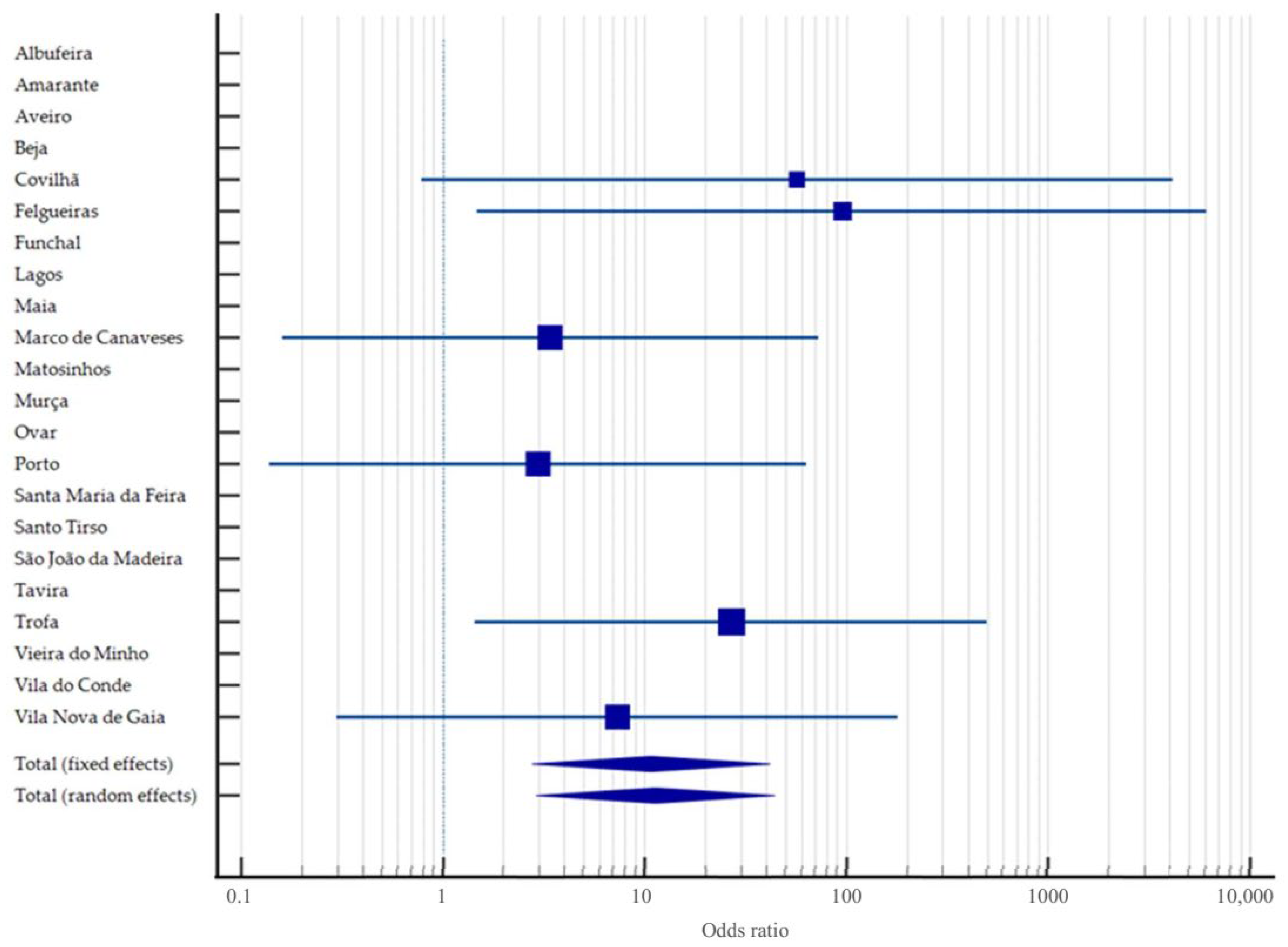
Figure 1. Forest plot representing the odds ratios and 95% confidence intervals for Brucella canis seropositivity in dogs across Portuguese municipalities, between 2013 and 2025, using Lagos as the reference group (OR = 1). Each square indicates the odds ratio estimate for a municipality, with the size proportional to its study weight under a fixed-effects model. Horizontal lines represent 95% confidence intervals. The pooled odds ratio is depicted as a diamond at the bottom of the graph, under both fixed-effects and random-effects models. Municipalities with no seropositive cases or with insufficient data are shown without a plotted estimate.
Zoonoticdis 05 00026 g001
Figure 2. Flow diagram of the identification and extraction process of articles included in the study. Adapted from Prisma guidelines 2020 [38].
Zoonoticdis 05 00026 g002
Figure 3. Forest plot showing individual and pooled prevalence estimates of Brucella spp. infection in animals in Portugal, based on a random-effects model. Each square represents the point estimate of prevalence for an individual study, with horizontal lines indicating 95% confidence intervals. The diamond at the bottom represents the overall pooled prevalence estimate (2.42%; 95% CI: 1.79–3.13), marked by a vertical dashed line. The x-axis represents prevalence expressed as a proportion (e.g., 0.1 = 10%). The studies included span a period from 2001 to 2024 and are based on data from [7,8,28,34,39,40,41].
Zoonoticdis 05 00026 g003
Figure 4. Forest plot showing individual and pooled prevalence estimates of Brucella spp. infection in humans in Portugal, based on a random-effects model. Each square represents the point estimate of prevalence for an individual study, with horizontal lines indicating the corresponding 95% confidence intervals. The red diamond at the bottom represents the overall pooled prevalence estimate (10.57%; 95% CI: 8.80–12.47), marked by a vertical dashed line. The x-axis represents prevalence values expressed as proportions (e.g., 0.1 = 10%). All included studies refer to data collected between 2002 and 2016, and are based on data from [42,43].
Zoonoticdis 05 00026 g004
Figure 5. Funnel plot assessing publication bias across all studies included in the meta-analysis of Brucella spp. prevalence in Portugal (both animal and human populations). Each circle represents an individual study, plotted according to its estimated prevalence (x-axis) and standard error (y-axis). The vertical line indicates the overall pooled prevalence estimate, and the triangular funnel illustrates the expected distribution of effect sizes in the absence of bias. Visual asymmetry suggests potential publication bias or small-study effects, supported by Egger’s regression test (intercept = 23.86, 95% CI: 11.23–36.51, p = 0.0004) and Begg’s test (Kendall’s Tau = −0.2629, p = 0.0077).
Zoonoticdis 05 00026 g005
Table 1. Seropositivity to Brucella canis by titre in 132 dogs between 2013 and 2025 in Portugal.
Titren%95% CI (%)
Negative10176.5268.60–82.93
1:50 (low positive)64.552.10–9.56
1:100 (moderate positive)96.823.10–11.50
1:200 (moderate positive)86.063.10–11.50
1:400 (high positive)75.302.60–10.54
1:800 (high positive)10.760.13–4.20
Total132100-
Sera were tested starting at a 1:50 dilution, with a titre of 50 defined as the threshold for seropositivity (titres are expressed as the reciprocal of the corresponding dilutions). CI confidence interval.
Table 2. Seropositivity to Brucella canis by region (NUTS 2) in 132 dogs between 2013 and 2025 in Portugal.
Regions (NUTS 2)n of Dogs Tested (%)% of Seropositive Dogs a
(n)
95% CI (%)
North109 (72.48)27.52 (30)20.01–36.56
Centre6 (83.33)16.67 (1)3.01–56.35
Alentejo2 (100)0.00 (0)-
Algarve11 (100)0.00 (0)-
Autonomous Region of Madeira4 (100)0.00 (0)-
CI confidence interval, NUTS 2 Nomenclature of Territorial Units for Statistics a χ2 = 6.27, df = 4, p = 0.18.
Table 3. Annual seropositivity of Brucella canis in dogs from the North region of Portugal (2017–2025), with national averages for comparison.
Region (NUTS 2)201720182019202020212022202320242025Average (%)
North9.7%74.2%9.7%0.0%0.0%0.0%0.0%3.2%0.0%7.4%
National Average (%)6.5%37.1%3.2%0.0%0.0%0.0%0.0%3.2%0.0%1.6%
NUTS 2 Nomenclature of Territorial Units for Statistics.
Table 4. Seropositivity a of Brucella canis within the season in 132 dogs between 2013 and 2025 in Portugal.
TitreSummer
n (%)
Winter
n (%)
Spring
n (%)
Autumn
n (%)
Total
n (%)
Negative29 (82.9%)25 (96.2%)23 (57.5%)24 (77.4%)101 (76.5%)
1:503 (8.6%)1 (3.8%)1 (2.5%)1 (3.2%)6 (4.5%)
1:1002 (5.7%)0 (0.0%)2 (5.0%)5 (16.1%)9 (6.8%)
1:2001 (2.9%)0 (0.0%)6 (15.0%)1 (3.2%)8 (6.1%)
1:4000 (0.0%)0 (0.0%)7 (17.5%)0 (0.0%)7 (5.3%)
1:8000 (0.0%)0 (0.0%)1 (2.5%)0 (0.0%)1 (0.8%)
Total35 (100%)26 (100%)40 (100%)31 (100%)132 (100%)
Winter, 21 December–19 March; spring, 20 March–20 June; summer, 21 June–21 September; autumn, 22 September–20 December. a χ2 = 18.1, df = 3, p < 0.001.
Table 5. Summary of included studies reporting Brucella spp. prevalence in animals and humans in Portugal (2001–2025). For each study, the reference, study period, sample size, estimated proportion of positive cases, 95% confidence interval (CI), and the corresponding weight (%) in both fixed-effects and random-effects meta-analytical models are presented. The fixed-effects model yields a pooled prevalence of 0.20% (95% CI: 0.19–0.20), while the random-effects model estimates a substantially higher pooled prevalence of 4.49% (95% CI: 3.77–5.27), reflecting considerable heterogeneity across studies, populations, and methodologies.
Ref.StudyStudy
Period
HostBrucella SpeciesSample Size
(n)
Positivity
(%)
95% CIWeight (%)
FixedRandom
[39]Coelho et al. (2015)2001GoatB. melitensis41,8254.364.16–4.560.682.41
[39]Coelho et al. (2015)2001SheepB. melitensis175,6665.845.73–5.952.862.41
[39]Coelho et al. (2015)2002GoatB. melitensis23,1623.233.00–3.460.382.41
[39]Coelho et al. (2015)2002SheepB. melitensis110,2754.454.33–4.581.792.41
[42]Pelerito et al. (2014)2002HumanBrucella spp.56915.9913.08–19.270.012.15
[39]Coelho et al. (2015)2003GoatB. melitensis21,7841.401.24–1.560.352.41
[39]Coelho et al. (2015)2003SheepB. melitensis93,3311.641.56–1.721.522.41
[42]Pelerito et al. (2014)2003HumanBrucella spp.54112.9410.23–16.060.012.14
[34]Caetano et al. (2014)2004BovineB. abortus340019.0017.69–20.360.062.36
[39]Coelho et al. (2015)2004GoatB. melitensis28,9851.371.24–1.510.472.41
[39]Coelho et al. (2015)2004SheepB. melitensis121,0051.331.26–1.391.972.41
[42]Pelerito et al. (2014)2004HumanBrucella spp.60112.6510.09–15.570.012.16
[39]Coelho et al. (2015)2005GoatB. melitensis43,1431.261.16–1.370.702.41
[39]Coelho et al. (2015)2005SheepB. melitensis197,6671.191.14–1.243.222.41
[42]Pelerito et al. (2014)2005HumanBrucella spp.66710.208.00–12.750.012.18
[39]Coelho et al. (2015)2006GoatB. melitensis47,6971.020.93–1.110.782.41
[39]Coelho et al. (2015)2006SheepB. melitensis206,6280.770.74–0.813.362.41
[42]Pelerito et al. (2014)2006HumanBrucella spp.6339.327.17–11.860.012.17
[39]Coelho et al. (2015)2007GoatB. melitensis51,2980.750.68–0.830.832.41
[39]Coelho et al. (2015)2007SheepB. melitensis226,7990.380.35–0.403.692.41
[42]Pelerito et al. (2014)2007HumanBrucella spp.61310.608.28–13.320.012.17
[42]Pelerito et al. (2014)2008HumanBrucella spp.7075.804.19–7.790.012.20
[34]Caetano et al. (2014)2009BovineB. abortus29303.002.42–3.690.052.36
[34]Caetano et al. (2014)2009BovineB. abortus33240.810.54–1.180.052.36
[34]Caetano et al. (2014)2009BovineB. abortus23320.090.01–0.310.042.34
[34]Caetano et al. (2014)2009BovineB. abortus1771.130.14–4.020.001.73
[42]Pelerito et al. (2014)2009HumanBrucella spp.5267.995.82–10.640.012.13
[42]Pelerito et al. (2014)2010HumanBrucella spp.3616.934.53–10.050.012.02
[42]Pelerito et al. (2014)2010HumanBrucella spp.2425.009.77–46.710.00040.64
[42]Pelerito et al. (2014)2011HumanBrucella spp.3308.185.46–11.680.011.99
[42]Pelerito et al. (2014)2011HumanBrucella spp.3333.3317.96–51.830.0010.79
[42]Pelerito et al. (2014)2012HumanBrucella spp.19511.287.21–16.580.0031.78
[42]Pelerito et al. (2014)2012HumanBrucella spp.24712.558.69–17.340.0041.88
[42]Pelerito et al. (2014)2012HumanBrucella spp.852.350.29–8.240.0011.33
[42]Pelerito et al. (2014)2013HumanBrucella spp.205.000.13–24.870.00030.56
[40]DGAV (2022)2016BovineB. abortus849,2520.040.03–0.0413.822.41
[40]DGAV (2022)2017BovineB. abortus820,0440.040.04–0.0513.342.41
[40]DGAV (2022)2018BovineB. abortus817,7210.030.03–0.0413.302.41
[40]DGAV (2022)2019BovineB. abortus811,9450.050.05–0.0613.212.41
[40]DGAV (2022)2020BovineB. abortus737,0930.030.02–0.0311.992.41
[40]DGAV (2022)2020BovineB. abortus698,5590.020.02–0.0311.372.41
[43]Pelerito et al. (2017)2009–2016HumanB. melitensis23137.226.20–8.350.042.34
[43]Pelerito et al. (2017)2009–2016HumanB. melitensis25916.6012.28–21.700.0041.90
[28]Djokic et al. (2023)2013–2014DogB. canis629.683.64–19.880.0011.14
[7]Pires et al. (2024)2016–2023Wild BoarBrucella spp.65021.6918.58–25.060.012.18
[28]Djokic et al. (2023)2018–2019DogB. canis1947.3724.45–71.140.00030.54
[8]Ruano et al. (2024)2022–2023Wild BoarB. melitensis33226.5121.84–31.600.011.99
[41]Cavaco et al. (2025)2022–2024DolphinB. ceti595.091.06–14.150.0011.11
[41]Cavaco et al. (2025)2022–2024DolphinB. ceti5923.7313.62–36.600.0011.11
Total (fixed effects) 6,145,9470.200.19–0.20100.00100.00
Total (random
effects) b
6,145,9474.49 a3.77–5.27100.00100.00
CI, confidence interval. a Q = 76,024.62, df = 30, p < 0.0001. b I2 = 99.96%, 95% CI: 99.96–99.96.
Disclaimer/Publisher’s Note: The statements, opinions and data contained in all publications are solely those of the individual author(s) and contributor(s) and not of MDPI and/or the editor(s). MDPI and/or the editor(s) disclaim responsibility for any injury to people or property resulting from any ideas, methods, instructions or products referred to in the content.