Article

Isolation and Characterization of a Novel Orthomyxovirus from a Bothriocroton hydrosauri Tick Removed from a Blotched Blue-Tongued Skink (Tiliqua nigrolutea) in Tasmania, Australia

1
Australian Centre for Disease Preparedness, Geelong 3220, Australia
2
Australian Rickettsial Reference Laboratory, University Hospital Geelong, Geelong 3220, Australia
*
Author to whom correspondence should be addressed.
Zoonotic Dis. 2025, 5(2), 9; https://doi.org/10.3390/zoonoticdis5020009
Submission received: 6 March 2025 / Revised: 28 March 2025 / Accepted: 31 March 2025 / Published: 10 April 2025

Simple Summary

Human-biting arthropods such as ticks are of interest to public health authorities around the world, owing to their potential to transmit a variety of pathogens, including bacteria and viruses that cause significant morbidity and mortality. In Australia, at least 70 species of soft and hard ticks have been documented. Of these, a variety of tick species have been reported to bite humans, with at least six species known to transmit pathogens during feeding. Endemic Australian diseases caused by bacterial pathogens transmitted by ticks include (i) Q Fever caused by Coxiella burnetii; (ii) Queensland Tick Typhus due to Rickettsia australis; (iii) Flinders Island Spotted Fever, caused by Rickettsia honei and (iv) Australian Spotted Fever due to R. honei subsp marmionii.

Abstract

Active and passive surveillance, followed by gene sequencing, continue to be used to identify a diverse range of novel bacteria, viruses, and other microorganisms in ticks with the potential to cause disease in vertebrate hosts following tick bite. In this study, we describe the isolation and characterization of a novel virus from Bothriocroton hydrosauri ticks collected from a blotched blue-tongue, Tiliqua nigrolutea. In an attempt to isolate rickettsia, the inoculation of Vero cell cultures with tick extracts led to the isolation of a virus, identified as a novel tick Orthomyxovirus by electron microscopy and gene sequencing. Transmission electron microscopic analysis revealed that B. hydrosauri tick virus-1 (BHTV-1) is a spherical orthomyxovirus, 85 nm in size. Multiple developmental stages of the virus were evident in vitro. Analysis of putative BHTV-1 amino acid sequences derived from a genomic analysis of virus-infected host cell extracts revealed the presence of six putative RNA segments encoding genes, sharing the closest sequence similarity to viral sequences belonging to the arthropod-borne Thogotovirus genus within the Orthomyxoviridae. Thogotoviruses are an emerging cause of disease in humans and animals following tick bite. The detection of this new thogotovirus, BHTV-1, in B. hydrosauri, a competent vector for human tick-borne infectious diseases, warrants follow-up investigation to determine its prevalence, host range, and pathogenic potential.

1. Introduction

While bacterial tick-borne diseases in Australia are widely recognized, less is known about the potential for viral pathogens transmitted by Australian ticks. Thus far, at least six viruses have been isolated from human-biting ticks in Australia [1,2,3,4]. These include (i) Ixodes holocyclus Iflavirus, a member of the Iflavirus genus of positive, sense single-stranded RNA (ssRNA) viruses, detected in I. holocyclus ticks from Queensland and northern New South Wales [5]; (ii) a diversity of viruses isolated from Argas robertsi ticks in the Northern Territory and South-East Queensland [6,7,8]; (iii) and two viruses, Saumarez Reef virus and Upolu virus (UPOV), isolated from Ornithodoros capensis ticks parasitizing sooty terns (Onychoprion fuscatus) on the Great Barrier Reef, Queensland [9,10]. UPOV, notably, was initially classified as a Bunyavirus, with molecular analyses subsequently reclassifying it as a member of the Orthomyxoviridae family [11]. While these viruses have been detected in ticks that bite humans, their pathogenic potential is unclear. It should nevertheless be noted that many of these viruses otherwise belong to families of viruses with well-recognized human pathogens, including the Orthomyxiviridae and Flaviviridae [3].
The preparation of an appropriate public health response to tick-borne diseases relies on the use of state-of-the-art laboratory methods to identify potential pathogenic agents, understand the epidemiology of the putative agents, and determine their actual pathogenic potential [12]. Given that the majority of pathogens associated with tick-borne diseases are obligate intracellular parasites, these methodologies inevitably include a combination of traditional laboratory methods including cell culture and microscopy coupled with molecular methods, including broad-range PCR assays and next-generation sequencing. The use of the latter approaches has revealed an increasing level of microbial diversity harbored by human-biting ticks [12]; while these approaches are useful, studies to isolate the organism are still required if the pathogenic potential of these novel tick-borne microorganisms is to be assessed properly.
A recent molecular study documented a growing number of rickettsiae, obligate intracellular bacterial pathogens from the Order Rickettsiales [13], in the Australian reptile tick, Bothriocroton hydrosauri. Bothriocroton hydrosauri is the established reservoir of Rickettsia honei, the etiological agent of Flinders Island Spotted Fever in Southern Australia [14]. A molecular survey of this tick species on the Australian mainland recently revealed that this tick may carry additional rickettsial agents with novel rickettsiae detected that were genetically distinct from R. honei [15]. Similar agents were also documented in Amblyomma ticks in Western Australia [16] and the Northern Territory [17].
To explore the diversity of novel rickettsiae hinted at during molecular screening [15], in the current study, we attempted to isolate novel rickettsial agents from B. hydrosauri. Instead of culturing a rickettsial agent as expected, we instead successfully isolated and subsequently characterized a novel orthomyxovirus harbored by this human-biting reptile tick. In many parts of the world, tick-transmitted viral infections are well recognized. However, this is not yet the case in Australia. This is the first Australian tick virus that may be a human pathogen.

2. Materials and Methods

2.1. Viral Isolation and Culturing

Ethics approval was received from the Tasmanian Department of wildlife, as part of a wider study in Flinders Island Spotted Fever, a newly identified human infection in Tasmania. As studies later showed, ticks from reptiles were responsible for this rickettsial infection when they bite humans. There are several publications on this. Permission was given to collect lizards and snakes, to remove their ticks, and to release them at the same place they were captured from. This was performed. There was no ethical breach. A live B. hydrosauri tick was collected from a blotched blue-tongue skink (Tiliqua nigrolutea) at Mount Stuart, Hobart, Tasmania, and transported to the Australian Rickettsial Reference Laboratory (ARRL) for further analysis.
The live tick was homogenized in Phosphate-Buffered Saline (PBS) using a plastic pestle. The homogenate was filtered through a 0.22 µm filter, with another 1 mL of PBS passed through to wash. The culture medium (RPMI supplemented with 3% fetal calf serum, 4 mM L-glutamine, and 25 mM HEPES) was removed from a 25 cm2 non-vented tissue culture flask containing a fresh, confluent monolayer of Vero cells, and 500 µL of tick homogenate was added. The flask was centrifuged at room temperature (500× g for 30 min) to promote the attachment of any organisms present in the cells. Following centrifugation, 9 mL of medium was added, and the flask was incubated at 35 °C.
After 6 days of incubation, a cytopathic effect was observed by microscopy. Remaining adherent cells were detached from the flask into the medium using a cell scraper, and 1 mL of this material was passaged into flasks containing fresh, confluent monolayers of Vero cells with 9 mL of culture medium. After a further 6 days of incubation at 35 °C, a cytopathic effect was observed again. Flask contents were harvested, and aliquots were frozen at −80 °C in cell culture freezing medium (CCFM; Gibco PTY LTD, North Fitzroy, Melbourne, Victoria, Australia).

2.2. PCR Screening

Several PCR assays were used to investigate the source of the cytopathic effect observed in cell culture. The possible presence of bacterial agents was examined using a eubacterial 16S rRNA PCR [18]. The presence of specific potential rickettsial agents was determined using a rickettsiae-specific qPCR, as previously described [19].

2.3. Transmission Electron Microscopy (TEM)

After the development of the cytopathic effect, cell cultures inoculated with passaged media, as described above in Section 2.1, were processed for TEM thin-section and negative contrast analyses according to the procedures described in [20].

2.4. Genome Sequencing

Viral RNA from 100 µL of frozen tissue culture supernatant from infected Vero cells was extracted with the Zymo Direct-zol Miniprep kit without DNase digestion, and total RNA was concentrated to a volume of 16 µL with Zymo’s RNA clean and concentrator kit. Sample isothermal amplification and Illumina Nextera XT library preparation were performed, and samples were sequenced on the Illumina MiniSeq Sequencing System, generating 150 bp paired-end (PE) reads.

2.5. Genome Annotation, Alignment, and Phylogenetic Analyses

A bioinformatics pipeline for viral whole genome de novo assembly was set up. Host subtraction was omitted, and an updated version of CLC Genomics Workbench (v20.0.4) was used for read trimming. Geneious Prime (v2020.2.3) was used for read error correction and normalization (BB Norm v38.84), and SPAdes “MµLti Cell” (v3.13.0) for de novo assembly. Additional de novo assemblies were also performed with alternative SPAdes data source options, including “RNA”, “Single Cell”, and “Metagenome” (default parameters), as well as MIRA v4.0 (default parameters). Assembled contigs were then queried against the National Center for Biotechnology Information (NCBI) non-redundant database with BlastX to identify virus-encoding contigs. Contigs for each genome segment were then aligned to closely related viruses from the Thogtovirus genus at both the nucleotide and amino acid levels to verify assemblies, and overlapping contigs were joined, if required, to attempt to obtain the complete viral genome. Final genome segments were verified by mapping back-trimmed reads to identify misassembled regions.
Final genome sequences (submitted to Genbank) were subject to BlastX and BlastN to support viral identification. Phylogenetic analyses were performed using a set of orthomyxovirus sequences retrieved from Genbank (www.ncbi.nlm.nih.gov) (accessed on 10 January 2025) to clarify the origin of open reading frames (ORFs) within each genome segment and to identify potential recombination events. All sequences were aligned using the Clustal algorithm (as implemented in MEGA3; [21]) at the nucleotide and amino acid level with additional manual editing to ensure the highest possible quality of alignment. Neighbor-joining analysis at the amino acid level was performed due to the observed high variability of the underlying nucleotide sequences of members of the family Orthomyxoviridae. The statistical significance of tree topologies were evaluated through the bootstrap resampling of the sequences 1000 times.

3. Results

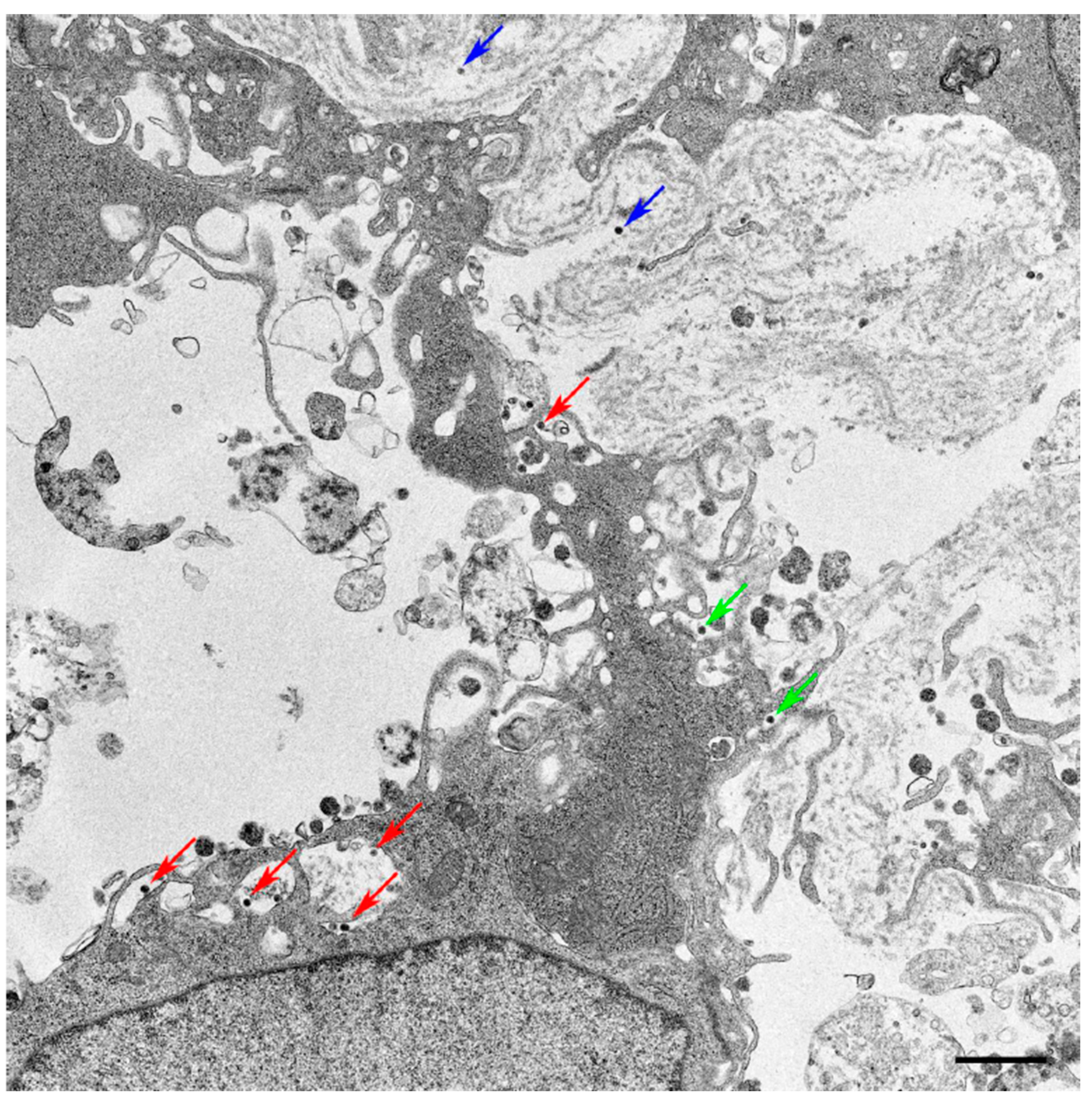
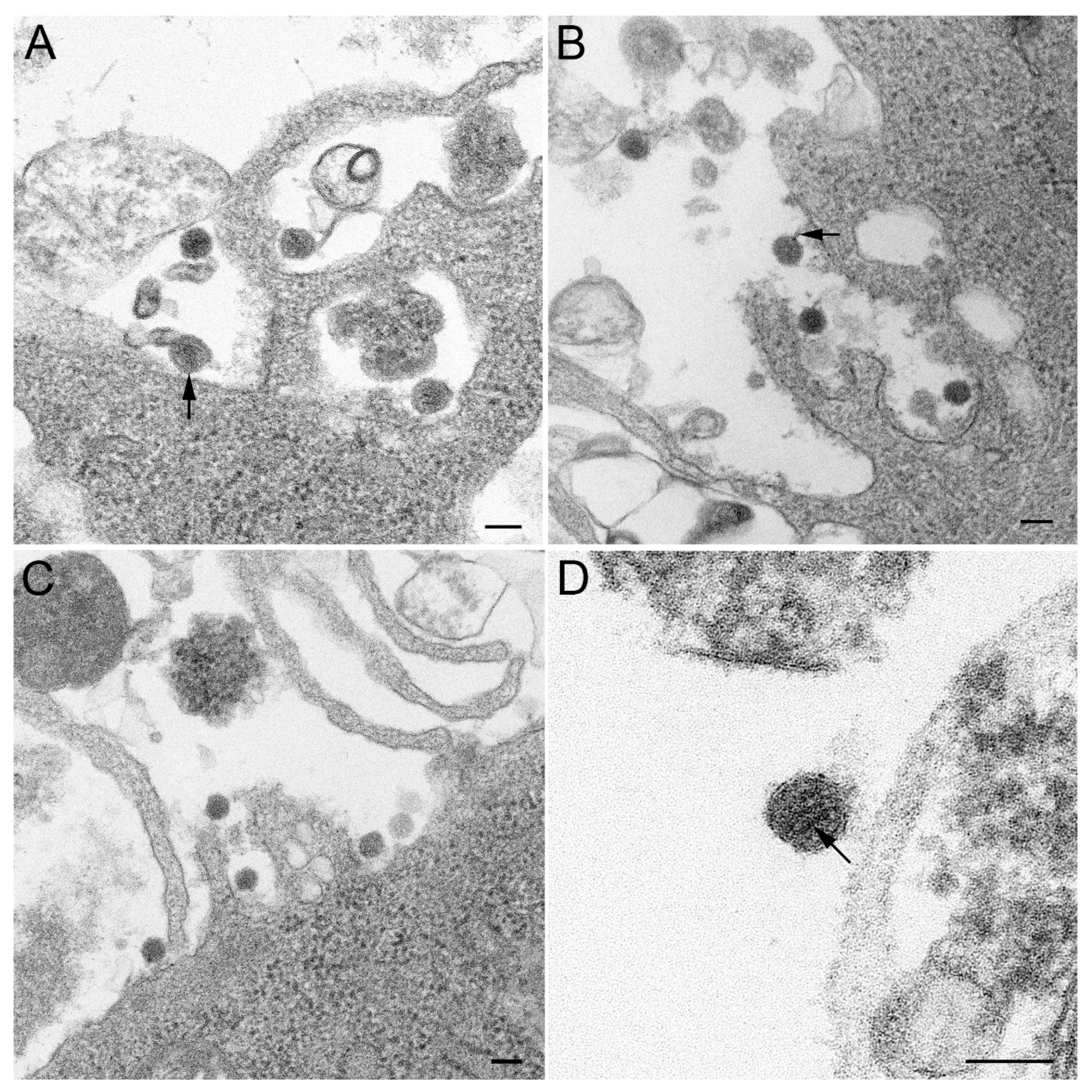
3.1. Isolation and Morphology of Novel Tick Virus

A cytopathic effect was observed by microscopy six days after the inoculation of the Vero cell culture with the filtered tick homogenate. To characterize the potential intracellular pathogen, supernatant extracts were screened by PCR. Cellular extracts were negative in the initial screening broad-range eubacterial and rickettsial-specific PCR tests. Further characterization using random PCR primers followed by sequencing showed that the supernatant contained an Orthomyxovirus in the Thogotovirus genus.
Further investigation by TEM (JEOL, Tokyo, Japan) revealed the presence of icosahedral- to spherical-shaped virus particles measuring approximately 85 nm in diameter. The particles observed were located (1) within invaginations of the plasma membrane, (2) at the surface of the plasma membrane, or (3) amongst extracellular debris (Figure 1). At higher magnifications, strands of internal ribonucleic protein were visible (Figure 2D). ‘Tether’ or stalk-like structures were visible between some viral particles and host cell membranes (Figure 2A–D). The ‘tethers’ were similar in appearance to those present in Upolu and Aransas Bay virus micrographs [11]. The identification and purpose of these tether-like structures are yet to be investigated; it is possible they may be an envelope resulting from the budding process. However, no virus particles were seen classically budding at the cell surface.

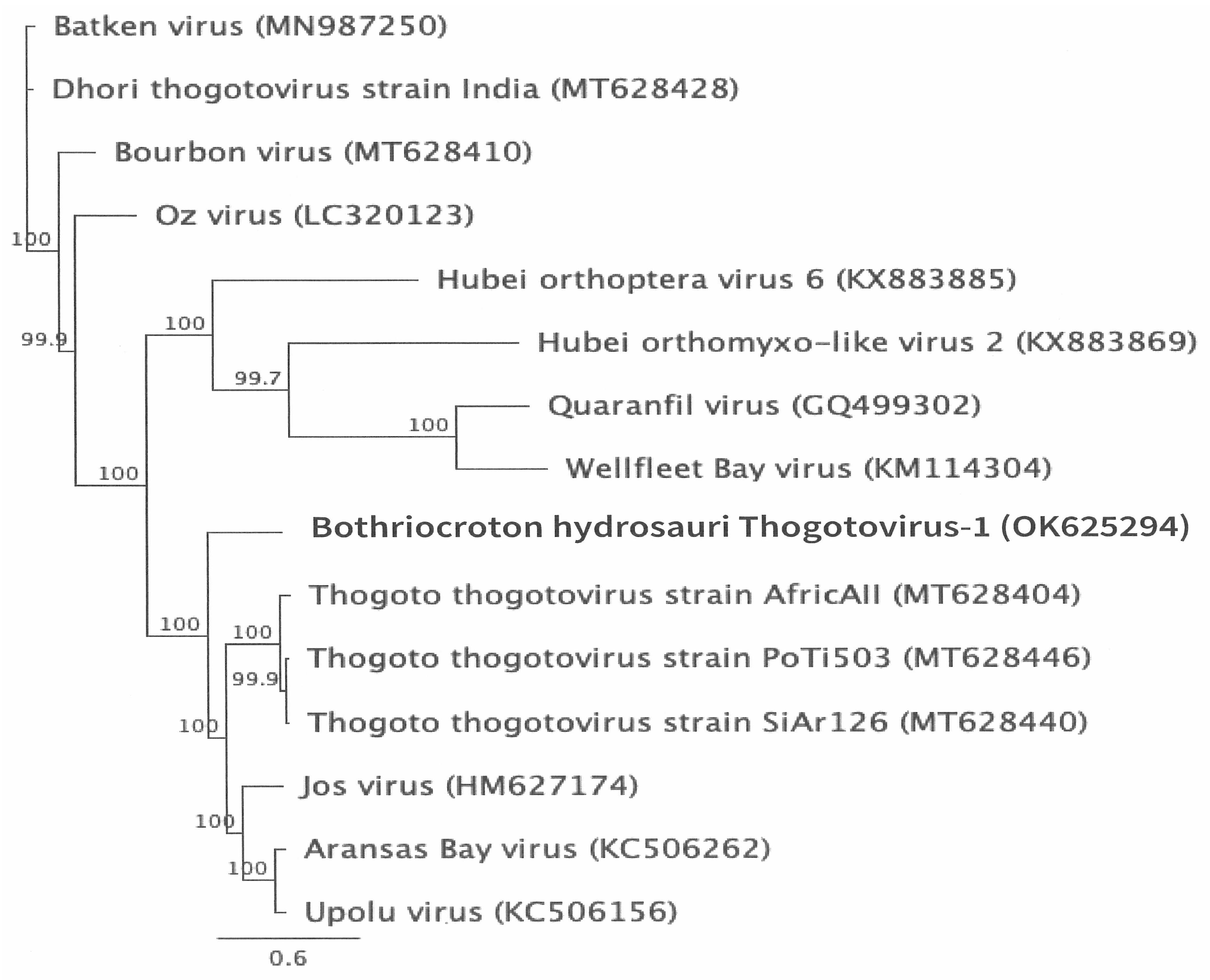
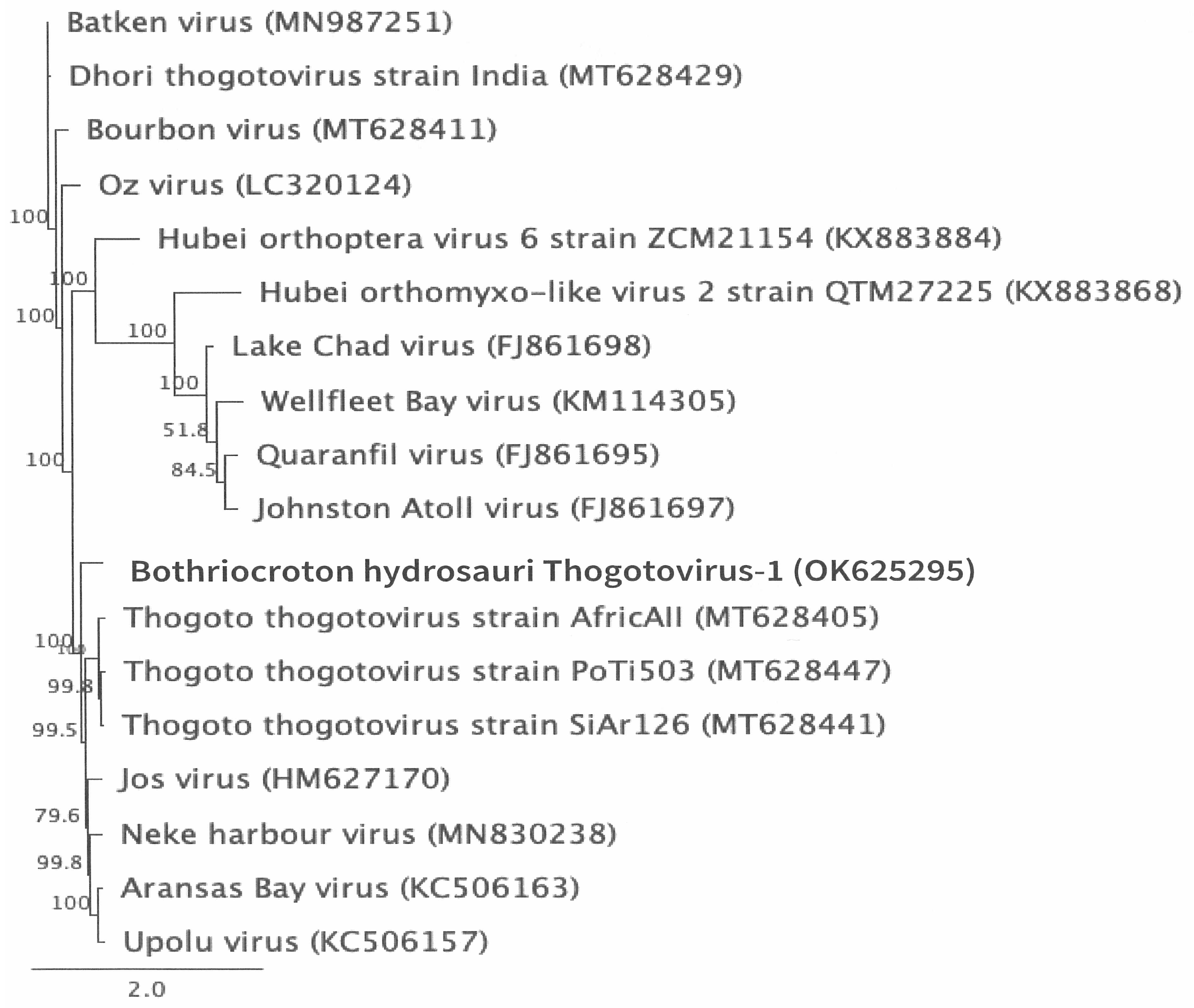
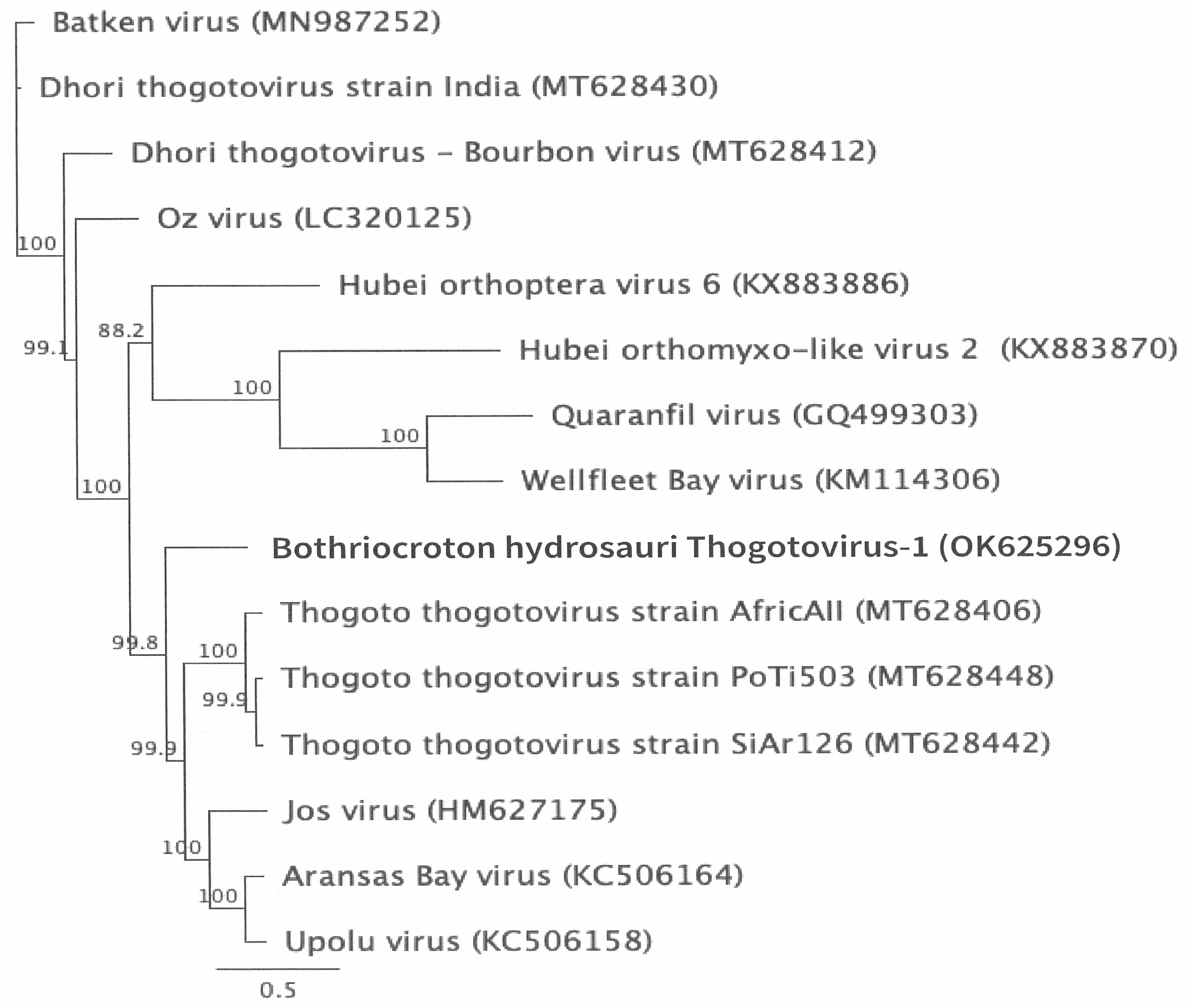
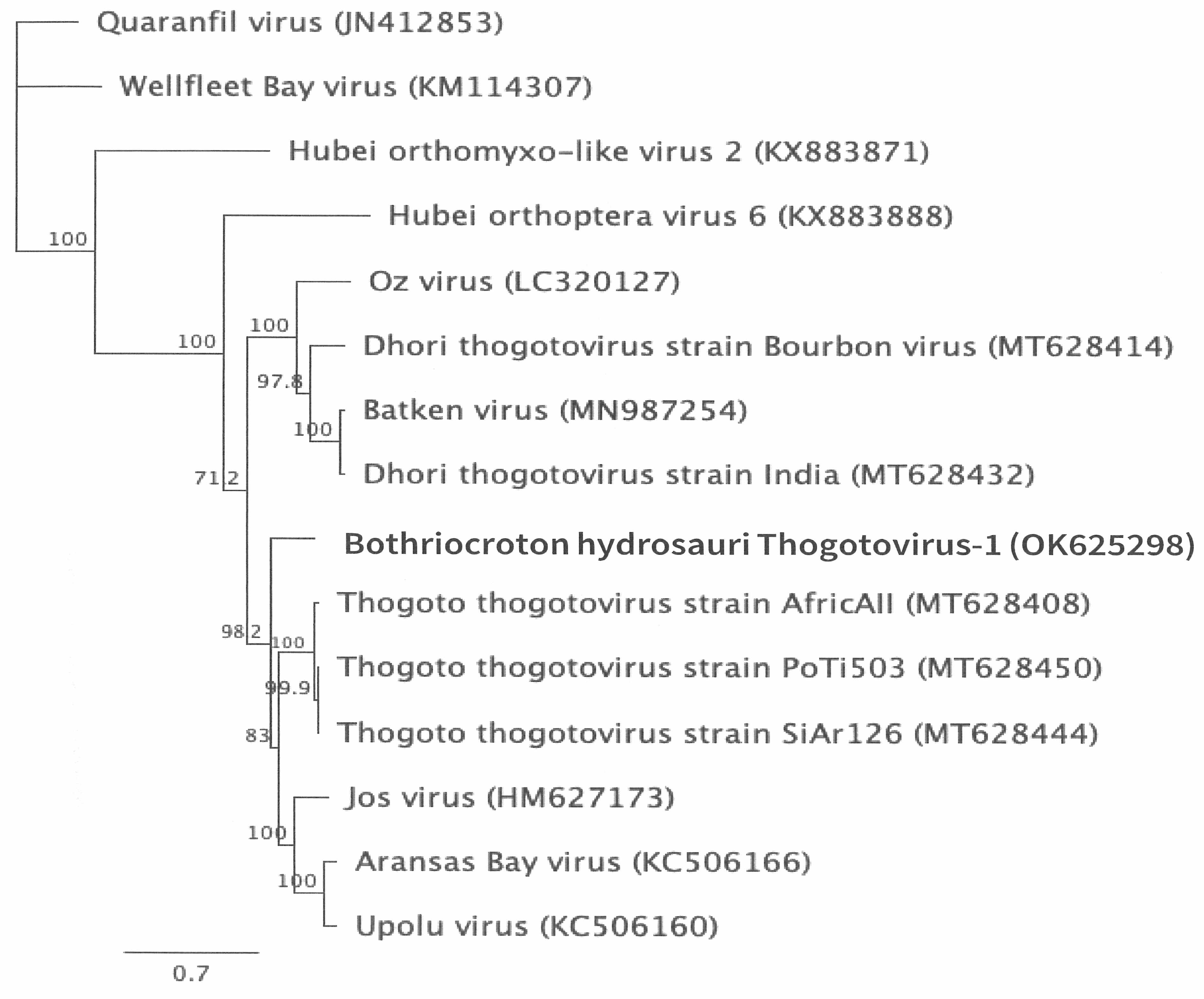
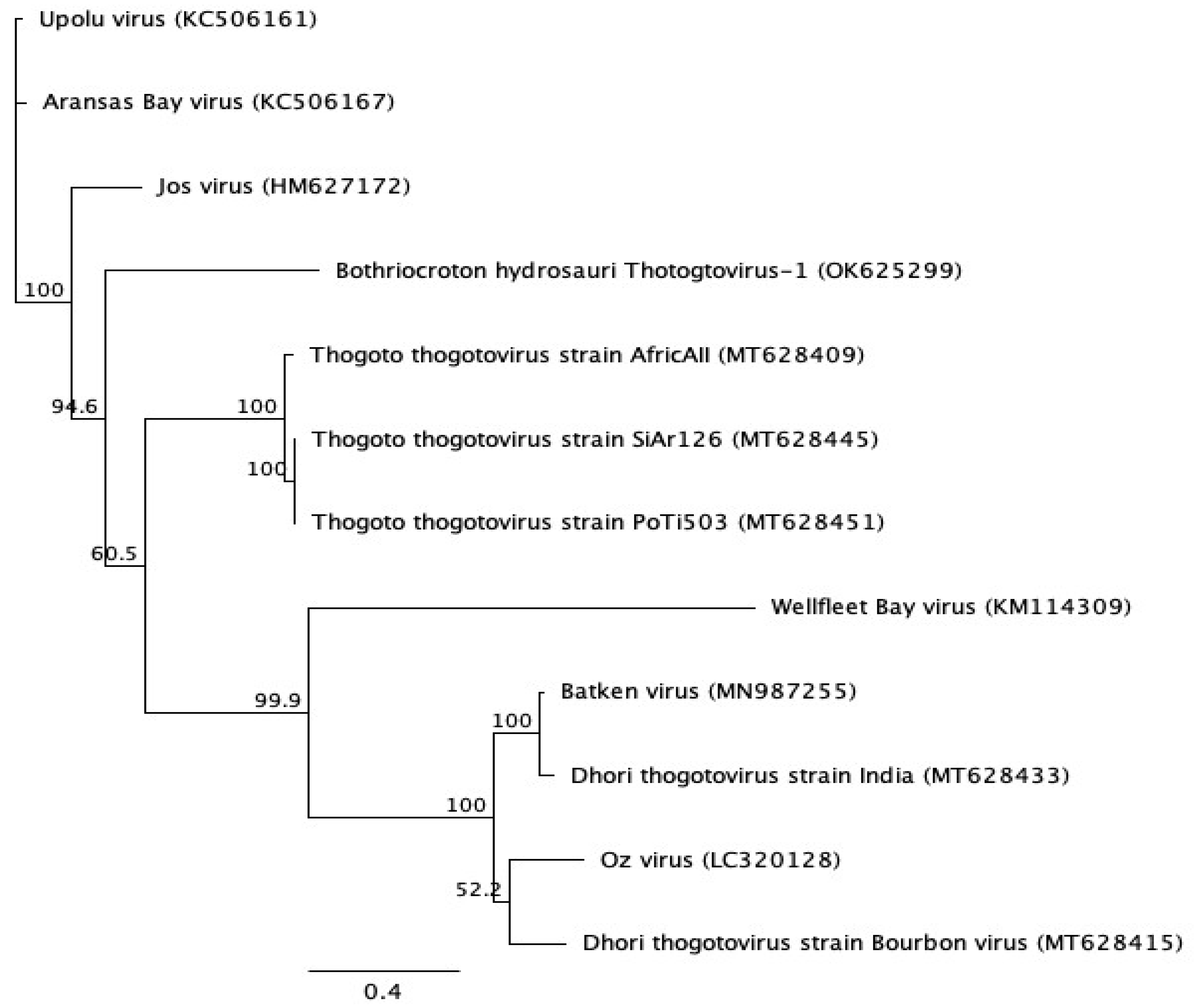
3.2. Genomic Characterization and Phylogenetic Analysis of the Novel Tick Virus

The total RNA extracted from the cell culture supernatant of Vero-infected cells was sequenced to characterize the putative virus cultured. The SPAdes MµLti-Cell de novo assembly of 6,493,760 trimmed, normalized Illumina PE reads led to the assembly of 39 contigs with BLASTn and BLASTx analysis used to identify 8 X contigs that contained viral ORFs_. Additional de novo assemblies with MIRA and SPAdes (a total of 456 contigs; 166 viral encoding genes) were performed to join and/or extend ORFs within the original virus containing contigs. Genome analysis resulted in a final total of six viral genome segments of 2140 bp (segment 1), 2193 bp (segment 2), 1955 bp (segment 3), 1612 bp (segment 4), 1427 bp (segment 5), and 966 bp (segment6). The putative identities of the viral ORFs within each of the six genome segments, as determined by BLASTx analysis, are presented in Table 1. Each ORF was found to be the most similar to viruses in the genus Thogotovirus within the family Orthomyxoviridae, including (i) ORF1, a complete coding sequence (781 amino acids (aa)) sharing the highest similarity with a polymerase basic protein-2 (PB2) from a Jos virus; (ii) ORF2, a complete coding sequence (712 aa) sharing 70.1% similarity with Jos virus polymerase basic protein-1 (PB1); (iii) ORF3, a complete coding sequence (629 aa) sharing 48.9% similarity with Aransas Bay virus polymerase acidic subunit (PA); (iv) ORF4, a partial coding sequence for an Upolu virus glycoprotein-encoding (GP); (v) ORF5, a complete coding sequence (454 aa) with the highest similarity with a Thogotovirus nucleoprotein (NP); and (vi) ORF6, a complete coding sequence (309 aa) sharing 45.7% sequence similarity with a Jos virus matrix protein long (ML). In addition, assembled data revealed a contig encoding a 269-amino-acid truncated, spliced variant of the matrix protein (M), as described for the closely related Jos virus M protein [22]. Based on this sequence analysis, for the remainder of this manuscript, this novel virus is referred to as Bothriocroton hydrosauri Thogotivirus-1 (BHTV-1).
To gain further insight into the genetic relationship between this BHTV-1 and other closely related viruses, phylogenetic trees were constructed for the individual protein sequences (ORF-1, ORF-2, ORF-3, ORF-4, ORF-5, ORF-6; Figure 3, Figure 4, Figure 5, Figure 6, Figure 7 and Figure 8). As predicted by BLASTx searching, phylogenetic analyses of each of these ORFs revealed that the novel virus, BHTV-1, is most closely related to other tick-borne viruses (Dhorivirus, Bourbonvirus, Josvirus) in the family Orthomyxoviridae, genus Thogtovirus. Phylogenetic analysis of the putative BHTV-1 ORF-6, however, revealed that this ORF amino acid sequence is most closely related to viruses in the genus Quaranjavirus within the family Orthomyxoviridae (Figure 4), suggesting a possible reassortment event between BHTV-1 and viruses within the Quaranjavirus genus. The most conserved gene within the Orthomyxovirus family was ORF2, the PA gene segment.

4. Discussion

In the current study, we identified a novel tick-borne orthomyxovirus, BHTV-1, during attempts to isolate novel rickettsial agents from B. hydrosauri ticks. No rickettsia was detected during this study. The genetic characterization of this novel virus shows that it is closely related to viruses within the Thogotovirus genus of the Orthomyxoviridae family. Like other members of the Orthomyxoviridae family, Thogotoviruses are single-stranded, negative-sense segmented RNA viruses (ssRNA) [21]. These viruses have been reported in a range of host species, including rodents [22], domesticated livestock [23,24], and humans [25,26]. In the latter host, viruses from this genus have been linked to serious infection outcomes, including fever and neurological symptoms [27]. Virus isolation and molecular studies suggest that the primary reservoir for thogotoviruses are hard tick species [28,29], with transmission to humans being the result of tick bite [27]. The isolation of BHTV-1 represents the second detection of a putative Thogotovirus genus virus from an Australian hard tick with UPOV previously isolated from O. capnesis ticks infesting sooty terns [9]. Human-biting B. hydrosauri ticks have already been proven to be competent reservoirs of tick-borne diseases, with this tick species being the host of R. honei, the causative agent of Flinders Island Spotted Fever in Australia [14,30]. Its detection in a tick removed from a blotched, blue-tongued skink likely suggests that the virus can potentially infect reptiles as well, which is an observation that is yet to be described for thogotoviruses. At present, the human health significance of the detection and description of BHTV-1 is unclear but given that (a) B. hydrosauri are human-biting ticks responsible for the transmission of tick-borne diseases and (b) there is growing evidence around the world for the pathogenic potential of thogotoviruses, further research into the prevalence and host range of this virus is warranted.
Orthomyxovirus genomes consist of six segments (thogotoviruses) to eight segments (influenza A viruses) [31]. Our genomic and phylogenetic analysis identified thogotovirus homologues for six of the ORFs described in thogotovirus family members, including viral polymerase complex proteins, (polymerase basic subunit 1 (PB1), polymerase basic subunit 2 (PB2), polymerase acidic subunit (PA)), a surface glycoprotein, a nucleoprotein, and matrix protein [11]. Phylogenetic analysis of the predicted amino acid sequences for each of these ORFs revealed that four of six BHTV-1 proteins shared the closest phylogenetic relationships with thogotovirus and Jos virus. Experimental infection studies have shown that tick-derived Dhori virus can cause fulminant, systemic, and fatal disease in infected mice [32,33], while the latter thogotovirus has been linked to a human tick bite-associated death reported in the United States in 2014 [27]. In contrast to the results for the other BHTV-1 proteins, phylogenetic analysis of the predicted glycoprotein and revealed closest similarity with viruses within the Thogotovirus genus, Orthomyxoviridae family, Upolu and Aransus Bay. Given the potential geographic and host overlap between viruses from each of these orthomyxovirus genera, it is conceivable that co-infection (presumably in a tick) and potential reassortment events between species may have occurred within the evolutionary history of BHTV-1.
An obvious limitation of this study was that viral isolation was performed on only a subset of B. hydrosauri ticks isolated from blotched blue-tongued skinks. Furthermore, while viral sequences were verified by mapping trimmed reads to reveal misassembled regions, RACE-PCR and traditional PCR and Sanger sequencing are required to confirm the BHTV-1 segments sequenced and to obtain complete genome ends.

5. Conclusions

In this study, a novel thogotovirus, BHTV-1, was isolated from a reptile-infesting B. hydrosauri tick in Tasmania. B. hydrosauri ticks are competent vectors for tick-borne diseases in Australia and have a growing reputation for causing disease in humans and animals. This may be the first known report of reassortment between different genera of orthomyxoviruses, in this case between one thogotoviruses genus and another. The effect of the reassortant on the pathogenicity of the virus is unknown, and further studies are required to determine the prevalence and potential for these viruses to cause tick-borne diseases in Australia and elsewhere.

Author Contributions

Conceptualization, S.G., J.S. and G.V.; virus isolation, G.V.; virus growth and sample preparation, P.S.; gene sequencing and analysis, M.T.; electron microscopy, S.C.; support and funding for testing at the Australian Centre for Disease Preparedness, G.M.; manuscript preparation, P.S.; review and editing of manuscript, all authors. All authors have read and agreed to the published version of the manuscript.

Funding

Funding for this research was provided by the Australian Rickettsial Reference Laboratory and the Australian Centre for Disease Preparedness.

Institutional Review Board Statement

Not applicable.

Informed Consent Statement

Not applicable.

Data Availability Statement

The original contributions presented in this study are included in the article. Further inquiries can be directed to the corresponding author(s). Sequence data has been uploaded to Genbank.

Acknowledgments

We thank Adam Polkinghorne for assistance in preparing this manuscript and Clare Holmes for technical assistance in the preparation of samples for electron microscopy.

Conflicts of Interest

S.G. and J.S. are Directors of the Australian Rickettsial Reference Laboratory, a not-for-profit diagnostic and research laboratory that undertakes diagnostic testing for Rickettsia, Coxiella, and Borrelia on human patient specimens. The Australian Government and individual patients pay for these tests. The other authors declare no conflicts of interest.

References

  1. Rochlin, I.; Toledo, A. Emerging tick-borne pathogens of public health importance: A mini-review. J. Med. Microbiol. 2020, 69, 781–791. [Google Scholar] [CrossRef] [PubMed]
  2. Barker, S.C.; Walker, A.R.; Campelo, D. A list of the 70 species of Australian ticks; diagnostic guides to and species accounts of Ixodes holocyclus (paralysis tick), Ixodes cornuatus (southern paralysis tick) and Rhipicephalus australis (Australian cattle tick); and consideration of the place of Australia in the evolution of ticks with comments on four controversial ideas. Int. J. Parasitol. 2014, 44, 941–953. [Google Scholar] [CrossRef] [PubMed]
  3. Dehhaghi, M.; Kazemi Shariat Panahi, H.; Holmes, E.C.; Hudson, B.J.; Schloeffel, R.; Guillemin, G.J. Human Tick-Borne Diseases in Australia. Front. Cell. Infect. Microbiol. 2019, 9, 3. [Google Scholar] [CrossRef] [PubMed]
  4. Graves, S.R.; Stenos, J. Tick-borne infectious diseases in Australia. Med. J. Aust. 2017, 206, 320–324. [Google Scholar] [CrossRef]
  5. O’Brien, C.A.; Hall-Mendelin, S.; Hobson-Peters, J.; Deliyannis, G.; Allen, A.; Lew-Tabor, A.; Rodriguez-Valle, M.; Barker, D.; Barker, S.C.; Hall, R.A. Discovery of a novel iflavirus sequence in the eastern paralysis tick Ixodes holocyclus. Arch. Virol. 2018, 163, 2451–2457. [Google Scholar] [CrossRef]
  6. Doherty, R.; Carley, J.; Filippich, C.; Kay, B. Isolation of virus strains related to Kao Shuan virus from Argas robertsi in Northern Territory, Australia. Search 1976, 7, 484–486. [Google Scholar]
  7. St George, T.; Cybinski, D.; Jmain, A.; Mckilligan, N.; Kemp, D. Isolation of a new arbovirus from the tick Argas robertsi from a cattle egret (Bubulcus ibis coromandus) colony in Australia. Aust. J. Biol. Sci. 1984, 37, 85–90. [Google Scholar] [CrossRef]
  8. Gauci, P.J.; McAllister, J.; Mitchell, I.R.; Cybinski, D.; St George, T.; Gubala, A.J. Genomic Characterisation of Vinegar Hill Virus, An Australian Nairovirus Isolated in 1983 from Argas Robertsi Ticks Collected from Cattle Egrets. Viruses 2017, 9, 373. [Google Scholar] [CrossRef]
  9. Doherty, R.; Whitehead, R.H.; Wetters, E.J. Isolation of viruses from Ornithodoros capensis Neumann from a tern colony on the Great Barrier Reef, North Queensland. Aust. J. Sci. 1968, 31, 363–364. [Google Scholar]
  10. George, T.S.; Standfast, H.; Doherty, R.; Carley, J.; Fillipich, C.; Brandsma, J. The isolation of Saumarez Reef virus, a new flavivirus, from bird ticks Ornithodoros capensis and Ixodes eudyptidis in Australia. Immun. Cell Biol. 1977, 55, 493. [Google Scholar] [CrossRef]
  11. Briese, T.; Chowdhary, R.; Travassos da Rosa, A.; Hutchison, S.K.; Popov, V.; Street, C.; Tesh, R.B.; Lipkin, W.I. Upolu virus and Aransas Bay virus, two presumptive bunyaviruses, are novel members of the family Orthomyxoviridae. J. Virol. 2014, 88, 5298–5309. [Google Scholar] [CrossRef] [PubMed]
  12. Madison-Antenucci, S.; Kramer, L.D.; Gebhardt, L.L.; Kauffman, E. Emerging Tick-Borne Diseases. Clin. Microbiol. Rev. 2020, 33. [Google Scholar] [CrossRef]
  13. Parola, P.; Paddock, C.D.; Socolovschi, C.; Labruna, M.B.; Mediannikov, O.; Kernif, T.; Abdad, M.Y.; Stenos, J.; Bitam, I.; Fournier, P.E.; et al. Update on tick-borne rickettsioses around the world: A geographic approach. Clin. Microbiol. Rev. 2013, 26, 657–702. [Google Scholar] [CrossRef] [PubMed]
  14. Stenos, J.; Graves, S.; Popov, V.L.; Walker, D.H. Aponomma hydrosauri, the reptile-associated tick reservoir of Rickettsia honei on Flinders Island, Australia. Am. J. Trop. Med. Hyg. 2003, 69, 314–317. [Google Scholar] [CrossRef]
  15. Whiley, H.; Custance, G.; Graves, S.; Stenos, J.; Taylor, M.; Ross, K.; Gardner, M.G. Rickettsia Detected in the Reptile Tick Bothriocroton hydrosauri from the Lizard Tiliqua rugosa in South Australia. Pathogens 2016, 5, 41. [Google Scholar] [CrossRef]
  16. Tadepalli, M.; Hii, S.F.; Vincent, G.; Watharow, S.; Graves, S.; Stenos, J. Molecular evidence of novel spotted fever group Rickettsia species in Amblyomma albolimbatum ticks from the shingleback skink, Tiliqua rugosa, in southern Western Australia. Pathogens 2020, 10, 35. [Google Scholar] [CrossRef]
  17. Vilcins, I.M.; Fournier, P.E.; Old, J.M.; Deane, E. Evidence for the presence of Francisella and spotted fever group rickettsia DNA in the tick Amblyomma fimbriatum (Acari: Ixodidae), Northern Territory, Australia. J. Med. Entomol. 2009, 46, 926–933. [Google Scholar] [CrossRef]
  18. Edwards, U.; Rogall, T.; Blocker, H.; Emde, M.; Bottger, E.C. Isolation and direct complete nucleotide determination of entire genes. Characterization of a gene coding for 16S ribosomal RNA. Nucleic Acids Res. 1989, 17, 7843–7853. [Google Scholar] [CrossRef] [PubMed]
  19. Stenos, J.; Graves, S.R.; Unsworth, N.B. A highly sensitive and specific real-time PCR assay for the detection of spotted fever and typhus group Rickettsiae. Am. J. Trop. Med. Hyg. 2005, 73, 1083–1085. [Google Scholar] [CrossRef]
  20. Unsworth, N.; Stenos, J.; Graves, S.; Faa, A.; Cox, E.; Dyer, J.; Boutlis, C.; Lane, A.; Shaw, M.; Robson, J.; et al. Flinders Island Spotted Fever rickettsioses caused by “marmionii” strain of Rickettsia honei, Eastern Australia. Emerg. Infect. Dis. 2007, 13, 566–573. [Google Scholar] [CrossRef]
  21. Kumar, S.; Tamura, K.; Nei, M. MEGA3: Integrated software for Molecular Evolutionary Genetics Analysis and sequence alignment. Brief. Bioinform. 2004, 5, 150–163. [Google Scholar] [CrossRef] [PubMed]
  22. Bussetti, A.V.; Palacios, G.; Travassos da Rosa, A.; Savji, N.; Jain, K.; Guzman, H.; Hutchison, S.; Popov, V.L.; Tesh, R.B.; Lipkin, W.I. Genomic and antigenic characterization of Jos virus. J. Gen. Virol. 2012, 93, 293–298. [Google Scholar] [CrossRef]
  23. Karabatsos, N. International Catalogue of Arboviruses including certain other viruses of vertebrates, 1985. Am. J. Trop. Med. Hyg. 1978, 27, 372. [Google Scholar] [CrossRef] [PubMed]
  24. Ogen-Odoi, A.; Miller, B.R.; Happ, C.M.; Maupin, G.O.; Burkot, T.R. Isolation of thogoto virus (Orthomyxoviridae) from the banded mongoose, Mongos mungo (Herpestidae), in Uganda. Am. J. Trop. Med. Hyg. 1999, 60, 439–440. [Google Scholar] [CrossRef]
  25. Hubalek, Z.; Rudolf, I.; Nowotny, N. Arboviruses pathogenic for domestic and wild animals. Adv. Virus Res. 2014, 89, 201–275. [Google Scholar] [CrossRef] [PubMed]
  26. Lutomiah, J.; Musila, L.; Makio, A.; Ochieng, C.; Koka, H.; Chepkorir, E.; Mutisya, J.; Mulwa, F.; Khamadi, S.; Miller, B.R.; et al. Ticks and tick-borne viruses from livestock hosts in arid and semiarid regions of the eastern and northeastern parts of Kenya. J. Med. Entomol. 2014, 51, 269–277. [Google Scholar] [CrossRef]
  27. Komar, N.; Hamby, N.; Palamar, M.B.; Staples, J.E.; Williams, C. Indirect Evidence of Bourbon Virus (Thogotovirus, Orthomyxoviridae) Infection in North Carolina. N. C. Med. J. 2020, 81, 214–215. [Google Scholar] [CrossRef]
  28. Lambert, A.J.; Velez, J.O.; Brault, A.C.; Calvert, A.E.; Bell-Sakyi, L.; Bosco-Lauth, A.M.; Staples, J.E.; Kosoy, O.I. Molecular, serological and in vitro culture-based characterization of Bourbon virus, a newly described human pathogen of the genus Thogotovirus. J. Clin. Virol. 2015, 73, 127–132. [Google Scholar] [CrossRef]
  29. Kosoy, O.I.; Lambert, A.J.; Hawkinson, D.J.; Pastula, D.M.; Goldsmith, C.S.; Hunt, D.C.; Staples, J.E. Novel thogotovirus associated with febrile illness and death, United States, 2014. Emerg. Infect. Dis. 2015, 21, 760–764. [Google Scholar] [CrossRef]
  30. Savage, H.M.; Burkhalter, K.L.; Godsey, M.S., Jr.; Panella, N.A.; Ashley, D.C.; Nicholson, W.L.; Lambert, A.J. Bourbon Virus in Field-Collected Ticks, Missouri, USA. Emerg. Infect. Dis. 2017, 23, 2017–2022. [Google Scholar] [CrossRef]
  31. Ejiri, H.; Lim, C.K.; Isawa, H.; Fujita, R.; Murota, K.; Sato, T.; Kobayashi, D.; Kan, M.; Hattori, M.; Kimura, T.; et al. Characterization of a novel thogotovirus isolated from Amblyomma testudinarium ticks in Ehime, Japan: A significant phylogenetic relationship to Bourbon virus. Virus Res. 2018, 249, 57–65. [Google Scholar] [CrossRef] [PubMed]
  32. Stenos, J.; Roux, V.; Walker, D.; Raoult, D. Rickettsia honei sp. nov., the aetiological agent of Flinders Island spotted fever in Australia. Int. J. Syst. Bacteriol. 1998, 48 Pt 4, 1399–1404. [Google Scholar] [CrossRef] [PubMed]
  33. Li, G.; Wang, N.; Guzman, H.; Sbrana, E.; Yoshikawa, T.; Tseng, C.T.; Tesh, R.B.; Xiao, S.Y. Dhori virus (Orthomyxoviridae: Thogotovirus) infection of mice produces a disease and cytokine response pattern similar to that of highly virulent influenza A (H5N1) virus infection in humans. Am. J. Trop. Med. Hyg. 2008, 78, 675–680. [Google Scholar] [CrossRef] [PubMed]
Figure 1. TEM images of putative viral particles detected in infected Vero cells. Red arrows indicate viral particles within smooth membrane vesicles of the cytoplasm of Vero cells. Green arrows indicate viral particles associated with the plasma membrane or lytic vesicles. Blue arrows indicate released viral particles amongst extracellular debris. The scale bar represents 1 µm.
Zoonoticdis 05 00009 g001
Figure 2. Transmission electron micrographs of different viral developmental stages. (A) The arrow indicates the virus, with a putative envelope remnant. (B) The arrow indicates the ‘tether’ or stalk-like structure. (C) The virus associated with plasma membrane amongst filopodia. (D) Extracellular virus particle showing internal ribonucleic protein. The scale bar represents 100 nm.
Zoonoticdis 05 00009 g002
Figure 3. BHTV-1 segment 1 polymerase B2 gene phylogenetic tree compared to other orthomyxoviruses based on amino acid sequences.
Zoonoticdis 05 00009 g003
Figure 4. BHTV-1 segment 2 polymerase B1 protein gene phylogenetic tree compared to other orthomyxoviruses based on amino acid sequences.
Zoonoticdis 05 00009 g004
Figure 5. BHTV-1 segment 3 polymerase A protein gene phylogenetic tree compared to other orthomyxoviruses based on amino acid sequences.
Zoonoticdis 05 00009 g005
Figure 6. BHTV-1 segment 4 glycoprotein gene phylogenetic tree compared to other orthomyxoviruses based on amino acid sequences.
Zoonoticdis 05 00009 g006
Figure 7. BHTV-1 segment 5 nucleoprotein gene phylogenetic tree compared to other orthomyxoviruses based on amino acid sequences.
Zoonoticdis 05 00009 g007
Figure 8. BHTV-1 segment 6 matrix protein gene phylogenetic tree compared to other orthomyxoviruses based on amino acid sequences.
Zoonoticdis 05 00009 g008
Table 1. Highest percentage BLASTx hits to novel virus ORFs detected after genome assembly.
ORF
(Size bp)
% IdentityGenbank IDAccession Description
ORF1
(2346 bp)
54.4% AED98375.1 (95% coverage)Jos virus PB2
ORF2
(2139 bp)
70.1%AED98371.1
(96% coverage)
Jos virus PB1
ORF3
(1890 bp)
48.9%AHB34062.1
(96% coverage)
Aransas Bay virus PA
ORF4
(1582 bp)
41.1%AHB34057.1
(89% coverage)
Upolu virus GP
ORF5
(1365 bp)
59.6%YP_145809.1
(95% coverage)
Thogotovirus NP
ORF6
(921 bp)
45.7%AED98373.1
(78% coverage)
Jos virus ML
Disclaimer/Publisher’s Note: The statements, opinions and data contained in all publications are solely those of the individual author(s) and contributor(s) and not of MDPI and/or the editor(s). MDPI and/or the editor(s) disclaim responsibility for any injury to people or property resulting from any ideas, methods, instructions or products referred to in the content.

