Simple Summary
This review discusses how the presence of rashes and other skin lesions may support the clinical management of viral meningitis. It highlights that the various viruses that cause meningitis, such as enteroviruses (EV) and herpes viruses (HV), often result in specific skin lesions that can help establish an early and accurate diagnosis. EV, the most common cause of viral meningitis (especially in children), often manifests as hand-foot-and-mouth disease. HV viruses, including HSV and VZV, can cause vesicular rashes along specific nerve pathways. Other viruses, such as Chikungunya, Zika, Dengue and West Nile virus, can also induce meningitis along with distinct cutaneous lesions. This article emphasizes that examination of skin lesions should be part of the standard clinical evaluation when viral meningitis is suspected.
Abstract
Viral infections may vary from mild to severe, manifesting with a wide range of symptoms, including skin lesions, influenza-like symptoms, or meningitis/meningoencephalitis signs. Viruses that cause both skin lesions and meningitis comprise, e.g., Enteroviruses (EVs) and Herpes viruses (HV). EVs are responsible for approximately 90% of viral meningitis cases. They occur frequently among children under 3 years of age and are characterized by various types of rash. HV infections are responsible for up to 18% of viral meningitis, mostly among adults or older children. Most patients with viral meningitis recover entirely. However, the rates of serious complications and mortality may be as high as 74% and 10%, respectively, for particularly vulnerable neonatal or immunocompromised patients. Patients that present signs of encephalitis and/or are suspected to have HSV/VZV infection require immediate implementation of empiric acyclovir therapy before receiving the polymerase chain reaction (PCR) test results. The clinical picture of viral meningitis may differ depending on the virus, including the presence of both meningeal signs and skin lesions. Therefore, early identification of the etiological factor is necessary for early and proper treatment implementation. It is crucial to accurately differentiate between the causative agents, and this work focuses on answering the question of how skin lesions can assist in achieving a better and faster diagnosis. The aim of this review was to analyze the characteristics of skin lesions in the course of meningitis caused by various viral species. This can be helpful for physicians in the diagnostic process and subsequent treatment.
1. Introduction
Viral meningitis is a serious condition that affects the central nervous system (CNS), causing inflammation of the protective brain membranes known as meninges [1]. In 2022, there were 1747 cases of meningitis and/or encephalitis reported in Poland, among which 900 (52%) were of viral etiology. Their number increased by 99.1% compared to 2021 [2]. Aseptic meningitis refers to the inflammation of the meninges in a patient who has not been treated with antibiotics before obtaining cerebral spinal fluid for testing, and the patient’s routine bacterial cultures have negative results [3]. Viruses are the most common cause of aseptic meningitis, chiefly Enteroviruses (approximately 90% of viral meningitis cases) and Herpes viruses. Other viruses, such as Arboviruses and Coronaviruses, may also cause meningitis, but less frequently [4,5,6]. Typical symptoms of viral meningitis include fever, neck stiffness, headache, and photophobia. In addition, skin lesions may also be present in the course of the disease. The type of dermal lesion may depend on the causative etiology [7].
In this narrative review, we aimed to address the question of whether the identification of dermal manifestations in the course of viral meningitis may be beneficial for the differential diagnosis of the disease’s etiological factor. We explored the potential link between specific skin changes and the underlying viral cause, considering the age of the patient and the type of virus involved. This paper presents a synthesis of the existing literature and highlights the importance of including a skin examination as a component of the physical assessment.
2. Materials and Methods
Online databases including PubMed, Scopus and Google Scholar were screened for meta-analyses, systematic reviews, randomized controlled trials, cohort studies, case reports, case series and expert opinions that were published until November 2023.
The screening was performed using key words according to the MeSH vocabulary: skin lesions/changes/manifestations or dermal lesions/changes/manifestations or cutaneous lesions/changes/manifestations or eczema or rash together with viral/aseptic meningitis.
The inclusion criteria were research papers, such as meta-analyses, systematic reviews randomized-controlled trials, cohort studies, case reports or case series and expert opinions addressing the topic of viral meningitis occurring together with skin lesions of various types published in English in open access.
The exclusion criteria were language of the paper other than English, paper pursuing only the topic of skin lesions or only viral meningitis, papers addressing the topic of skin lesions in bacterial meningitis, not peer-reviewed papers, insufficiently detailed papers, and letters to the editor. All the data and articles based on their time, subject, and resources were classified, studied and summed up in narrative synthesis that can be helpful in clinical practice and for planning upcoming studies.
Enteroviruses (EVs) belong to one of the most extensive RNA virus families, Picornaviridae, which encompasses a variety of pathogens capable of infecting both humans and animals. Among the members of the Picornavirus family we can distinguish Coxsackieviruses A and B, Echoviruses, the newer numbered EVs 68–71, and Poliovirus [8].
EVs can cause a wide range of presentations including mild respiratory and gastrointestinal infections, herpangina, hand-foot-and-mouth disease (Figure 1), and, in more serious conditions, pleurodynia, hepatitis, myopericarditis, pancreatitis, encephalitis, or meningitis. Furthermore, Enteroviruses stand out as the primary causative agents of viral meningitis, representing approximately 90% of all cases in which an etiological agent has been identified [8,9].
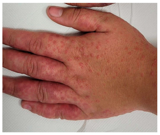
Figure 1.
Hand-foot-and-mouth disease resulting from EV infection, characterized by erythematous papules and vesicles on the dorsal surfaces of the hands. Visual material provided by Department of Children’s Infectious Diseases, Medical University of Warsaw, Warsaw, Poland.
In the course of EV infections, characteristic dermal lesions typically manifest as non-itchy, non-desquamating eruptions on the face, neck, chest, and extremities. These rashes may exhibit characteristics including maculopapular or morbilliform patterns but sometimes present as hemorrhagic, petechial, or vesicular. Fever commonly accompanies the above mentioned skin lesions, and there is also a potential concurrent onset of aseptic meningitis [10].
According to recent data, up to 39.1% of neonates diagnosed with aseptic meningitis may develop a rash presenting with small pink papules on the surface of the body and distributed to the sole. It should be noted that not all children in this study had meningitis. Additionally, this study was conducted on a small group (n = 23) of neonates [11]. Moreover, it was assessed that there was an association between the infection of echovirus with clinical manifestation of exanthem. In the report of the National Epidemiological Surveillance of Infectious Agents in Japan, exanthem was reported in 46.4% of cases of EV infections. It was observed that 90.1% of patients with these dermal manifestations were 2 years old or younger. Single cases that included both meningitis and exanthem were also reported [12].
There are reports available on a correlation between the onset of rash and aseptic meningitis induced by echovirus type 9. In 36% of cases (16 out of 44), a rash was observed, characterized as a widespread maculo-papular rash primarily found on the face, shoulders, and chest [13].
Herpesviridae is a large family of DNA viruses. There are eight known types of Human Herpesviruses (HHV), classified as HHV-1 through HHV-8, with two distinct variants of HHV-6: HHV-6A and HHV-6B. The most common types of meningitis causing viruses within this family include HHV-1 and 2, also called Herpes Simplex Viruses (HSV). However, there are also known cases of disease onset with the etiology of Varicella Zoster Virus (VZV/HHV-3), Epstein-Barr virus (EBV/HHV-4) or Human Herpesvirus 6 (HHV-6) [14].
In the course of Herpesvirus infections, characteristic skin lesions are observed. The emergence of specific dermal manifestation may indicate the diagnosis of an etiological factor.
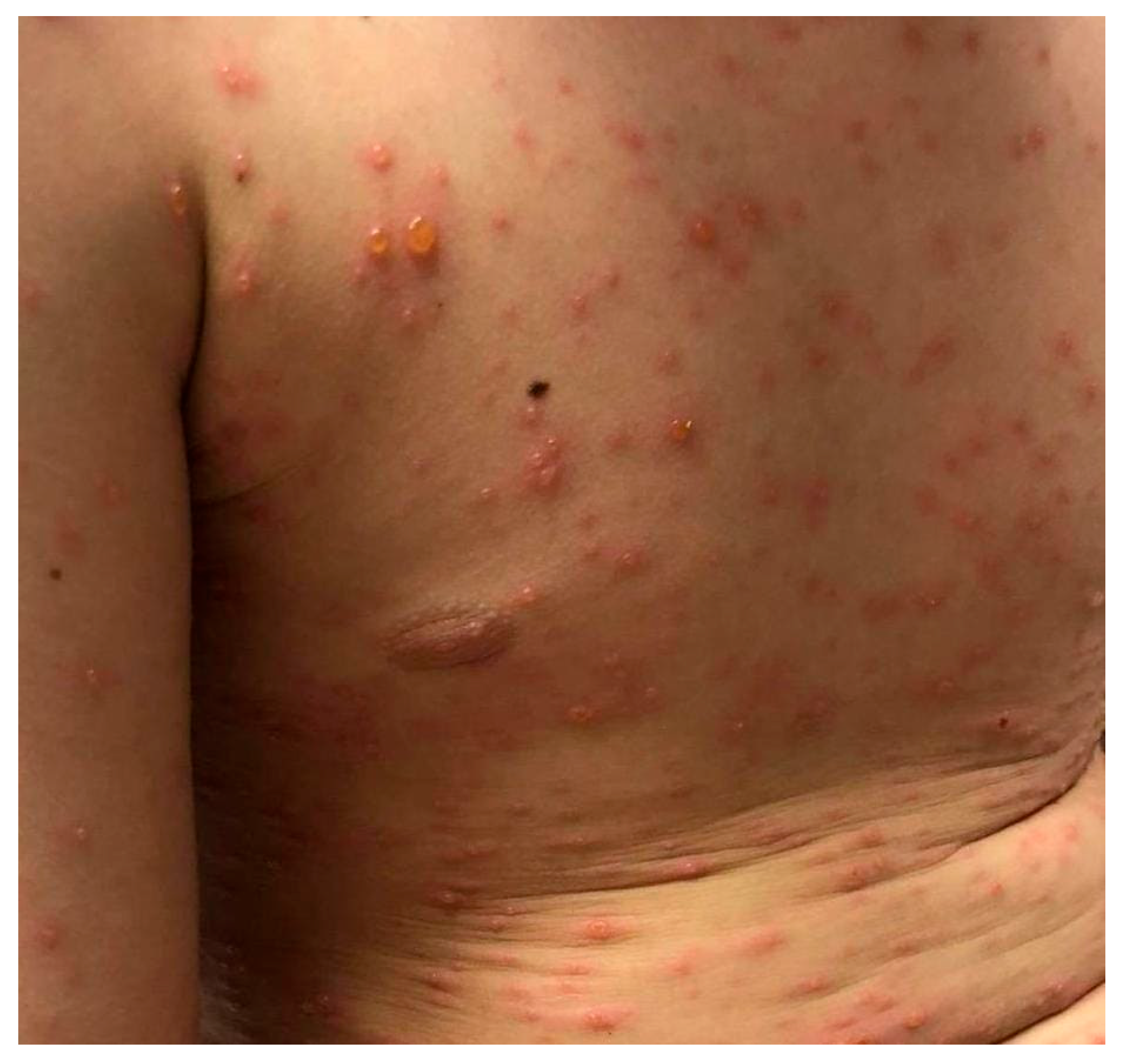
The majority of meningitis cases caused by HSV 1 or 2 occur without skin lesions. Kaewpoowat showed that these symptoms were present in only 1.3% of all patients with HSV meningitis [15]. However, there are reported cases in which the guide for the diagnosis of HSV meningitis was recurrent dermatomal vesicular skin lesions or herpetic whitlow [16,17]. The varicella rash manifests with vesicles and is often pruritic. The lesions initially appear as macules, progressing into papules, and evolving into vesicles (Figure 2 and Figure 3). In the final stage of the disease, crusted papules appear [18]. In rare cases, the rash does not have a typical course and it presents without itching or as erythematous [19].

Figure 2.
Maculopapular rash caused by primary VZV infection. Visual material provided by Department of Children’s Infectious Diseases, Medical University of Warsaw, Warsaw, Poland.

Figure 3.
Vesicles and maculopapular rash caused by primary VZV infection. Visual material provided by Department of Children’s Infectious Diseases, Medical University of Warsaw, Warsaw, Poland.
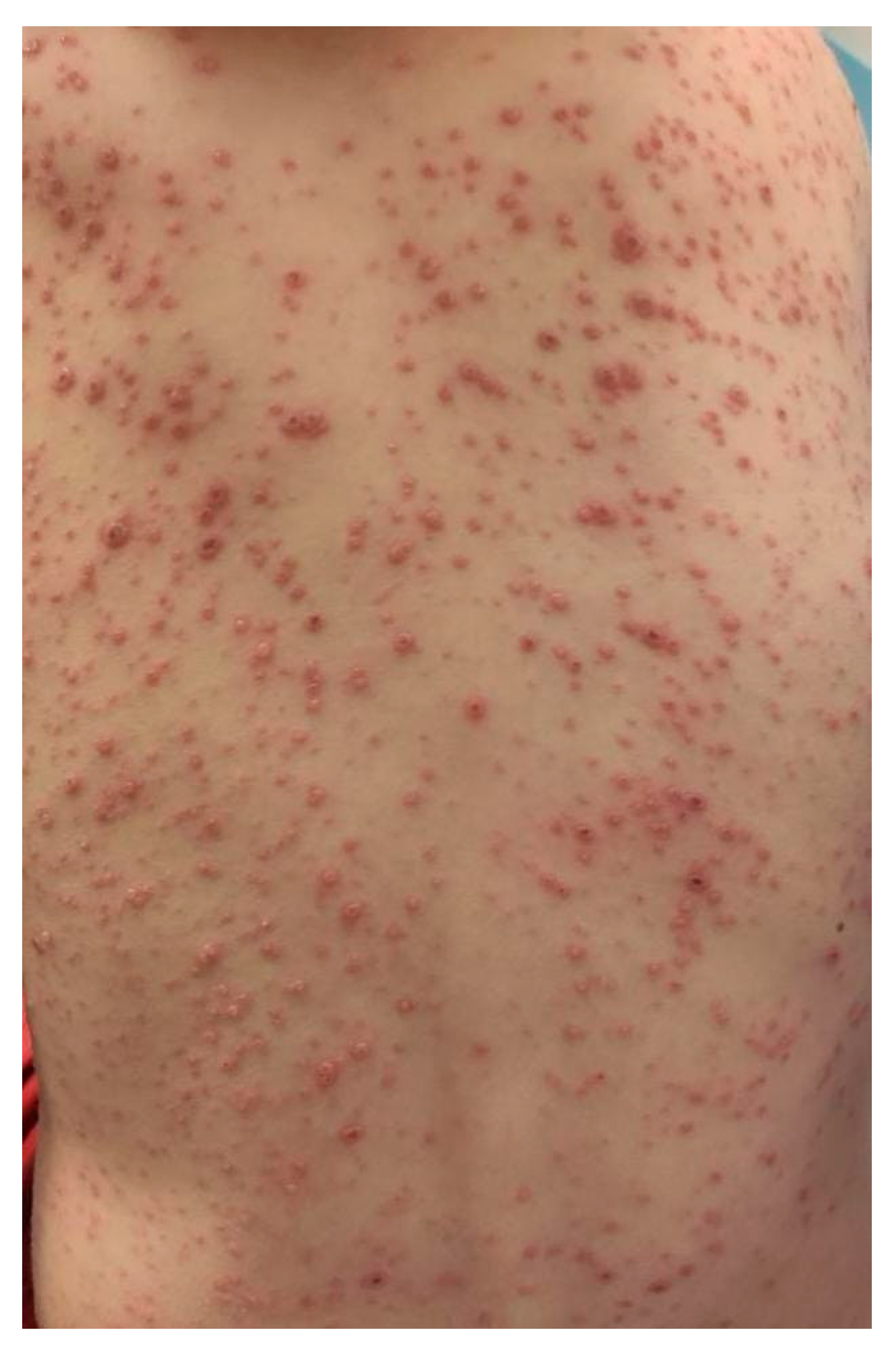
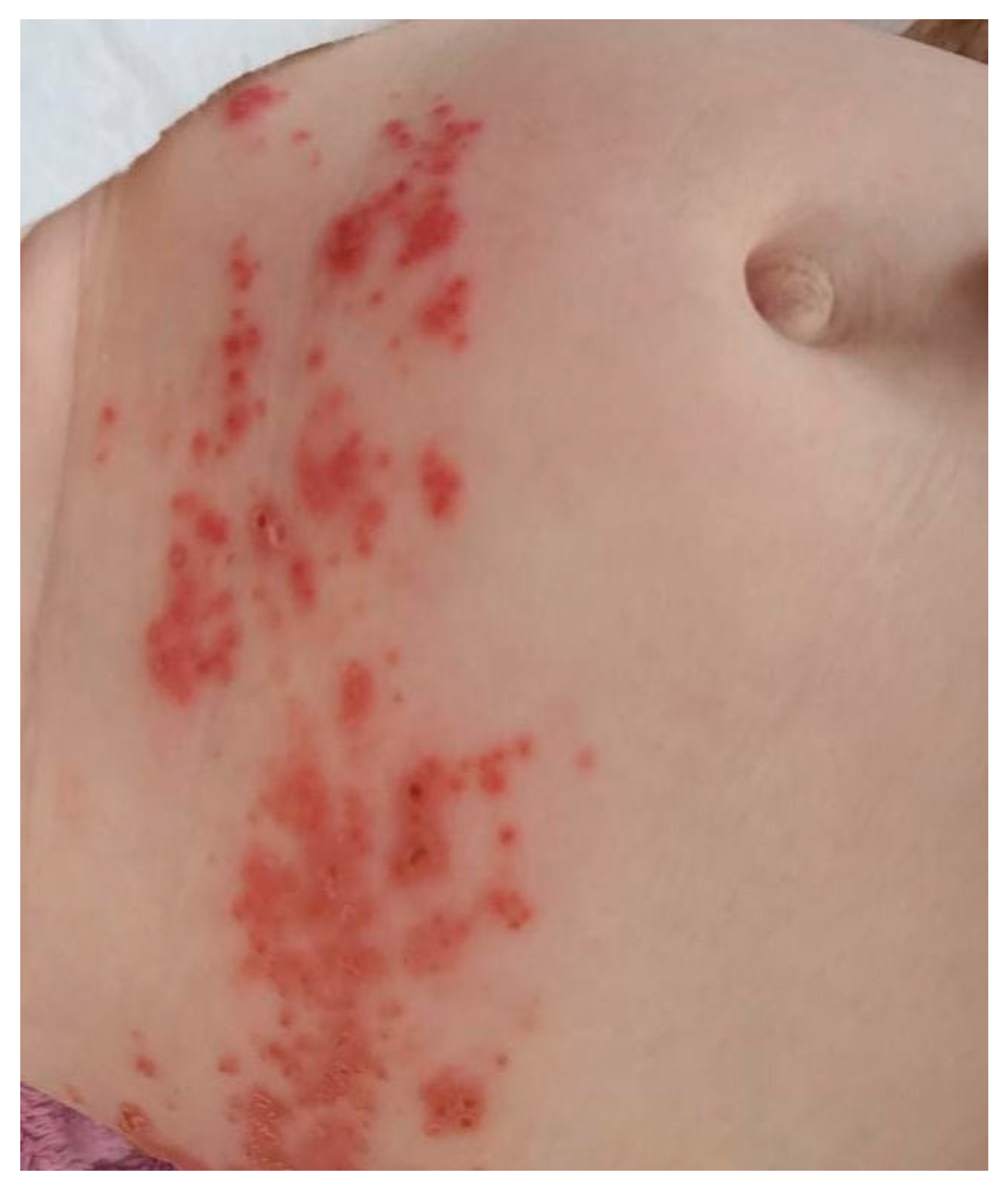
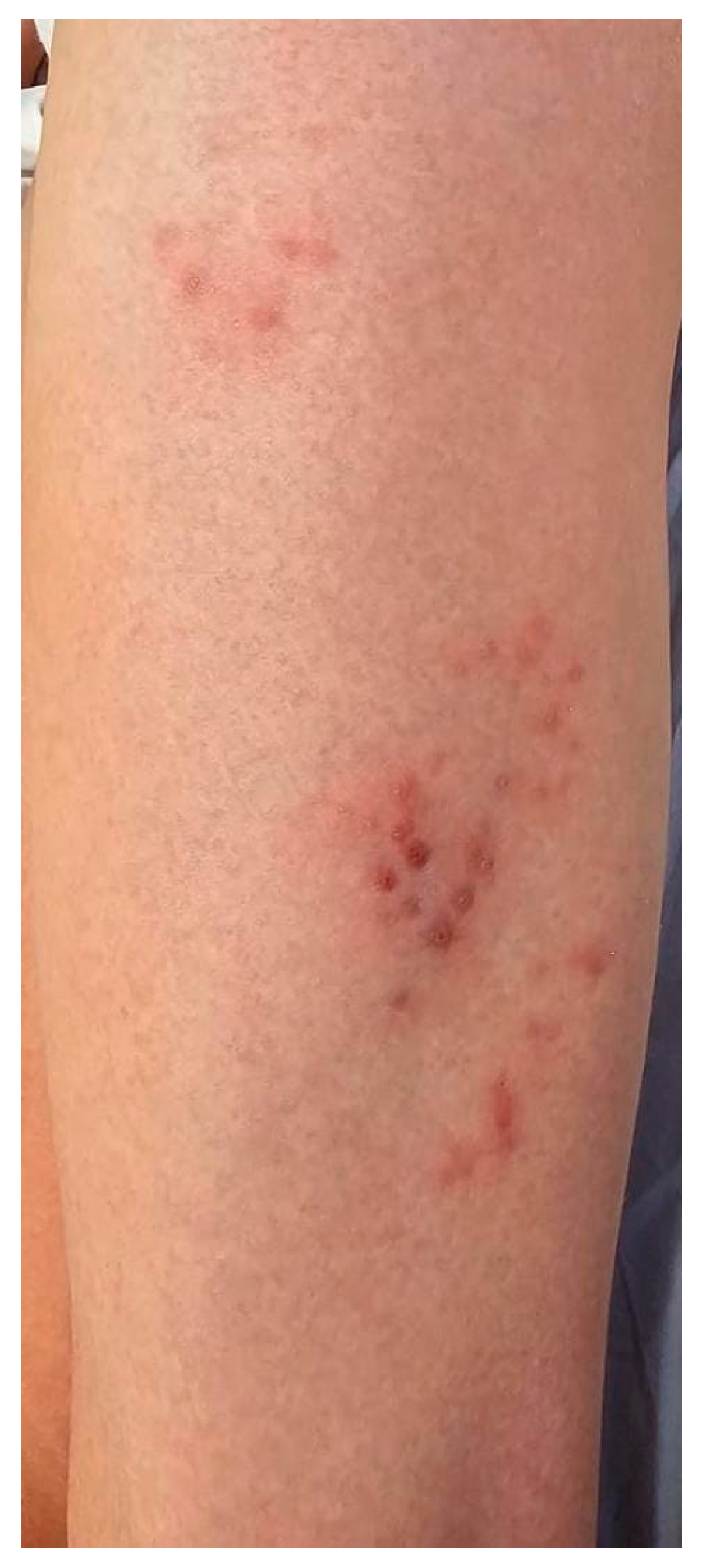
On the other hand, rash precedes papules and appears in a singular dermatome or spanning across neighborhood dermatomes (Figure 4 and Figure 5). The distribution of vesicular rash in herpes zoster aligns with the sensory territories associated with the affected ganglion [20].

Figure 4.
Papular rash appearing on the left side of the torso in the course of herpes zoster. Visual material provided by Department of Children’s Infectious Diseases, Medical University of Warsaw, Warsaw, Poland.
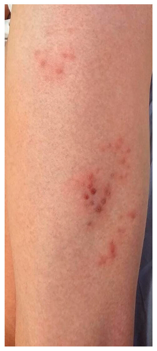
Figure 5.
Papular rash localized to the anterior tibial region, limited to a single dermatome, observed in the course of herpes zoster. Visual material provided by Department of Children’s Infectious Diseases, Medical University of Warsaw, Warsaw, Poland.
Herpes zoster ophthalmicus may occur as the involvement of the ophthalmic division (V1) of the trigeminal cranial nerve (V) with skin rash of the eye and ocular adnexa [21].
Another type of VZV infection is Ramsay Hunt Syndrome, also known as Herpes Zoster Oticus. In the course of this disease there is a characteristic triad of symptoms including facial nerve palsy, otalgia, and vesicular rash on the external auditory canal [22,23]. However, the course of the disease may differ and may also involve the vestibulocochlear nerve (VIII) due to its anatomical location [24].
The administration of acyclovir has become the gold standard in the treatment CNS HSV infections. Its introduction has significantly reduced mortality rates associated with CNS HSV infections, from approximately 20% in untreated cases to 6% in treated patients.
Visual materials were provided by the Department of Children’s Infectious Diseases, Medical University of Warsaw, Poland.
Arboviruses are transmitted by arthropods. These viruses belong to the families of Togaviridae, Bunyaviridae, Flaviviridae and Reoviridae [25].
Chikungunya fever is caused by Chikungunya virus (CHIKV), which belongs to the family Togaviridae, Alphavirus genus. CHIKV is transmitted mainly by mosquitoes. Meningitis in the course of CHIKV infection is a very rare but possible complication. The disease may be characterized by macular, maculopapular rash or generalized erythematous blanchable rash, which often appears approximately one day after the beginning of the disease and it usually lasts three to seven days [26]. The typical onset of the rash is located on the face, and neck. The rash usually spreads out on extremities and the torso [27]. Hyperpigmentation also may be observed [28]. Presence of rash is being reported in 33–88% of children with arbovirus (Av) infections. The incidence of skin lesions in adults varies from 35% to 50% [29,30].
West Nile virus (WNV) of the Flaviviridae family, genus Flavivirus, causes West Nile fever. WNV is spread by mosquitoes and usually has nonspecific symptoms. During the course of the disease, a generalized, maculopapular rash may be observed. The rash usually occurs from 5 to 12 days after infection. According to an analysis of 15 WNV cases, rash was located on the trunk in 14 (93%) patients, on upper extremities in 14 (93%) and on lower extremities in 12 (80%) [31]. It was reported that rash is present in 5% of cases but previous reports indicated the frequency of rash to be 14–28% [31,32]. Pruritus was reported in 33% of patients. According to the available data, rash may be an indicator of a lower risk of neuroinvasive disease and death [33].
Zika virus of the Flaviviridae family, genus Flavivirus is transmitted by mosquitoes. Meningitis is an extremely rare complication of Zika virus infection. Skin lesions, described as exanthem or rash, appear in 90% of patients. Typically, it presents as macular or morbilliform exanthema, which may be seen on the face, torso, limbs, and palms [34]. The exanthem was described as descending in 73% of adults and 76% of children. Pruritus was observed in 81% of adults and 83% of children. Cordel N., et al. showed that the median of the body surface area affected by exanthem was 45% and the most frequent sites affected with skin lesions were the face (95%), upper limbs (95%), torso (93%) and abdomen (90%) [35].
Colorado tick fever virus is a member of the Reoviridae family. The virus is transmitted by the Rocky Mountain wood tick. Approximately 15% of infections may present with petechial or maculopapular rash [36]. Most frequently, the skin lesion is described as a red, haloed papule. These skin lesions are also described as a transient, maculopapular rash on the face and upper and lower limbs [37,38].
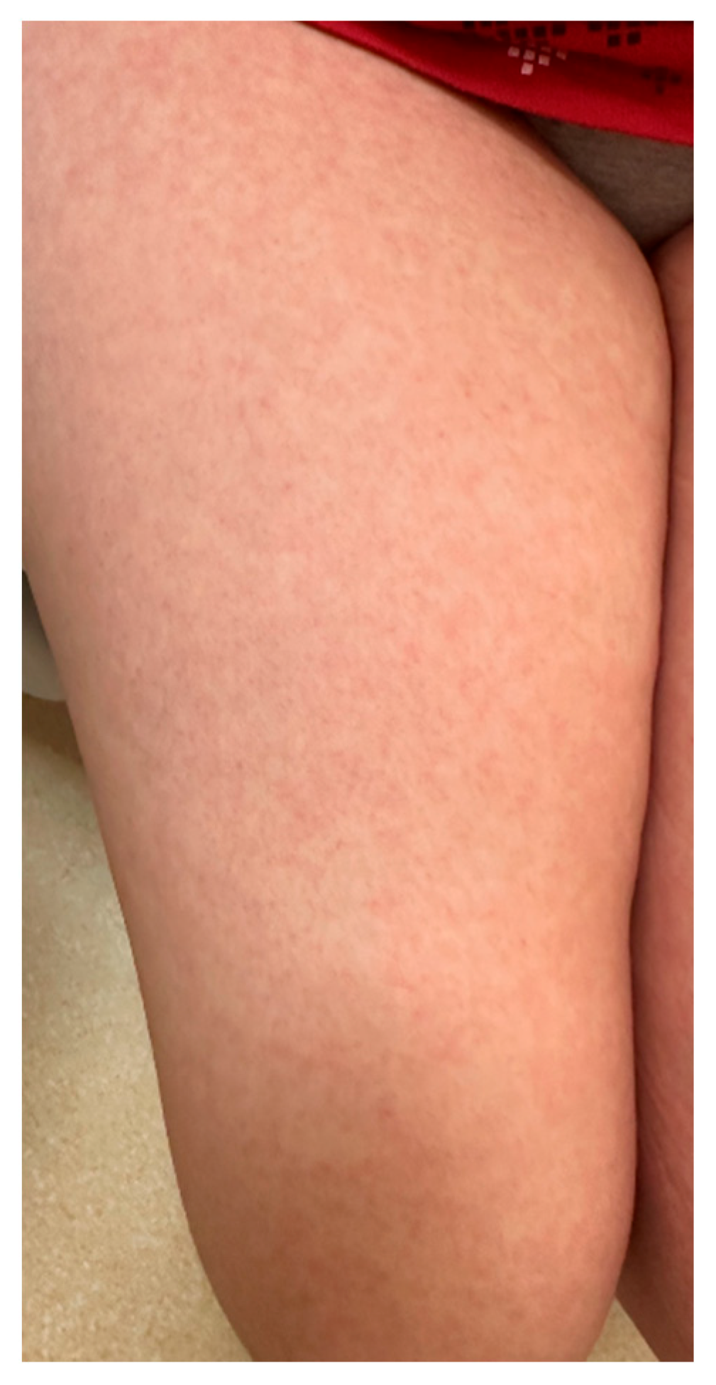
Dengue, caused by Flaviviridae virus, is usually spread by mosquitoes. This disease is mostly claimed not to be neurotropic, but there were a few case studies reporting central nervus system infection. In a prospective study that described neurologic complications of dengue, 47% of patients presented with neurologic involvement [39]. Dengue may be present with skin lesions including macular rash (Figure 6), which may develop up to five days after the beginning of fever. Skin lesions occur in 50% of cases, more frequently during primary infections than during a second episode of the disease. Usually, rash can be observed on the face, trunk and abdomen and may be accompanied by pruritus [40]. One study described petechial rash and dengue recovery rash as skin lesions during the infection [41].

Figure 6.
Macular rash on the lower extremities occurring in the course of dengue virus infection. Visual material provided by Department of Children’s Infectious Diseases, Medical University of Warsaw, Warsaw, Poland.
Oropouche virus is traditionally endemic to the Amazon region. However, since 2023, an increasing number of infections have been identified in the United States, likely associated with the growing popularity of international travel. Clinically, Oropouche virus disease presents with signs and symptoms that closely resemble those of other arboviral infections, such as Dengue, Zika, and Chikungunya. Most patients initially develop fever, myalgia, and headache, often accompanied by arthralgia, rash, gastrointestinal symptoms (including nausea, vomiting, or diarrhea), and, in some cases, conjunctivitis. While the disease is generally self-limiting and mild, rare cases of neuroinvasive manifestations, such as meningitis or meningoencephalitis, have been reported [42].
Acyclovir substantially reduces the morbidity and mortality of herpes simplex virus (HSV) encephalitis when administered promptly. In the absence of treatment, the mortality rate is approximately 70%, but early initiation of acyclovir therapy can reduce this to around 20% [43,44]. However, despite its effectiveness, acyclovir use carries the risk of adverse effects, including nephrotoxicity, leukopenia, and thrombocytopenia, which necessitate careful monitoring during therapy [45].
2.1. Cutaneous Manifestations in Bacterial Meningitis: Diagnostic Considerations
Bacterial meningitis (BM) is a rapidly progressing and potentially life-threatening central nervous system (CNS) infection. Nearly 50% of individuals with BM present to healthcare facilities within the first 24 h of symptom onset. The prognosis depends on host factors, pathogen virulence, and especially the timing of appropriate therapy initiation—delays significantly increase mortality. Although the classic triad of fever, neck stiffness, and altered mental status occurs in 41% of patients—more frequently in older adults—most (95%) present with at least two of the following: headache, fever, neck stiffness, and confusion. Less commonly, patients may experience seizures, focal deficits, cranial nerve palsies, or coma [46].
Skin lesions may also be present in bacterial meningitis. The best documented dermatological findings are those associated with Neisseria meningitidis infection. Skin changes are usually described as petechiae and palpable purpura. The prevalence of rash varies among studies. According to Heckenberg et al., who analyzed data from 258 patients with bacterial meningitis, a rash was present in 64% of patients and described mainly (91%) as petechial [47]. Another study identified skin changes in 176 out of 683 cases (26%), with petechial lesions found in 157 of those 176 patients (89%). The vast majority of infections in this cohort were caused by Neisseria meningitidis (162 cases), although other etiological agents included Streptococcus pneumoniae (8 cases), Staphylococcus aureus (2 cases), group B Streptococcus (2 cases), Haemophilus influenzae (1 case), and Listeria monocytogenes (1 case) [46].
2.2. The Role of Telemedicine in the Diagnosis of Skin Lesions
The expansion of telemedicine and remote video consultations presents an opportunity to enhance knowledge regarding the appearance of skin lesions based on photos or videos. The popularization of telemedicine has significantly improved access to healthcare for many individuals. Due to remote medical appointments, people living far from healthcare facilities can receive quicker and more convenient diagnoses [48,49].
Studies suggest that some dermal changes may be identified with similar accuracy during telemedicine appointments compared to face-to-face meetings [50]. However, in most cases, diagnosis through remote consultation shows lower efficiency. For this reason, these methods should be used with extreme caution, and if in any doubt, a patient with suspicious skin lesions should be referred to a healthcare facility.
3. Summary
Viral infections often manifest with diverse skin changes, providing crucial diagnostic insights into the underlying virus and potential complications like hearing loss or speech impairment. Enteroviruses (EVs) and Herpes viruses (HV) exhibit distinct dermatological manifestations. The introduction of acyclovir therapy has contributed to a reduction in mortality rates for central nervous system (CNS) herpes simplex virus (HSV) infections, from 20% (without treatment) to 6% (with treatment). However, it is important to remember that acyclovir therapy is associated with adverse effects, including nephrotoxicity, leukopenia, and thrombocytopenia. Therefore, incorporating cutaneous manifestations into the differential diagnosis may help guide earlier initiation of the appropriate therapy [44]. These rashes may occasionally manifest as hemorrhagic, petechial, or vesicular lesions, often accompanied by fever and concurrent aseptic meningitis. On the other hand, HV infections, particularly HSV and VZV, commonly present with vesicular eruptions, macules, or papules localized along specific dermatomes, reflecting the virus’s neural tropism. A summary of skin lesions caused by the mentioned viruses is presented in Table 1.

Table 1.
Characteristics of skin lesions in viral infections causing meningitis. Summary of the types of skin lesions, locations, and frequency of rash occurrence depending on the type of virus. The table presents a comparison of EVs, HHV-1, HHV-2, VZV/HHV-3, CHIKV, WNV, Zika virus, Colorado tick fever virus and Dengue virus.
4. Conclusions
The relationship between viral infections and dermatological manifestations, such as maculopapular rashes and vesicular lesions, underscores the significance of recognizing these skin changes as diagnostic clues for viral meningitis. EV infections frequently exhibit characteristic rashes, while HV infections, especially HSV and VZV, present with distinct patterns of skin lesions distributed along specific nerve pathways. Understanding these nuanced dermatological presentations facilitates early diagnosis and targeted management of viral meningitis. Skin examination, while not decisive on its own, can offer important diagnostic insights. Identifying cutaneous changes may assist in the clinical evaluation. In cases of suspected viral encephalitis, the presence of skin lesions typical for viruses from the Herpesviridae family allows for the early empirical initiation of acyclovir, which can reduce mortality. Although EVs are the most common etiologic agents of aseptic meningitis, the presence of cutaneous manifestations typical of HSV infections in such patients justifies the consideration of acyclovir as part of the initial treatment strategy. However, standardized recommendations in this area are still lacking. Moreover, the development of rapid diagnostic tools that combine rash patterns with laboratory testing is crucial for improving diagnostic accuracy and enabling timely treatment decisions.
Further exploration of the diverse spectrum of skin changes associated with viral infections is essential for refining diagnostic approaches and optimizing patient outcomes.
Author Contributions
Conceptualization, C.B. and M.P.-Ś.; methodology, A.M., W.G., A.Ł., C.B. and M.P.-Ś.; software, A.M., W.G. and A.Ł.; validation, C.B. and M.P.-Ś.; formal analysis, C.B. and M.P.-Ś.; investigation, A.M., W.G. and A.Ł.; resources, C.B. and M.P.-Ś.; data curation, A.M., W.G., A.Ł., C.B. and M.P.-Ś.; writing—original draft preparation, A.M., W.G. and A.Ł.; writing—review and editing, C.B. and M.P.-Ś.; visualization, A.M., W.G., A.Ł., C.B. and M.P.-Ś.; supervision, C.B. and M.P.-Ś.; project administration, C.B. All authors have read and agreed to the published version of the manuscript.
Funding
This research received no external funding.
Conflicts of Interest
The authors declare no conflicts of interest.
References
- Kohil, A.; Jemmieh, S.; Smatti, M.K.; Yassine, H.M. Viral meningitis: An overview. Arch. Virol. 2021, 166, 335–345. [Google Scholar] [CrossRef] [PubMed]
- Mrozowska-Nyckowska, K.; Zbrzeźniak, J.; Paradowska-Stankiewicz, I. Meningitis and encephalitis in Poland in 2022. Epidemiol. Rev./Przegląd Epidemiol. 2024, 78, 219–234. [Google Scholar] [CrossRef] [PubMed]
- Irani, D.N. Aseptic meningitis and viral myelitis. Neurol. Clin. 2008, 26, 635–655. [Google Scholar] [CrossRef]
- Kupila, L.; Vuorinen, T.; Vainionpaa, R.; Hukkanen, V.; Marttila, R.J.; Kotilainen, P. Etiology of aseptic meningitis and encephalitis in an adult population. Neurology 2006, 66, 75–80. [Google Scholar] [CrossRef]
- Logan, S.A.; MacMahon, E. Viral meningitis. BMJ 2008, 336, 36–40. [Google Scholar] [CrossRef]
- Moriguchi, T.; Harii, N.; Goto, J.; Harada, D.; Sugawara, H.; Takamino, J.; Ueno, M.; Sakata, H.; Kondo, K.; Myose, N.; et al. A first case of meningitis/encephalitis associated with SARS-Coronavirus-2. Int. J. Infect. Dis. 2020, 94, 55–58. [Google Scholar] [CrossRef]
- Rotbart, H.A. Viral meningitis. Semin. Neurol. 2000, 20, 277–292. [Google Scholar] [CrossRef] [PubMed]
- Sawyer, M.H. Enterovirus infections: Diagnosis and treatment. Semin. Pediatr. Infect. Dis. 2002, 13, 40–47. [Google Scholar] [CrossRef]
- De Crom, S.C.M.; Rossen, J.W.A.; Van Furth, A.M.; Obihara, C.C. Enterovirus and parechovirus infection in children: A brief overview. Eur. J. Pediatr. 2016, 175, 1023–1029. [Google Scholar] [CrossRef]
- Sridhar, A.; Karelehto, E.; Brouwer, L.; Pajkrt, D.; Wolthers, K.C. Parechovirus A Pathogenesis and the Enigma of Genotype A-3. Viruses 2019, 11, 1062. [Google Scholar] [CrossRef]
- Chen, W.; Dai, S.; Xu, L. Clinical characterization of benign enterovirus infection in neonates. Medicine 2021, 100, e25706. [Google Scholar] [CrossRef] [PubMed]
- Miyamura, K.; Yamashita, K.; Yamadera, S.; Kato, N.; Akatsuka, M.; Yamazaki, S. An epidemic of echovirus 18 in 1988 in Japan--high association with clinical manifestation of exanthem. A report of the National Epidemiological Surveillance of Infectious Agents in Japan. Jpn. J. Med. Sci. Biol. 1990, 43, 51–58. [Google Scholar] [CrossRef]
- Newman, C.R.; Smith, R.B. An outbreak of meningitis associated with ECHO virus type 9. J. Clin. Pathol. 1965, 18, 20–22. [Google Scholar] [CrossRef] [PubMed]
- Chadwick, D.R. Viral meningitis. Br. Med. Bull. 2005, 75–76, 1–14. [Google Scholar] [CrossRef]
- Kaewpoowat, Q.; Salazar, L.; Aguilera, E.; Wootton, S.H.; Hasbun, R. Herpes simplex and varicella zoster CNS infections: Clinical presentations, treatments and outcomes. Infection 2016, 44, 337–345. [Google Scholar] [CrossRef]
- Gonzales, N.; Tyler, K.L.; Gilden, D.H. Recurrent dermatomal vesicular skin lesions: A clue to diagnosis of herpes simplex virus 2 meningitis. Arch. Neurol. 2003, 60, 868–869. [Google Scholar] [CrossRef] [PubMed]
- Karpathios, T.; Moustaki, M.; Yiallouros, P.; Sarifi, F.; Tzanakaki, G.; Fretzayas, A. HSV-2 meningitis disseminated from a herpetic whitlow. Paediatr. Int. Child. Health 2012, 32, 121–122. [Google Scholar] [CrossRef]
- Heininger, U.; Seward, J.F. Varicella. Lancet 2006, 368, 1365–1376. [Google Scholar] [CrossRef]
- Kratlian, C.M.; Hopkins-Braddock, P.; Tristram, D. Case 4: Unexpected Rash in a 12-year-old Girl. Pediatr. Rev. 2020, 41, 256–258. [Google Scholar] [CrossRef]
- Dworkin, R.H.; Johnson, R.W.; Breuer, J.; Gnann, J.W.; Levin, M.J.; Backonja, M.; Betts, R.F.; Gershon, A.A.; Haanpää, M.L.; McKendrick, M.W.; et al. Recommendations for the management of herpes zoster. Clin. Infect. Dis. 2007, 44 (Suppl. S1), S1–S26. [Google Scholar] [CrossRef]
- Liesegang, T.J. Herpes Zoster Ophthalmicus: Natural History, Risk Factors, Clinical Presentation, and Morbidity. Ophthalmology 2008, 115 (Suppl. S2), S3–S12. [Google Scholar] [CrossRef]
- Bienkowski, C.; Kowalczyk, M.; Talarek, E.; Pokorska-Spiewak, M.; Kierdaszuk, B.; Marczynska, M. Meningitis and Ramsay-Hunt syndrome in a 17-year old girl. Neuro Endocrinol. Lett. 2019, 40, 149–151. [Google Scholar] [PubMed]
- Bieńkowski, C.; Talarek, E.; Pokorska-Śpiewak, M. The clinical course of herpes zoster is similar in immunocompetent and immunocompromised paediatric patients. J. Paediatr. Child. Health 2023, 59, 1112–1114. [Google Scholar] [CrossRef] [PubMed]
- Pietrzak, M.K.; Pluta, M.; Pokorska-Śpiewak, M.; Marczyńska, M. Meningitis in the Course of Herpes Zoster Ophthalmicus in an Immunocompetent Boy. Pediatr. Infect. Dis. J. 2023, 42, e361–e362. [Google Scholar] [CrossRef] [PubMed]
- Boroń-Kaczmarska, A.W.-D. Alicja Choroby zakaźne i pasożytnicze. T, 1st ed.; Boroń-Kaczmarska, A.W.-D., Ed.; PZWL Wydawnictwo Lekarskie: Warsaw, Poland, 2022; Volume 2. [Google Scholar]
- Rajapakse, S.; Rodrigo, C.; Rajapakse, A. Atypical manifestations of chikungunya infection. Trans. R. Soc. Trop. Med. Hyg. 2010, 104, 89–96. [Google Scholar] [CrossRef]
- Javelle, E.; Tiong, T.H.; Leparc-Goffart, I.; Savini, H.; Simon, F. Inflammation of the external ear in acute chikungunya infection: Experience from the outbreak in Johor Bahru, Malaysia, 2008. J. Clin. Virol. 2014, 59, 270–273. [Google Scholar] [CrossRef]
- Meena, S.S.; Arya, S.; Meena, D.; Matlani, M.; Salhan, M. Neonatal Chikungunya: A case series. Trop. Doct. 2021, 51, 103–105. [Google Scholar] [CrossRef]
- Ritz, N.; Hufnagel, M.; Gérardin, P. Chikungunya in Children. Pediatr. Infect. Dis. J. 2015, 34, 789–791. [Google Scholar] [CrossRef]
- Samra, J.A.; Hagood, N.L.; Summer, A.; Medina, M.T.; Holden, K.R. Clinical Features and Neurologic Complications of Children Hospitalized With Chikungunya Virus in Honduras. J. Child. Neurol. 2017, 32, 712–716. [Google Scholar] [CrossRef]
- Ferguson, D.D.; Gershman, K.; LeBailly, A.; Petersen, L.R. Characteristics of the rash associated with West Nile virus fever. Clin. Infect. Dis. 2005, 41, 1204–1207. [Google Scholar] [CrossRef]
- Watson, J.T.; Pertel, P.E.; Jones, R.C.; Siston, A.M.; Paul, W.S.; Austin, C.C.; Gerber, S.I. Clinical characteristics and functional outcomes of West Nile Fever. Ann. Intern. Med. 2004, 141, 360–365. [Google Scholar] [CrossRef] [PubMed]
- Huhn, G.D.; Dworkin, M.S. Rash as a prognostic factor in West Nile virus disease. Clin. Infect. Dis. 2006, 43, 388–389. [Google Scholar] [CrossRef]
- Derrington, S.M.; Cellura, A.P.; McDermott, L.E.; Gubitosi, T.; Sonstegard, A.M.; Chen, S.; Garg, A. Mucocutaneous Findings and Course in an Adult With Zika Virus Infection. JAMA Dermatol. 2016, 152, 691–693. [Google Scholar] [CrossRef]
- Cordel, N.; Birembaux, X.; Chaumont, H.; Delion, F.; Chosidow, O.; Tressieres, B.; Storck, C.H. Main Characteristics of Zika Virus Exanthema in Guadeloupe. JAMA Dermatol. 2017, 153, 326–328. [Google Scholar] [CrossRef]
- Yendell, S.J.; Fischer, M.; Staples, J.E. Colorado tick fever in the United States, 2002–2012. Vector Borne Zoonotic Dis. 2015, 15, 311–316. [Google Scholar] [CrossRef]
- Goodpasture, H.C.; Poland, J.D.; Francy, D.B.; Bowen, G.S.; HORN, K.A. Colorado tick fever: Clinical, epidemiologic, and laboratory aspects of 228 cases in Colorado in 1973–1974. Ann. Intern. Med. 1978, 88, 303–310. [Google Scholar] [CrossRef] [PubMed]
- Klasco, R. Colorado tick fever. Med. Clin. North. Am. 2002, 86, 435–440. [Google Scholar] [CrossRef]
- Sahu, R.; Verma, R.; Jain, A.; Garg, R.K.; Singh, M.K.; Malhotra, H.S.; Sharma, P.K.; Parihar, A. Neurologic complications in dengue virus infection: A prospective cohort study. Neurology 2014, 83, 1601–1609. [Google Scholar] [CrossRef] [PubMed]
- Cobra, C.; Rigau-Pérez, J.G.; Kuno, G.; Vomdam, V. Symptoms of dengue fever in relation to host immunologic response and virus serotype, Puerto Rico, 1990-1991. Am. J. Epidemiol. 1995, 142, 1204–1211. [Google Scholar] [CrossRef]
- Solomon, T.; Dung, N.M.; Vaughn, D.W.; Kneen, R.; Thao, L.T.T.; Raengsakulrach, B.; Loan, H.T.; Day, N.P.; Farrar, J.; Myint, K.S.; et al. Neurological manifestations of dengue infection. Lancet 2000, 355, 1053–1059. [Google Scholar] [CrossRef]
- Morrison, A. Oropouche Virus Disease Among U.S. Travelers—United States, 2024. MMWR Morb. Mortal. Wkly. Rep. 2024, 73, 769–773. [Google Scholar] [CrossRef]
- Hjalmarsson, A.; Blomqvist, P.; Sköldenberg, B. Herpes simplex encephalitis in Sweden, 1990–2001: Incidence, morbidity, and mortality. Clin. Infect. Dis. 2007, 45, 875–880. [Google Scholar] [CrossRef] [PubMed]
- Aboelezz, A.; Mahmoud, S.H. Acyclovir dosing in herpes encephalitis: A scoping review. J. Am. Pharm. Assoc. 2024, 64, 102040. [Google Scholar] [CrossRef]
- Kimberlin, D.W.; Lin, C.Y.; Jacobs, R.F.; Powell, D.A.; Corey, L.; Gruber, W.C.; Rathore, M.; Bradley, J.S.; Diaz, P.S.; Kumar, M.; et al. Safety and efficacy of high-dose intravenous acyclovir in the management of neonatal herpes simplex virus infections. Pediatrics 2001, 108, 230–238. [Google Scholar] [CrossRef] [PubMed]
- Van de Beek, D.; De Gans, J.; Spanjaard, L.; Weisfelt, M.; Reitsma, J.B.; Vermeulen, M. Clinical features and prognostic factors in adults with bacterial meningitis. N. Engl. J. Med. 2004, 351, 1849–1859. [Google Scholar] [CrossRef]
- Heckenberg, S.G.; de Gans, J.; Brouwer, M.C.; Weisfelt, M.; Piet, J.R.; Spanjaard, L.; van der Ende, A.; van de Beek, D. Clinical features, outcome, and meningococcal genotype in 258 adults with meningococcal meningitis: A prospective cohort study. Medicine 2008, 87, 185–192. [Google Scholar] [CrossRef] [PubMed]
- Sud, E.; Anjankar, A. Applications of Telemedicine in Dermatology. Cureus 2022, 14, e27740. [Google Scholar] [CrossRef]
- Palacholla, R.S.; Kvedar, J.C. Telemedicine for infectious disease care-how do we measure the true value? Ann. Transl. Med. 2019, 7 (Suppl. S6), S178. [Google Scholar] [CrossRef]
- Glines, K.R.; Haidari, W.; Ramani, L.; Akkurt, Z.M.; Feldman, S.R. Digital future of dermatology. Dermatol. Online J. 2020, 26. [Google Scholar] [CrossRef]
- Ihekwaba, U.K.; Kudesia, G.; McKendrick, M.W. Clinical features of viral meningitis in adults: Significant differences in cerebrospinal fluid findings among herpes simplex virus, varicella zoster virus, and enterovirus infections. Clin. Infect. Dis. 2008, 47, 783–789. [Google Scholar] [CrossRef]
- Lee, G.H.; Kim, J.; Kim, H.W.; Cho, J.W. Herpes simplex viruses (1 and 2) and varicella-zoster virus infections in an adult population with aseptic meningitis or encephalitis: A nine-year retrospective clinical study. Medicine 2021, 100, e27856. [Google Scholar] [CrossRef] [PubMed]

Disclaimer/Publisher’s Note: The statements, opinions and data contained in all publications are solely those of the individual author(s) and contributor(s) and not of MDPI and/or the editor(s). MDPI and/or the editor(s) disclaim responsibility for any injury to people or property resulting from any ideas, methods, instructions or products referred to in the content. |
© 2025 by the authors. Licensee MDPI, Basel, Switzerland. This article is an open access article distributed under the terms and conditions of the Creative Commons Attribution (CC BY) license (https://creativecommons.org/licenses/by/4.0/).