Review

Heartland Virus: An Evolving Story of an Emerging Zoonotic and Vector-Borne Disease

by
Emily K. Mantlo
1 and
Nicholas J. Haley
2,*
1
Department of Diagnostic Medicine/Pathobiology, College of Veterinary Medicine, Kansas State University, Manhattan, KS 66506, USA
2
Department of Microbiology and Immunology, College of Graduate Studies, Midwestern University, Glendale, AZ 85308, USA
*
Author to whom correspondence should be addressed.
Zoonotic Dis. 2023, 3(3), 188-202; https://doi.org/10.3390/zoonoticdis3030016
Submission received: 22 May 2023 / Revised: 21 July 2023 / Accepted: 27 July 2023 / Published: 29 July 2023
(This article belongs to the Special Issue Feature Papers of Zoonotic Diseases 2023)

Abstract

:

Simple Summary

Heartland virus is a tick-borne disease that was first identified in the Midwest United States in 2009. Since then, over 60 human cases of disease and several deaths have been reported. The true prevalence of disease in humans and domestic animals is not well understood. Though the disease is presumed to have an animal reservoir, specific infected animal hosts have not yet been identified. Several diagnostic approaches for Heartland virus infection have been developed, though there are currently no specific treatment or prevention options available. More research needs to be conducted on this newly emerging virus to better understand disease transmission, disease progression, treatment, and to facilitate the development of effective vaccines.

Abstract

Heartland virus (HRTV) is an emerging tick-borne bandavirus that is capable of causing severe disease characterized by acute thrombocytopenia and lymphopenia. The virus is endemic to the eastern United States and is carried by the Lone Star tick (Amblyomma americanum). Since its discovery in 2009, at least 60 human infections have been recorded across this area, with an overall 5–10% estimated mortality rate. All infections reported thus far have occurred following a known tick bite or exposure to tick-infested areas, but the possibility of nosocomial transmission has not been ruled out. Despite relatively high rates of seroprevalence among certain wildlife species such as white-tailed deer, the reservoir species for HRTV remains unknown, as the virus has never been isolated from any mammalian wildlife species. Furthermore, how the virus is transmitted to its vector species in nature remains unknown, though laboratory studies have confirmed both horizontal and vertical transmission of HRTV in A. americanum. In addition, the recent 2017 introduction of the Asian longhorned tick (Haemaphysalis longicornis) to the US has raised concerns about possible spillover of HRTV into a new tick species that has been confirmed to be a competent vector for HRTV in the laboratory. Thus, an increased awareness of its clinical presentation is needed, and further research is urgently required to establish the natural transmission cycle and develop new countermeasures for this novel zoonotic pathogen.

1. Introduction

In June of 2009, two men independently presented to Heartland Regional Medical Center in northwestern Missouri with fever, fatigue, and anorexia [1]. Both men were hospitalized, and over the following two weeks, they both developed severe thrombocytopenia and leukopenia. Because both men had reported a tick bite in days preceding clinical symptoms, Ehrlichia chaffeensis infection was initially suspected, and both patients were empirically treated with doxycycline. Symptoms failed to improve, and samples sent to the Centers for Disease Control & Prevention were negative for known tick-borne bacterial species. Electron microscopy and next-generation sequencing revealed the presence of a novel virus similar to severe fever with thrombocytopenia syndrome virus (SFTSV), another virus that was, coincidentally, first isolated in early 2009 in central China [2].
Since then, dozens of cases of HRTV infection have been reported in the United States, resulting in severe illness and several deaths. Because of its high morbidity and mortality, HRTV has been classified as a Category C Priority Pathogen by the National Institute of Allergy and Infectious Diseases (NIAID), and there are presently no approved treatments or vaccines for HRTV. Nevertheless, much remains unknown about HRTV, its animal reservoirs and susceptible hosts, and its transmission cycle. In this review, we summarize what is currently known about HRTV virology, clinical presentation, epidemiology, and transmission and present several future directions for research.

2. Viral Etiology

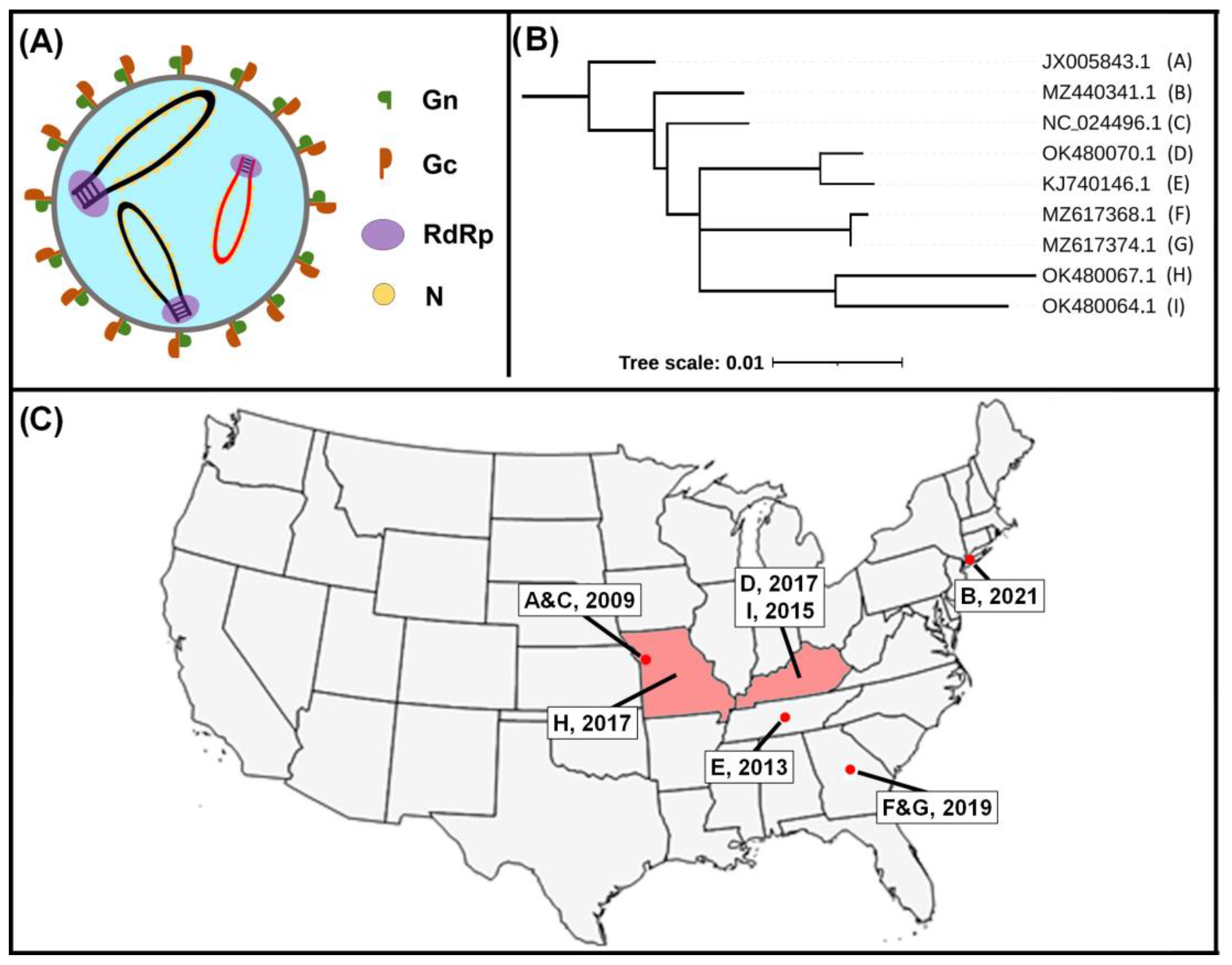
Originally classified as a Phlebovirus, HRTV was recently re-classified in the new Bandavirus genus alongside other similar viruses such as severe fever with thrombocytopenia syndrome virus (SFTSV), Bhanja virus (BHAV), and Lone Star virus (LSV) [3]. Among these, the most closely-related virus to HRTV is SFTSV, sharing 65–69% sequence homology. As a member of the Phenuiviridae family in the order Bunyavirales, HRTV is a single-stranded negative-sense RNA virus with a tri-segmented genome composed of small (S), medium (M), and large (L) segments (Figure 1). The L segment encodes the RNA-dependent RNA polymerase (RdRp); the M segment encodes the structural glycoproteins Gn and Gc; and the S segment encodes the nucleocapsid (N) and the nonstructural NSs protein. All three genome segments feature distinct 5′ and 3′ untranslated regions (UTRs) that form secondary structures which act as promoters for viral genome replication and transcription. Interestingly, though these UTR sequences differ between viruses, the RdRp and N proteins of HRTV can recognize SFTSV UTR sequences and initiate replication and transcription in a minigenome system [4]. This raises the possibility that reassortment of HRTV with other bandaviruses could lead to the emergence of novel pathogenic virus species.
The majority of antibodies generated against HRTV bind to either the N protein or the Gn/Gc proteins, and though most of these are specific for HRTV, at least one has been found to cross-react with SFTSV [5]. As Gn/Gc proteins are expressed on the surface of virions and are responsible for binding host cells and facilitating viral entry, antibodies generated against Gn/Gc are able to neutralize free-floating viruses. However, as with the related phleboviruses, the N protein is likely the immunodominant protein for HRTV [5]. Experimental HRTV inoculation in AG129 mice, which lack both α/β and γ interferon (IFN) receptors, does lead to the generation of a low-level neutralizing antibody titer. However, of nine different hybridoma clones created from these mice, all nine produced non-neutralizing monoclonal antibodies targeting N protein [5]. Many studies of the seroprevalence of HRTV in both human and animal populations have measured only neutralizing antibody titers [6,7,8], thus likely underestimating the true extent of exposure to HRTV.
Though not a major target of antibodies, the nonstructural NSs protein of HRTV likely plays a role in virulence and could therefore be a target for the development of novel antiviral compounds or rationally designed vaccines. Similar to the related phleboviruses, HRTV NSs acts as an IFN antagonist, inhibiting both Type I and Type III IFN responses [9]. HRTV NSs has been demonstrated to block phosphorylation of TANK-binding kinase 1 (TBK1) and signal transducer and activator of transcription 2 (STAT2), both important contributors in the IFN signaling pathway [10,11]. This leads to a downstream loss of expression of IFN-inducible antiviral effector genes. By comparison, NSs from the related, nonpathogenic Uukuniemi virus only weakly antagonizes the IFN response [11]. Deletion of the NSs of the related Rift Valley fever virus (RVFV) does not compromise viral replication in vitro but leads to attenuation in vivo [12,13]. Most recently, an HRTV mutant lacking NSs was successfully rescued, and this mutant demonstrated reduced replication kinetics in vitro and attenuation in vivo, highlighting the importance of NSs for HRTV replication and pathogenesis [14].
Figure 1. Structure of Heartland virus (HRTV) and the phylogeny and percent identity of reported isolates based on the S segment. (A) The structure of HRTV is similar to other members of the Bunyavirus order, including a tri-segmented genome coding for Gn and Gc glycoproteins, an RNA-dependent RNA polymerase (RdRp), and nucleocapsid (N) proteins. The S segment is highlighted in red. (B) HRTV S segment nucleotide sequences, denoted by Genbank accession numbers, were aligned using MAFFT, and trees were inferred by PhyML and plotted using Newick Display. The tree was rooted using the earliest HRTV clinical isolate. The workflow was performed by ngphylogeny.fr [15]. Letters in parentheses following accession numbers indicate isolate identification in subfigure C. (C) Source locations and year of recovery for isolates analyzed are in subfigure B. Where known, specific isolate sources are shown as a red dot, while states where isolates were recovered without location data are shown in pink.
Zoonoticdis 03 00016 g001

3. Epidemiology

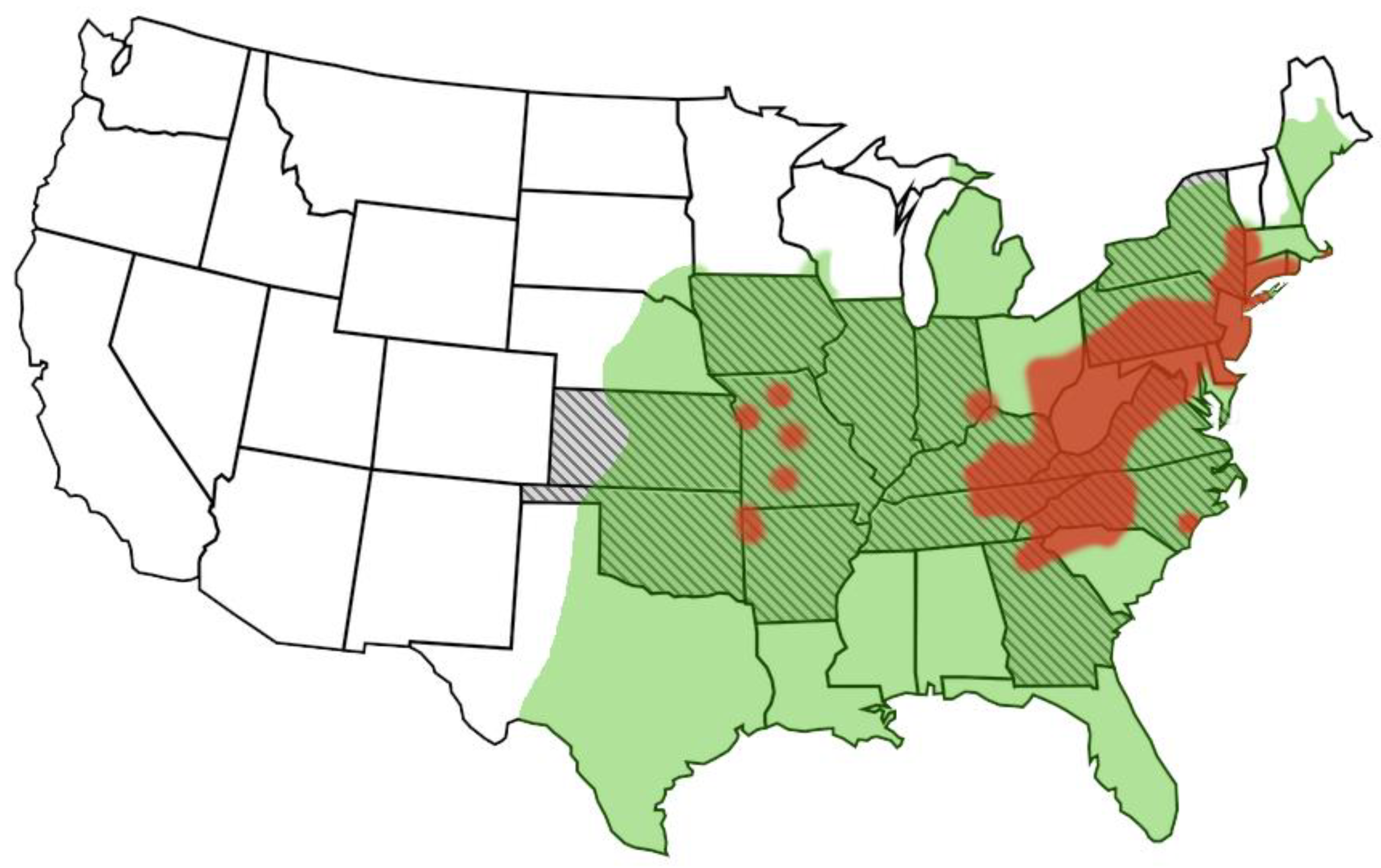
As of November of 2022, at least 60 cases of HRTV have been reported in the United States, primarily east of the Mississippi River [16]. These cases have occurred across 14 states, all within the geographical range of the lone star tick, Amblyomma americanum (Figure 2). Cases of HRTV infection in humans have been reported as far north as New York and as far south as Georgia. In addition, vertebrate animals with HRTV-specific neutralizing antibodies have been found in the southern states of Texas, Louisiana, and Florida and as far north as Maine, though no human cases have been recorded in these states [7,17]. Infections have been reported from April to September, with most occurring in the summer months, particularly June [18]. All known cases of HRTV infection have likely occurred following a tick bite. Most patients report removing a tick in the weeks leading up to their illnesses [19]. Those who do not remember a specific tick bite have reported spending time outdoors, and no infections have occurred in the winter months.
In contrast, the related SFTSV has been reported exclusively in parts of Asia, including China, Japan, Vietnam, and South Korea, with thousands of cases reported in these countries since its initial discovery in China in 2009 [2,20]. Like HRTV, SFTSV is transmitted primarily via tick bite, though direct animal to human and human to human transmission via blood and other bodily fluids has also been reported [21,22,23,24], raising concerns for the potential for direct anthropozoonotic and nosocomial transmission of HRTV as well.
Figure 2. Geographical distribution of Heartland virus and its vectors. Fourteen eastern and midwestern states have reported clinical cases of Heartland virus infection in humans (hatched lines) [16]. Distribution of the known vector, Amblyomma americanum [25], is shown in green, while reported locations of another competent vector, Haemaphysalis longicornis, is shown in red [26].
Zoonoticdis 03 00016 g002

4. Host Range

Evidence for either natural or experimental susceptibility to HRTV has been documented in a number of terrestrial mammalian and avian species—though no evidence for clinical infection has been reported in any species apart from humans. Based on serologic surveillance studies, several potential amplification hosts have been identified, including raccoon, white-tailed deer, coyotes, domestic dogs, and opossum (Figure 3). In HRTV endemic areas, seroprevalence ranges widely, with rates sometimes approaching 40–50% in select cases in raccoon, white-tailed deer, and coyotes [6,17]. Importantly, however, experimental challenge studies have failed to show any evidence of viremia or clinical symptoms in raccoons or white-tailed deer [27,28]. Several routes of inoculation in various animal species have been pursued, including intradermal, intracranial, subcutaneous, and intraperitoneal inoculation. To date, though, an experimental challenge using infected ticks has not yet been attempted. Thus, further studies are needed to take into account tick factors in disease transmission, which may influence transmission dynamics in susceptible host species [29]. Experimental infection in dogs, cats, coyotes, or avian species has so far not been evaluated. While several immunodeficient laboratory species do develop clinical disease upon experimental challenge, further discussed below, no evidence for viremia or clinical symptoms have been reported in challenge studies involving chickens and goats, though each of these species exhibited at least some evidence of seroconversion [6,27]. It may be possible that Amblyomma (or Haemophysalis) tick species themselves are the sole reservoir—as is the case for Crimean Congo Hemorrhagic Fever Virus, a related bunyavirus in the family Nairoviridae [30]. Though absent of clinical symptoms, deer and other species showing evidence of seroconversion may serve, in some respect, as facultative amplification hosts as a result of tick-to-tick transmission during co-feeding [18].
The lone exceptions to an apparent lack of infectivity and clinical diseases in animals include several immunodeficient laboratory species, with detection of viremia in AG129 mice, IFNAR−/− mice lacking type I interferon receptors, and STAT2 knockout hamsters, which are unresponsive to type I IFN signaling [27,31,32]. The immunodeficient mouse lines show various signs including weight loss, poor hair coat, and hematochezia, though they appear to lack many of the hematologic disturbances seen in human cases of HRTV. The STAT2 knockout hamsters infected with HRTV, in contrast, had elevated liver enzymes and leukocytosis, though they notably lacked any evidence of thrombocytopenia [31], a primary finding in humans as discussed in more detail in later sections.
The current findings of HRTV in wild and domestic animals are, to a degree, in contrast with those reported for SFTS virus, where comparatively high rates of seroprevalence as well as viral RNA have been reported in small ruminants, cattle, dogs, chickens, rodents, pigs, and a range of laboratory species [33,34]. Clinical disease resulting from SFTSV infection has also been reported in several animal species, including cats [24,35], cheetahs [36], dogs [37], and laboratory rodents [34], and importantly resulted in likely exposure and infection in veterinary professionals. Further investigations are warranted to determine whether SFTSV truly has an expanded host range compared to HRTV.

5. Viral Pathogenesis

Very little is known about the pathogenesis of HRTV infections in animal or human hosts. Due to the absence of wild-type animal models, much of the data available has been collected from immunocompromised rodent systems, introduced above. To date, the focus of most of these studies has been terminal in nature, centered on susceptibility or therapeutic development; however, a study in STAT2 knockout hamsters has provided some important insight into viral pathogenesis. Fortunately, additional insight into HRTV pathogenesis may be drawn from the more prolific studies available on SFTSV infections in various model systems.
In immunocompromised AG129 or IFNAR−/− mice inoculated intraperitoneally (IP) with HRTV, viremia typically began at 2-3 days post-infection (dpi) and peaked at roughly 4–5 dpi, waning by 7–9 dpi [27,32]. Fatality rates in HRTV-infected AG129 mice corresponded with inoculation dosage and ranged from 20 to 85%. Terminal pathology in these mice included enlarged, pale spleens, hepatic hemorrhage, and enlarged gall bladders. Immunohistochemistry also revealed HRTV antigen diffusely within splenic mononuclear cells, Kupfer cells, and renal interstitial cells. Postmortem analyses were not reported in IFNAR−/− mice.
Comparatively more is known about HRTV pathogenesis from a study utilizing STAT2 knockout hamsters, where timepoint studies provided more details on the kinetics of viral distribution in blood and other tissues [31]. In these hamsters, a low level of viremia was detected in IP-inoculated animals by 5 dpi, though no virus was detectable in tissues at this time. By 7 dpi, the infectious virus was detectable via cell culture at low levels in spleen, liver, and lymph nodes, among other tissues, though viremia at that time had begun to wane. A single animal with a pronounced infection was euthanized at day 9 in this study, with infectious virus and viral antigens (based on IHC) present in various tissues including spleen, kidney, heart, lung, small intestine, and lymph node. Notably, bone marrow in HRTV-inoculated hamsters in this study was apparently normal, outside of an increased myeloid to erythroid ratio, evidence of a response to increased demand for inflammatory cells elsewhere in the body in response to the infection.
In contrast to the sparsity of information available on HRTV pathogenesis, much more is known about the pathogenesis of SFTSV in a range of wild-type and immunodeficient model systems. Though many of the species investigated were of limited value for better understanding SFTSV infections in humans, lacking some key features of the disease process (summarized in a recent review by Sun et al. [34]), studies in ferrets [38], cats [39], and non-human primates [40,41,42] have found that these species may share some key features of disease in humans. Studies in rhesus macaques, for example, revealed that they have patterns of viremia, tissue distribution, and pathology which mirror those observed in human SFTSV infections [40,42]. Similar studies using HRTV are needed to establish an effective immunocompetent animal model for future research into HRTV pathogenesis and vaccine development.

6. Transmission and Vector Ecology

Transmission of HRTV likely relies heavily on the Lone Star tick, A. americanum, a vector known to transmit a wide range of diseases to both humans and animals, including Bourbon virus, canine and human ehrlichiosis, and Cytauxzoon felis [43]; HRTV has shown no evidence for direct animal to human transmission. Amblyomma americanum is a multi-host tick with an ever-expanding distribution that strongly overlaps the geographical presentation of human cases of HRTV, and surveillance studies have identified HRTV-infected ticks in areas proximal to known human cases and across the range of A. americanum occurrence. Virus or viral nucleic acids have been recovered from both nymph and adult stages of A. americanum [18,44,45,46,47,48,49], though to date, the virus has not been recovered from tick larvae collected in the field. Experimental studies have found larvae to be susceptible, however, with both transovarial and transstadial viral transmission reported [50]. These studies have also shown that naïve larvae may become infected when co-feeding on a host along with infected ticks in the nymph stage. Thus, ticks may become infected through vertical transmission via transovarial transmission, through co-feeding with other infected ticks or very likely through feeding on an infected host—though, as noted above, an effective animal model for proper tick feeding studies has yet to be identified. Based on our understanding of the life cycle of A. americanum, it seems likely that these ticks become infected either after feeding on an infected white-tailed deer—a frequent host for nymph and adult A. americanum with well-documented HRTV seroprevalence [6,8,27,28,43]—or other, as-yet unidentified reservoir species, or through transovarial transmission or co-feeding, as noted above. Ticks remain infected across life stage transitions, with the potential for persistence in ticks overwintering in suitable environments, ultimately transmitting the virus as they molt and find new, naïve hosts. The expanding range of this tick species, influenced by ongoing changes in climate [51] and urban interface dynamics [52,53], highlights the continued public threat of exposure to A. americanum and other tick species and the vector-borne zoonoses they may carry.
While A. americanum is likely the primary vector responsible for zoonotic transmission of HRTV, recent concern over invasive tick species has led to studies on the vector competence of the Asian longhorned tick (Haemaphysalis longicornis). This species of tick, native to Southeast Asia, was first reported in the United States in 2017 and has since been found in 18 different states—primarily those along the Appalachian Mountain range [26]—and is known to be a vector for the closely-related SFST virus [54]. Though field studies have so far failed to find any evidence of natural HRTV infection in this species [55], these ticks have been found to be experimentally susceptible and competent vectors, capable of horizontal transmission to mice (as evidenced by seroconversion), as well as transovarial transmission [56]. Like A. americanum, H. longicornis is a multi-host tick capable of overwintering under appropriate conditions [57] and thus should continue to be considered as a potential vector for HRTV.
Perhaps not surprisingly, H. longicornis is the principal vector for SFTS virus in Asia, though other potential vectors include Amblyomma testudinarium, Ixodes nippoensis, and Rhipicephalus microplus [58]—suggesting there may be other potential tick vectors for HRTV across North America as well. As with HRTV, transovarial and transstadial transmission of SFTSV are likely to be important factors in its seasonal persistence in the tick host [59], though the wide range of susceptible mammalian hosts is certainly as important in this virus’s natural life cycle.

7. Clinical Findings, Differential Diagnosis, Treatment, and Prognosis

The typical signalment for HRTV cases is an adult male over the age of 40—though most cases are over the age of 60, often with preexisting comorbidities—an outdoor lifestyle including farming, camping, or hunting, and very likely a history of tick exposure [1,19,44,60,61,62,63,64,65,66,67]. Clinical presentations are those which may mimic other regional tick-borne diseases most commonly seen in the summer months, such as Ehrlichia, Rickettsia, and Babesia, and include a short history of fever (≥38 °C), headache, fatigue, myalgia, nausea, anorexia, and non-bloody diarrhea. A localized rash, as may be seen with some other tick-borne diseases, has been reported in just a subset of cases, though a patient with a more diffuse maculopapular rash has also been reported [19,44,62]. Purpura has been reported on occasion as well [60]. Lymphadenopathy is also rare and often only detected via radiographic means [62,67]. Symptoms often present within 2 weeks of known tick exposure, often in summer months, though may vary based on geographic location and seasonal duration of tick activity [19].
Initial clinical evaluation often uncovers a mild to moderate leukopenia (1000–3000 cells/μL), influenced primarily by low neutrophil counts, and a mild thrombocytopenia (60–120,000/μL) [1,19,44,60,61,62,63,64,65,66,67]. Mild hyponatremia may be evident on metabolic panels, as well as mildly elevated alanine and aspartate aminotransferase levels. Packed cell volumes and red blood cell counts are usually normal to low-normal, with normal hemoglobin levels. Patient comorbidities often dictate additional clinical pathologic findings. Heartland virus RNA may be identified in blood samples collected in the first 7–10 days of symptoms [63,64], with IgM antibodies appearing after roughly 8–10 days of symptoms [19,63,68]; IgG antibodies are thought to appear within a similar timeframe [68]. In some cases, both IgM and IgG can be detected as early as 1 day after the onset of symptoms. Patients may require hospitalization for 1–2 weeks, and over that course leukopenia and thrombocytopenia may continue to worsen, with the nadir in both parameters often seen by the eighth day of symptoms. Red blood cell counts and hemoglobin most often remain stable, though low, and begin to recover along with other hematologic and metabolic parameters by two weeks following the onset of symptoms.
Primary differential diagnoses for HRTV infection would include other regionally endemic tick-borne diseases, including human monocytotropic ehrlichiosis (Ehrlichia chaffeensis) and related ehrlichial agents, Rocky Mountain Spotted Fever (Rickettsia rickettsii), human granulocytic anaplasmosis (Anaplasma phagocytophilum), Lyme disease (Borrelia burgdorferi), Bourbon virus, and potentially Powassan, Colorado tick fever, and SFTS viruses as well—pending clinical presentation and travel history. Potential, though less likely, differentials may include influenza, COVID-19, locally endemic fungal (e.g., Blastomyces and Histoplasma) or bacterial (e.g., Coxiella or Francisella) infections, and bone marrow neoplasia/dysplasia/aplasia syndromes. Molecular or serologic diagnostics targeting these agents and processes, including bone marrow aspirate [1,62,66], can be helpful in ruling in or out HRTV infection. Based on the wide range of differentials, treatment often begins empirically with tetracyclines or other antibiotics that effectively target bacterial tick-borne agents [1,60,63,69]. There are no specific treatments available specific for HRTV, although favipiravir, a nucleoside analogue and selective inhibitor of RNA-dependent RNA polymerases with in vitro activity against HRTV [31], has also been found to ameliorate disease in immunodeficient mice and hamsters [31,32]. This has likewise shown some promise for treating SFTSV infections in humans [70,71], indicating there may be some potential for using this drug in an off-label, emergency setting. Supportive therapy, including blood and/or platelet transfusions and intravenous fluids, is often necessary, as well as treatments addressing any underlying comorbidities.
The prognosis for HRTV infections is generally guarded and relies heavily on managing pre-existing conditions. Fatal infections have been reported and make up roughly 5–10% of published cases—most with serious underlying conditions [60,62,67]. Short-term fatigue, memory deficits, and anorexia following discharge, 4–6 weeks in duration, may be expected, with long-term sequelae reported on occasion as well [1].
To date, there have been no reports of symptomatic HRTV infections in domestic animals, though, as noted above, some have been found to be seropositive. Idiopathic or immune-mediated cases of thrombocytopenia are occasionally reported in dogs [72,73] and only rarely in cats [74,75], cattle [76], and horses [77], with primary differentials mirroring those noted above for humans—including regional tick-borne diseases and bone marrow disorders. Diagnostic approaches also follow those listed above for humans, and in areas where the disease has been reported in human populations, it would be important to consider adding HRTV screening to conventional testing protocols for tick-borne agents, especially when tests for more prevalent agents are negative.
As with HRTV, clinical cases of SFTSV most commonly present in elderly men, mainly in rural areas in individuals with links to farming and agriculture [58]. Clinical symptoms likewise include fever, with the addition of gastrointestinal and CNS signs. Blood profile and metabolic panel findings mirror those of HRTV, including thrombocytopenia and elevated liver enzymes [78]. Conservative treatment addressing underlying conditions is often applied, though both ribavirin and favipiravir have shown some potential for treatment in a clinical setting [79]. Additional prospective, randomized trials may help reveal the utility of antiviral treatments—though the importance of early differentiation and diagnosis in the evaluation of various treatment options cannot be understated [79]. Fatality rates are similar between cases of HRTV and SFTSV, with regional variation from 5–50%; patients most commonly succumb as a result of multiple organ failure [78,80]. Poor prognostic indicators in cases of SFTSV include age, CNS symptoms and other comorbidities, and higher viral loads [80]—the latter a potential avenue to explore in cases with HRTV.

8. Diagnosis and Surveillance

Several approaches have been developed to diagnose and detect HRTV infections in humans, animals, and vector species, and largely mirror those used in the diagnosis of SFTSV. Perhaps the most widely used approach is nucleic acid-based testing approaches such as quantitative reverse transcriptase-PCR (qRT-PCR), though antibody-based assays and virus isolation relying on the virus’s ability to replicate in Vero E6 cells have also been used to effectively diagnose and detect HRTV infections in a range of species. Other cell lines may be useful for virus isolation, though they have thus far only been used in propagation experiments [9,14,81].
Two different sets of primers and probes, targeting the S segment of the HRTV genome, have been developed for qRT-PCR and have been used extensively in both human diagnosis and pooled tick surveillance [19,44,45,46,48,49,82,83]. A third primer and probe set has been developed for use as part of a panel for screening insects for several different vector-borne diseases, though to date has not been used in field studies or to detect infections in humans [84]. Conventional RT-PCR, targeting each of the three segments of the HRTV genome, has also been described as a diagnostic option for humans [65]. Finally, an isothermal recombinase polymerase amplification with lateral flow (RPA-LF) assay, a rapid nucleic acid detection assay that permits amplification and product observation without the need for expensive instrumentation, has been developed to allow a more convenient point-of-care approach for diagnosing infections, though its use in field cases of HRTV infection has not yet been examined [85].
Immunologic assays have also been developed and employed to detect current HRTV infection or past exposure in both humans and animals. The simplest of these is a standard serum neutralization assay, whereby serially diluted serum samples are assessed for plaque reduction-based neutralization (PRNT) ability in Vero E6 cell culture. The PRNT assay has been used to screen both human and animal serum as part of surveillance studies, though it is not capable of differentiating recent and past exposure without paired samples [6,7,17,19,61]. A second approach, using microspheres capable of differentiating IgG and IgM antibody classes that target HRTV, has been developed which can help distinguish cases with recent or historical exposure [68]. Finally, experimental enzyme-linked immunoadsorption (ELISA, targeting general anti-HRTV antibodies) and immunohistochemistry (IHC, recognizing HRTV nucleoprotein) approaches have been developed for use in evaluating experimentally challenged animals and human post-mortem samples, though they have not seen wide-scale use [5,27,31,62].

9. Prevention

At present, prevention of HRTV infections in humans relies on general tick avoidance practices. Preventing tick bites in people is best achieved through common measures including (1) avoiding tick-endemic areas, e.g., tall grasses or heavily brushy or wooded areas, (2) tick repellants, including synthetic options such as permethrin or DEET or natural products like oil of lemon eucalyptus, and importantly (3) performing a full body check and thoroughly examining clothes and pets when returning from areas where ticks are known to be found [86]. Effective tick preventatives and acaricides are also widely available for use in large and small domestic species, though pet owners should consult their veterinarian to identify the most appropriate product for a given species [87].
Given the relatively sparse numbers of HRTV cases, motivation for the development of a vaccine has been lacking. A recent study, however, has identified an effective expression platform for HRTV-like particles which may be useful in future vaccine testing and production [88]. This platform, used to develop constructs for a range of different arboviruses, employs a vaccinia virus backbone with two HRTV gene segments of interest—the Gn/Gc polyprotein and N protein genes—under an inducible promoter, a control mechanism that restricts viral production in the absence of tetracycline. This allows for the efficient production of virus-like particles in vitro, while restricting in vivo replication. Without suitable animal models, this construct has not yet been evaluated for efficacy in preventing HRTV infections.
Several approaches to vaccine development for SFTSV have recently been reported, including DNA vaccines targeting Gn and Gc proteins, attenuated recombinant viral vaccines, and recombinant vesicular stomatitis virus-based vaccines [89]. Each of these have shown in vitro and, more importantly, in vivo effectivity in animal model species, with some evidence for the potential for cross-protection against HRTV infection [90], affording optimism for the future development of effective vaccines against HRTV, where necessary.
In addition, a reverse genetics system for HRTV has recently been established that may provide a valuable tool for the development of rationally designed live-attenuated vaccines [14]. Deletion of the NSs gene of HRTV produced a virus that was fully attenuated in AG129 mice. Furthermore, inoculation of mice with this mutant provided 100% protection from subsequent wild-type challenge. The addition of other key attenuating mutations to the HRTV genome using this reverse genetics platform may provide even safer, more efficacious vaccine candidates. The success of live-attenuated vaccines for related RVFV highlights the need for the continued development of such vaccines for bandaviruses such as HRTV.

10. Conclusions and Future Directions

Much has been learned about Heartland virus since its initial discovery in the United States over a decade ago. Cell culture systems have been developed which have allowed for effective isolation of the virus, which have also proven useful in serum neutralization assays. Sequencing of the segmented RNA genome has allowed for comparison to known viruses—including the closely-related severe fever with thrombocytopenia syndrome virus, as well as for the crucial development of nucleic acid-based testing assays. Identification of the viral vector(s) responsible for transmission, as well as potential reservoir species, has given us a better understanding of disease ecology, potential geographic range, and approaches to prevention. Case numbers since its discovery have fortunately been rare, though they have provided clinicians with a general understanding of the typical clinical presentation and possible treatment protocols.
Though much has been learned about HRTV, there remain a number of unanswered questions. Perhaps the most pressing knowledge gap that needs to be addressed is the confirmation of true reservoir or amplification species, as well as identification of an immunocompetent animal model system. Although several wild animal hosts have been found to be seropositive, as noted above, none so far have demonstrated viremia and thus a potential for transmitting the virus to naïve ticks. Additional serosurveillance studies in areas where HRTV infections have been reported, as well as where the A. americanum vector are prevalent, are warranted and should include additional targets such as rodent and avian species—species that are intricately involved in the transmission cycle of a range of historic and emerging arboviruses and are known or suspected hosts for A. americanum [91,92]. Identification of reservoir species would also assist in the establishment of suitable animal model systems, where more could be learned about viral pathogenesis, treatment, and prevention options—including vaccine development tracking important groundwork laid by SFTSV research—as well as vector transmission factors. Fundamental research on the viral protein structure/function and virulence determinants is importantly lacking as well, though the recent development of a reverse genetics system should aid future studies once more suitable animal model systems are identified.
Serosurveillance studies should likewise continue in human and domestic animal populations, with additional consideration for the development of screening assays (e.g., ELISA +/− indirect fluorescence assays) for non-neutralizing antibodies directed against the N protein, to better assess the distribution and clinical impact of HRTV infection. An important target to consider would be healthcare providers responsible for caring for HRTV-infected patients, to help rule out the potential for nosocomial transmission. Cases of idiopathic or immune thrombocytopenia (ITP) are occasionally encountered in both human and veterinary practice [73,93,94], and the addition of HRTV nucleic acid amplification tests to common screening protocols for cases with ITP is also warranted, along with investigations into the relationship between viral load and clinical outcomes. Although a multiplex qRT-PCR has been developed for screening insect vectors for HRTV and other arboviruses, a panel that incorporates HRTV would also be important to help differentiate causes of thrombocytopenia and fever in humans and other species. Agents to include in such a multiplex might include Ehrlichia chaffeensis and ewingii, Rickettsia rickettsii, Babesia species, and perhaps SFTSV, as they are important differentials with overlapping clinical symptoms. Seroprevalence and infection rates may be low across humans and domestic species, suggesting some level of barrier to natural infection; however, it seems likely that exposures greatly exceed the frequency of clinical cases, as is the case with many other arboviruses [95].
Finally, continued surveillance for closely related viruses in wild species would also help clarify the origin and evolutionary trajectory of HRTV, as it has with the related SFTS virus [96]. Additionally, closely related bandaviruses may be uncovered that would allow a more complete understanding of species susceptibility, disease transmission, and the evolution of this genus. Because of the propensity of segmented bunyaviruses to reassort in vivo [97,98], it would be worthwhile to consider investigations into pathogenic properties of HRTV reassortments with related viruses, including SFTSV, to better predict future potential for bandavirus-related zoonoses.

Author Contributions

Both E.K.M. and N.J.H. contributed to the writing of this review and production of its figures. All authors have read and agreed to the published version of the manuscript.

Funding

This work is funded in part by the Center on Emerging and Zoonotic Infectious Diseases (CEZID) of the National Institutes of General Medical Sciences under award number P20GM130448.

Institutional Review Board Statement

Not Applicable.

Informed Consent Statement

Not Applicable.

Data Availability Statement

Not Applicable.

Acknowledgments

The authors would like to thank Juergen Richt, who helped facilitate the completion of this manuscript.

Conflicts of Interest

The authors declare no conflict of interest. The funders had no role in the writing of the manuscript or in the decision to publish.

References

  1. McMullan, L.K.; Folk, S.M.; Kelly, A.J.; MacNeil, A.; Goldsmith, C.S.; Metcalfe, M.G.; Batten, B.C.; Albariño, C.G.; Rollin, S.R.Z.E.; Nicholson, W.L.; et al. A new phlebovirus associated with severe febrile illness in Missouri. N. Engl. J. Med. 2012, 367, 834–841. [Google Scholar] [CrossRef] [PubMed]
  2. Yu, X.J.; Liang, M.F.; Zhang, S.Y.; Liu, Y.; Li, J.D.; Sun, Y.L.; Zhang, L.; Zhang, Q.F.; Popov, V.L.; Li, C.; et al. Fever with thrombocytopenia associated with a novel bunyavirus in China. N. Engl. J. Med. 2011, 364, 1523–1532. [Google Scholar] [CrossRef] [PubMed] [Green Version]
  3. Kuhn, J.H.; Adkins, S.; Alkhovsky, S.V.; Avsic-Zupanc, T.; Ayllon, M.A.; Bahl, J.; Balkema-Buschmann, A.; Ballinger, M.J.; Bandte, M.; Beer, M.; et al. 2022 taxonomic update of phylum Negarnaviricota (Riboviria: Orthornavirae), including the large orders Bunyavirales and Mononegavirales. Arch. Virol. 2022, 167, 2857–2906. [Google Scholar] [CrossRef] [PubMed]
  4. Ren, F.; Zhou, M.; Deng, F.; Wang, H.; Ning, Y.J. Combinatorial Minigenome Systems for Emerging Banyangviruses Reveal Viral Reassortment Potential and Importance of a Protruding Nucleotide in Genome "Panhandle" for Promoter Activity and Reassortment. Front. Microbiol. 2020, 11, 599. [Google Scholar] [CrossRef] [Green Version]
  5. Calvert, A.E.; Brault, A.C. Development and Characterization of Monoclonal Antibodies Directed Against the Nucleoprotein of Heartland Virus. Am. J. Trop. Med. Hyg. 2015, 93, 1338–1340. [Google Scholar] [CrossRef] [Green Version]
  6. Bosco-Lauth, A.M.; Panella, N.A.; Root, J.J.; Gidlewski, T.; Lash, R.R.; Harmon, J.R.; Burkhalter, K.L.; Godsey, M.S.; Savage, H.M.; Nicholson, W.L.; et al. Serological investigation of heartland virus (Bunyaviridae: Phlebovirus) exposure in wild and domestic animals adjacent to human case sites in Missouri 2012–2013. Am. J. Trop. Med. Hyg. 2015, 92, 1163–1167. [Google Scholar] [CrossRef] [Green Version]
  7. Clarke, L.L.; Ruder, M.G.; Mead, D.G.; Howerth, E.W. Heartland Virus Exposure in White-Tailed Deer in the Southeastern United States, 2001–2015. Am. J. Trop. Med. Hyg. 2018, 99, 1346–1349. [Google Scholar] [CrossRef] [Green Version]
  8. Lindsey, N.P.; Menitove, J.E.; Biggerstaff, B.J.; Turabelidze, G.; Parton, P.; Peck, K.; Basile, A.J.; Kosoy, O.I.; Fischer, M.; Staples, J.E. Seroprevalence of Heartland Virus Antibodies in Blood Donors, Northwestern Missouri, USA. Emerg. Infect. Dis. 2019, 25, 358–360. [Google Scholar] [CrossRef]
  9. Feng, K.; Deng, F.; Hu, Z.; Wang, H.; Ning, Y.J. Heartland virus antagonizes type I and III interferon antiviral signaling by inhibiting phosphorylation and nuclear translocation of STAT2 and STAT1. J. Biol. Chem. 2019, 294, 9503–9517. [Google Scholar] [CrossRef]
  10. Ning, Y.J.; Feng, K.; Min, Y.Q.; Deng, F.; Hu, Z.; Wang, H. Heartland virus NSs protein disrupts host defenses by blocking the TBK1 kinase-IRF3 transcription factor interaction and signaling required for interferon induction. J. Biol. Chem. 2017, 292, 16722–16733. [Google Scholar] [CrossRef]
  11. Rezelj, V.V.; Li, P.; Chaudhary, V.; Elliott, R.M.; Jin, D.Y.; Brennan, B. Differential Antagonism of Human Innate Immune Responses by Tick-Borne Phlebovirus Nonstructural Proteins. mSphere 2017, 2, e00234-17. [Google Scholar] [CrossRef] [PubMed] [Green Version]
  12. Bird, B.H.; Albarino, C.G.; Hartman, A.L.; Erickson, B.R.; Ksiazek, T.G.; Nichol, S.T. Rift valley fever virus lacking the NSs and NSm genes is highly attenuated, confers protective immunity from virulent virus challenge, and allows for differential identification of infected and vaccinated animals. J. Virol. 2008, 82, 2681–2691. [Google Scholar] [CrossRef] [PubMed] [Green Version]
  13. Ikegami, T.; Won, S.; Peters, C.J.; Makino, S. Rescue of infectious rift valley fever virus entirely from cDNA, analysis of virus lacking the NSs gene, and expression of a foreign gene. J. Virol. 2006, 80, 2933–2940. [Google Scholar] [CrossRef] [Green Version]
  14. Taniguchi, S.; Inagaki, T.; Tajima, S.; Suzuki, T.; Yoshikawa, T.; Fukushi, S.; Park, E.S.; Fujii, H.; Morikawa, S.; Tani, H.; et al. Reverse Genetics System for Heartland Bandavirus: NSs Protein Contributes to Heartland Bandavirus Virulence. J. Virol. 2022, 96, e0004922. [Google Scholar] [CrossRef]
  15. Lemoine, F.; Correia, D.; Lefort, V.; Doppelt-Azeroual, O.; Mareuil, F.; Cohen-Boulakia, S.; Gascuel, O. NGPhylogeny.fr: New generation phylogenetic services for non-specialists. Nucleic Acids Res. 2019, 47, W260–W265. [Google Scholar] [CrossRef] [Green Version]
  16. Heartland Virus Disease (Heartland) Statistics and Maps. Available online: https://www.cdc.gov/heartland-virus/statistics/index.html (accessed on 21 July 2023).
  17. Riemersma, K.K.; Komar, N. Heartland Virus Neutralizing Antibodies in Vertebrate Wildlife, United States, 2009-2014. Emerg. Infect. Dis. 2015, 21, 1830–1833. [Google Scholar] [CrossRef] [PubMed] [Green Version]
  18. Brault, A.C.; Savage, H.M.; Duggal, N.K.; Eisen, R.J.; Staples, J.E. Heartland Virus Epidemiology, Vector Association, and Disease Potential. Viruses 2018, 10, 498. [Google Scholar] [CrossRef] [Green Version]
  19. Staples, J.E.; Pastula, D.M.; Panella, A.J.; Rabe, I.B.; Kosoy, O.I.; Walker, W.L.; Velez, J.O.; Lambert, A.J.; Fischer, M. Investigation of Heartland Virus Disease Throughout the United States, 2013–2017. Open Forum Infect. Dis. 2020, 7, ofaa125. [Google Scholar] [CrossRef] [Green Version]
  20. Bopp, N.E.; Kaiser, J.A.; Strother, A.E.; Barrett, A.D.T.; Beasley, D.W.C.; Benassi, V.; Milligan, G.N.; Preziosi, M.-P.; Reece, L.M. Baseline mapping of severe fever with thrombocytopenia syndrome virology, epidemiology and vaccine research and development. npj Vaccines 2020, 5, 111. [Google Scholar] [CrossRef]
  21. Fang, X.; Hu, J.; Peng, Z.; Dai, Q.; Liu, W.; Liang, S.; Li, Z.; Zhang, N.; Bao, C. Epidemiological and clinical characteristics of severe fever with thrombocytopenia syndrome bunyavirus human-to-human transmission. PLoS Negl. Trop. Dis. 2021, 15, e0009037. [Google Scholar] [CrossRef]
  22. Chen, Y.; Jia, B.; Huang, R.; Yan, X.; Xiong, Y.; Yong, L.; Chao, W. Occupational Severe Fever With Thrombocytopenia Syndrome Following Needle-Stick Injury. Infect. Control Hosp. Epidemiol. 2017, 38, 760–762. [Google Scholar] [CrossRef] [PubMed] [Green Version]
  23. Tang, X.; Wu, W.; Wang, H.; Du, Y.; Liu, L.; Kang, K.; Huang, X.; Ma, H.; Mu, F.; Zhang, S.; et al. Human-to-human transmission of severe fever with thrombocytopenia syndrome bunyavirus through contact with infectious blood. J. Infect. Dis. 2013, 207, 736–739. [Google Scholar] [CrossRef] [PubMed] [Green Version]
  24. Yamanaka, A.; Kirino, Y.; Fujimoto, S.; Ueda, N.; Himeji, D.; Miura, M.; Sudaryatma, P.E.; Sato, Y.; Tanaka, H.; Mekata, H.; et al. Direct Transmission of Severe Fever with Thrombocytopenia Syndrome Virus from Domestic Cat to Veterinary Personnel. Emerg. Infect. Dis. 2020, 26, 2994–2998. [Google Scholar] [CrossRef] [PubMed]
  25. Monzón, J.D.; Atkinson, E.G.; Henn, B.M.; Benach, J.L. Population and Evolutionary Genomics of Amblyomma americanum, an Expanding Arthropod Disease Vector. Genome Biol. Evol. 2016, 8, 1351–1360. [Google Scholar] [CrossRef] [PubMed] [Green Version]
  26. The Asian Longhorned Tick: What You Need to Know and What You Can Do. Available online: https://www.aphis.usda.gov/aphis/maps/animal-health/asian-longhorned-tick (accessed on 21 July 2023).
  27. Bosco-Lauth, A.M.; Calvert, A.E.; Root, J.J.; Gidlewski, T.; Bird, B.H.; Bowen, R.A.; Muehlenbachs, A.; Zaki, S.R.; Brault, A.C. Vertebrate Host Susceptibility to Heartland Virus. Emerg. Infect. Dis. 2016, 22, 2070–2077. [Google Scholar] [CrossRef] [Green Version]
  28. Clarke, L.L.; Ruder, M.G.; Mead, D.; Howerth, E.W. Experimental Infection of White-Tailed Deer (Odocoileus virginanus) with Heartland Virus. Am. J. Trop. Med. Hyg. 2018, 98, 1194–1196. [Google Scholar] [CrossRef] [Green Version]
  29. Maqbool, M.; Sajid, M.S.; Saqib, M.; Anjum, F.R.; Tayyab, M.H.; Rizwan, H.M.; Rashid, M.I.; Rashid, I.; Iqbal, A.; Siddique, R.M.; et al. Potential Mechanisms of Transmission of Tick-Borne Viruses at the Virus-Tick Interface. Front. Microbiol. 2022, 13, 846884. [Google Scholar] [CrossRef]
  30. Gargili, A.; Estrada-Peña, A.; Spengler, J.R.; Lukashev, A.; Nuttall, P.A.; Bente, D.A. The role of ticks in the maintenance and transmission of Crimean-Congo hemorrhagic fever virus: A review of published field and laboratory studies. Antivir. Res. 2017, 144, 93–119. [Google Scholar] [CrossRef]
  31. Westover, J.B.; Rigas, J.D.; Van Wettere, A.J.; Li, R.; Hickerson, B.T.; Jung, K.H.; Miao, J.; Reynolds, E.S.; Conrad, B.L.; Nielson, S.; et al. Heartland virus infection in hamsters deficient in type I interferon signaling: Protracted disease course ameliorated by favipiravir. Virology 2017, 511, 175–183. [Google Scholar] [CrossRef]
  32. Fujii, H.; Tani, H.; Egawa, K.; Taniguchi, S.; Yoshikawa, T.; Fukushi, S.; Yamada, S.; Harada, S.; Kurosu, T.; Shimojima, M.; et al. Susceptibility of Type I Interferon Receptor Knock-Out Mice to Heartland Bandavirus (HRTV) Infection and Efficacy of Favipiravir and Ribavirin in the Treatment of the Mice Infected with HRTV. Viruses 2022, 14, 1668. [Google Scholar] [CrossRef]
  33. Huang, X.Y.; Du, Y.H.; Wang, H.F.; You, A.G.; Li, Y.; Su, J.; Nie, Y.F.; Ma, H.X.; Xu, B.L. Prevalence of severe fever with thrombocytopenia syndrome virus in animals in Henan Province, China. Infect. Dis. Poverty 2019, 8, 56. [Google Scholar] [CrossRef] [PubMed] [Green Version]
  34. Sun, J.; Min, Y.-Q.; Li, Y.; Sun, X.; Deng, F.; Wang, H.; Ning, Y.-J. Animal Model of Severe Fever With Thrombocytopenia Syndrome Virus Infection. Front. Microbiol. 2022, 12, 797189. [Google Scholar] [CrossRef] [PubMed]
  35. Ando, T.; Nabeshima, T.; Inoue, S.; Tun, M.M.N.; Obata, M.; Hu, W.; Shimoda, H.; Kurihara, S.; Izumikawa, K.; Morita, K.; et al. Severe Fever with Thrombocytopenia Syndrome in Cats and Its Prevalence among Veterinarian Staff Members in Nagasaki, Japan. Viruses 2021, 13, 1142. [Google Scholar] [CrossRef] [PubMed]
  36. Matsuno, K.; Nonoue, N.; Noda, A.; Kasajima, N.; Noguchi, K.; Takano, A.; Shimoda, H.; Orba, Y.; Muramatsu, M.; Sakoda, Y.; et al. Fatal Tickborne Phlebovirus Infection in Captive Cheetahs, Japan. Emerg. Infect. Dis. 2018, 24, 1726–1729. [Google Scholar] [CrossRef] [Green Version]
  37. Ishijima, K.; Tatemoto, K.; Park, E.; Kimura, M.; Fujita, O.; Taira, M.; Kuroda, Y.; Mendoza, M.V.; Inoue, Y.; Harada, M.; et al. Lethal Disease in Dogs Naturally Infected with Severe Fever with Thrombocytopenia Syndrome Virus. Viruses 2022, 14, 1963. [Google Scholar] [CrossRef]
  38. Park, S.-J.; Kim, Y.-I.; Casel, M.A.; Kim, E.-H.; Kim, S.-M.; Yu, K.-M.; Rollon, R.; Jang, S.-G.; Jeong, H.W.; Choi, Y.K. Infection Route Impacts the Pathogenesis of Severe Fever with Thrombocytopenia Syndrome Virus in Ferrets. Viruses 2022, 14, 1184. [Google Scholar] [CrossRef]
  39. Park, E.S.; Shimojima, M.; Nagata, N.; Ami, Y.; Yoshikawa, T.; Iwata-Yoshikawa, N.; Fukushi, S.; Watanabe, S.; Kurosu, T.; Kataoka, M.; et al. Severe Fever with Thrombocytopenia Syndrome Phlebovirus causes lethal viral hemorrhagic fever in cats. Sci. Rep. 2019, 9, 11990. [Google Scholar] [CrossRef] [Green Version]
  40. Jin, C.; Jiang, H.; Liang, M.; Han, Y.; Gu, W.; Zhang, F.; Zhu, H.; Wu, W.; Chen, T.; Li, C.; et al. SFTS virus infection in nonhuman primates. J. Infect. Dis. 2015, 211, 915–925. [Google Scholar] [CrossRef] [Green Version]
  41. Li, Y.H.; Huang, W.W.; He, W.Q.; He, X.Y.; Wang, X.H.; Lin, Y.L.; Zhao, Z.J.; Zheng, Y.T.; Pang, W. Longitudinal analysis of immunocyte responses and inflammatory cytokine profiles in SFTSV-infected rhesus macaques. Front. Immunol. 2023, 14, 1143796. [Google Scholar] [CrossRef]
  42. Jin, C.; Liang, M.; Han, Y.; Jiang, H.; Li, C.; Zhu, H.; Wei, Q.; Qin, C.; Li, D. Pathogenesis of emerging severe fever with thrombocytopenia syndrome virus in a non-human primate model (P3021). J. Immunol. 2013, 190, 55.11. [Google Scholar] [CrossRef]
  43. McClung, K.L.; Little, S.E. Amblyomma americanum (Lone star tick). Trends Parasitol. 2023, 39, 70–71. [Google Scholar] [CrossRef]
  44. Tuten, H.C.; Burkhalter, K.L.; Noel, K.R.; Hernandez, E.J.; Yates, S.; Wojnowski, K.; Hartleb, J.; Debosik, S.; Holmes, A.; Stone, C.M. Heartland Virus in Humans and Ticks, Illinois, USA, 2018–2019. Emerg. Infect. Dis. 2020, 26, 1548–1552. [Google Scholar] [CrossRef] [PubMed]
  45. Aziati, I.D.; Jnr, D.M.; Antia, A.; Joshi, A.; Aviles-Gamboa, A.; Lee, P.; Harastani, H.; Wang, D.; Adalsteinsson, S.A.; Boon, A.C.M. Prevalence of Bourbon and Heartland viruses in field collected ticks at an environmental field station in St. Louis County, Missouri, USA. Ticks Tick Borne Dis. 2023, 14, 102080. [Google Scholar] [CrossRef] [PubMed]
  46. Newman, B.C.; Sutton, W.B.; Moncayo, A.C.; Hughes, H.R.; Taheri, A.; Moore, T.C.; Schweitzer, C.J.; Wang, Y. Heartland Virus in Lone Star Ticks, Alabama, USA. Emerg. Infect. Dis. 2020, 26, 1954–1956. [Google Scholar] [CrossRef] [PubMed]
  47. Romer, Y.; Adcock, K.; Wei, Z.; Mead, D.G.; Kirstein, O.; Bellman, S.; Piantadosi, A.; Kitron, U.; Vazquez-Prokopec, G.M. Isolation of Heartland Virus from Lone Star Ticks, Georgia, USA, 2019. Emerg. Infect. Dis. 2022, 28, 786–792. [Google Scholar] [CrossRef]
  48. Savage, H.M.; Godsey, M.S., Jr.; Tatman, J.; Burkhalter, K.L.; Hamm, A.; Panella, N.A.; Ghosh, A.; Raghavan, R.K. Surveillance for Heartland and Bourbon Viruses in Eastern Kansas, June 2016. J. Med. Entomol. 2018, 55, 1613–1616. [Google Scholar] [CrossRef] [Green Version]
  49. Savage, H.M.; Godsey, M.S.; Lambert, A.; Panella, N.A.; Burkhalter, K.L.; Harmon, J.R.; Lash, R.R.; Ashley, D.C.; Nicholson, W.L. First detection of heartland virus (Bunyaviridae: Phlebovirus) from field collected arthropods. Am. J. Trop. Med. Hyg. 2013, 89, 445–452. [Google Scholar] [CrossRef]
  50. Godsey, M.S.; Savage, H.M.; Burkhalter, K.L.; Bosco-Lauth, A.M.; Delorey, M.J. Transmission of Heartland Virus (Bunyaviridae: Phlebovirus) by Experimentally Infected Amblyomma americanum (Acari: Ixodidae). J. Med. Entomol. 2016, 53, 1226–1233. [Google Scholar] [CrossRef] [Green Version]
  51. Raghavan, R.K.; Peterson, A.T.; Cobos, M.E.; Ganta, R.; Foley, D. Current and Future Distribution of the Lone Star Tick, Amblyomma americanum (L.) (Acari: Ixodidae) in North America. PLoS ONE 2019, 14, e0209082. [Google Scholar] [CrossRef]
  52. Gregory, N.; Fernandez, M.P.; Diuk-Wasser, M. Risk of tick-borne pathogen spillover into urban yards in New York City. Parasites Vectors 2022, 15, 288. [Google Scholar] [CrossRef]
  53. Noden, B.H.; Roselli, M.A.; Loss, S.R. Effect of Urbanization on Presence, Abundance, and Coinfection of Bacteria and Protozoa in Ticks in the US Great Plains. J. Med. Entomol. 2022, 59, 957–968. [Google Scholar] [CrossRef] [PubMed]
  54. Zhang, X.; Zhao, C.; Cheng, C.; Zhang, G.; Yu, T.; Lawrence, K.; Li, H.; Sun, J.; Yang, Z.; Ye, L.; et al. Rapid Spread of Severe Fever with Thrombocytopenia Syndrome Virus by Parthenogenetic Asian Longhorned Ticks. Emerg. Infect. Dis. 2022, 28, 363–372. [Google Scholar] [CrossRef]
  55. Cumbie, A.N.; Trimble, R.N.; Eastwood, G. Pathogen Spillover to an Invasive Tick Species: First Detection of Bourbon Virus in Haemaphysalis longicornis in the United States. Pathogens 2022, 11, 454. [Google Scholar] [CrossRef] [PubMed]
  56. Raney, W.R.; Perry, J.B.; Hermance, M.E. Transovarial Transmission of Heartland Virus by Invasive Asian Longhorned Ticks under Laboratory Conditions. Emerg. Infect. Dis. 2022, 28, 726–729. [Google Scholar] [CrossRef]
  57. Whitlow, A.M.; Schurch, R.; Mullins, D.; Eastwood, G. The Influence of Southwestern Virginia Environmental Conditions on the Potential Ability of Haemaphysalis longicornis, Amblyomma americanum, and Amblyomma maculatum to Overwinter in the Region. Insects 2021, 12, 1000. [Google Scholar] [CrossRef] [PubMed]
  58. Wang, J.-N.; Li, T.-Q.; Liu, Q.-M.; Wu, Y.-Y.; Luo, M.-Y.; Gong, Z.-Y. Vectors, Hosts, and the Possible Risk Factors Associated with Severe Fever with Thrombocytopenia Syndrome. Can. J. Infect. Dis. Med. Microbiol. 2021, 2021, 8518189. [Google Scholar] [CrossRef]
  59. Zhuang, L.; Sun, Y.; Cui, X.M.; Tang, F.; Hu, J.G.; Wang, L.Y.; Cui, N.; Yang, Z.D.; Huang, D.D.; Zhang, X.A.; et al. Transmission of Severe Fever with Thrombocytopenia Syndrome Virus by Haemaphysalis longicornis Ticks, China. Emerg. Infect. Dis. 2018, 24, 868–871. [Google Scholar] [CrossRef] [Green Version]
  60. Muehlenbachs, A.; Fata, C.R.; Lambert, A.J.; Paddock, C.D.; Velez, J.O.; Blau, D.M.; Staples, J.E.; Karlekar, M.B.; Bhatnagar, J.; Nasci, R.S.; et al. Heartland virus-associated death in tennessee. Clin. Infect. Dis. 2014, 59, 845–850. [Google Scholar] [CrossRef] [Green Version]
  61. Pastula, D.M.; Turabelidze, G.; Yates, K.F.; Jones, T.F.; Lambert, A.J.; Panella, A.J.; Kosoy, O.I.; Velez, J.O.; Fisher, M.; Staples, E.; et al. Notes from the field: Heartland virus disease—United States, 2012–2013. MMWR 2014, 63, 270–271. [Google Scholar]
  62. Fill, M.A.; Compton, M.L.; McDonald, E.C.; Moncayo, A.C.; Dunn, J.R.; Schaffner, W.; Bhatnagar, J.; Zaki, S.R.; Jones, T.F.; Shieh, W.J. Novel Clinical and Pathologic Findings in a Heartland Virus-Associated Death. Clin. Infect. Dis. 2017, 64, 510–512. [Google Scholar] [CrossRef]
  63. Hevey, M.A.; O’Halloran, J.A.; Jagger, B.W.; Staples, J.E.; Lambert, A.J.; Panella, A.J.; Kosoy, O.I.; Turabelidze, G.; Raymer, D.S.; Ewald, G.A.; et al. Heartland virus infection in a heart transplant recipient from the Heartland. Transpl. Infect. Dis. 2019, 21, e13098. [Google Scholar] [CrossRef] [PubMed]
  64. Decker, M.D.; Morton, C.T.; Moncayo, A.C. One Confirmed and 2 Suspected Cases of Heartland Virus Disease. Clin. Infect. Dis. 2020, 71, 3237–3240. [Google Scholar] [CrossRef] [PubMed]
  65. Dupuis, A.P., 2nd; Prusinski, M.A.; O’Connor, C.; Maffei, J.G.; Ngo, K.A.; Koetzner, C.A.; Santoriello, M.P.; Romano, C.L.; Xu, G.; Ribbe, F.; et al. Heartland Virus Transmission, Suffolk County, New York, USA. Emerg. Infect. Dis. 2021, 27, 3128–3132. [Google Scholar] [CrossRef] [PubMed]
  66. Ahlers, C.G.; Matthews, H.; Perez, R.; Naggie, S. Secondary hemophagocytic lymphohistiocytosis due to Heartland virus. BMJ Case Rep. 2022, 15, e253082. [Google Scholar] [CrossRef]
  67. Liu, S.; Kannan, S.; Meeks, M.; Sanchez, S.; Girone, K.W.; Broyhill, J.C.; Martines, R.B.; Bernick, J.; Flammia, L.; Murphy, J.; et al. Fatal Case of Heartland Virus Disease Acquired in the Mid-Atlantic Region, United States. Emerg. Infect. Dis. 2023, 29, 992–996. [Google Scholar] [CrossRef]
  68. Basile, A.J.; Horiuchi, K.; Goodman, C.H.; Kosoy, O.; Panella, A.J.; Velez, J.O.; Pastula, D.M.; Brault, A.C.; Staples, J.E.; Calvert, A.E. Development of diagnostic microsphere-based immunoassays for Heartland virus. J. Clin. Virol. 2021, 134, 104693. [Google Scholar] [CrossRef]
  69. Biggs, H.M.; Behravesh, C.B.; Bradley, K.K.; Dahlgren, F.S.; Drexler, N.A.; Dumler, J.S.; Folk, S.M.; Kato, C.Y.; Lash, R.R.; Levin, M.L.; et al. Diagnosis and Management of Tickborne Rickettsial Diseases: Rocky Mountain Spotted Fever and Other Spotted Fever Group Rickettsioses, Ehrlichioses, and Anaplasmosis—United States. MMWR Recomm. Rep. 2016, 65, 1–44. [Google Scholar] [CrossRef] [Green Version]
  70. Song, R.; Chen, Z.; Li, W. Severe fever with thrombocytopenia syndrome (SFTS) treated with a novel antiviral medication, favipiravir (T-705). Infection 2020, 48, 295–298. [Google Scholar] [CrossRef]
  71. Suemori, K.; Saijo, M.; Yamanaka, A.; Himeji, D.; Kawamura, M.; Haku, T.; Hidaka, M.; Kamikokuryo, C.; Kakihana, Y.; Azuma, T.; et al. A multicenter non-randomized, uncontrolled single arm trial for evaluation of the efficacy and the safety of the treatment with favipiravir for patients with severe fever with thrombocytopenia syndrome. PLoS Negl. Trop. Dis. 2021, 15, e0009103. [Google Scholar] [CrossRef]
  72. Simpson, K.; Chapman, P.; Klag, A. Long-term outcome of primary immune-mediated thrombocytopenia in dogs. J. Small Anim. Pr. 2018, 59, 674–680. [Google Scholar] [CrossRef]
  73. Lewis, D.C.; Meyers, K.M. Canine idiopathic thrombocytopenic purpura. J. Vet. Intern. Med. 1996, 10, 207–218. [Google Scholar] [CrossRef] [PubMed]
  74. Best, M.P.; Fry, D.R. Primary immune-mediated thrombocytopenia and immune-mediated neutropenia suspected in a 21-week-old Maine Coon cat. Aust. Vet. J. 2014, 92, 250–253. [Google Scholar] [CrossRef] [PubMed]
  75. Garon, C.L.; Scott, M.A.; Selting, K.A.; Cohn, L.A. Idiopathic thrombocytopenic purpura in a cat. J. Am. Anim. Hosp. Assoc. 1999, 35, 464–470. [Google Scholar] [CrossRef]
  76. Yasuda, J.; Okada, K.; Sato, J.; Sato, R.; Tachibana, Y.; Takashima, K.; Naito, Y. Idiopathic thrombocytopenia in Japanese black cattle. J. Vet. Med. Sci./Jpn. Soc. Vet. Sci. 2002, 64, 87–89. [Google Scholar] [CrossRef] [Green Version]
  77. Larson, V.L.; Perman, V.; Stevens, J.B. Idiopathic thrombocytopenic purpura in two horses. J. Am. Vet. Med. Assoc. 1983, 183, 328–330. [Google Scholar] [PubMed]
  78. Seo, J.-W.; Kim, D.; Yun, N.; Kim, D.-M. Clinical Update of Severe Fever with Thrombocytopenia Syndrome. Viruses 2021, 13, 1213. [Google Scholar] [CrossRef] [PubMed]
  79. Zhang, Y.; Huang, Y.; Xu, Y. Antiviral Treatment Options for Severe Fever with Thrombocytopenia Syndrome Infections. Infect. Dis. Ther. 2022, 11, 1805–1819. [Google Scholar] [CrossRef] [PubMed]
  80. Dualis, H.; Zefong, A.C.; Joo, L.K.; Dadar Singh, N.K.; Syed Abdul Rahim, S.S.; Avoi, R.; Jeffree, M.S.; Hassan, M.R.; Ibrahim, M.Y.; Omar, A. Factors and outcomes in Severe Fever with Thrombocytopenia Syndrome (SFTS): A systematic review. Ann. Med. Surg. 2021, 67, 102501. [Google Scholar] [CrossRef]
  81. Xia, T.; Wu, X.; Hong, E.; Jung, K.; Lai, C.-J.; Kwak, M.-J.; Seo, H.; Kim, S.; Jiang, Z.; Cha, I.; et al. Glucosylceramide is essential for Heartland and Dabie bandavirus glycoprotein-induced membrane fusion. PLoS Pathog. 2023, 19, e1011232. [Google Scholar] [CrossRef]
  82. Savage, H.M.; Godsey, M.S., Jr.; Panella, N.A.; Burkhalter, K.L.; Ashley, D.C.; Lash, R.R.; Ramsay, B.; Patterson, T.; Nicholson, W.L. Surveillance for Heartland Virus (Bunyaviridae: Phlebovirus) in Missouri During 2013: First Detection of Virus in Adults of Amblyomma americanum (Acari: Ixodidae). J. Med. Entomol. 2016, 53, 607–612. [Google Scholar] [CrossRef]
  83. Savage, H.M.; Godsey, M.S., Jr.; Panella, N.A.; Burkhalter, K.L.; Manford, J.; Trevino-Garrison, I.C.; Straily, A.; Wilson, S.; Bowen, J.; Raghavan, R.K. Surveillance for Tick-Borne Viruses Near the Location of a Fatal Human Case of Bourbon Virus (Family Orthomyxoviridae: Genus Thogotovirus) in Eastern Kansas, 2015. J. Med. Entomol. 2018, 55, 701–705. [Google Scholar] [CrossRef]
  84. Warang, A.; Zhang, M.; Zhang, S.; Shen, Z. A panel of real-time PCR assays for the detection of Bourbon virus, Heartland virus, West Nile virus, and Trypanosoma cruzi in major disease-transmitting vectors. J. Vet. Diagn. Investig. 2021, 33, 1115–1122. [Google Scholar] [CrossRef] [PubMed]
  85. Shelite, T.R.; Bopp, N.E.; Moncayo, A.; Reynolds, E.S.; Thangamani, S.; Melby, P.C.; Bloch, K.; Aguilar, P.V.; Travi, B.L. Isothermal Recombinase Polymerase Amplification-Lateral Flow Point-of-Care Diagnostic Test for Heartland Virus. Vector Borne Zoonotic Dis. 2021, 21, 110–115. [Google Scholar] [CrossRef]
  86. Preventing Tick Bites. Available online: https://www.cdc.gov/ticks/avoid/on_people.html (accessed on 21 July 2023).
  87. Preventing Ticks on Your Pets. Available online: https://www.cdc.gov/ticks/avoid/on_pets.html (accessed on 21 July 2023).
  88. Wang, Y.; Griffiths, A.; Brackney, D.E.; Verardi, P.H. Generation of Multiple Arbovirus-like Particles Using a Rapid Recombinant Vaccinia Virus Expression Platform. Pathogens 2022, 11, 1505. [Google Scholar] [CrossRef] [PubMed]
  89. Yoshikawa, T. Vaccine Development for Severe Fever with Thrombocytopenia Syndrome. Viruses 2021, 13, 627. [Google Scholar] [CrossRef]
  90. Dong, F.; Li, D.; Wen, D.; Li, S.; Zhao, C.; Qi, Y.; Jangra, R.K.; Wu, C.; Xia, D.; Zhang, X.; et al. Single dose of a rVSV-based vaccine elicits complete protection against severe fever with thrombocytopenia syndrome virus. NPJ Vaccines 2019, 4, 5. [Google Scholar] [CrossRef] [Green Version]
  91. Kollars, T.M., Jr.; Oliver, J.H., Jr.; Durden, L.A.; Kollars, P.G. Host association and seasonal activity of Amblyomma americanum (Acari: Ixodidae) in Missouri. J. Parasitol. 2000, 86, 1156–1159. [Google Scholar] [CrossRef] [PubMed]
  92. Childs, J.E.; Paddock, C.D. The ascendancy of Amblyomma americanum as a vector of pathogens affecting humans in the United States. Annu. Rev. Entomol. 2003, 48, 307–337. [Google Scholar] [CrossRef] [PubMed] [Green Version]
  93. Rand, M.L.; Wright, J.F. Virus-associated idiopathic thrombocytopenic purpura. Transfus. Sci. 1998, 19, 253–259. [Google Scholar] [CrossRef]
  94. Stasi, R.; Willis, F.; Shannon, M.S.; Gordon-Smith, E.C. Infectious causes of chronic immune thrombocytopenia. Hematol. Oncol. Clin. North. Am. 2009, 23, 1275–1297. [Google Scholar] [CrossRef]
  95. Jones, R.; Kulkarni, M.A.; Davidson, T.M.V.; Talbot, B. Arbovirus vectors of epidemiological concern in the Americas: A scoping review of entomological studies on Zika, dengue and chikungunya virus vectors. PLoS ONE 2020, 15, e0220753. [Google Scholar] [CrossRef] [PubMed]
  96. Shi, J.; Hu, S.; Liu, X.; Yang, J.; Liu, D.; Wu, L.; Wang, H.; Hu, Z.; Deng, F.; Shen, S. Migration, recombination, and reassortment are involved in the evolution of severe fever with thrombocytopenia syndrome bunyavirus. Infect. Genet. Evol. 2017, 47, 109–117. [Google Scholar] [CrossRef] [PubMed]
  97. Sall, A.A.; Zanotto, P.M.; Sene, O.K.; Zeller, H.G.; Digoutte, J.P.; Thiongane, Y.; Bouloy, M. Genetic reassortment of Rift Valley fever virus in nature. J. Virol. 1999, 73, 8196–8200. [Google Scholar] [CrossRef] [PubMed] [Green Version]
  98. Calzolari, M.; Chiapponi, C.; Bellini, R.; Bonilauri, P.; Lelli, D.; Moreno, A.; Barbieri, I.; Pongolini, S.; Lavazza, A.; Dottori, M. Isolation of three novel reassortant phleboviruses, Ponticelli I, II, III, and of Toscana virus from field-collected sand flies in Italy. Parasit. Vectors 2018, 11, 84. [Google Scholar] [CrossRef] [PubMed] [Green Version]
Figure 3. Life cycle of Amblyomma americanum and putative transmission cycle of Heartland virus. Amblyomma americanum are known to be multi-host parasites, feeding on a range of mammalian and ground-dwelling avian hosts across its life stages. Heartland virus (HRTV) has been shown to be passaged both transovarially, from adult females to eggs, and transstadially, between life stages of an individual tick, as well as through co-feeding on a given host. Clinical HRTV disease has been reported in humans, though to date has not been observed in domestic animals. Suspect reservoirs—animals shown to be HRTV seropositive in surveillance studies—include deer, coyotes, and raccoon, while additional potential reservoir species known to host A. americanum may include small rodents, turkeys, and other ground-dwelling birds.
Zoonoticdis 03 00016 g003
Disclaimer/Publisher’s Note: The statements, opinions and data contained in all publications are solely those of the individual author(s) and contributor(s) and not of MDPI and/or the editor(s). MDPI and/or the editor(s) disclaim responsibility for any injury to people or property resulting from any ideas, methods, instructions or products referred to in the content.