Article

RNase P Ribozyme Effectively Inhibits Human CC-Chemokine Receptor 5 Expression and Human Immunodeficiency Virus 1 Infection

1
School of Public Health, University of California, Berkeley, CA 94720, USA
2
Program in Comparative Biochemistry, University of California, Berkeley, CA 94720, USA
*
Author to whom correspondence should be addressed.
These authors contribute equally to this study.
Zoonotic Dis. 2023, 3(2), 93-103; https://doi.org/10.3390/zoonoticdis3020009
Submission received: 4 January 2023 / Revised: 4 April 2023 / Accepted: 6 April 2023 / Published: 9 April 2023
(This article belongs to the Special Issue Feature Papers of Zoonotic Diseases 2021–2022)

Abstract

:

Simple Summary

Acquired immunodeficiency syndrome (AIDS), which is caused by human immunodeficiency virus (HIV), is a significant global public health concern. It is critical to generate new compounds and explore novel strategies against HIV infection to eliminate HIV-associated diseases such as AIDS. RNA enzymes, or ribozymes, represent a novel class of gene-targeting molecules with promising therapeutic potentials for the treatment of human diseases. In this report, we showed that an engineered ribozyme, called RNase P ribozyme, was able to shut down the expression of a cellular co-receptor for HIV and suppress HIV infection in human cells. Furthermore, our results suggest that the constructed anti-HIV ribozyme is highly specific, only inhibiting the expression of its target HIV co-receptor but not other co-receptors for HIV. These results demonstrate the utility of RNase P ribozyme as a gene-targeting agent for anti-HIV therapy and, furthermore, facilitate the development of new drugs and novel approaches for the treatment and prevention of HIV infection and AIDS.

Abstract

Developing novel antiviral agents and approaches is essential for the treatment against human and zoonotic viruses. We had previously produced RNase P-based ribozyme variants capable of efficiently cleaving mRNA in vitro. Here, engineered ribozymes were constructed from an RNase P ribozyme variant to target the mRNA encoding human CC-chemokine receptor 5 (CCR5), an HIV co-receptor. The constructed ribozyme efficiently cleaved the CCR5 mRNA in vitro. In cells expressing the engineered ribozyme, CCR5 expression diminished by more than 90% and the infection of HIV (R5 strain Ba-L) decreased by 200-fold. The ribozyme-expressing cells resistant to R5 strain Ba-L still supported the infection of HIV X4 strain IIIB due to its use of CXCR4 instead of CCR5 as the co-receptor. Thus, the ribozyme is specific against CCR5 but not CXCR4. This indicates that RNase P ribozyme is effective and specific against CCR5 to diminish HIV infection, and also displays the viability of developing engineered RNase P ribozymes against human and zoonotic viruses.

1. Introduction

Ribozymes derived from ribonuclease P (RNase P) catalytic RNAs are promising novel RNA-based gene interference agents [1,2]. By base-pairing with their mRNA target, RNase P-based ribozymes can cleave targeted mRNA substrates efficiently. For example, M1GS ribozyme was generated from catalytic M1 RNA of the RNase P holoenzyme in E. coli [1,2]. Previously, M1GS ribozymes were shown to be capable of cleaving the human cytomegalovirus (HCMV) mRNA sequence encoding the capsid scaffolding protein (CSP) in vitro [3]. We observed a noteworthy decline in HCMV CSP expression and viral replication in M1GS-expressing cells.
Novel therapeutic agents, including engineered ribozymes derived from hammerhead and hairpin ribozymes, have been tested for anti-HIV applications [4,5,6]. Few studies have investigated the possibility of using RNase P ribozyme against human and zoonotic viruses including HIV. Macrophage tropic (M-tropic or R5) HIV strains (e.g., HIV Ba-L) use CC-chemokine receptor 5 (CCR5) as the foremost co-receptor of viral entry, while T cell tropic (T-tropic or X4) HIV strains (e.g., HIV IIIB) use CXCR4 as the major co-receptor [7,8]. Previous clinical [9,10] and gene targeting studies [11,12,13,14] indicated that even a partial reduction in CCR5 can yield benefits such as protection against HIV-1 infection or slower disease progression. As such, targeting CCR5 would be a promising avenue for the development of novel anti-HIV gene therapy.
In the present report, we engineered M1GS ribozymes to target CCR5 mRNA. Anti-CCR5 ribozymes were derived from an engineered M1 RNA previously chosen for cleaving mRNA substrates efficiently in vitro [15]. Our engineered ribozyme effectively diminished cellular CCR5 level and HIV infection. Our study also established the viability of developing RNase P ribozymes for gene-targeting anti-HIV treatment.

2. Materials and Methods

2.1. HIV and Cells

HIV and human cells used for HIV infection were either from Dr. Shibo Jiang or the NIH AIDS Research and Reference Reagents Program [16].

2.2. Synthesis of M1GS RNA and Its Substrate

Constructs for M1 RNA, and its mutants, V858 (with mutations A86 → G86 and G316 → U316) and C102 (with mutations A347C348 → C347U348, C353C354C355G356 → G353G354A355U356) [15,17], were used to obtain the DNAs for M1-Rb, A-Rb, and I-Rb, respectively, by PCR with primers AP5 (5′-GGAATTCTAATACGACTCACTATAG-3′) and Rb-CCR-3 (5′-CCCGCTCGAGAAAAAATGGTGCAGCCCGCCTCCTGCCTTGTGGAATTGTG-3′). The DNA coding for C-Rb that targeted HCMV capsid scaffolding protein was derived from V858 by PCR with primers AP5 and M1AP3 (5′-CCCGCTCGAGAAAAAATGGTGTCCGGATGGGAGCGTTATGTGGAATTGTG-3′). We obtained the ccr5-38 coding sequence by PCR with primers AP5 and sCCR5-38 (5′-CGGGATCCGAGGCAGGAGGCGGGCTGCGATTTGCTTCTCTATAGTGAGTCGTATTA-3′).

2.3. Analysis In Vitro

Ribozymes and ccr5-38 were synthesized by T7 RNA polymerase. The cleavage of [32P]-labeled ccr5-38 by ribozymes was performed in the cleavage solution (100 mM MgCl2, 100 mM NH4Cl, and 50 mM Tris, pH 7.5) and analyzed with a phosphorimager [15]. The kobs and (kcat/Km)s values were assessed [15,17]. M1GS and ccr5-38 were mixed in the binding solution (0.1% bromophenol blue, 0.1% xylene cyanol, 100 mM NH4Cl, 3% glycerol, 50 mM Tris, pH 7.5, 100 mM CaCl2), and the Kd values were assessed as described previously [15].

2.4. Ribozyme and Human Gene Expression in Human Cells

DNAs for M1GS molecules inserted into LXSN vector and driven by the U6 RNA promoter were introduced to PA317 cells [18,19]. Human PM1 cells were infected with culture supernatants, collected from transfected PA317 cells and cloned under the selection of 800 µg/mL neomycin (Thermo Fisher, Waltham, MA, USA). The expression of ribozymes, and actin, CXCR4, and CCR5 mRNAs was studied by Northern blot analyses, while the protein levels of actin, CXCR4, and CCR5 were studied by Western blot analyses with antibodies, as described previously [15,20]. We studied ribozyme cytotoxicity by assessing cell viability for 90 days with an MTT assay (Sigma, St Louis, MO, USA) [21].

2.5. Studies of the Anti-HIV Effect

Parental PM1 cells and ribozyme-expressing cells were mixed with HIV (MOI = 0.05). We collected culture media at specified times. The culture supernatants were examined for p24 expression in a p24 ELISA assay. Intracellular HIV RNA were quantified with qRT-PCR, amplifying the HIV tat sequence. Different RNA specimens were isolated at 48–72 h post-infection, treated with DNase, and then reverse-transcribed [22]. The synthesized cDNA was mixed with the qPCR reactants and SYBR Green (1:50,000), and primers 5-TA-5 (5′-CATCCAGGAAGTCAGCCT-3′) and 3-TA-3 (5′-TTCCTGCCATAGGAGATGC-3′) [22]. The qPCR experiments were run in a Bio-Rad iCycler. Similarly, actin mRNA levels were examined by qRT-PCR, as noted previously [23].

2.6. Statistical Analysis

We performed experiments in triplicate and repeated them three times. Data were analyzed using GraphPad Prism software. We carried out statistical analyses with the analysis of variance (ANOVA). Differences with p < 0.05 were considered significant statistically.

3. Results

3.1. Cleaving CCR5 mRNA In Vitro by RNase P Ribozyme

The ribozymes were designed to target the CCR mRNA position 82 nucleotides downstream from its first AUG codon. This position may be exposed to ribozyme binding because of its proximity to the ribosome binding site and its accessibility for dimethyl sulphate modification in our experiments. Further sequence analyses revealed that this sequence shared no sequence homology [24,25] with those of other CC-chemokine receptors.
An active ribozyme, A-Rb, was constructed by linking to M1GS variant V-858 using a guide sequence complementary to the target CCR5 mRNA sequence. V-858 was a M1GS ribozyme variant selected to slice an mRNA efficiently in vitro, and contained two mutations (i.e., A86 → G86 and G316 → U316) at the M1 RNA sequence [15]. Inactive ribozyme I-Rb was developed from A-Rb, containing several point mutations in the P4 active domain known to abolish M1 RNA catalytic activity [20]. The DNA sequences for these ribozymes were generated and used as templates for the in vitro synthesis of ribozymes A-Rb and I-Rb.
Kinetic analyses of the in vitro cleavage reaction of A-Rb and I-Rb with a 38-nucleotide-long CCR5 mRNA substrate, ccr5-38, revealed that A-Rb is highly active. Indeed, ribozyme A-Rb was 30 times better than M1-Rb, which was derived from the wildtype M1 RNA sequence, in cleaving the substrate ccr5-38 (Table 1). As expected, I-Rb was not catalytically active. However, I-Rb bound to substrate ccr5-38 as well as A-Rb because its binding affinity to ccr5-38, when assayed as the Kd values, was not different from that of A-Rb (Table 1). Thus, the antisense effect was represented by I-Rb in our experiments.
A control ribozyme, C-Rb, was developed from A-Rb via linking ribozyme V-858, with a guide sequence aimed at the mRNA sequence coding for human cytomegalovirus (HCMV) capsid scaffolding protein. We observed no cleavage of substrate ccr5-38 by C-Rb in vitro (Table 1). Ribozyme C-Rb served as the control to assess whether the MGS ribozyme could affect the CCR5 mRNA with an incorrect guide sequence.

3.2. Anti-CCR5 Ribozyme Expression in Human Cells

DNAs for the M1GS ribozymes were inserted into the LXSN vector and downstream from the U6 RNA promoter [19,26,27]. Cell clones containing these vectors and with ribozyme expression were derived from human PM1 cells [16], with ribozyme expression confirmed by Northern blot analyses using H1 RNA expression as a loading control (Figure 1). The constructed cell lines were identical in growth and viability for 90 days when compared to those introduced with only LXSN vector without M1GS sequence (Supplemental Figure S1), suggesting that RNase P ribozyme expression would induce no meaningful cytotoxicity.

3.3. Ribozyme-Mediated Inhibition of Human CCR5 Expression

CCR5 and CXCR4 mRNA expression was measured by Northern blot analyses using actin mRNA as an internal control (Figure 2). CCR5 mRNA level decreased by 95 ± 5% (average of three experiments) in cells with active ribozyme A-Rb (Figure 2, lane 8). In contrast, the CCR5 mRNA level decreased by less than 10% in cells with inactive ribozyme I-Rb and control ribozyme C-Rb (Figure 2, lanes 6 and 7). Thus, the ribozyme-mediated cleavage of CCR5 mRNA resulted in a substantial reduction in CCR5 mRNA expression in A-Rb-expressing cells. CXCR4 mRNA expression exhibited no significant changes among all these cells (Figure 2).
CCR5 and CXCR4 protein levels were assessed by Western blot analysis with antibodies recognizing CCR5 and CXCR4 using actin protein as an internal control (Figure 3). The expression of CCR5 decreased by 94 ± 5% in cells that expressed A-Rb, and by less than 10% in cells that expressed I-Rb, and C-Rb. CXCR4 protein expression exhibited no significant changes among all these cells (Figure 3). These results suggest that ribozyme A-Rb is specific against CCR5 but not CXCR4.

3.4. Anti-HIV Effects of M1GS in Cells

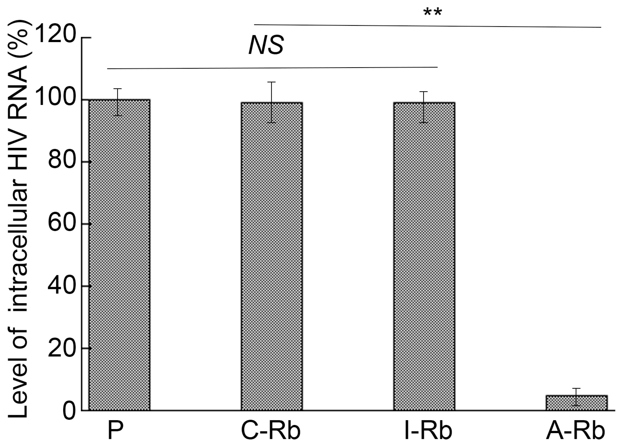
Decreased expression of CCR5 could lead to enhanced cellular protection against an M-tropic HIV strain (e.g., HIVBa-L) infection due to the usage of CCR5 as a co-receptor [28,29]. HIVBa-L was introduced to ribozyme-expressing cells with subsequent harvest. Experiments with qRT-PCR amplifying the HIV tat sequence were used to quantify intracellular HIV RNA, using actin mRNA expression as an internal control. The levels of total HIV-1 intracellular RNA decreased by 92% ± 7% in cells expressing A-Rb, and by less than 10% in I-Rb and C-Rb (Figure 4).
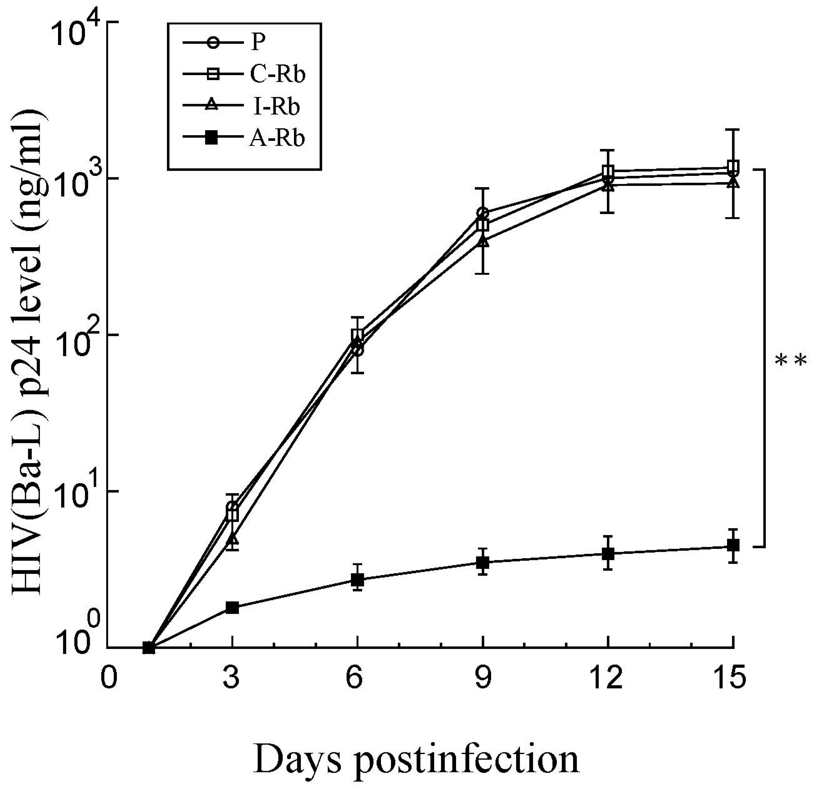
Progression quantification of HIVBa-L infection was assayed via viral p24 level present in the supernatant (Figure 5). HIVBa-L replication substantially diminished in cells expressing A-Rb but not in those with I-Rb or C-Rb during the infection. At 12 days post-infection, the levels of HIVBa-L p24 diminished by 200-fold in A-Rb expressing cells, while no reduction was found in those expressing I-Rb or C-Rb compared to PM1 cells with ribozymes (Figure 5). While I-Rb exhibited no catalytic activity, both it and A-Rb bound to the CCR5 mRNA sequence (Table 1). Thus, CCR5 mRNA cleavage by A-Rb in A-Rb-expressing cells resulted in a decrease in HIV infection.

3.5. Strain-Specific Anti-HIV Effects of M1GS Ribozymes

PM1 cells simultaneously express CCR5, a co-receptor for the M-tropic HIV strains (e.g., HIVBa-L), and CXCR4, a co-receptor for the T-tropic strains (e.g., HIVIIIB) [16,28]. Due to this aspect, the presence of constructed ribozymes with a high specificity against CCR5 expression should not impede PM1 cells from supporting infection from T-tropic strains, since the ribozymes would not impact other proteins such as CXCR4. Here, we used T-tropic HIVIIIB strain and M-tropic HIVBa-L strain to infect cells, examining HIV production via p24 levels in culture supernatants (Figure 6). On day 15 post-infection, A-Rb expressing cells exhibited a 200-fold decrease in p24 level when they were infected with HIVBa-L (Figure 5, Figure 6). However, these cells were susceptible to HIVIIIB as we found no difference in HIV p24 levels in the IIIB-infected cells with A-Rb, I-Rb, and C-Rb (Figure 6). Thus, the A-Rb mediated inhibition of viral replication is specific because the anti-CCR5 ribozyme-expressing cells could support infection of the X4 strain IIIB despite protective measures against R5 strain Ba-L. Ribozyme-mediated cleavage of target mRNA appeared to be critical for decreasing HIVBa-L infection, because both HIVBa-L and HIVIIIB strains efficiently infected and replicated in cells expressing the “inactive” ribozyme I-Rb, which displayed similar binding affinity to CCR5 mRNA as A-Rb but had no cleavage events.

4. Discussion

RNA-based gene interference strategies embody novel therapeutic agents for clinical applications [9,10,30,31,32,33]. The RNase P ribozyme used in our study is unique as it was derived from a variant of the catalytic M1 RNA [1,2]. This variant, V-858, was generated by in vitro selection of M1GS ribozyme mutants displaying efficient cleavage of target mRNA in vitro [15].
Here, M1GS ribozymes A-Rb and I-Rb that target cellular CCR5 mRNA were derived from variant V-858. Active ribozyme A-Rb efficiently cleaved CCR5 mRNA sequences in vitro with 30-fold greater efficiency than the wild-type ribozyme M1-Rb. V-858 has two point mutations (i.e., A86 → G86 and G316 → U316) in the M1 RNA sequence. How these mutations increase the ribozyme cleavage activity has not been studied. Further studies on these issues may offer insight into the mechanism facilitating the efficient cleavage by M1GS ribozymes against their target mRNAs.
In human cells, A-Rb induced decreases in both CCR5 mRNA and protein expression by more than 90%, and led to a more than 200-fold reduction in HIV infection. On the contrary, we only observed a minor CCR5 expression decrease and HIV infection inhibition in cells expressing the inactive ribozyme I-Rb or control ribozyme C-Rb. C-Rb targeted an unrelated mRNA, and I-Rb was able to bind to the CCR5 mRNA sequence similarly to A-Rb but was catalytically inactive. Thus, targeted cleavage by A-Rb results in CCR5 expression decrease and HIV infection inhibition, but not an antisense effect or other nonspecific effects of the ribozymes.
The results imply the RNase P ribozyme to be specific. To start, we observed no substantial distinction in cellular viability and growth between parental cells and ribozyme-expressing cells when they were allowed to grow for three months. Thus, ribozyme expression did not lead to substantial cytotoxicity. Second, the anti-HIV effect by ribozyme A-Rb was due to its induced CCR5 expression reduction. CCR5 expression and HIV infection decreased only in cells expressing A-Rb but not in those expressing I-Rb or C-Rb. There was a correlation between the decreased CCR5 levels and the extent of the HIV infection reduction. Third, ribozyme A-Rb only diminished CCR5 expression but had no effect on the expression of other targets including H1 RNA, actin, and CXCR4 (e.g., Figure 2 and Figure 3). The A-Rb-expressing cells were also still able to support the infection of T tropic HIVIIIB strain despite resistance against infection of the M-tropic HIVBa-L strain. Thus, the ribozyme specifically affects its mRNA target (i.e., CCR5) but not those of other targets (e.g., CXCR4).
Several potential issues may need to be addressed to develop M1GS RNAs for practical applications. First, M1GS ribozymes need to have specific delivery into target tissues and cell types. Efforts have been made to generate novel viral expression vectors such as lentiviral expression vectors. Furthermore, progress has been made to develop cell type- and tissue-specific expression systems. These studies should help to address the ribozyme delivery and expression in vivo. Second, more active ribozymes are needed to increase their cleavage and targeting activity. RNase P ribozyme variants with enhanced cleavage activity have been engineered by different methods including evolution in vitro approaches [1,2]. In this study, we showed that a ribozyme variant with two novel point mutations exhibited about 30-fold higher cleavage activity than that of the wildtype M1RNA sequence. The generation of M1GS ribozymes with better cleavage activity would enable the improvement of these gene-targeting molecules for applications.
Additional studies are needed to explore M1GS RNAs for anti-HIV therapy. For example, it is necessary to confirm if M1GS RNA (e.g., A-Rb) is also effective in reducing viral infection in primary cells known to be infected by HIV in vivo, such as the activated and resting CD4+ T cells [5,6]. We need to demonstrate if M1GS ribozyme is active in cleaving its target mRNA (i.e., CCR5 mRNA) and shutting down the CCR5 expression in these cells. Further efforts may also be needed to investigate how to deliver the ribozyme expression cassettes to these important HIV reservoir cells, and how to express M1GS ribozymes at a high level in these cells. Lentiviral vectors may represent a promising class of vectors used for the delivery of M1GS into HIV reservoir cells.
Several clinical studies using ribozymes for anti-HIV therapy have been carried out using a gene therapy approach by expressing ribozymes via retroviral and lentiviral vectors in CD4+ cells or CD34+ hematopoietic stem cells, which differentiate into hematopoietic lineage cells including CD4+ T cells [33,34,35,36]. These studies indicated that the gene-delivered ribozymes are safe and feasible for anti-HIV therapy [37]. However, these studies encountered several obstacles including limited efficacy of the ribozymes and their suboptimal kinetics, low ribozyme expression due to chromatin silencing of the integrated expression cassettes, and poor engraftment of transduced cells [4,5,6]. Future challenges also include the minimization of immune toxicity and stimulation associated with ribozymes, and the large-scale manufacturing of these M1GS-containing lentiviral vectors to be used for investigating the anti-HIV activity of M1GS RNAs in clinical studies. Collectively, these studies would reveal whether M1GS RNAs can serve as anti-HIV therapeutics.
Anti-CCR5 RNA-based methods (e.g., ribozymes and RNAi) have led to CCR5 expression decrease and HIV infection reduction [11,12,13,14]. A potential issue associated with treatment against HIV infection is the resistant mutant emergence [38,39,40]. The anti-CCR5 RNase P ribozyme-based approach focuses on a cellular gene, targeting the entry step of HIV [41,42], and therefore should minimize the generation of viral mutations. Of note, an inhibition of p24 expression of about 200-fold may not be sufficient to abolish HIV infection completely in vivo. A combinatorial approach, such as the use of ribozymes and RNAi together, is a better anti-HIV strategy [9,10,43]. Moreover, better M1GS ribozymes with enhanced activity will be produced by genetic engineering and in vitro evolution process [44,45]. These studies will help to develop RNase P ribozymes for gene-targeting applications.

Supplementary Materials

The following supporting information can be downloaded at https://www.mdpi.com/article/10.3390/zoonoticdis3020009/s1. Figure S1: Viability of the parental PM1 cells (PM1) and PM1 cells with empty LXSN vector (P) or PM1 cells expressing ribozymes C-Rb, I-Rb, and A-Rb after culturing for 90 days.

Author Contributions

Conceptualization, B.Y., Y.L., Y.-C.C. and F.L.; methodology, B.Y., Y.L., Y.-C.C. and F.L; validation, B.Y., Y.L., Y.-C.C. and F.L.; formal analysis, B.Y., Y.L., Y.-C.C., I.Z. and F.L.; investigation, B.Y., Y.L., Y.-C.C., I.Z. and F.L; data curation, B.Y., Y.L., Y.-C.C., I.Z. and F.L; writing, B.Y., Y.L., Y.-C.C., I.Z. and F.L.; supervision, F.L.; project administration, F.L.; funding acquisition, F.L. All authors have read and agreed to the published version of the manuscript.

Funding

This research was supported by a Start Up fund from the University of California at Berkeley.

Institutional Review Board Statement

Not applicable.

Informed Consent Statement

Not applicable.

Data Availability Statement

Not applicable.

Acknowledgments

Gratitude goes to Adam Smith and Hao Gong for advice and technical assistance.

Conflicts of Interest

The authors declare no conflict of interest.

References

  1. Gopalan, V.; Vioque, A.; Altman, S. RNase P: Variations and uses. J. Biol. Chem. 2002, 277, 6759–6762. [Google Scholar] [CrossRef] [Green Version]
  2. Raj, S.M.; Liu, F. Engineering of RNase P ribozyme for gene-targeting applications. Gene 2003, 313, 59–69. [Google Scholar] [CrossRef] [PubMed]
  3. Kawa, D.; Wang, J.; Yuan, Y.; Liu, F. Inhibition of viral gene expression by human ribonuclease P. RNA 1998, 4, 1397–1406. [Google Scholar] [CrossRef] [PubMed] [Green Version]
  4. Burnett, J.C.; Rossi, J.J. RNA-based therapeutics: Current progress and future prospects. Chem. Biol. 2012, 19, 60–71. [Google Scholar] [CrossRef] [Green Version]
  5. Henderson, L.J.; Reoma, L.B.; Kovacs, J.A.; Nath, A. Advances toward Curing HIV-1 Infection in Tissue Reservoirs. J. Virol. 2020, 94, e00375-19. [Google Scholar] [CrossRef] [PubMed]
  6. Richman, D.D.; Margolis, D.M.; Delaney, M.; Greene, W.C.; Hazuda, D.; Pomerantz, R.J. The challenge of finding a cure for HIV infection. Science 2009, 323, 1304–1307. [Google Scholar] [CrossRef] [Green Version]
  7. Doranz, B.J.; Berson, J.F.; Rucker, J.; Doms, R.W. Chemokine receptors as fusion cofactors for human immunodeficiency virus type 1 (HIV-1). Immunol. Res. 1997, 16, 15–28. [Google Scholar] [CrossRef]
  8. Berger, E.A. HIV entry and tropism: The chemokine receptor connection. Aids 1997, 11, S3–S16. [Google Scholar]
  9. Chung, J.; Rossi, J.J.; Jung, U. Current progress and challenges in HIV gene therapy. Future Virol. 2011, 6, 1319–1328. [Google Scholar] [CrossRef] [Green Version]
  10. Zhou, J.; Rossi, J.J. Current progress in the development of RNAi-based therapeutics for HIV-1. Gene Ther. 2011, 18, 1134–1138. [Google Scholar] [CrossRef] [Green Version]
  11. Feng, Y.; Leavitt, M.; Tritz, R.; Duarte, E.; Kang, D.; Mamounas, M.; Gilles, P.; Wong-Staal, F.; Kennedy, S.; Merson, J.; et al. Inhibition of CCR5-dependent HIV-1 infection by hairpin ribozyme gene therapy against CC-chemokine receptor 5. Virology 2000, 276, 271–278. [Google Scholar] [CrossRef] [Green Version]
  12. Cagnon, L.; Rossi, J.J. Downregulation of the CCR5 beta-chemokine receptor and inhibition of HIV-1 infection by stable VA1-ribozyme chimeric transcripts. Antisense Nucleic Acid. Drug. Dev. 2000, 10, 251–261. [Google Scholar] [CrossRef] [PubMed]
  13. Anderson, J.; Akkina, R. CXCR4 and CCR5 shRNA transgenic CD34+ cell derived macrophages are functionally normal and resist HIV-1 infection. Retrovirology 2005, 2, 53. [Google Scholar] [CrossRef] [Green Version]
  14. Song, E.; Lee, S.-K.; Dykxhoorn, D.M.; Novina, C.; Zhang, D.; Crawford, K.; Cerny, J.; Sharp, P.A.; Lieberman, J.; Manjunath, N.; et al. Sustained small interfering RNA-mediated human immunodeficiency virus type 1 inhibition in primary macrophages. J. Virol. 2003, 77, 7174–7181. [Google Scholar] [CrossRef] [PubMed] [Green Version]
  15. Kilani, A.F.; Trang, P.; Jo, S.; Hsu, A.; Kim, J.; Nepomuceno, E.; Liou, K.; Liu, F. RNase P ribozymes selected in vitro to cleave a viral mRNA effectively inhibit its expressionin cell culture. J. Biol. Chem. 2000, 275, 10611–10622. [Google Scholar] [CrossRef] [PubMed] [Green Version]
  16. Lusso, P.; Cocchi, F.; Balotta, C.; Markham, P.D.; Lioue, A.; Farci, P.; Pal, R.; Gallo, R.C.; Reitz, M.S. Growth of macrophage-tropic and primary human immunodeficiency virus type 1 (HIV-1) isolates in a unique CD4+ T-cell clone (PM1): Failure to downregulate CD4 and to interfere with cell-line-tropic HIV-1. J. Virol. 1995, 69, 3712–3720. [Google Scholar] [CrossRef] [Green Version]
  17. Kim, J.J.; Kilani, A.F.; Zhan, X.; Altman, S.; Liu, F. The protein cofactor allows the sequence of an RNase P ribozyme to diversify by maintaining the catalytically active structure of the enzyme. RNA 1997, 3, 613–623. [Google Scholar]
  18. Miller, A.D.; Rosman, G.J. Improved retroviral vectors for gene transfer and expression. Biotechniques 1989, 7, 980–990. [Google Scholar]
  19. Liu, F.; Altman, S. Inhibition of viral gene expression by the catalytic RNA subunit of RNase P from Escherichia coli. Genes Dev. 1995, 9, 471–480. [Google Scholar] [CrossRef] [Green Version]
  20. Sun, X.; Chen, W.; He, L.; Sheng, J.; Liu, Y.; Vu, G.-P.; Yang, Z.; Li, W.; Trang, P.; Wang, Y.; et al. Inhibition of human cytomegalovirus immediate early gene expression and growth by a novel RNase P ribozyme variant. PLoS ONE 2017, 12, e0186791. [Google Scholar] [CrossRef] [PubMed] [Green Version]
  21. Deng, Q.; Liu, Y.; Li, X.; Yan, B.; Sun, X.; Tang, W.; Trang, P.; Yang, Z.; Gong, H.; Wang, Y.; et al. Inhibition of human cytomegalovirus major capsid protein expression and replication by ribonuclease P-associated external guide sequences. RNA 2019, 25, 645–655. [Google Scholar] [CrossRef] [PubMed]
  22. Zeng, W.; Vu, G.-P.; Bai, Y.; Chen, Y.-C.; Trang, P.; Lu, S.; Xiao, G.; Liu, F. RNase P-associated external guide sequence effectively reduces the expression of human CC-chemokine receptor 5 and inhibits the infection of human immunodeficiency virus 1. Biomed. Res. Int. 2013, 2013, 509714. [Google Scholar] [CrossRef] [PubMed] [Green Version]
  23. Daftarian, P.M.; Kumar, A.; Kryworuchko, M.; Diaz-Mitoma, F. IL-10 production is enhanced in human T cells by IL-12 and IL-6 and in monocytes by tumor necrosis factor-alpha. J. Immunol. 1996, 157, 12–20. [Google Scholar] [CrossRef] [PubMed]
  24. Deng, H.K.; Unutmaz, D.; KewalRamani, V.N.; Littman, D.R. Expression cloning of new receptors used by simian and human immunodeficiency viruses. Nature 1997, 388, 296–300. [Google Scholar] [CrossRef]
  25. Liao, F.; Lee, H.-H.; Farber, J.M. Cloning of STRL22, a new human gene encoding a G-protein-coupled receptor related to chemokine receptors and located on chromosome 6q27. Genomics 1997, 40, 175–180. [Google Scholar] [CrossRef] [Green Version]
  26. Bertrand, E.; Castanotto, D.; Zhou, C.; Carbonnelle, C.; Lee, N.S.; Good, P.; Chatterjee, S.; Grange, T.; Pictet, R.; Kohn, D.; et al. The expression cassette determines the functional activity of ribozymes in mammalian cells by controlling their intracellular localization. RNA 1997, 3, 75–88. [Google Scholar]
  27. Yuan, Y.; Hwang, E.S.; Altman, S. Targeted cleavage of mRNA by human RNase, P. Proc. Natl. Acad. Sci. USA 1992, 89, 8006–8010. [Google Scholar] [CrossRef] [Green Version]
  28. Berger, E.A.; Murphy, P.M.; Farber, J.M. Chemokine receptors as HIV-1 coreceptors: Roles in viral entry, tropism, and disease. Annu. Rev. Immunol. 1999, 17, 657–700. [Google Scholar] [CrossRef]
  29. Bieniasz, P.D.; Cullen, B.R. Chemokine receptors and human immunodeficiency virus infection. Front. Biosci. 1998, 3, 44–58. [Google Scholar]
  30. Scherer, L.J.; Rossi, J.J. Approaches for the sequence-specific knockdown of mRNA. Nat. Biotechnol. 2003, 21, 1457–1465. [Google Scholar] [CrossRef]
  31. Santoro, S.W.; Joyce, G.F. A general purpose RNA-cleaving DNA enzyme. Proc. Natl. Acad. Sci. USA 1997, 94, 4262–4266. [Google Scholar] [CrossRef] [PubMed] [Green Version]
  32. Stein, C.A.; Cheng, Y.C. Antisense oligonucleotides as therapeutic agents—Is the bullet really magical? Science 1993, 261, 1004–1012. [Google Scholar] [CrossRef] [Green Version]
  33. Wong-Staal, F.; Poeschla, E.M.; Looney, D.J. A controlled, Phase 1 clinical trial to evaluate the safety and effects in HIV-1 infected humans of autologous lymphocytes transduced with a ribozyme that cleaves HIV-1 RNA. Hum. Gene Ther. 1998, 9, 2407–2425. [Google Scholar] [CrossRef]
  34. Amado, R.G.; Mitsuyasu, R.T.; Rosenblatt, J.D.; Ngok, F.K.; Bakker, A.; Cole, S.; Chorn, N.; Lin, L.-S.; Bristol, G.; Boyd, M.P.; et al. Anti-human immunodeficiency virus hematopoietic progenitor cell-delivered ribozyme in a phase I study: Myeloid and lymphoid reconstitution in human immunodeficiency virus type-1-infected patients. Hum. Gene Ther. 2004, 15, 251–262. [Google Scholar] [CrossRef] [Green Version]
  35. Macpherson, J.L.; Boyd, M.P.; Arndt, A.J.; Todd, A.V.; Fanning, G.C.; Ely, J.A.; Elliott, F.; Knop, A.; Raponi, M.; Murray, J.; et al. Long-term survival and concomitant gene expression of ribozyme-transduced CD4+ T-lymphocytes in HIV-infected patients. J. Gene Med. 2005, 7, 552–564. [Google Scholar] [CrossRef]
  36. Michienzi, A.; Castanotto, D.; Lee, N.; Li, S.; Zaia, J.A.; Rossi, J.J. RNA-mediated inhibition of HIV in a gene therapy setting. Ann. N. Y. Acad. Sci. 2003, 1002, 63–71. [Google Scholar] [CrossRef] [PubMed]
  37. DiGiusto, D.L.; Krishnan, A.; Li, L.; Li, H.; Li, S.; Rao, A.; Mi, S.; Yam, P.; Stinson, S.; Kalos, M.; et al. RNA-based gene therapy for HIV with lentiviral vector-modified CD34(+) cells in patients undergoing transplantation for AIDS-related lymphoma. Sci. Transl. Med. 2010, 2, 36ra43. [Google Scholar] [CrossRef] [PubMed] [Green Version]
  38. Autran, B.; Carcelain, G.; Li, T.S.; Blanc, C.; Mathez, D.; Tubiana, R.; Katlama, C.; Debre, P.; Leibowitch, J. Positive effects of combined antiretroviral therapy on CD4 + T cell homeostasis and function in advanced HIV disease. Science 1997, 277, 112–116. [Google Scholar] [CrossRef]
  39. Yeni, P. Update on HAART in HIV. J. Hepatol. 2006, 44, S100–S103. [Google Scholar] [CrossRef]
  40. Palella, F.J., Jr.; Delaney, K.M.; Moorman, A.C.; Loveless, M.O.; Fuhrer, J.; Satten, G.A.; Aschman, D.J.; Holmberg, S.D.; the HIV Outpatient Study Investigators. Declining morbidity and mortality among patients with advanced human immunodeficiency virus infection. HIV Outpatient Study Investigators. N. Engl. J. Med. 1998, 338, 853–860. [Google Scholar] [CrossRef]
  41. Freed, E.O.; Martin, M.A. HIVs and their replication. In Fields Virology; Knipe, D.M., Howley, P.M., Griffin, D.E., Martin, M.A., Lamb, R.A., Roizman, B., Straus, S.E., Eds.; Lippincott-William & Wilkins: Philadelphia, PA, USA, 2007; pp. 2108–2185. [Google Scholar]
  42. Kuritzkes, D.R.; Walker, B.D. HIV-1: Pathogenesis, Clinical Manifestations, and Treatment. In Fields Virology; Knipe, D.M., Howley, P.M., Griffin, D.E., Martin, M.A., Lamb, R.A., Roizman, B., Straus, S.E., Eds.; Lippincott-William & Wilkins: Philadelphia, PA, USA, 2007; pp. 2188–2214. [Google Scholar]
  43. Strayer, D.S.; Akkina, R.; Bunnell, B.A.; Dropulic, B.; Planelles, V.; Pomerantz, R.J.; Rossi, J.J.; Zaia, J.A. Current status of gene therapy strategies to treat HIV/AIDS. Mol. Ther. 2005, 11, 823–842. [Google Scholar] [CrossRef] [PubMed]
  44. Zhou, T.; Kim, J.; Kilani, A.F.; Kim, K.; Dunn, W.; Jo, S.; Nepomuceno, E.; Liu, F. In vitro selection of external guide sequences for directing RNase P-mediated inhibition of viral gene expression. J. Biol. Chem. 2002, 277, 30112–30120. [Google Scholar] [CrossRef] [PubMed] [Green Version]
  45. Yuan, Y.; Altman, S. Selection of guide sequences that direct efficient cleavage of mRNA by human ribonuclease P. Science 1994, 263, 1269–1273. [Google Scholar] [CrossRef] [PubMed]
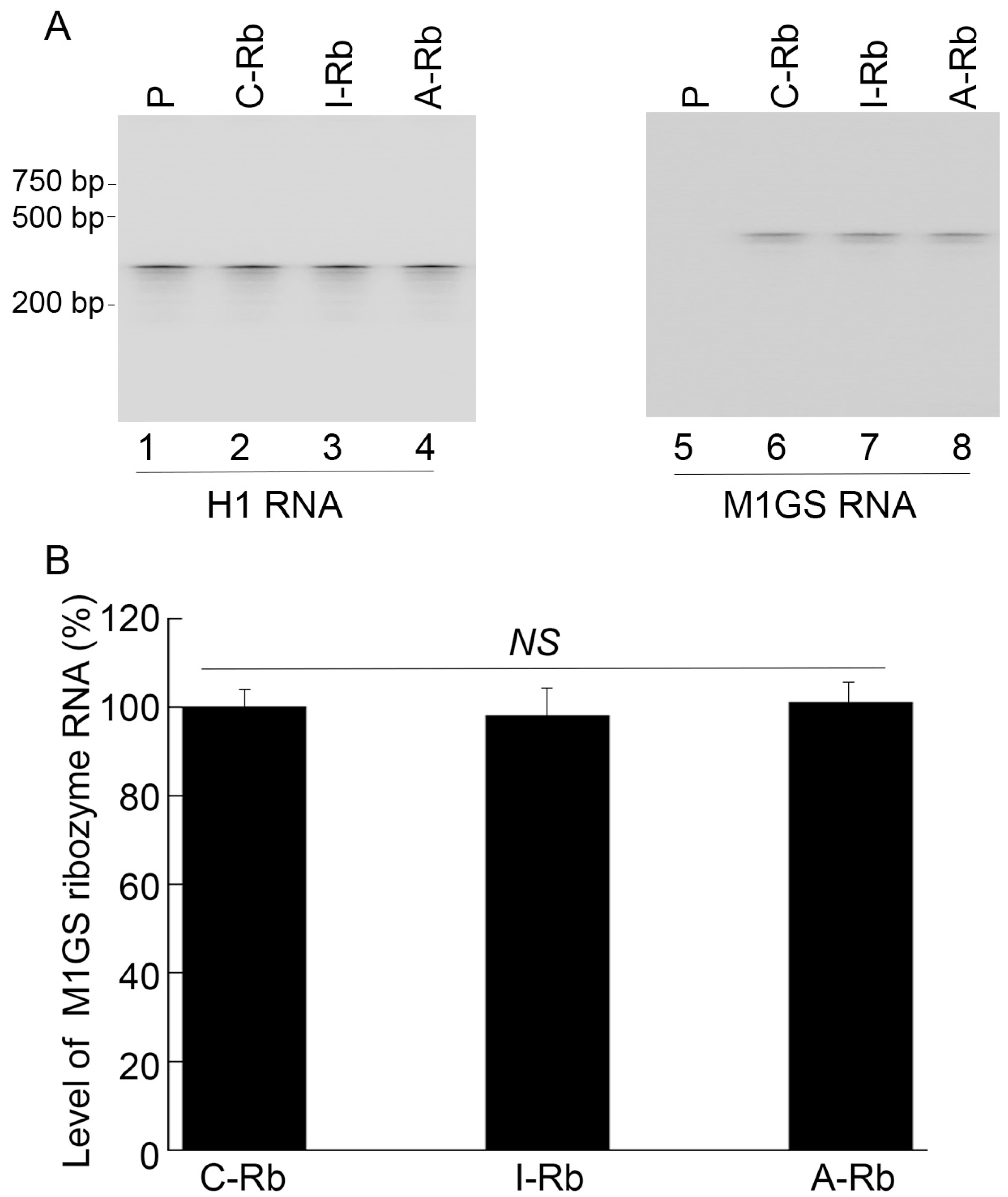
Figure 1. Northern blot analyses of RNase P ribozyme and H1 RNA expression in human cells. (A). We used total RNA samples (30 µg) extracted from PM1 cells with LXSN empty vector (P, lanes 1 and 5) and a cloned cell line producing C-Rb (lanes 2 and 6), I-Rb (lanes 3 and 7), and A-Rb (lanes 4 and 8). (B). Results are conveyed in % in comparison to those in PM1 cells producing C-Rb, and indicated as mean ± SD. NS, not significant. We performed experiments in triplicate and repeated them three times.
Zoonoticdis 03 00009 g001
Figure 2. Northern blot analyses of CCR5 (lanes 5–8), CXCR4 (lanes 9–12), and actin mRNA expression (lanes 1–4). (A). We used total RNA samples (40 µg) isolated from PM1 cells without ribozyme (P) and with C-Rb, I-Rb, and A-Rb. (B). Results are conveyed in % in comparison to the control, PM1 cells without ribozymes (P), and indicated as mean ± SD. ** p < 0.05. NS, not significant. We performed experiments in triplicate and repeated them three times.
Zoonoticdis 03 00009 g002
Figure 3. Western blot analyses of CCR5 (lanes 5–8), CXCR4 (lanes 9–12), and actin protein expression (lanes 1–4). (A). We used protein samples (30 µg) isolated from PM1 cells without ribozyme (P) and with C-Rb, I-Rb, and A-Rb. (B). Results are conveyed in % in comparison to the control, PM1 cells without ribozymes (P), and indicated as mean ± SD. ** p < 0.05. NS, not significant. We performed experiments in triplicate and repeated them three times.
Zoonoticdis 03 00009 g003
Figure 4. Quantifying intracellular HIV RNA by a qRT-PCR assay amplifying the HIV tat sequence. RNA samples were from HIVBa-L-infected PM1 cells without ribozymes (P) or with C-Rb, I-Rb, and A-Rb. Results are conveyed in % in comparison to the control, PM1 cells without ribozymes (P), and indicated as mean ± SD. ** p < 0.05. NS, not significant. We performed experiments in triplicate and repeated them three times.
Zoonoticdis 03 00009 g004
Figure 5. HIVBa-L growth in PM1 cells without (P) or with C-Rb, I-Rb, and A-Rb. 5 × 10⁵ cells were infected with HIV-1Ba-L (MOI = 0.02–0.1). A p24 antigen assay was used to quantify HIV production. Results are indicated as mean ± SD. ** p < 0.05. We performed experiments in triplicate and repeated them three times.
Zoonoticdis 03 00009 g005
Figure 6. Supernatant HIV-1 p24 protein levels in cells having R5 strain HIVBa-L (A) or X4 strain HIVIIIB at 15 days post-infection (B). A p24 antigen assay was used to quantify p24 expression in supernatants isolated from cultures of cells without ribozymes (P) or with C-Rb, I-Rb, and A-Rb. Results are indicated as mean ± SD. ** p < 0.05. NS, not significant. We performed experiments in triplicate and repeated them three times.
Zoonoticdis 03 00009 g006
Table 1. Values of overall cleavage rate [(kcat/Km)s] and binding dissociation constant (Kd) in M1GS reactions with substrate ccr5-38. Experimental details are described in Materials and Methods.
Enzyme(kcat/Km)s
(µM−1·min−1)
Kd (nM)
M1-Rb0.21 ± 0.080.31 ± 0.08
A-Rb6.5 ± 2.50.35 ± 0.09
I-Rb<5 × 10−50.32 ± 0.08
C-Rb<5 × 10⁻⁵ND
“ND” not determined.
Disclaimer/Publisher’s Note: The statements, opinions and data contained in all publications are solely those of the individual author(s) and contributor(s) and not of MDPI and/or the editor(s). MDPI and/or the editor(s) disclaim responsibility for any injury to people or property resulting from any ideas, methods, instructions or products referred to in the content.