
Corynebacterium lactis: Antimicrobial Resistance and Impact on Invertebrate Model Systems

 ,
, 
Abstract
1. Introduction
2. Materials and Methods
2.1. Bacteria and Growth Conditions
2.2. Transformation of C. lactis
2.3. Antimicrobial Resistance Testing
2.4. Infection of Caenorhabditis Elegans
2.4.1. Fluorescence Microscopy
2.4.2. Survival Assay
2.5. Chemotactic Behavior
2.6. Infection of Greater Wax Moth (Galleria mellonella) Larvae
2.7. Genome Screening
3. Results
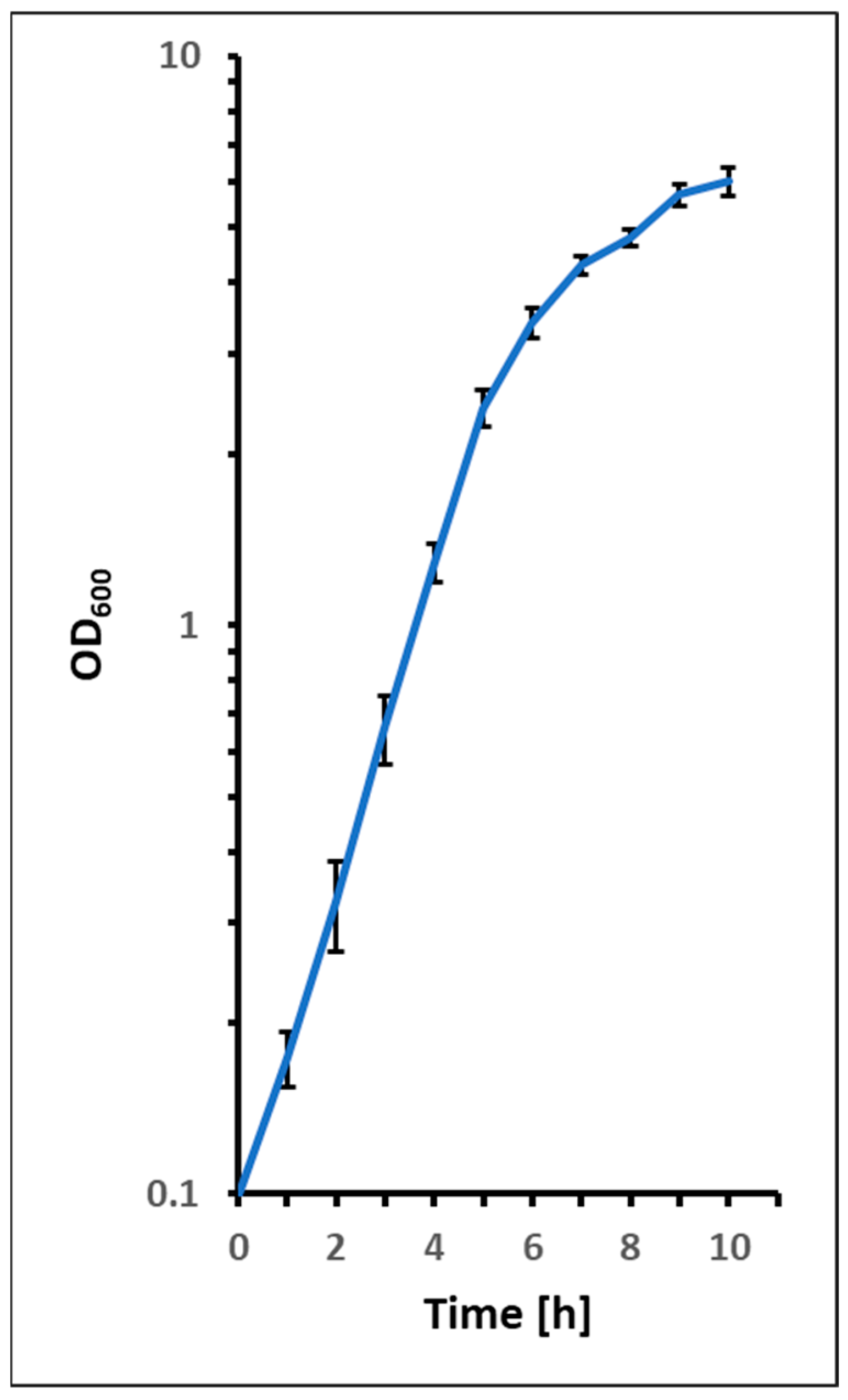
3.1. Growth of C. lactis
3.2. Antibiotic Susceptibility and Resistance of C. lactis
3.3. Interaction with Invertebrate Infection Model Systems
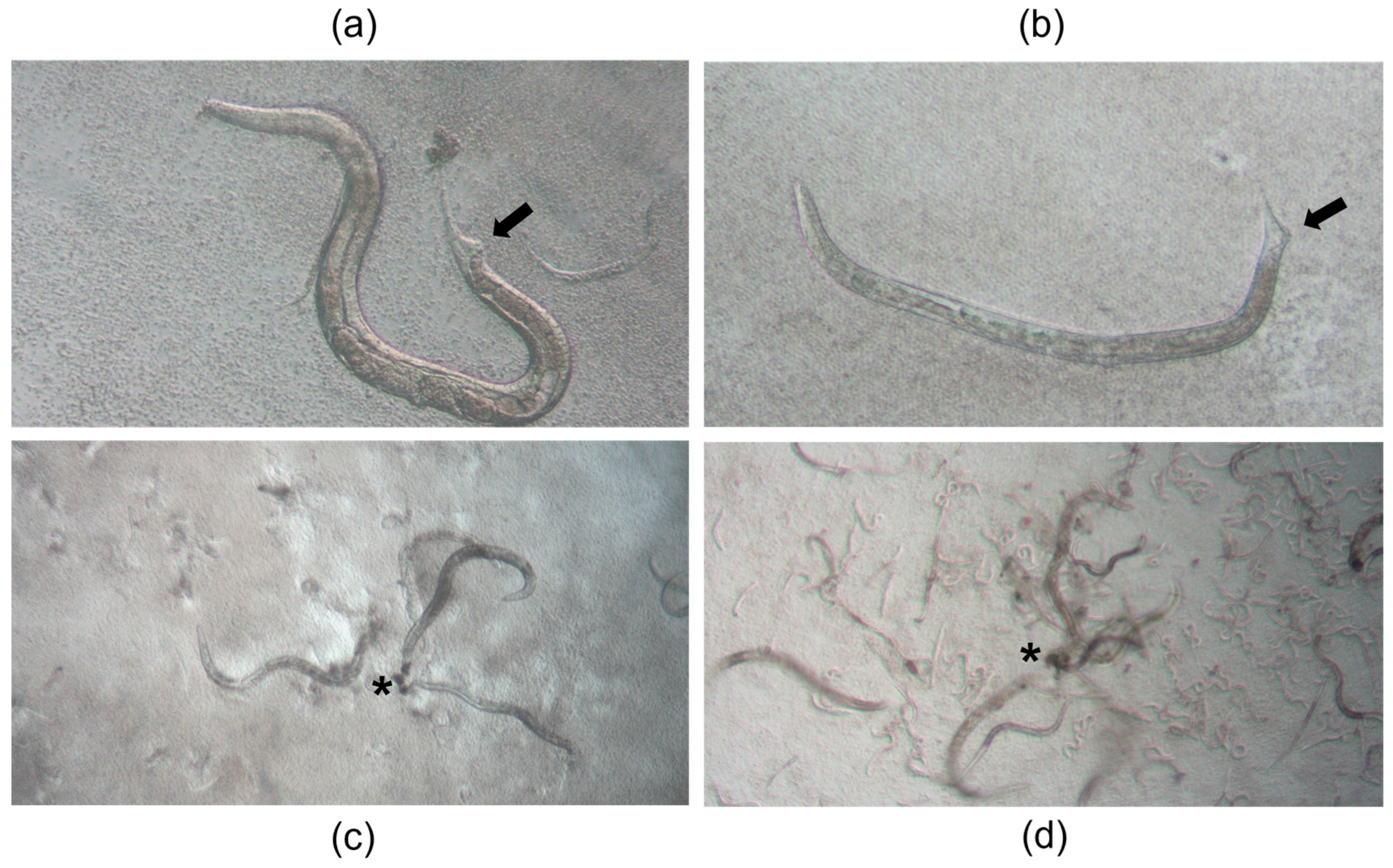
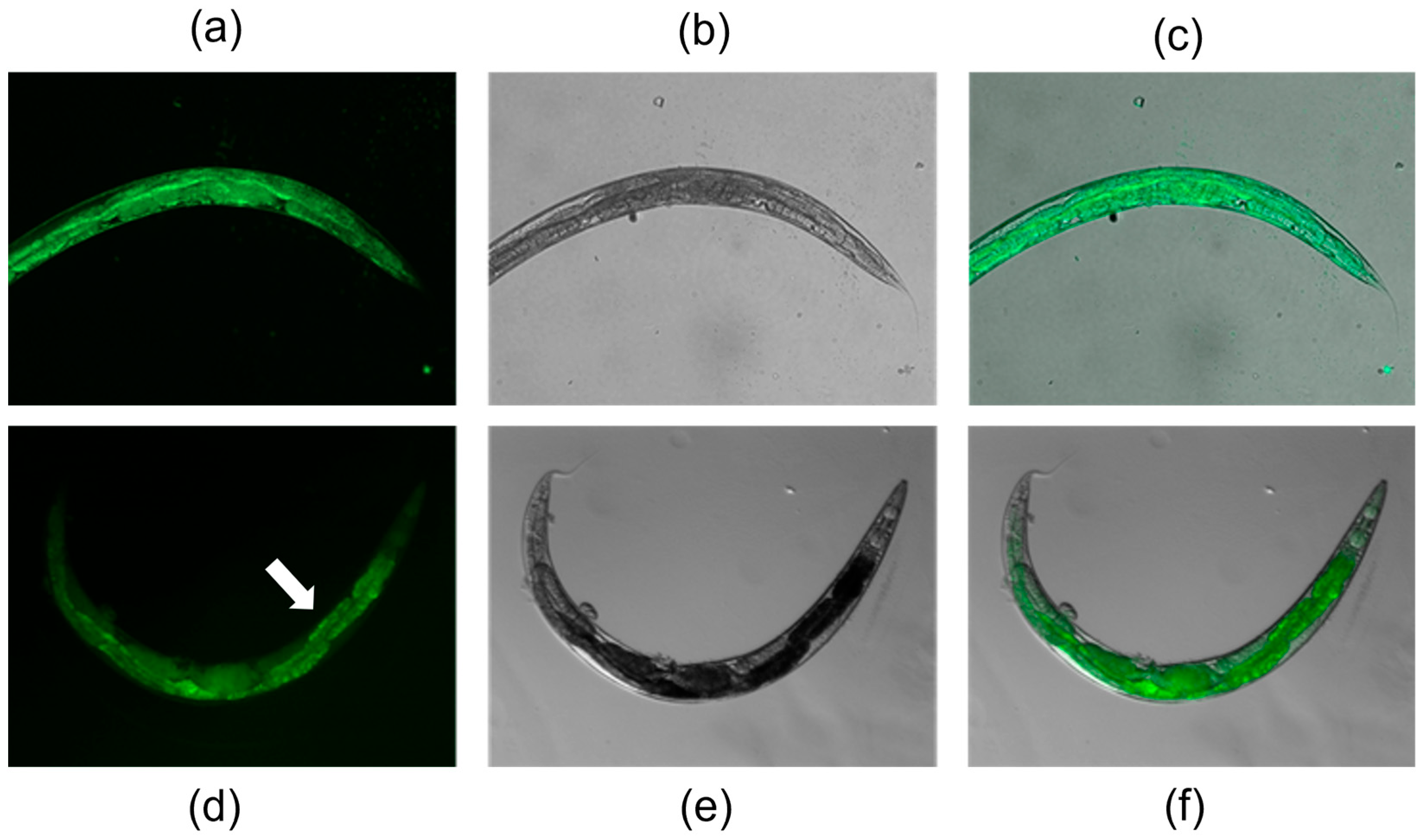
3.3.1. Colonization of C. elegans
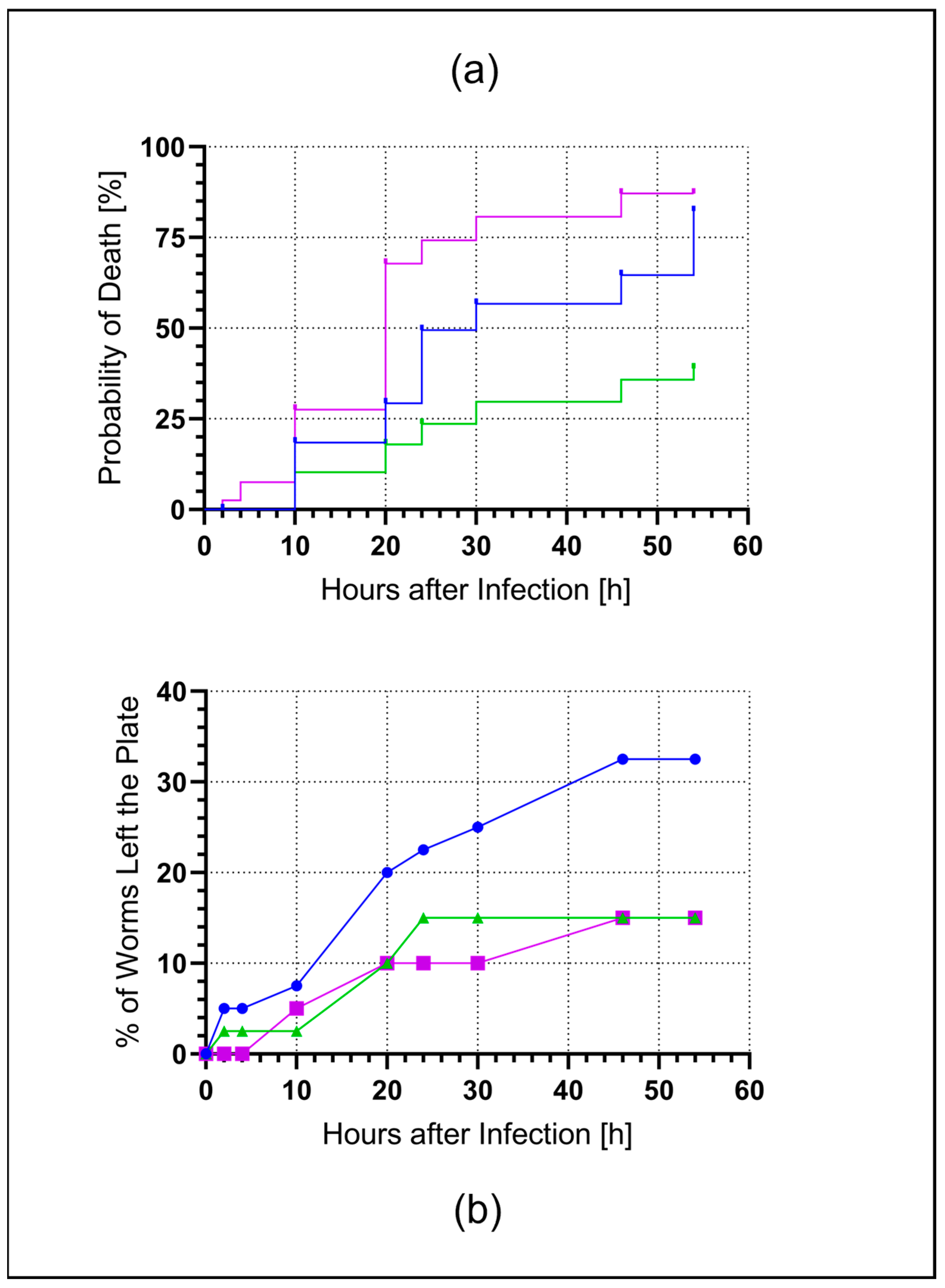
3.3.2. C. elegans Survival Assay
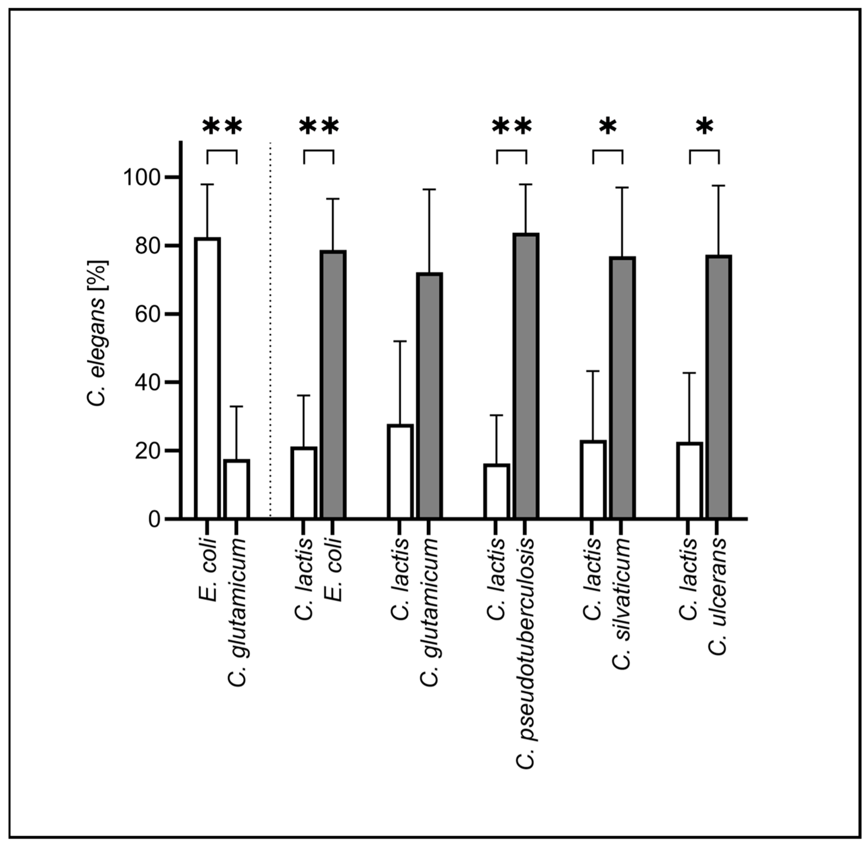
3.3.3. Chemotactic Behavior of C. elegans
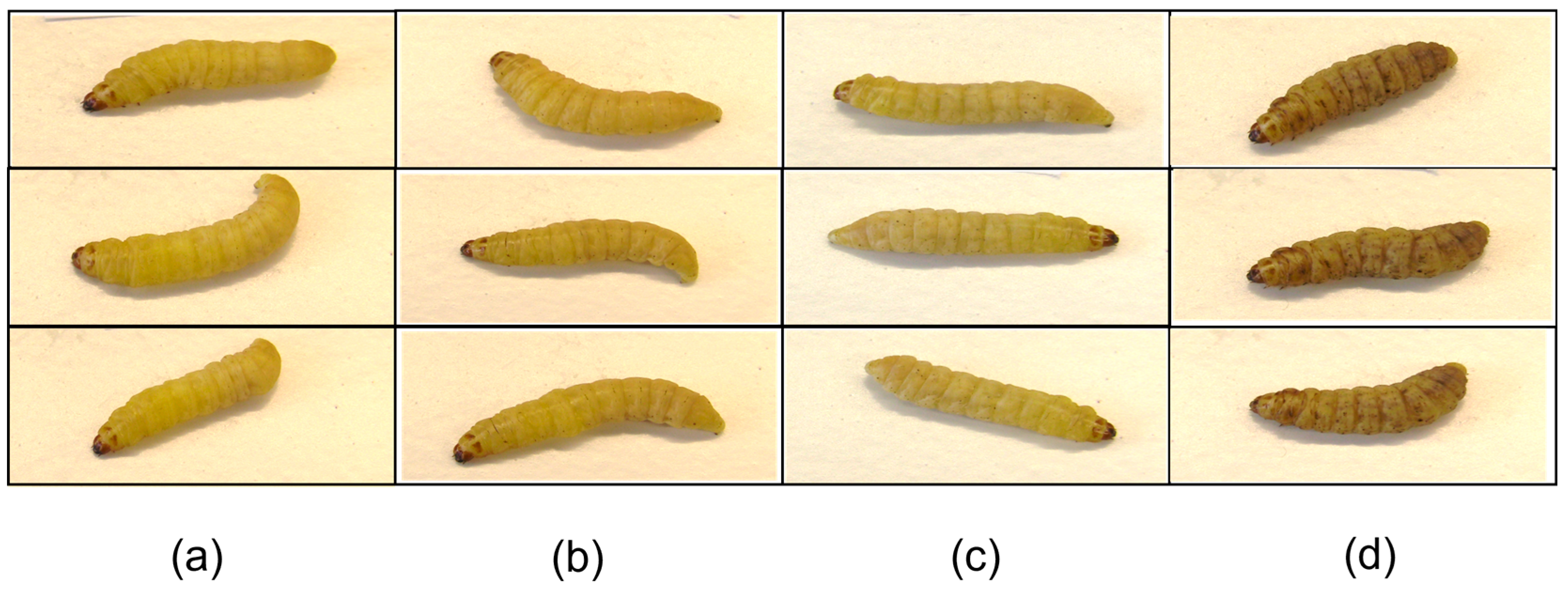
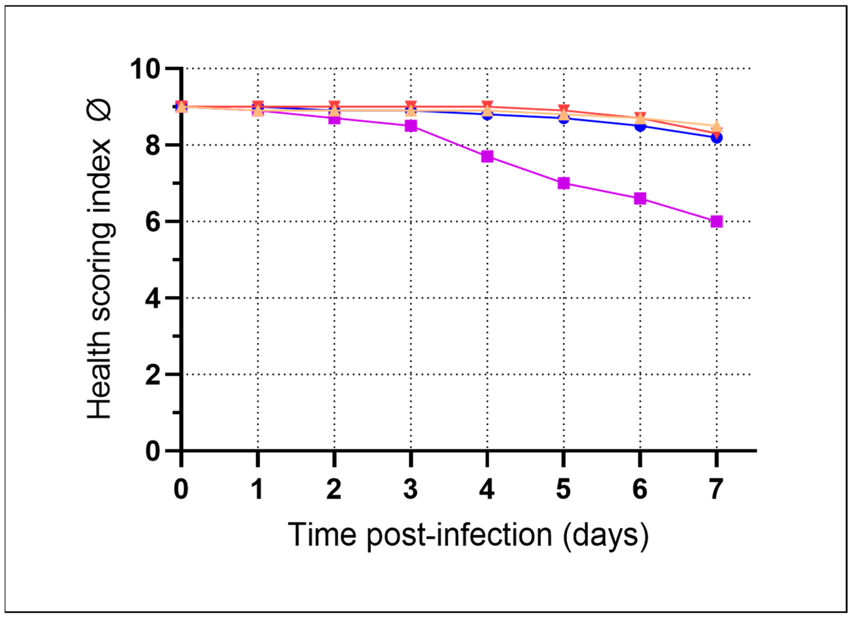
3.3.4. Infection of G. mellonella Larvae
3.4. Putative Virulence Factors
4. Discussion
5. Conclusions
Supplementary Materials
Author Contributions
Funding
Institutional Review Board Statement
Informed Consent Statement
Data Availability Statement
Acknowledgments
Conflicts of Interest
Abbreviations
| BHI | Brain Heart Infusion |
| Dar | Deformed anal region |
| EUCAST | European Committee on Antimicrobial Susceptibility Testing |
| FBS | Fetal Bovine Serum |
| MIC | Minimal inhibitory concentration |
| OD600 | Optical density at 600 nm |
| SD | Standard deviation |
References
- Ventura, M.; Canchaya, C.; Tauch, A.; Chandra, G.; Fitzgerald, G.F.; Chater, K.F.; van Sinderen, D. Genomics of Actinobacteria: Tracing the evolutionary history of an ancient phylum. Microbiol. Mol. Biol. Rev. 2007, 71, 495–548. [Google Scholar] [CrossRef] [Scilit]
- Whitman, W.B.; Oren, A.; Chuvochina, M.; da Costa, M.S.; Garrity, G.M.; Rainey, F.A.; Rossello-Mora, R.; Schink, B.; Sutcliffe, I.; Trujillo, M.E.; et al. Proposal of the suffix -ota to denote phyla. Addendum to ‘Proposal to include the rank of phylum in the International Code of Nomenclature of Prokaryotes’. Int. J. Syst. Evol. Microbiol. 2018, 68, 967–969. [Google Scholar] [CrossRef] [Scilit] [PubMed]
- Rahlwes, K.C.; Sparks, I.L.; Morita, Y.S. Cell Walls and Membranes of Actinobacteria. Subcell. Biochem. 2019, 92, 417–469. [Google Scholar] [CrossRef] [Scilit] [PubMed]
- Parte, A.C.; Sardà Carbasse, J.; Meier-Kolthoff, J.P.; Reimer, L.C.; Göker, M. List of Prokaryotic names with standing in Nomenclature (LPSN) moves to the DSMZ. Int. J. Syst. Evol. Microbiol. 2020, 70, 5607–5612. [Google Scholar] [CrossRef] [Scilit]
- Genus Corynebacterium. Available online: https://lpsn.dsmz.de/genus/corynebacterium (accessed on 7 January 2026).
- Ray, D.; Anand, U.; Jha, N.K.; Korzeniewska, E.; Bontempi, E.; Proćków, J.; Dey, A. The soil bacterium, Corynebacterium glutamicum, from biosynthesis of value-added products to bioremediation: A master of many trades. Environ. Res. 2022, 213, 113622. [Google Scholar] [CrossRef] [Scilit] [PubMed]
- Zha, J.; Zhao, Z.; Xiao, Z.; Eng, T.; Mukhopadhyay, A.; Koffas, M.A.; Tang, Y.J. Biosystem design of Corynebacterium glutamicum for bioproduction. Curr. Opin. Biotechnol. 2023, 79, 102870. [Google Scholar] [CrossRef] [Scilit]
- Sangal, V.; Hoskisson, P.A. Evolution, epidemiology and diversity of Corynebacterium diphtheriae: New perspectives on an old foe. Infect. Genet. Evol. 2016, 43, 364–370. [Google Scholar] [CrossRef] [Scilit]
- Parveen, S.; Bishai, W.R.; Murphy, J.R. Corynebacterium diphtheriae: Diphtheria Toxin, the tox Operon, and Its Regulation by Fe2+ Activation of apo-DtxR. Microbiol. Spectr. 2019, 7. [Google Scholar] [CrossRef] [Scilit]
- Sharma, N.C.; Efstratiou, A.; Mokrousov, I.; Mutreja, A.; Das, B.; Ramamurthy, T. Diphtheria. Nat. Rev. Dis. Primers 2019, 5, 81. [Google Scholar] [CrossRef] [Scilit]
- Gilbert, R.; Stewart, F.C. Corynebacterium ulcerans; A pathogenic microorganism resembling Corynebacterium diphtheriae. J. Lab. Clin. Med. 1927, 12, 756–761. [Google Scholar]
- Hacker, E.; Antunes, C.A.; Mattos-Guaraldi, A.L.; Burkovski, A.; Tauch, A. Corynebacterium ulcerans, an emerging human pathogen. Future Microbiol. 2016, 11, 1191–1208. [Google Scholar] [CrossRef] [Scilit] [PubMed]
- Sing, A.; Luaces, L.M.; Dangel, A.; Deramo, S.; Bengs, K.; Melnikov, V.G.; Berger, A. Toxigenic Corynebacterium ulcerans in raw milk of a cow with acute mastitis: Case report and historical review on milk-transmitted diphtheria. Infection 2025, 20, 767–774. [Google Scholar] [CrossRef] [Scilit]
- Monaco, M.; Sacchi, A.R.; Scotti, M.; Mancini, F.; Riccio, C.; Errico, G.; Ratti, G.; Bondi, F.; Ciervo, A.; Pantosti, A. Respiratory diphtheria due to Corynebacterium ulcerans transmitted by a companion dog, Italy 2014. Infection 2017, 45, 903–905, Erratum in Infection 2017, 45, 931. https://doi.org/10.1007/s15010-017-1045-9. [Google Scholar] [CrossRef] [Scilit]
- Othieno, R.; Mark, K.; Etherson, M.; Foster, G.; Murray, S.; Kalima, P.; Fry, N.K.; Cameron, C.; Strachan, J. A case of cutaneous toxigenic Corynebacterium ulcerans likely acquired from a domestic dog. Access Microbiol. 2019, 1, e000025. [Google Scholar] [CrossRef] [Scilit]
- Hillan, A.; Gibbs, T.; Weaire-Buchanan, G.; Brown, T.; Pang, S.; McEvoy, S.P.; Parker, E. Zoonotic transmission of diphtheria toxin-producing Corynebacterium ulcerans. Zoonoses Public Health 2024, 71, 157–169. [Google Scholar] [CrossRef] [Scilit] [PubMed]
- Dorella, F.A.; Pacheco, L.G.; Oliveira, S.C.; Miyoshi, A.; Azevedo, V. Corynebacterium pseudotuberculosis: Microbiology, biochemical properties, pathogenesis and molecular studies of virulence. Vet. Res. 2006, 37, 201–218. [Google Scholar] [CrossRef] [Scilit] [PubMed]
- Baird, G.J.; Fontaine, M.C. Corynebacterium pseudotuberculosis and its role in ovine caseous lymphadenitis. J. Comp. Pathol. 2007, 137, 179–210. [Google Scholar] [CrossRef] [Scilit]
- Hiller, E.; Hörz, V.; Sting, R. Corynebacterium pseudotuberculosis: Whole genome sequencing reveals unforeseen and relevant genetic diversity in this pathogen. PLoS ONE 2024, 19, e0309282. [Google Scholar] [CrossRef] [Scilit]
- Lipsky, B.A.; Goldberger, A.C.; Tompkins, L.S.; Plorde, J.J. Infections caused by nondiphtheria corynebacteria. Rev. Infect. Dis. 1982, 4, 1220–1235. [Google Scholar] [CrossRef] [Scilit]
- Li, X.; Lv, H.; Wu, C.; Li, H.; Yi, W.; Niu, X.; Peng, Q.; Meng, C.; Yuan, Y.; Tian, S.; et al. Corynebacterium pseudotuberculosis phospholipase D targets mitochondrial sphingomyelin and induces NLRP3-GSDMD axis-mediated pyroptosis in macrophages to promote infection. Vet. Res. 2025, 56, 198. [Google Scholar] [CrossRef] [Scilit]
- Dangel, A.; Berger, A.; Rau, J.; Eisenberg, T.; Kämpfer, P.; Margos, G.; Contzen, M.; Busse, H.J.; Konrad, R.; Peters, M.; et al. Corynebacterium silvaticum sp. nov., a unique group of NTTB corynebacteria in wild boar and roe deer. Int. J. Syst. Evol. Microbiol. 2020, 70, 3614–3624. [Google Scholar] [CrossRef] [Scilit] [PubMed]
- Möller, J.; Busch, A.; Berens, C.; Hotzel, H.; Burkovski, A. Newly Isolated Animal Pathogen Corynebacterium silvaticum Is Cytotoxic to Human Epithelial Cells. Int. J. Mol. Sci. 2021, 22, 3549. [Google Scholar] [CrossRef] [Scilit]
- Berger, A.; Dangel, A.; Melnikov, V.G.; Bengs, K.; Rupp, T.; Mappes, H.J.; Schneider, C.; Sing, A. Human Infections by Novel Zoonotic Species Corynebacterium silvaticum, Germany. Emerg. Infect. Dis. 2025, 31, 1450–1454. [Google Scholar] [CrossRef] [Scilit]
- Available online: https://pubmed.ncbi.nlm.nih.gov/?term=corynebacterium+lactis (accessed on 7 January 2026).
- Wiertz, R.; Schulz, S.C.; Müller, U.; Kämpfer, P.; Lipski, A. Corynebacterium frankenforstense sp. nov. and Corynebacterium lactis sp. nov., isolated from raw cow milk. Int. J. Syst. Evol. Microbiol. 2013, 63, 4495–4501. [Google Scholar] [CrossRef] [Scilit] [PubMed]
- Antunes, J.M.; Ribeiro, M.G.; Demoner, L.d.C.; Ramos, J.N.; Baio, P.V.; Simpson-Louredo, L.; Santos, C.S.; Hirata, R., Jr.; Ferioli, R.B.; Romera, A.R.; et al. Cutaneous abscess caused by Corynebacterium lactis in a companion dog. Vet. Microbiol. 2015, 178, 163–166. [Google Scholar] [CrossRef] [Scilit]
- Abe, S.; Takayama, K.; Kinoshita, S. Taxonomical studies on glutamic acid producing bacteria. J. Gen. Microbiol. 1967, 13, 279–301. [Google Scholar] [CrossRef] [Scilit]
- Möller, J.; Bodenschatz, M.; Sangal, V.; Hofmann, J.; Burkovski, A. Multi-Omics of Corynebacterium Pseudotuberculosis 12CS0282 and an In Silico Reverse Vaccinology Approach Reveal Novel Vaccine and Drug Targets. Proteomes 2022, 10, 39. [Google Scholar] [CrossRef] [Scilit]
- Busch, A.; Möller, J.; Burkovski, A.; Hotzel, H. Genome sequence of a pathogenic Corynebacterium ulcerans strain isolated from a wild boar with necrotizing lymphadenitis. BMC Res. Notes 2019, 12, 692. [Google Scholar] [CrossRef] [Scilit]
- Trost, E.; Al-Dilaimi, A.; Papavasiliou, P.; Schneider, J.; Viehoever, P.; Burkovski, A.; de Castro Soares, S.; Silva Almeida, S.; Alves Dorella, F.; Miyoshi, A.; et al. Comparative analysis of two complete Corynebacterium ulcerans genomes and detection of candidate virulence factors. BMC Genom. 2011, 12, 383. [Google Scholar] [CrossRef] [Scilit] [PubMed]
- Brenner, S. The genetics of Caenorhabditis elegans. Genetics 1974, 77, 71–94. [Google Scholar] [CrossRef] [Scilit]
- Oram, D.M.; Avdalovic, A.; Holmes, R.K. Construction and characterization of transposon insertion mutations in Corynebacterium diphtheriae that affect expression of the diphtheria toxin repressor (DtxR). J. Bacteriol. 2002, 184, 5723–5732. [Google Scholar] [CrossRef] [Scilit] [PubMed]
- Knoppová, M.; Phensaijai, M.; Veselý, M.; Zemanová, M.; Nesvera, J.; Pátek, M. Plasmid vectors for testing in vivo promoter activities in Corynebacterium glutamicum and Rhodococcus erythropolis. Curr. Microbiol. 2007, 55, 234–239. [Google Scholar] [CrossRef] [Scilit]
- European Committee on Antimicrobial Susceptibility Testing (EUCAST). Antimicrobial Susceptibility Testing, EUCAST Disk Diffusion Method—Latest Version; EUCAST: Växjö, Sweden, 2019. [Google Scholar]
- de Bono, M.; Bargmann, C.I. Natural variation in a neuropeptide Y receptor homolog modifies social behavior and food response in C. elegans. Cell 1998, 94, 679–689. [Google Scholar] [CrossRef] [Scilit]
- Zhang, Y.; Lu, H.; Bargmann, C.I. Pathogenic bacteria induce aversive olfactory learning in Caenorhabditis elegans. Nature 2005, 438, 179–184. [Google Scholar] [CrossRef] [Scilit] [PubMed]
- Antunes, C.A.; Clark, L.; Wanuske, M.T.; Hacker, E.; Ott, L.; Simpson-Louredo, L.; de Luna, M.D.G.; Hirata, R.; Mattos-Guaraldi, A.L.; Hodgkin, J.; et al. Caenorhabditis elegans star formation and negative chemotaxis induced by infection with corynebacteria. Microbiology 2016, 162, 84–93. [Google Scholar] [CrossRef] [Scilit] [PubMed]
- Tsai, C.J.-Y.; Loh, J.M.S.; Proft, T. Galleria mellonella infection models for the study of bacterial diseases and for antimicrobial drug testing. Virulence 2016, 7, 214–229. [Google Scholar] [CrossRef] [Scilit] [PubMed]
- Liu, B.; Zheng, D.D.; Zhou, S.Y.; Chen, L.H.; Yang, J. VFDB 2022: A general classification scheme for bacterial virulence factors. Nucleic Acids Res. 2022, 50, D912–D917. [Google Scholar] [CrossRef] [Scilit]
- Pashou, E.; Reich, S.J.; Reiter, A.; Weixler, D.; Eikmanns, B.J.; Oldiges, M.; Riedel, C.U.; Goldbeck, O. Identification and Characterization of Corynaridin, a Novel Linaridin from Corynebacterium lactis. Microbiol. Spectr. 2023, 11, e0175622. [Google Scholar] [CrossRef] [Scilit]
- Ott, L.; McKenzie, A.; Baltazar, M.T.; Britting, S.; Bischof, A.; Burkovski, A.; Hoskisson, P.A. Evaluation of invertebrate infection models for pathogenic corynebacteria. FEMS Immunol. Med. Microbiol. 2012, 65, 413–421. [Google Scholar] [CrossRef] [Scilit]
- Souza, C.; Simpson-Louredo, L.; Mota, H.F.; Faria, Y.V.; Cabral, F.O.; Colodette, S.D.S.; Canellas, M.E.F.C.; Cucinelli, A.D.E.S.; Luna, M.D.G.; Santos, C.D.S.; et al. Virulence potential of Corynebacterium striatum towards Caenorhabditis elegans. Antonie Leeuwenhoek 2019, 112, 1331–1340. [Google Scholar] [CrossRef] [Scilit]
- Weerasekera, D.; Möller, J.; Kraner, M.E.; Azevedo Antunes, C.; Mattos-Guaraldi, A.L.; Burkovski, A. Beyond diphtheria toxin: Cytotoxic proteins of Corynebacterium ulcerans and Corynebacterium diphtheriae. Microbiology 2019, 165, 876–890. [Google Scholar] [CrossRef] [Scilit]
- Chen, Y.W.; Ton-That, H. Corynebacterium diphtheriae Virulence Analyses Using a Caenorhabditis elegans Model. Curr. Protoc. Microbiol. 2020, 58, e109. [Google Scholar] [CrossRef] [Scilit] [PubMed]
- Sant’Anna, L.O.; Cappelli, E.A.; Araújo, M.R.B.; Ramos, J.N.; Simpson-Lourêdo, L.; Cucinelli, A.D.E.S.; Baio, P.V.P.; Vieira, V.V.; Santos, L.S.D.; Mattos-Guaraldi, A.L. Virulence potential of the first Corynebacterium mycetoides strain isolated from human urine: A rare species of Corynebacterium. Microbes Infect. 2022, 24, 105001. [Google Scholar] [CrossRef] [Scilit] [PubMed]
- Araújo, M.R.B.; Lana, L.F.; Perini, H.F.; Viana, M.V.C.; Felice, A.G.; Carvalho Castro, D.L.; Sousa, E.G.; Silva, J.S.; Paula, J.A.; Brenig, B.; et al. Novel insights into the newly described Corynebacterium hesseae: First molecular and genomic characterization of a multidrug-resistant strain from blood culture. Braz. J. Microbiol. 2025, 56, 2587–2600. [Google Scholar] [CrossRef] [Scilit]
- Beydoun, S.; Sridhar, A.; Bhandari, M.; Kitto, E.S.; Leiser, S.F. Polyethylene glycol as an improved barrier to prevent fleeing in C. elegans. microPubl. Biol. 2024. [Google Scholar] [CrossRef] [Scilit]
- Tauch, A.; Kaiser, O.; Hain, T.; Goesmann, A.; Weisshaar, B.; Albersmeier, A.; Bekel, T.; Bischoff, N.; Brune, I.; Chakraborty, T.; et al. Complete genome sequence and analysis of the multiresistant nosocomial pathogen Corynebacterium jeikeium K411, a lipid-requiring bacterium of the human skin flora. J. Bacteriol. 2005, 187, 4671–4682. [Google Scholar] [CrossRef] [Scilit]
- Hansmeier, N.; Chao, T.C.; Daschkey, S.; Müsken, M.; Kalinowski, J.; Pühler, A.; Tauch, A. A comprehensive proteome map of the lipid-requiring nosocomial pathogen Corynebacterium jeikeium K411. Proteomics 2007, 7, 1076–1096. [Google Scholar] [CrossRef] [Scilit]
- Tauch, A.; Burkovski, A. Molecular armory or niche factors: Virulence determinants of Corynebacterium species. FEMS Microbiol. Lett. 2015, 362, fnv185. [Google Scholar] [CrossRef] [Scilit]
- Araújo, M.R.B.; Prates, F.D.; Ramos, J.N.; Sousa, E.G.; Bokermann, S.; Sacchi, C.T.; de Mattos-Guaraldi, A.L.; Campos, K.R.; Sousa, M.Â.B.; Vieira, V.V.; et al. Infection by a multidrug-resistant Corynebacterium diphtheriae strain: Prediction of virulence factors, CRISPR-Cas system analysis, and structural implications of mutations conferring rifampin resistance. Funct. Integr. Genom. 2024, 24, 145. [Google Scholar] [CrossRef] [Scilit]
- Sheldon, J.R.; Heinrichs, D.E. Recent developments in understanding the iron acquisition strategies of gram positive pathogens. FEMS Microbiol. Rev. 2015, 39, 592–630. [Google Scholar] [CrossRef] [Scilit] [PubMed]
- Corrêa, J.I.; Stocker, A.; Trindade, S.C.; Vale, V.; Brito, T.; Bastos, B.; Raynal, J.T.; de Miranda, P.M.; de Alcantara, A.C.; Freire, S.M.; et al. In vivo and in vitro expression of five genes involved in Corynebacterium pseudotuberculosis virulence. AMB Express 2018, 8, 89. [Google Scholar] [CrossRef] [Scilit]
- Antunes, C.A.; Sanches dos Santos, L.; Hacker, E.; Köhler, S.; Bösl, K.; Ott, L.; de Luna, M.d.G.; Hirata, R., Jr.; Azevedo, V.A.; Mattos-Guaraldi, A.L.; et al. Characterization of DIP0733, a multi-functional virulence factor of Corynebacterium diphtheriae. Microbiology 2015, 161, 639–647. [Google Scholar] [CrossRef] [Scilit] [PubMed]
- Ott, L.; Höller, M.; Gerlach, R.; Hensel, M.; Rheinlaender, J.; Schäffer, T.E.; Burkovski, A. Corynebacterium diphtheriae invasion-associated protein (DIP1281) is involved in cell surface organization, adhesion and internalization in epithelial cells. BMC Microbiol. 2010, 10, 2. [Google Scholar] [CrossRef] [Scilit]
- Kolodkina, V.; Denisevich, T.; Titov, L. Identification of Corynebacterium diphtheriae gene involved in adherence to epithelial cells. Infect. Genet. Evol. 2011, 11, 518–521. [Google Scholar] [CrossRef] [Scilit]
- Peixoto, R.S.; Antunes, C.A.; Lourêdo, L.S.; Viana, V.G.; Santos, C.S.D.; Fuentes Ribeiro da Silva, J.; Hirata, R., Jr.; Hacker, E.; Mattos-Guaraldi, A.L.; Burkovski, A. Functional characterization of the collagen-binding protein DIP2093 and its influence on host-pathogen interaction and arthritogenic potential of Corynebacterium diphtheriae. Microbiology 2017, 163, 692–701. [Google Scholar] [CrossRef] [Scilit]
- Pappenheimer, A.M., Jr. Diphtheria toxin. Annu. Rev. Biochem. 1977, 46, 69–94. [Google Scholar] [CrossRef] [Scilit] [PubMed]
- Moreira, L.O.; Mattos-Guaraldi, A.L.; Andrade, A.F. Novel lipoarabinomannan-like lipoglycan (CdiLAM) contributes to the adherence of Corynebacterium diphtheriae to epithelial cells. Arch. Microbiol. 2008, 190, 521–530. [Google Scholar] [CrossRef] [Scilit]
- Reardon-Robinson, M.E.; Ton-That, H. Disulfide-Bond-Forming Pathways in Gram-Positive Bacteria. J. Bacteriol. 2015, 198, 746–754. [Google Scholar] [CrossRef] [Scilit]
- Rogers, E.A.; Das, A.; Ton-That, H. Adhesion by pathogenic corynebacteria. Adv. Exp. Med. Biol. 2011, 715, 91–103. [Google Scholar] [CrossRef] [Scilit]
- Tenor, J.L.; McCormick, B.A.; Ausubel, F.M.; Aballay, A. Caenorhabditis elegans-based screen identifies Salmonella virulence factors required for conserved host-pathogen interactions. Curr. Biol. 2004, 14, 1018–1024. [Google Scholar] [CrossRef] [Scilit] [PubMed]



| Strain | Reference |
|---|---|
| Corynebacterium glutamicum ATCC13032 | [28] |
| Corynebacterium lactis RW3-42 | [26] |
| Corynebacterium pseudotuberculosis 12CS0282 | [29] |
| Corynebacterium silvaticum W25 | [30] |
| Corynebacterium ulcerans 809 | [31] |
| Escherichia coli OP50 | [32] |
| Antibiotics | Amount | Diameter of Inhibition Zone |
|---|---|---|
| Amoxicillin | 10 µg | 18 mm/20 mm |
| Ampicillin | 25 µg | 27 mm/28 mm |
| Azithromycin | 15 µg | -/- |
| Aztreonam | 30 µg | -/- |
| Cefalexin | 30 µg | 24 mm/26 mm |
| Cefazolin | 30 µg | 25 mm/25 mm |
| Cefepime | 30 µg | -/- |
| Cefoperazone | 30 µg | 7 mm/8 mm |
| Cefotaxime | 30 µg | -/- |
| Cefotetan | 30 µg | -/- |
| Cefoxitin | 30 µg | 19 mm/20 mm |
| Ceftriaxone | 30 µg | 17 mm/20 mm |
| Cefuroxime | 30 µg | 22 mm/25 mm |
| Chloramphenicol | 30 µg | 26 mm/27 mm |
| Ciprofloxacin | 5 µg | 25 mm/26 mm |
| Clindamycin | 2 µg | -/- |
| Cloxacillin | 5 µg | 9 mm/11 mm |
| Colistin | 25 µg | -/- |
| Doxycycline | 30 µg | 9 mm/10 mm |
| Erythromycin | 15 µg | -/- |
| Gentamicin | 10 µg | 22 mm/22 mm |
| Imipenem | 10 µg | 32 mm/34 mm |
| Kanamycin | 30 µg | 19 mm/21 mm |
| Lincomycin | 15 µg | -/- |
| Meropenem | 10 µg | 8 mm/9 mm |
| Metronidazole | 5 µg | -/- |
| Mupirocin | 5 µg | -/- |
| Nalidixic acid | 30 µg | -/- |
| Nitrofurantoin | 200 µg | 15 mm/20 mm |
| Oxacillin | 5 µg | 10 mm/11 mm |
| Penicillin G | 10 IU | 29 mm/34 mm |
| Streptomycin | 25 µg | -/- |
| Sulfamethoxazole | 100 µg | -/- |
| Tetracycline | 30 µg | -/- |
| Ticarcillin | 75 µg | 30 mm/32 mm |
| Trimethoprim | 2.5 µg | -/- |
| Vancomycin | 30 µg | 13 mm/15 mm |
| Antibiotics | MIC [µg mL−1] |
|---|---|
| Ampicillin | 0.125–0.19/0.125–0.19/0.094–0.125 |
| Chloramphenicol | 2/1/0.75 |
| Doxycycline | 3–4/2–3/2–3 |
| Gentamicin | 0.19–0.25/0.064–0.094/0.125 |
| Kanamycin | 0.75–1/0.75/0.75 |
| Tetracycline | 24–32/32–48/32 |
| Protein | Occurrence | Reference |
|---|---|---|
| ABC heme uptake system | + | [53] |
| ABC iron chelate uptake system | − | [53] |
| ABC iron siderophore uptake system | + | [53] |
| Acid phosphatase | + | [49] |
| Ceramidase | + | [50] |
| Cholesterol esterase | − | [49,50] |
| Cholesterol oxidase | + | [49] |
| CPP/CP40 | − | [54] |
| DIP0733 | + | [55] |
| DIP1281 | + | [56] |
| DIP1621 | + | [57] |
| DIP2093 | − | [58] |
| Diphtheria toxin | − | [59] |
| EmbC/MptC | + | [60] |
| MdbA | + | [61] |
| Mycolic acids | − | [26] |
| Neuraminidase H | − | [54] |
| Phospholipase D/ovis toxin | − | [54] |
| Ribosome-binding protein/Shiga-like protein | − | [31,44] |
| RhuM | + | [50] |
| SpaA-type adhesive pili | + | [62] |
| SpaH-type adhesive pili | + | [62] |
| Superoxide dismutase C | + | [54] |
Disclaimer/Publisher’s Note: The statements, opinions and data contained in all publications are solely those of the individual author(s) and contributor(s) and not of MDPI and/or the editor(s). MDPI and/or the editor(s) disclaim responsibility for any injury to people or property resulting from any ideas, methods, instructions or products referred to in the content. |
© 2026 by the authors. Licensee MDPI, Basel, Switzerland. This article is an open access article distributed under the terms and conditions of the Creative Commons Attribution (CC BY) license.
Share and Cite
Brake, E.; Gastiger, S.; Peter, D.; Schober, L.; Stuhlfauth, L.; Burkovski, A. Corynebacterium lactis: Antimicrobial Resistance and Impact on Invertebrate Model Systems. Bacteria 2026, 5, 18. https://doi.org/10.3390/bacteria5010018
Brake E, Gastiger S, Peter D, Schober L, Stuhlfauth L, Burkovski A. Corynebacterium lactis: Antimicrobial Resistance and Impact on Invertebrate Model Systems. Bacteria. 2026; 5(1):18. https://doi.org/10.3390/bacteria5010018
Chicago/Turabian StyleBrake, Ella, Susanne Gastiger, David Peter, Lara Schober, Laurin Stuhlfauth, and Andreas Burkovski. 2026. "Corynebacterium lactis: Antimicrobial Resistance and Impact on Invertebrate Model Systems" Bacteria 5, no. 1: 18. https://doi.org/10.3390/bacteria5010018
APA StyleBrake, E., Gastiger, S., Peter, D., Schober, L., Stuhlfauth, L., & Burkovski, A. (2026). Corynebacterium lactis: Antimicrobial Resistance and Impact on Invertebrate Model Systems. Bacteria, 5(1), 18. https://doi.org/10.3390/bacteria5010018