Article

Isolation and Identification of Pathogenic Bacteria Aeromonas veronii in Ctenopharyngodon idella (Grass Carp) and Chinese Herbal Medicine Antibacterial Experiment

1
Disease Control Laboratory, Jiangsu Freshwater Fisheries Research Institute, Nanjing 210017, China
2
College of Marine Science and Fisheries, Jiangsu Ocean University, Lianyungang 222005, China
*
Author to whom correspondence should be addressed.
Bacteria 2025, 4(3), 34; https://doi.org/10.3390/bacteria4030034
Submission received: 19 May 2025 / Revised: 3 July 2025 / Accepted: 9 July 2025 / Published: 12 July 2025

Abstract

Grass carp in aquaculture exhibited symptoms of bacterial infection leading to mortality. To investigate the cause of the disease and control grass carp infections, samples from diseased grass carp were collected, and a bacterial strain named XH-1 was isolated from the internal organs of the infected fish. Artificial infection experiments were conducted to determine whether the isolated strain XH-1 was the pathogenic bacterium. The biological characteristics of the isolated strain were studied through a 16S rRNA sequence analysis, physiological and biochemical identification, and phylogenetic tree construction. Extracts from 14 traditional Chinese herbs were tested to evaluate their bacteriostatic and bactericidal effects on the isolated strain. The regression infection experiment confirmed that the isolated strain XH-1 was the pathogenic bacterium causing the grass carp disease. Biological characterization studies identified the bacterium as Aeromonas veronii, which is clustered with A. veronii MW116767.1 on the phylogenetic tree. Among the 14 Chinese herbal extracts, Lignum sappa, Pericarpium granna, Artemisia argyi, Scutellaria baicalensis Georgi, Coptis chinensis, and Artemisiacapillaris thunb exhibited significant bacteriostatic effects on XH-1. Lignum sappa showed the highest sensitivity to A. veronii, with the largest inhibition zone diameter, and its minimum inhibitory concentration (MIC) and minimum bactericidal concentration (MBC) were 7.813 mg/mL and 15.625 mg/mL, respectively. As the concentration of Lignum sappa extract increased, its bacteriostatic and bactericidal effects strengthened. When the concentration exceeded 14 mg/mL, it maintained strong bactericidal activity over 32 h. This study on A. veronii XH-1 provides theoretical insights for the prevention of grass carp aquaculture diseases and the use of traditional Chinese herbs for treatment.

1. Introduction

Grass carp (Ctenopharyngodon idellus) is a highly distinctive aquaculture species in China, primarily distributed in the three major river systems: the Yangtze River, the Pearl River, and the Amur River. Its aquaculture yield has long ranked first in China’s freshwater farming industry [1]. It has a long aquaculture history in East Asia and is also one of the most important freshwater fish species in the word [2]. In China, grass carp is one of the most important freshwater aquaculture fish [3]. As fish farming becomes more intensive, infections caused by bacteria, viruses, and parasites are increasingly resulting in disease outbreaks among cultured fish. Within this spectrum, bacterial pathogens represent the most critical etiology, associated with elevated mortality and major economic damage [4]. Global epidemiological data indicate that 125 bacterial species are pathogenic to fish, with the highest disease burden attributed to the genera Aeromonas, Pseudomonas, and Enterococcus [5]. Among the Aeromonas, certain bacterial species can infect grass carp, leading to bacterial septicemia and enteritis, causing significant economic losses to the grass carp aquaculture industry. According to research reports, A. veronii can infect various fish species, such as grass carp (Ctenopharyngodon idella), crucian carp (Carassius auratus), blunt-snout bream (Megalobrama amblycephala), tilapia (Oreochromis niloticus), largemouth bass (Micropterus salmoides), and channel catfish (Ictalurus punctatus), causing symptoms like hemorrhage and ascites [6]. Humans, animals, and fish can all serve as hosts for A. veronii. The transmission and infection of A. veronii can occur through contact with contaminated water or feces via open wounds, the consumption of undercooked/raw bacterially contaminated aquatic products, or handling infected fish.
A. veronii belongs to the genus Aeromonas and the family Aeromonadaceae. It can commonly be isolated from various aquatic environments, seafood, meat, vegetables, and clinical samples [7,8]. Under the microscope, the bacterium is rod-shaped and Gram-negative [9]. It is the main agent of bacterial hemorrhagic septicemia and surface ulcers in freshwater fish, and it also causes many symptoms in immunocompromised patients, such as diarrhea, localized soft tissue infections, and septicemia [10,11,12,13]. This bacterium is also the primary culprit behind Motile Aeromonas Septicemia (MAS), causing significant economic losses to the aquaculture industry [14].
In the grass carp breeding ponds in Jiangning District, Nanjing City, Jiangsu Province, grass carp have been found to be infected with bacteria, resulting in deaths. The main clinical symptoms are the ulceration of the head, snout, and tail; hemorrhagic bullae; swelling of the anus; slow movement; and then death. At present, antibiotics are the most widely used treatment for bacterial diseases in grass carp. Yet, their excessive application not only pollutes the environment but also contributes to the development of drug resistance. Chinese herbal medicines, rich in bioactive substances, are cost-effective, have fewer side effects, leave minimal residues, and are less likely to cause drug resistance. They are now widely used in combating bacterial diseases, as they boost animal immunity and help fight bacterial infections [15].
Therefore, this study not only investigated the causes of diseases in grass carp but also conducted a series of studies on the application of Chinese herbal medicines. This research not only contributes to a more scientifically sound therapeutic strategy for the disease but also advances the theoretical foundation for utilizing Chinese herbal medicines in aquaculture disease management.

2. Materials and Methods

2.1. Fish

The diseased grass carp samples were collected from a grass carp aquaculture facility in Nanjing City, Jiangsu Province, and preserved in a 4 °C sampling box before being transported to the laboratory. The healthy grass carp samples were obtained from the Pukou Base of the Jiangsu Freshwater Fisheries Research Institute.

2.2. Chinese Herbal Medicines

The fourteen Chinese herbal medicines, including Pericarpium granna, Glycyrrhiza uralensis Fisch, Spatholobus suberectus, Rehmannia glutinosa, Andrographis paniculata, Coptis chinensis, Artemisia argyi, Scutellaria baicalensis Georgi, Artemisiacapillaris thunb, Lignum sappa, Dendranthema morifolium, Carthamus tinctorius, Cortex fraxini, and Isatis indigotica, were all purchased from the Nanjing Tongrentang Pharmacy.

2.3. Etiological Examination

The diseased grass carp were placed in a dissection tray, and their skin mucus, viscera, and gills were microscopically examined for parasitic infections. The body surface was disinfected with 75% ethanol, and the liver, kidney, and spleen of the diseased grass carp were aseptically collected and ground in 10 mL of sterile physiological saline. The ground suspension was streaked onto a general nutrient agar medium (HuanKai Microbial, Guangzhou, China) using an inoculation loop and incubated at 28 °C for 24 h. Bacterial morphology was observed, and single colonies were selected and re-streaked onto the general nutrient agar medium. This process was repeated three times to purify the bacteria and obtain a single bacterial strain.

2.4. Bacterial Identification

2.4.1. The Identification of the Morphological, Physiological, and Biochemical Characteristics of the Pathogenic Bacteria

The isolated bacteria were streaked onto a 5% sheep blood agar plate medium and incubated at 30 °C for 24 h to observe the presence of hemolytic zones. A sterile glass slide was used to smear the isolated bacteria, which were then stained with a Gram stain and observed under an optical microscope for imaging. Simultaneously, the isolated bacteria were inoculated into an LB liquid medium and incubated at 30 °C in a shaking incubator for 8 h. After centrifugation, the supernatant was discarded, and the pellet was fixed with 4% glutaraldehyde at room temperature for 2 h. The sample was rinsed three times with PBS (pH 7.3), followed by fixation with 1% osmium tetroxide for 2 h and another three rinses with PBS. The sample was then dehydrated using an acetone gradient, embedded in resin, and left overnight at 37 °C. Ultrathin sections were prepared and stained with uranyl acetate and lead citrate for observation and imaging under a Hitachi H-600 transmission electron microscope. Additionally, conventional biochemical identification tubes were used to determine the biochemical characteristics of the isolated bacteria.

2.4.2. 16S rRNA Sequence Analysis and Phylogenetic Tree Construction

The total DNA of the isolated bacteria was extracted following the instructions of the bacterial genomic DNA extraction kit. The concentration and purity of the DNA template were further measured using a spectrophotometer (Eppendorf, Hamburg, Germany). The extracted DNA of the isolated bacteria was used as the template, and 16S rDNA primers, synthesized by Sangon Biotech (Shanghai, China), were employed. The forward primer was 5′-AGTTTGATCMTGGCTCAG-3′, and the reverse primer was 5′-GGTTACCTTGTTACGACTT-3′. The PCR reaction conditions were as follows: pre-denaturation at 94 °C for 4 min; 30 cycles of denaturation at 94 °C for 25 s; annealing at 55 °C for 45 s; and extension at 72 °C for 1 min; followed by a final extension at 72 °C for 10 min. The PCR products were sequenced by Sangon Biotech (Shanghai), and the sequencing results were submitted to the GenBank database for genetic similarity analysis. Relevant sequences were selected for a multiple sequence alignment and a cluster analysis using MEGA6, and a phylogenetic tree was constructed using the neighbor-joining (NJ) method. A bootstrap analysis with 1000 replications was performed to assess the confidence levels of the tree branches.

2.4.3. Identification of Isolated Strains

The identification of the isolated strains was determined based on their morphology, biological characteristics, and a 16S rRNA sequence analysis, in accordance with Bergey’s Manual of Determinative Bacteriology, 9th Edition [16], Bergey’s Manual of Systematic Bacteriology [17], and related references.

2.5. Artificial Infection Experiment

Fifty healthy crucian carp were randomly divided into five groups, with 50 fish in each group. The isolated bacterial suspension was diluted with 0.9% physiological saline to concentrations of 4.1 × 10⁷, 4.1 × 10⁶, 4.1 × 10⁵, and 4.1 × 10⁴ CFU/mL, which were injected into experimental groups 1 to 4, respectively, with 0.1 mL administered to each fish. Group 5 was injected with 0.9% physiological saline as the control group. The fish were observed continuously for 14 days, and the onset of disease and mortality were recorded. Diseased grass carp exhibiting typical symptoms and nearing death were selected, dissected, and bacteria were isolated from them.

2.6. Antibacterial Test of Chinese Herbal Medicine

2.6.1. Preparation of Herbal Extracts

Take 30 g each of Pericarpium granna, Glycyrrhiza uralensis Fisch, Spatholobus suberectus, Rehmannia glutinosa, Andrographis paniculata, Coptis chinensis, Artemisia Argyi, Scutellaria baicalensis Georgi, Artemisiacapillaris thunb, Lignum sappa, Dendranthema morifolium, Carthamus tinctorius, Cortex fraxini, and Isatis indigotica. Soak each herb in 300 mL of distilled water for 1 h, then heat the mixture to boiling and simmer for 30 min. After the decoction becomes concentrated, filter the liquid. Add another 300 mL of distilled water to the residue and repeat the decoction process once. Combine the two decoctions and concentrate the mixture to a final volume of 30 mL, resulting in a final concentration of 1 g/mL for each herbal extract. Store the extracts at 4 °C in a refrigerator.

2.6.2. Determination of Antibacterial Zones of Herbal Extracts

According to the experimental method described by Ke Wenjie [18], evenly spread 150 µL of bacterial suspension on a nutrient agar plate. Then, create four wells on the plate, adding 100 µL of herbal extract to each of the three wells and 100 µL of sterile distilled water to the fourth well as a negative control. Label the plate with the corresponding herbal extract used. Incubate the plate at 32 °C for 24 h, measure the diameter of the inhibition zones, and record the results.

2.6.3. Determination of Minimum Inhibitory Concentration (MIC) and Minimum Bactericidal Concentration (MBC)

Based on the results of the in vitro antibacterial zone assay, select the herbal extracts for MIC determination. The MIC of the herbal extracts against XH-1 is determined using the two-fold dilution method described by Chen Xia [19]. For each herbal extract, prepare 15 sterile test tubes. Add 2 mL of the herbal extract to each of the first 13 tubes. Add 2 mL of bacterial suspension to the first tube and mix well, then transfer 2 mL to the second tube, and continue the serial dilution until the 13th tube. Add 2 mL of herbal extract to the 14th tube without a bacterial suspension as a negative control, and add 2 mL of bacterial suspension to the 15th tube without a herbal extract as a positive control. Incubate the tubes at 32 °C for 24 h. The lowest concentration of the herbal extract that inhibits bacterial growth is determined as the MIC.
The MBC is determined according to the method described by Gao Xiaohua [20]. Based on the MIC results, take 0.1 mL from the tubes showing no bacterial growth and spread the mixture onto a culture medium. Incubate at 32 °C for 48 h. The lowest concentration of the herbal extract that results in no bacterial colony formation on the medium is determined as the MBC.

2.6.4. The Efficacy Determination of the Most Potent Bactericidal Herbal Extract

Based on the previous experimental results, Lignum sappa extract was identified as the most effective herbal extract against A. veronii. According to the method described by Yang Yibin [21], dilute the Lignum sappa extract with sterile LB broth to concentrations of 12.00, 20.00, 28.00, and 36.00 mg/mL. Add 2 mL of each concentration of Lignum sappa extract and 2 mL of bacterial suspension (4.0 × 10⁶ CFU/mL) to each test tube. Incubate at 32 °C for 24 h and then perform bacterial counting.

3. Results

3.1. Clinical Signs, Isolation of Bacteria, and Biological Characteristics Study

The diseased grass carp showed no parasitic infestation on the body surface or gills. However, ulceration was observed on the head, snout, and fin bases (Figure 1A,B). The body surface exhibited extensive diffuse hemorrhaging (Figure 1F) accompanied by raised pustules (Figure 1C,E). Upon dissection, the liver appeared pale (Figure 1D).
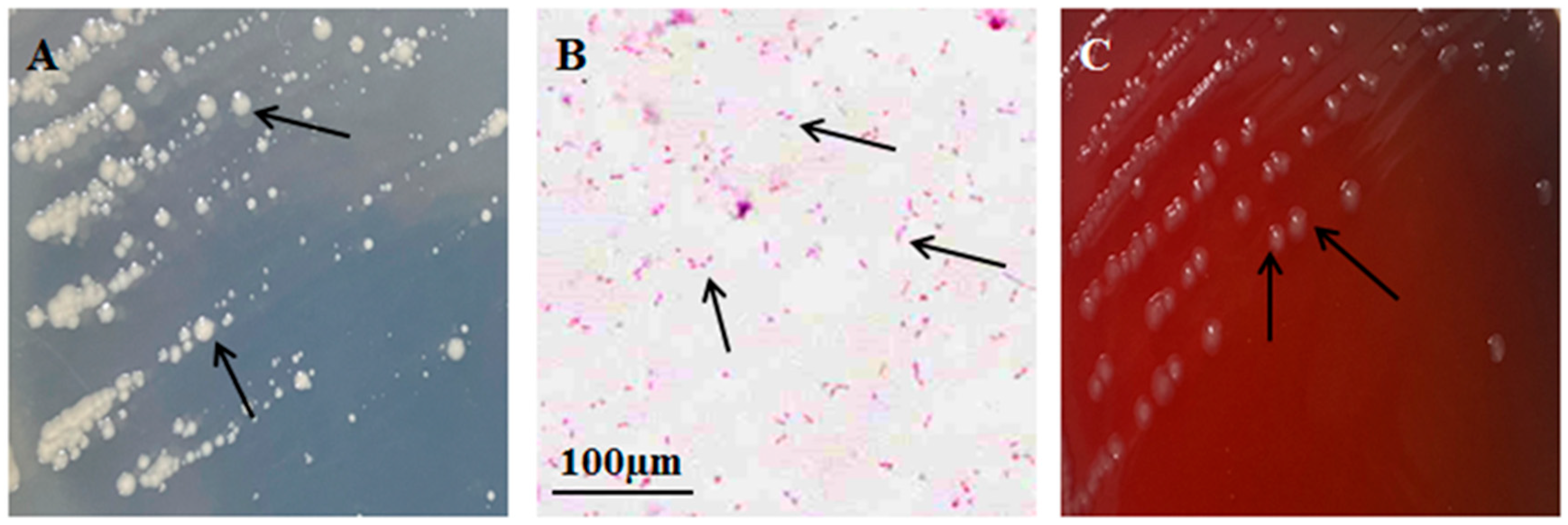
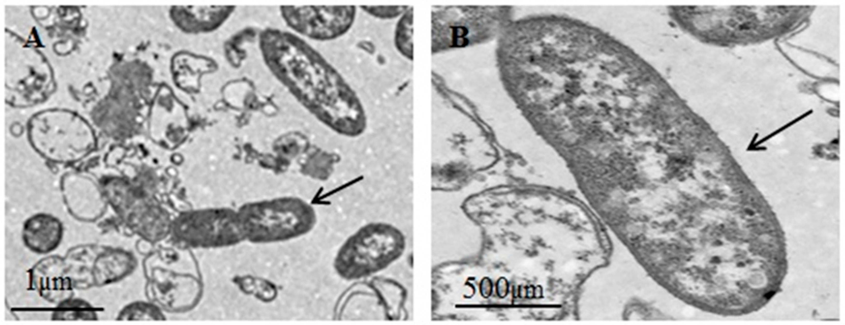
A bacterial strain, designated XH-1, was isolated from the internal organs of the diseased fish. On an ordinary agar medium, the isolated bacteria formed grayish-white, raised, circular colonies with smooth edges and a moist surface (Figure 2A). After Gram staining, XH-1 appeared as red spherical or rod-shaped cells under the microscope (Figure 2B). On a blood agar medium, the bacteria formed grayish-white, circular, convex colonies with smooth edges, appearing opaque and without a hemolytic ring, indicating the absence of hemolytic activity (Figure 2C). Under transmission electron microscopy, the bacteria exhibited an elliptical shape and lacked flagella (Figure 3).
The physiological and biochemical test results of the isolated bacteria are shown in Table 1. The isolated bacteria tested positive for oxidase, Voges–Proskauer (VP), indole, and methyl red tests. They were capable of fermenting glucose, mannitol, and arabinose but could not ferment inositol. They were unable to decompose salicin, esculin, or urea and did not produce hydrogen sulfide. The ornithine decarboxylase test was negative.

3.2. Cumulative Number of Deaths

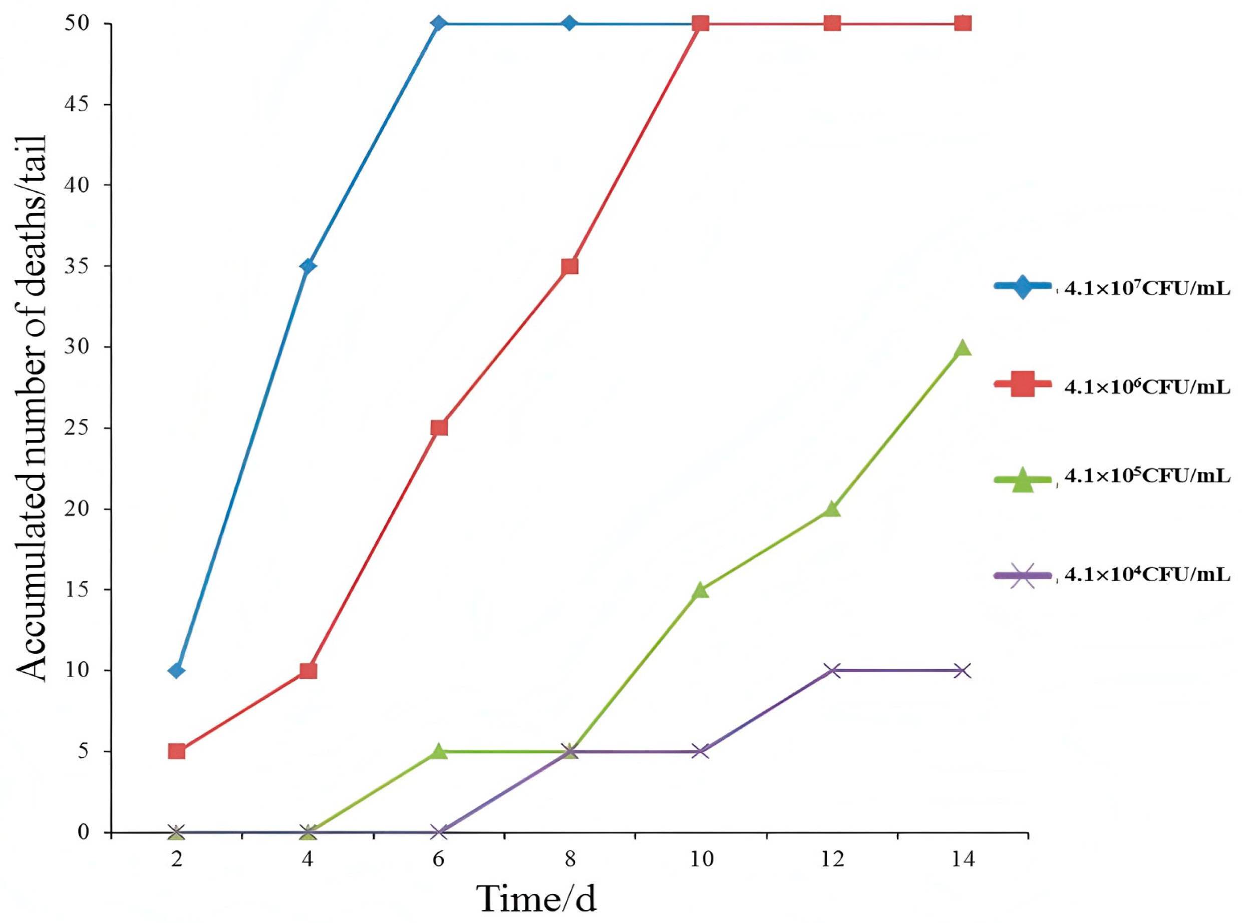
In the experimental group, the grass carp exhibited sluggish swimming 10 h after being inoculated with the isolated bacteria. On the second day of the experiment, mortality began to occur in the groups injected with bacterial concentrations of 4.1 × 10⁷ CFU/mL and 4.1 × 10⁶ CFU/mL. By the sixth day, all the grass carp in the group injected with 4.1 × 10⁷ CFU/mL had died. At the end of the experimental period, the mortality rates for experimental groups 1 to 4 were 100%, 100%, 60%, and 20%, respectively (Figure 4). The dead grass carp exhibited symptoms of diffuse skin hemorrhaging, a red and swollen anus, and the presence of numerous pustules and ulcers on the body surface. Dissection revealed intestines filled with yellowish ascites and a pale liver, consistent with the symptoms observed in naturally infected grass carp. A. veronii was isolated from the internal organs of the diseased fish. In contrast, no disease or mortality was observed in the control group.

3.3. 16S rRNA Sequence Analysis, Phylogenetic Tree Construction, and the Identification of the Isolated Strain

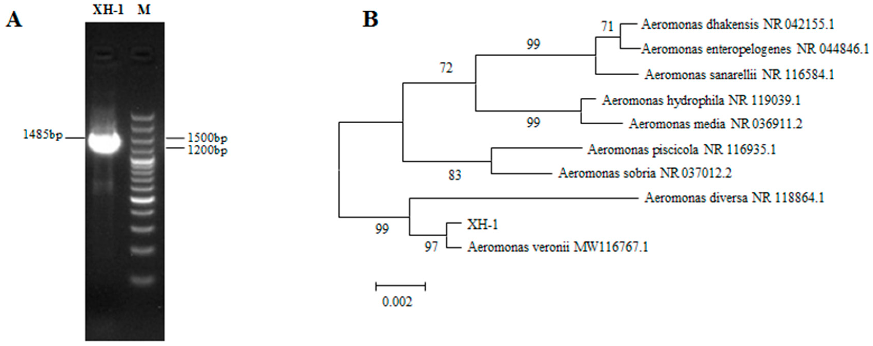
The PCR amplification product of the isolated strain was sequenced, yielding a fragment of 1485 bp (Figure 5A). The obtained 16S rDNA gene sequence of the isolated strain has been submitted to GenBank with the accession number SUB15430456 and was searched for homology in GenBank. The results showed that it naturally clustered with bacteria of the genus A. veronii, with a homology of 100% (Table 2). Partial 16S rDNA gene sequences of A. veronii were selected for phylogenetic analyses using the neighbor-joining method, and the phylogenetic tree is shown in Figure 5B. In the phylogenetic tree, the isolated strain XH-1 is clustered with A. veronii (MW116767.1) in a single branch, with a homology of 97%. Based on the morphological characteristics and the biological properties of the isolated strain XH-1, combined with the 16S rDNA sequence determination and the phylogenetic analysis results, it was concluded that the isolated strain XH-1 is A. veronii.

3.4. Sensitivity of XH-1 to 14 Chinese Herbal Medicines

The sensitivity of A. veronii XH-1 to 14 Chinese herbal medicines is shown in Table 3. The diameter of the inhibition zone was calculated as the average of three replicates. Among the 14 Chinese herbal medicines, the extracts of Lignum sappa, Pericarpium granna, Artemisia argyi, Scutellaria baicalensis Georgi, Coptis chinensis, and Artemisia capillaris exhibited inhibition zone diameters of greater than 15 mm, indicating high sensitivity [22]. The extracts of Spatholobus suberectus and Rehmannia glutinosa showed inhibition zone diameters between 10 and 15 mm, indicating moderate sensitivity. The extract of Glycyrrhiza uralensis Fisch had an inhibition zone diameter of less than 10 mm, indicating no sensitivity. No inhibition zones were observed for Andrographis paniculata, Dendranthema morifolium, Carthamus tinctorius, Cortex fraxini, and Isatis indigotica, indicating that these herbs had no effect on A. veronii.

3.5. Minimum Inhibitory Concentration (MIC) and Minimum Bactericidal Concentration (MBC)

The MIC and MBC of six highly sensitive Chinese herbal medicines against A. veronii are shown in Table 4. The MIC and MBC results of Lignum sappa extract and Pericarpium granna extract against A. veronii were identical, with an MIC of 7.813 mg/mL and an MBC of 15.625 mg/mL. In contrast, Coptis chinensis and Artemisia capillaris exhibited weaker bactericidal effects, both with both MIC and MBC values of 125.000 mg/mL and 250.000 mg/mL, respectively. Based on the results of the in vitro inhibition zone, MIC, and MBC tests, Lignum sappa extract was selected for further efficacy experiments.

3.6. Evaluation of the Efficacy of Chinese Herbal Medicines

The bactericidal effect of Lignum sappa extract on A. veronii increased with higher concentrations is shown in Table 5. When the concentration of Lignum sappa extract reached 18.00 mg/mL, the sterilization effect was 100%. At concentrations ranging from 14.00 to 18.00 mg/mL, the extract exhibited the strongest bactericidal effect within 8 h, and the effect persisted even after 32 h. At concentrations of 6.00 to 10.00 mg/mL, the extract showed bactericidal activity within 8 h; however, after 8 h, the bactericidal effect gradually weakened over time, allowing A. veronii to rapidly proliferate.

4. Discussion

Aeromonas spp. are primary pathogens of freshwater fish and are also believed to be secondary opportunistic pathogens of compromised or stressed hosts [23]. A. hydrophila, A. bestiarum, A. salmonicida, A. caviae, A. veronii, and A. jandaei all have been reported as pathogens of various fish species and cause several clinical signs, such as fin and tail rot, ulceration, exophthalmia, abdominal distention, ulceration, and bleeding [24,25,26,27,28,29]. A. veronii has the greatest range in virulence in the Aeromonads [30]. It contains two biovars, A. veronii biovar sobria and A. veronii biovar veronii, which have different biological characteristics with regard to esculin hydrolysis and ornithine decarboxylase [31]. The incidence of A. veronii from diseased fishes has been reported [27]. The symptoms of this disease are similar to Epizootic Ulcerative Syndrome (EUS). Although the specific role of Aeromonas in EUS is not yet clearly studied, numerous reports suggested that Aeromonas spp., mostly A. hydrophila and A. veronii, play a significant role in the infection process [25].
In this study, we isolated bacteria from the liver and intestine of farmed grass carp suffering from ulcers and identified A. veronii as the dominant bacteria by phenotypic characterization tests. Further, the 16S rRNA gene sequence homology of A. veronii with the reference strains was studied. The isolated bacteria XH-1 exhibited a 100% sequence homology with ten A. veronii reference strains. We also constructed a phylogenetic tree using the neighbor-joining method using the 16S rRNA gene sequence of the A. veronii strains. The isolates closely fit with A. veronii MW116767.1 within the same clade. The artificial infection challenge test was conducted to evaluate the pathogenic potential of the isolated bacteria, and it caused the same symptoms as the natural onset.
Antibiotic resistance is a great concern in the management of bacterial diseases [32]. Currently, the prevention and control of A. veronii infections primarily rely on the use of antibiotics, which not only pollute the aquatic environment but also contribute to the development of antibiotic resistance [33]. To address this issue, many researchers have conducted studies on Chinese herbal medicines. For instance, Mao Zhijuan investigated the antibacterial activity of five Chinese herbal extracts against Vibrio parahaemolyticus and found that Galla chinensis extract could inhibit the growth of Vibrio [34]. Jin Shan studied the sensitivity to 15 Chinese herbal medicines of Vibrio harveyi and discovered that Pericarpium granna, Sanguisorba officinalis, and Schisandra chinensis exhibited strong antibacterial effects, while Rheum palmatum, Prunus mume, Isatis indigotica, and Forsythia suspensa showed moderate effects [35].
Song Xuehong examined the in vitro antibacterial effects of nine common Chinese herbal medicines on Aeromonas and found that Euphorbia humifusa had the best antibacterial effect [36]. Yang Yibin studied the sensitivity to 41 Chinese herbal medicines of three sturgeon pathogens—Citrobacter freundii, Aeromonas hydrophila, and Yersinia ruckeri—and found that all three pathogens were highly sensitive to Prunus mume, Pericarpium granna, Sanguisorba officinalis, and Lycium barbarum [21]. These findings fully demonstrate that Chinese herbal medicines can be used to prevent and control bacterial infections in aquaculture, thereby curbing the spread and development of bacterial diseases.
In this study, the antibacterial effects of 14 Chinese herbal medicines on the grass carp pathogen XH-1 were investigated. It was found that Lignum sappa, Pericarpium granna, Artemisia argyi, Scutellaria baicalensis Georgi, Coptis chinensis, and Artemisia capillaris Thunb all exhibited antibacterial effects. Among them, Lignum sappa showed the best antibacterial effect, with the largest inhibition zone diameter. The MIC and MBC results were also consistent with the in vitro inhibition zone test results. Studies on Lignum sappa indicate that it has a pungent taste and neutral nature, primarily targeting the heart, liver, and spleen meridians. In traditional Chinese medicine, it is mainly used to treat blood stasis and pain-related conditions. Research has shown that Lignum sappa has various pharmacological effects, including blood sugar reduction; anti-complement activity; and anti-inflammatory, antioxidant, anti-tumor, and antibacterial properties [37,38,39]. However, its use as an antibacterial herbal medicine in aquaculture has not been reported. The findings of this study lay the foundation for the use of Chinese herbal medicines as alternatives to antibiotics for treating bacterial infections.

5. Conclusions

In the aquaculture industry, relying solely on antibiotics for the prevention and control of bacterial diseases has led to many adverse consequences, such as environmental pollution and the development of antibiotic resistance. Some drugs may also enter the human body through the consumption of fish, posing a threat to human health. In this study, we isolated pathogenic bacteria from diseased grass carp and studied the morphological and physiological biochemical characteristics. Through artificial infection, the symptoms of natural disease can be reproduced, and as the disease progresses, grass carp eventually die. We once again isolated A. veronii from the viscera of artificially infected grass carp, fulfilling Koch’s postulates. Through this investigation, we definitively determined the pathogen responsible for the grass carp disease outbreak and clarified its etiology. Our systematic study of the bacterial pathogen revealed important characteristics, yielding valuable scientific data that will significantly enhance future disease management approaches in grass carp farming.
Additionally, we conducted a series of experiments with Chinese herbal medicines. Based on the results, we found that the herb Lignum sappa exhibits antibacterial effects against A. veronii, the pathogenic bacterium affecting grass carp. Therefore, in future treatments, we can prioritize the use of Chinese herbal medicines as an alternative to antibiotics.
This discovery has transformed the current treatment paradigm for A. veronii infections in grass carp, which previously relied solely on antibiotic therapy. Moreover, it provides a novel therapeutic approach for managing bacterial diseases in grass carp aquaculture. In future aquaculture practices, antibiotics are not the only treatment option for A. veronii infections; Lignum sappa can also be used for prevention and control.

Author Contributions

Conceptualization, Y.Z.; Methodology, Y.Z. and H.J.; Validation, Y.Z. and H.J.; Investigation, H.X.; Resources, H.J.; Data Curation, G.L. and L.S.; Writing—Original Draft Preparation, Y.Z.; Writing—Review and Editing, Y.Z. and H.J.; Visualization, Y.Z. and L.S. All authors have read and agreed to the published version of the manuscript.

Funding

This work was supported by the earmarked fund for CARS-45 and the Jiangsu Agriculture Science and Technology Innovation Fund [CX (23)1007].

Institutional Review Board Statement

This study obtained the experimental animal specimens from the Jiangsu Institute of Freshwater Fisheries. The work was approved by the Ethics Committee (Animal Care and Use Committee of Jiangsu Ocean University, protocol: No. 2020-37, approval date 1 September 2019) for Reasoning and Using and operated by personnel during the experimental process. We strictly abided by the ethical norms of the Chinese Regulations on the Management of Experimental Animals and acted in accordance with the ethics committee of the Jiangsu Institute of Freshwater Fisheries with regard to the implementation of rules and regulations.

Informed Consent Statement

Not applicable.

Data Availability Statement

All the data generated or used during the study appear in the submitted article.

Conflicts of Interest

The authors declare no conflicts of interest.

References

  1. Zhang, M.L.; Lu, Z.J.; Zhan, F.F.; Li, Y.N.; Shi, F.; Zhao, L.J.; Lin, L.; Qin, Z.D. IgM expression and B lymphocyte antibacterial activity of grass carp (Ctenopharyngodon idella). J. Fish. China 2021, 45, 115–124. [Google Scholar] [CrossRef]
  2. Hong, Y.; Jiang, W.D.; Kuang, S.Y.; Hu, K.; Tang, L.; Liu, Y.; Jiang, J.; Zhang, Y.A.; Zhou, X.Q.; Feng, L. Growth, digestive and absorptive capacity and antioxidant status in intestine and hepatopancreas of subadult grass carp Ctenopharyngodon idellus fed graded levels of dietary threonine. J. Anim. Sci. Biotechnol. 2015, 6, 59–61. [Google Scholar] [CrossRef]
  3. Kong, W.G.; Huang, C.; Tang, Y.; Zhang, D.; Wu, Z.X.; Chen, X.X. Effect of Bacillus subtilis on Aeromonas hydrophila-induced intestinal mucosal barrier function damageand inflammation in grass carp (Ctenopharyngodon idella). Sci. Rep. 2017, 7, 1588. [Google Scholar] [CrossRef]
  4. El-Barbary, M.I. Serum Biochemical and Histopathological Changes Associated with Aeromonas hydrophila Isolated from Oreochromis niloticus and Sparus aurata with Multiple Antibiotic Resistance Index. J. Biol. Sci. 2017, 17, 222–234. [Google Scholar] [CrossRef]
  5. Lehane, L.; Rawlin, G. Topically acquired bacterial zoonoses from fish: A review. Med. J. Aust. 2000, 173, 256–259. [Google Scholar] [CrossRef] [PubMed]
  6. Ren, Y.; Gao, C.X.; Zeng, W.W.; Wang, Q.; Wang, Y.Y.; Li, Y.Y.; Yin, J.Y.; Chang, O.Q.; Shi, C.B. Pathogenicity and main phenotype characteristics of two Aeromonas veronii isolated from diseased grass carp. Chin. J. Prev. Vet. Med. 2019, 41, 145–150. Available online: http://yfsy.paperopen.com/#/digest?ArticleID=2897 (accessed on 8 July 2025).
  7. Qin, G.X.; Ai, X.H.; Xu, J.; Yang, Y.B. Dual RNA-seq of spleens extracted from channel catffsh infected with Aeromonas veronii reveals novel insights into host-pathogen interactions. Ecotoxicol. Environ. Saf. 2023, 252, 114609. [Google Scholar] [CrossRef]
  8. Monti, M.; Torri, A.; Amadori, E.; Rossi, A.; Bartolini, G.; Casadei, C.; Frassineti, G.L. Aeromonas veronii biovar veronii and sepsis-infrequent complication of biliary drainage placement: A case report. World J. Clin. Cases 2019, 7, 759–764. [Google Scholar] [CrossRef]
  9. Fernandez-Bravo, A.; Figueras, M.J. An update on the Genus Aermonas: Taxonomy, Epidemiology and pathogenicity. Microorganisms 2020, 8, 129. [Google Scholar] [CrossRef]
  10. Hatrongjit, R.; Kerdsin, A.; Takeuchi, D.; Wongsurawat, T.; Jenjaroenpun, P.; Chopjitt, P.; Boueroy, P.; Akeda, Y.; Hamada, S. Genomic Analysis of Aeromonas veronii C198, a Novel Mcr-3.41-Harboring isolate from a patient with septicemia in Thailand. Pathogens 2020, 9, 1031. [Google Scholar] [CrossRef]
  11. Li, T.; Raza, S.H.A.; Yang, B.; Sun, Y.; Wang, G.; Sun, W.; Qian, A.; Wang, C.; Kang, Y.; Shan, X. Aeromonas veronii infection in Commercial Freshwater Fish: A potential threat to Public Health. Animals 2020, 10, 608. [Google Scholar] [CrossRef] [PubMed]
  12. Tekedar, H.C.; Kumru, S.; Blom, J.; Perkins, A.D.; Griffin, M.J.; Abdelhamed, H.; Karsi, A.; Lawrence, M.L. Comparative genomics of Aeromonas veronii: Identification of a pathotype impacting aquaculture globally. PLoS ONE 2019, 14, e0221018. [Google Scholar] [CrossRef] [PubMed]
  13. Stanbaugh, C.L.; Abter, E.I.; Tompkins, A.L. Aeromonas veronii cellulitis, bacteremia, and sepsis in a patient with liver cirrhosis and end-stage renal disease following a minor abrasion with exposure to pond water: A case report and literature review. IDCases 2022, 30, e01631. [Google Scholar] [CrossRef]
  14. Dong, H.T.; Techatanakitarnan, C.; Jindakittikul, P.; Thaiprayoon, A.; Taengphu, S.; Charoensapsri, W.; Khunrae, P.; Rattanarojpong, T.; Senapin, S. Aeromonas jandaei and Aeromonas veronii caused disease and mortality in Nile tilapia, Oreochromis niloticus (L.). J. Fish Dis. 2017, 40, 1395–1403. [Google Scholar] [CrossRef]
  15. Wang, Y.E.; Xing, C.G.; Wang, G.L. Susceptibilities of Five Marine Pathogenic Vibrios to 34 Chinese Herbs. Fish. Sci. 2008, 27, 221–225. [Google Scholar] [CrossRef]
  16. Holt, J.G.; Kriey, N.R.; Sneath, P.H.A.; Staley, J.T. Bergey’s Manual of: Determinative Bacteriology, 9th ed.; Lippincott Williams and Wilkins: Baltimore, MA, USA, 1994. [Google Scholar]
  17. Krieg, N.R.; Holt, J.G. Bergey’s Manual of Systematic Bacteriology; Williams and Wilkins Baltimore: London, UK, 1984; Volume 1, pp. 545–548. [Google Scholar]
  18. Ke, W.J.; Qin, H.B.; Chen, Y.F.; Yang, Y.; Yu, H.; Guan, W.T.; Li, Z.Y.; Liu, F. Antibacterial effects of 25 kinds of Chinese herbal medicines on Proteus vulgaris in vitro. Fish. Sci. Technol. Inf. 2019, 46, 328–331. [Google Scholar] [CrossRef]
  19. Chen, X.; Zhang, Q.Z.; Li, C.T. Baeteriostasis of 50 traditional Chinese medicine herbs against Aeromonas hydrophila in vtro. Chin. J. Prev. Vet. Med. 2011, 33, 862–865. [Google Scholar] [CrossRef]
  20. Gao, X.H.; Xiao, Y.; Zhang, M.H.; An, W.; He, Z.K.; Shao, L.; Zhang, H.Q. In Vitro antibacterial effect of 13 Chinese herbal medicines on Pseudamonas fluoroscens. J. South. Agric. 2016, 47, 748–752. [Google Scholar] [CrossRef]
  21. Yang, Y.B.; Cao, H.P.; Xia, Y.T.; Yang, T.; Zhao, L.; Hu, K.; Yang, X.L. In Vitro inhibitory effect of 41 chinese herbs on three sturgeon-pathogenic bacteria. Freshw. Fish. 2013, 43, 81–84. [Google Scholar] [CrossRef]
  22. Wang, S. Handbook of Modern Diagnostics; Peking Union Medical College Press: Beijing, China, 1995. [Google Scholar]
  23. Jeney, Z.; Jeney, G. Recent achievements in studies on diseases of the common carp (Cyprinus carpio L.). Aquaculture 1995, 129, 397–420. [Google Scholar] [CrossRef]
  24. Kozinska, A.; Figueras, M.J.; Chacon, M.R.; Soler, L. Phenotypic characteristics and pathogenicity of Aeromonas genomospecies isolated from common carp (Cyprinus carpio L.). J. Appl. Microbiol. 2002, 93, 1034–1041. [Google Scholar] [CrossRef]
  25. Rahman, M.; Colque-Navarro, P.; Kühn, I.; Huys, G.; Swings, J.; Möllby, R. Identificationand characterization of pathogenic Aeromonas veronii biovar sobria associated with epizootic ulcerative syndrome in fish in Bangladesh. Appl. Environ. Microbiol. 2002, 68, 650–655. [Google Scholar] [CrossRef]
  26. Wahli, T.; Burr, S.E.; Pugovkin, D.; Mueller, O.; Frey, J. Aeromonas sobria, a causative agent of disease in farmed perch, Perca fluviatilis L. J. Fish Dis. 2005, 28, 141–150. [Google Scholar] [CrossRef] [PubMed]
  27. Hossain, M. Isolation of pathogenic bacteria from the skin ulcerous symptomatic gourami (Colisa lalia) through 16S rDNA analysis. Univ. J. Zool. 2008, 27, 21–24. [Google Scholar] [CrossRef]
  28. Martínez-Murcia, A.J.; Saavedra, M.J.; Mota, V.R.; Maier, T.; Stackebrandt, E.; Cousin, S. Aeromonas aquariorum sp. nov., isolated from the aquaria of ornamental fish. Int. J. Syst. Evol. Microbiol. 2008, 58, 1169–1175. [Google Scholar] [CrossRef]
  29. Sreedharan, K.; Philip, R.; Singh, I.S.B. Virulence potential and antibiotic susceptibility pattern of motile aeromonads associated with freshwater ornamental fish culture systems:a possible threat to public health. Braz. J. Microbiol. 2008, 43, 754–765. [Google Scholar] [CrossRef] [PubMed]
  30. Janda, J.M.; Kokka, R.P. The pathogenicity of Aeromonas strains relative to genospecies and pheospecies identification. FEMS Microbiol. Lett. 1991, 69, 29–33. [Google Scholar] [CrossRef]
  31. Altwegg, M.; Steigerwalt, A.G.; Altwegg-Bissig, R.; Luthy-Hottenstein, J.; Brenner, D.J. Biochemical identification of Aeromonas genospecies isolated from humans. J. Clin. Microbiol. 1990, 28, 258–264. [Google Scholar] [CrossRef]
  32. Zaki, M.M.; Eissa, A.E.; Saeid, S. Assessment of the Immune Status in Nile Tilapia (Oreochromis niloticus) Experimentally Challenged with Toxogenic/Septicemic Bacteria During Treatment Trial with Florfenicol and Enrofloxacin. Word J. Fish. Mar. Sci. 2011, 3, 21–36. [Google Scholar]
  33. Zhang, T.; Li, B. Antibiotic resistance in water environment: Frontiers of fundamental research, risk assessment and control strategies. Sci. Bull. 2020, 65, 2543–2554. [Google Scholar] [CrossRef]
  34. Mao, Z.J.; Zhuo, H.L. Studies on the Pathogen of the Bacteria Epidemic against Mud Crab Scylla serrata. Fish. Sci. 2001, 20, 8–11. [Google Scholar] [CrossRef]
  35. Jin, S.; Wang, G.L.; Zhao, Q.S.; Chen, H.Q. Medical study on provenion and cure for skin ulcer disease of marine cage-cultured sea-perch. J. Oceanogr. TaiWan Strait 2000, 19, 233–236. [Google Scholar] [CrossRef]
  36. Song, X.H.; Cai, C.F.; Ni, J.G. Extraction and Antibacterial Activity of Active Constituents from Nine Common Chinese Herbal Medicines. Reserv. Fish. 2001, 21, 38–40. [Google Scholar] [CrossRef]
  37. Settharaksa, S.; Monton, C.; Charoenchai, L. Optimization of Caesalpinia sappan L. heartwood extraction procedure to obtain the highest content of brazilin and greatest antibacterial activity. J. Integr. Med. 2019, 17, 351–358. [Google Scholar] [CrossRef]
  38. Choi, B.M.; Kim, B.R. Upregulation of heme oxygenase-1 by brazilin via the phosphatidylinositol 3-kinase/Akt and ERK pathways and its protective effect against oxidative injury. Eur. J. Pharmacol. 2008, 580, 12–18. [Google Scholar] [CrossRef]
  39. Batubara, I.; Mitsunaga, T.; Ohashi, H. Brazilin from Caesalpinia sappan wood as an antiacne agent. J. Wood Sci. 2010, 56, 77–81. [Google Scholar] [CrossRef]
Figure 1. Clinical signs observed in grass carp infected with A. veronii. Naturally diseased fish (AD). Artificially infected fish (E,F). The white circles and black arrows represent typical symptoms.
Bacteria 04 00034 g001
Figure 2. The morphology of XH-1 and hemolytic activity. Bacterial growth in LB medium (A). Gram staining (B). Bacterial growth in blood agar medium (C). The black arrows represent bacterial cells.
Bacteria 04 00034 g002
Figure 3. The morphology of XH-1 under transmission electron microscope. (A) Bacteria observed in mid-division; (B) bacterial cell body. The black arrows point to bacteria in the electron micrograph.
Bacteria 04 00034 g003
Figure 4. Artificial infection test results.
Bacteria 04 00034 g004
Figure 5. Agarose gel electrophoresis of 16S rRNA as species specific genes for PCR detection and phylogenetic construction of XH-1. (A) The agarose gel electrophoresis of 16S rRNA as species specific genes for PCR detection of XH-1; (B) The phylogenetic construction of XH-1. Lane M: DNA marker.
Bacteria 04 00034 g005
Table 1. The morphology and biochemical characteristics of XH-1.
ItemXH-1ItemXH-1
Gram stainEsculin hydrolysis
Oxidase+Hydrogen sulfide production
Voges–Proskauer+Arabinose fermentation+
Indole test+Inositol fermentation
Glucose fermentation+Ornithine decarboxylase
Mannitol fermentation+Urea
Salicin fermentationMethyl red+
Note: “+” means positive; “−” means negative.
Table 2. The 16S rRNA gene sequence homology of XH-1 with reference strains.
Homologous Strain16S rRNA Gene Homology (%)Gene Bank
Accession No.
A. veronii strain HD6454 chromosome, complete genome100CP079823.1
A. veronii strain 183026 chromosome, complete genome100CP072325.1
A. veronii strain AV040 chromosome, complete genome100CP095841.1
A. veronii strain HD6448 chromosome, complete genome100CP087266.1
A. veronii strain AEv1810 16S ribosomal RNA gene, partial sequence100ON063910.1
A. veronii strain SW3814 chromosome, complete genome100CP083461.2
A. veronii strain A29V chromosome, complete genome100CP080630.1
A. veronii strain Colony108 chromosome100CP070210.1
A. veronii strain Colony58 chromosome100CP070211.1
A. veronii strain Colony121 chromosome100CP070208.1
Table 3. Sensitivity of XH-1 to 14 Chinese herbal medicines.
Chinese Medicine NameInhibition Zone Diameter (mm)Sensitivity
Lignum sappa25.13 ± 0.24S
Pericarpium granna24.11 ± 0.18S
Artemisia argyi22.67 ± 0.12S
Scutellaria baicalensis Georgi19.64 ± 0.35S
Coptis chinensis17.33 ± 0.47S
Artemisia capillaris15.47 ± 0.32S
Spatholobus suberectus14.07 ± 0.34I
Rehmannia glutinosa12.53 ± 0.61I
Glycyrrhiza uralensis Fisch9.63 ± 0.16R
Andrographis paniculata0/
Dendranthema morifolium0/
Carthamus tinctorius0/
Cortex fraxini0/
Isatis indigotica0/
Note: S: highly susceptible (d > 15.00 mm); I: intermediately susceptible (15.00 mm ≥ d ≥ 10.00 mm); R: insusceptible (d < 10.00 mm).
Table 4. MIC and MRC measurement results (mg/mL).
Chinese Medicine NameMICMRC
Lignum sappa7.81315.625
Pericarpium granna7.81315.625
Artemisia argyi15.62531.250
Scutellaria baicalensis Georgi62.500125.000
Coptis chinensis125.000250.000
Artemisia capillaris125.000250.000
Table 5. Effect of time and concentration on drug efficacy.
Concentration of Drug (mg/mL)Primary Number of Bacteria (cfu/mL)Final Number of Bacteria (cfu/mL)
2 h8 h16 h32 h
6.002.00 × 1051.44 × 1051.68 × 1053.75 × 1056.79 × 105
10.005.66 × 1032.42 × 1047.14 × 1042.84 × 105
14.00007.80 × 1033.27 × 104
18.000000
Disclaimer/Publisher’s Note: The statements, opinions and data contained in all publications are solely those of the individual author(s) and contributor(s) and not of MDPI and/or the editor(s). MDPI and/or the editor(s) disclaim responsibility for any injury to people or property resulting from any ideas, methods, instructions or products referred to in the content.