Review

Deciphering the Structural and Functional Paradigms of Clostridioides difficile Toxins TcdA and TcdB

1
Division of Neurotherapeutics, Smt. Kishoritai Bhoyar College of Pharmacy, Kamptee, Nagpur 441002, MH, India
2
Department of Pharmaceutical Sciences, Rashtrasant Tukdoji Maharaj Nagpur University, Nagpur 440033, MH, India
*
Authors to whom correspondence should be addressed.
Bacteria 2025, 4(2), 21; https://doi.org/10.3390/bacteria4020021
Submission received: 19 February 2025 / Revised: 13 March 2025 / Accepted: 31 March 2025 / Published: 3 April 2025

Abstract

:
Clostridioides difficile Infection (CDI) continues to be a major cause of antibiotic-associated diarrhea and pseudomembranous colitis, fueled in large measure by virulence factors TcdA and TcdB. These giant glucosyltransferase toxins interfere with host cytoskeletal integrity and inflammatory signaling by inhibiting Rho GTPase; however, the detailed structural dynamics, receptor selectivity, and subcellular trafficking mechanisms remain in part unspecified. This review integrates recent insights from cryo-electron microscopy (cryo-EM) and X-ray crystallography to describe the quaternary architecture of TcdA/B, emphasizing conformational changes key to pore formation and endosomal escape. We also examine the genomic heterogeneity of hypervirulent C. difficile strains (e.g., ribotype 027), correlating toxin gene polymorphisms (e.g., tcdC mutations) with increased toxin production and virulence. Mechanistic explanations of toxin-driven inflammasome activation and epithelial barrier dysfunction are situated within host immune evasion mechanisms, including microbiota-derived bile acid regulation of toxin stability. Subsequent innovative therapeutic strategies, encompassing the utilization of engineered neutralizing antibodies that specifically target the autoprocessing domain alongside structure-guided small-molecule inhibitors, are subjected to a rigorous evaluation. By integrating structural biology, systems-level omics, and clinical epidemiology, this review establishes a comprehensive framework for understanding C. difficile toxin pathogenesis and guiding next-generation precision antimicrobials.

1. Introduction

In the United States alone, CDI infects nearly half a million people annually and results in approximately 29,000 deaths annually. Its most important determinants of virulence for CDI pathogenesis are two large exotoxins: toxin A (TcdA) and toxin B (TcdB). Toxins inhibit host cell functions, resulting in inflammation, cellular damage, and the hallmark of CDI syndrome [1].
The disturbance of gut microbiota by antibiotic exposure and proton pump inhibitors (PPIs) is a major contributor to the pathogenesis of Clostridium difficile infection (CDI). Antibiotics, especially clindamycin, fluoroquinolones, and cephalosporins, cause post-antibiotic dysbiosis, which is defined as a reduction in microbial diversity, loss of protective taxa, such as Bacteroidetes and Firmicutes, and overgrowth of opportunistic pathogens, such as Enterobacteriaceae. This perturbation impairs colonization resistance, providing an optimal setting for C. difficile growth. Similarly, PPIs are responsible for microbiota dysbiosis by elevating gastric pH, promoting survival of C. difficile spores, and causing microbial predominance toward Firmicutes over Bacteroidetes, a community known to be permissive to CDI. Leffler and Lamont et al. indicated that long-term PPI use leads to reduced microbial diversity and an overabundance of oral bacteria in the gut, further making individuals susceptible to infection. Due to the strong association between microbiota imbalance and CDI, therapeutic interventions, such as fecal microbiota transplantation (FMT) and probiotics, have been investigated to restore microbial balance and decrease CDI recurrence. Thus, reducing unnecessary antibiotic and PPI consumption is vital in preventing CDI as well as maintaining gut microbial homeostasis [2,3]. Structurally, TcdA and TcdB are composed of several domains, such as an N-terminal glucosyltransferase domain (GTD), a cysteine protease domain (CPD), a delivery or translocation domain, and a C-terminal combined repetitive oligopeptides (CROPs) domain [4]. The GTD is central to the inactivation of host Rho-family GTPases through a process of glucosylation, which brings about disruption of actin cytoskeleton dynamics and, therefore, undermines cell integrity. When they bind to host receptors, TcdA and TcdB are internalized by endocytosis and translocate their enzymatic domains into the cytosol, where they alter Rho GTPases, leading to the disassembly of actin filaments, disruption of tight junctions, and enhanced epithelial permeability. These effects initiate an inflammatory cascade with the release of cytokines and immune cell recruitment, which exacerbates tissue damage and contributes to the characteristic symptoms of CDI, such as diarrhea and pseudomembranous colitis. The widespread destruction of epithelium and immune response eventually result in complications, such as toxic megacolon and colonic perforation, in extreme cases, as shown in (Figure 1) [5]. The activation of inositol hexakisphosphate (InsP6) enables the CPD to instigate the release of the GTD into the host cytosol [6]. The delivery domain facilitates translocation across endosomal membranes of enzymatic components, including the CROPs domain involved in receptor binding and specificity. High-resolution data on these domains have more recently been obtained through structural characterization. For instance, a cryo-electron microscopy study of native TcdA has been resolved to a resolution of 2.8 Å, detailing the dynamic motion of the CROPs domain upon acidification of the environment, required for optimal receptor binding and subsequent endocytosis [7]. Furthermore, the structural and functional paradigms of Clostridioides difficile toxins A (TcdA) and B (TcdB) have been elucidated by X-ray crystallography, electron microscopy (EM), and biochemical assays. Both toxins are AB-type toxins comprising an N-terminal glucosyltransferase domain (GTD) inhibiting host Rho-family GTPases and a C-terminal receptor-binding domain composed of combined repetitive oligopeptides (CROPs) for host cell binding. Structural analyses show that TcdA and TcdB are receptor-mediated endocytosed, followed by pH-dependent pore formation to facilitate GTD translocation to the cytosol. Inositol hexakisphosphate (InsP6) activation triggers autoproteolysis, releasing the GTD to induce disruption of cytoskeletal integrity and epithelial barriers. Cryo-EM and SAXS studies show modular toxin conformations with extensive structural rearrangements to facilitate membrane insertion. These structural and functional paradigms of toxins are of utmost significance in the development of targeted antitoxin therapies [8]. These observations form the basis for the rational design of antitoxin modalities, such as neutralizing antibodies and small-molecule inhibitors, that inhibit the suppression of the pathogenic activities of TcdA and TcdB in CDI.

1.1. CTD Toxin

CTD belongs to the family of bipartite ADP-ribosylating clostridial toxins having two separate toxins components: CDTa (ezymatic ADP-ribosyltransferase), which modifies actin, and CDTb, which binds to host cells and translocates CDTa into the cytosol. Around 17 to 23% of C. difficile’s strains are used to produce the toxins. CTD has two unit of glucosylating toxins, toxin A (TcdA) and toxin B (TcdB), which are the main toxins along with a third binary toxin [9]. TcdA, an enterotoxin, triggers intestinal inflammation and elicits fluid secretion by compromising the integrity of the intestinal epithelial barrier and recruiting neutrophils, which enhances the inflammatory response. Toxin B (TcdB), a cytotoxin, causes cellular damage and initiates apoptosis by disrupting the cytoskeletal architecture of host cells, leading to cellular death and widespread damage to the colonic mucosa. In addition, C. difficile transferase (CDT), also known as a binary toxin, disrupts the actin cytoskeleton, thus enhancing the pathogenic activity of hypervirulent strains by promoting adherence to the intestinal epithelium and aiding in tissue invasion [10].

1.2. Regulation of Toxin Gene Loci (tcdA/tcdB)

C. difficile pathogenicity locus (PaLoc) controls its primary virulence factors, TcdA and TcdB, tightly. TcdR, a transcription activator, regulates their production and TcdC, a transcription repressor that inhibits RNA polymerase activity [11]. In addition, environmental factors, such as nutrient availability and temperature, also regulate toxin production, with carbon sources, such as glucose, being reported to repress tcdA/tcdB transcription [12].
More recent research has revealed a bimodal pattern of toxin gene expression in bacterial populations. Single-cell measurements with fluorescent reporters have revealed that tcdA transcription separates into two distinct subpopulations—ON and OFF—a phenomenon reported in many ribotypes [13]. Bimodality arises from a bistable regulatory process and not phase variation, where the autoregulatory positive feedback loop of TcdR provides a key switch. Random fluctuations in TcdR number dictate the selection of individual cells to become involved in toxin production. Disruption of this feedback loop by constitutive tcdR expression effectively eliminates bimodality, again highlighting the key role of TcdR in the emergence of bistable dynamics in gene expression [14].
Aside from TcdR and TcdC, global regulators, such as CodY and σD, also play a role in toxin production. CodY, a nutrient-level-responsive repressor, is efficient in toxin inhibition under abundant conditions but increases the ON/OFF ratio and overall toxin production when it is not active [15]. On the other hand, σD, associated with flagellar phase variation, induces tcdR transcription but not bistability. Even in strains with permanently fixed flgB orientations (ON or OFF), bimodal tcdA expression persisted but with fluctuations in the ON/OFF ratios, which indicates that σD is a modulator but not a strict regulator of bimodal expression [16,17].
In contrast to past models that held that toxin formation and sporulation were negatively related, current work has established that the two procedures can be run in parallel. Microscopic visualization has confirmed that mother cells harboring spores can form tcdA at the same time, thus abolishing the notion that toxin synthesis and sporulation are temporally separated [13,18]. The discovery suggests that C. difficile can pursue virulence and survival strategies simultaneously and, thus, optimize the survival potential while ensuring transmission using the formation of spores. The toxin gene expression regulatory pliability thus mimics the organism’s ability to adapt, meaning that various subpopulations pursue either host association or survival within environmental niches [19].
Bistable regulation of tcdA/tcdB is an important adaptive process enabling C. difficile to modulate toxin production according to changing environmental conditions. By integrating global regulatory signals, metabolic signals, and phase variation determinants, C. difficile can achieve a balance between persistence and pathogenicity [20]. Further exploration of how host immune defense systems and interspecies interactions influence bimodal toxin expression could yield insights into novel therapeutic approaches against this bistable switch. The utilization of single-cell analytical tools to probe infection biology will certainly be of ultimate importance in demystifying the stochastic and regulative aspects of C. difficile pathogenesis [21].

1.3. Evolution of Hypervirulent Strains

The development of hypervirulent strains of C. difficile, especially ribotype 027, has been driven by genetic mutation and recombination events within the PaLoc, a chromosomal locus encoding for the production of the homologous toxins TcdA and TcdB, which are essential for virulence. The toxins are highly sequence-diverse, making it difficult to devise diagnostics, therapeutics, and vaccines. Mansfield et al. (2020) [22] conducted a phylogenomic analysis of 8839 C. difficile isolates and characterized 7 TcdA subtypes (A1–A7) and 12 TcdB subtypes (B1–B12), thus providing a new toxin-based subtyping paradigm. Although the diversity of TcdA is mostly due to repeat expansions within its C-terminal repetitive region, the diversification of TcdB is marked by huge homologous recombination in its sequence, reflecting divergent evolutionary pressures [22].
Clinical validation of the subtyping system in 351 isolates at Brigham and Women’s Hospital showed that it could be used for stratifying TcdB to functional and antigenic groups. Subtypes are equivalent to large phenotypic differences, such as cell-rounding ability, receptor specificity (e.g., recognition by frizzled protein), and susceptibility to bezlotoxumab, the sole FDA-approved anti-TcdB monoclonal antibody [22].
Adding to these findings, Shen et al. (2020) [23] analyzed 3269 genomes, expanding TcdB subtyping to 12 varieties (B1–B8) on >5.03% amino acid divergence. Hypervirulent subtypes B1, B2, and B3 were most prevalent in clinical isolates, with enhanced toxicity in murine models, which could be accountable for their association with severe human disease. Notably, TcdB subtypes had geographical variation: B1 and B2 were most prevalent in North America, while B3 was prevalent in East Asia [23]. Subtype-specific functional variations were noted, e.g., reduced RhoA glucosylation in B3/B4 and reduced frizzled receptor binding in B2, most likely due to recombination-driven changes in key domains. Furthermore, ermB-positive ribotype 027 strains, reported by Aptekorz et al. (2017) [24], were multidrug-resistant (e.g., to erythromycin, clindamycin, and fluoroquinolones) and linked to elevated fecal lactoferrin levels—a biomarker of intestinal inflammation—correlating with more severe disease outcomes [24].
Phylogenetic discordance between C. difficile and TcdB subtype distribution reflects repeated horizontal gene transfer or recombination in PaLoc [25]. Evolutionary analyses also detected stronger positive selection in TcdB compared with TcdA, owing to host–pathogen arms races, with purifying selection preserving functional domains. These findings highlight the importance of combined subtyping and genomic surveillance to counter the rising threat of hypervirulent, multidrug-resistant strains and inform the design of next-generation therapeutics [26].

1.4. Horizontal Gene Transfer and Recombination Events

Horizontal gene transfer (HGT) and recombination events play a crucial role in defining C. difficile’s genetic diversity and virulence, particularly through PaLoc dissemination with the toxin genes tcdA and tcdB [27]. In a landmark report, Brouwer et al. demonstrated that C. difficile strain 630Δerm’s PaLoc can be transferred to non-toxigenic strains by a conjugation-like mechanism, converting the recipients into toxin producers. The observation has monumental clinical implications, since non-toxigenic strains are being considered for therapeutic targeting in C. difficile-associated diarrhea (CDAD) [28]. The coexistence of toxigenic and non-toxigenic strains in clinical settings offers opportunities for PaLoc spread, potentially undermining treatment effectiveness. Frighteningly, PaLoc transfer is followed by the co-transfer of conjugative transposons (CTns) with antibiotic resistance, as seen in transconjugants PaLoc386 and PaLoc37, where Tn5397 (tetracycline resistance) and PaLoc were co-transferred [29]. While CTns such as CTn1 and CTn5 typically excise and transfer autonomously, PaLoc itself lacks canonical mobile elements but instead transfers on variable-sized DNA blocks. This process is analogous to high-frequency recombination (Hfr)-mediated transfer, where integrated chromosomal oriT sites promote chromosomal DNA mobilization. In C. difficile, CTns in the vicinity of the PaLoc (e.g., CTn1, CTn2, and Tn5397) can act as facilitators of Hfr-like transfer, with homologous recombination integrating donor DNA into recipient genomes. Whole-genome analyses confirm that large DNA block transfers have been the driving force for C. difficile genome evolution in the past, such as in S-layer cassette recombination, to suggest broader HGT-mediated adaptability [30].
More support for toxin diversification through HGT is found in Monot et al. (2015) [31], who isolated three clinical isolates (RA09-070, SA10-050, and CD10-165) with unusual toxin profiles. RA09-070 was TcdA positive and TcdB negative, and SA10-050 and CD10-165 were TcdA negative but TcdB positive. Immunoassays and PCR assays failed to detect these strains as they had considerable sequence divergence for tcdA and tcdB. Genome sequencing showed non-conventional PaLoc arrangements: RA09-070 contained just tcdR and tcdA, bookended by a prophage-borne bhlA/uvib gene product with a holin-like fold, whereas SA10-050 and CD10-165 had tcdR, tcdB, and tcdE followed by a phage endolysin gene (cwlH). Remarkably, the PaLoc in these isolates was not inserted at non-canonical loci, ending the dogma of a conserved PaLoc insertion site. For example, RA09-070′s PaLoc was found in a 51-bp intergenic location between spoVAE and CD630_07760, while SA10-050 and CD10-165 showed PaLoc-CdtLoc collinearity, a novel genomic organization. These results infer that HGT allows C. difficile to acquire and relocate toxin modules, constructing hybrid PaLoc structures through the recombination of ancestral “mono-toxin” loci [31].
The evolutionary origin of the PaLoc is also complemented by phage-derived regulatory motifs. Mehner-Breitfeld et al. (2018) [32] detected TcdE isoforms (TcdE-M25/M27) with lambda phage holin-like folds, corroborating toxin secretion via non-lytic or lytic routes. The N-terminal extension of non-lytic isoform TcdE-M1 also inhibits premature lysis, modulating toxin release according to physiological conditions. PaLoc-encoded vestigial phage endolysin tcdL, a pseudogene, is also likely to interact with TcdB, suggesting a hijacked phage machinery for toxin export. Such strategies highlight how C. difficile exploits mobile genetic elements to control virulence, synchronizing toxin secretion with bacterial survival [32].

2. Structural Biology of TcdA/B: Cryo-EM-Derived Conformational Dynamics, Receptor-Binding Domains, and Pore-Forming Motifs

2.1. Cryo-EM-Derived Conformational Dynamics of TcdA and TcdB

Cryo-EM has revolutionized the structural description of the big clostridial toxins to enable near-atomic resolution models to capture the dynamic nature of TcdA and TcdB in different functional states [33].

Structural Organization and Domain Architecture

TcdA and TcdB share a modular domain structure composed of an N-terminal glucosyltransferase domain (GTD), an autoprocessing domain (APD), a delivery and receptor-binding domain (DRBD), and a C-terminal combined repetitive oligopeptide (CROP) domain. The high-resolution (2.8 Å) cryo-EM structure of native TcdA provided the first detailed insights into how these domains interact in a neutral pH environment [34].The study revealed that the CROP domain is highly flexible, undergoing significant positional shifts upon environmental acidification, which likely facilitates its detachment prior to membrane insertion.
Similarly, the structure of the full-length TcdB complex with chondroitin sulfate proteoglycan 4 (CSPG4) at neutral pH demonstrated a more compact conformation with extensive inter-domain contacts, particularly between the APD and DRBD [35]. CSPG4 is a critical receptor for C. difficile toxin B (TcdB), which binds to a composite site comprising the CPD, DRBD, hinge, and CROPs domains. Its junctional cell surface localization in epithelial cells, particularly in the colon, is critical for binding TcdB and subsequent cell entry and intoxication. This arrangement ensures structural stability before receptor engagement and subsequent pH-driven conformational shifts [36].

2.2. pH-Induced Conformational Changes and Receptor Dissociation

Endosomal acidification is of primary significance in triggering the structural rearrangements needed to activate the C. difficile toxins TcdA and TcdB. Cryo-EM analysis revealed that CSPG4 leaves TcdB at acidic pH by disrupting salt bridges and hydrophobic interactions, yet frizzled protein binding is retained in part [35]. These findings indicate pH-dependent conformational changes that regulate receptor interactions and, thus, toxin function.
Further studies on the structural stability of TcdB2 highlight the importance of amino acids 1769–1787 for receptor-binding activities. A deletion mutant of this region resulted in spontaneous autoprocessing with substantial loss of cell binding but with retention of enzymatic activity. Of note, even with the maintained enzymatic activity, the deletion resulted in a greater than 10,000-fold loss of cytotoxicity. The mutant protein continued to possess IP6-induced autoprocessing to an equivalent degree as full-length TcdB2. Structure analysis suggests that the region of amino acids 1769–1787 might be a stabilizing factor, influencing the structural conformation of the toxin and interaction with receptor-binding domains [37].
Autoprocessing in TcdB is initiated upon translocation into the cytosol of host cells, where IP6 binding induces conformational change allowing autoprocessing of intramolecular substrate by the active protease domain (APD) [38]. While mechanistic details are yet to be disclosed, certain hints indicate that the carboxy-terminal region of TcdB is involved in autoprocessing efficiency. Intriguing results were observed when the CROP domain of TcdA was replaced with that of TcdB, thus conferring increased resistance to IP6-induced autoprocessing [39]. Single-copy deletion mutants of the initial 1056 amino acids of TcdA were found to have increased autoprocessing relative to the full-length toxin. These findings indicate that certain structural elements are involved in controlling the availability of the protease domain in such a way that premature activation is prevented [37].
Three-dimensional structural analysis of TcdB1 and TcdA shows that deletion of the 1769–1787 region could lead to structural instability. The region in both toxins is positioned at the edge of a beta-sandwich fold converting into an alpha-helical turn, which would rationally connect it to the beta-solenoid CROP domain [40]. The glucosyltransferase domain (GTD) and APD positioning close to the region suggests that its deletion would disrupt their conformational assembly, ultimately affecting autoprocessing. In keeping with this, studies have shown that a synthetic peptide of the 1769–1787 region interacts with the CROP domain, again demonstrating that the region is associated with structural integrity and functional stability [41].
The deletion mutant TcdB2Δ1769–1787 has an impressive inability to bind to its known cellular receptors, CSPG4, PVRL3, and frizzled proteins [42]. However, the region 1769–1787 is not directly implicated in receptor interaction, and, therefore, the deletion can alter proximal structural elements necessary for receptor binding. Structural studies indicated that TcdB binds to frizzled proteins via a tricomponent complex of a palmitoleic acid lipid and the FZD-binding domain (TcdB-FBD), which is outside the deleted region. Similarly, CSPG4-binding sites in TcdB were found in the amino acids 1831–2366 region, which is carboxy-terminal to the deleted region. Therefore, it is likely that the internal deletion disrupts the structural conformation of proximal binding sites, preventing efficient receptor binding [43].
Interestingly, the TcdB2 Δ1769–1787 mutant elicits a strong neutralizing antibody response in mice, with potential application as a vaccine. Earlier studies demonstrated that the 1753–1852 region of TcdB2 blocks exposure of neutralizing epitopes in the proximal CROP domain. In contrast, that was not observed in TcdB1, demonstrating toxin variant functional divergence. The failure of the deletion mutant to engage with cells can enable improved antigen presentation by professional antigen-presenting cells, activating the immune response for prolonged durations [44]. Further, the structural stability of defined functional domains in the mutant offers a broad range of accessible epitopes, enhancing its immunogenicity. Importantly, TcdB2Δ1769–1787 lacks detectable cytotoxicity, even after administration at high doses in murine models. These findings suggest that some sequence elements function as structural locks, preventing premature activation and offering potential strategies for antitoxin therapeutic design [37].

2.3. Receptor-Binding Domains (RBDs) and Ligand Interactions

TcdA and TcdB exert their cytopathic effects through receptor-mediated endocytosis, relying on their receptor-binding domains (RBDs) for host cell recognition and internalization. Cryo-EM and biochemical studies have revealed that while TcdA primarily engages carbohydrate moieties, TcdB exhibits a broader specificity, interacting with multiple proteinaceous receptors, such as CSPG4, PVRL3, and frizzled proteins. These receptor interactions play a crucial role in determining toxin tropism, host cell susceptibility, and disease severity [45].

2.3.1. Ligand Specificity and Differential Receptor Utilization

TcdB is extremely basic in its receptor usage, binding multiple host proteins to enter cells. Cryo-EM structures of the TcdB with CSPG4 domain 1 (D1401–560) confirmed that the receptor engages the autoprocessing and delivery domains of TcdB specifically through the synergy of electrostatic and hydrophobic interactions. Follow-up studies have shown that CSPG4 is also a shared receptor for TcdB1 and TcdB2, the two largest isoform families responsible for more than 70% of clinical isolates. Notably, CSPG4 is the sole receptor for TcdB2, which cannot bind frizzled proteins. The finding underscores the importance of CSPG4 in TcdB pathogenesis and the potential of TcdB-mediated damage to CSPG4-expressing myofibroblasts as a contributor to disease progression.
In addition to CSPG4, frizzled-class receptors (FZD1–FZD7) are also important binding substrates of TcdB [46]. Cryo-EM structures of TcdB with the cysteine-rich domain of frizzled-2 (CRD2) showed a distinctive receptor interaction mode, where FZD2 binds to the toxin at a distinct binding site from CSPG4 [35]. FZD2 binding was partially inhibited by acidic pH, in contrast to CSPG4, suggesting differential receptor usage can modulate endosomal trafficking and cytotoxicity [47].
Mutational analysis also provided a demonstration of the importance of specific residues in receptor binding. Receptor affinity was decreased by 85–90% through alanine substitutions for required binding residues (R1475A, D1506A, W1510A), effectively abolishing CSPG4-dependent toxin internalization [48]. Furthermore, several structural modules of TcdB are involved in CSPG4 binding, including the CPD, DRBD, hinge, and CROP domains. A multi-unit binding interface like this demonstrates the sophistication of receptor binding and defines areas that are amenable to therapeutic targeting [49].

2.3.2. Role of Receptor-Binding Variants in Hypervirulence

Hypervirulent C. difficile strains also often harbor genetic polymorphisms in the TcdB receptor-binding domains (RBDs), which leads to enhanced receptor binding and altered host tropism. Genome-wide screening of 3269 C. difficile strains identified four new TcdB variants that harbored distinct mutations in the CSPG4 and frizzled-binding domains [23]. Functional assays revealed that hypervirulent ribotype 027 strains that harbored the TcdB2 variant exhibited a 2.4-fold enhancement in the binding affinity for CSPG4, which correlated with enhanced cytotoxicity against human colonic epithelial cells [50]. Bezlotoxumab, a TcdB monoclonal antibody, blocks CSPG4 binding allosterically and therefore offers a therapeutic window for the blockade of toxin-induced damage. Sequence differences in the bezlotoxumab-binding epitopes have, however, led to decreased efficacy against certain TcdB isoforms, and, therefore, alternative therapeutic strategies are required [51].
One such promising strategy is the design of decoy receptors that are mimics of the natural ligands of TcdB. Experiments have shown that recombinant Repeat1-Fc can inhibit TcdB toxicity in cell-based assays and protect mice from lethal toxin exposure. Since CSPG4 and FZD receptors are independent in the pathogenesis induced by TcdB, a bispecific decoy receptor with both Repeat1 and the CRD of FZD proteins could provide broad protection against various TcdB variants. The evolutionary conservation of CSPG4 as a TcdB receptor indicates that such decoy molecules would be less prone to escape mutations, thereby providing a sound therapeutic strategy to treat C. difficile infections [52].
Tissue Factor Pathway Inhibitor (TFPI) was found to be a key colonic crypt receptor of C. difficile Toxin B (TcdB) of hypervirulent clade 2 strains. TFPI was found to be a major host factor allowing TcdB4 entry into intestinal epithelial cells in a CRISPR-Cas9 screen. Cryo-EM structural analysis found that TcdB4 specifically binds to the Kunitz-2 (K2) domain of TFPI, similar to receptor–ligand binding with other large clostridial toxins. TFPI is strongly expressed in colonic glandular epithelium and, thus, was a prime candidate for TcdB-induced cytopathic effects. TFPI blockade greatly diminished TcdB-induced colonic tissue injury and should be an intriguing therapeutic target to treat hypervirulent C. difficile infection. TcdB is the main virulence factor of C. difficile, causing epithelial damage and disease. The study segregated TcdB variants into an FZD-binding group (TcdB1, TcdB3, TcdB5) and a TFPI-binding group (TcdB2, TcdB4, TcdB6, TcdB7). Surprisingly, hypervirulent variants TcdB2 and TcdB4 bind TFPI but not FZD, explaining their increased pathogenicity. The study identifies TFPI as a main mediator of C. difficile toxicity and therapeutic target candidate [53].
Together, these findings provide a mechanistic and structural rationale for the design of next-generation therapeutics against the receptor-binding domains of TcdB. Unraveling the interplay between receptor specificity, toxin evolution, and host susceptibility will be pivotal in driving precision medicine approaches in the treatment of C. difficile infections.

2.4. Pore-Forming Motifs and Membrane Insertion Mechanisms

The cytotoxic effects of TcdA and TcdB are contingent upon their ability to translocate the glucosyltransferase domain (GTD) across the endosomal membrane into the host cytosol. Translocation is facilitated by pH-dependent conformational transitions leading to the formation of a transmembrane pore. Recent cryo-electron microscopy experiments have shown the structural intricacies involved in such pore-forming motifs and membrane insertion and their involvement, emphasizing the complexity of interactions between domain rearrangement, pH-induced structural instability, and host lipids [52].

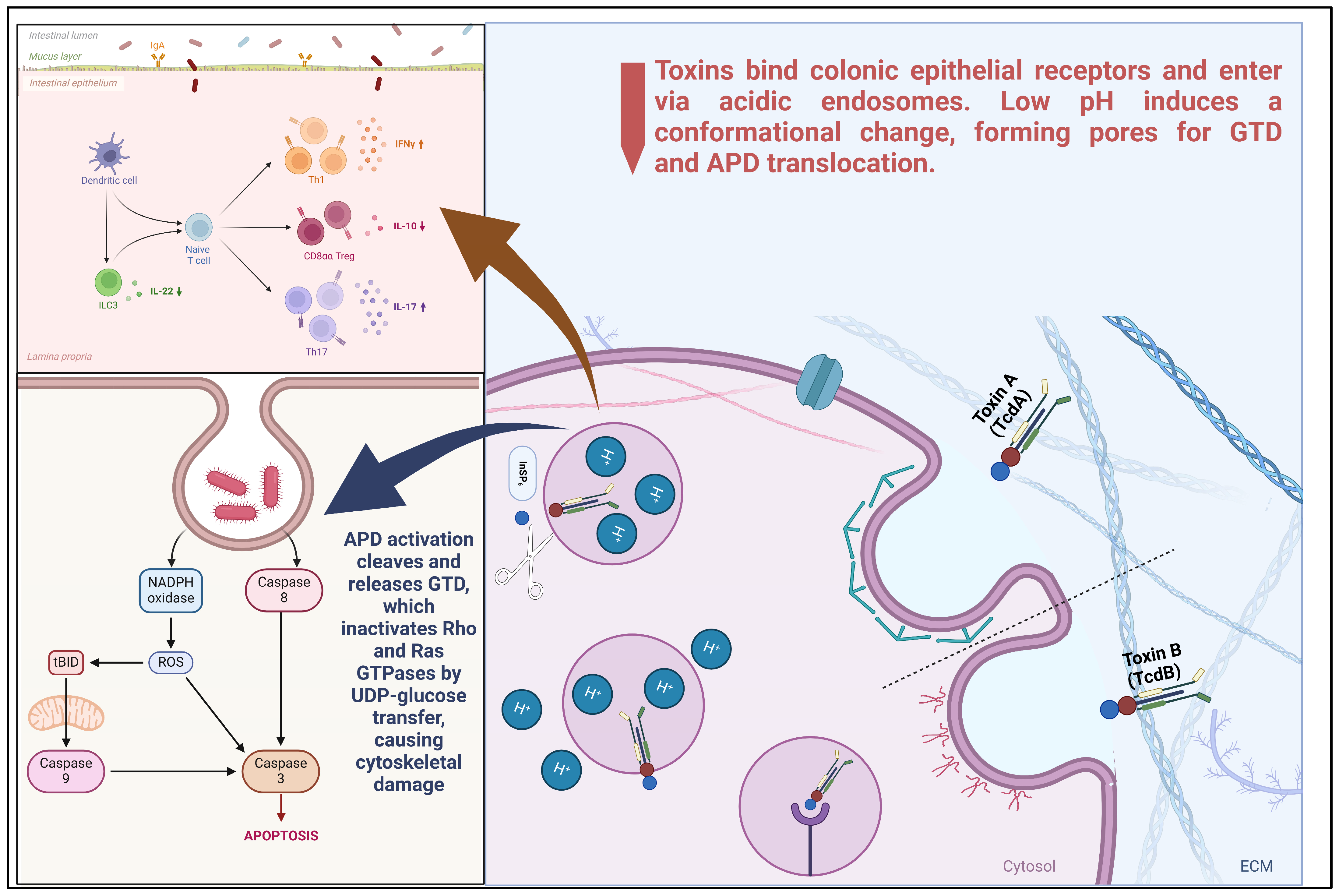
2.4.1. Receptor Binding and Endocytosis

The intoxication process begins with the specific binding of TcdA and TcdB to host cell surface receptors. TcdA is known to interact with glycans, glycoprotein 96 (gp96), sulfated glycosaminoglycans (sGAGs), and members of the low-density lipoprotein receptor (LDLR) family [54]. In contrast, TcdB targets receptors, such as (CSPG4), Nectin 3, and frizzled proteins (FZD1, FZD2, FZD7). Following receptor engagement, the toxins are internalized into the host cell via receptor-mediated endocytosis [55].
Once inside the endosome, the acidic environment induces significant conformational changes in the toxins. These pH-dependent alterations are crucial for exposing hydrophobic regions within the translocation domain, which were previously concealed at neutral pH. The exposure of these hydrophobic segments facilitates their insertion into the endosomal membrane, a critical step for subsequent pore formation [56].

2.4.2. Membrane Insertion and Pore Formation

The translocation domain of TcdA and TcdB plays a pivotal role in membrane insertion and pore formation. Studies have identified specific regions within this domain that are essential for these processes. Mutations in residues clustered between amino acids 1035 and 1107 significantly impair the toxin’s ability to form pores, thereby reducing its cytotoxicity by over 1000-fold. These findings highlight the importance of precise structural configurations in the translocation domain for effective membrane insertion and pore formation [57].
Following pore formation, the catalytic glucosyltransferase domain (GTD) and autoprocessing domain (APD) of the toxins translocate across the endosomal membrane into the host cell cytosol. This translocation is facilitated by the transmembrane pores formed earlier. Once in the cytosol, the APD undergoes autoprocessing, releasing the GTD. The liberated GTD then glucosylates host GTPases, disrupting various cellular functions and leading to cytopathic effects [58]. High-resolution cryo-electron microscopy studies have provided deeper insights into the structural dynamics of TcdA and TcdB. These studies reveal that at neutral pH, the receptor-binding domains, particularly the combined repetitive oligopeptides (CROPs), interact with the delivery and receptor-binding domains, maintaining a conformation that prevents premature membrane interaction [59]. Upon acidification, the CROPs undergo dynamic movements, facilitating the exposure of hydrophobic regions necessary for membrane insertion and pore formation [34].

3. Molecular Pathogenesis: Endocytic Trafficking, Cytosolic Delivery Mechanisms, and Rho GTPase Inactivation

3.1. Endocytic Trafficking of TcdA and TcdB

The internalization of TcdA and TcdB by the host cell is through a tightly controlled process involving receptor-mediated endocytosis, vesicular transport, and pH-dependent structural remodeling. The toxins employ more than one pathway for endocytosis to maximize uptake and translocation of their catalytic glucosyltransferase domain (GTD) into the host cell cytoplasm. Recent studies involving cryo-electron microscopy, live-cell imaging methods, and pharmacological inhibitors have provided detailed information on the endocytic pathways employed by the toxins [60].

3.1.1. Receptor-Mediated Endocytosis and Receptor Specificity

C. difficile toxins, TcdA and TcdB, utilize distinct receptor-mediated pathways for cell invasion. TcdA utilizes LRP1 to a significant degree as its major receptor, considering the strong inhibition of toxin binding and internalization in LRP1 knockout fibroblasts. LRP1, involved in the extracellular ligand clearance of molecules, such as lipoproteins, coagulation factors, and matrix metalloproteinases, specifically binds TcdA through its cluster II and IV domains. This is in agreement with earlier work demonstrating that most of the LRP1 ligands were associated with clusters II and IV. However, efforts to cross-link the LRP1-TcdA complex chemically were unsuccessful due to the rapid processing of LRP1. Further findings indicate LRP1 was rapidly internalized into fibroblasts as well as into Caco-2 cells and is in agreement with its function for facilitating TcdA uptake. Genetic ablation of LRP1 decreases cellular uptake of TcdA as indicated by inhibited Rac/Cdc42 glucosylation, dephosphorylation of PAK, and actin depolymerization induced by toxins. These findings collectively confirm the fact that TcdA is an LRP1 ligand, and it utilizes LRP1 constitutive endocytosis to become internalized by cells [61].
Although LRP1 is a major receptor, TcdA also binds to other receptor structures. LRP1 knockout by genetic means decreases the binding of TcdA to the cell surface by about 50%; however, this does not prevent TcdA from binding to LRP1-null mouse embryonic fibroblasts (MEFs), indicating alternative receptors. Potential candidates suggested are gp96, Lewis I, X, and Y glycans, and sulfated glucosaminoglycans [62]. TcdA enters LRP1-/- MEFs at high concentrations, suggesting the existence of other endocytic receptors, which may be from the LDL receptor family. LRP1 also co-associates with other membrane proteins, such as PDGF receptors, NMDA receptor subunits, TGFβ receptors, Frizzled1, and integrins, regulating their activity or their internalization. Of these, gp96, which co-associates with LRP1, is proposed as a co-receptor [63]. Inhibition or down-regulation of gp96 partially eliminates the biological activities of TcdA, suggesting it is involved. Experimental evidence, however, indicates that gp96 is not internalized and remains at the cell surface even in the presence of TcdAl thus, it may be a non-endocytic receptor. It is consistent with a model in which gp96 promotes TcdA binding to LRP1 but does not enhance its uptake. Instead, its presence enhances its uptake through LRP1-mediated endocytosis [64].
Compared to TcdA, TcdB has greater receptor specificity and binds to CSPG4, frizzled (FZD) 1/2/7, and NECTIN3 [65]. Cryo-EM analysis demonstrates that TcdB binding to CSPG4 is mainly at the junction of its translocation and combined repetitive oligopeptide (CROP) domains [66]. Mutational analysis identifies two important residues, Y1824 and N1839, in this area, whose mutation significantly compromises TcdB intoxication of Vero and CHO cells. These residues are vital for CSPG4 binding but are not required for NECTIN3 or FZD7 binding, further supporting CSPG4 as a major receptor. Cells naturally lacking CSPG4 or CSPG4 deletion using CRISPR/Cas9 are less susceptible to wild-type and mutant TcdB, highlighting its function in toxicity. Further experiments show that the minimal functional region for CSPG4 binding involves three short repeats at the C-terminal end of the translocation domain. Notably, although the CROP domain is important for TcdA receptor binding, TcdB’s CROP domain is not found to be crucial for its cellular interaction [67]. Binding studies demonstrate that TcdB CROPs do not bind to cell surfaces or competitively block TcdB intoxication, indicating a profound difference in receptor engagement mechanisms between TcdA and TcdB [68].
Structural-functional analysis of TcdA and TcdB receptor binding has wide therapeutic implications for neutralization. Notably, bezlotoxumab, a monoclonal TcdB antibody, efficiently inhibits CSPG4 binding and associated cytotoxicity. These observations are a testament to the importance of receptor specificity in toxin pathogenesis and provide a paradigm for the design of targeted therapeutics against CDI [69]. Nevertheless, identification of TcdB’s endocytic receptor remains enigmatic despite rigorous efforts and, as such, is a promising candidate for further investigation. The new two-receptor model hypothesizes that the large clostridial glucosylating toxins employ a non-endocytic receptor for cell association in addition to an endocytic receptor for the internalization of the toxins into cells. Mechanistic elucidation of these receptor-mediated pathways contains critical information regarding the pathogenesis of C. difficile and, as such, potential sites of intervention [70].

3.1.2. Clathrin-Independent and PACSIN2-Dependent Endocytosis of TcdA

The TcdA internalization pathway is quite distinct from that of TcdB, which is largely clathrin-mediated endocytosis (CME). Saturating analyses using RNAi knockdown technologies and pharmacologic inhibitors have determined that TcdA entry is clathrin-independent but completely PACSIN2-dependent [71]. The endocytic regulatory protein is associated with membrane curvature as well as with actin rearrangement, establishing its critical role in the cell intoxication mechanism mediated by TcdA [72].
TcdA and TcdB, being major virulence factors of C. difficile infection, depend on endocytosis and translocation into acidified endosomal compartments inside host cells. Whereas TcdB’s CME dependence on inactivation of Rac1 and rounding of cells is established, the endocytic process of TcdA was unclear until recent studies deciphered its PACSIN2 dependence. Perturbation experiments through RNAi-based and pharmacological inhibition demonstrated that TcdA internalization into Caco-2 cells is not CME dependent as CHC depletion had no effect on TcdA-induced cytotoxicity, and TcdA did not colocalize with clathrin markers [73].
Unlike earlier work indicating participation of CME in TcdA internalization, these observations are consistent with reports that inhibiting CME with chlorpromazine also affects clathrin-independent functions [74]. Notably, removal of CHC from HT-29 cells did not diminish TcdA-dependent glucosylation of Rac1, again indicating CME-independent internalization of TcdA. This discrepancy is testimony to the sophistication of TcdA endocytosis and necessitates multiple experimental strategies.
Clathrin-independent endocytic routes are divided based on dynamin dependence. Our research demonstrated that TcdA internalization is dynamin-dependent and that Cav1, cavin1, and PACSIN2 are components that are absolutely essential. While Cav1 and cavin1 participate in caveolae formation, our imaging experiments failed to find colocalization of TcdA with these proteins in Caco-2 or mouse embryonic fibroblast (MEF) cells, and, thus, TcdA entry appears to be caveolae-independent. Surprisingly, Caco-2 cells are devoid of the Cav1 α isoform essential for functional caveolae, which could be the reason for this finding [75].
PACSIN2 then becomes a dominant mediator of TcdA endocytosis, and knockdown decreases TcdA internalization by 80%. Being a BAR domain protein, PACSIN2 creates membrane curvature and dynamin recruitment for the vesicle scission, similar to the FEME pathway via endophilinA2. In both Caco-2 and MEF cells, TcdA colocalizes with PACSIN2, pointing to its indispensable role in the entry of the toxin and the resulting cytotoxicity [76].
The unique endocytic pathways used by TcdA and TcdB can account for their relative cytotoxicity, and the CME-dependent entry of TcdB allows for the generation of reactive oxygen species, in contrast to TcdA. The pathway of TcdA through PACSIN2 can be therapied, and the future lies in the determination of the receptor interactions and other host proteins participating in this pathway.

3.1.3. Endosomal Acidification and Toxin Translocation

The internalization of TcdA and TcdB by receptor-mediated endocytosis leads to targeting acidic endosomal compartments, where low pH induces the conformational changes necessary for membrane insertion and subsequent trafficking into the cytosol [77]. Ambroxol (Ax), an inhibitor of endosomal acidification, has been demonstrated to significantly reduce toxin-induced morphological changes and Rac1 glucosylation. Ax significantly inhibited cell rounding induced by toxins and conferred protection upon intestinal epithelial cells from the cytotoxic repercussions. Furthermore, Ax directly attenuated the intracellular enzymatic activity of the glucosyltransferase domain (GTD) of the toxins, independent of its influence on endosomal pH, thus further impairing the functionality of the toxins [78]. Bafilomycin A1 also inhibits endosomal acidification and prevents the translocation of the C. difficile toxins A (TcdA) and B (TcdB) into the cytosol. The inhibition inhibits conformational changes required for pore formation and toxin activation and, thus, protects host cells against TcdB-induced rounding and cytotoxicity Live-cell imaging has demonstrated that translocation of TcdB approximately 10 min after endocytosis corresponds to a rapid drop in endosomal pH, and cryo-EM analysis performed under acidic conditions shows TcdB in its open β-hairpin conformation characteristic of its membrane insertion state [79].
The pH-dependent activation through communication between the structural and functional elements highlights the complexity of C. difficile toxin translocation. A close understanding of such processes may indeed permit the design of therapeutic interventions aimed at endosomal acidification, thereby preventing the cytotoxic activity of such pathogenic bacterial toxins.

3.2. Cytosolic Delivery and Autoprocessing Mechanisms

C. difficile toxins, TcdA and TcdB, employ a sophisticated multistep process to infiltrate host cells and disrupt cellular functions. This process encompasses receptor binding, endocytosis, pore formation, translocation, autoprocessing, and subsequent glucosylation of host GTPases [80].
The initial phase involves the toxins binding to specific cell surface receptors. TcdA primarily interacts with glycan structures and proteins, such as sucrase–isomaltase and glycoprotein 96 (gp96), on the colonic epithelium. Additionally, sulfated glycosaminoglycans (sGAGs) and low-density lipoprotein receptors (LDLRs) have been identified as mediators for TcdA entry, suggesting that TcdA may engage multiple receptors to facilitate high-affinity cell entry. In contrast, TcdB targets receptors, including chondroitin sulfate proteoglycan 4 (CSPG4), frizzled proteins (FZD1, FZD2, FZD7), and poliovirus receptor-like 3 (PVRL3). These interactions are crucial for the internalization of the toxins via receptor-mediated endocytosis [8]. These changes facilitate the insertion of hydrophobic regions into the endosomal membrane, leading to pore formation. Notably, TcdA requires cholesterol-rich membranes for effective pore formation, whereas TcdB does not have this requirement. The formed pores allow the translocation of the N-terminal glucosyltransferase domain (GTD) and the autoprotease domain (APD) into the host cell cytosol [54].
Once translocated, the APD undergoes activation by inositol hexakisphosphate (InsP6), a cytosolic cofactor. Activation causes an autoprocessing reaction that cleaves and releases the GTD into the cytosol. Released GTD glucosylates Rho-family GTPases RhoA, Rac1, and Cdc42 at the conserved threonine residue. Modification causes inactivation of the GTPases, leading to cytoskeletal perturbations of the actin cytoskeleton, alterations in cell shape, barrier dysfunction, and eventual cell death [81].

3.3. Rho GTPase Inactivation by TcdA and TcdB

The cytotoxicity of C. difficile toxins TcdA and TcdB is mainly induced by Rho GTPase inactivation, which has crucial functions in controlling the organization of actin cytoskeleton, cell division, and immunomodulation signaling pathways. Toxins are involved in a glucosyltransferase reaction with irreversible inactivation of Rho-family GTPases, subsequently triggering a cascade of cellular aberrations. Recent structural and biochemistry studies have elucidated the enzymic mechanism of the glucosylation and its profound effects on host cell physiology [82].
Both TcdA and TcdB employ uridine diphosphate glucose (UDP-Glc) as a substrate for the catalysis of transferring a glucose moiety to a conserved threonine residue at the switch I region of Rho GTPases. Crystallographic investigations at high resolution have indicated that TcdB functions in a substitution nucleophilic internal manner, with nucleophilic attack and departure of the leaving group on the same face of the glucose ring [83]. Kinetic isotope effect analyses have further verified that glucosylation reaction is associated with a transition state of oxocarbenium phosphate ion pair, with dissociation of UDP being prior to threonine attack. Comparative structure analysis carried out between the two toxins has demonstrated that TcdA is able to work on a wider range of substrates, including both the Rho- and Rap-family of GTPases, whereas TcdB preferentially modifies RhoA, Rac1, and Cdc42. Crucially, the glucosyltransferase domain of TcdA is found to be more catalytically active after autoproteolysis, pointing toward cytosolic activation as part of optimization of enzymatic action [84].
Inactivation of the Rho GTPase through TcdA and TcdB significantly interferes with pivotal cellular processes. GDP/GTP cycling disruption interferes with the organization of the actin filaments, which leads to the depolymerization of the actin and cell rounding before eventual detachment [85]. Comparative studies between hypervirulent TcdB strains reported higher affinity toward Rac1 corresponding with increased cytotoxicity. Additionally, disturbance of RhoA has adverse effects on tight junctions found in epithelium, causing barrier dysfunction [86]. Experiments from human colonic organoids disclosed a significant fall in trans-epithelial electrical resistance (TEER) after Rac1 glucosylation, indicating heightened permeability. Additionally, suppression of Rac1 inhibits expression of cyclin D1 and causes G1-S phase arrest of the colonic epithelial cell cycle and is correlated with extended inactivation, which ultimately impairs repairing of epithelial layers in models using mice [87].
TcdA and TcdB act on the Rho-family GTPases (RhoA, Rac1, and Cdc42) with similar activities; TcdA, however, is more effective in modifying RhoA, and TcdB is faster in modifying Rac1. Although the two toxins have 51% sequence identity within their glucosyltransferase domains, both toxins employ similar strategies for the recognition of substrate, as inferred from structural alignments of their complexes with GTPases. This highlights their differential activities while acting on identical substrates [88].
Targeting the Rho GTPase inactivation process has tremendous therapeutic potential. Small molecule inhibitors, including Ambroxol, as mentioned earlier, have been reported to suppress TcdA/B enzymatic activity and decrease levels of Rac1 glucosylation by a significant amount. Human α-defensin-6 sequesters TcdA and TcdB into inactive complexes, preventing Rho-GTPase glucosylation and protecting epithelial barriers. This mechanism, alongside CRISPR/Cas9-mediated RhoA and Rac1 knockout, highlights the therapeutic potential of targeting host factors to mitigate toxin-induced injuries [89].

4. Microbiota-Mediated Resistance to C. difficile Infection

The gut microbiota plays a crucial role in protecting against CDI through a mechanism known as colonization resistance. Disruptions in microbiota composition, often due to antibiotic use, increase susceptibility to C. difficile colonization and toxin-mediated colonic damage. Recent studies have highlighted specific bacterial taxa, metabolic pathways, and immune interactions that contribute to microbiota-mediated resistance against C. difficile [90].

4.1. Role of Microbiota in Colonization Resistance

The gut microbiota plays a role in preventing CDI through a multi-component process of nutrient competition, production of antimicrobial metabolites, and immune system modulation of the host [91]. A healthy and dense microbial community is a natural defense against enteric pathogens. Disturbances by antibiotics, diet, or gastrointestinal disease can break this defense, thus increasing the risk of CDI by creating an environment conducive to C. difficile colonization and toxin production [92].
Antibiotic exposure is a major risk factor for CDI since it reduces the gut microbial diversity, especially within the Firmicutes and Bacteroidetes phyla, thus changing the gut metabolic environment in a way that promotes the development of C. difficile. Various studies have shown that about 60% of individuals with community-associated CDI had a history of antibiotic exposure [93]. In mouse models, Schubert et al. pointed out that interactions among various populations of bacteria are involved in colonization resistance, and this emphasizes the role played by microbial diversity. CDI patients have lower levels of protective bacteria, such as Bacteroidetes, Prevotella, and Bifidobacteria, and higher levels of Lactobacillaceae and Enterobacteriaceae [94].
Probiotic treatments for the restoration of protective gut microbiota populations are promising for the prevention and treatment of CDI. New evidence also indicates that dietary components, including high-fat and low-fiber Western diets, play a role in adverse effects on gut health by decreasing microbiota-accessible carbohydrates and SCFAs, which are protective against CDI. Excessive dietary zinc and underlying conditions, including inflammatory bowel disease (IBD), also contribute to gut microbiota dysbalances, enhancing CDI risk and severity [95].
Bile acids play a central role in modulating the composition of gut microbiota and CDI pathogenesis. Antibiotic-altered microbiota results in elevated primary bile acids, which activate C. difficile spore germination and growth and suppress secondary bile acids inhibiting its growth [96]. FMT can restore secondary bile acids and microbial community diversity and has therapeutic potential [97]. Secondary bile acid-producing bacteria, such as Clostridium scindens, that are involved in deoxycholate (DCA) and lithocholate (LCA) production have been targeted as major contributors to CDI resistance [98].
SCFAs, such as acetate, propionate, and butyrate, derived from microbiota during dietary fiber fermentation, keep the colon healthy and have immunomodulatory effects [99]. Research has demonstrated that butyrate improves intestinal epithelial barrier function and suppresses inflammation, which is CDI protective. Acetate, via immune signaling, improves antimicrobial function and repair of epithelium, further enhancing colonization resistance [100].
Competition for nutrients is a key process through which gut microbiota suppresses the growth of C. difficile. Commensal microbes restrict the supply of critical nutrients, including amino acids and carbohydrates. For example, Bacteroides, the taiotaomicron, breaks down sialic acid from mucosal carbohydrates, a nutrient that C. difficile uses. Reduction of such bacterial counts by antibiotics raises the supply of nutrients for C. difficile. Commensal bacteria that metabolize succinate reduce its supply, thus restricting the metabolism and growth of C. difficile [101].
Direct competition by commensal bacteria, in the form of the production of antimicrobial compounds, is also important. Thuricin CD, a bacteriocin that is active against C. difficile, is produced by Bacillus thuringiensis [99]. Growth of C. difficile is inhibited by Lactobacillus reuteri through the production of reuterin, and the co-administration of glycerol was found to enhance its activity. Combinatorial therapies involving antimicrobial compounds, such as durancin from Enterococcus durans and reuterin, have been found to demonstrate synergistic inhibitory activity against C. difficile in vitro. Gut microbiota, such as C. scindens and Clostridium sordelli, also produce tryptophan-derived antibiotics that, in combination with secondary bile acids, inhibit C. difficile [102].
Fecal microbiota studies have identified Bacteroides species as competitive and inhibitory bacteria that block colonization of C. difficile using host-derived nutrients that are necessary for C. difficile growth [103]. Eradication of Bacteroides using antibiotic treatment suppresses this defense mechanism, enhancing C. difficile growth. Therefore, maintaining a dense gut microbiota is essential to provide effective colonization resistance against C. difficile [104].

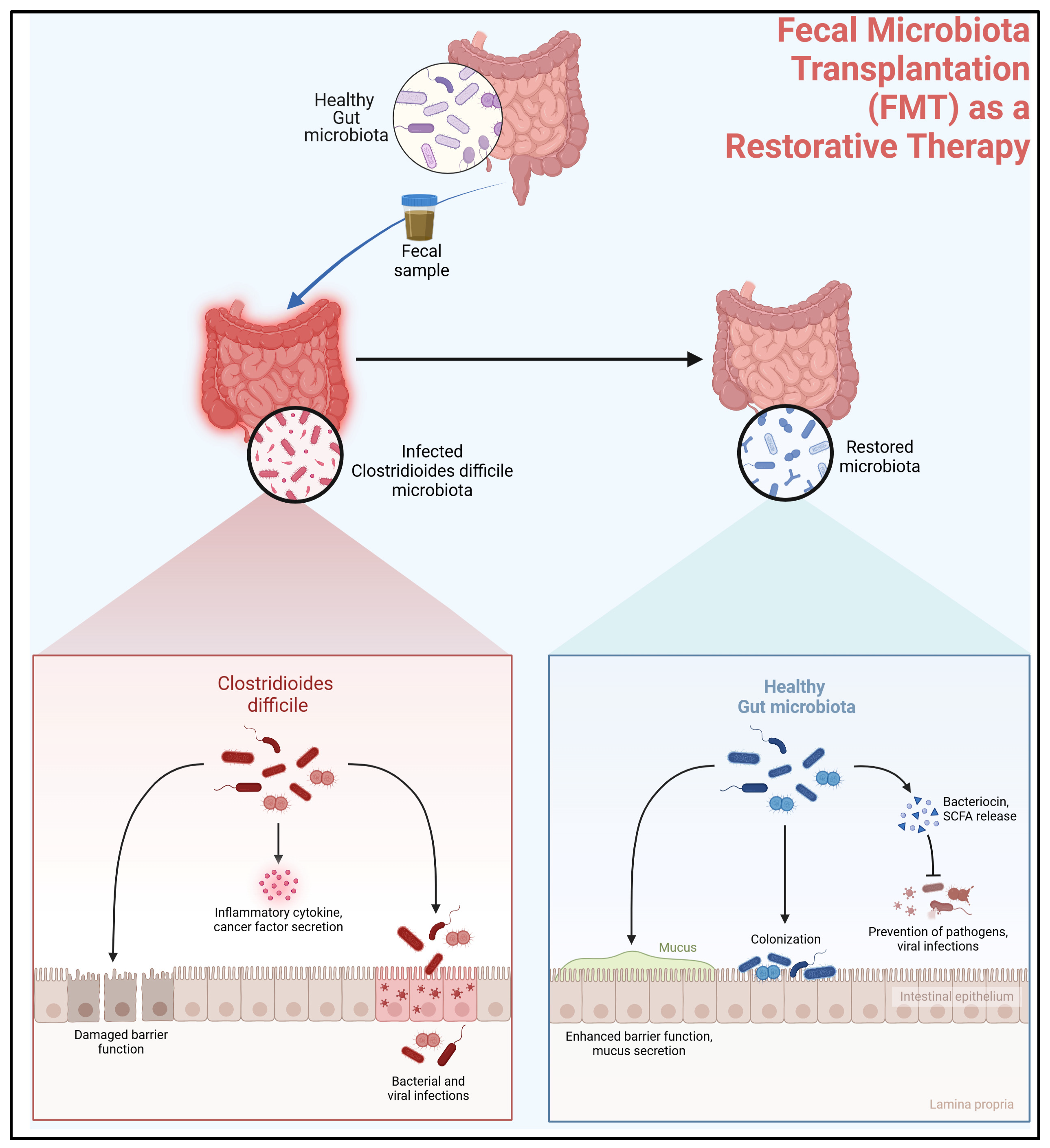
4.2. Fecal Microbiota Transplantation (FMT) as a Restorative Therapy

Fecal microbiota transplantation (FMT) (Figure 2) has been a highly effective treatment approach to the re-establishment of gut microbial diversity and alleviation of C. difficile infection, particularly in cases of recurrence [105]. The underlying mechanism of FMT is the re-introduction of a healthy and diverse microbial community into the infected gut, thus re-establishing colonization resistance to C. difficile [106]. CDI is frequently linked with alterations in gut microbiota composition as a result of antibiotic administration, which eliminates the populations of beneficial microbes and creates an environment conducive to the overgrowth of C. difficile. Through the transfer of stool from a donor with a healthy gut to a recipient, FMT allows for the re-establishment of a healthy gut microbiota, thus the alleviation of the resistance to pathogen overgrowth and recurrence [107].
Clinical trials have demonstrated that FMT is far more effective than traditional antibiotic therapy for the prevention of recurrent CDI [108]. While antibiotics, such as vancomycin and fidaxomicin, can eliminate an active infection, they are powerless to repair the underlying dysbiosis that makes patients susceptible to reinfection [109]. In contrast, FMT repairs the important bacterial communities, most notably the Firmicutes and Bacteroidetes phyla, necessary to maintain colonization resistance. Increasing evidence demonstrates that patients receiving FMT have rapid and persistent restoration of their microbiota, defined by a dramatic increase in bacterial diversity and metabolic activity suppressing the growth of C. difficile [110]. Furthermore, FMT allows for the restoration of metabolic end products, including short-chain fatty acids (SCFAs) and secondary bile acids, needed to maintain gut homeostasis and suppress the germination of C. difficile spores and toxin production [111].
The mechanisms of FMT efficacy go beyond the restoration of microbial diversity. FMT restoration of beneficial bacteria assists in immune modulation, dampening inflammatory reactions to CDI [112]. Research indicates that FMT restores host–microbiota interaction by modulating cytokine response and improving mucosal barrier function [113]. FMT also disturbs bile acid metabolism, transforming the composition from an antibiotic-altered primary bile acid-dominant environment supporting C. difficile colonization to one rich in secondary bile acids, which inhibit the growth of C. difficile. This metabolic transition is crucial in interrupting the cycle of recurrent infection and creating a gut environment that is adverse to the growth of C. difficile [114].
Despite its success, FMT is not without its challenges. The standardization of donor screening, stool preparation, and method of delivery are prime areas of research to determine safety and efficacy. There is also controversy regarding the best mode of delivery of FMT, from colonoscopic infusion to nasogastric or nasoduodenal tubes and oral encapsulated forms, each with varying levels of patient acceptability and success rates [115]. There are also concerns regarding the long-term effect of FMT, such as potential alteration in immune function or microbiota architecture beyond the treatment of CDI, which must be answered [116].
As a substitute or adjunct strategy, scientists are developing well-characterized microbial consortia and next-generation probiotics that replicate the therapeutic benefits of FMT but without the perils of direct stool transplantation [117]. These strategies aim to selectively replace critical microbial processes without the FMT donor-to-donor heterogeneity. Developments in microbiome therapeutics, such as bacterial cocktails and artificial microbiota preparations, have the potential to offer a more controlled and focused therapy for CDI patients in the future.

5. Future Directions: Integrative Models for Toxin–Host–Microbiome Crosstalk and Translational Challenges

The intricate interplay between C. difficile toxins, host immune responses, and the gut microbiome has emerged as a pivotal factor in understanding CDI pathogenesis and advancing therapeutic strategies [118]. Follow-up studies will be anticipated to employ integrative systems-level approaches, multi-omics, and computational modeling to uncover these complex interactions and translate findings into clinically applicable tools. In this section, the potential of these approaches is discussed, with a focus on current translational hurdles and future research avenues to meet the unmet needs in the treatment of CDI.

5.1. Systems Biology Approaches to Toxin–Host–Microbiome Crosstalk

A comprehensive understanding of CDI pathogenesis is an integrated study of C. difficile toxin remodulation of host metabolism, immune signaling, and microbiome organization. The advantages of multi-omics integration in revealing these processes have been identified in recent research. [103] for instance, employed transcriptomics and metabolomics to demonstrate how toxin-induced inflammation reprograms host tissue gene expression and gut microbiota composition to create a microenvironment that is favorable for the growth of C. difficile. Their research demonstrated how collagen degradation by matrix metalloproteinase releases peptides to be utilized as a source of nutrition by C. difficile and revealed a feedback process that sustains infection. These studies demonstrate the contribution of systems biology to the identification of key molecular pathways and microbial interactions that result in CDI development.
Apart from experimental approaches, computational modeling has also been very valuable as a predictive device for host–pathogen interaction and optimization of therapeutic intervention. A computational model, which comprised toxin expression pathways, host cell apoptosis, and MAPK signaling, was constructed and showed that biomodulation of toxin expression maximizes host survival greatly. These models provide not only mechanistic insights but also a platform for theoretically assessing interventions, thus facilitating the development of precision therapeutics [119].

5.2. Translational Challenges in Therapeutic Development

Despite tremendous progress in toxin-targeted therapies, there are a number of barriers to their application in a clinical setting. One of the greatest challenges is in reconstituting the microbiome. While FMT has been highly effective in the management of recurrent CDI, long-term safety concerns, optimal donor selection criteria, and standardization of procedural protocols are still unknown. Moreover, variability in toxin expression among different strains of C. difficile presents a considerable barrier. Hypervirulent bacteria, such as ribotype 027, have increased toxin expression and resistance to neutralizing techniques, further complicating the development of therapeutic strategies [120]. Moreover, host responses to toxins are based on genetic characteristics and the composition of the microbiome, necessitating personalized treatment. Experiments with gut epithelial models have shown varying responses to C. difficile toxins according to the genetic makeup of the host and the microbiome structure, highlighting the need for personalized therapeutic regimens [121].

5.3. Future Research Priorities

Future investigations pertaining to CDI must pioneer novel therapeutic strategies whilst simultaneously addressing extant limitations (Table 1). Among the most imperative domains is the formulation of targeted therapies that more effectively neutralize toxin activity, including engineered antibodies and small-molecule inhibitors. These strategies are designed to obstruct toxin–host interactions, mitigate cellular damage, and diminish the severity of the disease. Instead of FMT, one could consider synthetic microbiome-based therapy, which ensures a standardized and safer method for maintaining balance in the gastrointestinal tract. Another vital domain encompasses the identification of predictive biomarkers that facilitate the early detection of severe CDI, thereby enabling timely and targeted therapeutic interventions. Advances in immunotherapy, such as bispecific antibodies and toxin-targeting T-cell therapies, have the potential to further improve patient outcomes by modulating the immune response and decreasing recurrence rates. The challenges associated with the standardization of treatment, bioavailability, and delivery mechanisms must be systematically addressed to enhance therapeutic efficacy. Through the integration of precision medicine, biotechnology, and microbial therapeutics, forthcoming studies may yield more effective, accessible, and sustainable solutions for combating CDI [122]. These treatments could potentially augment current treatment while avoiding the limitations inherent in current toxin-neutralization mechanisms, as mentioned in the table below.

6. Conclusions

Clostridioides difficile infection (CDI) is a major global healthcare problem, largely due to the highly virulent toxins TcdA and TcdB. The current review consolidates an extensive overview of their structural and functional features, such as receptor binding, endocytosis, pH-dependent conformational changes, and immune modulatory functions within the host. Advances in cryo-electron microscopy (cryo-EM) and X-ray crystallography have provided richer structural insights into toxin dynamics and clear concepts, particularly in pore formation and auto processing mechanisms, which can lead to new therapeutic strategies. Genomic studies have also shed light on the evolutionary versatility of hypervirulent strains, with horizontal gene transfer as the driving force for the diversification of toxins.
Therapeutic methods of targeting C. difficile toxins have come a long way, with neutralizing antibodies, CRISPR gene editing, and microbiome restoration approaches being the preferred front-runners. FMT has shown great promise in the context of recurrent CDI, and engineered probionts and synthetic microbiome preparations are the directions to look out for in the future. Additionally, the role of gut microbiota in the control of toxin action highlights the necessity of holistic treatment approaches that account for both pathogen virulence and host resistance.
Despite these improvements, several obstacles remain, including toxin subtype heterogeneity, specificity of the host response, and the need for standardized microbiome therapy. Future research will necessitate bridging the identification of predictive biomarkers of the severity of CDI, refining further the development of immunotherapies, and using computational modeling to inform the selection of the most therapeutic interventions. The integration of structural biology, systems-level omics, and dynamics of host–microbiome interactions enable the formulation of precision medicine strategies, better addressing CDI. Identification of the more profound mechanistic understandings of TcdA and TcdB will play a pivotal role in the creation of novel targeted treatments and prevention approaches for future application.

Author Contributions

Conceptualization, M.Q. and A.T.; methodology, M.Q. and J.T.; software—validation, U.M.H., A.T. and J.T.; formal analysis, U.M.H.; investigation, T.P.; resources, D.T.; data curation, A.T.; writing—original draft preparation, M.Q. and T.P.; writing—review and editing, A.T.; visualization, U.M.H.; supervision, M.Q. and M.U.; project administration, M.U.; funding acquisition, D.T. All authors have read and agreed to the published version of the manuscript.

Funding

This research received no external funding.

Conflicts of Interest

The authors declare no conflicts of interest.

Abbreviations

APDAdaptor Protein Domain
BARBin/Amphiphysin/Rvs (domain)
CDClostridioides difficile
CDADClostridioides difficile-Associated Diarrhea
CDIClostridioides difficile Infection
CDTClostridioides difficile Transferase (binary toxin)
CHCClathrin Heavy Chain
CHOChinese Hamster Ovary (cells)
CMEClathrin-Mediated Endocytosis
CPDCysteine Protease Domain
CRDCysteine-Rich Domain
CROPCombined Repetitive Oligopeptides (domain)
DCADeoxycholic Acid
DNADeoxyribonucleic Acid
DRBDDouble RNA-Binding Domain
EMElectron Microscopy
FBDFrizzled Binding Domain
FDAFood and Drug Administration
FEMEFast Endophilin-Mediated Endocytosis
FMTFecal Microbiota Transplantation
FZDFrizzled (protein family)
GDPGuanosine Diphosphate
GTDGlucosyltransferase Domain
GTPGuanosine Triphosphate
HGTHorizontal Gene Transfer
HTHuman Toxin
IBDInflammatory Bowel Disease
IIIntrinsic Immunity
INDIAIndian National Drug Authority (possible context)
IVIntravenous
LCALithocholic Acid
LDLLow-Density Lipoprotein
LDLRLow-Density Lipoprotein Receptor
MAPKMitogen-Activated Protein Kinase
MEFMouse Embryonic Fibroblast
NMDAN-Methyl-D-Aspartate (receptor)
OFFInactive State
ONActive State
PAKp21-Activated Kinase
PCRPolymerase Chain Reaction
PDGFPlatelet-Derived Growth Factor
RNARibonucleic Acid
TEERTrans-Epithelial Electrical Resistance
UDPUridine Diphosphate

References

  1. Balsells, E.; Shi, T.; Leese, C.; Lyell, I.; Burrows, J.; Wiuff, C.; Campbell, H.; Kyaw, M.H.; Nair, H. Global burden of Clostridium difficile infections: A systematic review and meta-analysis. J. Glob. Health 2018, 9, 010407. [Google Scholar] [CrossRef] [PubMed]
  2. Stoian, M.; Andone, A.; Boeriu, A.; Bândilă, S.R.; Dobru, D.; Laszlo, S.Ș.; Corău, D.; Arbănași, E.M.; Russu, E.; Stoian, A. COVID-19 and Clostridioides difficile Coinfection Analysis in the Intensive Care Unit. Antibiotics 2024, 13, 367. [Google Scholar] [CrossRef] [PubMed]
  3. Leffler, D.A.; Lamont, J.T. Clostridium difficile Infection. N. Engl. J. Med. 2015, 372, 1539–1548. [Google Scholar] [CrossRef]
  4. Manse, J.S.; Baldwin, M.R. Binding and entry of Clostridium difficile toxin B is mediated by multiple domains. FEBS Lett. 2015, 589, 3945–3951. [Google Scholar] [CrossRef] [PubMed]
  5. Filić, V.; Mijanović, L.; Putar, D.; Talajić, A.; Ćetković, H.; Weber, I. Regulation of the Actin Cytoskeleton via Rho GTPase Signalling in Dictyostelium and Mammalian Cells: A Parallel Slalom. Cells 2021, 10, 1592. [Google Scholar] [CrossRef]
  6. Pruitt, R.N.; Chagot, B.; Cover, M.; Chazin, W.J.; Spiller, B.; Lacy, D.B. Structure-Function Analysis of Inositol Hexakisphosphate-induced Autoprocessing in Clostridium difficile Toxin A. J. Biol. Chem. 2009, 284, 21934–21940. [Google Scholar] [CrossRef]
  7. Zhou, Y.; Zhan, X.; Luo, J.; Li, D.; Zhou, R.; Zhang, J.; Pan, Z.; Zhang, Y.; Jia, T.; Zhang, X.; et al. Structural dynamics of the CROPs domain control stability and toxicity of Paeniclostridium sordellii lethal toxin. Nat. Commun. 2023, 14, 8426. [Google Scholar] [CrossRef]
  8. Kordus, S.L.; Thomas, A.K.; Lacy, D.B. Clostridioides difficile toxins: Mechanisms of action and antitoxin therapeutics. Nat. Rev. Microbiol. 2021, 20, 285–298. [Google Scholar] [CrossRef]
  9. Gerding, D.N.; Johnson, S.; Rupnik, M.; Aktories, K. Clostridium difficile binary toxin CDT. Gut Microbes 2013, 5, 15–27. [Google Scholar] [CrossRef]
  10. Alam, Z.; Madan, R. Clostridioides difficile Toxins: Host Cell Interactions and Their Role in Disease Pathogenesis. Toxins 2024, 16, 241. [Google Scholar] [CrossRef]
  11. Carter, G.P.; Douce, G.R.; Govind, R.; Howarth, P.M.; Mackin, K.E.; Spencer, J.; Buckley, A.M.; Antunes, A.; Kotsanas, D.; Jenkin, G.A.; et al. The Anti-Sigma Factor TcdC Modulates Hypervirulence in an Epidemic BI/NAP1/027 Clinical Isolate of Clostridium difficile. PLoS Pathog. 2011, 7, e1002317. [Google Scholar] [CrossRef] [PubMed]
  12. Majumdar, A.; Govind, R. Regulation of Clostridioides difficile toxin production. Curr. Opin. Microbiol. 2021, 65, 95–100. [Google Scholar] [CrossRef]
  13. Ransom, E.M.; Kaus, G.M.; Tran, P.M.; Ellermeier, C.D.; Weiss, D.S. Multiple factors contribute to bimodal toxin gene expression in Clostridioides (Clostridium) difficile. Mol. Microbiol. 2018, 110, 533–549. [Google Scholar] [CrossRef] [PubMed]
  14. Zhao, J.; Perkins, M.L.; Norstad, M.; Garcia, H.G. A bistable autoregulatory module in the developing embryo commits cells to binary expression fates. Curr. Biol. 2023, 33, 2851–2864.e11. [Google Scholar] [CrossRef]
  15. Martin-Verstraete, I.; Peltier, J.; Dupuy, B. The Regulatory Networks That Control Clostridium difficile Toxin Synthesis. Toxins 2016, 8, 153. [Google Scholar] [CrossRef] [PubMed]
  16. Anjuwon-Foster, B.R.; Tamayo, R. A genetic switch controls the production of flagella and toxins in Clostridium difficile. PLoS Genet. 2017, 13, e1006701. [Google Scholar] [CrossRef]
  17. Norris, M.A.W.; Plaskon, D.M.; Tamayo, R. Phase Variation of Flagella and Toxins in Clostridioides difficile is Mediated by Selective Rho-dependent Termination. J. Mol. Biol. 2024, 436, 168456. [Google Scholar] [CrossRef]
  18. Edwards, A.N.; McBride, S.M. Initiation of sporulation in Clostridium difficile: A twist on the classic model. FEMS Microbiol. Lett. 2014, 358, 110–118. [Google Scholar] [CrossRef]
  19. Buddle, J.E.; Fagan, R.P. Pathogenicity and virulence of Clostridioides difficile. Virulence 2023, 14, 2150452. [Google Scholar] [CrossRef]
  20. Hasan, K.; Alaribe, O.; Govind, R. Regulatory networks: Linking toxin production and sporulation in Clostridioides difficile. Anaerobe 2024, 91, 102920. [Google Scholar] [CrossRef]
  21. Lin, W.N.; Tay, M.Z.; Lu, R.; Liu, Y.; Chen, C.-H.; Cheow, L.F. The Role of Single-Cell Technology in the Study and Control of Infectious Diseases. Cells 2020, 9, 1440. [Google Scholar] [CrossRef] [PubMed]
  22. Mansfield, M.J.; Tremblay, B.J.-M.; Zeng, J.; Wei, X.; Hodgins, H.; Worley, J.; Bry, L.; Dong, M.; Doxey, A.C. Phylogenomics of 8,839 Clostridioides difficile genomes reveals recombination-driven evolution and diversification of toxin A and B. PLoS Pathog. 2020, 16, e1009181. [Google Scholar] [CrossRef]
  23. Shen, E.; Zhu, K.; Li, D.; Pan, Z.; Luo, Y.; Bian, Q.; He, L.; Song, X.; Zhen, Y.; Jin, D.; et al. Subtyping analysis reveals new variants and accelerated evolution of Clostridioides difficile toxin B. Commun. Biol. 2020, 3, 347. [Google Scholar] [CrossRef] [PubMed]
  24. Aptekorz, M.; Szczegielniak, A.; Wiechuła, B.; Harmanus, C.; Kuijper, E.; Martirosian, G. Occurrence of Clostridium difficile ribotype 027 in hospitals of Silesia, Poland. Anaerobe 2017, 45, 106–113. [Google Scholar] [CrossRef]
  25. Janezic, S.; Dingle, K.; Alvin, J.; Accetto, T.; Didelot, X.; Crook, D.W.; Lacy, D.B.; Rupnik, M. Comparative genomics of Clostridioides difficile toxinotypes identifies module-based toxin gene evolution. Microb. Genom. 2020, 6, e000449. [Google Scholar] [CrossRef]
  26. Pan, Z.; Zhang, Y.; Luo, J.; Li, D.; Zhou, Y.; He, L.; Yang, Q.; Dong, M.; Tao, L. Functional analyses of epidemic Clostridioides difficile toxin B variants reveal their divergence in utilizing receptors and inducing pathology. PLoS Pathog. 2021, 17, e1009197. [Google Scholar] [CrossRef]
  27. Knight, D.R.; Elliott, B.; Chang, B.J.; Perkins, T.T.; Riley, T.V. Diversity and Evolution in the Genome of Clostridium difficile. Clin. Microbiol. Rev. 2015, 28, 721–741. [Google Scholar] [CrossRef]
  28. Brouwer, M.S.; Roberts, A.P.; Hussain, H.; Williams, R.J.; Allan, E.; Mullany, P. Horizontal gene transfer converts non-toxigenic Clostridium difficile strains into toxin producers. Nat. Commun. 2013, 4, 2601. [Google Scholar] [CrossRef]
  29. Brouwer, M.S.M.; Warburton, P.J.; Roberts, A.P.; Mullany, P.; Allan, E. Genetic Organisation, Mobility and Predicted Functions of Genes on Integrated, Mobile Genetic Elements in Sequenced Strains of Clostridium difficile. PLoS ONE 2011, 6, e23014. [Google Scholar] [CrossRef]
  30. Newcomer, E.P.; Fishbein, S.R.S.; Zhang, K.; Hink, T.; Reske, K.A.; Cass, C.; Iqbal, Z.H.; Struttmann, E.L.; Burnham, C.-A.D.; Dubberke, E.R.; et al. Genomic surveillance of Clostridioides difficile transmission and virulence in a healthcare setting. mBio 2024, 15, e0330023. [Google Scholar] [CrossRef]
  31. Monot, M.; Eckert, C.; Lemire, A.; Hamiot, A.; Dubois, T.; Tessier, C.; Dumoulard, B.; Hamel, B.; Petit, A.; Lalande, V.; et al. Clostridium difficile: New Insights into the Evolution of the Pathogenicity Locus. Sci. Rep. 2015, 5, 15023. [Google Scholar] [CrossRef] [PubMed]
  32. Mehner-Breitfeld, D.; Rathmann, C.; Riedel, T.; Just, I.; Gerhard, R.; Overmann, J.; Brüser, T. Evidence for an Adaptation of a Phage-Derived Holin/Endolysin System to Toxin Transport in Clostridioides difficile. Front. Microbiol. 2018, 9, 2446. [Google Scholar] [CrossRef]
  33. Murata, K.; Wolf, M. Cryo-electron microscopy for structural analysis of dynamic biological macromolecules. Biochim. Biophys. Acta (BBA)-Gen. Subj. 2018, 1862, 324–334. [Google Scholar] [CrossRef]
  34. Aminzadeh, A.; Larsen, C.E.; Boesen, T.; Jørgensen, R. High-resolution structure of native toxin A from Clostridioides difficile. Embo Rep. 2021, 23, e53597. [Google Scholar] [CrossRef]
  35. Jiang, M.; Shin, J.; Simeon, R.; Chang, J.-Y.; Meng, R.; Wang, Y.; Shinde, O.; Li, P.; Chen, Z.; Zhang, J. Structural dynamics of receptor recognition and pH-induced dissociation of full-length Clostridioides difficile Toxin B. PLoS Biol. 2022, 20, e3001589. [Google Scholar] [CrossRef]
  36. Childress, K.O.; Cencer, C.S.; Tyska, M.J.; Lacy, D.B. Nectin-3 and shed forms of CSPG4 can serve as epithelial cell receptors for Clostridioides difficile TcdB. mBio 2023, 14, e0185723. [Google Scholar] [CrossRef] [PubMed]
  37. Bland, S.J.; Larabee, J.L.; Shadid, T.M.; Lang, M.L.; Ballard, J.D. Deletion of a 19-Amino-Acid Region in Clostridioides difficile TcdB2 Results in Spontaneous Autoprocessing and Reduced Cell Binding and Provides a Nontoxic Immunogen for Vaccination. Infect. Immun. 2019, 87, e00210-19. [Google Scholar] [CrossRef] [PubMed]
  38. Lanis, J.M.; Hightower, L.D.; Shen, A.; Ballard, J.D. TcdB from hypervirulent Clostridium difficile exhibits increased efficiency of autoprocessing. Mol. Microbiol. 2012, 84, 66–76. [Google Scholar] [CrossRef]
  39. Zhang, Y.; Hamza, T.; Gao, S.; Feng, H. Masking autoprocessing of Clostridium difficile toxin A by the C-terminus combined repetitive oligo peptides. Biochem. Biophys. Res. Commun. 2015, 459, 259–263. [Google Scholar] [CrossRef]
  40. Pruitt, R.N.; Chambers, M.G.; Ng, K.K.-S.; Ohi, M.D.; Lacy, D.B. Structural organization of the functional domains of Clostridium difficile toxins A and B. Proc. Natl. Acad. Sci. USA 2010, 107, 13467–13472. [Google Scholar] [CrossRef]
  41. Larabee, J.L.; Hauck, G.D.; Ballard, J.D. Cell-penetrating peptides derived from Clostridium difficile TcdB2 and a related large clostridial toxin. J. Biol. Chem. 2018, 293, 1810–1819. [Google Scholar] [CrossRef] [PubMed]
  42. Hwang, J.; Cho, K.-H.; Song, H.; Yi, H.; Kim, H.S. Deletion Mutations Conferring Substrate Spectrum Extension in the Class A β-Lactamase. Antimicrob. Agents Chemother. 2014, 58, 6265–6269. [Google Scholar] [CrossRef] [PubMed]
  43. Zheng, Y.; Yang, Q.; Luo, J.; Zhang, Y.; Li, X.; He, L.; Ma, C.; Tao, L. Identification of a hemorrhagic determinant in Clostridioides difficile TcdA and Paeniclostridium sordellii TcsH. Microbiol. Spectr. 2024, 12, e0035424. [Google Scholar] [CrossRef]
  44. Larabee, J.L.; Bland, S.J.; Hunt, J.J.; Ballard, J.D. Intrinsic Toxin-Derived Peptides Destabilize and Inactivate Clostridium difficile TcdB. mBio 2017, 8, e00503-17. [Google Scholar] [CrossRef]
  45. Zhang, K.; Zhou, Q.; Gu, H.; Yang, M.; Lin, X.; Wang, M.; Zhai, H.; Zhang, F.; Luo, Y.; Chen, L.; et al. TcdB From Hypervirulent Clostridioides difficile Induces Neuronal Loss and Neurotransmitter Alterations in the Intrinsic Enteric Nervous System. J. Infect. Dis. 2024. [Google Scholar] [CrossRef]
  46. Tao, L.; Zhang, J.; Meraner, P.; Tovaglieri, A.; Wu, X.; Gerhard, R.; Zhang, X.; Stallcup, W.B.; Miao, J.; He, X.; et al. Frizzled proteins are colonic epithelial receptors for C. difficile toxin B. Nature 2016, 538, 350–355. [Google Scholar] [CrossRef]
  47. Gupta, P.; Zhang, Z.; Sugiman-Marangos, S.N.; Tam, J.; Raman, S.; Julien, J.-P.; Kroh, H.K.; Lacy, D.B.; Murgolo, N.; Bekkari, K.; et al. Functional defects in Clostridium difficile TcdB toxin uptake identify CSPG4 receptor-binding determinants. J. Biol. Chem. 2017, 292, 17290–17301. [Google Scholar] [CrossRef]
  48. Gray, V.E.; Hause, R.J.; Fowler, D.M. Analysis of Large-Scale Mutagenesis Data To Assess the Impact of Single Amino Acid Substitutions. Genetics 2017, 207, 53–61. [Google Scholar] [CrossRef]
  49. Doyle, D.A.; DeAngelis, P.L.; Ballard, J.D. CSPG4-dependent cytotoxicity for C. difficile TcdB is influenced by extracellular calcium and chondroitin sulfate. mSphere 2024, 9, e0009424. [Google Scholar] [CrossRef]
  50. Mentula, S.; Laakso, S.; Lyytikäinen, O.; Kirveskari, J. Differentiating virulent 027 and non-027 Clostridium difficile strains by molecular methods. Expert Rev. Mol. Diagn. 2015, 15, 1225–1229. [Google Scholar] [CrossRef]
  51. Gerding, D.N.; Kelly, C.P.; Rahav, G.; Lee, C.; Dubberke, E.R.; Kumar, P.N.; Yacyshyn, B.; Kao, D.; Eves, K.; Ellison, M.C.; et al. Bezlotoxumab for Prevention of Recurrent Clostridium difficile Infection in Patients at Increased Risk for Recurrence. Clin. Infect. Dis. 2018, 67, 649–656. [Google Scholar] [CrossRef] [PubMed]
  52. Krueger, E.; Brown, A.C. Inhibition of bacterial toxin recognition of membrane components as an anti-virulence strategy. J. Biol. Eng. 2019, 13, 4. [Google Scholar] [CrossRef]
  53. Luo, J.; Yang, Q.; Zhang, X.; Zhang, Y.; Wan, L.; Zhan, X.; Zhou, Y.; He, L.; Li, D.; Jin, D.; et al. TFPI is a colonic crypt receptor for TcdB from hypervirulent clade 2 C. difficile. Cell 2022, 185, 980–994.e15. [Google Scholar] [CrossRef] [PubMed]
  54. Pourliotopoulou, E.; Karampatakis, T.; Kachrimanidou, M. Exploring the Toxin-Mediated Mechanisms in Clostridioides difficile Infection. Microorganisms 2024, 12, 1004. [Google Scholar] [CrossRef]
  55. Kinsolving, J.; Bous, J.; Kozielewicz, P.; Košenina, S.; Shekhani, R.; Grätz, L.; Masuyer, G.; Wang, Y.; Stenmark, P.; Dong, M.; et al. Structural and functional insight into the interaction of Clostridioides difficile toxin B and FZD7. Cell Rep. 2024, 43, 113727. [Google Scholar] [CrossRef]
  56. Kim, J.-S.; Choi, D.-K.; Shin, J.-Y.; Shin, S.-M.; Park, S.-W.; Cho, H.-S.; Kim, Y.-S. Endosomal acidic pH-induced conformational changes of a cytosol-penetrating antibody mediate endosomal escape. J. Control. Release 2016, 235, 165–175. [Google Scholar] [CrossRef] [PubMed]
  57. Zhang, Z.; Park, M.; Tam, J.; Auger, A.; Beilhartz, G.L.; Lacy, D.B.; Melnyk, R.A. Translocation domain mutations affecting cellular toxicity identify the Clostridium difficile toxin B pore. Proc. Natl. Acad. Sci. USA 2014, 111, 3721–3726. [Google Scholar] [CrossRef]
  58. Chandrasekaran, R.; Lacy, D.B. The role of toxins in Clostridium difficile infection. FEMS Microbiol. Rev. 2017, 41, 723–750. [Google Scholar] [CrossRef]
  59. Chen, P.; Zeng, J.; Liu, Z.; Thaker, H.; Wang, S.; Tian, S.; Zhang, J.; Tao, L.; Gutierrez, C.B.; Xing, L.; et al. Structural basis for CSPG4 as a receptor for TcdB and a therapeutic target in Clostridioides difficile infection. Nat. Commun. 2021, 12, 3748. [Google Scholar] [CrossRef]
  60. Chandrasekaran, R.; Kenworthy, A.K.; Lacy, D.B. Clostridium difficile Toxin A Undergoes Clathrin-Independent, PACSIN2-Dependent Endocytosis. PLoS Pathog. 2016, 12, e1006070. [Google Scholar] [CrossRef]
  61. Schöttelndreier, D.; Langejürgen, A.; Lindner, R.; Genth, H. Low Density Lipoprotein Receptor-Related Protein-1 (LRP1) Is Involved in the Uptake of Clostridioides difficile Toxin A and Serves as an Internalizing Receptor. Front. Cell. Infect. Microbiol. 2020, 10, 565465. [Google Scholar] [CrossRef]
  62. Tucker, K.D.; Wilkins, T.D. Toxin A of Clostridium difficile binds to the human carbohydrate antigens I, X, and Y. Infect. Immun. 1991, 59, 73–78. [Google Scholar] [CrossRef]
  63. Maier, W.; Bednorz, M.; Meister, S.; Roebroek, A.; Weggen, S.; Schmitt, U.; Pietrzik, C.U. LRP1 is critical for the surface distribution and internalization of the NR2B NMDA receptor subtype. Mol. Neurodegener. 2013, 8, 25. [Google Scholar] [CrossRef]
  64. Tao, L.; Tian, S.; Zhang, J.; Liu, Z.; Robinson-McCarthy, L.; Miyashita, S.-I.; Breault, D.T.; Gerhard, R.; Oottamasathien, S.; Whelan, S.P.J.; et al. Sulfated glycosaminoglycans and low-density lipoprotein receptor contribute to Clostridium difficile toxin A entry into cells. Nat. Microbiol. 2019, 4, 1760–1769. [Google Scholar] [CrossRef]
  65. Henkel, D.; Tatge, H.; Schöttelndreier, D.; Tao, L.; Dong, M.; Gerhard, R. Receptor Binding Domains of TcdB from Clostridioides difficile for Chondroitin Sulfate Proteoglycan-4 and Frizzled Proteins Are Functionally Independent and Additive. Toxins 2020, 12, 736. [Google Scholar] [CrossRef] [PubMed]
  66. Kuijper, E.J.; van Dissel, J.T.; Wilcox, M.H. Clostridium difficile: Changing epidemiology and new treatment options. Curr. Opin. Infect. Dis. 2007, 20, 376–383. [Google Scholar] [CrossRef]
  67. Orrell, K.E.; Mansfield, M.J.; Doxey, A.C.; Melnyk, R.A. The C. difficile toxin B membrane translocation machinery is an evolutionarily conserved protein delivery apparatus. Nat. Commun. 2020, 11, 432. [Google Scholar] [CrossRef]
  68. Marquardt, I.; Jakob, J.; Scheibel, J.; Hofmann, J.D.; Klawonn, F.; Neumann-Schaal, M.; Gerhard, R.; Bruder, D.; Jänsch, L. Clostridioides difficile Toxin CDT Induces Cytotoxic Responses in Human Mucosal-Associated Invariant T (MAIT) Cells. Front. Microbiol. 2021, 12, 752549. [Google Scholar] [CrossRef]
  69. Chen, P.; Jin, R. Receptor binding mechanisms of Clostridioides difficile toxin B and implications for therapeutics development. FEBS J. 2021, 290, 962–969. [Google Scholar] [CrossRef]
  70. Schorch, B.; Song, S.; van Diemen, F.R.; Bock, H.H.; May, P.; Herz, J.; Brummelkamp, T.R.; Papatheodorou, P.; Aktories, K. LRP1 is a receptor for Clostridium perfringens TpeL toxin indicating a two-receptor model of clostridial glycosylating toxins. Proc. Natl. Acad. Sci. USA 2014, 111, 6431–6436. [Google Scholar] [CrossRef]
  71. Pinheiro, D.H.; Vélez, A.M.; Fishilevich, E.; Wang, H.; Carneiro, N.P.; Valencia-Jiménez, A.; Valicente, F.H.; Narva, K.E.; Siegfried, B.D. Clathrin-dependent endocytosis is associated with RNAi response in the western corn rootworm, Diabrotica virgifera virgifera LeConte. PLoS ONE 2018, 13, e0201849. [Google Scholar] [CrossRef] [PubMed]
  72. Paparella, A.S.; Aboulache, B.L.; Harijan, R.K.; Potts, K.S.; Tyler, P.C.; Schramm, V.L. Inhibition of Clostridium difficile TcdA and TcdB toxins with transition state analogues. Nat. Commun. 2021, 12, 6285. [Google Scholar] [CrossRef] [PubMed]
  73. Castro-Córdova, P.; Otto-Medina, M.; Montes-Bravo, N.; Brito-Silva, C.; Lacy, D.B.; Paredes-Sabja, D. Redistribution of the Novel Clostridioides difficile Spore Adherence Receptor E-Cadherin by TcdA and TcdB Increases Spore Binding to Adherens Junctions. Infect. Immun. 2023, 91, e00476-22. [Google Scholar] [CrossRef]
  74. Dutta, D.; Donaldson, J.G. Search for inhibitors of endocytosis: Intended specificity and unintended consequences. Cell. Logist. 2012, 2, 203–208. [Google Scholar] [CrossRef]
  75. Mayor, S.; Parton, R.G.; Donaldson, J.G. Clathrin-Independent Pathways of Endocytosis. Cold Spring Harb. Perspect. Biol. 2014, 6, a016758. [Google Scholar] [CrossRef]
  76. Wint, H.; Li, J.; Abe, T.; Yamada, H.; Higaki, T.; Nasu, Y.; Watanabe, M.; Takei, K.; Takeda, T. Pacsin 2-dependent N-cadherin internalization regulates the migration behaviour of malignant cancer cells. J. Cell Sci. 2023, 136, jcs260827. [Google Scholar] [CrossRef]
  77. Papatheodorou, P.; Kindig, S.; Badilla-Lobo, A.; Fischer, S.; Durgun, E.; Thuraisingam, T.; Witte, A.; Song, S.; Aktories, K.; Chaves-Olarte, E.; et al. The Compound U18666A Inhibits the Intoxication of Cells by Clostridioides difficile Toxins TcdA and TcdB. Front. Microbiol. 2021, 12, 784856. [Google Scholar] [CrossRef]
  78. Heber, S.; Barthold, L.; Baier, J.; Papatheodorou, P.; Fois, G.; Frick, M.; Barth, H.; Fischer, S. Inhibition of Clostridioides difficile Toxins TcdA and TcdB by Ambroxol. Front. Pharmacol. 2022, 12, 809595. [Google Scholar] [CrossRef]
  79. Ruhe, F.; Olling, A.; Abromeit, R.; Rataj, D.; Grieschat, M.; Zeug, A.; Gerhard, R.; Alekov, A. Overexpression of the Endosomal Anion/Proton Exchanger ClC-5 Increases Cell Susceptibility toward Clostridium difficile Toxins TcdA and TcdB. Front. Cell. Infect. Microbiol. 2017, 7, 67. [Google Scholar] [CrossRef]
  80. Peritore-Galve, F.C.; Shupe, J.A.; Cave, R.J.; Childress, K.O.; Washington, M.K.; Kuehne, S.A.; Lacy, D.B. Glucosyltransferase-dependent and independent effects of Clostridioides difficile toxins during infection. PLoS Pathog. 2022, 18, e1010323. [Google Scholar] [CrossRef]
  81. Di Bella, S.; Ascenzi, P.; Siarakas, S.; Petrosillo, N.; Di Masi, A. Clostridium difficile Toxins A and B: Insights into Pathogenic Properties and Extraintestinal Effects. Toxins 2016, 8, 134. [Google Scholar] [CrossRef] [PubMed]
  82. Chen, S.; Sun, C.; Wang, H.; Wang, J. The Role of Rho GTPases in Toxicity of Clostridium difficile Toxins. Toxins 2015, 7, 5254–5267. [Google Scholar] [CrossRef]
  83. Paparella, A.S.; Cahill, S.M.; Aboulache, B.L.; Schramm, V.L. Clostridioides difficile TcdB Toxin Glucosylates Rho GTPase by an SNi Mechanism and Ion Pair Transition State. ACS Chem. Biol. 2022, 17, 2507–2518. [Google Scholar] [CrossRef]
  84. Pruitt, R.N.; Chumbler, N.M.; Rutherford, S.A.; Farrow, M.A.; Friedman, D.B.; Spiller, B.; Lacy, D.B. Structural Determinants of Clostridium difficile Toxin A Glucosyltransferase Activity. J. Biol. Chem. 2012, 287, 8013–8020. [Google Scholar] [CrossRef] [PubMed]
  85. Stieglitz, F.; Gerhard, R.; Hönig, R.; Giehl, K.; Pich, A. TcdB of Clostridioides difficile Mediates RAS-Dependent Necrosis in Epithelial Cells. Int. J. Mol. Sci. 2022, 23, 4258. [Google Scholar] [CrossRef]
  86. Quesada-Gómez, C.; López-Ureña, D.; Chumbler, N.; Kroh, H.K.; Castro-Peña, C.; Rodríguez, C.; Orozco-Aguilar, J.; González-Camacho, S.; Rucavado, A.; Guzmán-Verri, C.; et al. Analysis of TcdB Proteins within the Hypervirulent Clade 2 Reveals an Impact of RhoA Glucosylation on Clostridium difficile Proinflammatory Activities. Infect. Immun. 2016, 84, 856–865. [Google Scholar] [CrossRef]
  87. Petersen, L.; Stroh, S.; Schöttelndreier, D.; Grassl, G.A.; Rottner, K.; Brakebusch, C.; Fahrer, J.; Genth, H. The Essential Role of Rac1 Glucosylation in Clostridioides difficile Toxin B-Induced Arrest of G1-S Transition. Front. Microbiol. 2022, 13, 846215. [Google Scholar] [CrossRef]
  88. Chen, B.; Liu, Z.; Perry, K.; Jin, R. Structure of the glucosyltransferase domain of TcdA in complex with RhoA provides insights into substrate recognition. Sci. Rep. 2022, 12, 9028. [Google Scholar] [CrossRef]
  89. Barthold, L.; Heber, S.; Schmidt, C.Q.; Gradl, M.; Weidinger, G.; Barth, H.; Fischer, S. Human α-Defensin-6 Neutralizes Clostridioides difficile Toxins TcdA and TcdB by Direct Binding. Int. J. Mol. Sci. 2022, 23, 4509. [Google Scholar] [CrossRef]
  90. Seekatz, A.M.; Young, V.B. Clostridium difficile and the microbiota. J. Clin. Investig. 2014, 124, 4182–4189. [Google Scholar] [CrossRef]
  91. Kachrimanidou, M.; Tsintarakis, E. Insights into the Role of Human Gut Microbiota in Clostridioides difficile Infection. Microorganisms 2020, 8, 200. [Google Scholar] [CrossRef] [PubMed]
  92. Lesniak, N.A.; Schubert, A.M.; Flynn, K.J.; Leslie, J.L.; Sinani, H.; Bergin, I.L.; Young, V.B.; Schloss, P.D. The Gut Bacterial Community Potentiates Clostridioides difficile Infection Severity. mBio 2022, 13, e0118322. [Google Scholar] [CrossRef] [PubMed]
  93. Webb, B.J.; Subramanian, A.; Lopansri, B.; Goodman, B.; Jones, P.B.; Ferraro, J.; Stenehjem, E.; Brown, S.M. Antibiotic Exposure and Risk for Hospital-Associated Clostridioides difficile Infection. Antimicrob. Agents Chemother. 2020, 64, e02169-19. [Google Scholar] [CrossRef]
  94. Schubert, A.M.; Sinani, H.; Schloss, P.D. Antibiotic-Induced Alterations of the Murine Gut Microbiota and Subsequent Effects on Colonization Resistance against Clostridium difficile. mBio 2015, 6, e00974-15. [Google Scholar] [CrossRef]
  95. Mills, J.P.; Rao, K.; Young, V.B. Probiotics for prevention of Clostridium difficile infection. Curr. Opin. Gastroenterol. 2018, 34, 3–10. [Google Scholar] [CrossRef]
  96. McMillan, A.S.; Theriot, C.M. Bile acids impact the microbiota, host, and C. difficile dynamics providing insight into mechanisms of efficacy of FMTs and microbiota-focused therapeutics. Gut Microbes 2024, 16, 2393766. [Google Scholar] [CrossRef] [PubMed]
  97. Weingarden, A.R.; Chen, C.; Bobr, A.; Yao, D.; Lu, Y.; Nelson, V.M.; Sadowsky, M.J.; Khoruts, A. Microbiota transplantation restores normal fecal bile acid composition in recurrent Clostridium difficile infection. Am. J. Physiol. Liver Physiol. 2014, 306, G310–G319. [Google Scholar] [CrossRef]
  98. Kisthardt, S.C.; Thanissery, R.; Pike, C.M.; Foley, M.H.; Theriot, C.M. The microbial-derived bile acid lithocholate and its epimers inhibit Clostridioides difficile growth and pathogenicity while sparing members of the gut microbiota. J. Bacteriol. 2023, 205, e0018023. [Google Scholar] [CrossRef]
  99. Rea, M.C.; Sit, C.S.; Clayton, E.; O’Connor, P.M.; Whittal, R.M.; Zheng, J.; Vederas, J.C.; Ross, R.P.; Hill, C. Thuricin CD, a posttranslationally modified bacteriocin with a narrow spectrum of activity against Clostridium difficile. Proc. Natl. Acad. Sci. USA 2010, 107, 9352–9357. [Google Scholar] [CrossRef]
  100. Ney, L.-M.; Wipplinger, M.; Grossmann, M.; Engert, N.; Wegner, V.D.; Mosig, A.S. Short chain fatty acids: Key regulators of the local and systemic immune response in inflammatory diseases and infections. Open Biol. 2023, 13, 230014. [Google Scholar] [CrossRef]
  101. Horrocks, V.; King, O.G.; Yip, A.Y.G.; Marques, I.M.; McDonald, J.A.K. Role of the gut microbiota in nutrient competition and protection against intestinal pathogen colonization. Microbiology 2023, 169, 001377. [Google Scholar] [CrossRef] [PubMed]
  102. Engevik, M.A.; Danhof, H.A.; Shrestha, R.; Chang-Graham, A.L.; Hyser, J.M.; Haag, A.M.; Mohammad, M.A.; Britton, R.A.; Versalovic, J.; Sorg, J.A.; et al. Reuterin disrupts Clostridioides difficile metabolism and pathogenicity through reactive oxygen species generation. Gut Microbes 2020, 12, 1795388. [Google Scholar] [CrossRef]
  103. Fletcher, J.R.; Pike, C.M.; Parsons, R.J.; Rivera, A.J.; Foley, M.H.; McLaren, M.R.; Montgomery, S.A.; Theriot, C.M. Clostridioides difficile exploits toxin-mediated inflammation to alter the host nutritional landscape and exclude competitors from the gut microbiota. Nat. Commun. 2021, 12, 462. [Google Scholar] [CrossRef]
  104. Deng, H.; Yang, S.; Zhang, Y.; Qian, K.; Zhang, Z.; Liu, Y.; Wang, Y.; Bai, Y.; Fan, H.; Zhao, X.; et al. Bacteroides fragilis Prevents Clostridium difficile Infection in a Mouse Model by Restoring Gut Barrier and Microbiome Regulation. Front. Microbiol. 2018, 9, 2976. [Google Scholar] [CrossRef] [PubMed]
  105. Cho, Y.-S. Fecal Microbiota Transplantation Is Effective for the Treatment of Partially Treated Clostridioides difficile Infection. Gut Liver 2021, 15, 1–2. [Google Scholar] [CrossRef] [PubMed]
  106. Yadegar, A.; Pakpour, S.; Ibrahim, F.F.; Nabavi-Rad, A.; Cook, L.; Walter, J.; Seekatz, A.M.; Wong, K.; Monaghan, T.M.; Kao, D. Beneficial effects of fecal microbiota transplantation in recurrent Clostridioides difficile infection. Cell Host Microbe 2023, 31, 695–711. [Google Scholar] [CrossRef]
  107. Wang, L.; Cao, Y.; Lou, E.; Zhao, X.; Chen, X. The role of gut fungi in Clostridioides difficile infection. Biomed. J. 2023, 47, 100686. [Google Scholar] [CrossRef]
  108. Wortelboer, K.; Nieuwdorp, M.; Herrema, H. Fecal microbiota transplantation beyond Clostridioides difficile infections. EBioMedicine 2019, 44, 716–729. [Google Scholar] [CrossRef]
  109. Song, J.H.; Kim, Y.S. Recurrent Clostridium difficile Infection: Risk Factors, Treatment, and Prevention. Gut Liver 2019, 13, 16–24. [Google Scholar] [CrossRef]
  110. Gupta, S.; Allen-Vercoe, E.; Petrof, E.O. Fecal microbiota transplantation: In perspective. Ther. Adv. Gastroenterol. 2016, 9, 229–239. [Google Scholar] [CrossRef]
  111. Ouyang, Z.-R.; Niu, X.-R.; Wang, W.-G.; Zhao, J.-H. The role of short-chain fatty acids in Clostridioides difficile infection: A review. Anaerobe 2022, 75, 102585. [Google Scholar] [CrossRef]
  112. Karimi, M.; Shirsalimi, N.; Hashempour, Z.; Omran, H.S.; Sedighi, E.; Beigi, F.; Mortezazadeh, M. Safety and efficacy of fecal microbiota transplantation (FMT) as a modern adjuvant therapy in various diseases and disorders: A comprehensive literature review. Front. Immunol. 2024, 15, 1439176. [Google Scholar] [CrossRef]
  113. Yang, Y.; An, Y.; Dong, Y.; Chu, Q.; Wei, J.; Wang, B.; Cao, H. Fecal microbiota transplantation: No longer cinderella in tumour immunotherapy. EBioMedicine 2024, 100, 104967. [Google Scholar] [CrossRef]
  114. Mullish, B.H.; Allegretti, J.R. The contribution of bile acid metabolism to the pathogenesis of Clostridioides difficile infection. Ther. Adv. Gastroenterol. 2021, 14, 17562848211017725. [Google Scholar] [CrossRef]
  115. Porcari, S.; Benech, N.; Valles-Colomer, M.; Segata, N.; Gasbarrini, A.; Cammarota, G.; Sokol, H.; Ianiro, G. Key determinants of success in fecal microbiota transplantation: From microbiome to clinic. Cell Host Microbe 2023, 31, 712–733. [Google Scholar] [CrossRef] [PubMed]
  116. Gulati, A.S.; Nicholson, M.R.; Khoruts, A.; Kahn, S.A. Fecal Microbiota Transplantation Across the Lifespan: Balancing Efficacy, Safety, and Innovation. Am. J. Gastroenterol. 2022, 118, 435–439. [Google Scholar] [CrossRef]
  117. Petrof, E.O.; Khoruts, A. From Stool Transplants to Next-Generation Microbiota Therapeutics. Gastroenterology 2014, 146, 1573–1582. [Google Scholar] [CrossRef]
  118. Alam, Z.; Markantonis, J.E.; Fallon, J.T. Host Immune Responses to Clostridioides difficile Infection and Potential Novel Therapeutic Approaches. Trop. Med. Infect. Dis. 2023, 8, 506. [Google Scholar] [CrossRef]
  119. Tomar, N.; De, R.K. Modeling host-pathogen interactions: H. sapiens as a host and C. difficile as a pathogen. J. Mol. Recognit. 2012, 25, 474–485. [Google Scholar] [CrossRef] [PubMed]
  120. Cheng, Y.-W.; Phelps, E.; Ganapini, V.; Khan, N.; Ouyang, F.; Xu, H.; Khanna, S.; Tariq, R.; Friedman-Moraco, R.J.; Woodworth, M.H.; et al. Fecal microbiota transplantation for the treatment of recurrent and severe Clostridium difficile infection in solid organ transplant recipients: A multicenter experience. Am. J. Transplant. 2018, 19, 501–511. [Google Scholar] [CrossRef]
  121. Jafari, N.V.; Kuehne, S.A.; Minton, N.P.; Allan, E.; Bajaj-Elliott, M. Clostridium difficile-mediated effects on human intestinal epithelia: Modelling host-pathogen interactions in a vertical diffusion chamber. Anaerobe 2016, 37, 96–102. [Google Scholar] [CrossRef] [PubMed]
  122. Nibbering, B.; Gerding, D.N.; Kuijper, E.J.; Zwittink, R.D.; Smits, W.K. Host Immune Responses to Clostridioides difficile: Toxins and Beyond. Front. Microbiol. 2021, 12, 804949. [Google Scholar] [CrossRef]
  123. Raeisi, H.; Azimirad, M.; Nabavi-Rad, A.; Aghdaei, H.A.; Yadegar, A.; Zali, M.R. Application of recombinant antibodies for treatment of Clostridioides difficile infection: Current status and future perspective. Front. Immunol. 2022, 13, 972930. [Google Scholar] [CrossRef]
  124. Di Bella, S.; Sanson, G.; Monticelli, J.; Zerbato, V.; Principe, L.; Giuffrè, M.; Pipitone, G.; Luzzati, R. Clostridioides difficile infection: History, epidemiology, risk factors, prevention, clinical manifestations, treatment, and future options. Clin. Microbiol. Rev. 2024, 37, e0013523. [Google Scholar] [CrossRef]
  125. Pruitt, R.N.; Lacy, D.B. Toward a structural understanding of Clostridium difficile toxins A and B. Front. Cell. Infect. Microbiol. 2012, 2, 28. [Google Scholar] [CrossRef]
  126. Fujimoto, K.; Uematsu, S. Phage therapy for Clostridioides difficile infection. Front. Immunol. 2022, 13, 1057892. [Google Scholar] [CrossRef]
Figure 1. Mechanism of action of TcdA and TcdB, where toxins bind colonic epithelial receptors, enter via acidic endosomes, and undergo pH-induced conformational changes, leading to pore formation, GTD and APD translocation, cytoskeletal damage, and apoptosis induction.
Bacteria 04 00021 g001
Figure 2. Demonstrates the process and therapeutic mechanism of fecal microbiota transplantation (FMT) in the recovery of gut microbiota homeostasis in C. difficile-infected patients. The left panel (red) represents a C. difficile-infected gut, in which dysbiosis causes overgrowth of pathogenic bacteria. This results in barrier dysfunction, release of inflammatory cytokines, and enhanced susceptibility to bacterial and viral infections. The right panel (blue) represents a recovered microbiota after FMT, in which normal gut microbes increase mucus secretion, colonization resistance, and production of bacteriocins and SCFAs, suppressing overgrowth of pathogens and fortifying the intestinal barrier.
Bacteria 04 00021 g002
Table 1. Targeted therapies against Clostridioides difficile toxins.
TherapyTargeted Toxin Domain (TcdA/TcdB)MechanismAdvantagesClinical
Relevance
LimitationReference
Recombinant sdAbsCROPs domain (TcdB)Blocks CSPG4/PVR receptor bindingNeutralizes hypervirulent RT027/RT078 strainsPhase IIStandardization needed, Limited efficacy against TcdAB+ strains[123]
BezlotoxumabEnzymatic Domain (TcdB)Masking CSPG4 binding recurrenceApprovedHigh cost, only IV route[124]
Glucosyltransferase Inhibitors DXD motif
(TcdA/TcdB)
Blocks Rho/Ras GTPase inactivationOral bioavailabilityPreclinicalOff-target effects on host GTPases[125]
Bacteriophage therapyC. difficle cell lysisSelectively targets C. difficles cellsHighly specific, bacterial burden without disrupting microbiotaPreclinicalLimited data, potential for phage resistance[126]
means decrease.
Disclaimer/Publisher’s Note: The statements, opinions and data contained in all publications are solely those of the individual author(s) and contributor(s) and not of MDPI and/or the editor(s). MDPI and/or the editor(s) disclaim responsibility for any injury to people or property resulting from any ideas, methods, instructions or products referred to in the content.

