Article

Antimicrobial Resistance Patterns and Serological Diversity of Shigella Species from Patient Isolates at University Teaching Hospital in Lusaka, Zambia

by
Mike Nundwe
1,
Joseph Yamweka Chizimu
2,*,
John Mwaba
3,
Misheck Shawa
4,
Rodrick S. Katete
1,5,
Mable Mwale Mutengo
1,
Ruth Nakazwe
3,
Namwiinga R. Mulunda
1,
Cephas Sialubanje
6,
Mox Malama Kalumbi
1,
Yamba Kaunda
2,
Rapheal Chanda
3,
Herman Chambaro
7,
Harvey K. Kamboyi
8,
Thoko Kapalamula
9,
Steward Mudenda
10,
Freeman W. Chabala
1,
Bernard M. Hang’ombe
11,
Roma Chilengi
2,
Chie Nakajima
12 and
Yasuhiko Suzuki
12,*
add Show full author list remove Hide full author list
1
Institute of Basic and Biomedical Sciences, Levy Mwanawasa Medical University, Lusaka 10101, Zambia
2
Zambia National Public Health Institute, Lusaka 10101, Zambia
3
Department of Pathology and Microbiology, University Teaching Hospitals, Lusaka 10101, Zambia
4
Hokudai Center for Zoonosis Control in Zambia, Hokkaido University, Lusaka 10101, Zambia
5
Department of Biomedical Sciences, Faculty of Health Sciences, Mzuzu University, Luwinga 105212, Malawi
6
School of Public Health, Levy Mwanawasa Medical University, Lusaka 10101, Zambia
7
Department of Veterinary Services, Ministry of Fisheries and Livestock, Lusaka 10101, Zambia
8
Division of Infection and Immunity, International Institute for Zoonosis Control, Hokkaido University, Sapporo 001-0020, Japan
9
Department of Pathobiology, Faculty of Veterinary Medicine, Lilongwe University of Agriculture and Natural Resources, P.O. Box 219, Lilongwe 207225, Malawi
10
Department of Pharmacy, School of Health Sciences, University of Zambia, Lusaka 10101, Zambia
11
Department of Para-Clinical Studies, School of Veterinary Medicine, University of Zambia, Lusaka 10101, Zambia
12
Division of Bioresources, Hokkaido University International Institute for Zoonosis Control, Sapporo 001-0020, Japan
*
Authors to whom correspondence should be addressed.
Bacteria 2025, 4(2), 18; https://doi.org/10.3390/bacteria4020018
Submission received: 6 December 2024 / Revised: 7 March 2025 / Accepted: 20 March 2025 / Published: 2 April 2025

Abstract

:
Background: Shigella species are the leading cause of human shigellosis. In Zambia, more than 30% of children experiencing diarrhea are infected with Shigella species. The increasing resistance of Shigella species to the recommended therapy is of great concern. Therefore, this study investigated the antibiotic resistance profiles and phenotypic and genotypic characteristics of Shigella isolates at the largest referral hospital in Zambia. Methodology: Of the forty-eight archived presumptive Shigella isolates, thirty-two were serologically confirmed and subjected to antimicrobial susceptibility testing using the Kirby Bauer disk diffusion method. Thereafter, polymerase chain reaction was performed to detect the bla genes. Results: Most isolates were Shigella flexneri (16/32, 50%) and Shigella sonnei (14/32, 44%), while Shigella boydii and Shigella dysenteriae were rare. High resistance rates were noted for sulfamethoxazole/trimethoprim (78%) and tetracycline (75%), while 15.6% of the isolates showed resistance to ciprofloxacin and/or azithromycin. The blaTEM gene encoding beta-lactamase was detected in 7/32 (22%) of isolates. Conclusions: In this study, a significant number of multidrug-resistant isolates were identified. Additionally, Shigella species resistant to the World Health Organization-recommended drugs call for strengthened laboratory diagnosis and close monitoring of these pathogens to guide the clinical management of shigellosis.

1. Introduction

Shigella species are small Gram-negative rod-shaped bacteria belonging to the Enterobacteriaceae family. The genus Shigella consists of four species—Shigella dysenteriae, Shigella flexneri, Shigella boydii, and Shigella sonnei—all of which are pathogenic to humans and cause shigellosis, a severe form of bacillary dysentery characterized by diarrhea, fever, and abdominal pain [1,2]. The four Shigella species are classified into serotypes and subserotypes based on the O-antigen of their cell envelope lipopolysaccharides [3]. S. dysenteriae comprises 15 serotypes, with Shigella dysenteriae type 1 being the most pathogenic, as well as S. flexneri (19 serotypes and subserotypes), S. boydii (19 serotypes), and S. sonnei (one serotype) [3,4,5,6]. Globally, Shigella species cause approximately 80 to 165 million cases of infection and 600,000 deaths annually [7]. In Zambia, more than 30% of children experiencing diarrhea are infected with Shigella species [8,9]. Generally, shigellosis is self-limiting, but antibiotic use shortens the duration of symptoms and the shedding of pathogens, consequently lowering the likelihood of transmission [2]. In essence, shigellosis prevails in developing nations and spreads through the consumption of contaminated food, inadequate sanitation, or direct person-to-person contact [10]. Shigella infections affect everyone, but high-risk groups include children, the elderly, and immune-compromised persons. Shigella species have a notable resistance to stomach acid, and an infectious dose can be as low as 10 bacteria [11]. Once ingested, Shigella invades the epithelial cells and secretes virulence factors that induce inflammatory colitis; this decreases colonic absorption or net colonic secretion, leading to the accumulation of fluids in the colon, causing watery diarrhea [12,13]. Consequently, damage to the epithelial cells lead to vascular damage to the intestines and hemorrhage characterized by blood and fecal leukocytes in stool [10]. Treatment of Shigella infections with antibiotics plays a vital role in reducing the prevalence and death rates of the disease [14].
Several risk factors can increase antimicrobial resistance (AMR) in developing countries, including the prescription of substandard imported antimicrobial drugs, antimicrobial misuse and overuse, and self-prescription [15,16,17,18]. Drug-resistant Shigella species, on the other hand, have become a global concern. Since 2013, Shigella infections have been progressively resistant [7]. Unfortunately, Shigella is challenging to control because it easily spreads between people, including through sexual activity [19]. According to the World Health Organization (WHO) treatment guidelines for shigellosis, ciprofloxacin is recommended as a first-line option, and either pivmecillinam, ceftriaxone, or azithromycin (in adults only) as a second-line antibiotic with zinc supplementation [20]. Nevertheless, these recommended antibiotics, with a wide range of effectiveness in diverse clinical scenarios, face a challenge due to the escalating drug resistance observed in Shigella species, particularly in developing nations [21].
In Zambia, access to clean water and sanitation infrastructure is limited; the majority of the population has inadequate sanitary facilities, with only 64% using basic drinking water services and 33% using basic sanitation services, while 10% practice open defecation [22]. Additionally, only 24% of the population can access basic hygiene services such as hand washing facilities with soap and water [22]. Therefore, contaminated water sources, inadequate sewage disposal, and limited access to healthcare facilities increase the frequency and spread of Shigella. Public health efforts in Zambia involve initiatives to improve water and sanitation infrastructure, promote hygiene practices, and enhance healthcare services to diagnose and treat Shigella infections promptly [23,24]. Surveillance and monitoring systems have been put in place to track the incidence of Shigella and implement appropriate interventions [25]. While other studies have reported AMR in Shigella species isolated from children in Zambia [9,26,27], reports on Shigella species in adults are rare, and there is inadequate information on the circulating serotypes and their resistance patterns in the country. This study, therefore, investigates the antibiotic resistance profiles and phenotypic and genotypic characteristics of Shigella isolates at the University Teaching Hospital, the largest referral hospital in Zambia.

2. Materials and Methods

2.1. Isolation of Shigella Species, Biochemical Tests, and Detection of Strain Serotypes

Suspected Shigella isolates previously collected in 2021 from 48 patients in 10 health facilities from three districts (Table S1) were identified using standard microbiological methods [28]. Briefly, the isolates were plated on xylose lysine deoxycholate (XLD) agar (Himedia, Mumbai, India) and MacConkey agar (MAC) (Himedia, Mumbai, India) and aerobically incubated for 24 h at 37 °C; the obtained colonies (pink and colorless colonies from XLD agar plates and MAC plates, respectively) were identified using the standard biochemical tests, including triple sugar iron (TSI), lysine iron agar (LIA) urease, and indole and motility. Serological identification was performed by slide agglutination as described by the manufacturer (Mast Group Ltd., Bootle, UK). The information on the archived isolates such as age, sex, and residence was collected from the laboratory registers.

2.2. Antimicrobial Susceptibility Testing (AST): The Kirby–Bauer Disk Diffusion Method

The bacterial isolates identified as Shigella species underwent antimicrobial susceptibility testing using the Kirby–Bauer disk diffusion method, as detailed by [29]. The following antimicrobial agents were included: ampicillin (AMP 10 µg), ciprofloxacin (CIP 10 µg), tetracycline (TET 30 µg), amoxicillin-clavulanic acid (AMC 20 µg), sulfamethoxazole/trimethoprim (SXT 25 µg), cefepime (FEP 30 µg), azithromycin (AZM 15 µg), ceftriaxone (CRO 30 µg), and imipenem (IPM 10 µg) (Mast Group Ltd., Bootle, UK). A bacterial suspension equivalent to 0.5 McFarland standard [30], was prepared and inoculated on Mueller–Hinton agar plates. According to the Kirby–Bauer disk diffusion method described by [29], inoculation involves dipping a sterile swab in a 0.5 McFarland-standardized bacterial suspension and pressing against the tube to remove excess inoculum, but it does not accurately measure the inoculum on the swab, thus we could not determine the amount plated. The plates were allowed to dry for 10 min, and then antimicrobial disks were placed on the medium. The culture plates were incubated at 37 °C for 24 h. The results were interpreted using guidelines stipulated by the Clinical and Laboratory Standards Institute (CLSI) [31]. For quality control, a whole-genome sequenced laboratory strain of Escherichia coli (ATCC 25922), recommended by CLSI for antimicrobial susceptibility testing, was utilized.

2.3. DNA Extraction, Polymerase Chain Reaction, and Gel Electrophoresis

Genomic DNA was extracted from overnight cultures prepared in Luria–Bertani (LB) agar (Himedia, Mumbai, India) using a HiPurA Stool DNA Purification Kit (Himedia, Mumbai, India). Conventional Polymerase Chain Reaction (PCR) was performed using Takara PCR Thermal Cycler Dice Touch (Takara, Japan). Primers (TEM-F; 5′-CCCCGAAGAACTTTTC-3′ and TEM-R; 5′-AGAAGTGGCCTGCAACTTT-3′; CTX-MA1; 5′-CSATGTGCAGYACCAGTAA-3′, CTX-MA2; 5′-CCGCRATATGRTTGGTGGTG-3′) [32,33,34] were used to screen for bla genes using KOD One Master Mix (TOYOBO, Osaka, Japan). The PCR conditions were as follows: an initial denaturation of 95 °C for 2 min, 25 cycles, 98 °C for 10 s, 59.5 °C for 5 s, 68 °C for 1 s, and a final hold at 4 °C to infinity. The amplicons were visualized under UV light after 30 min of electrophoresis at 100 volts using 1.5% agarose gel. For the PCR positive control, a previously characterized strain of Escherichia coli using whole genome sequencing with Strain-ID, Zam_UTH_03 was employed [35].

2.4. Analysis of AMR Profiles

Microsoft Excel version 21 was used to record the obtained data from the Kirby–Bauer disk diffusion method. The data were then exported into R software version 4.4.0, where the resistant profiles for all antibiotics, frequency tables, and graphs were generated. The R package dplyr was used for data manipulation before visualizing with ComplexHeatmap.

3. Results

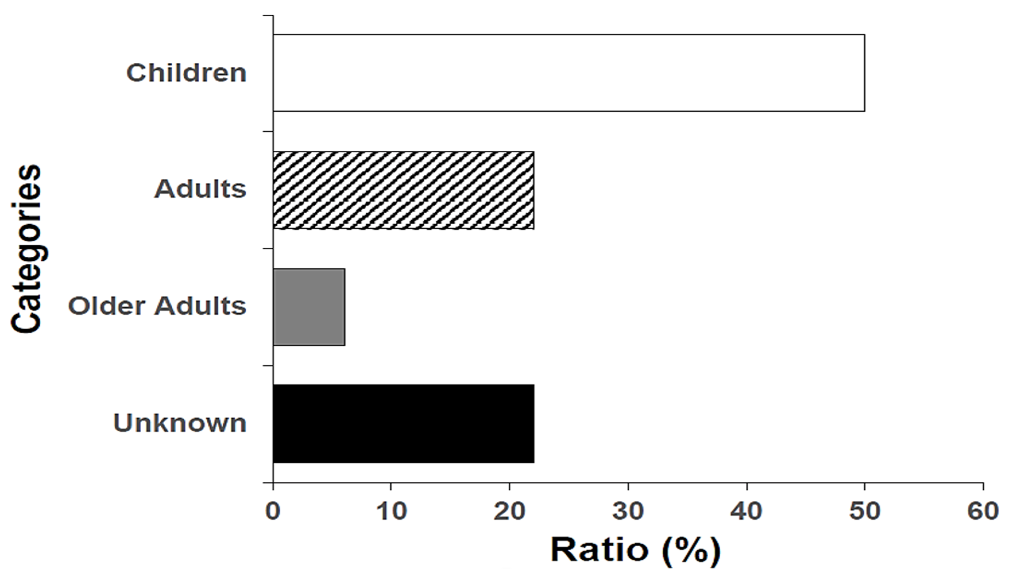
3.1. Demographic Features and Shigella Isolate Serogroup Distribution

Of the 48 presumptive Shigella isolates, 32 (66.7%) tested positive on species-specific serology. The 32 seropositive isolates exhibited a diverse age distribution; 16 (16/32, 50%) from children (1–12 years), seven (7/32, 22%) from adults (18–59 years old), and two (2/32, 6%) from older adults (60 years and above), while seven (7/32, 22%) had missing age data (Figure 1). The serogroup-based species classification of Shigella isolates is depicted in Table 1. Of these, the most predominant serogroup was Group B, S. flexneri (16/32, 50%), followed by Group D, S. sonnei 14 (14/32, 44%), and both Group A, S. dysenteriae, and Group C, S. boydii (1/32, 3%).

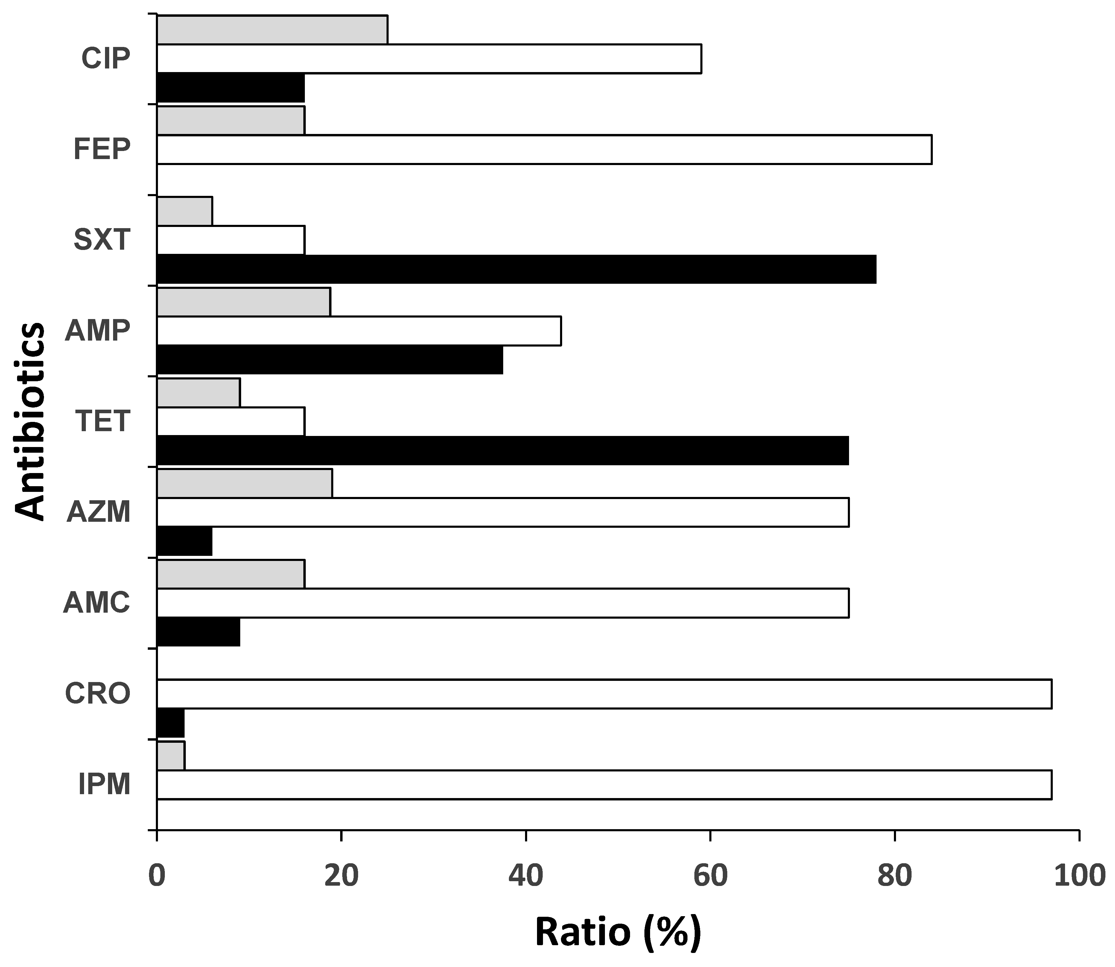
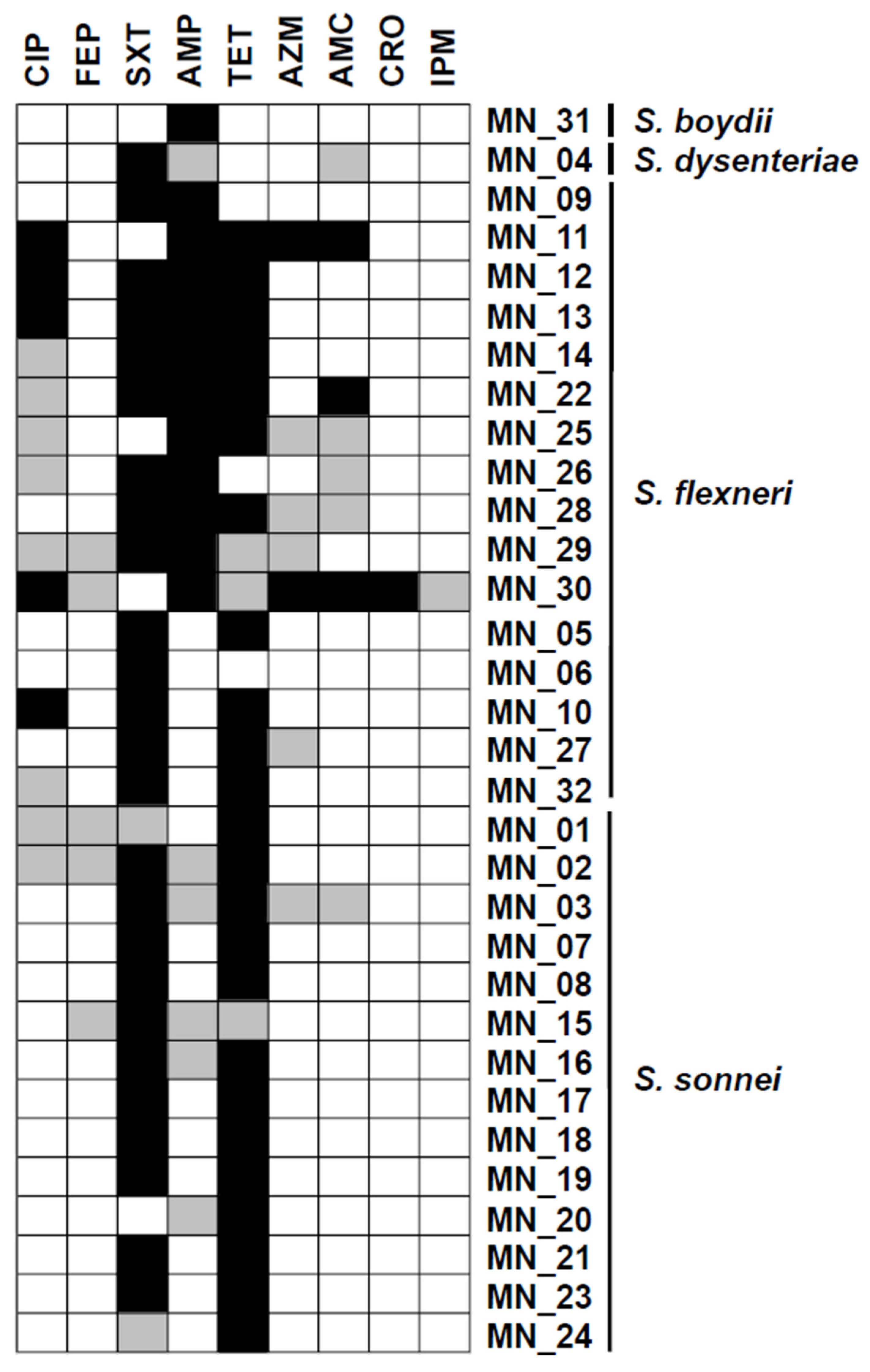
3.2. Antibiotic Resistance Rates to Commonly Used Antibiotics and WHO Priority Shigella Isolates

Generally, the resistance rates varied across different antibiotic classes, with the highest being sulfamethoxazole/trimethoprim (SXT) (25/32, 78%) and tetracycline (TET) (24/32, 75%). Much lower resistance rates were observed against ampicillin (12/32, 38%), ciprofloxacin (CIP) (5/32, 16%), amoxicillin-clavulanic acid (AMC) (3/32, 9%), and azithromycin (AZM) (2/32, 6%) (Figure 2). Notably, resistance to third-generation cephalosporins (3GCs) was low, with only one (1/32, 3%) isolate resistant to ceftriaxone. The ceftriaxone-resistant isolate also exhibited intermediate resistance to the drug of last resort, imipenem. Among the 16 S. flexneri isolates, 13 (13/16, 81%) were resistant to SXT, 11 (11/16, 69%) to ampicillin (AMP), and 11 (11/16, 69%) to TET. Resistance to first- and second-line treatment (CIP and AZM, respectively) was detected only in S. flexneri isolates. Contrarily, resistance among S. sonnei species was observed only against SXT (11/14 78.5%) and TET (13/14, 93%), with occasional intermediate resistance to AMC, cefepime (FEP), AMP, AZM, and CIP. Two isolates (MN_12 and MN_13) from the UTH Adult Hospital shared the same AMR pattern (Figure 3).

3.3. Proportion of Multidrug Resistance Shigella Species

Multidrug resistance (MDR) was defined as non-susceptibility to at least one agent in three or more antimicrobial classes [36]. The Shigella MDR phenotypes detected in this study are shown in Table 2. There were six combinations of multidrug-resistant phenotypes involving eight (8/32, 25%) isolates, all being S. flexneri. The most predominant MDR phenotypes were SXT/TET/AMP/CIP and SXT/AMP/TET (2/8, 25%, each). Phenotypes AMC/TET/SXT, AMC/AZM/CIP/TET, CIP/AZM/AMC/CRO/TET, and CIP/TET/SXT all had one (1/8, 13%, each).

3.4. β-Lactamase-Encoding Genes Were Detected Among Shigella Species

Genomic DNA from the 32 clinical isolates of Shigella was screened for blaCTX-M and blaTEM genes by PCR using previously described primers [32,33,34]. The expected bands (i.e., 480 bp) for the blaTEM gene were obtained in seven (7/32, 22%) isolates (Figure S1). The blaTEM gene was present in seven out of twelve (7/12, 58.3%) AMP-resistant isolates, and only in one out of three (1/3, 33.3%) AMC-resistant isolates (Figure 3). However, none of the strains tested positive for the blaCTX-M gene.

4. Discussion

This study revealed the sero-diversity and resistance profiles of clinical Shigella isolates from the highest referral hospital in Zambia. The clinical isolates exhibited a diverse age distribution, the majority (16/32, 50%) being children (1–12 years), followed by adults (7/32, 22%) (18–59). According to a recent CDC report, Shigella is most predominant in children globally [2]. This study, along with others conducted in different locations [37,38] also identified S. sonnei as the most predominant species among children. In this study, the most predominant serogroup was S. flexneri (16/32, 50%), and the least predominant were S. dysenteriae and S. boydii, at 3% (1/32) each. This is similar to what is reported elsewhere, in Iran [39], China [3,40], and India [41]. The second most predominant species in this study was S. sonnei, which has been reported to be the most prevalent species in the developed world [42]. The lower proportion of S. sonnei observed in this study and in many developing countries could be related to cross-immunity associated with frequent exposure to Plesiomonas shigelliodes [43]. Nevertheless, there has been a recent shift towards increasing rates of S. sonnei infections being reported in low- or middle-income countries [14], which is attributed to increased international travel [42,44].
Generally, the resistance rates varied across different antibiotic classes, the highest being SXT (25/32, 78%) and TET (24/32, 75%). The observed high resistance level could be related to antimicrobial overuse. For instance, SXT is often used as prophylaxis for Pneumocystis jirovecii pneumonia in HIV-exposed infants [45] and those with very low CD4 counts. Additionally, this drug class has been on the market for a long time [46], giving microorganisms time to develop resistance [47]. In addition, the low cost of this drug and poor restrictions on its access have led to overuse and misuse by the general public [48].
Although TET is not among the recommended treatments for Shigella infection, our results show a high resistance rate, often coinciding with SXT resistance. This observation could be caused by the co-selection of genes encoding resistance to the two drug classes. For instance, the tet and dfr genes have been reported to co-exist on the same plasmid of chromosomal insertion [35], suggesting that selection pressure created by one drug could be selected for both. Notably, resistance to 3GCs was low, with only one (1/32, 3%) isolate resistant to ceftriaxone. The ceftriaxone-resistant isolate also exhibited intermediate resistance to the drug of last resort, imipenem, suggesting the need for close monitoring.
This study reports MDR Shigella with a prevalence of 25% (8/32). Despite the lower prevalence of MDR reported in this study compared to other studies reported elsewhere [49,50], there is a need for monitoring the observed phenomenon. Curiously, all the MDR phenotypes were observed only in S. flexneri isolates, and majority of the isolates with similar MDR profiles were from the same health facility. Furthermore, these facilities are located in densely populated urban areas with inadequate sanitation and hygiene services [51,52]. This may suggest the clonal spread of multidrug-resistant S. flexneri as a nosocomial or community-acquired infection. This could also indicate the spread of MDR among S. flexneri via the horizontal gene transfer of AMR-gene-carrying plasmids. Therefore, further investigations, such as molecular epidemiology using whole-genome sequencing and detailed demographic history, are needed to confirm this observation.
WHO treatment guidelines for shigellosis recommend CIP as a first-line option, together with either pivmecillinam, CRO, or AZM (in adults only) as a second-line antibiotic with zinc supplementation [16]. Similarly, the Zambia Standard Treatment Guidelines recommend including ciprofloxacin in treating shigellosis in both adults and children [53], while AZM is reserved for second-line treatment in adults. High resistance to CIP and AZM was detected in this study and was only observed in S. flexneri isolates. In addition, intermediate resistance was observed in both S. flexneri and S. sonnei. The observed resistance to CIP and AZM is of great concern, and it must be closely monitored to prevent the situation from escalating and causing challenges in managing shigellosis. Our results suggest that the current treatment regimens may be ineffective, thus there is a need to strengthen Shigella surveillance and revise shigellosis management guidelines based on updated antibiograms.
The blaTEM gene was present in seven (7/32, 22%) Shigella isolates, suggesting β-lactamase-mediated resistance. β-lactamase inhibitors such as clavulanic acid inhibit β-lactamases by irreversibly binding to the active site, rendering the enzyme ineffective [54]. Expectedly, most (6/7, 85.7%) of the blaTEM-positive isolates were susceptible or only exhibited intermediate resistance to AMC. The observation that the blaTEM gene was not found in all the penicillin-resistant isolates highlights that other mechanisms are responsible for β-lactam resistance. Therefore, future studies should explore more robust approaches like whole-genome sequencing.

5. Conclusions

We characterized 32 Shigella isolates collected in 2021 from patients at the UTH. Speciation of the isolates revealed a serological diversity of the four Shigella species—S. flexneri, S. sonnei, S. boydii, and S. dysenteriae. We found that S. flexneri was the most predominant isolated species, with half exhibiting MDR with a prevalence of 25% (8/32). Consequently, the resistance of Shigella species to CIP and AZM is of public health concern, as these are the recommended treatment options for shigellosis. The blaTEM gene was detected in 58.3% (7/12) AMP-resistant strains, but none possessed the blaCTX-M gene. Hence, there is a need for systematic AMR surveillance for Shigella to guide clinical management in Zambia. Additionally, we recommend improved sanitation services as key to infection prevention and control, particularly in endemic areas with a high prevalence of shigellosis.

6. Limitation of This Study

The small sample size affected the generalization of the findings and prevented us from identifying significant differences or relationships. Furthermore, the absence of comprehensive epidemiological data hindered the contextualization of the results, making it challenging to account for confounding variables such as socioeconomic factors and underlying clinical conditions. Consequently, future studies should involve larger sample sizes and robust epidemiological data to make meaningful statistical inferences.

Supplementary Materials

The following supporting information can be downloaded at https://www.mdpi.com/article/10.3390/bacteria4020018/s1. Figure S1. Agarose gel electrophoresis results for the blaTEM gene of the 14 representative Shigella isolates: M—100 bp molecular weight marker (HiGenoMB-Himedia, Mumbai, India); PC—positive control; NC—negative control. Table S1: Metadata and susceptibility patterns for the studied isolates.

Author Contributions

Conceptualization, M.N., J.Y.C., J.M., B.M.H., R.C. (Roma Chilengi), C.N. and Y.S.; methodology, M.N., J.Y.C., M.S., R.S.K., M.M.K., R.C. (Rapheal Chanda), H.K.K., T.K., B.M.H., R.C. (Roma Chilengi), C.N. and Y.S.; software M.S., Y.K., R.C. (Rapheal Chanda), H.C., H.K.K. and F.W.C.; validation, M.N., J.Y.C., J.M., M.M.M., R.N., C.S., M.M.K., R.C. (Rapheal Chanda), H.K.K., T.K., S.M., F.W.C., B.M.H., R.C. (Roma Chilengi), C.N. and Y.S.; formal analysis, M.N., J.Y.C., J.M., M.S., R.S.K., M.M.M., N.R.M., M.M.K., B.M.H. and C.N.; investigation, M.N., J.Y.C., J.M., M.S., R.S.K., M.M.M., N.R.M., C.S., M.M.K., Y.K., H.C., S.M., F.W.C., R.C. (Roma Chilengi) and Y.S.; resources, M.M.M., R.N., N.R.M., B.M.H., R.C. (Roma Chilengi), C.N. and Y.S.; data curation, M.N., J.M., R.S.K., R.N., N.R.M., C.S., M.M.K., Y.K., H.C., S.M., F.W.C., R.C. (Roma Chilengi) and Y.S.; writing—original draft preparation, M.N., J.Y.C., J.M. and N.R.M.; writing—review and editing, M.N., M.S., R.S.K., M.M.M., R.N., C.S., Y.K., R.C. (Rapheal Chanda), H.C., H.K.K., T.K., S.M., F.W.C., B.M.H., R.C. (Roma Chilengi), C.N. and Y.S.; visualization M.N., C.S., M.M.K., Y.K., R.C. (Rapheal Chanda), H.C., H.K.K., T.K. and S.M.; supervision, J.Y.C., J.M. and M.S.; project administration, J.Y.C. and M.S.; funding acquisition, C.N. and Y.S. All authors have read and agreed to the published version of the manuscript.

Funding

This work was supported in part by the Japan Agency for Medical Research and Development (AMED) under Grant Numbers JP23wm0125008 and JP233fa627005 to Y.S.

Institutional Review Board Statement

Ethics approval for this study was obtained from Levy Mwanawasa Medical University Research Ethics Committee with the reference number LMMU-REC 00009/22 and the National Health Research Authority with the reference number NHRA000006/23/02/2023. Permission to use the clinical bacterial isolates was obtained from the University Teaching Hospital management.

Informed Consent Statement

Not applicable.

Data Availability Statement

The data used in this study can be accessed by request from the corresponding author.

Acknowledgments

We wish to thank the management of the University Teaching Hospital, Lusaka, for permission to carry out this study. We also wish to acknowledge the members of staff at the Bacteriology Laboratory of UTH for providing support during sample processing.

Conflicts of Interest

The authors declare no conflicts of interest.

References

  1. Anderson, M.; Sansonetti, P.J.; Marteyn, B.S. Shigella Diversity and Changing Landscape: Insights for the Twenty-First Century. Front. Cell. Infect. Microbiol. 2016, 6, 45. [Google Scholar] [CrossRef]
  2. Hale, T.L.; Keusch, G.T. Shigella. In Medical Microbiology; University of Texas Medical Branch at Galveston: Galveston, TX, USA, 1996. Available online: https://www.ncbi.nlm.nih.gov/books/NBK8038/ (accessed on 29 January 2025).
  3. Liu, H.; Zhu, B.; Qiu, S.; Xia, Y.; Liang, B.; Yang, C.; Dong, N.; Li, Y.; Xiang, Y.; Wang, S.; et al. Dominant serotype distribution and antimicrobial resistance profile of Shigella spp. in Xinjiang, China. PLoS ONE 2018, 13, e0195259. [Google Scholar] [CrossRef]
  4. Pieper, R.; Zhang, Q.; Parmar, P.P.; Huang, S.; Clark, D.J.; Alami, H.; Donohue-Rolfe, A.; Fleischmann, R.D.; Peterson, S.N.; Tzipori, S. The Shigella dysenteriae serotype 1 proteome, profiled in the host intestinal environment, reveals major metabolic modifications and increased expression of invasive proteins. Proteomics 2009, 9, 5029–5045. [Google Scholar] [CrossRef] [PubMed]
  5. Sethuvel, D.M.; Ragupathi, N.D.; Anandan, S.; Veeraraghavan, B. Update on: Shigella new serogroups/serotypes and their antimicrobial resistance. Lett. Appl. Microbiol. 2016, 64, 8–18. [Google Scholar] [CrossRef]
  6. Liu, J.; Pholwat, S.; Zhang, J.; Taniuchi, M.; Haque, R.; Alam, M.; Ochieng, J.B.; Jones, J.A.; Platts-Mills, J.A.; Tennant, S.M.; et al. Evaluation of Molecular Serotyping Assays for Shigella flexneri Directly on Stool Samples. J. Clin. Microbiol. 2021, 59, e02455-20. [Google Scholar] [CrossRef] [PubMed]
  7. CDC. CDC Yellow Book 2023; CDC: Atlanta, GA, USA, 2023. [Google Scholar]
  8. Miti, S.; Chilyabanyama, O.N.; Chisenga, C.C.; Chibuye, M.; Bosomprah, S.; Mumba, C.; Chitondo, S.; Siziya, S.; Cohen, D.; Chilengi, R.; et al. Sensitivity and predictive value of dysentery in diagnosing shigellosis among under five children in Zambia. PLoS ONE 2023, 18, e0279012. [Google Scholar] [CrossRef]
  9. Chisenga, C.C.; Bosomprah, S.; Simuyandi, M.; Mwila-Kazimbaya, K.; Chilyabanyama, O.N.; Laban, N.M.; Bialik, A.; Asato, V.; Meron-Sudai, S.; Frankel, G.; et al. Shigella-specific antibodies in the first year of life among Zambian infants: A longitudinal cohort study. PLoS ONE 2021, 16, e0252222. [Google Scholar] [CrossRef]
  10. Aslam, A.; Okafor, C.N. Shigella; StatPearls Publishing: Treasure Island, FL, USA, 2022. [Google Scholar]
  11. DuPont, H.L.; Levine, M.M.; Hornick, R.B.; Formal, S.B. Inoculum Size in Shigellosis and Implications for Expected Mode of Transmission. J. Infect. Dis. 1989, 159, 1126–1128. [Google Scholar] [CrossRef]
  12. Rout, W.; Formal, S.; Giannella, R.; Dammin, G. Pathophysiology of Shigella Diarrhea in the Rhesus Monkey: Intestinal Transport, Morphological, and Bacteriological Studies. Gastroenterology 1975, 68, 270–278. [Google Scholar] [CrossRef]
  13. Mattock, E.; Blocker, A.J. How Do the Virulence Factors of Shigella Work Together to Cause Disease? Front. Cell. Infect. Microbiol. 2017, 7, 64. [Google Scholar] [CrossRef]
  14. Ranjbar, R.; Farahani, A. Shigella: Antibiotic-Resistance Mechanisms And New Horizons For Treatment. Infect. Drug Resist. 2019, 12, 3137–3167. [Google Scholar] [CrossRef] [PubMed]
  15. Sharma, A.; Singh, A.; Dar, M.A.; Kaur, R.J.; Charan, J.; Iskandar, K.; Haque, M.; Murti, K.; Ravichandiran, V.; Dhingra, S. Menace of antimicrobial resistance in LMICs: Current surveillance practices and control measures to tackle hostility. J. Infect. Public Health 2021, 15, 172–181. [Google Scholar] [CrossRef] [PubMed]
  16. Sono, T.M.; Yeika, E.; Cook, A.; Kalungia, A.; Opanga, S.A.; Acolatse, J.E.E.; Sefah, I.A.; Jelić, A.G.; Campbell, S.; Lorenzetti, G.; et al. Current rates of purchasing of antibiotics without a prescription across sub-Saharan Africa; rationale and potential programmes to reduce inappropriate dispensing and resistance. Expert Rev. Anti-Infect. Ther. 2023, 21, 1025–1055. [Google Scholar] [CrossRef]
  17. Sulis, G.; Sayood, S.; Gandra, S. Antimicrobial resistance in low- and middle-income countries: Current status and future directions. Expert Rev. Anti-Infect. Ther. 2021, 20, 147–160. [Google Scholar] [CrossRef]
  18. Otaigbe, I.I.; Elikwu, C.J. Drivers of inappropriate antibiotic use in low- and middle-income countries. JAC-Antimicrobial Resist. 2023, 5, dlad062. [Google Scholar] [CrossRef]
  19. Amanda, G.-W.; Kayla, V.E.; Naeemah, L. Shigellosis: Travel-Associated Infections & Diseases. 2023. Available online: https://wwwnc.cdc.gov/travel/yellowbook/2024/infections-diseases/shigellosis (accessed on 29 January 2025).
  20. Williams, P.C.M.; Berkley, J.A. Guidelines for the treatment of dysentery (shigellosis): A systematic review of the evidence. Paediatr. Int. Child Health 2018, 38, S50–S65. [Google Scholar] [CrossRef]
  21. Klein, N.C.; Cunha, B.A. Third-generation cephalosporins. Med. Clin. N. Am. 1995, 79, 705–719. [Google Scholar] [CrossRef]
  22. UNICEF. Annual Report; For Every Child, Every Right; United Nations Children’s Fund: New York, NY, USA, 2018. [Google Scholar]
  23. Yeboah-Antwi, K.; MacLeod, W.B.; Biemba, G.; Sijenyi, P.; Höhne, A.; Verstraete, L.; McCallum, C.M.; Hamer, D.H. Improving Sanitation and Hygiene through Community-Led Total Sanitation: The Zambian Experience. Am. J. Trop. Med. Hyg. 2019, 100, 1005–1012. [Google Scholar] [CrossRef]
  24. WHO; UNICEF. Simple Solution Improves Water and Sanitation in Zambian Health-Care Facilities. 2015. Available online: https://www.who.int/news-room/feature-stories/detail/simple-solution-improves-water-and-sanitation-in-zambian-health-care-facilities (accessed on 29 January 2025).
  25. INDEX GHS. Global Health Security Index scores for Zambia; INDEX GHS. 2021. Available online: https://ghsindex.org/wp-content/uploads/2021/12/Zambia.pdf (accessed on 29 January 2025).
  26. Chiyangi, H.; Muma, J.B.; Malama, S.; Manyahi, J.; Abade, A.; Kwenda, G.; Matee, M.I. Identification and antimicrobial resistance patterns of bacterial enteropathogens from children aged 0–59 months at the University Teaching Hospital, Lusaka, Zambia: A prospective cross sectional study. BMC Infect. Dis. 2017, 17, 117. [Google Scholar] [CrossRef]
  27. Tuttle, J.; Ries, A.A.; Chimba, R.M.; Perera, C.U.; Bean, N.H.; Griffin, P.M. Antimicrobial-Resistant Epidemic Shigella Dysenteriae Type 1 in Zambia: Modes of Transmission. J. Infect. Dis. 1995, 171, 371–375. [Google Scholar] [CrossRef]
  28. Niyogi, S.K. Shigellosis. J. Microbiol. 2005, 43, 133–143. [Google Scholar] [PubMed]
  29. Hudzicki, J. Kirby-Bauer Disk Diffusion Susceptibility Test Protocol. Am. Soc. Microbiol. 2009, 15, 1–23. [Google Scholar]
  30. McFaland, J. Nephelometer: An instrument for media used for estimating the number of bacteria in suspensions used for calculating the opsonic index and for vaccines. J. Am. Med. Assoc. 1907, 49, 1176–1178. [Google Scholar]
  31. CLSI. Performance Standards for Antimicrobial Susceptibility Testing, 31st ed.; CLSI: Philadelphia, PA, USA, 2021. [Google Scholar]
  32. Mabilat, C.; Courvalin, P. Development of “oligotyping” for characterization and molecular epidemiology of TEM beta-lactamases in members of the family Enterobacteriaceae. Antimicrob. Agents Chemother. 1990, 34, 2210–2216. [Google Scholar] [CrossRef]
  33. Cai, W.; Fu, Y.; Zhang, W.; Chen, X.; Zhao, J.; Song, W.; Li, Y.; Huang, Y.; Wu, Z.; Sun, R.; et al. Synergistic effects of baicalein with cefotaxime against Klebsiella pneumoniae through inhibiting CTX-M-1 gene expression. BMC Microbiol. 2016, 16, 181. [Google Scholar] [CrossRef]
  34. Pokhrel, R.H.; Thapa, B.; Kafle, R.; Shah, P.K.; Tribuddharat, C. Co-existence of beta-lactamases in clinical isolates of Escherichia coli from Kathmandu, Nepal. BMC Res. Notes 2014, 7, 694. [Google Scholar] [CrossRef]
  35. Shawa, M.; Furuta, Y.; Mulenga, G.; Mubanga, M.; Mulenga, E.; Zorigt, T.; Kaile, C.; Simbotwe, M.; Paudel, A.; Hang’ombe, B.; et al. Novel chromosomal insertions of ISEcp1-blaCTX-M-15 and diverse antimicrobial resistance genes in Zambian clinical isolates of Enterobacter cloacae and Escherichia coli. Antimicrob. Resist. Infect. Control 2021, 10, 79. [Google Scholar] [CrossRef]
  36. Magiorakos, A.-P.; Srinivasan, A.; Carey, R.B.; Carmeli, Y.; Falagas, M.E.; Giske, C.G.; Harbarth, S.; Hindler, J.F.; Kahlmeter, G.; Olsson-Liljequist, B.; et al. Multidrug-resistant, extensively drug-resistant and pandrug-resistant bacteria: An international expert proposal for interim standard definitions for acquired resistance. Clin. Microbiol. Infect. 2012, 18, 268–281. [Google Scholar] [CrossRef]
  37. Gharpure, R.; Marsh, Z.A.; Tack, D.M.; Collier, S.A.; Strysko, J.; Ray, L.; Payne, D.C.; Garcia-Williams, A.G. Disparities in Incidence and Severity of Shigella Infections Among Children—Foodborne Diseases Active Surveillance Network (FoodNet), 2009-2018. J. Pediatr. Infect. Dis. Soc. 2021, 10, 782–788. [Google Scholar] [CrossRef]
  38. McCrickard, L.S.; Crim, S.M.; Kim, S.; Bowen, A. Disparities in severe shigellosis among adults—Foodborne diseases active surveillance network, 2002–2014. BMC Public Health 2018, 18, 221. [Google Scholar] [CrossRef]
  39. Nikfar, R.; Shamsizadeh, A.; Darbor, M.; Khaghani, S.; Moghaddam, M. A Study of prevalence of Shigella species and antimicrobial resistance patterns in paediatric medical center, Ahvaz, Iran. Iran. J. Microbiol. 2017, 9, 277–283. [Google Scholar]
  40. Zhu, Z.; Wang, W.; Cao, M.; Zhu, Q.; Ma, T.; Zhang, Y.; Liu, G.; Zhou, X.; Li, B.; Shi, Y.; et al. Virulence factors and molecular characteristics of Shigella flexneri isolated from calves with diarrhea. BMC Microbiol. 2021, 21, 214. [Google Scholar] [CrossRef]
  41. Taneja, N.; Mewara, A. Shigellosis: Epidemiology in India. Indian J. Med. Res. 2016, 143, 565–576. [Google Scholar] [CrossRef] [PubMed]
  42. Drews, S.J.; Lau, C.; Andersen, M.; Ferrato, C.; Simmonds, K.; Stafford, L.; Fisher, B.; Everett, D.; Louie, M. Laboratory based surveillance of travel-related Shigella sonnei and Shigella flexneri in Alberta from 2002 to 2007. Glob. Health 2010, 6, 20. [Google Scholar] [CrossRef]
  43. Sack, D.; Hoque, S.; Etheridge, M.; Huq, A. Is protection against shigellosis induced by natural infection with Plesiomonas shigelloides? Lancet 1994, 343, 1413–1415. [Google Scholar] [CrossRef] [PubMed]
  44. Toro, C.; Arroyo, A.; Iglesias, N.; Sarria, A.; Enríquez, A.; Baquero, M.; de Guevara, C.L. Shigellosis in Subjects with Traveler’s Diarrhea Versus Domestically Acquired Diarrhea: Implications for Antimicrobial Therapy and Human Immunodeficiency Virus Surveillance. Am. J. Trop. Med. Hyg. 2015, 93, 491–496. [Google Scholar] [CrossRef]
  45. D’souza, A.W.; Moodley-Govender, E.; Berla, B.; Kelkar, T.; Wang, B.; Sun, X.; Daniels, B.; Coutsoudis, A.; Trehan, I.; Dantas, G. Cotrimoxazole Prophylaxis Increases Resistance Gene Prevalence and α-Diversity but Decreases β-Diversity in the Gut Microbiome of Human Immunodeficiency Virus–Exposed, Uninfected Infants. Clin. Infect. Dis. 2019, 71, 2858–2868. [Google Scholar] [CrossRef]
  46. Shambaugh, G.E. History of Sulfonamides. Arch. Otolaryngol. Head Neck Surg. 1966, 83, 1–2. [Google Scholar] [CrossRef]
  47. Yeta, K.I.; Michelo, C.; Jacobs, C. Antimicrobial Resistance among Pregnant Women with Urinary Tract Infections Attending Antenatal Clinic at Levy Mwanawasa University Teaching Hospital (LMUTH), Lusaka, Zambia. Int. J. Microbiol. 2021, 2021, 8884297. [Google Scholar] [CrossRef]
  48. Ryan, M.; Griffin, S.; Chitah, B.; Walker, A.S.; Mulenga, V.; Kalolo, D.; Hawkins, N.; Merry, C.; Barry, M.G.; Chintu, C.; et al. The cost-effectiveness of cotrimoxazole prophylaxis in HIV-infected children in Zambia. AIDS 2008, 22, 749–757. [Google Scholar] [CrossRef]
  49. Ud-Din, A.I.M.S.; Wahid, S.U.H.; Latif, H.A.; Shahnaij, M.; Akter, M.; Azmi, I.J.; Hasan, T.N.; Ahmed, D.; Hossain, M.A.; Faruque, A.S.G.; et al. Changing Trends in the Prevalence of Shigella Species: Emergence of Multi-Drug Resistant Shigella sonnei Biotype g in Bangladesh. PLoS ONE 2013, 8, e82601. [Google Scholar] [CrossRef] [PubMed]
  50. Salleh, M.Z.; Zuraina, N.M.N.N.; Hajissa, K.; Ilias, M.I.; Singh, K.K.B.; Deris, Z.Z. Prevalence of Multidrug-Resistant and Extended-Spectrum Beta-Lactamase-Producing Shigella Species in Asia: A Systematic Review and Meta-Analysis. Antibiotics 2022, 11, 1653. [Google Scholar] [CrossRef] [PubMed]
  51. Ministry of Finance and National Planning. Socio-Economic Transformation for Improved Livelihoods. 2022. Available online: https://www.mot.gov.zm/?wpdmpro=eighth-national-development-plan-8ndp-2022-2026&wpdmdl=972&refresh=6799924eb4f741738117710 (accessed on 29 January 2025).
  52. Sukwa, N.; Bosomprah, S.; Somwe, P.; Muyoyeta, M.; Mwape, K.; Chibesa, K.; Luchen, C.C.; Silwamba, S.; Mulenga, B.; Munyinda, M.; et al. The Incidence and Risk Factors for Enterotoxigenic E. coli Diarrheal Disease in Children under Three Years Old in Lusaka, Zambia. Microorganisms 2024, 12, 698. [Google Scholar] [CrossRef] [PubMed]
  53. Ministry of Health Zambia NFC. Standard Treatment Guidelines, Essential Medicines List, Essential Laboratory Supplies for Zambia, 5th ed.; Zambia Ministry of Health: Lusaka, Zambia, 2020. [Google Scholar]
  54. Neu, H.C.; Fu, K.P. Clavulanic Acid, a Novel Inhibitor of β-Lactamases. Antimicrob. Agents Chemother. 1978, 14, 650–655. [Google Scholar] [CrossRef]
Figure 1. Shigella isolates categorized by age distribution. White, hatched, gray, and black bars denote children, adults, older adults, and unknown age groups, respectively.
Bacteria 04 00018 g001
Figure 2. Antimicrobial susceptibility testing (AST) profiles of Shigella isolates (n = 32) to commonly used antibiotics. AMC: amoxicillin-clavulanic acid. AZM: azithromycin. IPM: imipenem. CRO: ceftriaxone. FEP: cefepime. CIP: ciprofloxacin. AMP: ampicillin. TET: tetracycline. SXT: sulfamethoxazole/trimethoprim. White, gray, and black bars denote susceptible, intermediate, and resistant phenotypes, respectively.
Bacteria 04 00018 g002
Figure 3. Heatmap showing Shigella isolates and their AST results to antibiotics. AMC: amoxicillin-clavulanic acid. AZM: azithromycin. IPM: imipenem. CRO: ceftriaxone. FEP: cefepime. CIP: ciprofloxacin. AMP: ampicillin. TET: tetracycline. SXT: sulfamethoxazole/trimethoprim. White, gray, and black boxes denote susceptible, intermediate, and resistant phenotypes, respectively.
Bacteria 04 00018 g003
Table 1. Shigella isolate serogroup distribution (n = 32).
Name of SpeciesMost Prevalent Age Category Number (n)Percentage (%)
S. flexneriChildren (6/16; 37.5%) *1650
S. sonneiChildren (10/14; 71.4%)1444
S. dysenteriaeUnknown (1; 100%)13
S. boydiiOlder adult (1; 100%)13
Total32100
* In brackets are the proportions and percentages for the most prevalent age category.
Table 2. MDR phenotype patterns and facility of referral for S. flexneri isolates.
MDR PhenotypeStrain IDFacility of OriginNo. of IsolatesPercentage (%)
SXT/TET/AMP/CIPMN_12UTH Adult225
MN_13UTH Adult
SXT/AMP/TETMN_14Matero Hospital225
MN_28UTH Paeds
AMC/TET/SXTMN_22UTH Paeds112.5
CIP/TET/SXTMN-10Chilanga Hospital112.5
CIP/AZM/AMC/CRO/TETMN_11UTH Adult112.5
AMC/AZM/CIP/TETMN_30State House Clinic112.5
Disclaimer/Publisher’s Note: The statements, opinions and data contained in all publications are solely those of the individual author(s) and contributor(s) and not of MDPI and/or the editor(s). MDPI and/or the editor(s) disclaim responsibility for any injury to people or property resulting from any ideas, methods, instructions or products referred to in the content.

