Article

Comparative Analysis of Oral Prevotella intermedia, Tannerella forsythia, Streptococcus sanguinis, and Streptococcus mutans in Patients with Esophageal Squamous Cell Carcinoma and Healthy Controls in Mthatha, South Africa

by
Yolanda Yolisa Nokamatye
,
Gabriel Tchuente Kamsu
and
Eugene Jamot Ndebia
*
Department of Human Biology, Faculty of Medicine and Health Sciences, Walter Sisulu University, Mthatha 5100, South Africa
*
Author to whom correspondence should be addressed.
Bacteria 2025, 4(1), 11; https://doi.org/10.3390/bacteria4010011
Submission received: 3 January 2025 / Revised: 4 February 2025 / Accepted: 12 February 2025 / Published: 1 March 2025

Abstract

:
The microbiome plays a crucial role in cancer development, influencing fundamental processes such as cell proliferation, apoptosis, immune system regulation, and host metabolism. Recent studies have highlighted a possible relationship between esophageal cancer and the oral microbiota, making oral microflora a possible risk factor. The bacteria Prevotella intermedia, Tannerella forsythia, Streptococcus sanguinis, and Streptococcus mutans, implicated in various oral pathologies, were of interest in this study, which was initiated to examine their potential role in the etiology of esophageal squamous cell carcinoma (ESCC). To achieve this, a case-control design was used, with whole saliva samples collected from 24 healthy controls and 24 patients with esophageal squamous cell carcinoma. DNA was then extracted, and real-time PCR was performed to quantify the presence of the targeted bacteria in both groups. The results showed that all the bacteria studied were present in the saliva of both patients with ESCC and healthy controls. However, expression levels were significantly higher in patients with ESCC. Specifically, a marked increase in the presence of P. intermedia, T. forsythia, S. sanguinis, and S. mutans was observed in the patients with cancer compared to the healthy controls. In short, this study highlights a significant imbalance in the microbial flora, with an increased abundance of selected bacteria in patients with ESCC. The monitoring of these bacteria could thus be exploited to track patients who are at risk. Their integration into diagnostic and therapeutic strategies would offer new prospects for the early diagnosis and improved prognosis of patients at risk of ESCC.

1. Introduction

Esophageal carcinoma (EC) is the eighth most common cancer globally and the sixth leading cause of cancer-related deaths [1]. It mainly presents as esophageal squamous cell carcinoma (ESCC) and esophageal adenocarcinoma (EAC), classified based on their origin: ESCC originates from squamous epithelial cells in the upper esophagus and is more common in developing countries, while EAC originates from glandular cells in the lower esophagus and is predominantly seen in developed countries [2,3,4]. In South Africa, particularly in the Eastern Cape province, ESCC has a high prevalence, notably in rural areas like Transkei, and is linked to factors such as the consumption of Fusarium verticillioides contaminated food [5]. Risk factors for EC include tobacco use [6], alcohol consumption [7], low fruit and vegetable intake, gastroesophageal reflux disease (GERD), Barrett’s esophagus, and others [5,8,9].
The oral microbiome, consisting of over 700 bacterial species [10], is thought to influence overall health [11,12], including the development of EC. Dysbiosis of the oral microbiome has been linked to various cancers, including those of the digestive system [13,14]. Bacterial infections may contribute to cancer through mechanisms like chronic inflammation, anti-apoptotic activity, and the production of carcinogenic substances [15,16,17,18]. Certain oral bacteria, including Streptococcus mutans and Streptococcus sanguinis are notable for their role in oral biofilm formation, and their involvement in inflammation and immune modulation may influence cancer development [19,20]. Prevotella intermedia, associated with periodontal disease, has also been implicated in other cancers [21], while Tannerella forsythia, a bacterium frequently found in oral dysbiosis, is linked to inflammation [22,23]. These bacteria, through their role in dysbiosis, may alter the local environment in the esophagus, contributing to cancer progression or increasing cancer risk through inflammatory and carcinogenic pathways [24].
Furthermore, the oral microbiota has been associated with precancerous lesions and cancer progression, including the enhancement of alcohol metabolism, which increases cancer risk [25,26,27]. Given the similarity between the esophageal and oral microbiomes, the oral microbiome has been studied as a potential biomarker for gastrointestinal malignancies, including ESCC [28]. This study aimed to analyze and compare the presence and abundance of specific oral bacteria (Streptococcus mutans, Streptococcus sanguinis, Prevotella intermedia, and Tannerella forsythia) in the oral microbiome of patients with ESCC and healthy controls in Mthatha, Eastern Cape, South Africa. The goal was to assess differences in the oral microbiome between these groups, specifically regarding the abundance of these bacteria.

2. Materials and Methods

2.1. Study Design and Population

This study was a case–control comparing and analyzing the presence and abundance of P. intermedia, T. forsythia, S. mutans, and S. sanguinis within the oral microbiome of patients diagnosed with ESCC (cases) and healthy individuals (controls). Cases included patients endoscopically diagnosed and histologically confirmed for ESCC at the Oncology Department of the Nelson Mandela Academic Hospital. The control group was made up of healthy volunteers recruited from around the hospital, with no family history or association with any form of cancer.

2.2. Inclusion and Exclusion Criteria

The inclusion criteria for cases were as follows: patients diagnosed with squamous cell esophageal cancer, specifically men and women aged 30 years and over. The exclusion criteria included edentulous patients, pregnant women, patients with chronic medical conditions, and those receiving antibiotic treatment. Regarding the controls, the inclusion criteria stipulated that participants needed to be male or female, living near the NMAH, and aged 30 or over. The exclusion criteria included participants with esophageal cancer, those with chronic conditions, those on antibiotic treatment, and participants who had not given informed consent.

2.3. Data Collection

Data were collected using a structured questionnaire. Participants were assisted in completing the questionnaire where necessary, particularly for those without writing skills. The demographic information collected included age, gender, marital status, education level, smoking status, and alcohol consumption. The questionnaire was labeled with a unique code assigned to each participant, ensuring the anonymity and confidentiality of the data.
The sample size was calculated using the formula for a case-control study [29], considering a significance level (α) of 0.05 and a power (1-β) of 0.70. With expected exposure proportions of 0.2 for controls and 0.3 for cases, the required sample size was determined to be 48 (24 cases and 24 controls). To reduce selection bias, all participants meeting the inclusion criteria were recruited. All patients diagnosed with ESCC at the Nelson Mandela Hospital during the study period were included, and healthy controls were matched to the patients with ESCC according to age, geographical location (Mthatha), and gender.

2.4. Sample Collection

Unstimulated saliva was collected from all participants following the protocol described by Jo et al. [30]. Before collection, the participants rinsed their mouths with mineral water to remove food debris and then waited approximately 30 min to allow natural saliva production and minimize sample dilution. A minimum of 1 mL of saliva was collected from the participants by allowing it to pool in the mouth and spit into a sample tube. Samples were immediately stored in a cooler containing ice and transported to the laboratory, where they were stored at −80 °C within 3 h of collection to ensure DNA stability for subsequent analysis.

2.5. DNA Extraction and Quantification

According to the manufacturer’s protocol, DNA extraction from the saliva samples was performed using the ZymoBIOMICS DNA Extraction Miniprep Kit (Zymo Research, 17062 Murphy Ave, Irvine, CA 92614, USA). Then, DNA concentration and purity were assessed using a nanodrop spectrophotometer (Thermo Scientific, NND-1 ND-ONE-W-1PR22, Bangkok, Thailand). To prevent degradation by chemical and enzymatic processes, extracted DNA was stored as a precipitate in ethanol at −80 °C [31].

2.6. Probe/Primer Sets for Real-Time PCR

Bacteria-specific TaqMan probe and primer sets were designed from the species-specific regions on the 16S rRNA and were utilized to detect P. intermedia, T. forsythia, S. mutans, and S. sanguinis (Table 1). Additionally, as reported by Barbadoro et al. [32], a universal primer pair based on the conserved region of the 165S rRNA gene was used to quantify the total amount of eubacterial species in the specimens. The TaqMan probes and primers were ordered and synthesized from Inqaba biotec (Inqaba Biotechnical Industries (Pty) Ltd., Pretoria, South Africa).

2.7. Amplification of DNA Through Real-Time Polymerase Chain Reaction

Real-time PCR was performed using a CFX 96 Real-Time System, and all sample were run in duplicates. Each PCR was carried out in a total volume of 20 μL, consisting of 10 μL of Luna Universal Probe qPCR Master Mix, 0.8 μL each of forward and reverse primers, 0.4 µL of probe, 2 μL of template DNA solution, and an appropriate dose of sterilized nuclease-free water. Amplification was conducted on the CFX 96 Real-Time System using a specified thermocycling program. The fluorescence signal was acquired at the annealing temperature to monitor intensity. Fluorescence data were analyzed using CFX Manager Dx 3.1 (v3.1.3090.1022) Win C216125 software. The bacterial DNA level was quantified by qRT-PCR and converted to theoretical cell numbers as previously described by Barbadoro et al. [22,32].

2.8. Ethical Considerations

Ethical approval for this study was granted by the Research and Ethics Committee of the Faculty of Medicine and Health Sciences, Walter Sisulu University (protocol code 038/2022; approval date: 26 October 2022) for ethical and biosafety clearance. Additionally, approval was obtained from the Eastern Cape Health Research Committee (protocol number EC_202211_004; approval date: 7 November 2022).

2.9. Statistical Analysis

Data management, including compilation, aggregation, quality control, and coding, was carried out using Excel 2016. Data analysis was performed with Epi Info 7 (CDC, Atlanta, GA, USA). The log transformation was applied to quantitative variables to achieve a normal distribution. Means were compared using Student’s t-test and ANOVA. Significant factors were further adjusted using a multiple linear regression model. A confidence level of 95% was selected, with statistical significance defined as a p-value ≤ 0.05.

3. Results

3.1. Sociodemographic Parameters of Study Population

Table 2 compares sociodemographic characteristics between the healthy control and ESCC groups. The results reveal that patients with ESCC were predominantly older, with 75% aged 65 or over, and had a lower level of education, with 83.33% having only primary education. All participants in both groups were black South Africans from the Xhosa ethnic group. Additionally, patients with ESCC showed a higher prevalence of missing teeth (66.66% with 1–3 missing teeth) and were more exposed to risk behaviors such as smoking (70.83%) and alcohol consumption (66.66%) compared to the healthy controls. In contrast, healthy controls were characterized by a younger population, a higher level of education, and more favorable oral health habits, such as more frequent tooth brushing.

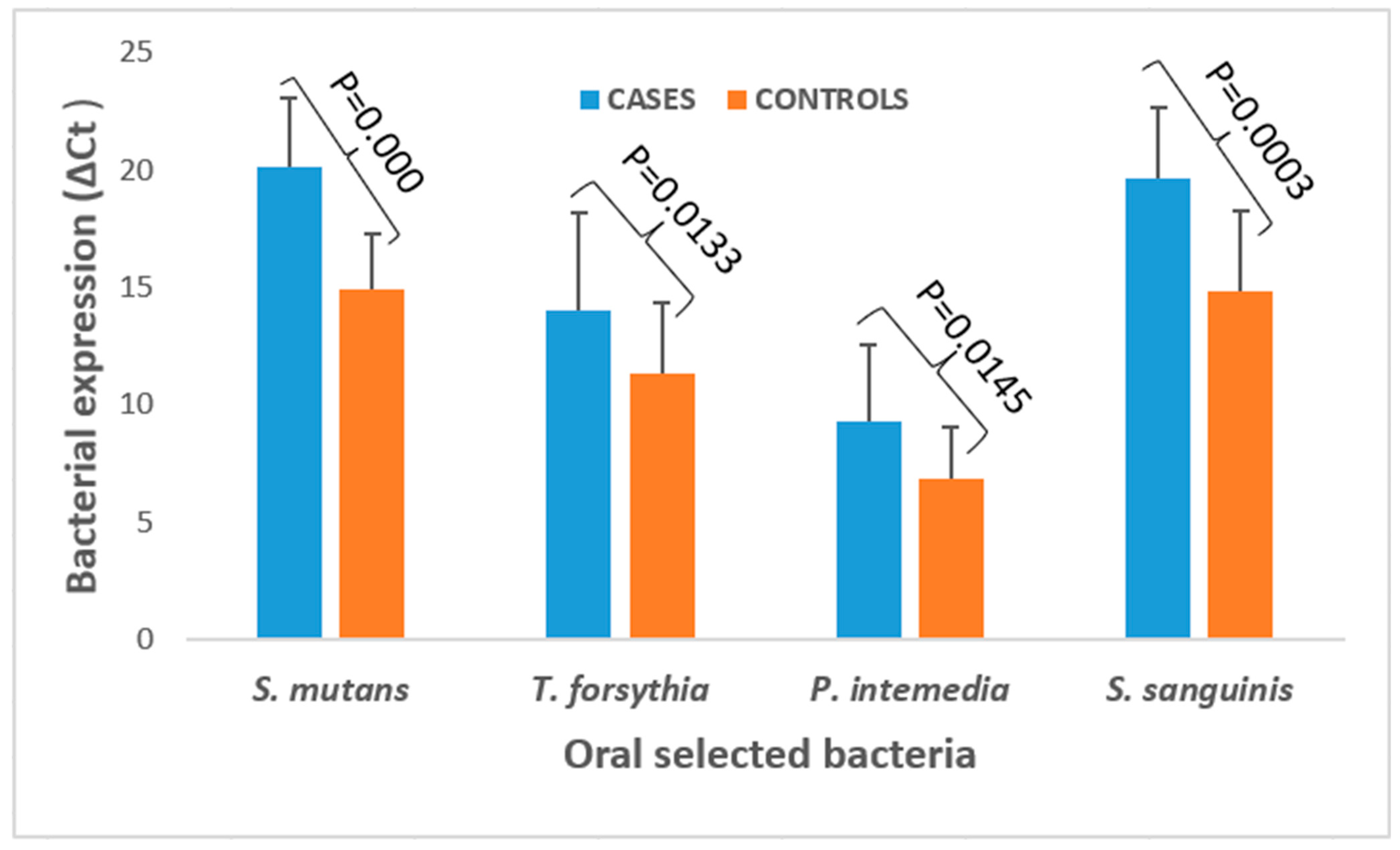
3.2. Bacterial Expression and Quantification in ESCC Cases and Healthy Controls

Figure 1 illustrates the distribution of S. mutans, S. sanguinis, P. intermedia, and T. forsythia bacteria and their bacterial load in the saliva of patients with ESCC and healthy controls in Mthatha, South Africa. The analysis of the figure revealed the presence of these bacteria in both groups, but the bacterial load was significantly higher in the patients with ESCC than in the healthy controls. The results also showed a significantly higher abundance of bacteria in the ESCC group than healthy controls. However, their expression levels were significantly higher in patients with ESCC, with p-values below 0.05 in all cases, underlining the significant association between these bacteria and ESCC.

3.3. Association Between Sociodemographic Characteristics and Bacterial Expression Levels of Selected Oral Bacteria in Healthy Controls and Patients with ESCC

Table 3 and Table 4 present an in-depth analysis of the demographic and behavioral parameters related to the presence of selected oral bacteria. The results in Table 3 reveal significant differences for several of the variables studied. Age shows a marked distinction for S. mutans (p = 0.0004) and S. sanguinis (p = 0.0087). Education level is also significant, particularly for S. mutans (p = 0.0000) and T. forsythia (p = 0.0146). Additionally, smoking status emerges as a determining factor, with notable differences for S. mutans (p = 0.0001) and T. forsythia (p = 0.0297). These results emphasize the importance of these demographic and behavioral factors in the presence of the studied oral bacteria. However, the multivariate analysis between age, education level, and smoking status did not reveal any association between these factors and the varying levels of expression of the selected bacteria (Table 4).

4. Discussion

Many years ago, the role played by microorganisms in cancer was ignored, but the finding that Helicobacter pylori bacteria promote gastric cancer brought back attention on this topic in 1994 [33]. The role of microbiome in the carcinogenesis of several cancers has since been demonstrated in that it influences various processes, such as host cell proliferation and apoptosis, the modulation of immune system activity, and its impact on host metabolism [34]. This work was conceived, focusing specifically on four bacteria of the oral microbiome: P. intermedia, T. forsythia, S. sanguinis, and S. mutans. This study compared their presence and abundance in the saliva of patients with ESCC and healthy controls to better understand their potential role in the development of ESCC.
The present study revealed the presence of the bacteria P. intermedia, T. forsythia, S. sanguinis, and S. mutans in the saliva of ESCC cases and healthy controls. This presence in both groups suggests that these bacteria are part of the natural habitat of the oral cavity and that an imbalance in their concentrations, in combination with other risk factors, could play a role in the development of ESCC. These findings are corroborated by the work of Könönen et al. [35], Zhu et al. [36], and Lemos et al. [37], who observed that these bacteria are present in low concentrations in the oral cavity of infants, but their abundance increases in healthy adults, even more so under pathological conditions. Bacteria of the genus Streptococcus are the first to colonize the oral cavity after birth, contributing to the establishment of the oral microbiome, and they dominate the soft tissues of the mouth [38]. These observations align with the results of the present study, which showed that these bacteria (S. mutans and S. sanguinis) were also present in the healthy controls.
This study also found that P. intermedia (p = 0.0152), S. mutans, and S. sanguinis (p < 0.001), as well as T. forsythia (p = 0.0146), were significantly more expressed in the patients with ESCC than in the healthy controls. The changes in the oral microflora observed in this study could be linked, on the one hand, to immune dysfunctions induced by ESCC, which cause immunodepression. On the other hand, these modifications promote the invasion of bacteria into the esophagus, where they attack it, thus contributing to the aggravation or onset of ESCC. This altered immune system reduces the body’s ability to control bacterial proliferation, thus facilitating disease progression [39]. These observations align with the work of Castañeda-Corzo et al. [40] and Yano et al. [41], who also observed an increased frequency of P. intermedia in oropharyngeal cancers and ESCC. P. intermedia produces volatile sulfur compounds, such as hydrogen sulfide (H2S) and methyl mercaptan (CH3SH), which are genotoxic agents that induce genomic instability, promote tumor proliferation, and stimulate angiogenesis. Thus, an imbalance in the oral microbiome, as observed in patients with ESCC, leads to an increase in volatile sulfur compounds in the oral cavity, accentuating their toxicity.
Yano et al. [41] and Chiang et al. [42] also observed an increased abundance of S. mutans and S. sanguinis in saliva samples from patients with ESCC. S. sanguinis is an oral cavity bacterium associated with healthy dental biofilm [36]. Although S. mutans was slightly more abundant than S. sanguinis in this study, it is known for its resistance to hydrogen peroxide (H2O2) produced by S. sanguinis, which disrupts bacterial equilibrium and could contribute to tumor progression [36]. S. sanguinis produces hydrogen peroxide (H2O2) and mutacins, which inhibit the growth of S. mutans, a cariogenic bacterium. However, S. mutans possesses an H2O2 resistance system, which allows it to produce acids, creating an acidic environment that reduces the population of S. sanguinis (which is acid-sensitive) in saliva [36]. Moreover, the hyperproduction of H2O2 could generate oxidative stress, causing genetic damage in cells in contact with bacteria, which could promote the initiation or progression of ESCC.
Saliva from patients with ESCC was shown to contain significantly (p < 0.001) more T. forsythia DNA than the healthy controls. These results align with those of Kawasaki et al. [43], who also identified T. forsythia as one of the oral bacteria associated with esophageal cancer. Furthermore, Lara et al. [44] have reported that T. forsythia, in association with other bacteria, plays a key role in triggering periodontal disease by enhancing its virulent factors, enabling it to become pathogenic.

5. Conclusions

The level of oral bacteria S. mutans, S. sanguinis, P. intermedia, and T. forsythia was significantly higher in patients with ESCC compared to the control group. The increasing number of selected oral bacteria during the development of ESCC may serve as a biomarker in the future. This study demonstrated the differences in the oral microbiota between patients with esophageal cancer and healthy controls. However, it also raised the prospect of future research to further our understanding of the role of these bacteria in the development and/or complications of esophageal tumors, particularly in the South African population.

6. Limitations

A major limitation of this work is the small sample size. A larger sample and studies in more targeted clinical contexts could further investigate these results and provide a better understanding of the factors underlying these relatively small variations in bacterial expression.

Author Contributions

Conceptualization, Y.Y.N. and E.J.N.; methodology, Y.Y.N. and G.T.K.; software, G.T.K.; validation, E.J.N.; formal analysis, G.T.K. and E.J.N.; investigation, Y.Y.N.; resources, Y.Y.N.; data curation, E.J.N.; writing—original draft preparation, Y.Y.N. and G.T.K.; writing—review and editing, G.T.K. and E.J.N.; visualization, G.T.K.; supervision, E.J.N.; project administration, E.J.N. and Y.Y.N.; and funding acquisition, E.J.N. All authors have read and agreed to the published version of the manuscript.

Funding

This study was supported by the Chemical Industries Education and Training Authority (CHIETA) and the Strategic Health Innovation Partnerships (SHIP) of the Medical Research Council (MRC), fundings attributed to Prof. Eugene Jamot Ndebia.

Institutional Review Board Statement

This study was conducted in accordance with the Declaration of Helsinki and approved by the Institutional Ethics Committee of the Faculty of Medicine and Health Sciences, Walter Sisulu University (protocol code 038/2022; approval date: 26 October 2022) and Eastern Cape Health Research Committee (protocol number EC_202211_004; approval date: 7 November 2022).

Informed Consent Statement

Informed consent was obtained from all the subjects involved in this study.

Data Availability Statement

The data are available upon request from the corresponding author.

Acknowledgments

The authors wish to express their sincere gratitude to the participants of this study and to the dedicated staff of the Oncology Unit at Nelson Mandela Academic Hospital for their invaluable contributions.

Conflicts of Interest

The authors declare no conflicts of interest.

References

  1. Peters, B.A.; Wu, J.; Pei, Z.; Yang, L.; Purdue, M.P.; Freedman, N.D.; Jacobs, E.J.; Gapstur, S.M.; Hayes, R.B.; Ahn, J. Oral Microbiome Composition Reflects Prospective Risk for Esophageal Cancers. Cancer Res. 2017, 77, 6777–6787. [Google Scholar] [CrossRef]
  2. Napier, K.J.; Scheerer, M.; Misra, S. Esophageal cancer: A Review of epidemiology, pathogenesis, staging workup and treatment modalities. World J. Gastrointest. Oncol. 2014, 6, 112–120. [Google Scholar] [CrossRef] [PubMed]
  3. Kamangar, F.; Malekzadeh, R.; Dawsey, S.M. Esophageal cancer: A global perspective. J. Clin. Gastroenterol. 2019, 53, 461–466. [Google Scholar]
  4. Drahos, J.; Zhang, Y.; Risch, H.A. Epidemiology of esophageal adenocarcinoma. J. Surg. Oncol. 2020, 121, 257–265. [Google Scholar]
  5. Alaouna, M.; Hull, R.; Penny, C.; Dlamini, Z. Esophageal cancer genetics in South Africa. Clin. Exp. Gastroenterol. 2019, 12, 157–177. [Google Scholar] [CrossRef] [PubMed]
  6. Kamsu, G.T.; Ndebia, E.J. Uncovering Risks Associated with Smoking Types and Intensities in Esophageal Cancer within High-Prevalence Regions in Africa: A Comprehensive Meta-analysis. Cancer Epidemiol Biomarkers Prev. 2024, 33, 874–883. [Google Scholar] [CrossRef] [PubMed]
  7. Ndebia, E.J.; Kamsu, G.T. Drinking patterns, alcoholic beverage types, and esophageal cancer risk in Africa: A comprehensive systematic review and meta-analysis. Front. Oncol. 2023, 13, 1310253. [Google Scholar] [CrossRef] [PubMed]
  8. di Pietro, M.; Alzoubaidi, D.; Fitzgerald, R.C. Barrett’s esophagus and cancer risk: How research advances can impact clinical practice. Gut Liver 2014, 8, 356–370. [Google Scholar] [CrossRef]
  9. Ferndale, L.; Aldous, C.; Hift, R.; Thomson, S. Gender Differences in Oesphageal Squamous Cell Carcinoma in a South African Tertiary Hospital. Int. J. Environ. Res. Public Health 2020, 17, 7086. [Google Scholar] [CrossRef]
  10. Deo, P.N.; Deshmukh, R. Oral microbiome: Unveiling the fundamentals. J. Oral Maxillofac. Pathol. 2019, 23, 122–128. [Google Scholar] [CrossRef]
  11. D’Agostino, S.; Ferrara, E.; Valentini, G.; Stoica, S.A.; Dolci, M. Exploring Oral Microbiome in Healthy Infants and Children: A Systematic Review. Int. J. Environ. Res. Public Health 2022, 19, 11403. [Google Scholar] [CrossRef] [PubMed]
  12. D’Agostino, S.; Valentini, G.; Iarussi, F.; Dolci, M. Effect of Probiotics Lactobacillus rhamnosus and Lactobacillus plantarum on Caries and Periodontal Diseases: A Systematic Review. Dent. J. 2024, 12, 102. [Google Scholar] [CrossRef] [PubMed]
  13. Fan, X.; Peters, B.A.; Jacobs, E.J.; Gapstur, S.M.; Purdue, M.P.; Freedman, N.D.; Alekseyenko, A.V.; Wu, J.; Yang, L.; Pei, Z.; et al. Drinking alcohol is associated with variation in the human oral microbiome in a large study of American adults. Microbiome 2018, 24, 59. [Google Scholar] [CrossRef] [PubMed]
  14. Yu, Y.; Xia, L.; Wang, Z.; Zhu, T.; Zhao, L.; Fan, S. A cross-cohort study identifies potential oral microbial markers for esophageal squamous cell carcinoma. iScience 2024, 27, 111453. [Google Scholar] [CrossRef]
  15. Karpiński, T.M. Role of Oral Microbiota in Cancer Development. Microorganisms 2019, 7, 20. [Google Scholar] [CrossRef] [PubMed]
  16. Mantovani, A.; Allavena, P.; Sica, A.; Balkwill, F. Cancer-related inflammation. Nature 2008, 454, 436–444. [Google Scholar] [CrossRef] [PubMed]
  17. Zhang, Y.; Wang, X.; Li, H.; Ni, C.; Du, Z.; Yan, F. Human oral microbiota and its modulation for oral health. Biomed. Pharmacother. 2018, 99, 883–893. [Google Scholar] [CrossRef]
  18. Ahn, H.J.; Lee, D.S. Helicobacter pylori in gastric carcinogenesis. World J. Gastrointest. Oncol. 2015, 7, 455–465. [Google Scholar] [CrossRef] [PubMed]
  19. Gao, S.; Zhang, Z.; Sun, K.; Li, M.X.; Qi, Y.J. Upper gastrointestinal tract microbiota with oral origin in relation to oesophageal squamous cell carcinoma. Ann. Med. 2023, 55, 2295401. [Google Scholar] [CrossRef] [PubMed]
  20. Thomas, R.; Gupta, V.; Kwan, B. Second look at Streptococcus sanguinis and the colon. BMJ Case Rep. 2018, 2018, bcr2018224799. [Google Scholar] [CrossRef] [PubMed]
  21. Liang, W.; Zhou, Z.; Gao, Q.; Zhu, Z.; Zhu, J.; Lin, J.; Wen, Y.; Qian, F.; Wang, L.; Zhai, Y.; et al. Tumor-derived Prevotella intermedia aggravates gastric cancer by enhancing Perilipin 3 expression. Cancer Sci. 2024, 115, 1141–1153. [Google Scholar] [CrossRef] [PubMed]
  22. Chukkapalli, S.S.; Rivera-Kweh, M.F.; Velsko, I.M.; Chen, H.; Zheng, D.; Bhattacharyya, I.; Gangula, P.R.; Lucas, A.R.; Kesavalu, L. Chronic oral infection with major periodontal bacteria Tannerella forsythia modulates systemic atherosclerosis risk factors and inflammatory markers. Pathog. Dis. 2015, 73, ftv009. [Google Scholar] [CrossRef]
  23. Sato, K.; Takahashi, N.; Kato, T. Tannerella forsythia induces inflammation and oxidative stress in human oral epithelial cells. J. Periodontal Res. 2017, 52, 451–460. [Google Scholar]
  24. Yamamoto, T.; Kato, T.; Takahashi, N. Association between periodontal disease and risk of oesophageal cancer. Cancer Epidemiol. Biomark. Prev. 2012, 21, 2013–2021. [Google Scholar]
  25. Galvão-Moreira, L.V.; da Cruz, M.C. Oral microbiome, periodontitis and risk of head and neck cancer. Oral Oncol. 2016, 53, 17–19. [Google Scholar] [CrossRef] [PubMed]
  26. Rodriguez, F.D.; Covenas, R. Biomechanical Mechanisms Associating Alcohol Use Disorders with Cancers. Cancers 2021, 13, 3548. [Google Scholar] [CrossRef] [PubMed]
  27. Zhao, H.; Chu, M.; Huang, Y. Association between oral microbiome and risk of oesophageal cancer. Gut Microbes 2018, 9, 548–558. [Google Scholar]
  28. May, M.; Abrams, J.A. Emerging Insights into the Esophageal Microbiome. Curr. Treat. Options Gastroenterol. 2018, 16, 72–85. [Google Scholar] [CrossRef] [PubMed]
  29. Bland, M. An Introduction to Medical Statistics; Oxford University Press: Oxford, UK, 2000; 448p. [Google Scholar]
  30. Jo, R.; Nishimoto, Y.; Umezawa, K.; Yama, K.; Aita, Y.; Ichiba, Y.; Murakami, S.; Kakizawa, Y.; Kumagai, T.; Yamada, T.; et al. Comparison of oral microbiome profiles in stimulated and unstimulated saliva, tongue, and mouth-rinsed water. Sci. Rep. 2019, 9, 16124. [Google Scholar] [CrossRef] [PubMed]
  31. Heikrujam, J.; Kishor, R.; Mazumder, P.B. The Chemistry Behind Plant DNA Isolation Protocols. In Biochemical Analysis Tools—Methods for Bio-Molecules Studies; Boldura, O.M., Baltă, C., Awwad, N.S., Eds.; IntechOpen Limited: London, UK, 2020; 206p. [Google Scholar] [CrossRef]
  32. Barbadoro, P.; Ponzio, E.; Coccia, E.; Prospero, E.; Santarelli, A.; Santarelli, A.; Rappelli, G.G.L.; D’Errico, M.M. Association between hypertension, oral microbiome and salivary nitric oxide: A case-control study. Nitric Oxide Biol. Chem. 2021, 106, 66–71. [Google Scholar] [CrossRef] [PubMed]
  33. Wroblewski, L.E.; Peek, R.M., Jr.; Wilson, K.T. Helicobacter pylori and gastric cancer: Factors that modulate disease risk. Clin Microbiol Rev. 2010, 23, 713–739. [Google Scholar] [CrossRef]
  34. Hou, K.; Wu, Z.X.; Chen, X.Y.; Wang, J.Q.; Zhang, D.; Xiao, C.; Zhu, D.; Koya, B.J.; Wei, L.; Li, J.; et al. Microbiota in health and diseases. Signal Transduct. Target. Ther. 2022, 7, 135. [Google Scholar] [CrossRef]
  35. Könönen, E.; Fteita, D.; Gursoy, U.K.; Gursoy, M. Prevotella species as oral residents and infectious agents with potential impact on systemic conditions. J. Oral Microbiol. 2022, 14, 2079814. [Google Scholar] [CrossRef] [PubMed]
  36. Zhu, B.; Macleod, L.C.; Kitten, T.; Xu, P. Streptococcus sanguinis biofilm formation & interaction with oral pathogens. Future Microbiol. 2018, 13, 915–932. [Google Scholar] [CrossRef]
  37. Lemos, J.A.; Palmer, S.R.; Zeng, L.; Wen, Z.T.; Kajfasz, J.K.; Freires, I.A.; Abranches, J.; Brady, L.J. The Biology of Streptococcus mutans. Microbiol. Spectr. 2019, 7, GPP3-0051. [Google Scholar] [CrossRef]
  38. Abranches, J.; Zeng, L.; Kajfasz, J.K.; Palmer, S.R.; Chakraborty, B.; Wen, Z.T.; Richards, V.P.; Brady, L.J.; Lemos, J.A. Biology of Oral Streptococci. Microbiol. Spect. 2018, 6, GPP3-0042. [Google Scholar] [CrossRef]
  39. Hiam-Galvez, K.J.; Allen, B.M.; Spitzer, M.H. Systemic immunity in cancer. Nat. Rev. Cancer 2021, 21, 345–359. [Google Scholar] [CrossRef] [PubMed]
  40. Castañeda-Corzo, G.J.; Infante-Rodríguez, L.F.; Villamil-Poveda, J.C.; Bustillo, J.; Cid-Arregui, A.; García-Robayo, D.A. Association of Prevotella intermedia with oropharyngeal cancer: A patient-control study. Heliyon 2023, 9, e14293. [Google Scholar] [CrossRef]
  41. Yano, Y.; Etemadi, A.; Abnet, C.C. Microbiome and Cancers of the Esophagus: A Review. Microorganisms 2021, 9, 1764. [Google Scholar] [CrossRef]
  42. Chiang, H.C.; Hughes, M.; Chang, W.L. The role of microbiota in esophageal squamous cell carcinoma: A review of the literature. Thorac. Cancer 2023, 14, 2821–2829. [Google Scholar] [CrossRef]
  43. Kawasaki, M.; Ikeda, Y.; Ikeda, E.; Takahashi, M.; Tanaka, D.; Nakajima, Y.; Arakawa, S.; Izumi, Y.; Miyake, S. Oral infectious bacteria in dental plaque and saliva as risk factors in patients with esophageal cancer. Cancer 2021, 127, 512–519. [Google Scholar] [CrossRef] [PubMed]
  44. Lara, E.S.M.; Moyeda, A.L.G.; Ibarra, K.I.J.; Moreno, N.P.F.; Depraect, N.E.Z.; Flores, P.F.; Delgado, K.N.R.; Soto, J.M.S. Tannerella forsythia: A periodontopathic pathogen review. Int. J. Appl. Dent. Sci. 2023, 9, 163–166. [Google Scholar] [CrossRef]
Figure 1. Repartition of bacterial expressions among ESCC and healthy control groups.
Bacteria 04 00011 g001
Table 1. Sequences of primers and probes used for real-time PCR [32].
BacteriaPrimers and Probes
Streptococcus sanguinis5′-GTGTCAATTCCCAGAAAAG-3′ 54.0
5′-ATTATTGGCTGATGTGGAGTC-3′ 45.3
5′-HEX-AGATGACCACCACCGT-BHQ1-3′ 50.0
Streptococcus mutans5′-TCACCAGAAAAGACAAAAGTTAC-3′
5′-AACTACTAACCAAGCCCAAC-3′
5′-Cy5-TAGCCGCAGCAATCAATG-BHQ-3′
Prevotella intermedia5′-TCCACCGATGAATCTTTGGTC-3′
5′-ATCCAACCTTCCCTCCACTC-3′
5′-FAM-CGTCAGATGCCATATGTGGACAACATCG-TAMRA-3′
Tannerella forsythia5′-AGCGATGGTAGCAATACCTGTC-3′
5′-TTCGCCGGGTTATCCCTC-3′
5′-FAM-TGAGTAACGCGTATGTAAACCTGCCCGC-TAMRA-3′
Universal5′-TCCTACGGGAGGCAGCAGT-3′
5′-GGACTACCAGGGTATCTAATCCTGTT-3′
5′-FAM-CGTATTACCGCGGCTGCTGGCAC-TAMRA-3′
Table 2. Sociodemographic characteristic differences between patients with ESSC and healthy controls.
ParametersGroupsHealthy Controls
n = 24(%)
Cases (ESCC)
n = 24(%)
p-Value
AgeMean51.3466.42/
GenderFemale11 (45.83)13 (54.16)0.773
Male13 (54.16)11(45.83)
Level of educationPrimary1 (4.16)20 (83.33)0.000
Secondary23 (95.83)4 (16.66)
Marital statusSingle17 (70.83)19 (79.16)0.740
Married7 (29.16)5 (20.83)
Teeth brushingOnce11 (45.83)14(58.33)0.772
≥213 (54.16)10 (41.66)
Missing teeth07 (29.16))4 (16.66)0.666
1–313 (54.16)16 (66.66)
>34 (16.66)4 (16.66)
Smoking statusNever20 (83.33)7 (29.16)0.000
Ever4 (16.66)17 (70.83)
Alcohol consumptionNever12 (50)8 (33.33)0.380
Ever12 (50)16 (66.66)
Table 3. Univariate analysis of association between sociodemographic characteristics and bacterial expression levels of selected oral bacteria in the study population.
ParametersGroupsS. mutansS. sanguinisP. intermediaT. forsythia
Log (Mean ± SD)p-ValueLog (Mean ± SD)p-ValueLog (Mean ± SD)p-ValueLog (Mean ± SD)p-Value
AgeMean/0.000/0.009/0.131/0.367
GenderFemale1.25 ± 0.100.1891.25 ± 0.130.0760.89 ± 0.160.6031.10 ± 0.130.248
Male1.22 ± 0.081.19 ± 0.120.86 ± 0.171.06 ± 0.11
Education levelPrimary1.30 ± 0.090.0001.28 ± 0.120.0040.92 ± 0.180.0691.13 ± 0.120.014
Secondary1.19 ± 0.071.18 ± 0.130.84 ± 0.141.04 ± 0.10
Marital statusSingle1.24 ± 0.090.2071.22 ± 0.130.8250.88 ± 0.170.5341.07 ± 0.120.275
Married1.20 ± 0.121.23 ± 0.140.85 ± 0.161.12 ± 0.13
Teeth brushingOnce1.25 ± 0.090.2790.86 ± 0.170.5890.86 ± 0.170.5891.10 ± 0.140.737
≥21.22 ± 0.100.89 ± 0.160.89 ± 0.161.07 ± 0.11
Missing teeth01.24 ± 0.080.6921.24 ± 0.110.6430.92 ± 0.210.5211.07 ± 0.110.777
1–31.23 ± 0.101.22 ± 0.140.86 ± 0.151.08 ± 0.13
>31.21 ± 0.111.18 ± 0.120.87 ± 0.151.11 ± 0.12
Smoking statusNever1.19 ± 0.080.0001.19 ± 0.140.0600.85 ± 0.170.3171.05 ± 0.100.030
Ever1.30 ± 0.081.26 ± 0.110.90 ± 0.161.12 ± 0.13
Alcohol consumptionNever1.21 ± 0.110.0971.22 ± 0.150.9740.88 ± 0.160.8321.07 ± 0.100.724
Ever1.25 ± 0.081.22 ± 0.120.87 ± 0.171.09 ± 0.13
SD: standard deviation; p-value from the chi-squared test of difference in the categorical distribution between cases and controls.
Table 4. Multivariate analysis of the association between sociodemographic characteristics and bacterial expression levels of selected oral bacteria in the study population.
Tannerella forsythiaStreptococcus mutansStreptococcus sanguinisPrevotella intermedia
VariableAdjusted Coefficient (95% CI)p-ValueAdjusted Coefficient (95% CI)p-ValueAdjusted Coefficient (95% CI)p-ValueAdjusted Coefficient (95% CI)p-Value
Age//−0.026 (−0.090–0.038)0.417−0.010 (−0.120–0.101)0.862//
Education Level−0.049 (−0.163–0.065)0.392−0.017 (−0.083–0.049)0.614−0.002 (−0.115–0.111)0.970//
Smoking status0.044 (−0.038–0.127)0.2830.043 (−0.005–0.090)0.076////
95% CI: 95% confidence interval.
Disclaimer/Publisher’s Note: The statements, opinions and data contained in all publications are solely those of the individual author(s) and contributor(s) and not of MDPI and/or the editor(s). MDPI and/or the editor(s) disclaim responsibility for any injury to people or property resulting from any ideas, methods, instructions or products referred to in the content.

