Article

Air–Liquid-Interface-Differentiated Human Nose Epithelium: The Benchmark Culture Model for SARS-CoV-2 Infection

by
Sarah L. Harbach
1,2,†,
Bang M. Tran
1,†,
Abderrahman Hachani
2,3,
Samantha Leigh Grimley
2,
Damian F. J. Purcell
2,
Georgia Deliyannis
2,
Joseph Torresi
2,
Julie L. McAuley
2,* and
Elizabeth Vincan
1,4,*
1
Department of Infectious Diseases, Peter Doherty Institute for Infection and Immunity, The University of Melbourne, Melbourne, VIC 3000, Australia
2
Department of Microbiology and Immunology, Peter Doherty Institute for Infection and Immunity, The University of Melbourne, Melbourne, VIC 3000, Australia
3
Centre for Pathogen Genomics, Peter Doherty Institute for Infection and Immunity, The University of Melbourne, Melbourne, VIC 3000, Australia
4
Victorian Infectious Diseases Reference Laboratory, Royal Melbourne Hospital at the Peter Doherty Institute for Infection and Immunity, Melbourne, VIC 3000, Australia
*
Authors to whom correspondence should be addressed.
These authors contributed equally to this work.
Organoids 2025, 4(3), 21; https://doi.org/10.3390/organoids4030021
Submission received: 25 July 2025 / Revised: 5 September 2025 / Accepted: 17 September 2025 / Published: 18 September 2025

Abstract

COVID-19 has triggered the rapid adoption of human organoid-based tissue culture models to overcome the limitations of the commonly used Vero cell line that did not fully recapitulate SARS-CoV-2 infection of human tissues. As the primary site of SARS-CoV-2 infection, the human nasal epithelium (HNE) cultivated in vitro and differentiated at air–liquid interface (ALI) is an ideal model to study infection processes and for testing anti-viral antibodies and drugs. However, the need for primary basal cells to establish the ALI-HNE limits the scalability of this model system. To try and bypass this bottleneck, we devised an ALI-differentiated form of the human adenocarcinoma cell line Calu-3, reported to model most aspects of authentic SARS-CoV-2 infection, including viral entry. The ALI-Calu-3 were tested for infection by a panel of SARS-CoV-2 variants, including ancestral (VIC01) and early pandemic lineages (VIC2089, Beta, Delta), and Omicron subvariants (BA2.75, BA4, BA5, XBB1.5). All tested lineages infected the ALI-HNE. In stark contrast, infection of the ALI-Calu-3 by Omicron subvariants BA4 and XBB1.5 was reduced. These data support the use of ALI-Calu-3 as a complementary, intermediary model for most but not all SARS-CoV-2 lineages, and places the ALI-HNE as the benchmark culture model for SARS-CoV-2 infection.

1. Introduction

Severe acute respiratory syndrome coronavirus 2 (SARS-CoV-2), the causative agent of the pandemic coronavirus disease 2019 (COVID-19), has undergone diverse patterns of evolution since the first reports of the outbreak from Wuhan, China, in December 2019 [1,2]. The virus has naturally evolved as it transmitted globally among the human population, resulting in the emergence of new variants that outcompeted the circulating virus and became the new, dominant circulating virus. Based on the altered virulence potential and risk to public health, the World Health Organization (WHO) has characterized clusters of dominating variants as variants of concern (VOC), with a quick succession of Alpha, Beta and Delta VOC identified between September 2020 and May 2021. The Omicron lineage was identified as a VOC when it first emerged in late 2021 in Africa, and the sub-lineage BA1 rapidly spread to other countries, replacing earlier circulating VOC [3]. It was soon recognized that the fusogenicity and pathogenicity of Omicron was attenuated [4], and that the variant efficiently evaded vaccine-induced and convalescent COVID-19 anti-viral antibodies [5]. The various Omicron subvariants arose worldwide through convergent evolution, where the same amino acid substitutions occurred independently in different human populations to evade innate and adaptive immunity [6,7,8]. This is perhaps not surprising given global vaccination programs with vaccines that were associated with rapidly waning antibody responses, and that evasion of anti-viral immunity induced by vaccination and natural SARS-CoV-2 infection is a primary driver of viral evolution. Furthermore, given the attenuated pathogenicity of Omicron, infection of individuals with more than one Omicron variant helped drive the emergence of new subvariants through recombination. For example, the SARS-CoV-2 XBB sub-lineage is thought to be derived from the recombination of two distinct Omicron subvariants [9].
Investigating viral replication, cell tropism, immune evasion and host cell–pathogen interaction relies on physiologically relevant human tissue culture models. The canonical cell surface receptor for SARS-CoV-2 is the angiotensin-converting enzyme 2 (ACE2) paired with the membrane serine protease TRMPSS2 [10]. Viral entry into primary epithelial cells is mediated via processing of the Spike protein by TMPRSS2 to trigger membrane fusion [11]. The expression of ACE2 in the human respiratory tract is highest in the nasal epithelium [12]. While the first human SARS-CoV-2 isolate was propagated in Vero cells [13], it was soon recognized that SARS-CoV-2 entry into Vero cells did not recapitulate this mode of entry but occurred via an endocytic route sensitive to inhibition by hydroxychloroquine [11], thereby making the interpretation of microneutralization assays based on standard Vero cell cultures less predictive of viral neutralization in vivo [14]. Based on its anti-viral activity in Vero cells, hydroxychloroquine was repurposed to treat COVID-19 but did not protect against SARS-CoV-2 infection in clinical trials [15]. Rigorous testing of anti-viral activity necessitates physiological models of infection. All human SARS-CoV-2 isolates tested, including the Omicron subvariants, are reported to infect primary human epithelial cells, including nasal epithelia [16,17,18], via TMPRSS2-mediated cleavage and membrane fusion [17]. Notably, the more recent Omicron subvariants have evolved towards increased fitness in the upper respiratory tract [18,19].
The nose is considered the first site of infection for SARS-CoV-2 [12], making human nasal epithelium (HNE) cultures the ideal tissue culture model. Culture of HNE requires a non-invasive nasal turbinate brush sample as the source of basal cells, which are then expanded and seeded onto TranswellTM inserts. The HNE are then differentiated at air–liquid interface (ALI) into a pseudostratified epithelium that is reminiscent of the human tissue [16]. The limitation for the exclusive adoption of ALI-HNE is the reliance on the continued supply of primary basal cells and, consequently, the lack of scalability. The human adenocarcinoma cell line Calu-3 [20] expresses ACE2 and TMPRSS2 [21,22,23]. Calu-3 cells recapitulate SARS-CoV-2 viral entry into human epithelium. Propagation of SARS-CoV-2 in this cell line, unlike Vero E6 cells, did not select for mutations that affect viral pathogenicity [22,24]. These favorable characteristics of Calu-3 cells have led to the wide-spread adoption of the cell line for propagating and characterizing SARS-CoV-2.
Three-dimensional (3D) culture of continuous cell lines in matrix alters their characteristics to better model tissue phenotypes [25,26]. In this study, we sought to evaluate polarized Calu-3 cells as an intermediary model of SARS-CoV-2 infection to complement the ALI-HNE model. Calu-3 cells were differentiated on TranswellTM inserts at ALI, akin to ALI-HNE, and tested for susceptibility to infection by clinical isolates of SARS-CoV-2 variants. We demonstrate that ALI-HNE remain the benchmark for SARS-CoV-2 infections as this model supported replication of all SARS-CoV-2 variants tested (VIC01, VIC2089, Beta, Delta, BA2.75, BA4, BA5, XBB1.5). Surprisingly, the Omicron subvariants BA4 and XBB1.5 replicated poorly in the ALI-Calu-3, indicating that ALI-HNE must be incorporated into the panel of tissue culture models used to isolate and characterize emerging SARS-CoV-2 variants.

2. Materials and Methods

2.1. Cell Culture

Vero cells [African Green Monkey Kidney, clone CCL-81 were supplied by the European Collection of Cell Cultures (ECACC; Salisbury, UK) as catalog number 84113001, and purchased from CellBank Australia (Westmead, NSW, Australia)] were cultured in MEM (GibcoTM #11-095-080, Waltham, MA, USA), the Huh7 cells (human hepatocellular carcinoma) were cultured in Dulbecco’s Modified Eagle Medium (DMEM) (Gibco™ #11995073), while Calu-3 cells (ATCC Manassas, Virginia, USA; HTB-55) were cultured in Advanced DMEM/F12 (GibcoTM #12634010). Base media were supplemented with 10% fetal bovine serum (FBS) and 1% Glutamax (Gibco™ #35050061), 100 U/mL penicillin and 100 μg/mL streptomycin, and 10 mM HEPES (N-2-hydroxyl piperazine-N′-2-ethane sulfonic acid, Gibco #15630130), unless indicated otherwise. The identity of the human cell lines and mycoplasma-free status was certified by the Cell Line Identification Service at the Garvan Institute of Medical Research, NSW, Australia.
Spheroid cultures were established by resuspending cell pellets in ice cold Cultrex Reduced Growth Factor Basement Membrane Extract, Type 2 (BME2) (R&D Systems #3533-010-02, Minneapolis, MN, USA) and plating the cell suspension onto pre-warmed chamber slides (Nunc Lab-Tek with removable chamber #177399, Thermo Scientific, Waltham, MA, USA). Concentration of seeded cells was calculated so that each chamber contained 5000 cells in 50 μL of BME2. After the matrix was set, the chambers were filled with medium and incubated for seven days, with medium changed every two to three days.

2.2. Procurement of Human Material and Informed Consent

Study approval was received from the Medicine and Dentistry Human Ethics Sub-Committee, University of Melbourne (HREC/2023-14208-37805-4, approval date 22 March 2023). Written donor consent was obtained from all participants prior to nasal brush collection.

2.3. Primary Nasal Epithelium Culture and Differentiation

Nasal brush samples were collected using Cytology brushes (McFarlane Medical, Ringwood, VIC, Australia, #33009-SA, EndoScan+). The cytology brushes were placed into a 50 mL centrifuge tube containing 20 mL DMEM (Gibco™ #11995073) supplemented with 20% FBS and 100 µg/mL Primocin (InvivoGen 10515 Vista Sorrento Pkwy San Diego, CA, USA, #ant-pm-1) and kept on ice until processing. The attached cells were dislodged from the cytology brush and centrifuged at 300 g for 5 min at 4 °C. Supernatant was carefully removed and the pellet resuspended in TrypLE Express (GibcoTM # 12604013) and incubated in 37 °C water bath for 10 min. Cells were washed again in DMEM 20% FBS, centrifuged, and resuspended in 2 mL of complete PneumaCultTM-ExPlus (STEMCELL Technologies, Vancouver, BC, Canada) supplemented with Primocin, before seeding onto a PureCol-S (Advanced BioMatrix, Carlsbad, CA, USA) coated well of a six-well plate. Medium was changed every two days until formation of epithelial cell patches. Cells were then detached with TripLETM Express Enzyme (GibcoTM) and seeded onto T25 tissue culture flasks precoated with PureCol-S for expansion.
To initiate mucociliary differentiation at ALI, 150,000 cells/insert were seeded onto TranswellTM membranes (6.5 mm polyester membrane with 0.4 μm pore size, Corning Transwell, Sigma-Aldrich Saint Louis, MO, USA, #CLS3470) pre-coated with PureCol-S. Cells were incubated and submerged in PneumaCultTM ExPlus (STEMCELL Technologies) until confluency. Typically, this takes four to seven days. Cultures were then switched to ALI conditions by removing apical media and adding PneumaCultTM ALI medium (STEMCELL Technologies) to the basal chamber. The basal medium was replaced three times per week, for three to four weeks, during which time beating cilia and mucous production were monitored visually by light microscopy.

2.4. Polarization of Calu-3 Cells

Calu-3 cells were detached from T75 tissue culture flasks when 70–80% confluent and seeded at 200,000 cells/insert onto TranswellTM membranes (6.5 mm Corning, Sigma-Aldrich #CLS3470) pre-coated with PureCol-S and differentiated at ALI, as we detailed in our recent protocol chapter [27]. Briefly, the cells were cultured and submerged in medium until confluent, as indicated by trans epithelial electrical resistance (TEER) measured with Millicell ERS-2 instrument. The apical medium was removed, while the basal medium was replaced three times a week to achieve apical–basal polarization [27].

2.5. SARS-CoV-2 Propagation and Infection

SARS-CoV-2 clinical isolates hCoV-19/Australia/VIC01/2020 [13] (referred to as VIC01), hCoV-19/Australia/VIC2089/2020 (referred to as VIC2089), hCoV-19/Australia/VIC17991/2020 (referred to as Beta), hCoV-19/Australia/VIC18440/2021 (referred to as Delta), and Omicron clinical isolates hCoV-19/Australia/VIC65806/2022 (referred to as BA2.75), hCoV-19/Australia/VIC55437/2022 (referred to as BA4), hCoV-19/Australia/VIC61194/2022 (referred to as BA5), and hCoV-19/Australia/VIC00006714/2023 (referred to as XBB1.5) were propagated in Calu-3 cell monolayer cultures in Advanced DMEM/F12 (GibcoTM #12634028), supplemented with HEPES, Glutamax, penicillin (100 IU/mL), streptomycin (100 IU/mL) and 1 μg/mL TPCK-treated trypsin (Trypsin Worthington) at 37 °C in a humidified CO2 incubator. The viral supernatants were prepared and titrated as we previously detailed [16,28]. Briefly, the supernatant was harvested five days post-inoculation and clarified by centrifugation at 2000 g for 5 min, then aliquoted and stored at −80 °C until use. Virus stock titers were determined via the method of Reed and Muench [29] using a 50% Tissue Culture Infectious Dose (TCID50) assay in Vero cells after observation of virus-induced cytopathic effect (CPE) at five days post-inoculation. Work with infectious SARS-CoV-2 virus was performed in a Class II Biosafety Cabinet under BSL-3 containment.
ALI-HNE and ALI-Calu-3 cells were infected as we previously detailed [16]. Briefly, virus was added to the apical surface at an MOI of 0.02 in 60 µL of inoculum per insert. After virus adsorption for 2 h at 37 °C, the inoculum was washed off with calcium- and magnesium-containing PBS (PBS++). Then, 200 µL PBS++ was added to the apical surface and harvested after 10 min incubation at 37 °C and stored at −80 °C as the t0 sample. Apical 200 µL PBS++ washes were harvested in the same way at the indicated time points. Apical PBS++ wash samples were assayed for infectious virus by performing a TCID50 assay in Vero cells and observing virus-induced CPE five days after inoculation.

2.6. Immunofluorescence and Confocal Microscopy

For cells cultured in chamber slides, at the experimental endpoints, cells were washed three times with PBS++ at room temperature before fixation with 4% paraformaldehyde (PFA) (Electron Microscopy Sciences, Hatfield, PA, USA) for 30 min. PFA was removed and neutralized by adding 100 mM sterile-filtered Glycine and incubating for 10 min. Permeabilization was performed by incubating cells with 0.5% Triton-X for 30 min. Samples were blocked with blocking buffer (filtered sterile PBS++ supplemented with 0.1% BSA, 0.2% Triton-X, 0.05% Tween 20, and 10% normal goat serum) for 2 h at room temperature. Cells were incubated with anti-zonula occludens (ZO)-1 antibody (Invitrogen #339100, Waltham, MA, USA at 1:100 in blocking buffer) overnight at 4 °C, before being washed thrice and subsequently incubated with goat anti-mouse Alexa Fluor 488 (Invitrogen #A11001 at 1:450 dilution) and DAPI. Chamber slides were washed again with PBS++ before removing the chambers and gasket. FluoroSave Reagent (EMD, Millipore, Billerica, MA, USA) was applied prior to sealing the coverslip with nail polish.
At the indicated experimental endpoints, the cells on inserts were processed for confocal immunofluorescence microscopy, as we previously detailed [16]. Briefly, membranes were washed, fixed with 4% paraformaldehyde and permeabilized with 0.5% Triton-X in PBS++, as for the chamber slides above. At this stage, the filters were excised from the inserts using a sharp scalpel, cut in half (for test and control primary antibodies) and transferred to Eppendorf tubes for immunofluorescence staining. The membranes were incubated with phalloidin (Invitrogen #A30106 at 1:400 dilution), antibodies against ZO-1 (Invitrogen #339100 at 1:100) and SARS Nucleocapsid protein (Novus Biologicals #NB100-56683, Centennial, CO, USA; 1:200 dilution). The secondary antibodies used for immunofluorescence were goat anti-mouse Alexa Fluor 488 (Invitrogen #A11001, 1:450 dilution) and goat anti-rabbit Alexa Fluor 647 (Invitrogen #A21244, 1:450 dilution). Filters were then transferred to slides, incubated with DAPI, and mounted in FluoroSave Reagent (EMD, Millipore, Burlington, MA, USA); coverslips were sealed with nail polish.
The confocal microscopy imaging was acquired on Zeiss LSM 780 system (Oberkochen, Baden-Wurttemberg, Germany). The acquired Z-sections were stacked and processed using ImageJ software Fiji v2.15 (NIH, Bethesda, MD, USA).

2.7. Statistical Analyses

Plotting of graphical data and statistical analyses were performed using GraphPad Prism version 10 software (La Jolla, CA, USA). All graphs show mean ± standard error of the mean (SEM).

3. Results

3.1. Calu-3 Cells Polarize Along the Apical–Basal Axis When Embedded in Matrix

Continuous cell lines established from human epithelial cancers can be differentiated, or polarized along the apical–basal axis, to adopt characteristics that are more akin to their tissue of origin [25]. Notably, virus–cell interactions are different when the same cell line is grown attached as a monolayer to tissue culture plastic, compared to being embedded in 3D matrix [26]. To confirm the capacity of Calu-3 cells to polarize along the apical–basal axis, the cells were embedded in 3D matrix in parallel with the Huh-7 cell line, known to form spheroids lined by polarized cells [26], and Vero cells, commonly used for SARS-CoV-2 isolation and propagation. Immunofluorescence staining of the monolayer 2D and matrix-embedded 3D cultures for the tight junction protein ZO-1 revealed that Calu-3 cells, like the Huh7 cells, form spheroids in the 3D matrix that are lined by polarized cells with their apical surface towards the lumen. Under the same culture conditions, and consistent with findings from a previous study [30], Vero cells formed cell aggregates but not spheroids (Figure 1).

3.2. Calu-3 Cells Polarize on TranswellTM Inserts at ALI and Support Replication of Most, but Not All, SARS-CoV-2 Variants Tested

The demonstration that matrix-embedded Calu-3 cells polarize along the apical–basal axis when embedded in 3D matrix (Figure 1) supported our approach to polarize Calu-3 cells on TranswellTM inserts to provide direct access to the apical surface. SARS-CoV-2 entry into cells is via the apical surface of epithelia [16,31]; consequently, spheroids with a luminal apical surface are not the optimal culture model because access to the apical surface requires microinjection of the pathogen. Thus, we differentiated the Calu-3 cells at ALI on TranswellTM inserts following protocols we had established for the ALI-HNE [16,27]. We demonstrated the Calu-3 cells polarize along the apical–basal axis, forming an epithelial sheet on the TranswellTM membranes with apical expression of ZO-1 [27].
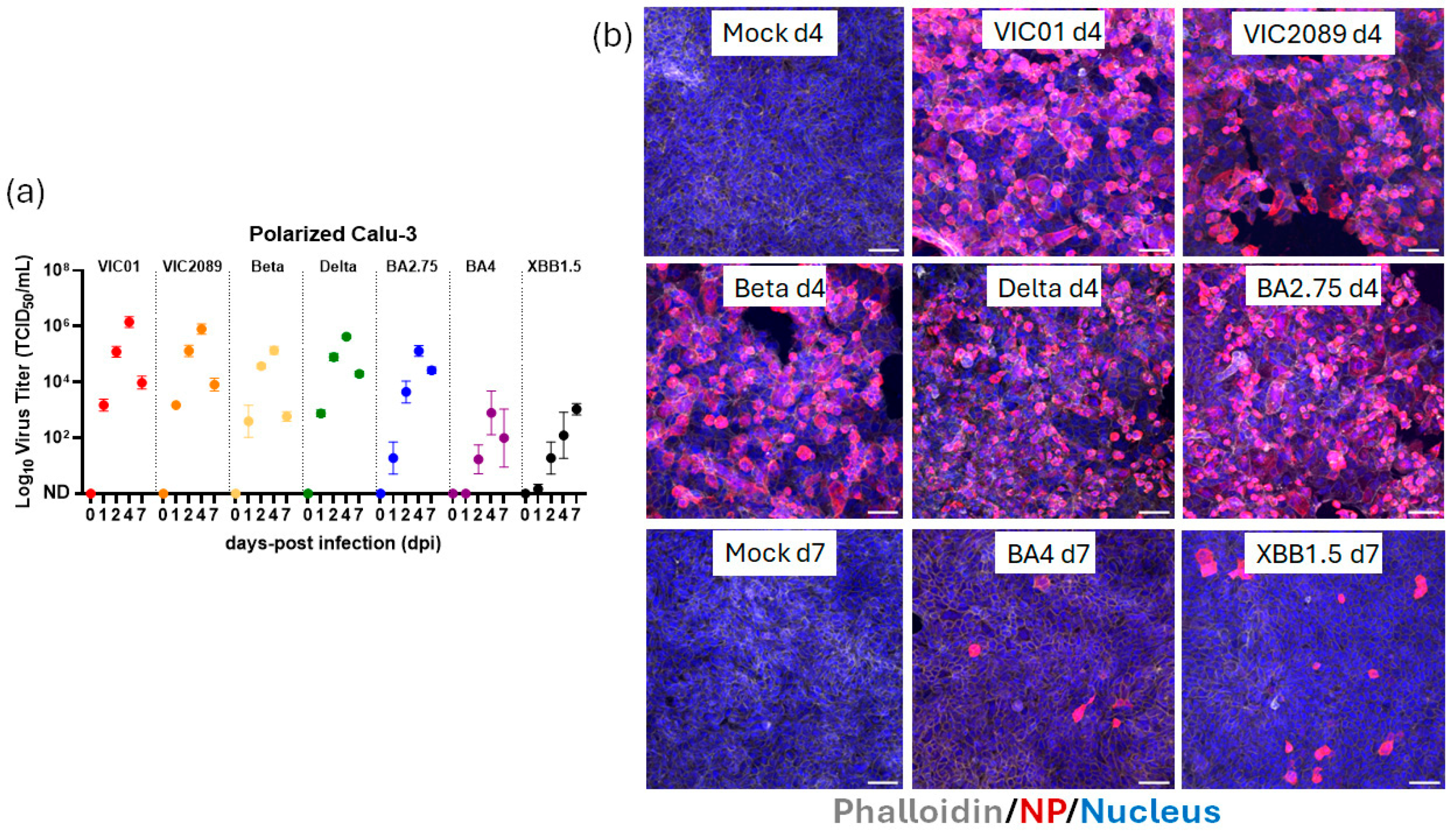
Here, we test the susceptibility of the polarized ALI-Calu-3 cells to infection by various SARS-CoV-2 clinical isolates, including the ancestral variant VIC01 [13], a clinical isolate (VIC2089) harboring the N501Y mutation that increases binding to mouse ACE2 [32,33] and has been adopted for in vivo mouse studies [34,35]; early VOC Beta and Delta; an early Omicron variant (BA2.75); and more recent Omicron subvariants (BA4 and XBB1.5). At the indicated time points post-infection, apical washes were assessed for their viral load using a TCID50 assay. Production of infectious virus was robust following infection with VIC01, VIC2089, Beta, Delta and BA2.75, with peak viral load occurring at four days post-infection (Figure 2a). A reduction in virus titer was observed at seven days post-infection, likely representing virus-induced CPE and cell exhaustion. In stark contrast, virus production with BA.4 and XBB1.5 was consistently 1–2 logs lower than VIC01 titers from one to four days following infection (Figure 2a, p < 0.05 two-way ANOVA with Dunnett’s multiple comparisons test). TranswellTM membranes at four and seven days post-infection (n = 3 per time point) were processed for imaging (Figure 2). Confocal microscopy of immunolabelled infected cells at four days post-infection confirmed robust infection by VIC01, VIC 2089, Beta, Delta and Omicron BA2.75 (Figure 2b and Supplementary Figure S1). Consistent with the reduced virus production results (Figure 2a), fewer infected cells were detected at four days post-infection with BA.4 and XBB1.5 (images not shown), a trend that was maintained at seven days post-infection (Figure 2b and Supplementary Figure S2). Increasing the incubation time from four to seven days did not increase virus production to levels seen with VIC01, for example, indicating that it was not simply a slower replication time course for BA.4 and XBB1.5 in the polarized Calu-3 model.

3.3. ALI-HNE Support Replication of All SARS-CoV-2 Variants

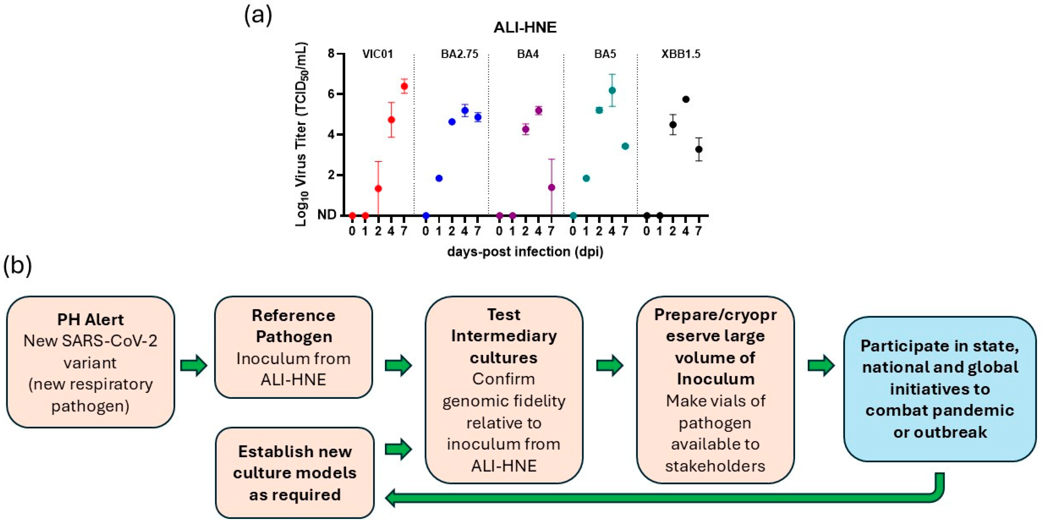
Given the poor replication seen with Omicron BA4 and XBB1.5 variants in the ALICalu-3 model, we sought to assess whether this was due to an intrinsic viral replication defect, or due to host cell specificity. Thus, we tested the susceptibility of ALI-HNE to infection by a panel of SARS-CoV-2 variants (VIC01, BA2.75, BA4, BA5 and XBB1.5) (Figure 3). We included the Omicron BA5 variant as it is closely related to BA4 [36]. Following the same infection protocol as for the ALI-Calu-3, the infectious replication kinetics produced by the ALI-HNE were similar for each SARS-CoV-2 variant, including BA4 and XBB1.5 (Figure 3a). The peak of viral production occurred at four days post-inoculation, with the average titer of all variants tested falling within 1-log of each other (p > 0.05 two-way ANOVA with Tukey’s multiple comparisons test). Reduction in the infectious viral titers at seven days reflects cell death due to virus-induced CPE. Consistent with our previous report [16], virus-induced cytopathology and decreased virus production was not seen with VIC01 (Figure 3a).
Our results indicate that the Omicron subvariants have developed important replication fitness advantages for productive infection of human nasal epithelial cell cultures over lung epithelial cells. Collectively, these data highlight the importance of including ALI-HNE as an integral component when selecting tissue culture models for SARS-CoV-2 infection. ALI-HNE need to be included into an isolation and characterization pipeline for an effective public health response to a new variant, if the findings generated in tissue culture are to translate to human infection and clinical trial (Figure 3b).

4. Discussion

Since the emergence of the SARS-CoV-2 Omicron lineage in late 2021, a plethora of Omicron subvariants have been identified due to the rapid spread of the lineage globally and mutations through convergent evolution and recombination. As of December 2024, the WHO has deemed Omicron subvariant JN-1 as a “variant of interest” (VOI) and, as of May 2025, six other subvariants as “variants under monitoring” (VUM) [37]. Notably, BA2.86/JN1 lineages harbor more than 30 additional mutations when compared to earlier Omicron variants, highlighting the increased viral fitness of this lineage endowed with a higher potential to evade neutralizing antibodies [38]. These and other more recent Omicron variants no longer replicate efficiently in Vero cells, necessitating the adoption of alternative cell lines for virus propagation [38]. As the SARS-CoV-2 Omicrons evolve, direct comparison of infection of human nasal epithelial and lung organoids revealed increased fitness in the upper respiratory tract compared to the lung [38]. Despite the robust infection of polarized ALI-Calu-3 that we observed with historical SARS-CoV-2 isolates and early Omicron subvariants, replication of the more recent Omicron subvariants was reduced. The reduced replication of Omicron BA4 and XBB1.5 in ALI-Calu-3 cells that we report here may reflect the inherent properties of the cell line. Calu-3 cells were established from human lung adenocarcinoma tissue [20], thus the cell line’s origin is human lung. This is consistent with increased fitness for the upper respiratory tract. Such viral evolution places human nasal epithelium cultures, like ALI-HNE, at the forefront for viral isolation and characterization against which other cell culture models should be benchmarked. Furthermore, unlike cell lines, ALI-HNE contain several epithelium cell types, and more accurately recapitulate the human nasal epithelium [39].
Other research groups have tested SARS-CoV-2 variants and subvariants and found that all variants replicated in the human nasal cultures [17,18,38], which is consistent with our data. Nonetheless, polarized continuous cell lines are still advantageous when used as an intermediary culture model to complement primary upper respiratory tract culture, but must be pre-screened for infectivity. Including intermediary cell culture models in the repertoire of cell culture models helps alleviate the scalability limitations for human nasal epithelium. Passage of SARS-CoV-2 viral stocks in Calu-3 has been shown to eliminate viruses with accumulated mutations in the furin cleavage site that arise through long-term viral propagation in Vero cells [24]. These mutations affect pathogenicity, transmission and drug/antibody sensitivity [21,22,40]. Thus, the stocks of SARS-CoV-2 isolates we tested were propagated in Calu-3 monolayer cultures. However, given the continued evolution and the emergence of new variants and subvariants [37] with the potential for altered cell tropism and pathology, as already seen with the Omicron subvariants [18,19], it remains paramount to test alternative primary and continuous cell lines and benchmark them against infection of human nasal epithelium. Furthermore, culture in 3D matrix is a robust method to test the capacity of a continuous cell line to polarize along the apical–basal axis before selection as an intermediary ALI culture model.
Moving forward, the pipeline for culture of SARS-CoV-2 must incorporate human nasal cultures, like the ALI-HNE, and infection with a reference virus as there is donor-to-donor variation in generating an ALI-HNE-based model of SARS-CoV-2 infection [16]. Notably, the WHO recommends human organoids for the culture of pathogens [41], while drug regulators acknowledge human organoids as a new pre-clinical pathway for drug development [42], placing human organoid culture at the center of the infectious disease and drug development fields. During the development of vaccines and neutralizing antibody-based treatments to combat COVID-19, the ALI-HNE-based neutralization assay proved to be a discerning assay for recapitulating inhibition of infection in vivo in mice [43] and patient response to treatment with antibody [44].

5. Conclusions

Collectively, our data demonstrate the utility of the polarized Calu-3 model of SARS-CoV-2 infection. The polarized Calu-3 cells were robustly infected by most SARS-CoV-2 variants and could serve as an intermediary model to compliment the ALI-HNE. However, not all the emerging SARS-CoV-2 variants infected the polarized Calu-3 cells efficiently, despite robust infection of ALI-HNE. Consequently, before adopting a continuous cell line, susceptibility to infection by a panel of SARS-CoV-2 variants, including historical and emerging clinical isolates, should be conducted.

Supplementary Materials

The following supporting information can be downloaded at: https://www.mdpi.com/article/10.3390/organoids4030021/s1, Figure S1: Four days post-infection of polarized Calu-3 cells with SARS-CoV-2 variants. Shown are individual channels [DAPI, phalloidin and nucleoprotein (NP)] and the merged images for the day four confocal immunofluorescence data in Figure 2. Also shown is the staining of the filters with negative control antibody (Ab) as a merged image. Scale bar = 50 μm; Figure S2: Shown are individual channels [DAPI, phalloidin and nucleoprotein (NP)] and the merged images for the day seven confocal immunofluorescence data in Figure 2. Also shown is the staining of the filters with negative control antibody (Ab) as a merged image. Scale bar = 50 μm.

Author Contributions

Conceptualization, E.V.; methodology, B.M.T., S.L.H., J.L.M., A.H. and S.L.G.; validation, E.V.; formal analysis, B.M.T., S.L.H. and J.L.M.; investigation, B.M.T., S.L.H. and J.L.M.; resources, E.V., J.T., G.D. and D.F.J.P.; data curation, B.M.T., E.V. and J.L.M.; writing—original draft preparation, E.V., B.M.T. and J.L.M.; writing—review and editing, S.L.H., A.H., S.L.G., D.F.J.P., G.D. and J.T.; visualization, B.M.T., E.V. and J.L.M.; supervision, E.V., A.H., J.T., G.D. and D.F.J.P.; project administration, E.V.; funding acquisition, E.V., B.M.T. and J.T. All authors have read and agreed to the published version of the manuscript.

Funding

This work was supported by a grant from the Victorian Medical Research Acceleration Fund (Victorian Government and the Royal Melbourne Hospital Foundation, grant number GA-F3744277-2925) awarded to E.V. The salary for B.M.T. was supported by an Ideas grant from the National Health and Medical Research Council (NHMRC) of Australia awarded to E.V., B.M.T. and J.T. (grant number APP1181580) and a foundation grant from the Cummings Global Centre for Pandemic Therapeutics (CGCPT, grant #CGCPT00007) awarded to E.V. Funding for S.L.H. and A.H. was awarded by the CASS foundation (grant #10077) and by NHMRC (GNT2018880). Funding for S.G., D.P. and J.L.M. was provided by Medical Research Future Funds (grant numbers 2015317 and CTI000025).

Institutional Review Board Statement

The study was conducted in accordance with the Declaration of Helsinki and approved by the Medicine and Dentistry Human Ethics Sub-Committee, University of Melbourne (HREC/2023-14208-37805-4, approval date 22 March 2023).

Informed Consent Statement

Informed consent was obtained from all donors of nasal brush samples involved in the study. Donor brush samples are de-identified at time of collection.

Data Availability Statement

The original contributions presented in this study are included in the article and Supplementary Materials. Further inquiries can be directed to the corresponding authors.

Acknowledgments

We thank the Biological Optical Microscopy Platform (BOMP), University of Melbourne and the Victorian Centre for Functional Genomics (VCGF) for their assistance. We acknowledge our public health partners and the Victorian Department of Human Services, the major funder of the Victorian Infectious Diseases Reference Laboratory.

Conflicts of Interest

The authors declare no conflicts of interest. The funders had no role in the design of the study; in the collection, analyses, or interpretation of data; in the writing of the manuscript; or in the decision to publish the results.

References

  1. Huang, C.; Wang, Y.; Li, X.; Ren, L.; Zhao, J.; Hu, Y.; Zhang, L.; Fan, G.; Xu, J.; Gu, X.; et al. Clinical features of patients infected with 2019 novel coronavirus in Wuhan, China. Lancet 2020, 395, 497–506. [Google Scholar] [CrossRef]
  2. Zhu, N.; Zhang, D.; Wang, W.; Li, X.; Yang, B.; Song, J.; Zhao, X.; Huang, B.; Shi, W.; Lu, R.; et al. A Novel Coronavirus from Patients with Pneumonia in China, 2019. N. Engl. J. Med. 2020, 382, 727–733. [Google Scholar] [CrossRef]
  3. Quarleri, J.; Galvan, V.; Delpino, M.V. Omicron variant of the SARS-CoV-2: A quest to define the consequences of its high mutational load. Geroscience 2022, 44, 53–56. [Google Scholar] [CrossRef]
  4. Suzuki, R.; Yamasoba, D.; Kimura, I.; Wang, L.; Kishimoto, M.; Ito, J.; Morioka, Y.; Nao, N.; Nasser, H.; Uriu, K.; et al. Attenuated fusogenicity and pathogenicity of SARS-CoV-2 Omicron variant. Nature 2022, 603, 700–705. [Google Scholar] [CrossRef] [PubMed]
  5. Liu, L.; Iketani, S.; Guo, Y.; Chan, J.F.; Wang, M.; Liu, L.; Luo, Y.; Chu, H.; Huang, Y.; Nair, M.S.; et al. Striking antibody evasion manifested by the Omicron variant of SARS-CoV-2. Nature 2022, 602, 676–681. [Google Scholar] [CrossRef] [PubMed]
  6. Ito, J.; Suzuki, R.; Uriu, K.; Itakura, Y.; Zahradnik, J.; Kimura, K.T.; Deguchi, S.; Wang, L.; Lytras, S.; Tamura, T.; et al. Convergent evolution of SARS-CoV-2 Omicron subvariants leading to the emergence of BQ.1.1 variant. Nat. Commun. 2023, 14, 2671. [Google Scholar] [CrossRef] [PubMed]
  7. Bouhaddou, M.; Reuschl, A.K.; Polacco, B.J.; Thorne, L.G.; Ummadi, M.R.; Ye, C.; Rosales, R.; Pelin, A.; Batra, J.; Jang, G.M.; et al. SARS-CoV-2 variants evolve convergent strategies to remodel the host response. Cell 2023, 186, 4597–4614.e4526. [Google Scholar] [CrossRef]
  8. Khan, K.; Lustig, G.; Romer, C.; Reedoy, K.; Jule, Z.; Karim, F.; Ganga, Y.; Bernstein, M.; Baig, Z.; Jackson, L.; et al. Evolution and neutralization escape of the SARS-CoV-2 BA.2.86 subvariant. Nat. Commun. 2023, 14, 8078. [Google Scholar] [CrossRef]
  9. Tamura, T.; Ito, J.; Uriu, K.; Zahradnik, J.; Kida, I.; Anraku, Y.; Nasser, H.; Shofa, M.; Oda, Y.; Lytras, S.; et al. Virological characteristics of the SARS-CoV-2 XBB variant derived from recombination of two Omicron subvariants. Nat. Commun. 2023, 14, 2800. [Google Scholar] [CrossRef]
  10. Avdonin, P.P.; Rybakova, E.Y.; Trufanov, S.K.; Avdonin, P.V. SARS-CoV-2 Receptors and Their Involvement in Cell Infection. Biochem. Suppl. Ser. A Membr. Cell Biol. 2023, 17, 1–11. [Google Scholar] [CrossRef]
  11. Beumer, J.; Geurts, M.H.; Lamers, M.M.; Puschhof, J.; Zhang, J.; van der Vaart, J.; Mykytyn, A.Z.; Breugem, T.I.; Riesebosch, S.; Schipper, D.; et al. A CRISPR/Cas9 genetically engineered organoid biobank reveals essential host factors for coronaviruses. Nat. Commun. 2021, 12, 5498. [Google Scholar] [CrossRef]
  12. Hou, Y.J.; Okuda, K.; Edwards, C.E.; Martinez, D.R.; Asakura, T.; Dinnon, K.H., 3rd; Kato, T.; Lee, R.E.; Yount, B.L.; Mascenik, T.M.; et al. SARS-CoV-2 Reverse Genetics Reveals a Variable Infection Gradient in the Respiratory Tract. Cell 2020, 182, 429–446.e14. [Google Scholar] [CrossRef]
  13. Caly, L.; Druce, J.; Roberts, J.; Bond, K.; Tran, T.; Kostecki, R.; Yoga, Y.; Naughton, W.; Taiaroa, G.; Seemann, T.; et al. Isolation and rapid sharing of the 2019 novel coronavirus (SARS-CoV-2) from the first patient diagnosed with COVID-19 in Australia. Med. J. Aust. 2020, 212, 459–462. [Google Scholar] [CrossRef] [PubMed]
  14. Rosenke, K.; Jarvis, M.A.; Feldmann, F.; Schwarz, B.; Okumura, A.; Lovaglio, J.; Saturday, G.; Hanley, P.W.; Meade-White, K.; Williamson, B.N.; et al. Hydroxychloroquine Proves Ineffective in Hamsters and Macaques Infected with SARS-CoV-2. bioRxiv 2020. [Google Scholar] [CrossRef]
  15. Boulware, D.R.; Pullen, M.F.; Bangdiwala, A.S.; Pastick, K.A.; Lofgren, S.M.; Okafor, E.C.; Skipper, C.P.; Nascene, A.A.; Nicol, M.R.; Abassi, M.; et al. A Randomized Trial of Hydroxychloroquine as Postexposure Prophylaxis for COVID-19. N. Engl. J. Med. 2020, 383, 517–525. [Google Scholar] [CrossRef]
  16. Tran, B.M.; Grimley, S.L.; McAuley, J.L.; Hachani, A.; Earnest, L.; Wong, S.L.; Caly, L.; Druce, J.; Purcell, D.F.J.; Jackson, D.C.; et al. Air-Liquid-Interface Differentiated Human Nose Epithelium: A Robust Primary Tissue Culture Model of SARS-CoV-2 Infection. Int. J. Mol. Sci. 2022, 23, 835. [Google Scholar] [CrossRef]
  17. Mykytyn, A.Z.; Breugem, T.I.; Geurts, M.H.; Beumer, J.; Schipper, D.; van Acker, R.; van den Doel, P.B.; van Royen, M.E.; Zhang, J.; Clevers, H.; et al. SARS-CoV-2 Omicron entry is type II transmembrane serine protease-mediated in human airway and intestinal organoid models. J. Virol. 2023, 97, e0085123. [Google Scholar] [CrossRef]
  18. Tanneti, N.S.; Patel, A.K.; Tan, L.H.; Marques, A.D.; Perera, R.; Sherrill-Mix, S.; Kelly, B.J.; Renner, D.M.; Collman, R.G.; Rodino, K.; et al. Comparison of SARS-CoV-2 variants of concern in primary human nasal cultures demonstrates Delta as most cytopathic and Omicron as fastest replicating. mBio 2024, 15, e0312923. [Google Scholar] [CrossRef] [PubMed]
  19. Wickenhagen, A.; Flagg, M.; Port, J.R.; Yinda, C.K.; Goldin, K.; Gallogly, S.; Schulz, J.E.; Lutterman, T.; Williamson, B.N.; Kaiser, F.; et al. Evolution of Omicron lineage towards increased fitness in the upper respiratory tract in the absence of severe lung pathology. Nat. Commun. 2025, 16, 594. [Google Scholar] [CrossRef] [PubMed]
  20. Shen, B.Q.; Finkbeiner, W.E.; Wine, J.J.; Mrsny, R.J.; Widdicombe, J.H. Calu-3: A human airway epithelial cell line that shows cAMP-dependent Cl- secretion. Am. J. Physiol. Lung Cell. Mol. Physiol. 1994, 266, L493–L501. [Google Scholar] [CrossRef]
  21. Mykytyn, A.Z.; Breugem, T.I.; Riesebosch, S.; Schipper, D.; van den Doel, P.B.; Rottier, R.J.; Lamers, M.M.; Haagmans, B.L. SARS-CoV-2 entry into human airway organoids is serine protease-mediated and facilitated by the multibasic cleavage site. eLife 2021, 10, e64508. [Google Scholar] [CrossRef]
  22. Lamers, M.M.; Mykytyn, A.Z.; Breugem, T.I.; Wang, Y.; Wu, D.C.; Riesebosch, S.; van den Doel, P.B.; Schipper, D.; Bestebroer, T.; Wu, N.C.; et al. Human airway cells prevent SARS-CoV-2 multibasic cleavage site cell culture adaptation. eLife 2021, 10, e66815. [Google Scholar] [CrossRef]
  23. Ren, X.; Glende, J.; Al-Falah, M.; de Vries, V.; Schwegmann-Wessels, C.; Qu, X.; Tan, L.; Tschernig, T.; Deng, H.; Naim, H.Y.; et al. Analysis of ACE2 in polarized epithelial cells: Surface expression and function as receptor for severe acute respiratory syndrome-associated coronavirus. J. Gen. Virol. 2006, 87, 1691–1695. [Google Scholar] [CrossRef] [PubMed]
  24. Baczenas, J.J.; Andersen, H.; Rashid, S.; Yarmosh, D.; Puthuveetil, N.; Parker, M.; Bradford, R.; Florence, C.; Stemple, K.J.; Lewis, M.G.; et al. Propagation of SARS-CoV-2 in Calu-3 Cells to Eliminate Mutations in the Furin Cleavage Site of Spike. Viruses 2021, 13, 2434. [Google Scholar] [CrossRef]
  25. Lee, G.Y.; Kenny, P.A.; Lee, E.H.; Bissell, M.J. Three-dimensional culture models of normal and malignant breast epithelial cells. Nat. Methods 2007, 4, 359–365. [Google Scholar] [CrossRef]
  26. Collett, S.; Torresi, J.; Silveira, L.E.; Truong, V.K.; Christiansen, D.; Tran, B.M.; Vincan, E.; Ramsland, P.A.; Elbourne, A. Investigating virus-host cell interactions: Comparative binding forces between hepatitis C virus-like particles and host cell receptors in 2D and 3D cell culture models. J. Colloid Interface Sci. 2021, 592, 371–384. [Google Scholar] [CrossRef]
  27. Harbach, S.L.; Tran, B.M.; Kastrappis, G.; Tran, H.; Grimley, S.L.; McAuley, J.L.; Hachani, A.; Vincan, E. Polarized Calu-3 Cells Serve as an Intermediary Model for SARS-CoV-2 Infection. In Methods in Molecular Biology; Springer: New York, NY, USA, 2025. [Google Scholar] [CrossRef]
  28. Lee, J.Y.H.; Best, N.; McAuley, J.; Porter, J.L.; Seemann, T.; Schultz, M.B.; Sait, M.; Orlando, N.; Mercoulia, K.; Ballard, S.A.; et al. Validation of a single-step, single-tube reverse transcription loop-mediated isothermal amplification assay for rapid detection of SARS-CoV-2 RNA. J. Med. Microbiol. 2020, 69, 1169–1178. [Google Scholar] [CrossRef]
  29. Reed, L.J.; Muench, H. A simple method of estimating fifty per cent endpoints. Am. J. Hyg. 1938, 27, 493–497. [Google Scholar] [CrossRef]
  30. To, C.Z.; Bhunia, A.K. Three Dimensional Vero Cell-Platform for Rapid and Sensitive Screening of Shiga-Toxin Producing Escherichia coli. Front. Microbiol. 2019, 10, 949. [Google Scholar] [CrossRef]
  31. Lamers, M.M.; Beumer, J.; van der Vaart, J.; Knoops, K.; Puschhof, J.; Breugem, T.I.; Ravelli, R.B.G.; Paul van Schayck, J.; Mykytyn, A.Z.; Duimel, H.Q.; et al. SARS-CoV-2 productively infects human gut enterocytes. Science 2020, 369, 50–54. [Google Scholar] [CrossRef]
  32. Starr, T.N.; Greaney, A.J.; Hilton, S.K.; Ellis, D.; Crawford, K.H.D.; Dingens, A.S.; Navarro, M.J.; Bowen, J.E.; Tortorici, M.A.; Walls, A.C.; et al. Deep Mutational Scanning of SARS-CoV-2 Receptor Binding Domain Reveals Constraints on Folding and ACE2 Binding. Cell 2020, 182, 1295–1310.e20. [Google Scholar] [CrossRef]
  33. Gu, H.; Chen, Q.; Yang, G.; He, L.; Fan, H.; Deng, Y.Q.; Wang, Y.; Teng, Y.; Zhao, Z.; Cui, Y.; et al. Adaptation of SARS-CoV-2 in BALB/c mice for testing vaccine efficacy. Science 2020, 369, 1603–1607. [Google Scholar] [CrossRef]
  34. Pymm, P.; Redmond, S.J.; Dolezal, O.; Mordant, F.; Lopez, E.; Cooney, J.P.; Davidson, K.C.; Haycroft, E.R.; Tan, C.W.; Seneviratna, R.; et al. Biparatopic nanobodies targeting the receptor binding domain efficiently neutralize SARS-CoV-2. iScience 2022, 25, 105259. [Google Scholar] [CrossRef]
  35. Bader, S.M.; Cooney, J.P.; Sheerin, D.; Taiaroa, G.; Harty, L.; Davidson, K.C.; Mackiewicz, L.; Dayton, M.; Wilcox, S.; Whitehead, L.; et al. SARS-CoV-2 mouse adaptation selects virulence mutations that cause TNF-driven age-dependent severe disease with human correlates. Proc. Natl. Acad. Sci. USA 2023, 120, e2301689120. [Google Scholar] [CrossRef]
  36. Meijers, M.; Ruchnewitz, D.; Eberhardt, J.; Luksza, M.; Lassig, M. Population immunity predicts evolutionary trajectories of SARS-CoV-2. Cell 2023, 186, 5151–5164.e13. [Google Scholar] [CrossRef] [PubMed]
  37. WHO. Tracking SARS-CoV-2. Available online: https://www.who.int/activities/tracking-SARS-CoV-2-variants (accessed on 5 June 2025).
  38. Planas, D.; Staropoli, I.; Michel, V.; Lemoine, F.; Donati, F.; Prot, M.; Porrot, F.; Guivel-Benhassine, F.; Jeyarajah, B.; Brisebarre, A.; et al. Distinct evolution of SARS-CoV-2 Omicron XBB and BA.2.86/JN.1 lineages combining increased fitness and antibody evasion. Nat. Commun. 2024, 15, 2254. [Google Scholar] [CrossRef] [PubMed]
  39. Chang, J.J.; Grimley, S.L.; Tran, B.M.; Deliyannis, G.; Tumpach, C.; Nguyen, A.N.T.; Steinig, E.; Zhang, J.; Schroder, J.; Caly, L.; et al. Uncovering strain- and age-dependent innate immune responses to SARS-CoV-2 infection in air-liquid-interface cultured nasal epithelia. iScience 2024, 27, 110009. [Google Scholar] [CrossRef] [PubMed]
  40. Lau, S.Y.; Wang, P.; Mok, B.W.; Zhang, A.J.; Chu, H.; Lee, A.C.; Deng, S.; Chen, P.; Chan, K.H.; Song, W.; et al. Attenuated SARS-CoV-2 variants with deletions at the S1/S2 junction. Emerg. Microbes Infect. 2020, 9, 837–842. [Google Scholar] [CrossRef]
  41. WHO. Research Response to Pathogen X During a Pandemic. Available online: https://www.who.int/news-room/events/detail/2024/01/19/default-calendar/Research-response-to-pathogen-X-during-a-pandemic (accessed on 11 June 2025).
  42. Wadman, M. FDA no longer has to require animal testing for new drugs. Science 2023, 379, 127–128. [Google Scholar] [CrossRef]
  43. Carrera Montoya, J.; Collett, S.; Fernandez Ruiz, D.; Earnest, L.; Edeling, M.A.; Yap, A.H.Y.; Wong, C.Y.; Cooney, J.P.; Davidson, K.C.; Roberts, J.; et al. Human Nasal Epithelium Organoids for Assessing Neutralizing Antibodies to a Protective SARS-CoV-2 Virus-like Particle Vaccine. Organoids 2024, 3, 18–31. [Google Scholar] [CrossRef]
  44. Wan, Z.X.; Li, C.; Zhou, Y.; Chiu, M.C.; Huang, J.J.; Zhang, S.X.; Zhu, X.X.; Lan, Q.S.; Deng, Y.L.; Xue, W.; et al. Organoid-based neutralization assays reveal a distinctive profile of SARS-CoV-2 antibodies and recapitulate the real-world efficacy. Proc. Natl. Acad. Sci. USA 2025, 122, e2509616122. [Google Scholar] [CrossRef] [PubMed]
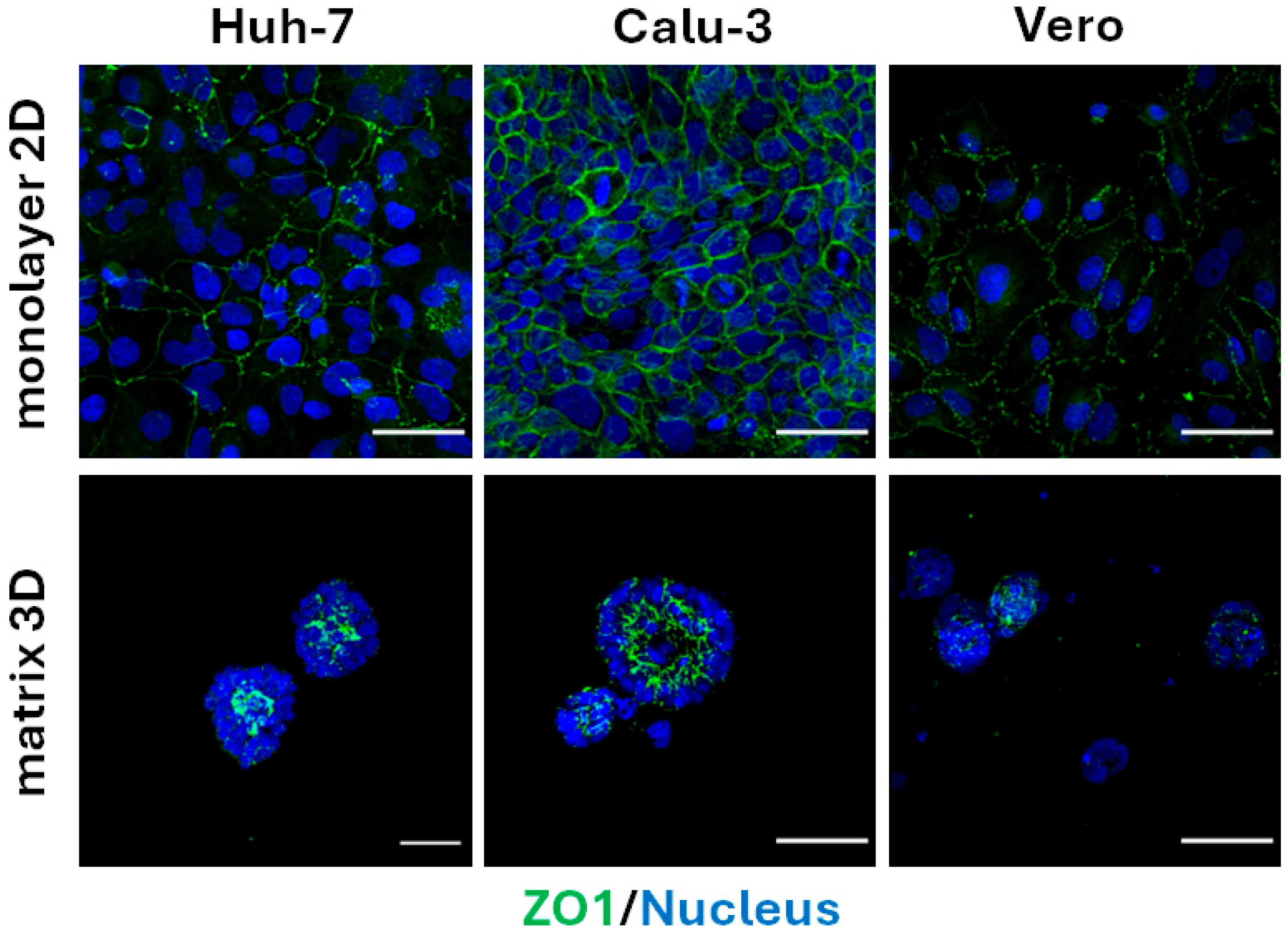
Figure 1. Calu-3 cells form spheroids in 3D matrix. Huh-7, Calu-3 and Vero cells were cultured as a monolayer (2D) on tissue culture plastic or embedded in matrix (3D). Confocal immunofluorescence staining of the cells in 2D and 3D for the tight junction protein ZO-1 (green) and nuclei (blue) revealed spheroid formation by Huh-7 and Calu-3 cells but not Vero cells. The 3D images are stacked Z-sections at the center of the spheroids and cell aggregates. Scale bar = 50 µm.
Organoids 04 00021 g001
Figure 2. Infection of polarized Calu-3 cells with SARS-CoV-2 variants. (a) Polarized Calu-3 epithelial sheets on TranswellTM inserts were infected with the indicated SARS-CoV-2 isolates. Infectious viral load in the apical washes harvested at the indicated times post-infection were quantified by TCID50 assay on Vero cells (mean ± SEM, n = 6 for days zero to four and n = 3 for day seven post-infection); ND = virus-induced CPE was not detected. (b) At four and seven days post-infection, the TranswellTM membranes were processed for immunofluorescence (IF) confocal microscopy (n = 3 per time point). (c) Shown are merged IF images at four days post-infection for VIC01, VIC2089, Beta, Delta and BA2.75, and seven days post-infection for BA4 and XBB1.5 (phalloidin for F actin, gray; anti-nucleoprotein for SARS-CoV-2 infection, red and DAPI for nuclei, blue). Scale bar = 50 µm.
Organoids 04 00021 g002
Figure 3. Infection of ALI-HNE with SARS-CoV-2 variants. (a) ALI-HNE epithelial sheets on TranswellTM inserts were infected with the indicated SARS-CoV-2 isolates. Infectious viral loads in the apical washes at the indicated times post-infection were quantified by TCID50 assay (mean ± SEM, n = 2). (b) A proposed pipeline for public health response to a new SARS-CoV-2 variant, using ALI-HNE as the benchmark against which other culture models are tested (public health, PH).
Organoids 04 00021 g003
Disclaimer/Publisher’s Note: The statements, opinions and data contained in all publications are solely those of the individual author(s) and contributor(s) and not of MDPI and/or the editor(s). MDPI and/or the editor(s) disclaim responsibility for any injury to people or property resulting from any ideas, methods, instructions or products referred to in the content.