Article

Phenotypic and Genotypic Characterization of Enterococcus spp. from Poultry in Chile: Antimicrobial Resistance, Biofilm, and Virulence Genes


1
Facultad de Medicina Veterinaria y Agronomía, Universidad de las Américas, Campus Maipú, 5 de Abril 620, Santiago 7500975, Chile
2
Laboratorio de Patología Aviar, Departamento de Patología Animal, Facultad de Ciencias Veterinarias y Pecuarias, Universidad de Chile, Santiago 8820808, Chile
3
Departamento de Ciencia Animal, Facultad de Ciencias Veterinarias y Pecuarias, Universidad de Chile, Santiago 8820808, Chile
*
Author to whom correspondence should be addressed.
Poultry 2026, 5(4), 52; https://doi.org/10.3390/poultry5040052
Submission received: 25 May 2026 / Revised: 16 July 2026 / Accepted: 21 July 2026 / Published: 24 July 2026

Abstract

Enterococcus spp. are increasingly recognized as important opportunistic pathogens in poultry production and represent a potential One Health concern because of their virulence potential and capacity to acquire antimicrobial resistance determinants. This study characterized Enterococcus isolates recovered from clinical poultry cases in Chile, focusing on species distribution, virulence-associated genes, antimicrobial resistance patterns and genes, biofilm formation, and cytolysin activity. A total of 39 isolates were identified using the VITEK® 2 Compact system and confirmed by PCR targeting the tuf gene. Seven Enterococcus species were detected, with E. faecalis predominating (61.5%) and E. faecium following (15.4%). The virulence-associated genes asa1, gelE, and cylA were detected in 97.4%, 97.4%, and 48.7% of the isolates, respectively. Phenotypic antimicrobial susceptibility testing showed resistance mainly to tetracycline (61.5%) and erythromycin (17.9%), whereas all isolates were susceptible to vancomycin, ampicillin, chloramphenicol, and linezolid. Molecular analysis identified tetM (71.8%), ermB (66.7%), and tetL (30.8%) as the most predominant resistance genes. A significant association was observed between tetM and phenotypic tetracycline resistance (Fisher’s exact test, p = 0.010). However, no association was found between ermB and erythromycin resistance. Biofilm formation was observed in 66.7% of the isolates, although most were weak biofilm producers. Despite the relatively high prevalence of cylA, β-hemolytic activity was detected in only 5.1% of isolates, indicating an incomplete genotype–phenotype concordance. Overall, poultry-associated Enterococcus spp. circulating in Chile harbored multiple virulence and antimicrobial resistance determinants that may contribute to their persistence and adaptation in poultry production systems. These findings provide the first baseline molecular epidemiological data on clinical poultry-associated Enterococcus spp. isolates in Chile and support the need for continued surveillance and prudent antimicrobial use in veterinary medicine.

1. Introduction

Enterococci are Gram-positive, facultative anaerobic bacteria belonging to the family Enterococcaceae. They are common members of the intestinal microbiota of humans, mammals, birds, and poultry and are also widely distributed in the environment, reflecting their remarkable ecological adaptability [1,2,3]. Although generally regarded as commensals, enterococci are increasingly recognized as opportunistic pathogens and important reservoirs of antimicrobial resistance determinants in both human and veterinary medicine [4,5,6].
In poultry, several Enterococcus species have been associated with clinical diseases, particularly Enterococcus faecalis, Enterococcus faecium, and Enterococcus cecorum [7]. E. faecalis and E. faecium are commonly associated with omphalitis, septicemia, endocarditis, and amyloidosis, whereas E. cecorum has emerged as an important cause of locomotor disorders, including vertebral osteomyelitis, arthritis, femoral head necrosis, and spondylitis, resulting in substantial economic losses in broiler and breeder herds [7,8,9,10,11,12,13]. Other species, including Enterococcus hirae, Enterococcus durans, Enterococcus gallinarum, Enterococcus casseliflavus, and Enterococcus avium, have been sporadically associated with poultry diseases [7,14].
The pathogenicity of enterococci is associated with several virulence determinants that facilitate colonization, persistence, and tissue invasion of the host. Among the best-characterized are aggregation substance (asa1), cytolysin (cylA), and gelatinase (gelE), which contribute to bacterial adhesion, biofilm formation, cytotoxicity, and extracellular matrix degradation [3,15,16]. Biofilm formation further enhances bacterial persistence by increasing tolerance to environmental stress, host immune defense, disinfectants, and antimicrobial agents [3,17].
Enterococci also possess an exceptional ability to acquire and disseminate antimicrobial resistance determinants through horizontal gene transfer, making them important reservoirs of resistance genes within poultry production systems and along the food chain [6,18]. Of particular concern is the emergence of vancomycin-resistant enterococci (VRE), although their occurrence in poultry varies considerably among countries and production systems [19]. These characteristics underscore the relevance of poultry-associated enterococci within the One Health framework linking animal health, food safety, and public health.
Despite their importance as emerging poultry pathogens, information on the epidemiology, species diversity, virulence-associated genes, antimicrobial resistance, and biofilm-forming capacity of clinical poultry-associated Enterococcus spp. isolates in Chile remains extremely limited. Consequently, the epidemiological characteristics and resistance profiles of these bacteria in Chilean poultry production are poorly understood. Therefore, the present study aimed to provide the first comprehensive phenotypic and genotypic characterization of Enterococcus spp. isolated from clinical poultry cases in Chile by evaluating species distribution, antimicrobial susceptibility, antimicrobial resistance genes, virulence-associated genes, biofilm formation, and cytolysin activity in these isolates. These findings provide baseline epidemiological information to support future surveillance, antimicrobial stewardship, and molecular epidemiological studies in the poultry industry in Chile.

2. Results

2.1. Bacterial Isolation and Identification

After 24 h of incubation, the isolates produced cream-colored colonies with smooth edges on blood agar, measuring up to 1.5 mm in diameter. Among the 39 poultry isolates analyzed using the VITEK® 2 Compact system, seven Enterococcus species were identified: 24 (61.5%) E. faecalis, 6 (15.4%) E. faecium, 3 (7.7%) E. durans, 2 (5.1%) E. avium, 2 (5.1%) E. hirae, 1 (2.6%) E. cecorum, and 1 (2.6%) E. gallinarum. Genus-level confirmation was performed using Enterococcus-specific primers targeting the elongation factor (tuf) gene, and all isolates tested positive. Figure 1 illustrates the distribution of Enterococcus species among the analyzed isolates.

2.2. Virulence Genes

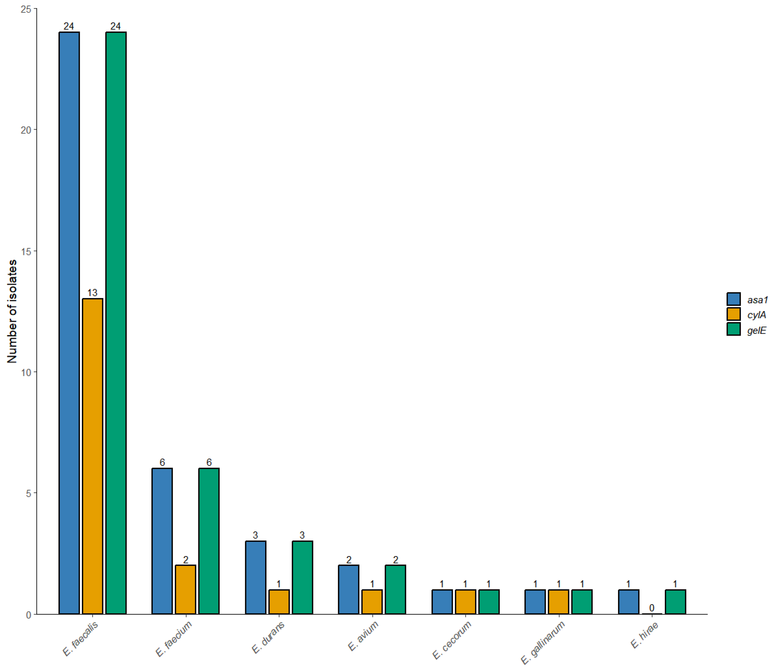
Virulence-associated genes were highly prevalent among the Enterococcus isolates in this study. The asa1 gene (aggregation substance) was detected in 38 of 39 isolates (97.4%) and was present in all E. faecalis strains. The gelE gene (gelatinase) was detected in 38 of the 39 isolates (97.4%), including all isolates of E. faecalis, E. faecium, E. durans, E. cecorum, E. gallinarum, and E. avium, and one of the two E. hirae isolates. The cylA gene (cytolysin activator) was detected in 19 of the 39 isolates (48.7%). Specifically, cylA was identified in 54.2% of E. faecalis, 33.3% of E. faecium, 33.3% of E. durans, 50% of E. avium, and 100% of both E. cecorum and E. gallinarum. The cylA gene was not detected in either E. hirae isolate. The frequency of cylA was compared among the Enterococcus species groups. Although cylA was more frequently detected in E. faecalis isolates (54.2%) than in E. faecium (33.3%) and other Enterococcus species (44.4%), no statistically significant association was observed between the species group and the presence of cylA (Fisher’s exact test, p = 0.514). Figure 2 shows the distribution of virulence genes among the Enterococcus isolates.

2.3. Assessment of Antimicrobial Susceptibility Patterns

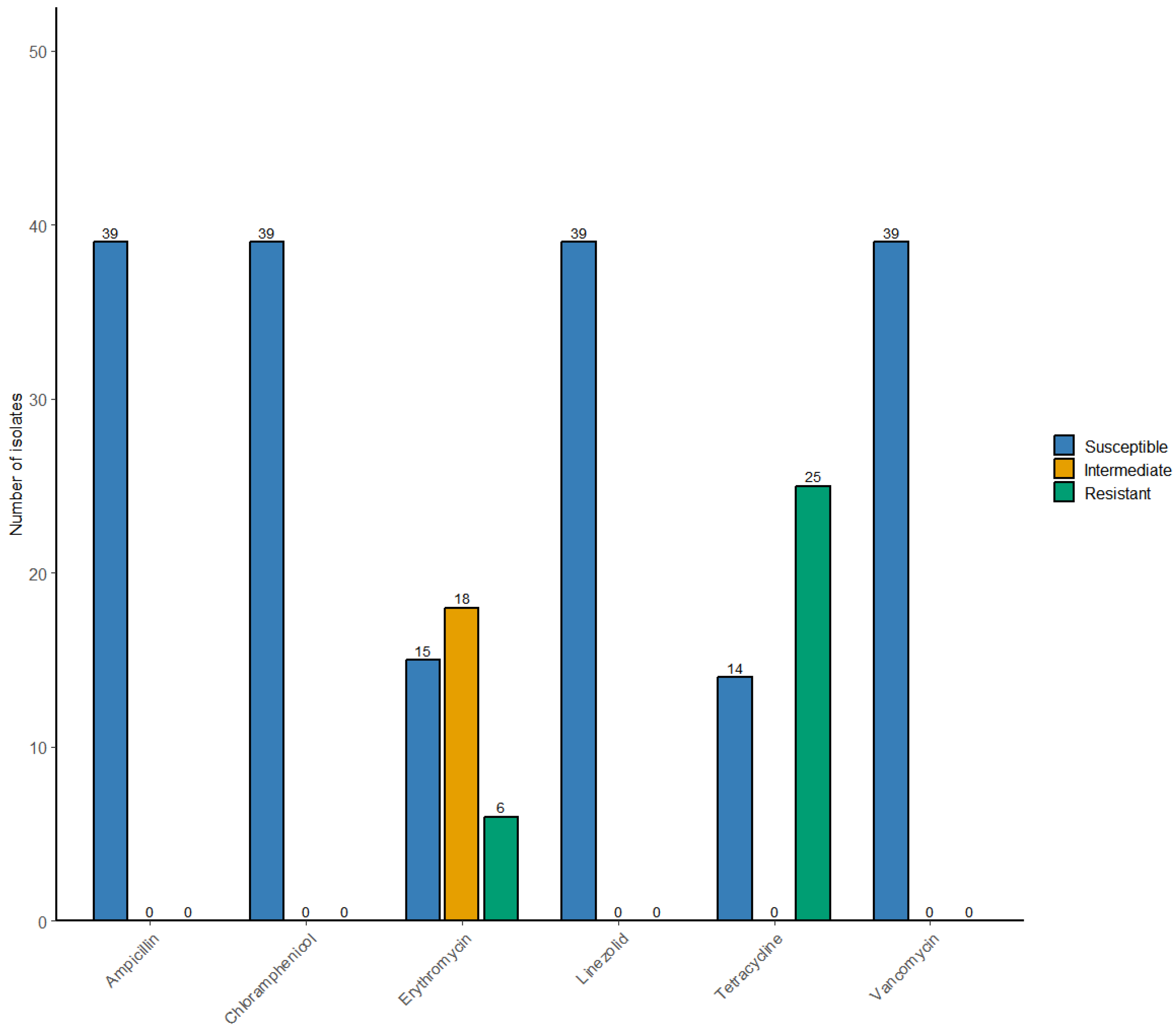
Antimicrobial susceptibility testing performed using the VITEK® 2 Compact system revealed variability in resistance patterns among the Enterococcus isolates. Phenotypic resistance was primarily observed against tetracycline, with 24 of 39 isolates (61.5%) classified as tetracycline-resistant. Resistance to erythromycin was detected in seven isolates (17.9%), whereas an additional 17 isolates (43.6%) exhibited intermediate susceptibility to this antimicrobial agent. All isolates (39/39; 100%) were susceptible to vancomycin, ampicillin, linezolid, and chloramphenicol (Figure 3). No resistance was observed against these antimicrobial agents. Based on the resistance patterns, 11 isolates (28.2%) were fully susceptible to all antimicrobials evaluated, whereas 28 isolates (71.8%) were resistant to at least one antimicrobial agent. The antimicrobial resistance patterns of the 39 Enterococcus isolates are shown in Table 1.

2.4. Antimicrobial Resistance Genes

A total of 11 antimicrobial resistance genes were investigated among the 39 Enterococcus isolates (Table 2). The tetracycline resistance gene tetM was the most frequently detected determinant, being identified in 28/39 isolates (71.8%), followed by ermB in 26/39 isolates (66.7%), and tetL in 12/39 isolates (30.8%). The tetA and optrA genes were detected in five (12.8%) and three (7.7%) isolates, respectively, whereas ermA was detected in two isolates (5.1%) and tetB in one isolate (2.6%). None of the isolates carried vanA, vanB, pbp5, or cat genes.
Because E. faecalis represented most of the isolates, it also accounted for the highest number of resistance gene detections, whereas multiple resistance determinants were also identified among the remaining Enterococcus species (Table 2).
The associations between antimicrobial resistance genes and their corresponding phenotypic resistance profiles are summarized in Table 3. A statistically significant association was observed between the presence of tetM and phenotypic resistance to tetracycline (Fisher’s exact test, p = 0.010). In contrast, no significant association was detected between ermB presence and phenotypic resistance to erythromycin (Fisher’s exact test, p = 0.388). All isolates were phenotypically susceptible to vancomycin, none carried vanA or vanB, and all isolates remained susceptible to linezolid despite the detection of optrA in three isolates; therefore, statistical analyses for these antimicrobial resistance determinants were not informative.

2.5. Biofilm Formation

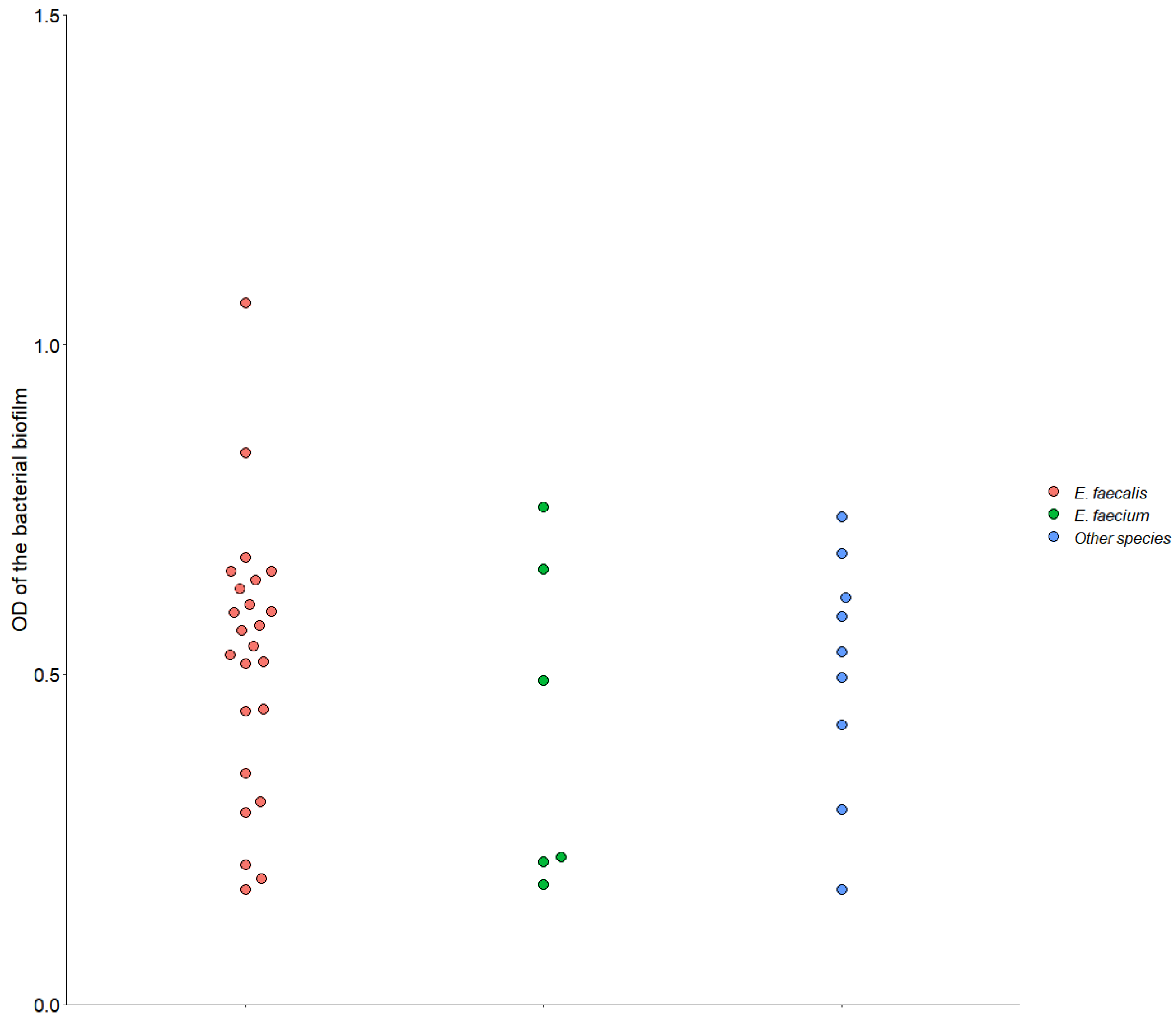
According to the OD570 values and the biofilm classification criteria proposed by Stepanović et al., the isolates were classified as non-biofilm producers (13/39, 33.3%), weak biofilm producers (25/39, 64.1%), and moderate biofilm producers (1/39, 2.6%), whereas no isolate was classified as a strong biofilm producer. Biofilm formation was observed in 26 of the 39 Enterococcus isolates (66.7% of isolates). Among the biofilm-producing isolates, 17 were identified as E. faecalis, three as E. faecium, two as E. avium, and one each as E. hirae, E. durans, E. cecorum, and E. gallinarum (Figure 4).
No significant differences in quantitative biofilm production (OD570 values) were observed among the three Enterococcus species groups (Kruskal–Wallis test, p > 0.05). Similarly, no significant association was detected between Enterococcus species groups and biofilm formation when isolates were classified as biofilm producers or nonproducers (χ2 test, p = 0.626). No significant association was observed between biofilm formation and cylA gene presence (Fisher’s exact test, p = 0.734). Because asa1 and gelE were detected in nearly all isolates (97.4%), their association with biofilm formation was not statistically evaluated because of the limited variability of these variables.

2.6. Hemolytic Activity

Among the 39 isolates examined, α-, β-, and γ-hemolysis was observed in 15 (38.5%), 2 (5.1%), and 22 (56.4%) isolates, respectively. Although cylA was detected in 19 isolates (48.7%), only two isolates exhibited β-hemolysis, and no significant association was observed between the presence of cylA and β-hemolysis (Fisher’s exact test, p = 0.231). The isolate-level phenotypic and genotypic characteristics are shown in Supplementary Table S1.

3. Discussion

The present study is the first to comprehensively phenotypically and genotypically characterize Enterococcus spp. recovered from clinical poultry cases in Chile. The predominance of E. faecalis (61.5%), followed by E. faecium (15.4%), agrees with previous reports identifying these species as the most common enterococci associated with septicemia, omphalitis, osteomyelitis, and locomotor disorders in poultry [20,21,22]. The detection of additional species, including E. durans, E. hirae, E. avium, E. gallinarum, and E. cecorum, further demonstrates the diversity of enterococci involved in poultry diseases [3,7,23,24].
The virulence-associated genes asa1, gelE, and cylA were highly prevalent among the isolates, indicating that they harbor important genetic determinants associated with bacterial colonization and persistence. The widespread detection of asa1 (97.4%) and gelE (97.4%) is consistent with previous reports describing these genes as major virulence determinants in poultry-associated Enterococcus spp. [20,21,25]. The aggregation substance (asa1) is associated with bacterial adhesion, conjugative plasmid transfer, and biofilm development, whereas gelatinase (gelE) contributes to extracellular matrix degradation, tissue invasion, and biofilm maturation [15,16,24,25,26]. Nevertheless, because gene expression and enzymatic activity were not evaluated, these findings should be interpreted as evidence of genetic potential for virulence rather than confirmation of biological activity.
The cylA gene was detected in nearly half of the isolates, a frequency comparable to that reported for poultry-associated enterococci from other countries [21,27,28,29]. Cytolysin is associated with increased virulence, tissue damage, and enhanced colonization in both animals and humans [16,27]. However, despite the relatively high prevalence of cylA, only two isolates exhibited β-hemolysis, and no significant association was observed between cylA carriage and the β-hemolytic phenotype. Similar discrepancies between genotype and phenotype have been reported previously and may reflect differences in gene regulation, environmental conditions, quorum-sensing mechanisms, or variations in the hemolysis assay conditions [15,25]. Collectively, these findings indicate that the presence of virulence-associated genes alone is insufficient to predict their phenotypic expression under the evaluated in vitro conditions.
Phenotypic antimicrobial susceptibility testing showed that resistance among poultry-associated Enterococcus spp. isolates was primarily directed against tetracycline (61.5%), whereas resistance to erythromycin was less frequent (17.9%). In contrast, all isolates remained susceptible to vancomycin, ampicillin, chloramphenicol, and linezolid. These findings are consistent with previous reports describing tetracycline resistance as the predominant antimicrobial resistance phenotype among poultry-associated enterococci, reflecting the long-standing use of tetracyclines in animal production and the consequent selection pressure for resistant strains [20,21,28,30,31,32,33,34,35,36,37,38,39,40,41]. The universal susceptibility to vancomycin is encouraging from both veterinary and public health perspectives and agrees with reports indicating that glycopeptide resistance remains uncommon among poultry-associated Enterococcus spp. in countries where avoparcin has been discontinued [40,41,42,43].
The molecular findings largely supported the phenotypic susceptibility results by identifying tetM (71.8%), ermB (66.7%), and tetL (30.8%) as the predominant antimicrobial resistance genes. Consistent with the phenotypic analysis in poultry isolates from Saudi Arabia and China, where tetM, tetL, and ermB were also the most frequently detected resistance determinants, supporting the widespread dissemination of these genes among poultry-associated enterococci despite geographical differences [21,28,40,41]. Consistent with the phenotypic analysis, a significant association was observed between the presence of tetM and tetracycline resistance, confirming its role as the principal genetic determinant of tetracycline resistance in the isolates analyzed. The widespread occurrence of tetM is expected because this gene is commonly associated with integrative conjugative elements (ICEs), including Tn916, Tn925, Tn5386, and several related mobile genetic elements, which facilitate horizontal gene transfer among enterococci and other Gram-positive bacteria [18,42,43]. Likewise, ermB is frequently associated with Tn3 family transposons, particularly Tn917, as well as with composite and conjugative elements such as Tn1545, Tn6002, and Tn3872, which contribute to the horizontal dissemination of macrolide resistance among enterococci and other Gram-positive bacteria [18,42,43]. Although ermB was highly prevalent, no significant association was observed between its presence and phenotypic erythromycin resistance. This apparent genotype–phenotype discordance may reflect inducible gene expression, differences in promoter activity, gene integrity, or other regulatory mechanisms influencing ermB expression [40,44,45].
Likewise, all isolates remained phenotypically susceptible to vancomycin, consistent with the absence of the acquired glycopeptide resistance operons vanA and vanB [6,18]. Similarly, although optrA was detected in three isolates, no phenotypic resistance to linezolid was observed, indicating that the presence of resistance genes does not necessarily predict phenotypic resistance [18]. Collectively, these findings emphasize that molecular detection of antimicrobial resistance genes complements, but does not replace, phenotypic susceptibility testing and highlights the importance of integrating both approaches for the accurate characterization of antimicrobial resistance in Enterococcus spp.
Beyond their importance as poultry pathogens, enterococci are recognized as opportunistic zoonotic bacteria because they are capable of colonizing a wide range of animal hosts and humans and may serve as reservoirs of virulence and antimicrobial resistance determinants. Poultry-associated Enterococcus spp. have been detected throughout the food production chain, raising concerns regarding the potential dissemination of clinically relevant resistance genes through direct animal contact or the food chain [6,18,42,46]. Although the present study did not include human, food, or environmental isolates and therefore cannot infer transmission between these compartments, the high prevalence of virulence-associated genes and mobile antimicrobial resistance determinants observed among clinical poultry isolates underscores the importance of continued surveillance within poultry production systems. Integrating molecular epidemiology across animal, food, environmental, and human sectors will be essential to better understand the potential contribution of poultry-associated enterococci to the broader epidemiology of antimicrobial resistance within a One Health framework [18,42,43].
Biofilm formation was observed in 66.7% of the isolates, although most strains exhibited weak biofilm-forming phenotype. Biofilm formation is recognized as an important virulence and persistence mechanism in enterococci because it enhances bacterial survival under environmental stress and contributes to increased tolerance to disinfectants, host immune responses, and antimicrobial agents [17,41,42]. Although no strong biofilm-producing isolates were identified, even weak biofilm production may facilitate long-term persistence on poultry farm surfaces, in water systems, on equipment, and in host tissues, thereby contributing to the persistence and dissemination of Enterococcus spp. within poultry production systems [43,44,45].
The relatively high frequency of biofilm formation among E. faecalis isolates is consistent with the recognized adaptability and persistence of this species in poultry-associated environments [16,42,45]. However, no significant associations were observed between the Enterococcus species groups and biofilm production or between biofilm formation and the presence of the cylA gene [47]. Likewise, although asa1 and gelE have been associated with biofilm development in previous studies [15,16,25], their almost universal distribution among the isolates precluded a meaningful statistical evaluation. These findings suggest that biofilm formation in poultry-associated Enterococcus spp. isolates is influenced by multiple genetic and regulatory mechanisms rather than by the presence of a single virulence-associated gene, supporting the multifactorial nature of enterococcal pathogenicity [16,21,25,28,47,48,49,50].
An important strength of the present study is that all Enterococcus isolates were recovered from independent clinical poultry cases submitted for diagnostic investigation and were associated with gross pathological lesions observed during necropsy. In contrast to previous studies based primarily on isolates recovered from healthy birds, cloacal swabs, fecal samples, or environmental sources [21,22,28], the present study focused exclusively on disease-associated isolates obtained from vertebral osteomyelitis, femoral head necrosis, hepatitis, yolk sac infection, cellulitis, and other clinical conditions. These findings provide valuable baseline information on the virulence-associated characteristics, antimicrobial resistance profiles, and biofilm-forming capacity of clinically relevant poultry-associated Enterococcus spp. in Chile.
This study has some limitations that should be considered when interpreting the results. Although isolates were collected from multiple poultry production systems, companies, and geographical regions in Chile, the relatively small sample size and limited number of isolates representing some Enterococcus species reduced the statistical power for interspecific comparisons and precluded the application of more complex multivariable analyses. In addition, detailed flock-level information, including antimicrobial use, management practices, and longitudinal epidemiological data, was not consistently available for all clinical cases, limiting the evaluation of potential risk factors associated with antimicrobial resistance and virulence profiles. Furthermore, only three virulence-associated genes (asa1, gelE, and cylA) were investigated; therefore, the virulence repertoire of the isolates may not have been fully represented. Future studies incorporating broader virulence gene panels, gene expression analyses, and whole-genome sequencing will be required to provide a more comprehensive assessment of pathogenic potential. Nevertheless, an important strength of this study is that all isolates originated from independent clinical poultry cases associated with gross pathological lesions, providing the first comprehensive phenotypic and genotypic characterization of clinically relevant poultry-associated Enterococcus spp. in Chile. These findings establish an important baseline for future studies incorporating larger isolate collections, whole-genome sequencing, and prospective epidemiological analyses.

4. Conclusions

This study provides the first comprehensive phenotypic and genotypic characterization of Enterococcus spp. recovered from clinical poultry cases in Chile. E. faecalis was the predominant species, showing the highest frequency of virulence-associated genes and the greatest diversity of antimicrobial resistance determinants. The widespread detection of asa1, gelE, tetM, and ermB, combined with the high prevalence of biofilm formation, suggests that clinically relevant poultry-associated Enterococcus spp. circulating in Chile possess multiple genetic determinants associated with bacterial persistence and adaptation.
Phenotypic antimicrobial susceptibility testing showed that resistance was primarily associated with tetracycline and, to a lesser extent, erythromycin, whereas all isolates remained susceptible to vancomycin, ampicillin, chloramphenicol, and linezolid. Molecular characterization supported these findings by identifying tetM and ermB as the predominant resistance genes. The significant association between tetM and phenotypic tetracycline resistance, and the lack of association between ermB and erythromycin resistance, highlights the importance of combining phenotypic and molecular approaches for accurate antimicrobial resistance characterization in Enterococcus spp.
A major strength of this study is that all isolates originated from independent clinical poultry cases associated with gross pathological lesions rather than from healthy birds or environmental samples, providing findings that are directly relevant to avian pathology. Overall, these results provide valuable baseline epidemiological data on clinically relevant poultry-associated Enterococcus spp. in Chile and provide a foundation for future genomic, epidemiological, and surveillance studies aimed at enhancing our understanding and control of enterococcal infections in poultry production.

5. Materials and Methods

5.1. Study Design and Bacterial Strains

This was an observational descriptive study based on routine veterinary diagnostic submissions. A total of 39 Enterococcus isolates were recovered from independent clinical poultry cases submitted to the Avian Pathology Laboratory, Faculty of Veterinary and Animal Sciences, University of Chile, between July 2024 and July 2025. The isolates originated from 12 commercial poultry companies comprising 30 different farms located in the Arica and Parinacota, Valparaíso, Metropolitan, O’Higgins, and Biobío regions of Chile. The poultry production systems represented included broiler breeders (n = 21), commercial broilers (n = 6), commercial laying hens (n = 11), and turkeys (n = 1). Samples were collected during routine diagnostic investigations of disease outbreaks and individual clinical cases were examined. Necropsies were performed by trained veterinary pathologists, and bacteriological samples were aseptically collected from lesions or affected organs exhibiting gross pathological changes consistent with bacterial infections.
To ensure isolate independence and avoid duplicate sampling, only one Enterococcus isolate was included from each necropsied bird. Consequently, each isolate represented an independent clinical case and was considered a unit of analysis. Because the primary objective of the study was the phenotypic and genotypic characterization of clinical Enterococcus isolates rather than the estimation of farm-level prevalence or epidemiological risk factors, clustering by farm, company, or production system was not considered in the statistical analyses. Cloacal, fecal, environmental, and duplicate isolates were excluded. Samples were inoculated onto 5% sheep blood agar and MacConkey agar (Oxoid Ltd., Basingstoke, UK) and incubated aerobically at 37 °C for 24 h. Colonies exhibiting morphological characteristics compatible with Enterococcus spp. were selected for phenotypic and molecular characterization.

5.2. Identification of Enterococci

Pure subcultures were generated from single colonies on blood agar plates and identified based on colony morphology, Gram staining, catalase testing, oxidase testing, and biochemical characterization using the VITEK® 2 Compact identification system according to the manufacturer’s recommendations (bioMérieux, Marcy-l’Étoile, Lyon, France).

5.3. DNA Extraction

Colonies grown overnight on blood agar plates were placed in a test tube containing 2 mL of tryptic soy broth (TSB; Merck KGaA, Darmstadt, Germany). Bacteria were harvested by centrifugation, and the bacterial genomic DNA was extracted and purified according to the manufacturer’s recommendations using the PureLinkTM Genomic DNA Mini Kit (Invitrogen, Waltham, MA, USA). Purified DNA was stored at −20 °C for further analysis.

5.4. Molecular Identification of Enterococci

The tuf gene, which encodes the elongation factor Tu, was included as a genus-specific molecular marker because of its high level of conservation among Enterococcus species and its widely validated reliability for molecular diagnosis and genus-level confirmation of Enterococcus. Molecular identification was performed by PCR in a final reaction volume of 25 μL containing 0.5 μM of each primer, 2.5 μL of 10× reaction buffer, 1.25 mM dNTP mix, 0.75 μL of 50 mM MgCl2, 2.5 U of DNA polymerase (PlatinumTM Taq DNA Polymerase, Invitrogen, Waltham, MA, USA), 2.5 μL of extracted DNA, and ultrapure water to adjust the final volume. The PCR conditions were as follows: initial denaturation at 94 °C for 2 min, followed by 30 cycles of denaturation at 94 °C for 30 s, annealing at 55 °C for 30 s, and extension at 72 °C for 30 s, with a final extension at 72 °C for 7 min. PCR products were resolved by electrophoresis on a 1% agarose gel (Invitrogen, Waltham, MA, USA) stained with GelRed® Nucleic Acid Stain (Millipore, Burlington, MA, USA) and visualized using a TransLum SOLO transilluminator (Biotop, Jing’an District, Shanghai, China). The primer sequences used in this study are listed in Table 4.

5.5. Molecular Detection of Virulence Factor Genes

Virulence-associated genes, including asa1 (aggregation substance), gelE (gelatinase), and cylA (cytolysin activator), were screened using polymerase chain reaction (PCR). The asa1, gelE, and cylA genes were selected because they represent three of the most extensively characterized virulence determinants in Enterococcus spp. asa1 encodes an aggregation substance involved in bacterial adhesion and conjugation, gelE encodes an extracellular gelatinase associated with tissue degradation and biofilm development, and cylA is part of the cytolysin operon that is responsible for cytolytic activity. These genes are among the most frequently investigated virulence markers in epidemiological studies of poultry-associated Enterococcus spp. [53]. Multiplex PCR was performed in a final reaction volume of 50 μL, containing 0.1 μM of each primer, 2.5 μL of 10× buffer, 2.5 mM dNTP mix, 1.5 μL of 50 mM MgCl2, 5 U of DNA polymerase (PlatinumTM Taq DNA Polymerase, Invitrogen, Waltham, MA, USA), 5 μL of extracted DNA, and ultrapure water to reach a total volume of 50 μL. The amplification protocol consisted of an initial denaturation step at 94 °C for 5 min, followed by 30 cycles of denaturation at 94 °C for 1 min, annealing at 56 °C for 1 min, and extension at 72 °C for 1 min, with a final extension step at 72 °C for 10 min. The primer sequences used in this study are listed in Table 4.

5.6. Antimicrobial Susceptibility Testing

Antimicrobial susceptibility testing was performed using the VITEK® 2 Compact system (bioMérieux, Marcy-l’Étoile, Lyon, France) according to the manufacturer’s instructions. Two antimicrobial susceptibility testing cards were used in this study: AST-P663 and AST-GP80. The AST-P663 card included ampicillin, erythromycin, linezolid, tetracycline, and vancomycin, whereas the AST-GP80 card was used to complement the susceptibility testing and included chloramphenicol among the evaluated antimicrobial agents.
The antimicrobial agents included in the final analysis were ampicillin, erythromycin, linezolid, tetracycline, vancomycin, and chloramphenicol. Susceptibility results were interpreted according to the Clinical and Laboratory Standards Institute (CLSI) Performance Standards for Antimicrobial Susceptibility Testing (M100, 35th Edition, 2025) [54]. Quality control procedures were performed using E. faecalis ATCC 29212. The quality control results were within the CLSI acceptable ranges. The isolates were categorized as susceptible, intermediate, or resistant according to the CLSI breakpoints. Intermediate isolates were considered non-susceptible only for descriptive purposes and were not included as resistant in the genotype–phenotype association analysis.

5.7. Molecular Detection of Antimicrobial Resistance Genes

Molecular detection of antimicrobial resistance genes was performed using conventional PCR to investigate the genetic determinants associated with the antimicrobial resistance phenotypes observed among the Enterococcus isolates. The genes evaluated included vanA and vanB (vancomycin resistance), ermA and ermB (erythromycin resistance), tetA, tetB, tetM, and tetL (tetracycline resistance), cat (chloramphenicol resistance), optrA (linezolid resistance), and pbp5 (ampicillin resistance). PCR amplification was performed using gene-specific primers under the amplification conditions described in Table 5. PCR products were separated by electrophoresis on 1.5% agarose gels (Invitrogen, Waltham, MA, USA) stained with GelRed® Nucleic Acid Stain (Millipore, Burlington, MA, USA) and visualized under ultraviolet illumination using a TransLum SOLO transilluminator (Biotop, Jing’an District, Shanghai, China).

5.8. Quantitative Biofilm Assay

The ability of Enterococcus isolates to form biofilms was quantitatively evaluated. Briefly, isolates from overnight cultures were grown in TSB supplemented with 1% glucose and incubated at 37 °C for 24 h. The culture density was adjusted to approximately 0.5 McFarland standard, and each culture was subsequently diluted 1:10 in sterile TSB. Aliquots of 200 μL of each dilution were inoculated into three wells of sterile 96-well polystyrene microtiter plates (Sigma-Aldrich, St. Louis, MO, USA). Sterile TSB served as the negative control, and E. faecalis ATCC 29212 was used as the positive control. The plates were incubated at 37 °C for 48 h and washed thrice with sterile phosphate-buffered saline (PBS). Biofilms were fixed with 200 μL methanol for 20 min, air-dried for 30 min, and stained with 2% crystal violet for 15 min. The wells were then rinsed with sterile deionized water, air-dried, and the bound dye solubilized with 150 μL of acetic acid. Optical density (OD) was measured at 570 nm using a microplate reader (EpochTM, BioTek Instruments, Winooski, VT, USA). The optical density cut-off value (ODc), calculated as the mean OD of the negative control plus three standard deviations, according to Stepanović et al. [60], was 0.3490. Isolates were classified as non-biofilm producers (optical density [OD] ≤ ODc), weak biofilm producers (ODc < OD ≤ 2 × ODc), moderate biofilm producers (2 × ODc < OD ≤ 4 × ODc), or strong biofilm producers (OD > 4 × ODc).

5.9. Hemolysin Activity

For hemolysin screening, Enterococcus isolates were streaked onto soy agar supplemented with 5% sheep blood and incubated at 37 °C for 24 h. A clear zone around the colonies (β-hemolysis) or a greenish discoloration (α-hemolysis) was considered positive, whereas the absence of any visible hemolytic zone (γ-hemolysis) was considered negative [61].

5.10. Statistical Analysis

Statistical analyses were performed using R ((version 4.4.1; R Foundation for Statistical Computing, Vienna, Austria; https://www.r-project.org)). For comparative analyses, the isolates were grouped into three categories: Group 1, E. faecalis; Group 2, E. faecium; and Group 3, other Enterococcus species. Differences in quantitative biofilm production (OD570 values) among the groups were assessed using the Kruskal–Wallis test. As no statistically significant differences were detected, post-hoc pairwise comparisons were not performed.
Associations between Enterococcus species groups and biofilm formation (biofilm producer versus non-producer), as well as between Enterococcus species groups and the presence of the cylA gene, were evaluated using Pearson’s chi-square test or Fisher’s exact test. Associations between cylA and β-hemolysis and between antimicrobial resistance genes (tetM and ermB) and their corresponding phenotypic resistance profiles were assessed using Fisher’s exact test. Odds ratios (ORs) with 95% confidence intervals (95% CIs) were calculated for the genotype–phenotype associations involving antimicrobial resistance genes. Because of the small sample size and sparse contingency tables, Fisher’s exact test was preferred whenever the expected cell frequencies were <5. Two-sided p-values < 0.05 were considered to be statistically significant.

Supplementary Materials

The following supporting information can be downloaded at: https://www.mdpi.com/article/10.3390/poultry5040052/s1, Table S1: Summary of main findings for Enterococcus spp. isolated from clinical poultry cases.

Author Contributions

Conceptualization, L.C. and F.N.; methodology, L.C., P.R., S.G., P.T. and F.N.; investigation, L.C., F.N., P.T., P.C. and H.H.; writing—original draft preparation, L.C.; writing—review and editing, L.C., F.N., P.C., S.G., P.T. and H.H.; visualization, L.C. and F.N.; supervision, L.C. All authors have read and agreed to the published version of the manuscript.

Funding

This research was funded by internal funds from the Avian Pathology Laboratory, Department of Animal Pathology, Faculty of Veterinary and Animal Sciences, Universidad de Chile.

Institutional Review Board Statement

Samples analyzed in this study were obtained as part of routine veterinary diagnostic procedures and no experimental animal manipulation was performed. Therefore, according to institutional and national regulations, ethical committee approval was not required.

Informed Consent Statement

Not applicable.

Data Availability Statement

The original contributions presented in this study are included in the article and Supplementary Material. Further inquiries can be directed to the corresponding author.

Conflicts of Interest

The authors declare no conflicts of interest.

References

  1. Gilmore, M.S.; Clewell, D.B.; Ike, Y.; Shankar, N. (Eds.) Enterococci: From Commensals to Leading Causes of Drug Resistant Infection [Internet]; Massachusetts Eye and Ear Infirmary: Boston, MA, USA, 2014. [Google Scholar] [PubMed]
  2. Byappanahalli, M.N.; Nevers, M.B.; Korajkic, A.; Staley, Z.R.; Harwood, V.J. Enterococci in the environment. Microbiol. Mol. Biol. Rev. 2012, 76, 685–706. [Google Scholar] [CrossRef] [Scilit] [PubMed] [PubMed Central]
  3. Lebreton, F.; Willems, R.J.L.; Gilmore, M.S. Enterococcus Diversity, Origins in Nature, and Gut Colonization. In Enterococci: From Commensals to Leading Causes of Drug Resistant Infection [Internet]; Gilmore, M.S., Clewell, D.B., Ike, Y., Shankar, N., Eds.; Massachusetts Eye and Ear Infirmary: Boston, MA, USA, 2014. [Google Scholar] [PubMed]
  4. Arias, C.A.; Murray, B.E. The rise of the Enterococcus: Beyond vancomycin resistance. Nat. Rev. Microbiol. 2012, 10, 266–278. [Google Scholar] [CrossRef] [Scilit] [PubMed] [PubMed Central]
  5. Van Tyne, D.; Gilmore, M.S. Friend turned foe: Evolution of enterococcal virulence and antibiotic resistance. Annu. Rev. Microbiol. 2014, 68, 337–356. [Google Scholar] [CrossRef] [Scilit] [PubMed] [PubMed Central]
  6. Hollenbeck, B.L.; Rice, L.B. Intrinsic and acquired resistance mechanisms in enterococcus. Virulence 2012, 3, 421–433. [Google Scholar] [CrossRef] [Scilit] [PubMed] [PubMed Central]
  7. Souillard, R.; Laurentie, J.; Kempf, I.; Le Caër, V.; Le Bouquin, S.; Serror, P.; Allain, V. Increasing incidence of Enterococcus-associated diseases in poultry in France over the past 15 years. Vet. Microbiol. 2022, 269, 109426. [Google Scholar] [CrossRef] [Scilit] [PubMed]
  8. Wigmore, S.M.; Greenhill, A.R.; Bean, D.C. Isolation and characterization of enterococci from poultry reveals high incidence of Enterococcus thailandicus in Victoria, Australia. J. Appl. Microbiol. 2024, 135, lxae194. [Google Scholar] [CrossRef] [Scilit] [PubMed]
  9. Wood, A.M.; MacKenzie, G.; McGiliveray, N.C.; Brown, L.; Devriese, L.A.; Baele, M. Isolation of Enterococcus cecorum from bone lesions in broiler chickens. Vet. Rec. 2002, 150, 27. [Google Scholar] [PubMed]
  10. Devriese, L.A.; Cauwerts, K.; Hermans, K.; Wood, A.M. Enterococcus cecorum septicemia as a cause of bone and joint lesions resulting in lameness in broiler chickens. Vlaams Diergeneeskd. Tijdschrift. 2002, 71, 219–221. [Google Scholar] [CrossRef] [Scilit]
  11. Jung, A.; Rautenschlein, S. Comprehensive report of an Enterococcus cecorum infection in a broiler flock in Northern Germany. BMC Vet. Res. 2014, 10, 311. [Google Scholar] [CrossRef] [Scilit] [PubMed] [PubMed Central]
  12. Kense, M.J.; Landman, W.J.M. Enterococcus cecorum infections in broiler breeders and their offspring: Molecular epidemiology. Avian Pathol. 2011, 40, 603–612. [Google Scholar] [CrossRef] [Scilit] [PubMed]
  13. Higuita, J.; Arango, M.; Forga, A.; Cortes, D.; Graham, D. An Updated Review of Enterococcus cecorum Infections in Poultry. Avian Dis. 2025, 68, 404–411. [Google Scholar] [CrossRef] [Scilit] [PubMed]
  14. Stalker, M.J.; Brash, M.L.; Weisz, A.; Ouckama, R.M.; Slavic, D. Arthritis and osteomyelitis associated with Enterococcus cecorum infection in broiler and broiler breeder chickens in Ontario, Canada. J. Vet. Diagn. Investig. 2010, 22, 643–645. [Google Scholar] [CrossRef] [Scilit] [PubMed]
  15. Eaton, T.J.; Gasson, M.J. Molecular screening of Enterococcus virulence determinants and potential for genetic exchange between food and medical isolates. Appl. Environ. Microbiol. 2001, 67, 1628–1635. [Google Scholar] [CrossRef] [Scilit] [PubMed] [PubMed Central]
  16. Fisher, K.; Phillips, C. The ecology, epidemiology and virulence of Enterococcus. Microbiology 2009, 155, 1749–1757. [Google Scholar] [CrossRef] [Scilit] [PubMed]
  17. Mohamed, J.A.; Huang, D.B. Biofilm formation by enterococci. J. Med. Microbiol. 2007, 56, 1581–1588. [Google Scholar] [CrossRef] [Scilit] [PubMed]
  18. Werner, G.; Coque, T.M.; Franz, C.M.; Grohmann, E.; Hegstad, K.; Jensen, L.; van Schaik, W.; Weaver, K. Antibiotic resistant enterococci—Tales of a drug resistance gene trafficker. Int. J. Med. Microbiol. 2013, 303, 360–379. [Google Scholar] [CrossRef] [Scilit] [PubMed]
  19. Lee, T.; Pang, S.; Abraham, S.; Coombs, G.W. Molecular characterization and evolution of the first outbreak of vancomycin-resistant Enterococcus faecium in Western Australia. Int. J. Antimicrob. Agents 2019, 53, 814–819. [Google Scholar] [CrossRef] [Scilit] [PubMed]
  20. Stępień-Pyśniak, D.; Hauschild, T.; Dec, M.; Marek, A.; Urban-Chmiel, R.; Kosikowska, U. Phenotypic and genotypic characterization of Enterococcus spp. from yolk sac infections in broiler chicks with a focus on virulence factors. Poult. Sci. 2021, 100, 100985. [Google Scholar] [CrossRef] [Scilit] [PubMed] [PubMed Central]
  21. Alzahrani, O.M.; Fayez, M.; Alswat, A.S.; Alkafafy, M.; Mahmoud, S.F.; Al-Marri, T.; Almuslem, A.; Ashfaq, H.; Yusuf, S. Antimicrobial Resistance, Biofilm Formation, and Virulence Genes in Enterococcus Species from Small Backyard Chicken Flocks. Antibiotics 2022, 11, 380. [Google Scholar] [CrossRef] [Scilit] [PubMed] [PubMed Central]
  22. Mwikuma, G.; Kainga, H.; Kallu, S.A.; Nakajima, C.; Suzuki, Y.; Hang’ombe, B.M. Determination of the Prevalence and Antimicrobial Resistance of Enterococcus faecalis and Enterococcus faecium Associated with Poultry in Four Districts in Zambia. Antibiotics 2023, 12, 657. [Google Scholar] [CrossRef] [Scilit] [PubMed] [PubMed Central]
  23. Jung, A.; Chen, L.R.; Suyemoto, M.M.; Barnes, H.J.; Borst, L.B. A Review of Enterococcus cecorum Infection in Poultry. Avian Dis. 2018, 62, 261–271. [Google Scholar] [CrossRef] [Scilit] [PubMed]
  24. Stępień-Pyśniak, D.; Marek, A.; Banach, T.; Adaszek, Ł.; Pyzik, E.; Wilczyński, J.; Winiarczyk, S. Prevalence and antibiotic resistance of Enterococcus strains isolated from poultry. Acta Vet. Hung. 2016, 64, 148–163. [Google Scholar] [CrossRef] [Scilit] [PubMed]
  25. Fiore, E.; Van Tyne, D.; Gilmore, M.S. Pathogenicity of Enterococci. Microbiol. Spectr. 2019, 7, 10-1128. [Google Scholar] [CrossRef] [Scilit] [PubMed] [PubMed Central]
  26. Pinkston, K.L.; Gao, P.; Diaz-Garcia, D.; Sillanpää, J.; Nallapareddy, S.R.; Murray, B.E.; Harvey, B.R. The Fsr quorum-sensing system of Enterococcus faecalis modulates surface display of the collagen-binding MSCRAMM Ace through regulation of gelE. J. Bacteriol. 2011, 193, 4317–4325. [Google Scholar] [CrossRef] [Scilit] [PubMed] [PubMed Central]
  27. Chow, J.W.; Thal, L.A.; Perri, M.B.; Vazquez, J.A.; Donabedian, S.M.; Clewell, D.B.; Zervos, M.J. Plasmid-associated hemolysin and aggregation substance production contribute to virulence in experimental enterococcal endocarditis. Antimicrob. Agents Chemother. 1993, 37, 2474–2477. [Google Scholar] [CrossRef] [Scilit] [PubMed] [PubMed Central]
  28. Yu, L.; Liu, Y.; Liu, M.; Li, Z.; Li, L.; Wang, F. Research Note: Molecular characterization of antimicrobial resistance and virulence gene analysis of Enterococcus faecalis in poultry in Tai’an, China. Poult. Sci. 2022, 101, 101763. [Google Scholar] [CrossRef] [Scilit] [PubMed] [PubMed Central]
  29. Jian, Z.; Qian, Y.; He, S.; Zhao, R.; Li, K.; Cha, J.; Ning, Z.; Ye, Y.; Bao, Z.; Wang, K.; et al. The gut resistome in poultry production: Microbial ecology, antibiotic use, and sustainable control approaches. Front. Microbiol. 2026, 17, 1768747. [Google Scholar] [CrossRef] [Scilit] [PubMed] [PubMed Central]
  30. Kerek, Á.; Szabó, Á.; Jerzsele, Á. Antimicrobial Susceptibility Profiles of Commensal Enterococcus spp. Isolates from Chickens in Hungarian Poultry Farms Between 2022 and 2023. Antibiotics 2024, 13, 1194. [Google Scholar] [CrossRef] [Scilit] [PubMed] [PubMed Central]
  31. Ribeiro, J.; Silva, V.; Monteiro, A.; Vieira-Pinto, M.; Igrejas, G.; Reis, F.S.; Barros, L.; Poeta, P. Antibiotic Resistance among Gastrointestinal Bacteria in Broilers: A Review Focused on Enterococcus spp. and Escherichia coli. Animals 2023, 13, 1362. [Google Scholar] [CrossRef] [Scilit] [PubMed] [PubMed Central]
  32. Alhothali, M.T.; Seemann, T.; Andersson, P.; Sherry, N.; Silver, J.D.; Howden, O.C.; Wilmot, M.; Siryj, W.; Veitch, M.G.; Howden, B.P.; et al. Rising burden of enterococcal bacteremia in Victoria, Australia: Population-based incidence and antimicrobial resistance trends from three decades of surveillance. Antimicrob. Agents Chemother. 2026, 70, e0152625. [Google Scholar] [CrossRef] [Scilit] [PubMed] [PubMed Central]
  33. Berktas, M.; Yaman, G.; Ozturk, O. vanC gene-related intrinsic teicoplanin resistance detected in Enterococcus casseliflavus and E. gallinarum strains by the BD phoenix automated microbiology system. J. Clin. Microbiol. 2008, 46, 2466. [Google Scholar] [CrossRef] [Scilit] [PubMed] [PubMed Central]
  34. Tyson, G.H.; Nyirabahizi, E.; Crarey, E.; Kabera, C.; Lam, C.; Rice-Trujillo, C.; McDermott, P.F.; Tate, H. Prevalence and Antimicrobial Resistance of Enterococci Isolated from Retail Meats in the United States, 2002 to 2014. Appl. Environ. Microbiol. 2018, 84, e01902-17. [Google Scholar] [CrossRef] [Scilit] [PubMed] [PubMed Central]
  35. Zaheer, R.; Cook, S.R.; Barbieri, R.; Goji, N.; Cameron, A.; Petkau, A.; Polo, R.O.; Tymensen, L.; Stamm, C.; Song, J.; et al. Surveillance of Enterococcus spp. reveals distinct species and antimicrobial resistance diversity across a One-Health continuum. Sci. Rep. 2020, 10, 3937, Erratum in Sci. Rep. 2020, 10, 13401. https://doi.org/10.1038/s41598-020-69044-5. [Google Scholar] [CrossRef] [Scilit] [PubMed] [PubMed Central]
  36. Torres, C.; Alonso, C.A.; Ruiz-Ripa, L.; León-Sampedro, R.; Del Campo, R.; Coque, T.M. Antimicrobial Resistance in Enterococcus spp. of animal origin. Microbiol. Spectr. 2018, 6, 10-1128. [Google Scholar] [CrossRef] [Scilit] [PubMed] [PubMed Central]
  37. Ngbede, E.O.; Raji, M.A.; Kwanashie, C.N.; Kwaga, J.K.P. Antimicrobial resistance and virulence profile of enterococci isolated from poultry and cattle sources in Nigeria. Trop. Anim. Health Prod. 2016, 49, 451–458, Erratum in Trop. Anim. Health Prod. 2017, 49, 885. https://doi.org/10.1007/s11250-017-1261-4. [Google Scholar] [CrossRef] [Scilit] [PubMed]
  38. Urban-Chmiel, R.; Marek, A.; Stępień-Pyśniak, D.; Wieczorek, K.; Dec, M.; Nowaczek, A.; Osek, J. Antibiotic Resistance in Bacteria—A Review. Antibiotics 2022, 11, 1079. [Google Scholar] [CrossRef] [Scilit] [PubMed] [PubMed Central]
  39. Jahantigh, M.; Samadi, K.; Dizaji, R.E.; Salari, S. Antimicrobial resistance and prevalence of tetracycline resistance genes in Escherichia coli isolated from lesions of colibacillosis in broiler chickens in Sistan, Iran. BMC Vet. Res. 2020, 16, 267. [Google Scholar] [CrossRef] [Scilit] [PubMed] [PubMed Central]
  40. Ahmadpoor, N.; Ahmadrajabi, R.; Esfahani, S.; Hojabri, Z.; Moshafi, M.H.; Saffari, F. High-Level Resistance to Erythromycin and Tetracycline and Dissemination of Resistance Determinants among Clinical Enterococci in Iran. Med. Princ. Pract. 2021, 30, 272–276. [Google Scholar] [CrossRef] [Scilit] [PubMed] [PubMed Central]
  41. Koskeroglu, K.; Onmaz, N.E.; Gundog, D.A.; Gungor, C.; Gungor, G.; Imre, K.; Morar, A. Tracking persistent and resistant Enterococcus faecalis and E. faecium from farm to fork: Biofilm-linked risks in antibiotic resistance of isolates. Vet. Res. Commun. 2026, 50, 100. [Google Scholar] [CrossRef] [Scilit] [PubMed] [PubMed Central]
  42. Willett, J.L.E.; Dunny, G.M. Insights into ecology, pathogenesis, and biofilm formation of Enterococcus faecalis from functional genomics. Microbiol. Mol. Biol. Rev. 2025, 89, e0008123. [Google Scholar] [CrossRef] [Scilit] [PubMed] [PubMed Central]
  43. Sandoe, J.A.T.; Witherden, I.R.; Cove, J.H.; Heritage, J.; Wilcox, M.H. Correlation between enterococcal biofilm formation in vitro and medical-device-related infection potential in vivo. J. Med. Microbiol. 2003, 52, 547–550. [Google Scholar] [CrossRef] [Scilit] [PubMed]
  44. Ogundipe, T.T.; Beitia, S.; Zhang, L.; Zhang, X.; Obe, T. Differences in microbial composition of litter and water line biofilm of broiler farms as influenced by water quality history. Poult. Sci. 2025, 104, 105832. [Google Scholar] [CrossRef] [Scilit] [PubMed] [PubMed Central]
  45. Silva, V.; Freitas, C.; Ribeiro, J.; Igrejas, G.; Poeta, P. Comparative analysis of antibiotic resistance and biofilm formation in Enterococcus spp. across One Health domains. FEMS Microbes 2025, 6, xtaf005. [Google Scholar] [CrossRef] [Scilit] [PubMed] [PubMed Central]
  46. Olsen, R.H.; Schønheyder, H.C.; Christensen, H.; Bisgaard, M. Enterococcus faecalis of human and poultry origin share virulence genes supporting the zoonotic potential of E. faecalis. Zoonoses Public Health 2012, 59, 256–263. [Google Scholar] [CrossRef] [Scilit] [PubMed]
  47. Reynolds, D.L.; Simpson, E.B.; Hille, M.M. A method for demonstrating the cytolysin/hemolysin of Enterococcus faecalis isolates of poultry origin. Poultry 2025, 4, 11. [Google Scholar] [CrossRef] [Scilit]
  48. Chakroborty, N.; Fahim, N.A.I.; Islam, M.S.; Rana, M.L.; Ferdous, F.B.; Atiq, M.N.; Sobur, M.A.; Ahammed, M.; Saha, S.; Rahman, M.T. Biofilm Formation, Virulence Traits, and Antimicrobial Resistance Profiles of Enterococcus faecalis in Layer Parent Stock in Bangladesh. Int. J. Microbiol. 2026, 2026, 4082070. [Google Scholar] [CrossRef] [Scilit] [PubMed] [PubMed Central]
  49. Reynolds, D.L.; Hille, M.M.; Jia, B. Review of Enterococcus faecalis Infections of Poultry. Avian Dis. 2024, 68, 412–420. [Google Scholar] [CrossRef] [Scilit] [PubMed]
  50. Tadesse, B.T.; Zhao, S.; Gu, L.; Jers, C.; Mijakovic, I.; Solem, C. Genome analysis reveals a biased distribution of virulence and antibiotic resistance genes in the genus Enterococcus and an abundance of safe species. Appl. Environ. Microbiol. 2025, 91, e0041525. [Google Scholar] [CrossRef] [Scilit] [PubMed] [PubMed Central]
  51. Ke, D.; Picard, F.J.; Martineau, F.; Ménard, C.; Roy, P.H.; Ouellette, M.; Bergeron, M.G. Development of a PCR assay for rapid detection of enterococci. J. Clin. Microbiol. 1999, 37, 3497–3503. [Google Scholar] [CrossRef] [Scilit] [PubMed] [PubMed Central]
  52. Vankerckhoven, V.; Van Autgaerden, T.; Vael, C.; Lammens, C.; Chapelle, S.; Rossi, R.; Jabes, D.; Goossens, H. Development of a multiplex PCR for the detection of asa1, gelE, cylA, esp, and hyl genes in enterococci and survey for virulence determinants among European hospital isolates of Enterococcus faecium. J. Clin. Microbiol. 2004, 42, 4473–4479. [Google Scholar] [CrossRef] [Scilit] [PubMed] [PubMed Central]
  53. Mareković, I.; Markanović, M.; Lešin, J.; Ćorić, M. Vancomycin-Resistant Enterococci: Current Understandings of Resistance in Relation to Transmission and Preventive Strategies. Pathogens 2024, 13, 966. [Google Scholar] [CrossRef] [Scilit] [PubMed] [PubMed Central]
  54. Clinical and Laboratory Standards Institute. Performance Standards for Antimicrobial Susceptibility Testing, 35th ed.; CLSI Supplement M100; Clinical and Laboratory Standards Institute: Malvern, PA, USA, 2025. [Google Scholar]
  55. Osman, K.M.; Badr, J.; Orabi, A.; Elbehiry, A.; Saad, A.; Ibrahim, M.D.S.; Hanafy, M.H. Poultry as a vector for emerging multidrug resistant Enterococcus spp.: First report of vancomycin (van) and the chloramphenicol–florfenicol (cat-fex-cfr) resistance genes from pigeon and duck faeces. Microb. Pathog. 2019, 128, 195–205. [Google Scholar] [CrossRef] [Scilit] [PubMed]
  56. Jensen, L.B.; Frimodt-Møller, N.; Aarestrup, F.M. Presence of erm gene classes in gram-positive bacteria of animal and human origin in Denmark. FEMS Microbiol. Lett. 1999, 170, 151–158. [Google Scholar] [CrossRef] [PubMed]
  57. Ng, L.-K.; Martin, I.; Alfa, M.; Mulvey, M. Multiplex PCR for the detection of tetracycline resistant genes. Mol. Cell. Probes 2001, 15, 209–215. [Google Scholar] [CrossRef] [Scilit] [PubMed]
  58. Wang, S.; Zhang, S.; Liu, Z.; Liu, P.; Shi, Z.; Wei, J.; Shao, D.; Li, B.; Ma, Z. Molecular characterization of enterohemorrhagic E. coli O157 isolated from animal fecal and food samples in Eastern China. Sci. World J. 2014, 2014, 946394. [Google Scholar] [CrossRef] [Scilit] [PubMed] [PubMed Central]
  59. Jureen, R.; Top, J.; Mohn, S.C.; Harthug, S.; Langeland, N.; Willems, R.J.L. Molecular characterization of ampicillin-resistant Enterococcus faecium isolates from hospitalized patients in Norway. J. Clin. Microbiol. 2003, 41, 2330–2336. [Google Scholar] [CrossRef] [Scilit] [PubMed] [PubMed Central]
  60. Stepanović, S.; Vuković, D.; Hola, V.; Di Bonaventura, G.; Djukić, S.; Ćirković, I.; Ruzicka, F. Quantification of biofilm in microtiter plates: Overview of testing conditions and practical recommendations for assessment of biofilm production by staphylococci. APMIS 2007, 115, 891–899. [Google Scholar] [CrossRef] [Scilit] [PubMed]
  61. De Vuyst, L.; Moreno, M.F.; Revets, H. Screening for enterocins and detection of hemolysin and vancomycin resistance in enterococci of different origins. Int. J. Food Microbiol. 2003, 84, 299–318. [Google Scholar] [CrossRef] [Scilit] [PubMed]
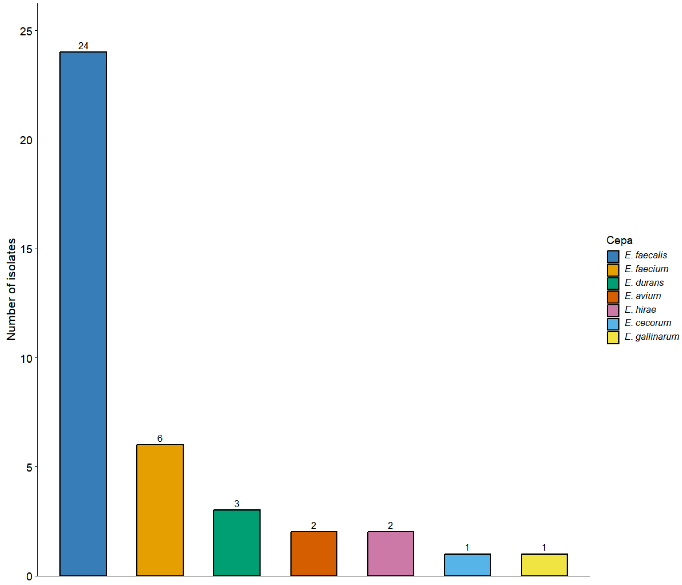
Figure 1. Distribution of Enterococcus species identified in poultry.
Poultry 05 00052 g001
Figure 2. Distribution of virulence genes among the Enterococcus isolates.
Poultry 05 00052 g002
Figure 3. Antimicrobial susceptibility of Enterococcus isolates recovered from poultry in Chile.
Poultry 05 00052 g003
Figure 4. Classification of biofilm formation among Enterococcus spp. isolates according to the Stepanović classification. Biofilm production was classified as non-producer (OD ≤ ODc), weak producer (ODc < OD ≤ 2 × ODc), moderate producer (2 × ODc < OD ≤ 4 × ODc), or strong producer (OD > 4 × ODc).
Poultry 05 00052 g004
Table 1. Antimicrobial resistance patterns among Enterococcus isolates recovered from poultry in Chile (N = 39).
Resistance PatternsNumber of Isolates% Isolates
Fully susceptible1128.2
ERY410.3
TCY2153.8
ERY + TCY37.7
Total39100
TCY, tetracycline; ERY, erythromycin.
Table 2. Distribution of antimicrobial resistance genes among Enterococcus isolates (n = 39).
Resistance GeneE.
faecalis (N = 24)
E. faecium (N = 6)E.
durans (N = 3)
E. avium (N = 2)E.
hirae (N = 2)
E.
cecorum (N = 1)
E.
gallinarum (N = 1)
Total
Enterococci (N = 39)
vanA00000000
vanB00000000
ermA10001002
ermB1632121126
pbp500000000
tetA20110105
tetB00010001
tetM1832211128
tetL811011012
optrA11100003
cat00000000
Table 3. Association between antimicrobial resistance genes and corresponding phenotypic resistance.
GenePhenotypic ResistanceOdds Ratio (95% CI)p-Value
tetMTetracycline resistance8.0 (1.7–37.5)0.010
ermBErythromycin resistance3.6 (0.4–31.0)0.388
Table 4. Primers for Enterococcus identification and virulence gene detection.
Target GenePrimer NamePrimer Sequence 5’ to 3’Amplicon Size (bp)Reference
tuftuf-FATGCCGACATTGAAAGAAAAAATT803[51]
tuf-RTCAATCTTTGGTTCCATCTCT
asa1ASA 11GCACGCTATTACGAACTATGA375[52]
ASA 12TAAGAAAGAACATCACCACGA
gelEGEL 11TATGACAATGCTTTTTGGGAT213
GEL 12AGATGCACCCGAAATAATATA
cylACYT 1ACTCGGGGATTGATAGGC688
CYT 2GCTGCTAAAGCTGCGCTT
bp = base pairs.
Table 5. Primers for detection of antimicrobial resistance genes.
Target GenePrimer NamePrimer Sequence 5’ to 3’Amplicon Size (bp)Reference
vanAvanAfGCGCGGTCCACTTGTAGATA314[55]
vanArTGAGCAACCCCCAAACAGTA
vanBvanBfAGACATTCCGGTCGAGGAAC220
vanBrGCTGTCAATTAGTGCGGGAA
catcatfGGATATGAAATTTATCCCTC486
catrCAATCATCTACCCTATGAAT
ermAermAfGCGGTAAACCCCTCTGAG434[56]
ermArGCCTGTCGGAATTGG
ermBermBfCATTTAACGACGAAACTGGC425
ermBrGGAACATCTGTGGTATGGCG
tetMtetMfGTGGACAAAGGTACAACGAG406[57]
tetMrCGGTAAAGTTCGTCACACAC
tetAtetAfGCTACATCCTGCTTGCCTTC210
tetArCATAGATCGCCGTGAAGAGG
tetBtetBfTTGGTTAGGGGCAAGTTTTG659
tetBrGTAATGGGCCAATAACACCG
tetLtetLfATAAATTGTTTCGGGTCGGTAAT1077
tetLrAACCAGCCAACTAATGACAATGAT
optrAoptrAfAGGTGGTCAGCGAACTAA1395[58]
optrArATCAACTGTTCCCATTCA
pbp5pbp5fAACAAAATGACAAACGGG779[59]
pbp5rTATCCTTGGTTATCAGGG
bp = base pairs.
Disclaimer/Publisher’s Note: The statements, opinions and data contained in all publications are solely those of the individual author(s) and contributor(s) and not of MDPI and/or the editor(s). MDPI and/or the editor(s) disclaim responsibility for any injury to people or property resulting from any ideas, methods, instructions or products referred to in the content.