Article

Insights into Stability and Selective Agglomeration in Binary Mixtures of Colloids: A Study on Gold Nanoparticles and Ultra-Small Quantum Dots

1
Institute for Energy and Materials Processes—Particle Science and Technology (EMPI-PST), University of Duisburg-Essen (UDE), 47057 Duisburg, Germany
2
Institute of Micro- and Nanostructure Research (IMN) & Center for Nanoanalysis and Electron Microscopy (CENEM), Interdisciplinary Center for Nanostructured Films (IZNF), Friedrich-Alexander University of Erlangen-Nuremberg (FAU), 91058 Erlangen, Germany
3
Centre for Nanointegration Duisburg-Essen (CENIDE), 47057 Duisburg, Germany
*
Author to whom correspondence should be addressed.
Submission received: 30 December 2024 / Revised: 5 March 2025 / Accepted: 10 March 2025 / Published: 19 March 2025

Abstract

:
Controlling the stability of colloidal nanoparticles in multicomponent systems is crucial for advancing formulations and separation processes. This study investigates the selective agglomeration approach for binary colloidal mixtures, providing both fundamental insights into stability/agglomeration mechanisms and a scalable separation strategy. First, we established a binary model system comprising gold nanoparticles (Au NPs) and ZnS quantum dots (QDs) to assess interparticle interactions. UV-visible spectroscopy revealed that impurities released from ZnS QDs, particularly thiol-based ligands and unbound Zn ions, triggered the aggregation of Au NPs depending on their surface stabilizers. Functionalization of Au NPs with bis(p-sulfonatophenyl) phenylphosphine (BSPP) significantly enhanced colloidal stability, with unpurified BSPP-functionalized Au NPs exhibiting superior resistance to agglomeration. Building on these insights, we applied selective agglomeration to separate a complex colloidal system consisting of InP/ZnS core–shell QDs and ZnS byproducts, a critical challenge in QD synthesis that is particularly relevant for post-processing of samples that originate from large-scale flow synthesis. By systematically tuning the ethanol concentration as a poor solvent, we successfully achieved composition-dependent fractionation. Optical and spectroscopic analyses confirmed that coarse fractions were enriched in InP/ZnS QDs, while fines fractions mainly contained pure ZnS QDs, with absorption peaks at 605 nm and 290 nm, respectively. Photoluminescence spectra further demonstrated a redshift in the coarse fractions, correlating with an increase in particle size. These results underscore the potential of selective agglomeration as a scalable, post-synthesis classification method, offering a framework for controlling stability and advancing post-synthesis separation strategies in colloidal multicomponent systems.

1. Introduction

Nanoparticles play a crucial role in advancing technologies across fields such as energy storage, catalysis, photonics, and biomedicine due to their unique optical, electronic, and catalytic properties that emerge at the nanoscale. In particular, semiconductor quantum dots (QDs) and noble metal nanoparticles, such as gold nanoparticles (Au NPs), are valued for their unique size-dependent functionalities, which enable applications in dye-sensitized solar cells [1,2,3,4,5], light-emitting diodes (LEDs) [6,7,8,9,10,11,12], and biomedical imaging [13,14,15,16]. However, these functionalities are often dependent not only on size but also on well-controlled characteristics, such as shape, surface chemistry, and composition, particularly in systems comprising particles under 10 nm [17,18,19].
Multidimensional classification in nanoparticle processing is essential for separating complex colloidal systems in more than one dimension, such as size and composition or size and surface functionality. This is critical to tailor nanoparticle properties for specific applications. While substantial progress has been made in the synthesis of these ultra-small nanoparticles, the field faces significant challenges in realizing multidimensional classification at the sub-10 nm scale [20,21,22,23]. Traditional methods, including chromatography and field-flow fractionation (FFF), offer limited resolution in differentiating nanoparticles based on more than one dimension, often leading to sample dilution and requiring extensive optimization for each unique system. While FFF shows promise for noble metals, it remains undeveloped for preparative, high-concentration separations of QDs [24,25]. Chromatography, though making rapid progress in the past years [26,27,28], still faces limitations when particles with close sizes and compositions are to be separated. Thus, these existing techniques have not yet achieved the scalability or multidimensional selectivity required for advanced nanoparticle applications.
To address these limitations, scalable methods such as size-selective agglomeration (SSA) are being explored. Originally developed for noble metals, SSA leverages selective destabilization by a poor solvent, facilitating aggregation based on differences in van der Waals interactions and particle size [29,30]. This technique has already shown promise in separating sub-5 nm ZnS QDs, establishing SSA as a scalable approach for the preparative separation of small nanoparticles [31,32,33]. However, multidimensional classification for practical, industrially relevant systems remains challenging, particularly in establishing model systems that allow fine control over stability and particle-particle interactions in binary and multinary mixtures. Despite significant progress in the synthesis and stability of one-component colloidal dispersions, such as Au NPs stabilized with various capping ligands [34,35], multicomponent systems introduce new complexities. For Au NPs, ligand surface interactions have been studied extensively for their role in enhancing stability and controlling self-assembly, leading to innovations across optics, catalysis, and biological applications [36,37,38,39,40,41,42]. In contrast, semiconductor QDs, valued for their tunable photoluminescence and high quantum yield, typically exhibit inherent stability due to steric effects originating from their surface ligands, even when only small-chain molecules are used [43,44,45]. Nevertheless, nanoparticle-based applications often require blending multiple nanoparticle types or face complex reaction mixtures with multiple byproducts, which complicates control over stability and interaction due to the diverse properties of each component [46,47].
In addressing these challenges, we summarize the key findings of two recent studies. In the first study [48], we focused on creating a stable binary model colloid of Au NPs and ZnS QDs, two materials with distinct interactions with light, to investigate stability and classification dynamics in a mixed colloidal environment. This model system was developed to meet the unique demands of binary colloidal mixtures, including the need for precise stability control over time. The successful stabilization and characterization of this system provided insights into managing interparticle interactions in complex colloidal environments, forming a foundation for more advanced multicomponent systems. This groundwork enabled us to systematically study the fundamental interactions in binary colloidal systems, setting the stage for multidimensional separation methods.
Building upon these foundational insights, our project extended SSA to a practical system: a mixture of InP/ZnS QDs synthesized for the use as core–shell structures but unintentionally containing pure ZnS QDs as a synthesis byproduct [49]. The presence of these ZnS impurities diminishes optical performance, adversely affecting photoluminescence and quantum yield [50]. To address this, we employed SSA to separate the InP/ZnS QDs and ZnS QDs into distinct fractions based on their size and composition. These findings demonstrate the potential of selective agglomeration not only as a scalable technique for nanoparticle separation but, as we could also unravel the scalability of the method to gram-scale separation [33], also as a cornerstone for realizing continuous, multidimensional classification of complex colloidal systems.
Our work contributes to the field by providing an efficient, scalable methodology for the post-synthesis classification of multicomponent colloidal systems. By developing SSA for complex mixtures and addressing both an understanding of colloidal stability in model systems and the application of selective agglomeration for multidimensional classification, our research paves the way for further advancements in nanoparticle processing, thereby enhancing the applicability and scalability of multidimensional separation techniques across industrial and research applications.

2. Materials and Methods

2.1. Materials

The chemicals used in the works summarized in this study included zinc acetate dihydrate (ZnAc·2H2O, 98%, Alfa Aesar, Karlsruhe, Germany), thioglycerol (3-mercapto-1,2-propanediol, 98%, Merck (Sigma-Aldrich) KGaA, Taufkirchen, Germany), dimethylformamide (DMF, 98.8%, Carl Roth GmbH & Co., KG, Karlsruhe, Germany), sodium hydroxide (NaOH, 98%, Carl Roth GmbH & Co., KG), sodium sulfide nonahydrate (Na2S·9H2O, 98%, Merck (Sigma-Aldrich), hydrogen tetrachloroaurate (III) trihydrate (HAuCl₄·3H2O, 99.99%), and Au foil (0.5 mm thickness, 99.99% purity, AGOSI). Additional agents included tannic acid, sodium citrate tribasic dihydrate (99%), bis(p-sulfonatophenyl) phenylphosphine dipotassium salt (BSPP, 97%), ethanol (EtOH, 99.98%, VWR International GmbH, Germany), and methanol (MeOH, 99.9%, Carl Roth GmbH & Co., KG, 99.9%). These reagents were used as received, without further purification, for the liquid-phase synthesis of nanoparticles. Millipore water was prepared with a Milli-Q® Reference (18.2 MΩ·cm, Merck, Taufkirchen, Germany). Other chemicals used were indium chloride (InCl3, 99.99%), zinc chloride (ZnCl2, 98%), tris(diethylamino)phosphine (PDEA, 97%), and zinc diethyldithiocarbamate (ZDEC, 97%), all obtained from Sigma-Aldrich. Oleylamine (OLA, 80–90%) was sourced from Acros Organics, while analytical-grade ethanol and n-hexane (≥99%) were purchased from VWR Chemicals.

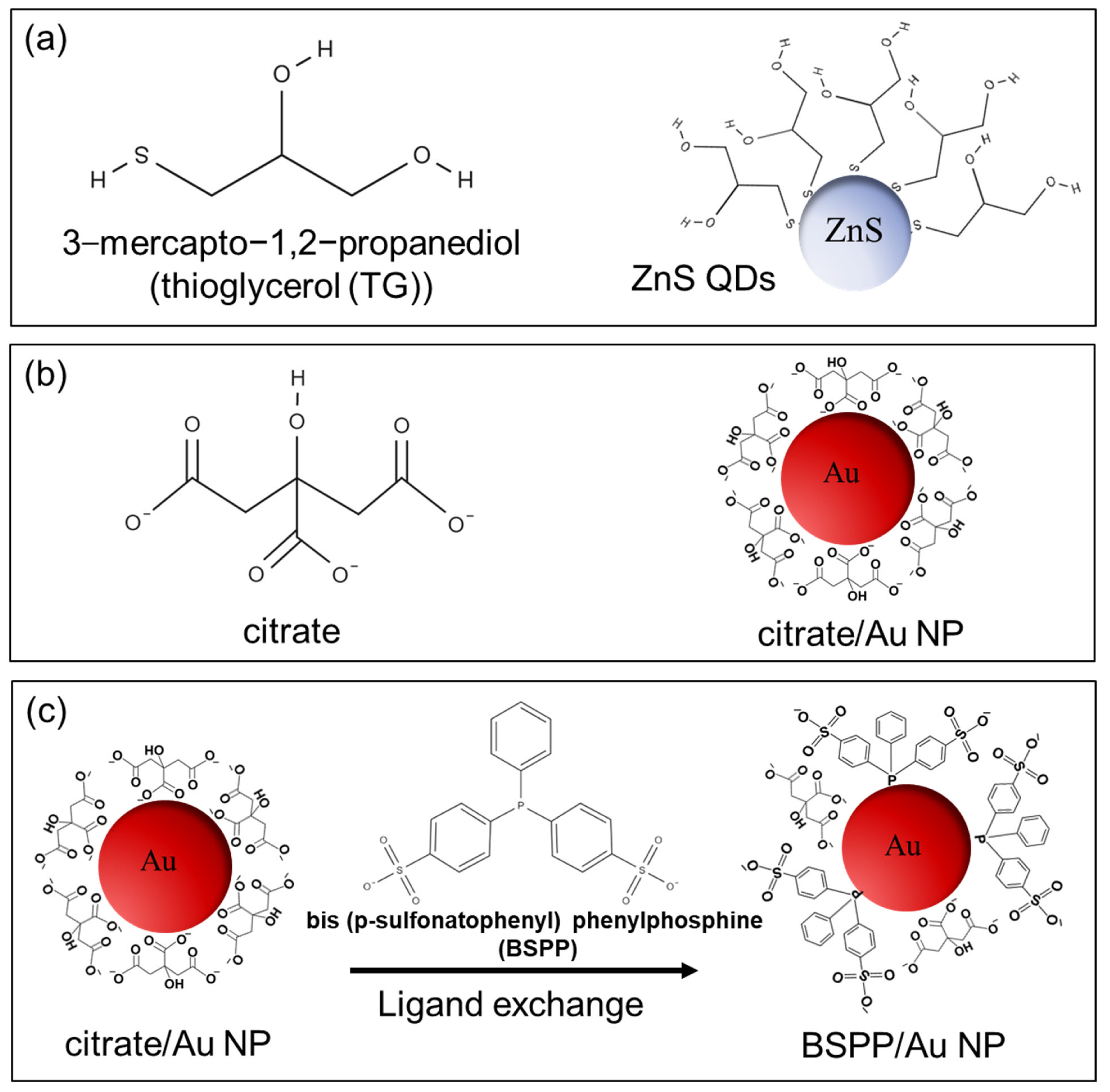
2.2. Nanoparticles Used to Establish Binary Colloidal Mixtures

ZnS QDs below 5 nm in size and spherical Au NPs below 20 nm were selected as the components for forming the binary colloid [48]. Figure 1 presents the schematic of the nanoparticles and the structures of the ligand molecules utilized. ZnS QDs, capped with thioglycerol (TG) (Figure 1a), were synthesized based on a modified protocol from Nanda et al., adapted by Komada and Mori [51,52]. Initially, the synthesized ZnS QDs were isolated in powder form before their redispersion in Millipore water. In contrast, rather because of their more-pronounced van der Waals adhesion (larger diameter and larger Hamaker constant), Au NPs needed to remain in a dispersed, wet state to avoid irreversible agglomeration. As such, the Au NPs were synthesized directly in the aqueous phase. Citrate-capped Au NPs were prepared using established methods [53,54,55,56] labeled as citrate/Au NP (Figure 1b). Detailed synthesis steps for both ZnS QDs and Au NPs are available in the Supporting Information (SI S1 and SI S2). After the synthesis of citrate/Au NPs, the citrate ligands with multiple carboxylic groups adhered to the Au NP surfaces through van der Waals forces (Figure 1b) [57]. Then, these citrate/Au NPs were functionalized with the bis(p-sulfonatophenyl) phenylphosphine (BSPP) ligand (see Figure 1c). To achieve this, the citrate/Au NPs dispersion was stirred overnight with an excess of BSPP (final concentration of 0.2 mg/mL) [58]. The resulting dispersion, now containing Au NPs functionalized with BSPP, is labeled as BSPP/Au NP (Figure 1c-right). All details on the ligand exchange process and confirmation of successful surface modification from citrate to BSPP are provided in the Supporting Information (SI S3, Figures S1–S3). The BSPP molecule is a water-soluble phosphine ligand containing sulfonate/thiol groups that bind to the surface via the phosphorus lone electron pair [59,60]. Moreover, the bulky aromatic rings of BSPP provide a steric hindrance, which increases the colloidal stability of Au NPs [61,62,63,64,65].
The synthesis and ligand exchange processes were followed by purification through centrifugation to remove excess ligand molecules and other non-Au NP residues from the synthesis solution. This purification required careful optimization to retain the colloidal stability of the Au NPs, with different relative centrifugal forces (RCF) and centrifugation times tested for optimum results. Details of the purification steps are available in SI S4. Consistent with previous reports, all synthesized nanoparticles, including ZnS QDs and Au NPs after synthesis, exhibited sufficient stability as colloidal dispersions in Millipore water [31,33,53,54,55,66]. The characterization of the synthesized nanoparticles, along with properties such as pH and ionic strength of the dispersions, is available in the Supporting Information (SI5—Table S3, Figures S7 and S8).

2.3. Continuous Flow Synthesis and Post Synthesis Purification of InP/ZnS QDs

The InP/ZnS QDs used in this study were synthesized through a modified flow reactor method based on the approach by Wang et al. [50,67]. The process involved indium chloride (InCl3), zinc chloride (ZnCl2), and tris(diethylamino)phosphine (PDEA) in oleylamine (OLA) at high temperatures, followed by the addition of zinc diethyldithiocarbamate (ZDEC) to form the shell. Initially, 442.36 mg (2 mmol) of InCl3 and 1363 mg (10 mmol) of ZnCl2 were placed in a three-neck flask with 20 mL of OLA, degassed at 120 °C for one hour, and then cooled. After cooling, 2.2 mL (8 mmol) of PDEA was added. The mixture was stirred overnight before being transferred to a continuous tubular flow reactor that was set to 250 °C, with a flow rate of 0.1 mL/min, resulting in an average residence time of 36 min in which the particles formed by nucleation and growth.
The resulting InP core QDs were mixed with a ZDEC dispersion in OLA in a 2:3 volume ratio for 30 min, then processed again in the flow reactor at the same flow rate but with a lower reaction temperature of 220 °C. The final InP/ZnS QDs were collected at the reactor outlet.
To purify the InP/ZnS QDs, ethanol (analytical grade) was added in a volume ratio of 3:1 (ethanol to crude QDs dispersion) to induce flocculation. The QDs were separated by centrifugation at 10,000 rpm for 10 min and then re-dispersed in n-hexane. Additional ethanol (3:1 ratio to QDs in n-hexane) was added to flocculate the QDs again, followed by centrifugation under the same conditions. This purification cycle was repeated three times. The purified QDs were finally re-dispersed in n-hexane and stored in a glovebox at 5 °C for further use and characterization.

2.4. Characterizations

UV-visible absorption spectra were acquired using a UV-visible spectrometer (ThermoFisher Scientific Evolution 201, ThermoFisher Scientific, Dreieich, Germany), spanning wavelengths from 200 to 900 nm.
All measurements related to binary mixtures of Au NPs-ZnS QDs were conducted in a quartz glass cuvette with an optical path length of 2 mm (Hellma Analytics, Munich, Germany). The choice of the optical path length was optimized based on the optical properties of both ZnS QDs and Au NPs to prevent sample dilution. In the case of the QDs synthesized in the continuous flow reactor, because of their higher optical density after synthesis and washing, for each measurement, the samples were diluted with n-hexane and placed into a 1 mm quartz cuvette (Hellma Analytics) for analysis. Steady-state photoluminescence (PL) spectra were obtained at room temperature utilizing a Horiba Jobin YVON Fluorolog®-3 spectrofluorometer with an excitation wavelength of 450 nm.
Transmission electron microscopy (TEM) investigations of the QDs were carried out using a probe-corrected Thermo Fisher Scientific (TFS) Spectra 200 (operated at 200 kV) and FEI Titan Themis3 300 (operated at 300 kV), both equipped with Super-X detectors for energy-dispersive X-ray spectroscopy (EDXS). Details on the sample preparation and measurements can be found in our previous works [49,50].

3. Results and Discussion

Our initial focus was on developing and stabilizing a binary colloidal system composed of Au NPs and ZnS QDs. This model system was designed to investigate particle interactions and understand the underlying mechanisms of colloidal stability. Details can be found in Rezvani et al. [48]. Building on these findings, we aimed to tackle a practical challenge in nanoparticle separation by employing size-selective agglomeration (SSA). As described in our recent original work [49], we were able to separate a mixed colloid as it originated during continuous QD synthesis by SSA: InP/ZnS core–shell QDs from ZnS QDs that formed as a byproduct during the shell formation step.

3.1. Insights into the Stability of the Binary Mixture as Model System

In the following, the development of a stable binary colloid consisting of Au NPs and ZnS QDs is investigated. The key factors affecting stability, such as surface chemistry and impurity control, are examined to create a reproducible model system that can support future applications.

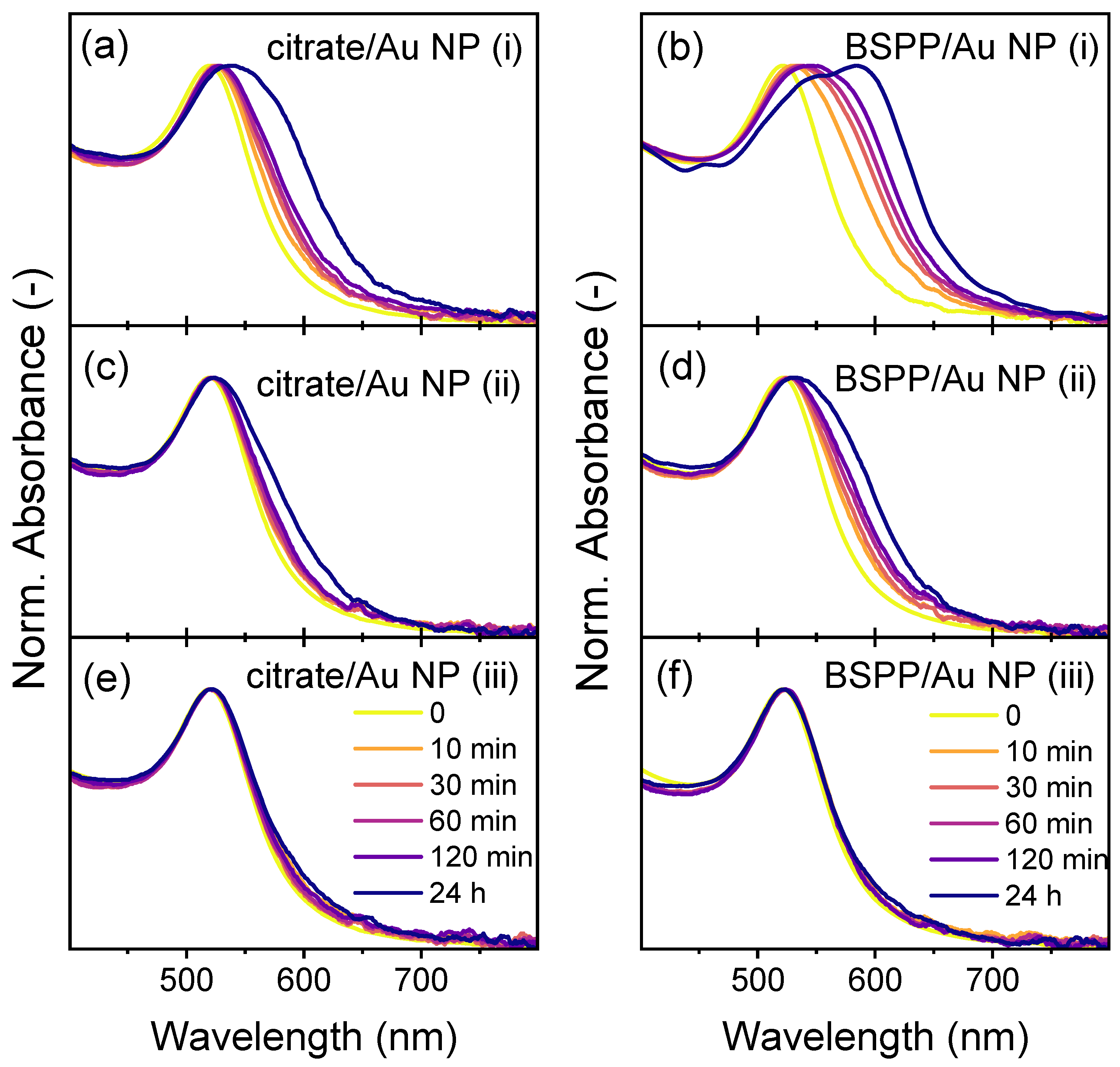
3.1.1. Stability Assessment of Individual Nanoparticles in Pure Water

The selection of Au NPs and ZnS QDs to make the binary mixture was guided by their distinct optical properties, water dispersibility, and ease of separation by centrifugation. UV-visible spectroscopy was employed to highlight the unique absorbance features of each nanoparticle, offering a reliable method to monitor their behavior in the mixture. As shown in Figure 2a, ZnS QDs exhibit a strong absorbance peak in the UV range (258 nm), whereas Figure 2b displays Au NPs with a characteristic surface plasmon resonance (SPR) band in the visible spectral range (519 nm). These well-separated spectral features allow for the straightforward differentiation of the nanoparticles in the binary mixture and facilitate tracking of their stability and potential agglomeration over time. Figure 2c showcases the UV-visible absorbance spectrum of the binary mixture. The characteristic peaks at 258 nm for ZnS QDs and 519 nm for Au NPs remain distinct in the mixture, demonstrating the feasibility of monitoring both components simultaneously. The inset photographs in Figure 2 further illustrate the distinct optical appearances of the dispersions. ZnS QDs, being ultra-small and colorless in water, yield a transparent dispersion, while Au NPs exhibit a vibrant red color due to their SPR in the visible spectrum. The binary mixture, containing both types of nanoparticles, maintains a red hue, confirming the coexistence of the two species and their successful dispersion (Figure 2c-inset).
Another critical factor in selecting these nanoparticles is their spectral response to agglomeration. For ZnS QDs, aggregation results in an increase of the absorbance spectra at longer wavelengths (>700 nm) due to the pronounced scattering of light in the presence of agglomerates [32,33], while for Au NPs, the SPR band broadens or shifts to higher wavelength as soon as the first doublets form [53,58,68,69,70,71]. This behavior provides a direct tool to monitor the progression of agglomeration and stability changes in real time. Additionally, the excellent dispersibility of both Au NPs and ZnS QDs in water ensures a simple system, minimizing external complexities and allowing us to attribute the observed changes directly to particle-particle interactions. Au NPs and agglomerates also exhibit distinct sedimentation behavior under centrifugation. In contrast, ZnS QDs, due to their smaller size and lower density, require massively higher centrifugation speeds for sedimentation that cannot be reached preparative with typical laboratory centrifuges. This enabled us to achieve the selective separation of each component, preserving their individual properties for further characterization.

3.1.2. Stability Assessment of Individual Nanoparticles in the Presence of a Solvent Mixture

The stability of individual components in binary colloidal dispersions is critical for understanding their interactions and separation potential. In our previous studies, we investigated the size classification of colloidal ZnS QDs through selective agglomeration and its scalability in detail [31,33], which includes the stability/agglomeration behavior of ZnS QDs in the presence of the second solvent. Thus, also in the case of the binary mixture, we wanted to examine the behavior of Au NPs under the influence of a second solvent. Ethanol was chosen as the poor solvent based on its previously demonstrated impact on the stability of ZnS QDs in our previous studies [31,33].
As mentioned in the Experimental section, citrate/Au NPs were synthesized in the liquid phase, resulting in colloidal dispersions containing both citrate/Au NPs and residual chemicals from the synthesis. To investigate the effect of Au NP surface chemistry, BSPP-stabilized Au NPs (BSPP/Au NPs) were prepared via ligand exchange and compared to citrate-stabilized Au NPs (citrate/Au NPs). Zeta potential measurements combined with UV-Vis absorption measurements after stepwise washing of the Au NPs were performed to verify the ligand exchange from citrate to BSPP. The results confirm successful surface modification and are detailed in the Supporting Information (SI S3, Figures S2 and S3). Thus, the dispersion after ligand exchange most probably includes BSPP/Au NPs, residual chemicals from the synthesis, released citrate ions during the ligand exchange, and excess BSPP. To eliminate potential stabilizing or destabilizing effects from dissolved impurities, the dispersions in both cases of citrate/Au NPs after synthesis and BSPP/Au NPs after ligand exchange were purified through centrifugation, where the supernatant was removed to the greatest extent possible (see SI S4 for details on purification). The purified flocculates were subsequently used to investigate the impact of ethanol addition on the stability of the Au NPs. Ethanol concentrations were varied between 25% to 100% v/v. Samples were prepared by initial redispersion of the purified Au NPs in the required volume of Millipore water to create a stable starting dispersion. Then, the required volume of ethanol was added slowly under stirring. We recorded the UV-Vis spectra of Au NPs at two time points: 10 min and 120 min post-ethanol addition. This protocol allowed us to compare the spectra before and after (10 min and 120 min) ethanol exposure. The SPR band of Au NPs served as a reliable indicator of aggregation, as any change, broadening, or increase/reduction in the intensity signifies alterations in the nanoparticle stability [53,58,68,69,70,71]. Observing no change in the SPR band suggests maintained nanoparticle stability. In contrast, alterations such as a red shift, broadening of the SPR band, or the emergence of a secondary absorption band at higher wavelengths are indicative of nanoparticle aggregation. Figure 3 shows UV-visible spectra measured after 10 and 120 min of addition of ethanol to both Au NPs dispersions, citrate/Au NPs, and BSPP/Au NPs.
Our results indicate that both citrate/Au NPs and BSPP/Au NPs remained stable in ethanol–water mixtures up to 95% v/v ethanol. The UV-visible spectra show no significant changes in the SPR band for these samples, confirming the absence of aggregation (Figure 3). However, in the presence of 100% ethanol, a notable shift and broadening of the SPR band were observed, indicating the onset of agglomeration. This behavior aligns with previous findings in the literature [58]. In agreement with literature reports [58], the primary cause of aggregation in pure ethanol appears to be linked to unremoved residues from the synthesis process, such as citrate or other ions, which destabilize the colloidal system in a non-aqueous medium. Note that the Au NP flocculates were not extensively washed with Millipore water before being transferred to ethanol, implying that some supernatant may be left along with the Au NPs after centrifugation. For lower ethanol concentrations, the initial dispersion of the purified Au NPs in even small volumes of Millipore water prior to ethanol addition was sufficient to reduce the concentration of impurities and prevent aggregation, even in the presence of 95% ethanol.
We hypothesize that this behavior arises from the interplay between electrostatic stabilization and solvent effects on surface interactions. When ethanol is added directly to the Au NPs, it introduces an abrupt change in the solvent polarity. Ethanol has a significantly lower dielectric constant (~24.5) compared to water (~78.5), which reduces the solvent’s ability to maintain charge separation. As a result, electrostatic repulsion between the Au NPs weakens, leading to rapid aggregation due to van der Waals attractive forces dominating interparticle interactions.
In contrast, when Au NPs are pre-dispersed in even a small volume of Millipore water, even a minimal dilution of residual ionic species can significantly alter their impact. Moreover, the high dielectric constant of water ensures that surface charges remain well-separated, maintaining strong repulsive forces between individual particles. The subsequent addition of ethanol then induces a more gradual transition to a lower-dielectric environment, allowing stabilizing forces to adapt, thereby preventing uncontrolled agglomeration. The gradual shift in the solvent polarity gives the stabilizing ligands more time to reorganize and prevent uncontrolled agglomeration.
This highlights the importance of the specifically chosen sequence of solvent mixing and the control of residual salts for maintaining the colloidal stability of nanoparticles in mixed-solvent environments. The results underscore the role of residual salts and the necessity of optimizing purification protocols and solvent handling strategies.

3.1.3. The Role of Dissolved Impurities

To systematically investigate the influence of synthesis residuals on the stability of Au NPs in the presence of ethanol, a three-step experiment was designed using citrate- and BSPP-capped Au NPs. The aim was to assess the extent to which residual impurities affect aggregation when ethanol is introduced. The samples were prepared according to three different procedures: (i) As-synthesized Au NPs that were left in the original synthesis solution, (ii) Au NPs in the original synthesis solution that were centrifuged, followed by removal of half of the supernatant (by volume), and addition of the same volume of Millipore water, (iii) Au NPs in the original synthesis solution that were centrifuged, followed by removal of all of the supernatant, and addition of the same volume of Millipore water. Afterward, ethanol was added to each sample to a final concentration of 87% v/v of ethanol, and the UV-visible spectra were recorded over a 24-hour period at room temperature.
The results, shown in Figure 4, reveal observable differences in the behavior of Au NPs based on the level of residual supernatant from synthesis being present. For Sample i, where no purification steps were applied, for both citrate and BSPP-capped Au NPs, the absorbance spectrum shows a substantial redshift over time, indicating aggregation. This is consistent with the high concentration of residual ions, particularly sodium ions (from trisodium citrate) and chloride ions (from HAuCl4), which likely induce agglomeration in the ethanol-rich environment. For Sample ii, where half of the supernatant was replaced with Millipore water, the degree of redshift in the SPR band was reduced, but agglomeration was still observed, suggesting that the partial removal of impurities was not sufficient to fully stabilize the colloidal dispersion. In contrast, Sample iii, where the supernatant was completely replaced with Millipore water, exhibited no redshift, maintaining a high colloidal stability throughout the experiment. This shows that the presence of sodium chloride and other ionic species in the supernatant creates conditions that promote interparticle interactions, leading to agglomeration. This finding aligns with previous literature reports, which have highlighted the destabilizing effects of similar ionic impurities in colloidal systems [58].
In principle, this salt effect could be confirmed by completely removing the residual salt solution from the centrifuged precipitates and then testing the dispersibility of the nanoparticles in ethanol. However, simply drying the precipitates does not effectively remove the salt and results in irreversible aggregates that are no longer dispersible, even in Millipore water. Previous studies have attempted to address this issue by washing the Au NP precipitates with Millipore water and performing additional centrifugation steps to remove as many residuals as possible before transferring the particles into ethanol [58]. In our case, however, we observed instability in Au NPs during the second centrifugation round, which led us to avoid this approach. Instead, we relied on the complete removal of the supernatant (as shown by Sample iii) followed by redispersion in Millipore water and the addition of 87% v/v ethanol. Based on our findings here, this method proved to be sufficient for achieving a good level of stability in the Au NP dispersion. For our ultimate goal of creating stable binary mixtures of ZnS QDs and Au NPs, followed by the selective agglomeration of ZnS QDs by the addition of ethanol as a poor solvent while maintaining the stability of Au NPs, this approach strikes a practical balance between purification and maintaining colloidal stability.
This finding demonstrates that the stability of one component (Au NPs) can be strongly influenced by the presence of impurities in the dispersion medium, which could also be introduced by the other component (ZnS QDs). In the case of ZnS QDs, during synthesis or post-synthesis purification, surface ligands or other adsorbed chemicals may be released into the dispersion. These released chemicals, such as thiol-based ligands or unbound Zn ions, can act as additional impurities when combined with Au NPs in the binary mixture, potentially lowering their colloidal stability. By understanding the specific interactions between impurities released from ZnS QDs and Au NPs, we can develop targeted approaches to enhance the stability of the binary system.

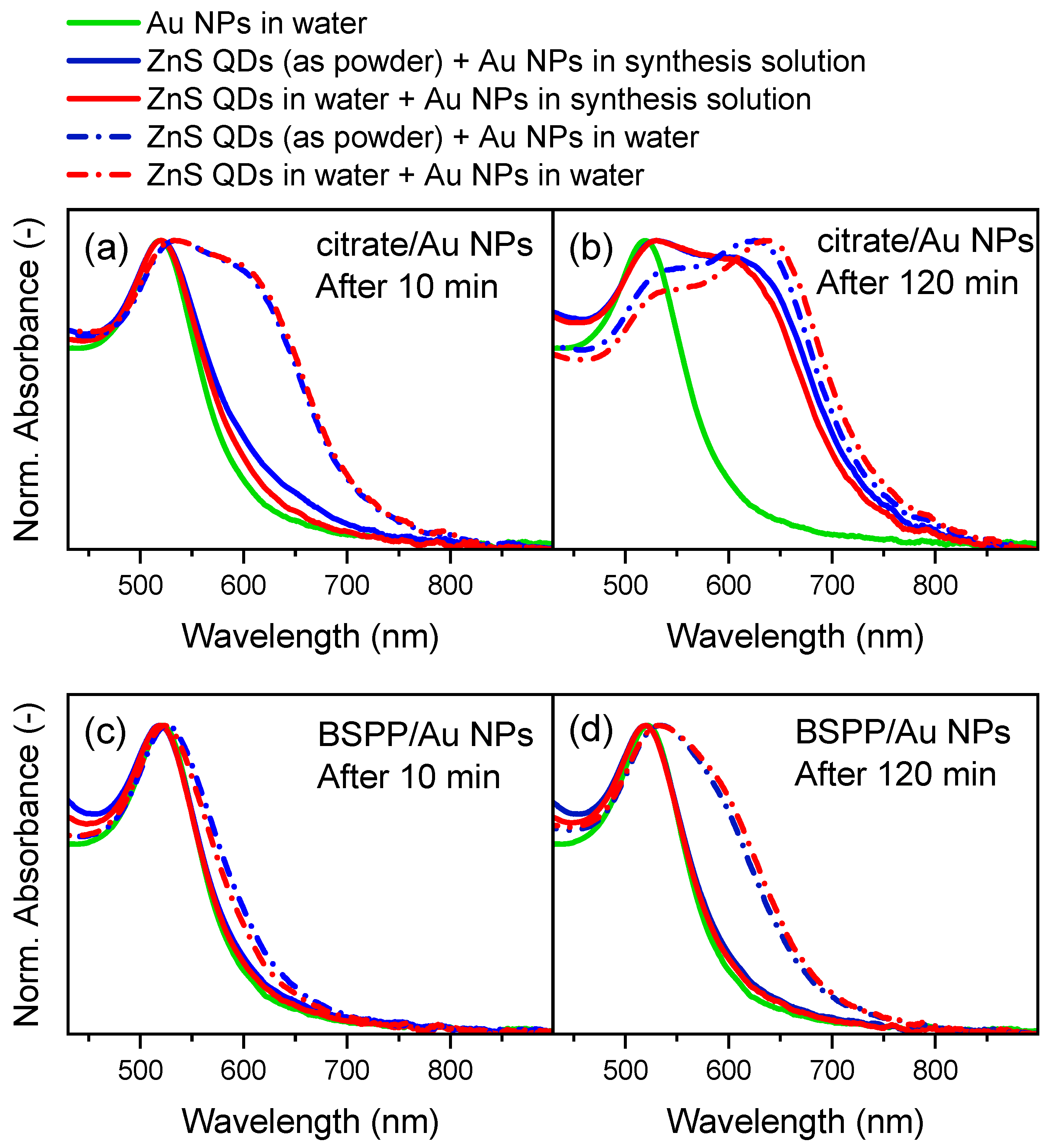
3.1.4. Establishing the Binary Mixture

Building upon the findings regarding individual component stability, we aimed to establish a binary mixture where ZnS QDs and Au NPs coexist without undesired agglomeration. The formation of a stable binary colloidal system comprising ZnS QDs and Au NPs necessitates the careful consideration of all factors influencing the stability of each component. We investigated the impact of surface ligands, residual synthesis byproducts, and the method of combining ZnS QDs with Au NPs on the overall stability of the mixture.
To assess the stability of the binary mixture, we prepared Au NPs with two different surface ligands: citrate and BSPP. For each ligand type, two sample conditions were evaluated: (i) unpurified dispersion: Au NPs retained in their original synthesis medium, containing residual byproducts, and (ii) purified dispersion: Au NPs purified by centrifugation to remove synthesis residuals, followed by redispersion in Millipore water. Afterward, the ZnS QDs were introduced into the Au NP dispersions using two methods: (i) as powder: direct addition of dry ZnS QD powder to the Au NP dispersion, and (ii) pre-dispersed in Millipore water: addition of ZnS QDs pre-dispersed in Millipore water to the Au NP dispersion. This resulted in four distinct combinations for each ligand type, varying in the presence/absence of synthesis residuals and the way ZnS QDs were added as a powder or pre-dispersed.
The stability of each binary mixture was monitored for all four samples by recording UV-visible spectra at 10 min and 120 min after mixing, as shown in Figure 5, focusing on the SPR band of Au NPs in the visible region. The absorption band of ZnS QDs, located in the UV region, remained unchanged across all experiments, indicating their stability within the mixtures (see SI6, Figure S9).
The UV-visible spectra (Figure 5) reveal that the method of ZnS QD addition, whether as powder (Figure 5-blue lines) or pre-dispersed (Figure 5-red lines), had negligible impact on the final stability of the binary mixtures. However, the choice of the surface ligand and the presence of synthesis residuals introduced via the Au NPs significantly influenced the stability. Samples with citrate/Au NPs, both in the original synthesis solution (Figure 5a,b-solid lines) and after purification (Figure 5a,b-dash dot lines), exhibited notable changes in the SPR band over time, suggesting aggregation and reduced stability. The sample with BSPP/Au NPs in the original synthesis solution (Figure 5c,d-solid lines) demonstrated remarkable stability, with no change in the SPR band even after 120 min. In contrast, purified BSPP/Au NPs (Figure 5c,d-dash dot lines) showed signs of instability, indicating that the removal of synthesis residuals adversely affected their stability.
The enhanced stability of BSPP-capped Au NPs in theS presence of synthesis residuals suggests that certain residuals may play a stabilizing role. BSPP is known to effectively stabilize Au NPs by forming a robust ligand shell that prevents agglomeration. Residual components in the original synthesis solution, such as unreacted BSPP or other stabilizing agents, might contribute additional steric or electrostatic stabilization, enhancing the overall stability of Au NPs. Conversely, the purification process removes these residual stabilizers, potentially leaving the BSPP-capped Au NPs more susceptible to destabilizing interactions, leading to agglomeration.
In our recent study, we also investigated the effect of TG molecules (surface ligand of ZnS QDs) and zinc acetate (one of the synthesis components) on the stability of Au NPs with surface chemistries [48]. We hypothesize that TG molecules and zinc acetate are released from the surface of the ZnS QDs into the mixture, triggering rather likely irreversible agglomeration of citrate-stabilized Au NPs under the tested conditions [58,72]. The TG molecule neutralizes the negative charge of citrate without providing steric stabilization, reducing electrostatic stability and resulting in agglomeration. A similar mechanism was reported by Chegel et al., where citrate-capped Au NPs agglomerated upon exposure to 6-mercapto-1-hexanol (MCH), a thiol-containing molecule [72]. For BSPP-functionalized Au NPs, agglomeration occurred only with washed particles, likely due to incomplete citrate displacement or the formation of a secondary BSPP layer without full ligand exchange. During the purification process, surface ligands that are not chemically bound desorb, thereby reducing the stability when mixed with ZnS QDs. In the context of forming stable binary mixtures of ZnS QDs and Au NPs, maintaining certain synthesis residuals appears beneficial, particularly for BSPP-capped Au NPs. This insight is valuable for designing colloidal systems where controlled stability is essential, such as in applications involving selective agglomeration or the targeted assembly of nanoparticle components. Thus, the most stable system was the binary mixture prepared using ZnS QDs and BSPP-capped Au NPs directly from the original synthesis solution without purification.
For further investigation over a longer time window, we studied four different binary mixtures by mixing a dispersion of ZnS QDs in Millipore water with (i) as-synthesized citrate/Au NPs (retained in their original synthesis medium, containing residual byproducts), (ii) purified citrate/Au NPs (purified by centrifugation to remove synthesis residuals, followed by redispersion in Millipore water), (iii) as-synthesized BSPP/Au NPs (retained in their original synthesis medium, containing residual byproducts), and iv) purified BSPP/Au NPs (purified by centrifugation to remove synthesis residuals, followed by redispersion in Millipore water). These four binary mixtures were monitored over a 6-hour-period, as presented in Figure 6. Binary mixtures containing as-synthesized citrate/Au NPs (Figure 6a-II), purified citrate/Au NPs (Figure 6b-II), and purified BSPP/Au NPs (Figure 6c-II) exhibited notable changes in the SPR band over time, including a redshift and the appearance of a second absorption band at higher wavelengths (~600 nm). These spectral changes indicate aggregation and reduced stability of Au NPs. In contrast, the binary mixture prepared with as-synthesized BSPP/Au NPs (Figure 6d-II) demonstrated remarkable stability, showing no redshift or broadening of the SPR band at 519 nm, nor the emergence of any secondary absorption band at higher wavelengths, even after prolonged mixing with ZnS QDs. Furthermore, the sustained red color of the dispersion over time, as shown in Figure 6d-III, corroborates the stability of this binary system compared to the other investigated mixtures (Figure 6a-III,b-III,c-III).
In the UV range, a slight redshift and decrease in intensity were observed for the ZnS absorption band at 259 nm in the binary mixture containing as-synthesized BSPP/Au NPs (Figure 6d-I). We attribute this to the overlap between the ZnS QD absorption band and the absorption band at 268 nm, which corresponds to free BSPP molecules in the continuous phase of the dispersion. The UV-Vis spectra of BSPP in Millipore water illustrate an absorption band at 268 nm (SI S3—Figure S1). Moreover, in the other three mixtures, which do not contain free BSPP molecules in the dispersion, no redshift of the ZnS QD absorption band at 259 nm was observed (Figure 6a-I,b-I,c-I). Figure 6a-I shows a slight decrease in intensity (but no shift) of this band after 4 h, which might be due to the extensive aggregation of Au NPs in this mixture and the possibility of hetero-aggregation between Au NPs and ZnS QDs, leading to a reduction in the intensity of the absorption band of ZnS QDs.
To further investigate the stability/agglomeration in binary mixtures, the usual techniques like dynamic light scattering (DLS) and zeta potential measurements have limitations for our binary mixture. For the ZnS QD–Au NP binary mixture, using Au NPs directly in their synthesis solution (where we achieved the stable binary mixture) retains all residuals and excess ligands. Even with purified Au NPs, both particles release surface residuals or ligands into the dispersion medium, as noted in previous studies [73,74,75]. Under such dynamic conditions, and without the possibility of adding a background electrolyte, as this would further change the ligand composition at the particle surface, the zeta potential values fluctuate significantly, making stability/agglomeration assessments unreliable. While zeta potential data are provided in the Supporting Information (SI S7, Figures S10–S13), its high variability prevents clear interpretation. Since DLS is unsuitable for polydisperse systems like Au NPs and binary colloidal mixtures [76], we used analytical centrifugation (AC) to assess the PSD of four binary mixtures. The results are presented in the Supporting Information (SI S7, Figures S10–S13) and confirm the stability/agglomeration state by comparing the PSD of the binary mixtures with a reference sample of pure, stable Au NPs dispersion.
The successful stabilization of the binary mixture using as-synthesized BSPP/Au NPs within a 6-hour timeframe highlights its potential for subsequent processing steps, such as separation. This timeframe is sufficiently long to enable practical application without compromising colloidal stability. Achieving such a well-defined and stable binary colloid not only provides a robust model system for studying interactions in multicomponent systems but also lays the groundwork for developing technical formulations involving more complex mixtures.

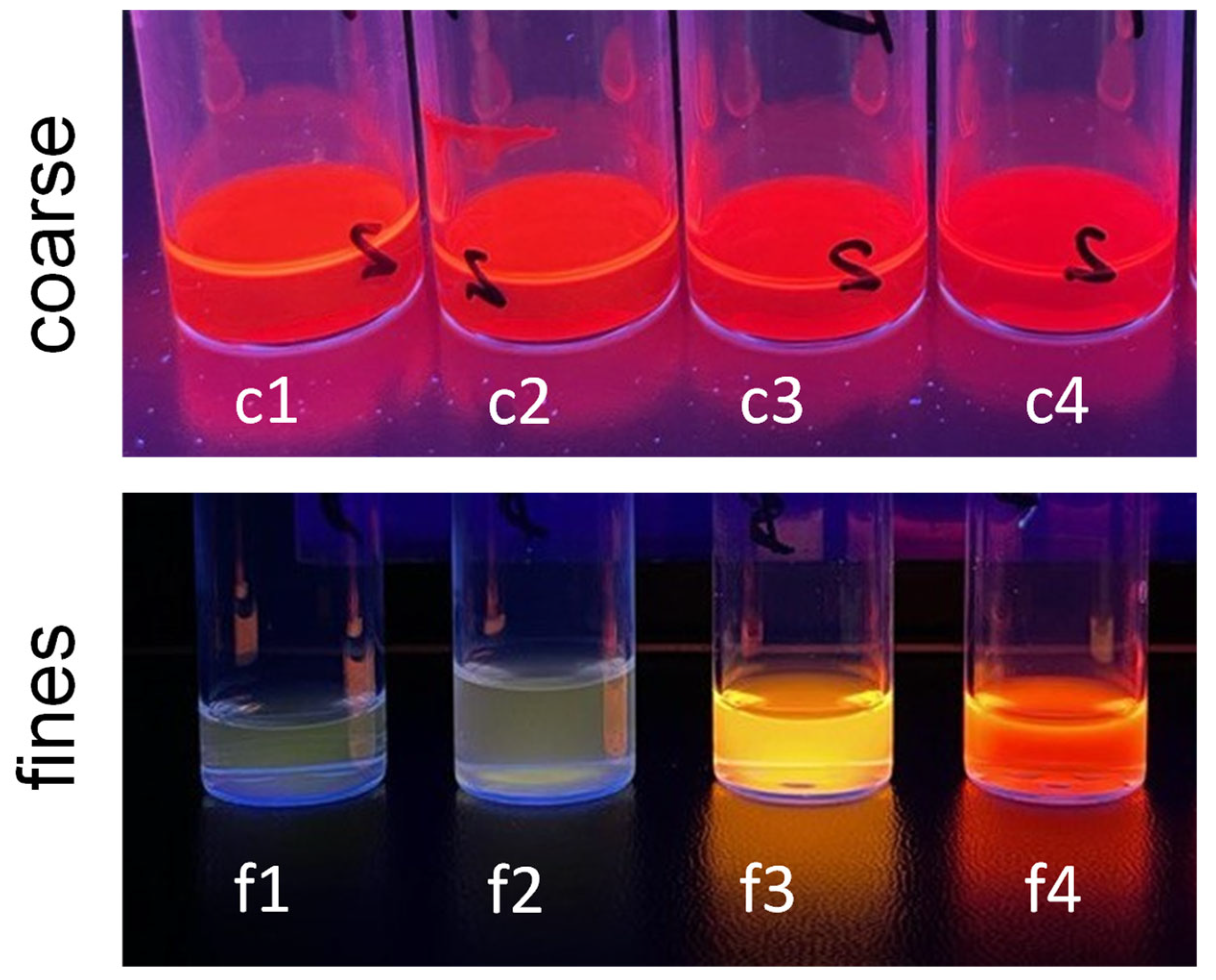
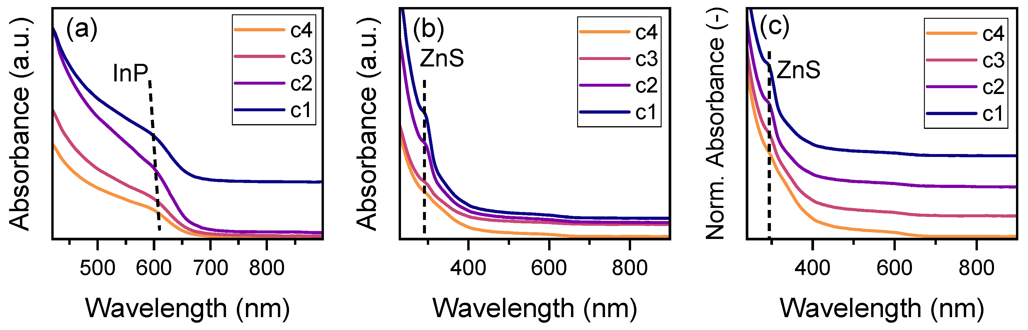
3.2. Separation of ZnS QDs from InP/ZnS Core–Shell QDs

The fundamental principles of stability and interaction, explored from the establishment of a stable binary colloidal system of ZnS QDs and Au NPs, were instrumental in tackling a more complex and application-driven challenge that is the separation of an as-synthesized mixture of InP/ZnS core–shell QDs and ZnS byproduct QDs. In our recent study, we introduced a custom-made tubular flow reactor for the continuous and scalable synthesis of InP cores and subsequent shelling, yielding InP/ZnS core–shell QDs [50,67]. Core passivation using an outer shell is a well-established and widely applied technique to enhance the optical properties of QDs. Therefore, following the synthesis of InP cores, a ZnS shell was subsequently grown over the InP QDs through the decomposition of ZDEC, which acts as a single-source precursor. However, further investigation revealed the unintended formation of pure ZnS QDs as a byproduct during the shelling process [50]. In this process, the synthesis involves mixing InP core QDs with the ZDEC shell precursor. Afterward, two potential reaction pathways can occur: (i) ZnS may nucleate heterogeneously and grow on the InP cores, leading to the formation of InP/ZnS core–shell QDs, or (ii) ZnS can nucleate homogeneously, resulting in the creation of separate ZnS QDs. While the homogeneous formation of ZnS QDs does not directly affect the photoluminescence quantum yield (PLQY) for excitation wavelengths between 375–485 nm (as this range lies beyond the ZnS absorption peak at approximately 350 nm) [49,50,77], it reduces the amount of shell precursor available for ZnS passivation. This limitation impacts the thickness and overall quality of the ZnS shell, which, in turn, affects the PLQY of the resulting InP/ZnS QDs [50].
In the following, we summarize how it was possible to investigate the post-synthesis separation of these QDs from our recent work by Wang et al. [50,67], containing a mixture of InP-ZnS core–shell QDs and ZnS byproduct QDs using SSA, to remove the ZnS byproduct QDs. The following sections will provide a brief discussion of the separation process, characterization results, and the broader implications of our findings. Details can be found in Rezvani et al. [49].

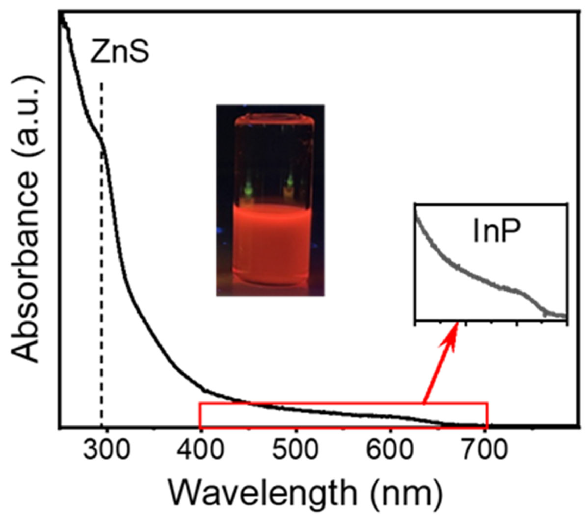
3.2.1. Characterization of the QDs After Continuous Core and Shell Synthesis

So far, the synthesis of QDs has involved the formation of InP core QDs, followed by their passivation with an outer shell of ZnS using ZDEC as a single-source precursor. The resulting QDs are covered by oleylamine as a surface ligand, ensuring their colloidal stability. The colloid collected at the outlet went through three purification steps of flocculation using ethanol, centrifugation, and redispersion in a good solvent, here n-hexane. As described in the Experimental section, this three-step procedure was repeated three times. Figure 7 shows that the UV-visible spectrum exhibits two characteristic absorption bands, one at around 290 nm and another broad band at around 605 nm. The inset in Figure 7 on the right provides an enlarged view of the absorption spectrum between 400 and 700 nm, clearly showing the latter band around 605 nm. It is the typical first excitonic band that occurs due to the quantum confinement effect of InP cores. The absorption band at 290 nm has been ascribed to the presence of ZnS QDs in our previous work [50]. The second inset in Figure 7 on the left shows a photograph of the final purified QDs dispersed in n-hexane under photoexcitation at 400 nm, emitting a dark red photoluminescence (PL). This purified QD dispersion was then used for the separation experiments and named feed QDs.
The high-angle annular dark-field (HAADF) scanning transmission electron microscopy (STEM) images (Figure 8a,b) further provide evidence of two types of nanoparticles with distinct sizes and shapes: larger tetrahedral particles (marked with red circles) and smaller spherical ones (marked with green circles). Energy-dispersive X-ray spectroscopy (EDXS) mapping (Figure 8c,e) identifies In, P, Zn, and S in the tetrahedral QDs, while only Zn and S are present in the smaller spherical ones.

3.2.2. Separation Procedure

The observed optical properties and the results of the HR-STEM studies call for a separation technique that enables the separation of the QD mixtures. Figure 9 illustrates the separation process based on SSA that was applied to the feed QDs.
In brief, 2 mL of feed QDs were provided in a 15 mL centrifuge tube. Then, we stepwise added a certain amount of ethanol as a poor solvent into these feed QDs. Each step, including the addition of 200 µL of ethanol, was followed by intense mixing for 30 s using a shaker (IKA Shaker, Vortex 2; Speed adjustment: scale 6~2500 rpm). Visually, this resulted in a slight turbidity (cloudiness), which is ascribed to the flocculation of the least-stable QDs. These flocculates were separated from the colloidally stable supernatant by centrifugation (4000 rpm, 10 min). The pellet (precipitate) was dispersed in 2 mL of n-hexane as a good solvent named the coarse fraction. To the supernatant, an excess amount of ethanol was added to flocculate all QDs. These flocculates were separated from the solvent mixture by centrifugation (10,000 rpm, 10 min) and dispersed in 2 mL of n-hexane as a good solvent named the fines fraction. The separation process was repeated using four different volumes of ethanol as a poor solvent, including 67%, 64%, 60%, and 56% final volume percentage of ethanol in the dispersion. Thus, we obtained four coarse (c1 to c4) and four fines (f1 to f4) fractions.
Figure 10 shows photographs of the collected fractions under photoexcitation at 400 nm. It unravels coarse fractions emitting in the red and fines fractions with no noticeable emission for f1 and f2, and emitting yellow and orange for f3 and f4, respectively.

3.2.3. Optical Properties After Separation

Figure 11a shows the UV-visible absorption spectra for coarse fractions in the visible region. The primary feature in the visible range of the absorption spectrum is the first excitonic peak, which occurs due to the quantum confinement effect of the InP cores. This peak typically appears in the range of 500 to 650 nm, depending on the size of the InP cores with a direct bulk band gap of 1.34 eV. Smaller QDs exhibit an absorption peak at shorter wavelengths, i.e., higher energies, while larger QDs absorb at longer wavelengths. Figure 11a shows a very slight redshift of this excitonic band from c1 to c4, which simultaneously becomes narrower. This means that by reducing the final concentration of ethanol, the cut size shifted to a larger size. The cut size is defined as the critical particle size at which the separation occurs, distinguishing smaller particles that remain in dispersion (fines) from larger ones that agglomerate and settle out (coarse). Thus, the coarse fraction shifts to larger particle sizes. To detect changes in the UV region, we diluted the coarse fraction. This was necessary due to the significantly higher absorption by ZnS QDs in this region (200–350 nm). Figure 11b shows the spectra in the UV region where pure ZnS QDs exhibit their absorption band. For bulk ZnS, the bandgap is ~3.6 eV (corresponding to ~345 nm in UV region). The direct bandgap of ZnS QDs is size-dependent due to the quantum confinement effect, meaning the bandgap increases as the particle size decreases. This shifts the absorption band to shorter wavelengths for smaller QDs. As shown in Figure 11b, there is a distinct band at the wavelength of 290 nm for c1 and c2. This absorption band of ZnS QDs is not detected in the spectra for c3 and c4, signifying the negligible presence of ZnS QDs in these fractions. Figure 11c illustrates this even clearer by showcasing the normalized spectra. To clearly distinguish between different spectra, we plotted them in a waterfall format, where each spectrum is slightly shifted vertically to ensure that overlapping regions remain distinguishable and to facilitate the comparison between different concentrations. The spectra with the original baseline are presented in Figure S14.
On the other hand, we analyzed the fines fractions to see how the population of ZnS QDs and InP/ZnS core–shell QDs evolved in the supernatants. Figure 12 shows the UV-visible spectra of the fines fractions. The characteristic excitonic peak of InP cores is detected for f4 at a wavelength of 575 nm and with significantly lower intensity at a wavelength of 570 nm for f3. In the spectra for f2 and f1, this band completely disappears, signifying the negligible presence of InP cores in f2 and f1. However, the absorption band at 290 nm in the spectra for f2 and f1 indicates the presence of ZnS QDs in both fines fractions.
The distinctive presence of the absorption band around 290 nm observed for coarse fractions c1 and c2, and then its disappearance observed for c3 and c4, indicates that we could separate ZnS QDs from the feed QDs samples 3 and 4 and isolate them in the fines fractions f3 and f4. This evidences the capabilities of selective agglomeration for both analytic and preparative separation.
Figure 13 presents the normalized PL spectra recorded for the four coarse and fines fractions under 450 nm excitation. It shows how the PL band shifts to higher wavelengths for the coarse fractions and lower wavelengths for the fines fractions. This is attributed to the shift in the particle size distribution to larger sizes for the coarse and smaller sizes for the fines compared to the feed QDs due to separation. We also monitored a redshift in the PL band from c1 to c4 for the coarse fractions (see Figure 13a). This corresponds to an increase in the particle size from c1 to c4, which is in line with our findings from the UV-visible analysis. As mentioned before, this means that the cut size shifts to a larger size. Figure 13b shows the PL spectra recorded for the fines fractions. The PL spectra show a strong redshift in the PL band for the fines fractions from f1 to f4, which corresponds to an increase in the particle size from f1 to f4. It is worth mentioning that Figure 13b shows the normalized PL spectra; however, absolutely, the fines fractions f1 and f2 exhibit a significantly lower PL intensity compared to f3 and f4 (see Figure 13c). This shows the negligible emission for f1 and f2 (see photos in Figure 10), indicating the negligible presence of InP cores in the fines fractions f1 and f2. This is in line with the findings from UV-visible spectra, confirming the absence of InP cores in fines fractions f1 and f2. Fines fractions f1 and f2 only include pure ZnS, which has a wide bandgap (~3.6 eV) and cannot absorb photons at 450 nm (~2.76 eV). Consequently, ZnS QDs do not emit in the visible range [65,66,67,68]. Hence, by applying selective agglomeration, we were able to separate a feed sample from continuous QD synthesis into fines and coarse fractions to better understand the underlying mixture. At the same time, we could achieve the separation of ZnS byproduct QDs from InP/ZnS core–shell QDs by controlling the solvent mixture. As selective agglomeration is a preparative technique enabling the isolation of powders [33], this also paves the way for product improvement, i.e., particles with optimized PLQY.

4. Conclusions

This study explored the stability and separation of binary and multicomponent colloidal nanoparticle systems to address critical challenges in advanced nanoparticle processing. By first establishing a robust binary model system of Au NPs and ZnS QDs, we demonstrated the intricate role of surface ligands, impurities, and solvent systems in managing interparticle interactions. Notably, BSPP-functionalized Au NPs in unpurified solutions exhibited superior stability, providing a practical pathway for maintaining stable colloidal mixtures.
Building on these insights, second, we applied size selective agglomeration (SSA) to a complex mixture of InP/ZnS core–shell QDs and ZnS byproducts originating from the continuous QD synthesis in a tubular flow reactor. The separation yielded size- and composition-dependent fractions, enabling the successful isolation of pure ZnS QDs in the fines fractions, while the target InP/ZnS QDs enriched in the coarse fractions. Spectroscopic and photoluminescent analyses confirmed the efficiency of this scalable technique, showing distinct shifts in the optical properties of the individual fractions that aligned very well with the underlying particle size distribution.
These findings emphasize SSA’s potential as a versatile and scalable method for the multidimensional classification of nanoparticles, advancing its applicability in separating and stabilizing complex colloidal systems, even preparative. This work enhances the understanding of colloidal stability and separation mechanisms and also provides a foundation for developing innovative solutions for nanoparticle-based technologies, particularly in applications requiring precise control over composition and size at the nanoscale.

Supplementary Materials

The following supporting information can be downloaded at: https://www.mdpi.com/article/10.3390/powders4010009/s1, SI 1. Synthesis of ZnS quantum dots; SI 2. Synthesis of citrate stabilized Au nanoparticles; SI 3. Functionalization of Au nanoparticles; SI 4. Purification of as-synthesized Au nanoparticles; SI 5. Characterization of Nanoparticles; SI 6. Establishing the Binary Mixture; SI 7. Stability assessment in binary mixtures via zeta potential measurements and analytical centrifugation (AC); SI 8. Optical properties of the QDs after separation. Table S1: Centrifugation parameters during the purification of as-synthesized citrate/Au NPs.; Figure S1: UV-Visible absorbance spectrum of citrate/Au NPs, before (black line) and after (red line) functionalization with BSPP. The band at 519 nm is the surface plasmon resonance (SPR) of Au NPs and the one at 268 nm corresponds to the free BSPP molecules in solution.; Figure S2: Proof of effective ligand exchange: UV-Visible absorbance spectrum of as-synthesized Au NPs compared to the spectra after one step of washing by centrifugation (blue line) and second step of washing (red line) for (a) citrate/Au NPs and (b) BSPP/Au NPs.; Figure S3: Zeta potential of purified Au NPs: (a) citrate/Au NPs and (b) BSPP/Au NPs.; Figure S4: Schematic of purification process employed for Au NPs.; Figure S5: Optimizing the centrifugation parameters for as-synthesized citrate/Au NPs: UV-Vis spectra measured for (a,c) dispersed pellet and (b,d) supernatant after centrifugation with RCF of (a,b) 8000 g and (c,d) 15,000 g with different centrifugal duration.; Table S2: Centrifugation parameters during the purification of as-synthesized BSPP/Au NPs.; Figure S6: Optimizing the centrifugation parameters for as-synthesized BSPP/Au NPs: UV-Vis spectra measured for (a,c) dispersed pellet and (b,d) supernatant after centrifugation with RCF of (a,b) 8000 g and (c,d) 15,000 g with different centrifugal duration.; Table S3: Properties of ZnS QDs (dispersed in Millipore water) and Au NPs directly measured after synthesis/functionalization without purification.; Figure S7: The zeta potential distribution showing negative zeta potential values for ZnS QDs in water.; Figure S8: Zeta potential of purified Au NPs: (a) citrate/Au NPs and (b) BSPP/Au NPs.; Figure S9: UV-visible spectrum (UV region) recorded for binary mixtures provided by mixing ZnS QDs as powder (blue lines) and pre-dispersed in Millipore water (red lines) with Au NPs dispersed in the synthesis solution (solid lines) and Millipore water (dash dot lines): citrate/Au NPs after (a) 10 min, (b) 120 min, and BSPP/Au NPs after (c) 10 min, (d) 120 min.; Figure S10: Stability assessment in the binary mixture of purified citrate/Au NPs and ZnS QDs: (a) particle size distribution measured using AC for purified citrate/Au NPs compared to binary mixture, (b) zeta potential measured for purified citrate/Au NPs, and (c) zeta potential measured for binary mixture after 2 h of mixing.; Figure S11: Stability assessment in the binary mixture of as-synthesized citrate/Au NPs and ZnS QDs: (a) particle size distribution measured using AC for purified citrate/Au NPs compared to binary mixture, (b) zeta potential measured for as-synthesized citrate/Au NPs, and (c) zeta potential measured for binary mixture after 2 h of mixing.; Figure S12: Stability assessment in the binary mixture of purified BSPP/Au NPs and ZnS QDs: (a) particle size distribution measured using AC for purified citrate/Au NPs compared to binary mixture, (b) zeta potential measured for purified BSPP/Au NPs, and (c) zeta potential measured for binary mixture after 2 h of mixing.; Figure S13: Stability assessment in the binary mixture of as-synthesized BSPP/Au NPs and ZnS QDs: (a) particle size distribution measured using AC for purified citrate/Au NPs compared to binary mixture, (b) zeta potential measured for as-synthesized BSPP/Au NPs, and (c) zeta potential measured for binary mixture after 2 h of mixing.; Figure S14: UV-visible absorbance spectra of coarse fractions c1 to c4: (a) first excitonic band for InP cores in the visible region (b) highlighting the absorption band of ZnS QDs in the UV region, and (c) normalized spectra (standard 0–1 normalization) highlighting the absorption band of ZnS QDs.

Author Contributions

A.R.: formal analysis, investigation, methodology, project administration, resources, validation, writing—original draft; A.K.: formal analysis, resources, writing—review and editing; B.A.Z.: formal analysis, resources, writing—review and editing; E.S.: resources, supervision, writing—review and editing; D.S.: funding acquisition, investigation, Project administration, resources, supervision, validation, writing—review and editing. All authors have read and agreed to the published version of the manuscript.

Funding

A.R. and D.S. gratefully acknowledge funding by the priority program PP 2045 “Highly specific and multidimensional fractionation of fine particle systems with technical relevance” (Project-ID SE 2526/2-2) provided by the Deutsche Forschungsgemeinschaft (DFG, German Research Foundation). A.R., A.K., B.A.Z., E.S. and D.S. gratefully acknowledge the financial support from the Collaborative Research Centre 1411 “Design of Particulate Products” (Project-ID 416229255) provided by the Deutsche Forschungsgemeinschaft (DFG, German Research Foundation).

Institutional Review Board Statement

Not applicable.

Informed Consent Statement

Not applicable.

Data Availability Statement

Acknowledgments

The authors thank Zhuang Wang (University of Duisburg-Essen, Germany) for supporting us with InP/ZnS core–shell QDs generated by continuous flow synthesis.

Conflicts of Interest

The authors declare no conflicts of interest.

References

  1. Choi, H.; Nahm, C.; Kim, J.; Kim, C.; Kang, S.; Hwang, T.; Park, B. Review paper: Toward highly efficient quantum-dot- and dye-sensitized solar cells. Curr. Appl. Phys. 2013, 13, S2–S13. [Google Scholar] [CrossRef]
  2. Ananthakumar, S.; Balaji, D.; Kumar, J.R.; Babu, S.M. Role of co-sensitization in dye-sensitized and quantum dot-sensitized solar cells. SN Appl. Sci. 2019, 1, 186. [Google Scholar] [CrossRef]
  3. Kouhnavard, M.; Ikeda, S.; Ludin, N.A.; Khairudin, N.B.A.; Ghaffari, B.V.; Mat-Teridi, M.A.; Ibrahim, M.A.; Sepeai, S.; Sopian, K. A review of semiconductor materials as sensitizers for quantum dot-sensitized solar cells. Renew. Sustain. Energy Rev. 2014, 37, 397–407. [Google Scholar] [CrossRef]
  4. Buhbut, S.; Itzhakov, S.; Tauber, E.; Shalom, M.; Hod, I.; Geiger, T.; Garini, Y.; Oron, D.; Zaban, A. Built-in quantum dot antennas in dye-sensitized solar cells. ACS Nano 2010, 4, 1293–1298. [Google Scholar] [CrossRef]
  5. Mora-Seró, I.; Giménez, S.; Fabregat-Santiago, F.; Gómez, R.; Shen, Q.; Toyoda, T.; Bisquert, J. Recombination in quantum dot sensitized solar cells. Acc. Chem. Res. 2009, 42, 1848–1857. [Google Scholar] [CrossRef]
  6. Kong, Y.L.; Tamargo, I.A.; Kim, H.; Johnson, B.N.; Gupta, M.K.; Koh, T.W.; Chin, H.A.; Steingart, D.A.; Rand, B.P.; McAlpine, M.C. 3D printed quantum dot light-emitting diodes. Nano Lett. 2014, 14, 7017–7023. [Google Scholar] [CrossRef]
  7. Qi, H.; Wang, S.; Jiang, X.; Fang, Y.; Wang, A.; Shen, H.; Du, Z. Research progress and challenges of blue light-emitting diodes based on II–VI semiconductor quantum dots. J. Mater. Chem. C Mater. 2020, 8, 10160–10173. [Google Scholar] [CrossRef]
  8. Jang, E.; Jang, H. Review: Quantum Dot Light-Emitting Diodes. Chem. Rev. 2023, 123, 4663–4692. [Google Scholar] [CrossRef]
  9. Yuan, S.; Liu, L.; Dong, X.; Li, X.; Yin, S.; Li, J. Synthesis of Eco-Friendly Narrow-Band CuAlSe2/Ga2S3/ZnS Quantum Dots for Blue Quantum Dot Light-Emitting Diodes. Coatings 2025, 15, 245. [Google Scholar] [CrossRef]
  10. Chen, T.; Yu, K.; Hu, H.; Li, Y.; Huang, W.; Li, R.; Qie, Y.; Lin, H.; Guo, T.; Li, F. Engineering Electron Transport Layer with Ionic Liquid for High-Performance Quantum Dot Light-Emitting Diodes. ACS Appl. Nano Mater. 2025, 8, 4573–4579. [Google Scholar] [CrossRef]
  11. Moon, H.; Lee, C.; Lee, W.; Kim, J.; Chae, H. Stability of Quantum Dots, Quantum Dot Films, and Quantum Dot Light-Emitting Diodes for Display Applications. Adv. Mater. 2019, 31, 1804294. [Google Scholar] [CrossRef] [PubMed]
  12. De Arquer, F.P.G.; Talapin, D.V.; Klimov, V.I.; Arakawa, Y.; Bayer, M.; Sargent, E.H. Semiconductor quantum dots: Technological progress and future challenges. Science 2021, 373, eaaz8541. [Google Scholar] [CrossRef] [PubMed]
  13. Zhang, X. Gold Nanoparticles: Recent Advances in the Biomedical Applications. Cell Biochem. Biophys. 2015, 72, 771–775. [Google Scholar] [CrossRef] [PubMed]
  14. Lai, S.F.; Chien, C.C.; Chen, W.C.; Chen, H.H.; Chen, Y.Y.; Wang, C.L.; Hwu, Y.; Yang, C.S.; Chen, C.Y.; Liang, K.S.; et al. Very small photoluminescent gold nanoparticles for multimodality biomedical imaging. Biotechnol. Adv. 2013, 31, 362–368. [Google Scholar] [CrossRef]
  15. Ghosh, S.; Sarkar, B.; Chakraborty, S.; Mostafavi, E. Gold nanoparticles for bio-imaging applications, Gold Nanoparticles, Nanomaterials and Nanocomposites. Sci. Technol. Appl. 2025, 831–867. [Google Scholar] [CrossRef]
  16. Ma, K.; Jiang, Q.; Yang, Y.; Zhang, F. Recent advances of versatile fluorophores for multifunctional biomedical imaging in the NIR-II region. J. Mater. Chem. B 2024, 13, 15–36. [Google Scholar] [CrossRef]
  17. Skrabalak, S.E.; Xia, Y. Pushing nanocrystal synthesis toward nanomanufacturing. ACS Nano 2009, 3, 10–15. [Google Scholar] [CrossRef]
  18. Rimer, J.D.; Chawla, A.; Le, T.T. Crystal engineering for catalysis. Annu. Rev. Chem. Biomol. Eng. 2018, 9, 283–309. [Google Scholar] [CrossRef]
  19. Kowalczyk, B.; Lagzi, I.; Grzybowski, B.A. Nanoseparations: Strategies for size and/or shape-selective purification of nanoparticles. Curr. Opin. Colloid. Interface Sci. 2011, 16, 135–148. [Google Scholar] [CrossRef]
  20. Yen, B.K.H.; Günther, A.; Schmidt, M.A.; Jensen, K.F.; Bawendi, M.G. A Microfabricated Gas–Liquid Segmented Flow Reactor for High-Temperature Synthesis: The Case of CdSe Quantum Dots. Angew. Chem. Int. Ed. 2005, 44, 5447–5451. [Google Scholar] [CrossRef]
  21. Nightingale, A.M.; Phillips, T.W.; Bannock, J.H.; De Mello, J.C. Controlled multistep synthesis in a three-phase droplet reactor. Nat. Commun. 2014, 5, 3777. [Google Scholar] [CrossRef] [PubMed]
  22. Akdas, T.; Haderlein, M.; Walter, J.; Zubiri, B.A.; Spiecker, E.; Peukert, W. Continuous synthesis of CuInS2 quantum dots. RSC Adv. 2017, 7, 10057–10063. [Google Scholar] [CrossRef]
  23. Vikram, A.; Kumar, V.; Ramesh, U.; Balakrishnan, K.; Oh, N.; Deshpande, K.; Ewers, T.; Trefonas, P.; Shim, M.; Kenis, P.J.A. A Millifluidic Reactor System for Multistep Continuous Synthesis of InP/ZnSeS Nanoparticles. ChemNanoMat 2018, 4, 943–953. [Google Scholar] [CrossRef]
  24. Contado, C. Field flow fractionation techniques to explore the “nano-world”. Anal. Bioanal. Chem. 2017, 409, 2501–2518. [Google Scholar] [CrossRef]
  25. Williams, S.K.R.; Runyon, J.R.; Ashames, A.A. Field-flow fractionation: Addressing the nano challenge. Anal. Chem. 2011, 83, 634–642. [Google Scholar] [CrossRef]
  26. Süß, S.; Bartsch, K.; Wasmus, C.; Damm, C.; Segets, D.; Peukert, W. Chromatographic property classification of narrowly distributed ZnS quantum dots. Nanoscale 2020, 12, 12114–12125. [Google Scholar] [CrossRef]
  27. Supper, M.; Birner, V.; Gromotka, L.; Peukert, W.; Kaspereit, M. Isolation and Purification of Single Gold Nanoclusters by Alternate Pumping Chromatography. Separations 2023, 10, 214. [Google Scholar] [CrossRef]
  28. Peukert, W.; Kaspereit, M.; Hofe, T.; Gromotka, L. Size exclusion chromatography (SEC), Particle Separation Techniques. Fundam. Instrum. Sel. Appl. 2022, 409–447. [Google Scholar] [CrossRef]
  29. Chemseddine, A.; Weller, H. Highly Monodisperse Quantum Sized CdS Particles by Size Selective Precipitation. Berichte Bunsenges. Phys. Chem. 1993, 97, 636–638. [Google Scholar] [CrossRef]
  30. Murray, C.B.; Norris, D.J.; Bawendi, M.G. Synthesis and Characterization of Nearly Monodisperse CdE (E = S, Se, Te) Semiconductor Nanocrystallites. J. Am. Chem. Soc. 1993, 115, 8706–8715. [Google Scholar] [CrossRef]
  31. Segets, D.; Lutz, C.; Yamamoto, K.; Komada, S.; Süß, S.; Mori, Y.; Peukert, W. Classification of zinc sulfide quantum dots by size: Insights into the particle surface-solvent interaction of colloids. J. Phys. Chem. C 2015, 119, 4009–4022. [Google Scholar] [CrossRef]
  32. Segets, D.; Komada, S.; Butz, B.; Spiecker, E.; Mori, Y.; Peukert, W. Quantitative evaluation of size selective precipitation of Mn-doped ZnS quantum dots by size distributions calculated from UV/Vis absorbance spectra. J. Nanoparticle Res. 2013, 15, 1486. [Google Scholar] [CrossRef]
  33. Menter, C.; Segets, D. Scalable classification of nanoparticles: A proof of principle for process design. Adv. Powder Technol. 2019, 30, 2801–2811. [Google Scholar] [CrossRef]
  34. Van Lehn, R.C.; Alexander-Katz, A. Ligand-mediated short-range attraction drives aggregation of charged monolayer-protected gold nanoparticles. Langmuir 2013, 29, 8788–8798. [Google Scholar] [CrossRef]
  35. Wang, Y.; Quinsaat, J.E.Q.; Ono, T.; Maeki, M.; Tokeshi, M.; Isono, T.; Tajima, K.; Satoh, T.; Sato, S.I.; Miura, Y.; et al. Enhanced dispersion stability of gold nanoparticles by the physisorption of cyclic poly(ethylene glycol). Nat. Commun. 2020, 11, 6089. [Google Scholar] [CrossRef]
  36. Aldewachi, H.; Woodroofe, N.; Gardiner, P. Study of the Stability of Functionalized Gold Nanoparticles for the Colorimetric Detection of Dipeptidyl Peptidase IV. Appl. Sci. 2018, 8, 2589. [Google Scholar] [CrossRef]
  37. Kang, H.; Buchman, J.T.; Rodriguez, R.S.; Ring, H.L.; He, J.; Bantz, K.C.; Haynes, C.L. Stabilization of Silver and Gold Nanoparticles: Preservation and Improvement of Plasmonic Functionalities. Chem. Rev. 2019, 119, 664–699. [Google Scholar] [CrossRef]
  38. Zhao, W.; Lee, T.M.H.; Leung, S.S.Y.; Hsing, I.M. Tunable stabilization of gold nanoparticles in aqueous solutions by mononucleotides. Langmuir 2007, 23, 7143–7147. [Google Scholar] [CrossRef]
  39. Alkilany, A.M.; Abulateefeh, S.R.; Mills, K.K.; Yaseen, A.I.B.; Hamaly, M.A.; Alkhatib, H.S.; Aiedeh, K.M.; Stone, J.W. Colloidal stability of citrate and mercaptoacetic acid capped gold nanoparticles upon lyophilization: Effect of capping ligand attachment and type of cryoprotectants. Langmuir 2014, 30, 13799–13808. [Google Scholar] [CrossRef]
  40. Barreto, Â.; Luis, L.G.; Girão, A.V.; Trindade, T.; Soares, A.M.V.M.; Oliveira, M. Behavior of colloidal gold nanoparticles in different ionic strength media. J. Nanoparticle Res. 2015, 17, 493. [Google Scholar] [CrossRef]
  41. Liu, Y.; Liu, L.; Yuan, M.; Guo, R. Preparation and characterization of casein-stabilized gold nanoparticles for catalytic applications. Colloids Surf. A Physicochem. Eng. Asp. 2013, 417, 18–25. [Google Scholar] [CrossRef]
  42. Li, Y.; Lan, J.Y.; Liu, J.; Yu, J.; Luo, Z.; Wang, W.; Sun, L. Synthesis of gold nanoparticles on rice husk silica for catalysis applications. Ind. Eng. Chem. Res. 2015, 54, 5656–5663. [Google Scholar] [CrossRef]
  43. Rossetti, R.; Nakahara, S.; Brus, L.E. Quantum size effects in the redox potentials, resonance Raman spectra, and electronic spectra of CdS crystallites in aqueous solution. J. Chem. Phys. 1983, 79, 1086–1088. [Google Scholar] [CrossRef]
  44. Marczak, R.; Segets, D.; Voigt, M.; Peukert, W. Optimum between purification and colloidal stability of ZnO nanoparticles. Adv. Powder Technol. 2010, 21, 41–49. [Google Scholar] [CrossRef]
  45. Segets, D.; Marczak, R.; Schäfer, S.; Paula, C.; Gnichwitz, J.F.; Hirsch, A.; Peukert, W. Experimental and theoretical studies of the colloidal stability of nanoparticles? A general interpretation based on stability maps. ACS Nano 2011, 5, 4658–4669. [Google Scholar] [CrossRef]
  46. Naithani, S.; Sharma, P.; Layek, S.; Thetiot, F.; Goswami, T.; Kumar, S. Nanoparticles and quantum dots as emerging optical sensing platforms for Ni(II) detection: Recent approaches and perspectives. Coord. Chem. Rev. 2025, 524, 216331. [Google Scholar] [CrossRef]
  47. Das, P.; Ganguly, S.; Marvi, P.K.; Hassan, S.; Sherazee, M.; Mahana, M.; Tang, X.; Srinivasan, S.; Rajabzadeh, A.R. Silicene-Based Quantum Dots Nanocomposite Coated Functional UV Protected Textiles With Antibacterial and Antioxidant Properties: A Versatile Solution for Healthcare and Everyday Protection. Adv. Healthc. Mater. 2025, 14, 2404911. [Google Scholar] [CrossRef]
  48. Rezvani, A.; Li, Y.; Neumann, S.; Anwar, O.; Rafaja, D.; Reichenberger, S.; Segets, D. Stability of binary colloidal mixtures of Au noble metal and ZnS semiconductor nanoparticles. Colloids Surf. A Physicochem. Eng. Asp. 2024, 682, 132832. [Google Scholar] [CrossRef]
  49. Rezvani, A.; Wang, Z.; Wegner, K.D.; Moradi, H.S.; Kichigin, A.; Zhou, X.; Gantenberg, T.; Schram, J.; Zubiri, B.A.; Spiecker, E.; et al. Separation of core-shell quantum dots from shelling-precursor-byproducts using a multiple-step selective agglomeration process: A case study on InP/ZnS core-shell dots synthesized in a tubular flow reactor. ChemRxiv 2024. [Google Scholar] [CrossRef]
  50. Wang, Z.; Wegner, K.D.; Stiegler, L.M.S.; Zhou, X.; Rezvani, A.; Odungat, A.S.; Zubiri, B.A.; Wu, M.; Spiecker, E.; Walter, J.; et al. Optimizing the Shelling Process of InP/ZnS Quantum Dots Using a Single-Source Shell Precursor: Implications for Lighting and Display Applications. ACS Appl. Nano Mater. 2024, 7, 24262–24273. [Google Scholar] [CrossRef]
  51. Komada, S.; Kobayashi, T.; Arao, Y.; Tsuchiya, K.; Mori, Y. Optical properties of manganese-doped zinc sulfide nanoparticles classified by size using poor solvent. Adv. Powder Technol. 2012, 23, 872–877. [Google Scholar] [CrossRef]
  52. Nanda, J.; Sapra, S.; Sarma, D.D.; Chandrasekharan, N.; Hodes, G. Size-selected zinc sulfide nanocrystallites: Synthesis, structure, and optical studies. Chem. Mater. 2000, 12, 1018–1024. [Google Scholar] [CrossRef]
  53. Liao, J.; Zhang, Y.; Yu, W.; Xu, L.; Ge, C.; Liu, J.; Gu, N. Linear aggregation of gold nanoparticles in ethanol. Colloids Surf. A Physicochem. Eng. Asp. 2003, 223, 177–183. [Google Scholar] [CrossRef]
  54. Slot, J.W.; Geuze, H.J. A new method of preparing gold probes for multiple-labeling cytochemistry. Eur. J. Cell Biol. 1985, 38, 87–93. Available online: https://europepmc.org/article/med/4029177 (accessed on 29 December 2024).
  55. Turkevich, J.; Stevenson, P.C.; Hillier, J. A study of the nucleation and growth processes in the synthesis of colloidal gold. Discuss. Faraday Soc. 1951, 11, 55–75. [Google Scholar] [CrossRef]
  56. Letzel, A.; Reich, S.; Rolo, T.D.S.; Kanitz, A.; Hoppius, J.; Rack, A.; Olbinado, M.P.; Ostendorf, A.; Gökce, B.; Plech, A.; et al. Time and Mechanism of Nanoparticle Functionalization by Macromolecular Ligands during Pulsed Laser Ablation in Liquids. Langmuir 2019, 35, 3038–3047. [Google Scholar] [CrossRef]
  57. Piella, J.; Bastús, N.G.; Puntes, V. Size-Controlled Synthesis of Sub-10-nanometer Citrate-Stabilized Gold Nanoparticles and Related Optical Properties. Chem. Mater. 2016, 28, 1066–1075. [Google Scholar] [CrossRef]
  58. Han, X.; Goebl, J.; Lu, Z.; Yin, Y. Role of salt in the spontaneous assembly of charged gold nanoparticles in ethanol. Langmuir 2011, 27, 5282–5289. [Google Scholar] [CrossRef]
  59. Yon, M.; Pibourret, C.; Marty, J.D.; Ciuculescu-Pradines, D. Easy colorimetric detection of gadolinium ions based on gold nanoparticles: Key role of phosphine-sulfonate ligands. Nanoscale Adv. 2020, 2, 4671–4681. [Google Scholar] [CrossRef]
  60. Moreira, H.; Grisolia, J.; Sangeetha, N.M.; Decorde, N.; Farcau, C.; Viallet, B.; Chen, K.; Viau, G.; Ressier, L. Electron transport in gold colloidal nanoparticle-based strain gauges. Nanotechnology 2013, 24, 095701. [Google Scholar] [CrossRef]
  61. Heuer-Jungemann, A.; Feliu, N.; Bakaimi, I.; Hamaly, M.; Alkilany, A.; Chakraborty, I.; Masood, A.; Casula, M.F.; Kostopoulou, A.; Oh, E.; et al. The role of ligands in the chemical synthesis and applications of inorganic nanoparticles. Chem. Rev. 2019, 119, 4819–4880. [Google Scholar] [CrossRef] [PubMed]
  62. Jin, Z.; Yeung, J.; Zhou, J.; Retout, M.; Yim, W.; Fajtová, P.; Gosselin, B.; Jabin, I.; Bruylants, G.; Mattoussi, H.; et al. Empirical Optimization of Peptide Sequence and Nanoparticle Colloidal Stability: The Impact of Surface Ligands and Implications for Colorimetric Sensing. ACS Appl. Mater. Interfaces 2023, 15, 20483. [Google Scholar] [CrossRef] [PubMed]
  63. Johnston, B.D.; Kreyling, W.G.; Pfeiffer, C.; Schäffler, M.; Sarioglu, H.; Ristig, S.; Hirn, S.; Haberl, N.; Thalhammer, S.; Hauck, S.M.; et al. Colloidal Stability and Surface Chemistry Are Key Factors for the Composition of the Protein Corona of Inorganic Gold Nanoparticles. Adv. Funct. Mater. 2017, 27, 1701956. [Google Scholar] [CrossRef]
  64. Yeung, J.; Jin, Z.; Ling, C.; Retout, M.; da Silva, E.B.; Damani, M.; Chang, Y.C.; Yim, W.; O’Donoghue, A.J.; Jokerst, J.V. An approach to zwitterionic peptide design for colorimetric detection of the Southampton norovirus SV3CP protease. Analyst 2023, 148, 4504. [Google Scholar] [CrossRef]
  65. Tsai, D.H.; Cho, T.J.; Delrio, F.W.; Gorham, J.M.; Zheng, J.; Tan, J.; Zachariah, M.R.; Hackley, V.A. Controlled formation and characterization of dithiothreitol-conjugated gold nanoparticle clusters. Langmuir 2014, 30, 3397–3405. [Google Scholar] [CrossRef]
  66. Balasubramanian, S.K.; Yang, L.; Yung, L.Y.L.; Ong, C.N.; Ong, W.Y.; Yu, L.E. Characterization; purification, and stability of gold nanoparticles. Biomaterials 2010, 31, 9023–9030. [Google Scholar] [CrossRef]
  67. Wang, Z.; Segets, D. Aminophosphine-based continuous liquid-phase synthesis of InP and InP/ZnS quantum dots in a customized tubular flow reactor. React. Chem. Eng. 2023, 8, 316–322. [Google Scholar] [CrossRef]
  68. Yang, Y.; Matsubara, S.; Nogami, M.; Shi, J. Controlling the aggregation behavior of gold nanoparticles. Mater. Sci. Eng. B 2007, 140, 172–176. [Google Scholar] [CrossRef]
  69. Keene, A.M.; Tyner, K.M. Analytical characterization of gold nanoparticle primary particles, aggregates, agglomerates, and agglomerated aggregates. J. Nanoparticle Res. 2011, 13, 3465–3481. [Google Scholar] [CrossRef]
  70. Gao, R.; Chen, J.; Fan, G.; Jiao, W.; Liu, W.; Liang, C.; Ren, H.; Wang, Y.; Ren, S.; Wei, Q.; et al. Optical properties of formation of gold nanoparticle aggregates deposited on quartz glass and application to SPR sensing. Opt. Mater. 2022, 125, 112104. [Google Scholar] [CrossRef]
  71. Malcolm, A.C.; Parnis, J.M.; Vreugdenhil, A.J. Size control and characterization of Au nanoparticle agglomeration during encapsulation in sol–gel matrices. J. Non Cryst. Solids 2011, 357, 1203–1208. [Google Scholar] [CrossRef]
  72. Chegel, V.; Rachkov, O.; Lopatynskyi, A.; Ishihara, S.; Yanchuk, I.; Nemoto, Y.; Hill, J.P.; Ariga, K. Gold nanoparticles aggregation: Drastic effect of cooperative functionalities in a single molecular conjugate. J. Phys. Chem. C 2012, 116, 2683–2690. [Google Scholar] [CrossRef]
  73. Le Goas, M.; Saber, J.; Bolívar, S.G.; Rabanel, J.M.; Awogni, J.M.; Boffito, D.C.; Banquy, X. (In)stability of ligands at the surface of inorganic nanoparticles: A forgotten question in nanomedicine? Nano Today 2022, 45, 101516. [Google Scholar] [CrossRef]
  74. Moreels, I.; Martins, J.C.; Hens, Z. Ligand Adsorption/Desorption on Sterically Stabilized InP Colloidal Nanocrystals: Observation and Thermodynamic Analysis. ChemPhysChem 2006, 7, 1028–1031. [Google Scholar] [CrossRef]
  75. Lin, W.; Walter, J.; Burger, A.; Maid, H.; Hirsch, A.; Peukert, W.; Segets, D. A general approach to study the thermodynamics of ligand adsorption to colloidal surfaces demonstrated by means of catechols binding to zinc oxide quantum dots. Chem. Mater. 2015, 27, 358–369. [Google Scholar] [CrossRef]
  76. Walter, J.; Thajudeen, T.; Süß, S.; Segets, D.; Peukert, W. New possibilities of accurate particle characterisation by applying direct boundary models to analytical centrifugation. Nanoscale 2015, 7, 6574–6587. [Google Scholar] [CrossRef]
  77. Alwany, A.B.; Youssef, G.M.; Saleh, E.E.; Samir, O.M.; Algradee, M.A.; Alnehia, A. Structural, optical and radiation shielding properties of ZnS nanoparticles QDs. Optik 2022, 260, 169124. [Google Scholar] [CrossRef]
Figure 1. Schemes of ligand molecules and nanoparticles used to create the binary mixture: (a) 3-mercapto-1,2-propanediol (thioglycerol (TG)) and ZnS QD, (b) citrate and citrate/Au NP, and (c) bis (p-sulfonatophenyl) phenylphosphine (BSPP) and BSPP/Au NP. Reproduced with permission from Elsevier [48].
Powders 04 00009 g001
Figure 2. UV-visible spectrum (main panel) and photograph (inset) of aqueous dispersions of: (a) ZnS QDs, (b) Au NPs, and (c) the binary mixture of ZnS QDs and Au NPs.
Powders 04 00009 g002
Figure 3. UV-visible spectrum recorded after the addition of 25% to 100% v/v ethanol: (a) citrate/Au NPs after 10 min, (b) citrate/Au NPs after 120 min, (c) BSPP/Au NPs after 10 min, and (d) BSPP/Au NPs after 120 min.
Powders 04 00009 g003
Figure 4. UV-visible spectrum recorded after addition of 87% v/v ethanol to: (a) citrate/Au NPs dispersed in the original synthesis solution, (b) BSPP/Au NPs dispersed in the original synthesis solution, (c) citrate/Au NPs dispersed in a 50% v/v mixture of synthesis solution and Millipore water, (d) BSPP/Au NPs dispersed in a 50% v/v mixture of synthesis solution and Millipore water, (e) citrate/Au NPs dispersed in pure Millipore water, and (f) BSPP/Au NPs dispersed in pure Millipore water.
Powders 04 00009 g004
Figure 5. UV-visible spectrum (visible region) recorded for binary mixtures provided by mixing ZnS QDs as powder (blue lines) and pre-dispersed in Millipore water (red lines) with Au NPs dispersed in the synthesis solution (solid lines) and Millipore water (dash dot lines): citrate/Au NPs after (a) 10 min, (b) 120 min, and BSPP/Au NPs after (c) 10 min, (d) 120 min.
Powders 04 00009 g005
Figure 6. Temporal evolution of the UV-visible spectra of the binary mixture of ZnS QDs (pre-dispersed in Millipore water) and (a) purified citrate/Au NPs, (b) as-synthesized citrate/Au NPs, (c) purified BSPP/Au NPs and (d) as-synthesized BSPP/Au NPs over 6 h: (I) the UV-signal from the ZnS QDs, (II) the visible-signal from the Au NPs, and (III) photographs of the binary mixture tracking the color change over time. Reproduced with permission from Elsevier [48].
Powders 04 00009 g006
Figure 7. UV−visible absorption spectrum (main panel) and photograph under photoexcitation at 400 nm (inset) of the feed QDs.
Powders 04 00009 g007
Figure 8. (a) Overview HAADF-STEM image, (b) high-resolution HAADF-STEM image, and (ce) corresponding EDXS elemental maps of the feed QDs.
Powders 04 00009 g008
Figure 9. Schematic illustration of the separation process applied on the feed QDs.
Powders 04 00009 g009
Figure 10. Photographs of the collected fractions under photoexcitation at 400 nm: (top) coarse factions c1 to c4, (bottom) fines fractions f1 to f4.
Powders 04 00009 g010
Figure 11. UV-visible absorbance spectra of coarse fractions c1 to c4: (a) first excitonic band for InP cores in the visible region (b) highlighting the absorption band of ZnS QDs in the UV region, and (c) normalized spectra (standard 0–1 normalization) highlighting the absorption band of ZnS QDs. All spectra are plotted in a waterfall format, where each spectrum is slightly shifted vertically for better clarity.
Powders 04 00009 g011
Figure 12. UV-visible absorbance spectra of fines fractions f1 to f4, highlighting the absorption band of ZnS QDs in the UV region (main panel) and the characteristic first excitonic band for InP cores in the visible region (inset).
Powders 04 00009 g012
Figure 13. PL spectra of collected fractions: (a) normalized spectra for coarse fractions c1 to c4, (b) normalized spectra for fines fractions f1 to f4, and (c) on a relative intensity scale for fines fractions f1 to f4 to show the intensity level. Excitation was at the wavelength of 450 nm.
Powders 04 00009 g013
Disclaimer/Publisher’s Note: The statements, opinions and data contained in all publications are solely those of the individual author(s) and contributor(s) and not of MDPI and/or the editor(s). MDPI and/or the editor(s) disclaim responsibility for any injury to people or property resulting from any ideas, methods, instructions or products referred to in the content.

