Proceeding Paper

Molecular Docking Study of Natural Compounds Targeting the β2-Adrenergic Receptor (β2-AR) †

Institute of General and Ecological Chemistry, Faculty of Chemistry, Lodz, University of Technology, 90-924 Lodz, Poland
*
Authors to whom correspondence should be addressed.
Presented at the 3rd International Electronic Conference on Biomedicines, 12–15 May 2025; Available online: https://sciforum.net/event/ECB2025.
Med. Sci. Forum 2025, 34(1), 3; https://doi.org/10.3390/msf2025034003
Published: 8 July 2025

Abstract

G-protein-coupled receptors (GPCRs) are vital transmembrane proteins that regulate a wide range of physiological processes by transmitting extracellular signals into intracellular responses. Among them, the β2-adrenergic receptor (β2-AR) plays a central role in bronchodilation, smooth muscle relaxation, and cardiovascular modulation, making it a key therapeutic target for diseases such as asthma, chronic obstructive pulmonary disease (COPD), and hypertension. This study explores the potential of natural bioactive compounds like ephedrine, quercetin, catechin, and resveratrol as alternative ligands for β2-AR through molecular docking analysis. Using AutoDock 4.6, these compounds were docked with the binding site of the β2-AR (PDB ID: 2RH1), and their binding affinities and interaction map were evaluated. Results showed that all compounds exhibited favorable binding energies and stable interactions with key receptor residues, with quercetin demonstrating the highest affinity. The findings suggest that these natural compounds may serve as promising leads for the development of safer, plant-derived modulators of β2-AR, supporting the role of computational approaches in natural product-based drug discovery. However, as docking cannot determine functional activity, these findings should be interpreted as preliminary and require experimental validation.

1. Introduction

G-protein-coupled receptors (GPCRs) are one of the largest and most diverse families of membrane proteins in the human genome. These receptors are responsible for converting a wide range of extracellular stimuli into intracellular signals, regulating various physiological processes such as neurotransmission, immune responses, cardiovascular functions, and hormonal signaling [1,2]. Among GPCRs, the β2-adrenergic receptor (β2-AR) is an important target due to its vital role in bronchodilation, smooth muscle relaxation, and cardiovascular regulation [3]. β2-AR is targeted in the treatment of conditions like asthma, chronic obstructive pulmonary disease (COPD), and hypertension. Current pharmacological treatments rely heavily on synthetic β2-agonists, which, while effective, are associated with various adverse effects, drug resistance, and long-term desensitization of the receptor [4,5,6].
With the limitations of synthetic therapeutics, there is increasing interest in exploring natural bioactive compounds as potential modulators of GPCRs. Natural products often possess favorable pharmacokinetic profiles, reduced side effects, and the capacity to modulate. Ephedrine, quercetin, catechin, and resveratrol are known for their antioxidant, anti-inflammatory, and cardioprotective properties [7,8,9,10,11]. The natural compounds were chosen based on their pharmacological relevance to β2-adrenergic receptor (β2-AR)-related pathways, particularly in cardiovascular and respiratory contexts. Quercetin and its metabolites have been shown to modulate β2-AR signaling, while resveratrol enhances β2-AR gene expression and β-agonist responses. Ephedrine, a known β2-AR agonist, exhibits direct receptor activation and was included as a positive control. Although catechin lacks direct binding data, it increases intracellular cAMP and demonstrates smooth muscle-modulating activity. These properties support the selection of these compounds for docking-based screening of potential β2-AR modulators [12,13,14,15,16]. This study aims to evaluate the binding potential of selected natural compounds through molecular docking by assessing their interactions with the β2-AR binding site and comparing their docking profiles to known agonists such as salbutamol and formoterol, thereby providing insight into their potential as natural β2-AR modulators [17,18].

2. Methods

2.1. Receptor and Ligand Preparation

The three-dimensional crystal structures of the human β2-adrenergic receptor (β2AR) in both its inactive and active conformations were retrieved from the Protein Data Bank (PDB IDs: 2RH1 and 3SN6, respectively) [19,20]. Receptor preparation was performed using AutoDock Tools, which included the removal of water molecules and co-crystallized ligands, the addition of polar hydrogen atoms, and the assignment of Gasteiger and Kollman charges. The binding site was defined based on the position of the co-crystallized ligand and supported by literature-reported active site residues [21].
The ligands ephedrine, quercetin, catechin, and resveratrol were selected based on their reported bioactivity and availability in natural sources. Their 3D structures were retrieved from the PubChem database in SDF format [22] and converted to PDBQT format after geometry optimization using AutoDock Tools. Polar hydrogen and appropriate charges like Kollaman and Gasteiger were added [23].

2.2. Docking Protocol
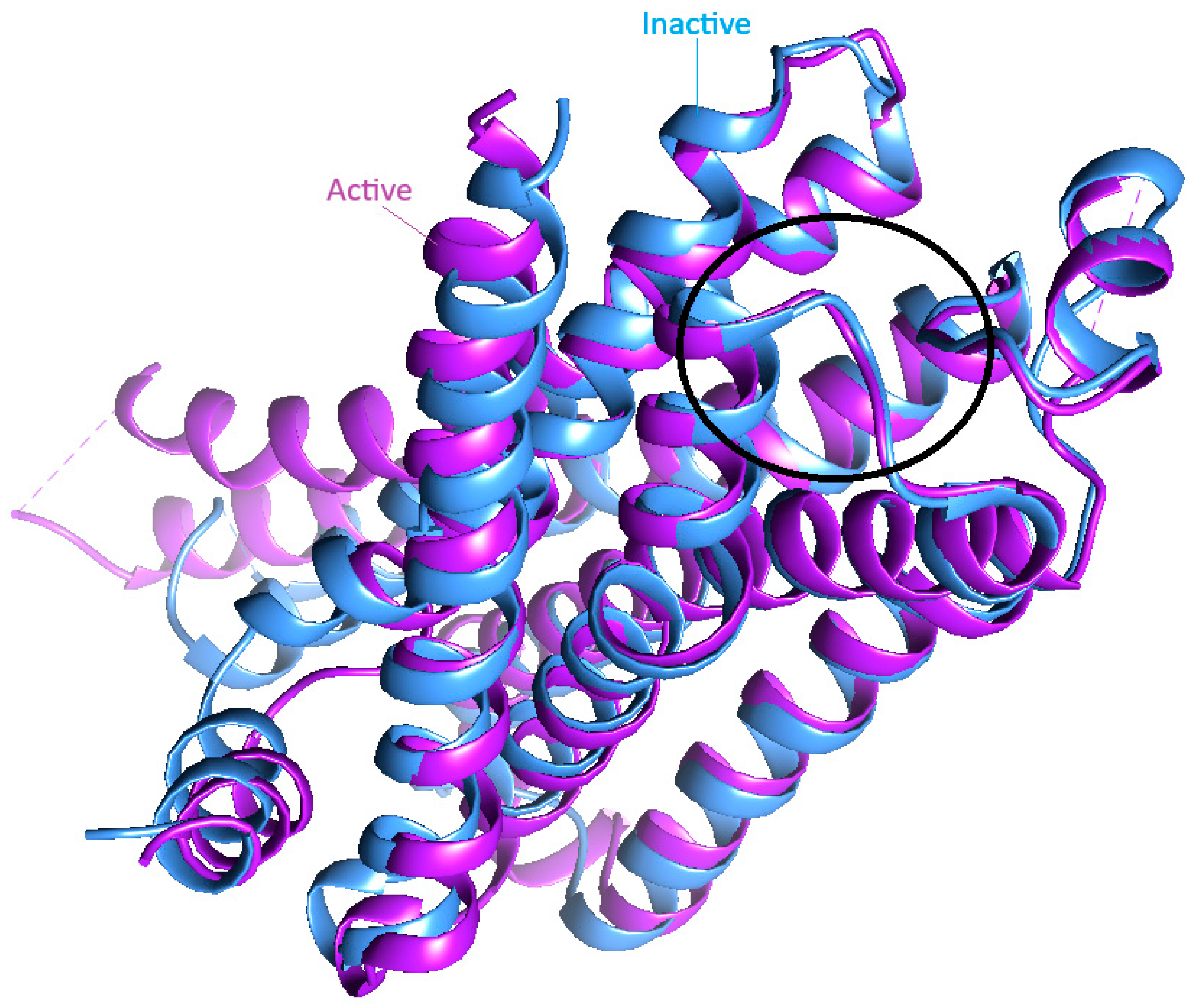
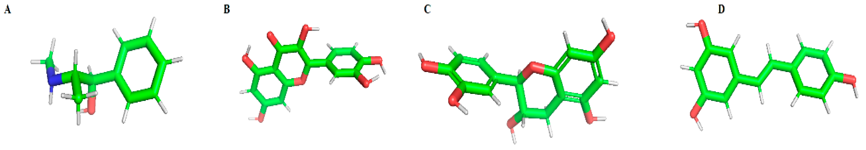
Molecular docking was performed using AutoDock 4.6, a widely used tool for flexible-ligand docking based on the Lamarckian Genetic Algorithm. The grid box was designed around the ligand-binding domain of the β2-AR, with dimensions large enough to accommodate the entire active site and potential allosteric regions. The results were ranked based on binding energy scores (kcal/mol) [21]. The top-scoring conformations were analyzed for hydrogen bonding and hydrophobic interactions with receptor residues using PyMOL 3.1 and Chimera 1.19. The interaction maps between the ligands and the β2-adrenergic receptor (β2-AR) were generated using LigPlot+ 2.2, illustrating hydrogen bonds and hydrophobic contacts within the binding site [24,25,26].

3. Results

A structural comparison of the β2-adrenergic receptor (β2-AR) in its active and inactive conformations was performed to evaluate conformational changes relevant to ligand docking. The alignment revealed that although there were significant rearrangements, especially in transmembrane helices TM5 and TM6, during receptor activation, the orthosteric ligand-binding pocket remained highly conserved. This suggests that docking simulations on both conformations can provide meaningful insights into ligand interaction patterns and potential conformation-sensitive binding behavior. As shown in the circled region in Figure 1, the overall shape and positioning of the key residues within the binding pocket are conserved between the two states. This indicates that the binding site of β2AR is structurally stable and does not undergo significant conformational shifts due to activation. These findings suggest that ligand binding alone does not induce major structural changes in the binding pocket and that receptor activation is likely driven by intracellular interactions such as G-protein coupling.
The results were ranked based on binding energy scores (kcal/mol) (Table 1).
Molecular docking simulations indicated that all four natural compounds, ephedrine, quercetin, catechin, and resveratrol, were able to bind within the β2-adrenergic receptor (β2-AR) binding site in both inactive and active conformations. Their binding profiles were compared to those of known β2-AR agonists, salbutamol and formoterol, to assess the relative interaction patterns and explore the potential of these natural compounds as modulators of β2-AR.
Based on the docking results, quercetin demonstrated the strongest binding affinity among the natural compounds in the inactive β2-AR conformation (−5.70 kcal/mol, 5 hydrogen bonds), although it exhibited reduced affinity in the active form (−4.00 kcal/mol, 3 hydrogen bonds), suggesting possible conformation-sensitive behavior. Catechin showed stable binding across both conformations (−5.37 kcal/mol in inactive and −4.65 kcal/mol in active), supported by five and four hydrogen bonds, respectively, indicating better conformational adaptability. Resveratrol also displayed consistent interaction patterns (−5.29 and −4.64 kcal/mol), forming three hydrogen bonds in both receptor states. Ephedrine, a known natural β2-AR agonist included as a positive control, demonstrated the weakest binding energies among the tested compounds (−4.66 and −4.04 kcal/mol), but still interacted with key residues like Asp113, aligning with its known biological activity.
Figure 2 depicts the 3D structures of the natural ligands. Figure 3 shows a LigPlot+ representation of the β2-adrenergic receptor (β2-AR) in complex with natural ligands.

4. Discussion

The results of this study display the potential of natural compounds as alternative ligands for β2-adrenergic receptor modulation. The β2-AR plays a crucial role in respiratory and cardiovascular physiology, and its therapeutic targeting is essential in diseases like asthma, COPD, and hypertension. However, synthetic β2-agonists, though effective, often bring challenges such as receptor desensitization and side effects like tremors, headaches, and elevated heart rate. Natural compounds offer a promising avenue due to their biocompatibility, lower toxicity, and multifaceted biological activities.
Among the tested natural compounds, quercetin emerged as a notable candidate due to its strong binding affinity in the inactive β2-AR conformation and its ability to form an extensive network of interactions with key residues involved in agonist recognition. Its planar aromatic structure, multiple hydroxyl groups, and high polarity contribute to stable hydrogen bonding and hydrophobic interactions within the binding pocket. Although quercetin showed reduced binding affinity in the active conformation, it continued to engage important receptor residues, suggesting conformation-dependent binding behavior and potential functional relevance.
Resveratrol, despite being structurally different, also showed strong interactions, suggesting that diverse chemical scaffolds can be effective in targeting GPCRs. These findings support the idea that structural diversity among natural products allows for unique binding strategies, potentially leading to the discovery of novel functional modulators.
Ephedrine, already known as a natural β-agonist, validated the docking setup through its interaction profile. Catechin, although less potent than quercetin, displayed stable interactions and may be useful in combination therapies or as a functional food component. The ability of these natural compounds to bind β2-AR and form key stabilizing interactions suggests that they could either mimic or modulate endogenous ligands, offering safer and more sustainable pharmacological options.
We recognize that minor variations in docking scores (<1 kcal/mol) do not necessarily correlate with differences in biological activity or potency. We emphasize that this study does not aim to predict functional outcomes such as agonism or antagonism, nor to claim therapeutic relevance. Instead, the primary goal was to assess whether the selected natural compounds can engage key residues critical for β2-AR recognition.
Molecular docking was used solely as an initial in silico screening method to guide future experimental validation. This approach highlights the utility of computational tools in early-stage natural product drug discovery, where virtual screening can help prioritize candidates and reduce the cost and time of subsequent in vitro or in vivo studies [17,27].

5. Conclusions

In summary, this computational study demonstrated the potential of naturally derived compounds to bind the β2-adrenergic receptor with favorable interaction profiles. Docking was performed using both inactive and active β2-AR conformations. The strongest binding affinity among the natural compounds was observed for quercetin in the inactive state, followed by catechin and resveratrol, while ephedrine served as a positive control due to its known pharmacological activity. Moreover, the results reveal that resveratrol and catechin maintained more consistent binding across both states, while quercetin showed conformation-sensitive behavior with reduced affinity in the active form. Formoterol and salbutamol were included as reference β2-agonists, and their stronger binding confirmed the reliability of the docking protocol. All compounds formed stabilizing interactions with key residues within the β2-AR active site, suggesting their viability as natural β2-AR modulators. Importantly, we acknowledge that docking results alone cannot determine receptor activation, antagonist vs. agonist activity, or therapeutic relevance.
These findings provide a foundation for future experimental research aimed at validating the bioactivity of these compounds and optimizing their pharmacokinetic profiles. The use of natural compounds to target GPCRs represents a promising and underexplored strategy in drug discovery, especially in the context of chronic respiratory and cardiovascular diseases. Further studies involving molecular dynamics simulations, ADME prediction, and biological assays are warranted to fully characterize the therapeutic potential of these ligands.

Author Contributions

Docking analysis, S.J.; supervision, J.B. All authors have read and agreed to the published version of the manuscript.

Funding

This research received no external funding.

Institutional Review Board Statement

Not applicable.

Informed Consent Statement

Not applicable.

Data Availability Statement

Data is contained within the article.

Conflicts of Interest

The authors declare no conflicts of interest.

References

  1. Schiöth, H.B.; Fredriksson, R. The GRAFS classification system of G-protein coupled receptors in comparative perspective. Gen. Comp. Endocrinol. 2005, 142, 94–101. [Google Scholar] [CrossRef]
  2. Rehman, S.; Rahimi, N.; Dimri, M. Biochemistry, G protein coupled receptors. In StatPearls; StatPearls Publishing: Treasure Island, FL, USA, 2023. [Google Scholar]
  3. Benovic, J.L. Novel β2-adrenergic receptor signaling pathways. J. Allergy Clin. Immunol. 2002, 110, S229–S235. [Google Scholar] [CrossRef] [PubMed]
  4. Tandale, A.; Joshi, M.; Sengupta, D. Structural insights and functional implications of inter-individual variability in β2-adrenergic receptor. Sci. Rep. 2016, 6, 24379. [Google Scholar] [CrossRef] [PubMed]
  5. Barnes, P.J. Biochemical basis of asthma therapy. J. Biol. Chem. 2011, 286, 32899–32905. [Google Scholar] [CrossRef]
  6. Hill, S.J. G-protein-coupled receptors: Past, present and future. Br. J. Pharmacol. 2006, 147, S27–S37. [Google Scholar] [CrossRef]
  7. Serrano-Marín, J.; Reyes-Resina, I.; Martínez-Pinilla, E.; Navarro, G.; Franco, R. Natural compounds as guides for the discovery of drugs targeting G-protein-coupled receptors. Molecules 2020, 25, 5060. [Google Scholar] [CrossRef]
  8. Saqib, U.; Kelley, T.T.; Panguluri, S.K.; Liu, D.; Savai, R.; Baig, M.S.; Schürer, S.C. Polypharmacology or promiscuity? Structural interactions of resveratrol with its bandwagon of targets. Front. Pharmacol. 2018, 9, 1201. [Google Scholar] [CrossRef] [PubMed]
  9. Grabarczyk, M.; Justyńska, W.; Czpakowska, J.; Smolińska, E.; Bielenin, A.; Glabinski, A.; Szpakowski, P. Role of plant phytochemicals: Resveratrol, curcumin, luteolin and quercetin in demyelination, neurodegeneration, and epilepsy. Antioxidants 2024, 13, 1364. [Google Scholar] [CrossRef]
  10. Georgiou, N.; Kakava, M.G.; Routsi, E.A.; Petsas, E.; Stavridis, N.; Freris, C.; Zoupanou, N.; Moschovou, K.; Kiriakidi, S.; Mavromoustakos, T. Quercetin: A potential polydynamic drug. Molecules 2023, 28, 8141. [Google Scholar] [CrossRef]
  11. Najafi, N.N.; Armide, N.; Akbari, A.; Rahimi, V.B.; Askari, V.R. Quercetin a promising functional food additive against allergic diseases: A comprehensive and mechanistic review. J. Funct. Foods 2024, 116, 106152. [Google Scholar] [CrossRef]
  12. Vansal, S.S.; Feller, D.R. Direct effects of ephedrine isomers on human β-adrenergic receptor subtypes. Biochem. Pharmacol. 1999, 58, 807–810. [Google Scholar] [CrossRef] [PubMed]
  13. Yashima, M.; Sato, Y.; Kazama, I. Catechin synergistically potentiates mast cell-stabilizing property of caffeine. Allergy Asthma Clin. Immunol. 2021, 17, 1. [Google Scholar] [CrossRef]
  14. Yamazaki, S.; Miyoshi, N.; Kawabata, K.; Yasuda, M.; Shimoi, K. Quercetin-3-O-glucuronide inhibits noradrenaline-promoted invasion of MDA-MB-231 human breast cancer cells by blocking β2-adrenergic signaling. Arch. Biochem. Biophys. 2014, 557, 18–27. [Google Scholar] [CrossRef] [PubMed]
  15. Tripathi, A.; Kumar, M.; Kaur, P.; Kumar, B.; Sagi, S.S. Efficacy of Quercetin as a potent sensitizer of β2-AR in combating the impairment of fluid clearance in lungs of rats under hypoxia. Respir. Physiol. Neurobiol. 2020, 273, 103334. [Google Scholar] [CrossRef] [PubMed]
  16. Chikazawa, M.; Sato, R. Identification of a Novel Function of Resveratrol and Genistein as a Regulator of β2-Adrenergic Receptor Expression in Skeletal Muscle Cells and Characterization of Promoter Elements Required for Promoter Activation. Mol. Nutr. Food Res. 2018, 62, 1800530. [Google Scholar] [CrossRef]
  17. Stanzione, F.; Giangreco, I.; Cole, J.C. Use of molecular docking computational tools in drug discovery. Prog. Med. Chem. 2021, 60, 273–343. [Google Scholar] [CrossRef]
  18. Roux, F.J.; Grandordy, B.; Douglas, J.S. Functional and binding characteristics of long-acting beta 2-agonists in lung and heart. Am. J. Respir. Crit. Care Med. 1996, 153, 1489–1495. [Google Scholar] [CrossRef]
  19. Cherezov, V.; Rosenbaum, D.M.; Hanson, M.A.; Rasmussen, S.G.; Thian, F.S.; Kobilka, T.S.; Choi, H.-J.; Kuhn, P.; Weis, W.I.; Kobilka, B.K. High-resolution crystal structure of an engineered human β2-adrenergic G protein–coupled receptor. Science 2007, 318, 1258–1265. [Google Scholar] [CrossRef]
  20. Rasmussen, S.G.; DeVree, B.T.; Zou, Y.; Kruse, A.C.; Chung, K.Y.; Kobilka, T.S.; Thian, F.S.; Chae, P.S.; Pardon, E.; Calinski, D. Crystal structure of the β2 adrenergic receptor–Gs protein complex. Nature 2011, 477, 549–555. [Google Scholar] [CrossRef]
  21. Morris, G.M.; Huey, R.; Lindstrom, W.; Sanner, M.F.; Belew, R.K.; Goodsell, D.S.; Olson, A.J. AutoDock4 and AutoDockTools4: Automated docking with selective receptor flexibility. J. Comput. Chem. 2009, 30, 2785–2791. [Google Scholar] [CrossRef]
  22. Kim, S.; Chen, J.; Cheng, T.; Gindulyte, A.; He, J.; He, S.; Li, Q.; Shoemaker, B.A.; Thiessen, P.A.; Yu, B. PubChem 2025 update. Nucleic Acids Res. 2025, 53, D1516–D1525. [Google Scholar] [CrossRef] [PubMed]
  23. El-Hachem, N.; Haibe-Kains, B.; Khalil, A.; Kobeissy, F.H.; Nemer, G. AutoDock and AutoDockTools for protein-ligand docking: Beta-site amyloid precursor protein cleaving enzyme 1 (BACE1) as a case study. In Neuroproteomics; Humana Press: New York, NY, USA, 2017; pp. 391–403. [Google Scholar] [CrossRef]
  24. Pettersen, E.F.; Goddard, T.D.; Huang, C.C.; Couch, G.S.; Greenblatt, D.M.; Meng, E.C.; Ferrin, T.E. UCSF Chimera—A visualization system for exploratory research and analysis. J. Comput. Chem. 2004, 25, 1605–1612. [Google Scholar] [CrossRef] [PubMed]
  25. Schrödinger, Inc. The PyMOL Molecular Graphics System, Version 1.8; Schrödinger, Inc.: New York, NY, USA, 2015. [Google Scholar]
  26. Wallace, A.C.; Laskowski, R.A.; Thornton, J.M. LIGPLOT: A program to generate schematic diagrams of protein-ligand interactions. Protein Eng. Des. Sel. 1995, 8, 127–134. [Google Scholar] [CrossRef] [PubMed]
  27. Meng, X.-Y.; Zhang, H.-X.; Mezei, M.; Cui, M. Molecular docking: A powerful approach for structure-based drug discovery. Curr. Comput.-Aided Drug Des. 2011, 7, 146–157. [Google Scholar] [CrossRef]
Figure 1. Structural comparison of the β2-adrenergic receptor (β2AR), a G-protein-coupled receptor (GPCR), in its active (PDB ID: 3SN6, shown in purple) and inactive (PDB ID: 2RH1, shown in blue) conformations. The black circle highlights the orthosteric binding site, which remains structurally conserved between the active and inactive states. This alignment illustrates that despite large conformational differences elsewhere in the protein, the binding pocket retains a similar architecture in both functional state.
Msf 34 00003 g001
Figure 2. Three-dimensional structures of natural ligands used in the study: (A) ephedrine, (B) quercetin, (C) catechin, and (D) resveratrol. Atoms are color-coded as follows: Carbon (C): green, Hydrogen (H): white, Oxygen (O): red, Nitrogen (N): blue (visible in ephedrine only). Molecular geometries were optimized and rendered using UCSF Chimera version 1.16.
Msf 34 00003 g002
Figure 3. LigPlot+ interaction diagrams illustrating the binding modes of natural ligands to the β2-adrenergic receptor (β2-AR): (A) ephedrine, (B) quercetin, (C) catechin, and (D) resveratrol. Hydrogen bonds are shown as green dashed lines with bond distances labeled in Ångströms. Red arcs represent non-ligand residues involved in hydrophobic contacts. Key interacting residues such as Asp113, Ser203, Asn312, and Tyr316 are involved in stabilizing the ligand within the orthosteric binding pocket.
Msf 34 00003 g003
Table 1. Binding energies and hydrogen bond interactions of compounds with β2-adrenergic receptor (β2-AR) in inactive (2RH1) and active (3SN6) conformations.
CompoundBinding Energy (Inactive—2RH1)
[kcal/mol]
H-Bonds (2RH1)Binding Energy (Active—3SN6)
[kcal/mol]
H-Bonds (3SN6)
Formoterol−7.862−7.533
Salbutamol−6.334−6.533
Catechin−5.375−4.654
Ephedrine−4.663−4.042
Quercetin−5.705−4.003
Resveratrol−5.293−4.643
Disclaimer/Publisher’s Note: The statements, opinions and data contained in all publications are solely those of the individual author(s) and contributor(s) and not of MDPI and/or the editor(s). MDPI and/or the editor(s) disclaim responsibility for any injury to people or property resulting from any ideas, methods, instructions or products referred to in the content.

