Proceeding Paper

The Electrochemical Behavior of Methotrexate upon Binding to the DNA of Different Cell Lines †

by
Douglas Vieira Thomaz
* and
Pierre Alexandre dos Santos
Faculty of Pharmacy, Federal University of Goiás, Goiânia 74690-900, GO, Brazil
*
Author to whom correspondence should be addressed.
Presented at the 1st International Electronic Conference on Cancers: Exploiting Cancer Vulnerability by Targeting the DNA Damage Response, 1–14 February 2021; Available online: https://iecc2021.sciforum.net/.
Med. Sci. Forum 2021, 3(1), 16; https://doi.org/10.3390/IECC2021-09215
Published: 29 January 2021

Abstract

:
Methotrexate (MTX) is a widely used anticancer agent whose DNA binding properties are well known. Despite its consolidated usage in the therapeutics of cancer, the physicochemical features of MTX binding to healthy and neoplastic DNA are still not fully understood. Therefore, this work showcases the electrochemical study of MTX binding to distinct DNA sequences through voltametric approaches.

1. Introduction

Methotrexate (MTX) is an anticancer agent whose main therapeutic property relies on the impairment of cellular metabolism [1]. This compound is considered an antifolate derivative due to its structural similarity to folic acid and its hinderance of folate-dependent enzymatic activity [2]. The literature reports that MTX might also interact with nucleic acids, which further supports its antineoplastic appeal [3].
Considering the distinctions in constitution and physicochemical behavior between healthy and neoplastic DNA sequences, it can be inferred that anticancer agents might couple differently according to the nature of the DNA segment [4]. This was supported by several authors [4,5,6]. In this regard, a comparative study between the binding of MTX to the DNA of distinct cell lines would likely provide remarkable information that sheds light on the pharmacodynamics of anticancer agents whose main biological target is the DNA.
Regarding the DNA-binding properties of anticancer agents, redox mechanisms may play a distinct role in their pharmacology [7,8,9]. Owing to the occurrence of electron transfer in covalent-based DNA-binding, and the impairment of charge transfer upon the stabilizing effect provided by intermolecular interactions, the detection of changes in faradaic currents may contribute with information regarding the physicochemistry of the anchoring of ligands to the DNA [7]. In this sense, electrochemical methods such as voltammetry may be useful for evaluating the dynamics of charge transfer in reaction media, and by consequence, provide information regarding the binding of anticancer agents to DNA [10,11,12].
Therefore, in view of the importance of differentiating the binding of anticancer agents to DNA from distinct cell lineages, this study showcases an investigation of the electrochemical behavior of anticancer drug MTX upon binding to the DNA of melanoma BRAFV600E (SK-Mel-28) and wild type (WM852) cell lines, and commercial double-stranded calf thymus DNA (ds-CT).

2. Materials and Methods

2.1. Reagents and Solutions

MTX (Sigma, St. Louis, MO, USA) was diluted in purified water (conductivity ≤ 0.1 µS cm−1) obtained from a Milli-Q purification system, Millipore S/A (Molsheim, France), in order to render a stock solution of 0.01 mol L−1. Potassium chloride, disodium hydrogen phosphate, potassium hydrogen phosphate, and sodium chloride were used to prepare 0.1 mol L−1 phosphate buffered saline (PBS) solution, pH 7.0. Furthermore, 1.5 g of ds-CT was diluted in 2 mL PBS, pH 7.0, in order to render a concentrated mixture for the studies.

2.2. DNA Extraction

The following cell lines were used in this study: melanoma BRAFV600E (SK-Mel-28) and wild type (WM852). The cells were incubated in culture plates with penicillin, streptomycin, and 10% fetal bovine serum in Eagle’s Minimum Essential Medium. Incubator temperature was maintained at 37 °C under 5% CO2 pressure until cell monolayer was formed. Thereafter, DNA was extracted according to standard protocols [13,14].

2.3. Electrochemical Assays

Voltametric experiments were carried out in a potentiostat/galvanostat Autolab III® integrated to GPES 4.9® software, Eco-Chemie, Utrecht, Netherlands. The measurements were performed in a 1.0 mL one-compartment/three-electrode system electrochemical cell consisting of glassy carbon electrode (GCE)—0.785 mm2 area, a Pt wire, and an Ag/AgCl/KClsat electrode (Lab solutions, São Paulo, Brazil), representing the working electrode, the counter electrode, and the reference electrode, respectively.
Experimental conditions for cyclic voltammetry (CV) were: a scan rate (υ) of 12.5, 25, 50, 100, 250, or 500 mV s−1; and a scan range of 0.5 to 1.2 V. All voltametric assays were performed in 0.1 mol L−1 PBS solution, pH 7.0.
The experiments consisted of two approaches, namely: (i) investigating the electrochemical behavior of MTX without DNA, and (ii) evaluating the electroanalytic signal of MTX upon the addition of DNA to the test solution. Then, 100 µL of MTX solution was added in the electrochemical cell containing 850 mL of PBS buffer (i), and was followed by the addition of 5 µL of a 7.8 ng mL−1 (WM852), 5.6 ng mL−1 (SK-Mel-28), or 0.75 g mL−1 (ds-CT) DNA solution (ii).
Thereafter, the mixture of MTX and DNA from each lineage was submitted to several CV cycles under crescent υ, in order to obtain the plots of the faradaic currents against the square root of the voltametric scan rate, as stated by Randles–Sevcik equation (1). This mathematical treatment was used because it allowed us to draw comparisons regarding diffusional aspects of the electrochemical reaction [15].
I p a υ 1 / 2 = 2.69   10 5   A   n 3 / 2   D 1 / 2   c
wherein Ipa is the anodic peak current, A is electrode area in cm2, n is the number of transferred electrons, D is the diffusion coefficient, c is the concentration of MTX in mol L−1, and υ is scan rate in V s−1.
All experiments were performed in triplicate, and strenuous cleaning of the working electrode surface was conducted before each assay. Furthermore, all voltametric data were analyzed and prepared with Origin 8® software.

3. Results and Discussion

3.1. Redox Behavior of MTX

In order to primarily investigate the redox behavior of MTX, CV was assayed on a bare MTX solution in PBS buffer, pH 7.0. Results are showcased in Figure 1.
The results show that MTX exhibits two anodic signals at Ep1a ≈ 0.9 V and Ep2a ≈ 1.05 V (Figure 1A), which correlate with the oxidation of electroactive moieties in MTX molecule (Figure 1B).
The findings showcased in Figure 1 are in consonance with the literature, wherein it has been reported that MTX undergoes irreversible electrooxidation at Ep1a ≈ 0.9 V and Ep2a ≈ 1.05 V [16,17,18]. The reaction was deemed irreversible, hence the absence of visible cathodic peaks in the voltammogram [19,20,21]. Considering that the peaks show broadening, it can be suggested that MTX oxidation products might undergo adsorption on the working electrode’s surface, which was in fact observed during the experiments, leading us to adopt a strenuous electrode surface renewal protocol. Furthermore, this behavior was also reported by other authors [16,22].
The proposed electrooxidation mechanism of MTX includes an irreversible demethylation step at Ep1a ≈ 0.9 V. This oxidation process leads to the collection of a single electron from MTX by the working electrode surface [23,24]. Moreover, the second proposed electrooxidation at Ep2a ≈ 1.05 V leads to the formation of an imine derivative, and follows 1:1 stoichiometry regarding MTX and the release of electrons—an equivalence of electrons and protons (i.e., a Nernstian process) [23]. These electrons rose the total charge, which was detected by the electric current detector coupled to the working electrode circuit. Each oxidation was followed by rises in the electric current, which is in consonance with the literature. Furthermore, other authors proposed similar oxidation mechanisms to anticancer drugs bearing structural similarity to MTX [25,26].

3.2. Plots of the Faradaic Currents against the Square Root of the Voltametric Scan Rate Created via the Randles–Sevcik Equation

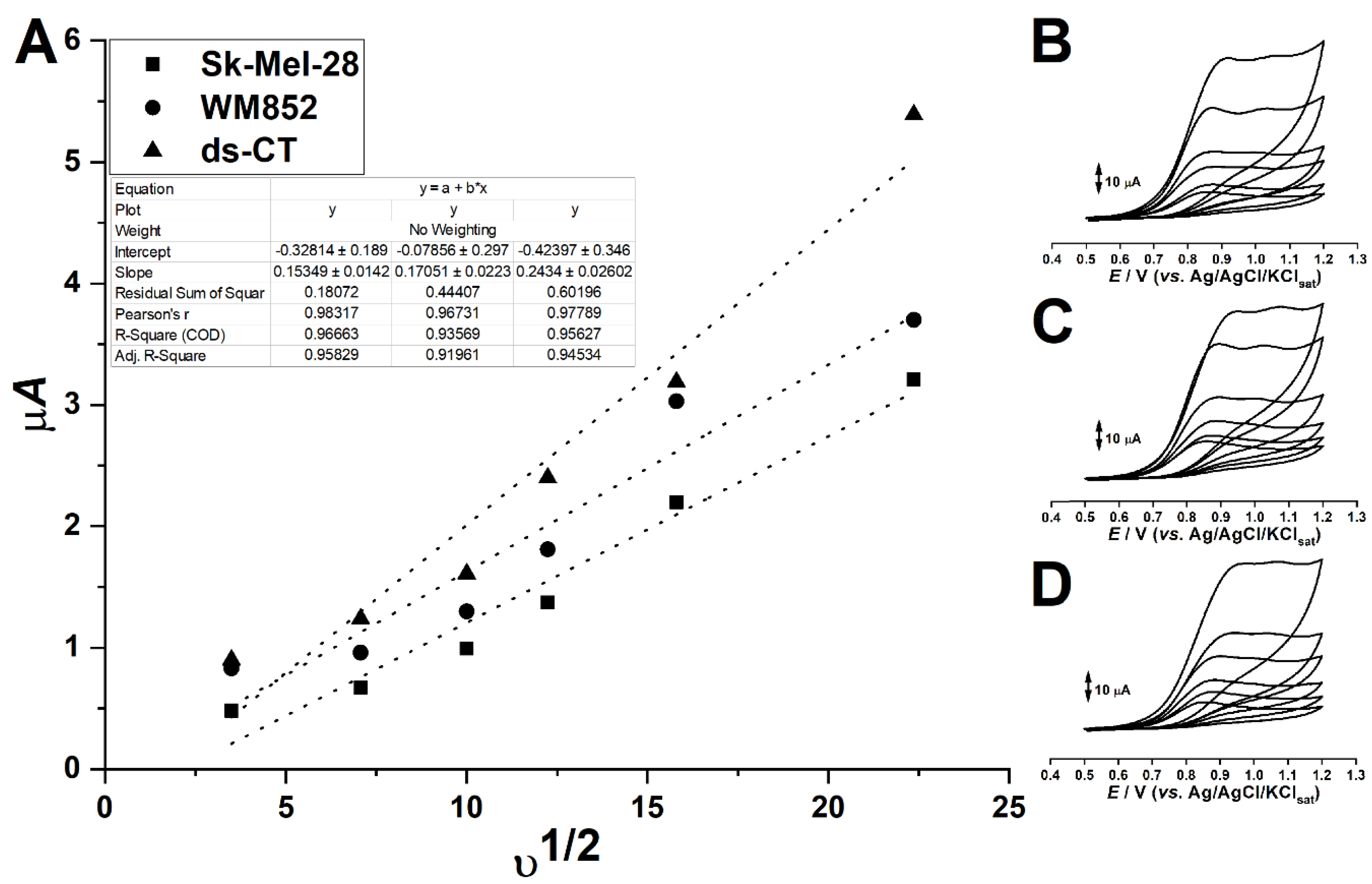
In order to draw information regarding the diffusional process of MTX-DNA adducts, plots of the faradaic currents against the square root of the voltametric scan rate were created according to Randles–Sevcik equation. The results are showcased in Figure 2.
The results show that the response of the MTX signal upon the addition of each DNA was distinct under Randles–Sevcik treatment (Figure 2A). It can be noticed that all plots have linear profiles. The response of MTX in the presence of SK-Mel-28 was the lowest, and in the presence of ds-CT it was the highest (Figure 2A–D).
The literature states that MTX electrooxidation is adsorption controlled [25], though the addition of DNA shifts the rate-defining step of the electrooxidation of a binding agent to the diffusion of adducts through the bulk solution [7]. In this sense, the Handles–Sevcik treatment can be applied even for molecules whose oxidation products promote extensive electrode fouling. Nonetheless, the presence of DNA in the solution leads to the formation of MTX-DNA adducts, which diffuse slowly through the bulk solution and take a long time to reach the working electrode’s surface [24]. Moreover, the DNA-binding leads to lesser amounts of faster-diffusing free MTX, thereby leading to smaller faradaic signals, as observed elsewhere with other intercalating agents [7].
Considering that the response of MTX to the addition of SK-Mel-28 was the smallest regarding all assayed DNAs, and that this DNA was nonetheless the most diluted among them all, it can be suggested that SK-Mel-28 DNA led to the smallest amount of free MTX in the reaction media, hence the higher affinity of this neoplastic DNA to the anticancer drug [27]. In this sense, our results are corroborated by the literature, which describe the DNA-binding properties of MTX [26,27]. Moreover, it was noteworthy that highly concentrated ds-CT DNA did not promote major changes in the response when compared to WM852 DNA. However, owing to the small differences in the signals (i.e., in the order of µA), further investigations are needed in order to fully explore the applicability of electrochemical methods in the study of the DNA-binding properties of MTX.

4. Conclusions

This work showcased an electrochemical study of MTX binding to distinct DNA sequences through voltametric approaches. Results showcased that MTX exhibits two irreversible anodic peaks at Ep1a ≈ 0.9 V and Ep2a ≈ 1.05 V, which correlate the oxidation of electroactive moieties in its structure. Moreover, the investigation of MTX binding to DNAs through the Handles–Sevcik equation showed that the MTX response to the presence of every type of DNA led to a linear profile. The response of MTX in the presence of SK-Mel-28 was the lowest; in the presence of ds-CT, the highest.

Author Contributions

Conceptualization, D.V.T. and P.A.d.S.; methodology D.V.T. and P.A.d.S.; formal analysis, D.V.T. and P.A.d.S.; investigation, D.V.T. and P.A.d.S.; resources, D.V.T. and P.A.d.S.; data curation, D.V.T. and P.A.d.S.; writing, D.V.T. and P.A.d.S. All authors have read and agreed to the published version of the manuscript.

Funding

This research was funded by Coordenação de Aperfeiçoamento de Pessoal de Nível Superior (CAPES) and Conselho Nacional de Desenvolvimento Científico e Tecnológico (CnPq).

Conflicts of Interest

Authors declare no conflict of interest.

References

  1. Mager, D.R. Methotrexate. Home Healthc. Now 2015, 33, 139–141. [Google Scholar] [CrossRef]
  2. Bleyer, W.A. The clinical pharmacology of methotrexate. New applications of an old drug. Cancer 1978. [Google Scholar] [CrossRef]
  3. Tian, H.; Cronstein, B. Understanding the Mechanisms of Action of Methotrexate. Bull. NYU Hosp. Jt. Dis. 2007, 65, 168–173. [Google Scholar] [PubMed]
  4. Palchaudhuri, R.; Hergenrother, P.J. DNA as a target for anticancer compounds: Methods to determine the mode of binding and the mechanism of action. Curr. Opin. Biotechnol. 2007, 18, 497–503. [Google Scholar] [CrossRef] [PubMed]
  5. Cheung-Ong, K.; Giaever, G.; Nislow, C. DNA-damaging agents in cancer chemotherapy: Serendipity and chemical biology. Chem. Biol. 2013, 20, 648–659. [Google Scholar] [CrossRef] [PubMed] [Green Version]
  6. Tacar, O.; Sriamornsak, P.; Dass, C.R. Doxorubicin: An update on anticancer molecular action, toxicity and novel drug delivery systems. J. Pharm. Pharmacol. 2013, 65, 157–170. [Google Scholar] [CrossRef] [PubMed]
  7. Thomaz, D.V.; de Oliveira, M.G.; Rodrigues, E.S.B.; da Silva, V.B.; Santos, P.A. Dos Physicochemical investigation of psoralen binding to double stranded dna through electroanalytical and cheminformatic approaches. Pharmaceuticals 2020, 13, 108. [Google Scholar] [CrossRef]
  8. Zunino, F.; Gambetta, R.; Di Marco, A.; Zaccara, A. Interaction of daunomycin and its derivatives with DNA. BBA Sect. Nucleic Acids Protein Synth. 1972. [Google Scholar] [CrossRef]
  9. Ozluer, C.; Kara, H.E.S. In vitro DNA binding studies of anticancer drug idarubicin using spectroscopic techniques. J. Photochem. Photobiol. B Biol. 2014. [Google Scholar] [CrossRef]
  10. Gorodetsky, A.A.; Buzzeo, M.C.; Barton, J.K. DNA-mediated electrochemistry. Bioconjug. Chem. 2008, 19, 2285–2296. [Google Scholar] [CrossRef] [Green Version]
  11. Kelley, S.O.; Barton, J.K.; Jackson, N.M.; Hill, M.G. Electrochemistry of methylene blue bound to a DNA-modified electrode. Bioconjug. Chem. 1997. [Google Scholar] [CrossRef]
  12. Boon, E.M.; Jackson, N.M.; Wightman, M.D.; Kelley, S.O.; Hill, M.G.; Barton, J.K. Intercalative stacking: A critical feature of DNA charge-transport electrochemistry. J. Phys. Chem. B 2003. [Google Scholar] [CrossRef] [Green Version]
  13. Elkins, K.M. DNA Extraction. In Forensic DNA Biology; Academic Press: Cambridge, MA, USA, 2013. [Google Scholar]
  14. Jennings, W.B.; Jennings, W.B. DNA Extraction. In Phylogenomic Data Acquisition; CRC Press: Boca Raton, FL, USA, 2016. [Google Scholar]
  15. Thomaz, D.V.; Filho, A.M.d.A.; Macedo, I.Y.L.; Rodrigues, E.S.B.; Gil, E.d.S. Predictive Modelling to Study the Electrochemical Behaviour of PdO, TiO2 and Perovskite-Type LaFeO3 Modified Carbon Paste Electrodes. Path Sci. 2019. [Google Scholar] [CrossRef]
  16. Gao, L.; Wu, Y.; Liu, J.; Ye, B. Anodic voltammetric behaviors of methotrexate at a glassy carbon electrode and its determination in spiked human urine. J. Electroanal. Chem. 2007. [Google Scholar] [CrossRef]
  17. Šelešovská, R.; Bandžuchová, L.; Navrátil, T. Voltammetric behavior of methotrexate using mercury meniscus modified silver solid amalgam electrode. Electroanalysis 2011. [Google Scholar] [CrossRef]
  18. Asbahr, D.; Figueiredo-Filho, L.C.S.; Vicentini, F.C.; Oliveira, G.G.; Fatibello-Filho, O.; Banks, C.E. Differential pulse adsorptive stripping voltammetric determination of nanomolar levels of methotrexate utilizing bismuth film modified electrodes. Sens. Actuators B Chem. 2013. [Google Scholar] [CrossRef]
  19. Antunes, R.S.; Ferraz, D.; Garcia, L.F.; Thomaz, D.V.; Luque, R.; Lobón, G.S.; Gil, E.d.S.; Lopes, F.M. Development of a polyphenol oxidase biosensor from Jenipapo fruit extract (Genipa americana L.) and determination of phenolic compounds in textile industrial effluents. Biosensors 2018, 8, 47. [Google Scholar] [CrossRef] [PubMed] [Green Version]
  20. Antunes, R.S.; Thomaz, D.V.; Garcia, L.F.; De Souza Gil, E.; Somerset, V.S.; Lopes, F.M. Determination of methyldopa and paracetamol in pharmaceutical samples by a low cost Genipa americana L. polyphenol oxidase based biosensor. Adv. Pharm. Bull. 2019. [Google Scholar] [CrossRef] [PubMed] [Green Version]
  21. Thomaz, D.V.; Leite, K.C.d.S.; Moreno, E.K.G.; Garcia, L.F.; Alecrim, M.F.; Macêdo, I.Y.L.; Caetano, M.P.; de Carvalho, M.F.; Machado, F.B.; de Souza Gil, E. Electrochemical study of commercial black tea samples. Int. J. Electrochem. Sci. 2018, 13, 5433–5439. [Google Scholar] [CrossRef]
  22. Geise, R.J.; Adams, J.M.; Barone, N.J.; Yacynych, A.M. Electropolymerized films to prevent interferences and electrode fouling in biosensors. Biosens. Bioelectron. 1991. [Google Scholar] [CrossRef]
  23. Lutterbeck, C.A.; Baginska, E.; Machado, Ê.L.; Kümmerer, K. Removal of the anti-cancer drug methotrexate from water by advanced oxidation processes: Aerobic biodegradation and toxicity studies after treatment. Chemosphere 2015. [Google Scholar] [CrossRef] [PubMed]
  24. Pontinha, A.D.R.; Jorge, S.M.A.; Chiorcea Paquim, A.M.; Diculescu, V.C.; Oliveira-Brett, A.M. In situ evaluation of anticancer drug methotrexate-DNA interaction using a DNA-electrochemical biosensor and AFM characterization. Phys. Chem. Chem. Phys. 2011. [Google Scholar] [CrossRef] [PubMed]
  25. Oliveira, G.G.; Janegitz, B.C.; Zucolotto, V.; Fatibello-Filho, O. Differential pulse adsorptive stripping voltammetric determination of methotrexate using a functionalized carbon nanotubes-modified glassy carbon electrode. Cent. Eur. J. Chem. 2013. [Google Scholar] [CrossRef]
  26. Wang, Y.; Liu, H.; Wang, F.; Gao, Y. Electrochemical oxidation behavior of methotrexate at DNA/SWCNT/Nafion composite film-modified glassy carbon electrode. J. Solid State Electrochem. 2012. [Google Scholar] [CrossRef]
  27. Martin, S.A.; McCarthy, A.; Barber, L.J.; Burgess, D.J.; Parry, S.; Lord, C.J.; Ashworth, A. Methotrexate induces oxidative DNA damage and is selectively lethal to tumour cells with defects in the DNA mismatch repair gene MSH2. EMBO Mol. Med. 2009. [Google Scholar] [CrossRef]
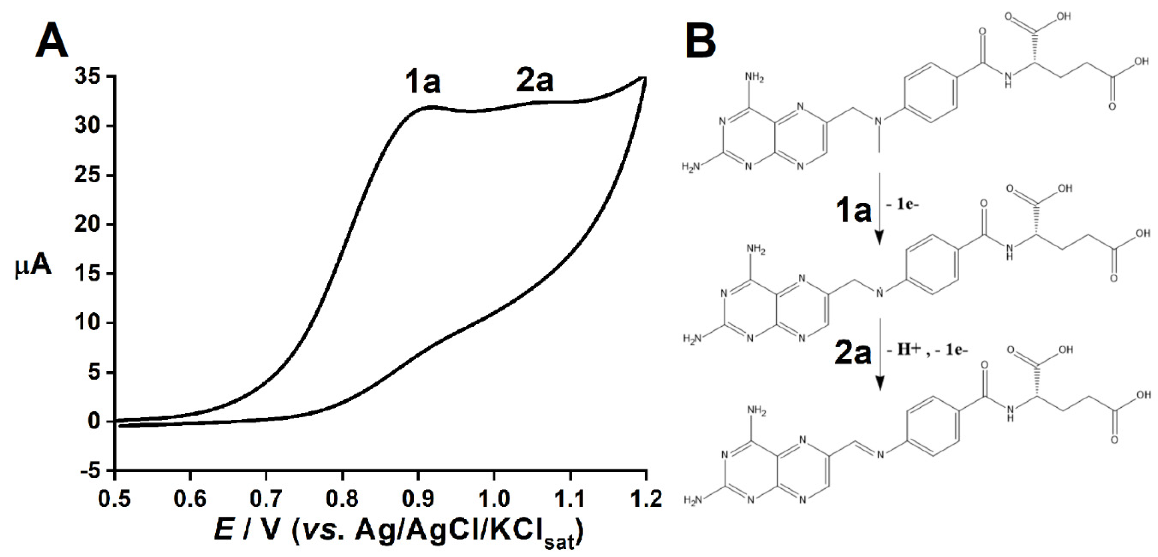
Figure 1. (A) The cyclic voltammogram of MTX showcasing two anodic processes (1a and 2a); (B) theroposed electrooxidation reaction for MTX. All voltametric assays were performed in 0.1 mol L−1 PBS solutions, pH 7.0.
Msf 03 00016 g001
Figure 2. (A) Handles–Sevcik plots of MTX signals in the presence of different DNA. Insert. Analytical parameters of each fitted curve. (B) Plot of MTX-WM852 at successive υ. (C) Plot of MTX-SK-Mel-28 at successive υ. (D) Plot of MTX-ds-CT at successive υ. All voltametric assays were performed in 0.1 mol L−1 PBS solution, pH 7.0, at υ of 12.5, 25, 50, 100, 250, and 500 mV s−1.
Msf 03 00016 g002
Publisher’s Note: MDPI stays neutral with regard to jurisdictional claims in published maps and institutional affiliations.