Proceeding Paper

External Cervical Resorption—The Commonly Misdiagnosed, Destructive Resorption—A Pilot Study †

by
Marta Alves Duarte
* and
João Albernaz Neves
Endodontics Department, Egas Moniz School of Health and Science, Caparica, 2829-511 Almada, Portugal
*
Author to whom correspondence should be addressed.
Presented at the 6th International Congress of CiiEM—Immediate and Future Challenges to Foster One Health, Almada, Portugal, 5–7 July 2023.
Med. Sci. Forum 2023, 22(1), 26; https://doi.org/10.3390/msf2023022026
Published: 14 August 2023

Abstract

:
External cervical resorption is a relatively uncommon, often misdiagnosed, destructive form of external resorption. It consists of an uncontrolled growth of resorptive tissue and consequent loss of mineralized tooth tissue, in an apical direction. The clastic cells are responsible for the process, which can lead to severe deterioration and, eventually, tooth loss. To date, however, an etiological factor has not yet been found. Dentists and students still have difficulties in diagnosing and treating it. In May 2023, a structural questionnaire about external cervical resorption was distributed via Google Forms to 104 people between dentists and fourth- and fifth-year students.

1. Introduction

External cervical tooth resorption is a relatively rare [1,2,3], often misdiagnosed [4,5], destructive form of external tooth resorption [6]. It is characterized by uncontrolled growth of resorptive tissue and consequent loss of mineralized tooth tissue in an apical direction [7,8,9]. For this reason, it can affect all areas of enamel, dentin and cementum [6]. The clastic cells are responsible for this process, which can lead to severe deterioration and eventual tooth loss [2,4,8,10]. However, there is no consensus on its origin and, to date, no etiological factor has been identified for this pathological resorption [6,7,10,11]. Even so, trauma [6], periodontal disease or orthodontic treatment may be predisposing factors [1,12]. In spite of this, there does not seem to be a significant difference in either gender or age [13]. Also, this pathological resorption is asymptomatic [3,13].
Usually, there are no obvious clinical signs [14]. However, there are some symptoms which are crucial to diagnosing this resorption. Firstly, as the name suggests, it occurs in the cervical part of the tooth [8], below the epithelial junction [1,12] but supra-osseous [11]. Furthermore, if the resorption is relatively small and the pulp is not involved, the sensibility tests are positive. In other words, the tooth is vital [6]. In general, it is shown as a restricted area, both clinically and radiographically. On radiographs, it may appear as a radiolucent [1], well-defined lesion [14], as shown in Figure 1. Generally, a radiopaque line separates the root canal from the cervical invasive resorption lesion [3]. In addition, in some cases it can be seen as a pinkish coronal discoloration [3,8]. However, for the diagnosis and assessment of a resorptive lesion, the use of cone beam computed tomography (CBCT) is recommended [4,11]. This helps to diagnose and assess the entry point [15]. For this reason, it may facilitate the selection of an appropriate treatment approach, especially in advanced cases [15].
If left untreated, it is expected to increase in size [16]. Therefore, treatment is mandatory, but the real question is: how? Nowadays, there is no well-established protocol for the treatment of cervical invasive reabsorption [4]. However, it should depend on the size, extent and dimension of the invasive area and its relationship to the root canal [8]. It can be treated externally, internally, with a combined approach externally and internally, externally with deliberate replantation or, when there is no other alternative, by extraction [4]. If possible, the infected tissue should be removed and sealed without compromising posterior rehabilitation. If the pulp is not involved and there is good access to the cavity, topical application of a 90% aqueous solution of trichloroacetic acid and manual or mechanical scaling [6,17] may be the answer. If the pulp is involved and the tooth is necrotic, endodontic treatment [6] is required as well as the previous treatment alternatives.

2. Materials and Methods

In this pilot study, a structured questionnaire was administered to one hundred and four people via Google Forms, in May 2023. The inclusion criteria were dentists and fourth- or fifth-year dentistry students. Of the one hundred and four responses, one hundred and one were usable. The questionnaire was used to assess the knowledge, perception and clinical experience towards external cervical resorption.

3. Results and Discussion

As shown in Figure 2a, among the volunteers, 99.0% claimed to be aware of tooth resorptions, although only 45.2%, as demonstrated in Figure 2b, were aware of external cervical resorptions. Of this last percentage, 21.8% were fourth-year students, 30.4% were fifth-year students and 47.8% were dentists. Thus, the results continue to progress as clinical experience grows. Only those who were aware of this pathology were included for further analysis.
As a result, the evidence shows that only 13% answered all the questions correctly, 30.4% gave one wrong answer and 56.6% gave at least two wrong answers. In addition, over 50% did not know how to radiographically identify a cervical invasive resorption. In fact, only 45.7% recognized this resorption as a well-demarcated radiolucent area on the radiograph, while 53.3% mistakenly identified it as undefined. Furthermore, 56.5% identified the lesion as supra-osseous, 37.0% as infra-osseous and 6.5% as osseous. It can also be seen that 89.1% classified it as tooth resorption, but 8.7% as bone resorption and 2.2% as a tissue lesion. A total of 69.6% considered CBCT to be the best diagnostic test, while 30.4% considered periapical radiographs. A total of 89.1% described this pathological resorption as asymptomatic, although 10.9% described it as painful. In addition, 54.4% associated it with a pinkish discoloration, 15.2% with a yellowish discoloration, another 15.2% with a brownish discoloration, 13.0% with a reddish discoloration and 2.2% with a grey discoloration.
After analysis, 45.7% felt comfortable diagnosing cervical invasive resorption, although 54.3% did not (Figure 3a). As well demonstrated in Figure 3, among the interviewees who felt comfortable diagnosing this lesion (45.7%), only 5 people (23.8%) got all the answers correct. In fact, 42.9% gave one wrong answer and 33.3% gave at least two wrong answers (Figure 3b). In addition, only 21.7% felt comfortable treating this lesion, as opposed to 78.3% who said they were not comfortable (Figure 3c).

4. Conclusions

Undoubtedly, external cervical resorption is a devastating form of external resorption and there are still difficulties among dentists and students in its diagnosis and treatment. For this reason, it remains crucial to educate this population about this type of pathology, so that it can be properly diagnosed and treated to avoid tooth loss and major complications.

Author Contributions

M.A.D. and J.A.N.; Conceptualization, M.A.D.; methodology, M.A.D.; validation, M.A.D. and J.A.N.; formal analysis, M.A.D.; investigation, M.A.D.; resources, M.A.D.; data curation, M.A.D. and J.A.N.; writing—original draft preparation, M.A.D.; writing—review and editing, M.A.D. and J.A.N.; visualization, M.A.D.; supervision, M.A.D. and J.A.N.; project administration, M.A.D.; funding acquisition, M.A.D. All authors have read and agreed to the published version of the manuscript.

Funding

This research received no external funding.

Institutional Review Board Statement

Not applicable.

Informed Consent Statement

Not applicable.

Data Availability Statement

Data Available at https://zenodo.org/record/8020464 (accessed on 9 June 2023).

Acknowledgments

This work was supported by Vanessa Machado, Madalena Caçador, Catarina Estácio, Matilde Guerra, Sandro Pinto, Mariana Machete, Rita Ferreira and Matilde Jardim. My thanks to Joana Costa for her incredible attention and availability.

Conflicts of Interest

The authors declare no conflict of interest.

References

  1. De Brito, G.M.; Campos, P.S.F.; Mariz, A.C.R.; Simões, D.; Machado, A.W. Invasive Cervical Resorption of Central Incisor during Orthodontic Treatment. Dent. Press J. Orthod. 2020, 25, 49–58. [Google Scholar] [CrossRef] [PubMed]
  2. Aiuto, R.; Fumei, G.; Lipani, E.; Garcovich, D.; Dioguardi, M.; Re, D. Conservative Therapy of External Invasive Cervical Resorption with Adhesive Systems: A 6-Year Follow-Up Case Report and Literature Review. Case Rep. Dent. 2022, 2022, 9620629. [Google Scholar] [CrossRef] [PubMed]
  3. Heithersay, G.S. Clinical, Radiologic, and Histopathologic Features of Invasive Cervical Resorption. Quintessence Int. 1999, 30, 27–37. [Google Scholar] [PubMed]
  4. Espona, J.; Roig, E.; Durán-Sindreu, F.; Abella, F.; Machado, M.; Roig, M. Invasive Cervical Resorption: Clinical Management in the Anterior Zone. J. Endod. 2018, 44, 1749–1754. [Google Scholar] [CrossRef] [PubMed]
  5. Alqedairi, A. Non-Invasive Management of Invasive Cervical Resorption Associated with Periodontal Pocket: A Case Report. World J. Clin. Cases 2019, 7, 863–871. [Google Scholar] [CrossRef] [PubMed]
  6. Heithersay, G.S. Invasive Cervical Resorption Following Trauma. Aust. Endod. J. 1999, 25, 79–85. [Google Scholar] [CrossRef] [PubMed]
  7. Rabinovich, I.M.; Snegirev, M.V.; Golubeva, S.A.; Markheev, C.I. External cervical tooth root resorption. Stomatologiia 2022, 101, 73–78. [Google Scholar] [CrossRef] [PubMed]
  8. Sarmento, E.B.; Tavares, S.J.; Thuller, K.A.; Falcao, N.P.; de Paula, K.M.; Antunes, L.A.; Gomes, C.C. Minimally Invasive Intervention in External Cervical Resorption: A Case Report with Six-Year Follow-Up. Int. J. Burns Trauma 2020, 10, 324–330. [Google Scholar] [PubMed]
  9. Tsaousoglou, P.; Markou, E.; Efthimiades, N.; Vouros, I. Characteristics and Treatment of Invasive Cervical Resorption in Vital Teeth. A Narrative Review and a Report of Two Cases. Br. Dent. J. 2017, 222, 423–428. [Google Scholar] [CrossRef]
  10. O’Mahony, A.; McNamara, C.; Ireland, A.; Sandy, J.; Puryer, J. Invasive Cervical Resorption and the Oro-Facial Cleft Patient: A Review and Case Series. Br. Dent. J. 2017, 222, 677–681. [Google Scholar] [CrossRef] [PubMed] [Green Version]
  11. Rotondi, O.; Waldon, P.; Kim, S.G. The Disease Process, Diagnosis and Treatment of Invasive Cervical Resorption: A Review. Dent. J. 2020, 8, 64. [Google Scholar] [CrossRef] [PubMed]
  12. Lo Giudice, G.; Matarese, G.; Lizio, A.; Lo Giudice, R.; Tumedei, M.; Zizzari, V.; Tetè, S. Invasive Cervical Resorption: A Case Series with 3-Year Follow-Up. Int. J. Periodontics Restor. Dent. 2016, 36, 103–109. [Google Scholar] [CrossRef] [Green Version]
  13. Jeng, P.-Y.; Lin, L.-D.; Chang, S.-H.; Lee, Y.-L.; Wang, C.-Y.; Jeng, J.-H.; Tsai, Y.-L. Invasive Cervical Resorption-Distribution, Potential Predisposing Factors, and Clinical Characteristics. J. Endod. 2020, 46, 475–482. [Google Scholar] [CrossRef] [PubMed]
  14. Vasconcelos, K.D.F.; de-Azevedo-Vaz, S.L.; Freitas, D.Q.; Haiter-Neto, F. CBCT Post-Processing Tools to Manage the Progression of Invasive Cervical Resorption: A Case Report. Braz. Dent. J. 2016, 27, 476–480. [Google Scholar] [CrossRef] [PubMed]
  15. Shemesh, A.; Levin, A.; Hadad, A.; Itzhak, J.B.; Solomonov, M. CBCT Analyses of Advanced Cervical Resorption Aid in Selection of Treatment Modalities: A Retrospective Analysis. Clin. Oral Investig. 2019, 23, 1635–1640. [Google Scholar] [CrossRef] [PubMed]
  16. Nosrat, A.; Dianat, O.; Verma, P.; Levin, M.D.; Price, J.B.; Aminoshariae, A.; Rizzante, F.A.P. External Cervical Resorption: A Volumetric Analysis on Evolution of Defects over Time. J. Endod. 2023, 49, 36–44. [Google Scholar] [CrossRef] [PubMed]
  17. Heithersay, G.S. Treatment of Invasive Cervical Resorption: An Analysis of Results Using Topical Application of Trichloracetic Acid, Curettage, and Restoration. Quintessence Int. 1999, 30, 96–110. [Google Scholar]
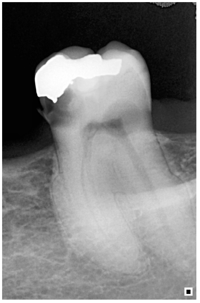
Figure 1. Cervical invasive resorption lesion, through radiograph.
Msf 22 00026 g001
Figure 2. (a) Percentage of respondents who know what tooth resorption is. (b) Percentage of subjects aware of cervical invasive resorption.
Msf 22 00026 g002
Figure 3. (a) Percentage of people, among those who know what an invasive cervical resorption is, who feel comfortable with the diagnosis. (b) This figure shows the number of people who gave all the correct answers, one wrong answer or at least two wrong answers, among the volunteers who consider themselves able to diagnose cervical invasive resorption. (c) This figure shows the percentage of volunteers who think they can diagnose invasive cervical resorption and who feel comfortable treating this external resorption.
Msf 22 00026 g003
Disclaimer/Publisher’s Note: The statements, opinions and data contained in all publications are solely those of the individual author(s) and contributor(s) and not of MDPI and/or the editor(s). MDPI and/or the editor(s) disclaim responsibility for any injury to people or property resulting from any ideas, methods, instructions or products referred to in the content.