Abstract
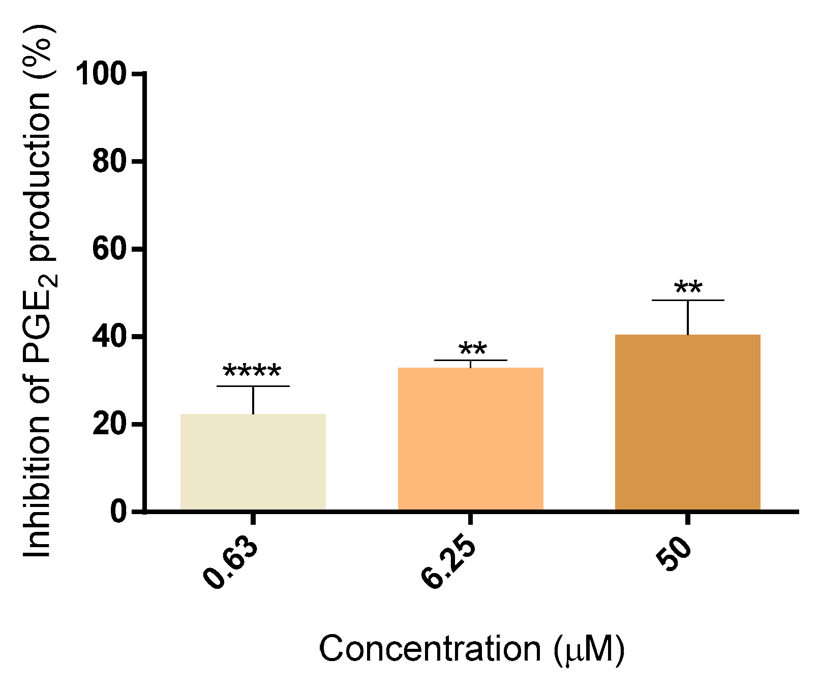
The drugs currently available as cyclooxygenase-2 (COX-2) inhibitors increase the risk of cardiovascular events, which justifies the search for new alternative anti-inflammatory molecules. This work aimed to evaluate the anti-inflammatory activity of the chalcones (polyhydroxylated aromatic compounds): 2′,3,4,4′,6′-pentahydroxychalcone (5OH), 2′,3,4,4′,6′-pentamethoxychalcone (5OMe), and 2′,3,4,4′-tetrahydroxychalcone (butein), at non-cytotoxic concentrations, in an experimental human whole blood model. The results obtained showed that none of the chalcones under study was cytotoxic to human blood cells. From the tested chalcones, butein was the only one showing a concentration-dependent inhibitory production of prostaglandin E2, via COX-2, in human blood (40 ± 8%, at 50 µM).
1. Introduction
Chalcones belong to the flavonoid family and occur naturally in edible plants, such as fruits and vegetables [1]. Chemically, chalcones are 1,3-diaryl-2-propen-1-ones, in which two aromatic rings are linked by a three-carbon α,β-unsaturated carbonyl system. The IUPAC name for chalcone is 1,3-diphenyl-2-propen-1-one. These structures have conjugated double bonds and a pair of fully delocalized π electrons on both benzene rings [2].
The chemical synthesis of chalcones is performed by condensation reactions via acid or basic catalysis. Of the classical reactions for chalcones synthesis, Claisen–Schmidt condensation stands out, due to its experimental simplicity and the highly efficient formation of the carbon-carbon double bond with low restriction to the complexity of the molecules. This reaction owes its name to the pioneering researchers who described the process in which a benzaldehyde and a methyl ketone are condensed in the presence of a catalyst, with the most widely used being a basic catalyst [1].
Chalcones display a large panel of biological activities described in the literature, such as anticancer, antioxidant, antidiabetic, antimicrobial, and anti-inflammatory properties, among many others [2]. Inflammation is a physiological mechanism of the organism in response to an infection or injury and is, therefore, a natural defense process in the human body. It is a complex process involving several signaling pathways. One of the most important pathways, focused on in this work, is the arachidonic acid (AA) cascade. AA exists in the phospholipid membrane of cells and when a stimulus occurs, especially in an inflammatory response, it is released from the cell membrane by the action of enzymes, such as phospholipase A2 or phospholipase C. Cyclooxygenases (COX) are enzymes that exist essentially under two isoforms—COX-1 and COX-2—and are involved in the metabolism of AA. COX-1 is constitutive, i.e., it is always expressed and is responsible for the synthesis of prostaglandins (PG), essential for the body’s homeostasis, for example, gastrointestinal protection and maintenance of renal blood flow. The COX-2 isoform is non-constitutive, i.e., it is hardly expressed under normal physiological conditions, but becomes expressed at high levels when pro-inflammatory stimuli are present. COX-2 is expressed by cells involved in the inflammatory process, such as macrophages, fibroblasts, and endothelial cells [3]. Both COX isoforms synthesize a common precursor: PGH2, which is an unstable substrate and is converted to other PG, such as PGE2 and PGF2, and prostacyclins [4]. PGE2 is an important pro-inflammatory mediator with a role in hyperalgesia, fever, and vascular permeability; inhibition of nitric oxide radical production, through inhibition of inducible nitric oxide synthase; and inhibition of nuclear factor kappa-light-chain-enhancer of activated B cells (NF-kB), a transcription factor (complex 4 protein) that modulates DNA transcription for the expression of pro-inflammatory genes, such as cytokine, chemokine, and adhesion molecule production [5].
The discovery of acetylsalicylic acid more than a century ago marked the beginning of the demand for non-steroidal anti-inflammatory drugs (NSAIDs), which currently top the rankings of best-selling and most widely used drugs on the world market. It was not until many years later that their mechanism of action was discovered: the irreversible inhibition of COX. This triggered the development of drugs that act on these same enzymes. However, the non-selective COX inhibitors have adverse effects, such as gastrointestinal bleeding, which is due to the inhibition of the production of protective PG of the gastric mucosa caused by constitutive COX-1 inhibition in the gastrointestinal tract [6]. This has promoted the search for selective inhibitors of COX-2. Although these drugs do not present so many adverse effects at the gastrointestinal level, they present another major adverse effect: an increased risk of cardiovascular events [7]. This adverse effect justifies the continuous search for new anti-inflammatory molecules that allow for maintaining drug effectiveness while reducing the risk of adverse effects.
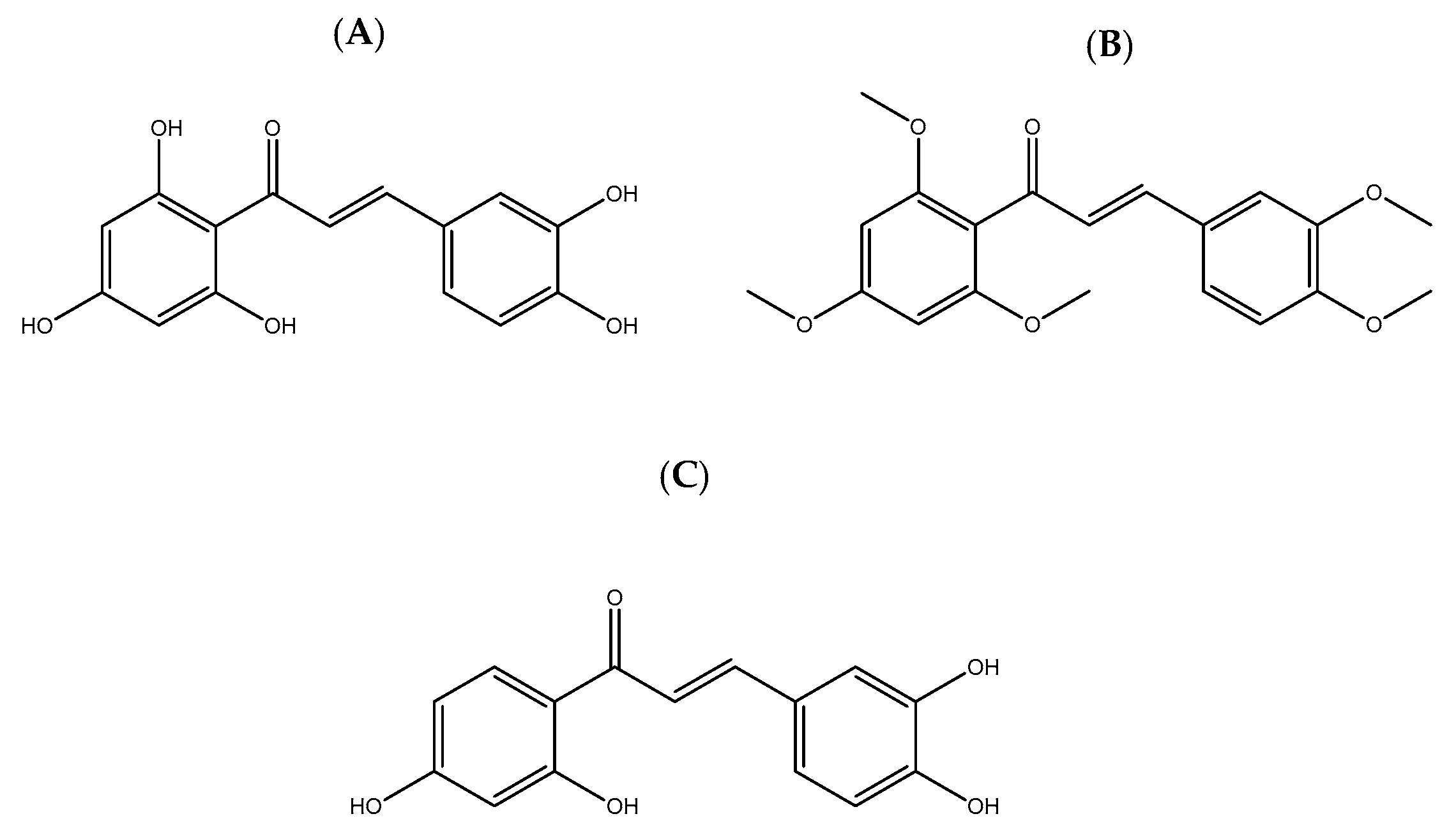
Regarding the anti-inflammatory activity of chalcones, several articles describe the mechanisms of action reflecting this activity, such as the suppression of the activity of COX [3]. The main objective of this work was to evaluate the anti-inflammatory and cytotoxic activity of the chalcones 2′,3,4,4′,6′-pentahydroxychalcone (5OH), 2′,3,4,4′,6′-pentamethoxychalcone (5OMe), and 2′,3,4,4′-tetrahydroxychalcone (butein) (Figure 1).
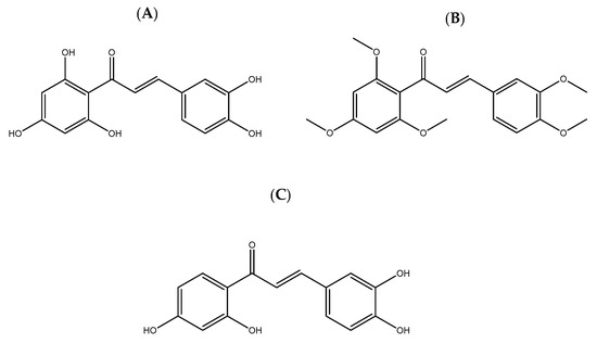
Figure 1.
Chemical structures of the chalcones under study: (A) 2′,3,4,4′,6′-pentahydroxychalcone, (B) 2′,3,4,4′,6′-pentamethoxychalcone, and (C) butein.
2. Material and Methods
To assess the anti-inflammatory activity of chalcones 5OH, 5OMe, and butein, the modulation of PG production in human whole blood was evaluated, as described in the literature [8]. Human blood was donated by healthy volunteers from the Centro Hospitalar Universitário de Santo António after explaining the project and execution of the assay and once the informed consent was obtained. Blood was collected in K3EDTA vacuum tubes after antecubital venipuncture of the donors.
The study of the effect of chalcones (5OH, 5OMe, and butein) on the viability of blood cells was carried out separately for erythrocytes and leukocytes. This study aimed to understand the cytotoxic potential of the compounds under study and what are the maximum innocuous concentrations that could be used in the anti-inflammatory activity assays.
Hemolysis is defined as the rupture of red blood cells resulting in the release of intracellular components into the plasma or serum [9]. Hemoglobin and methemoglobin are erythrocyte components that will be found in the plasma or serum when the erythrocytes are lysed [10]. The method used for this assay completely mimics the conditions of exposure to the chalcones (5OH, 5OMe, and butein) to be studied in the assay for the evaluation of the anti-inflammatory activity. Thus, the incubation was carried out for 5 h. To evaluate the percentage of hemolysis caused by the chalcones under study, the presence of hemoglobin and methemoglobin was measured by reading the absorbance at the wavelengths of maximum absorption of hemoglobin, 540 nm [11], and of methemoglobin, 630 nm [12]. Triton X-100, a non-ionic detergent that causes hemolysis [13], was used as a positive control.
Propidium iodide (PI) is a red fluorescent probe, which allows the distinction of viable cells from necrotic cells [14]. Necrotic cells exhibit disorganization in the phospholipid membrane, which allows PI to enter the cell and consequently intercalate into the DNA, leading to the production of a highly fluorescent cell adduct [15]. Therefore, viable cells, which have an intact cell membrane, keep PI outside, while necrotic cells are permeable to PI. As described previously for erythrocytes, the leukocyte viability assay completely mimics the experimental conditions of the first incubation phase of the assay for the evaluation of the anti-inflammatory capacity. Thus, human blood was incubated for 5 h with the compounds under study. To eliminate possible interferences, a lysis buffer was used that allows the lysis of the erythrocytes without interfering with the viability of the leukocytes. Subsequently, PI was added to mark the necrotic cells and thus show the viability of the leukocytes when exposed to the compounds under study. Flow cytometry was then performed. Fluorescence signals were collected for at least 25,000 cells/sample, where red fluorescence, due to PI staining, was followed on channel 3 (FL3) and presented as a dot plot. The population of leukocytes present was defined and subsequently, the percentage of necrosis was analyzed considering the percentage of PI-labeled leukocytes.
The modulation of PG production in human whole blood was performed in two phases. In the first phase, the blood is incubated in the presence of chalcones, thromboxane synthetase inhibitor to inhibit thromboxane synthetase [16], and acetylsalicylic acid to inhibit COX-1 [17]. Afterwards, lipopolysaccharide (LPS) is added to the prior reaction mixture to induce COX-2 expression in the incubation time of 5 h. At the end of this first part, centrifugation is performed at a low temperature to separate the supernatant, where PGE2 is found. In the second phase, the quantity of PGE2 produced in each sample is quantified in the supernatant using a commercially available enzyme-linked immunosorbent assay (ELISA) kit. The kit contains a 96-well microplate which is coated on the inside with an IgG goat anti-mouse antibody to which a monoclonal antibody to PGE2 will be attached. PGE2 will compete with the conjugate to bind to the antibody, i.e., if there is more PGE2, more will be bound to the antibody, however, if there is more conjugate, there will be more conjugate bound to the antibody. After a 2 h incubation period, the excess reagents are washed away and a substrate is added that will bind to the conjugate, resulting in a yellow-colored product. The enzymatic reaction is stopped after 45 min, and the absorbance is read at 405 nm. Thus, a lower concentration of PGE2 in the sample means more conjugate bound to the antibody, and consequently more substrate bound to the conjugate, resulting in a higher intensity of the yellow color. Therefore, if the chalcones under study inhibit the production of PGE2, there will be a higher absorbance. Celecoxib, a selective COX-2 inhibitor, was used as a positive control.
3. Results
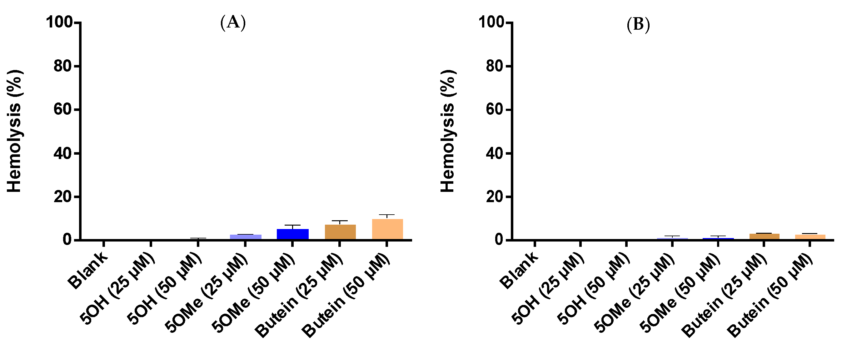
The effect of the chalcones under study on erythrocytes’ viability was evaluated by colorimetric determination of hemoglobin and methemoglobin present in plasma or serum at the time of erythrocyte lysis. Figure 2 shows the hemolysis (in %) induced by 5OH, 5OMe, and butein. By analyzing the graphs, it is possible to observe that hemolysis is in all cases lower than 15%. After statistical analysis of the results, it was found that there is no statistically significant difference between the results obtained for each tested concentration of the chalcones and blank (without chalcones), that is, there was no induction of hemolysis by these compounds. The positive control, Triton X-100, as expected, provoked hemolysis in all tested concentrations.
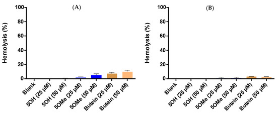
Figure 2.
Hemolysis (%) caused by 5OH, 5OMe, and butein, calculated from the detection of (A) hemoglobin and (B) methemoglobin. Results are expressed as mean ± standard error of the mean (SEM) (n ≥ 3).
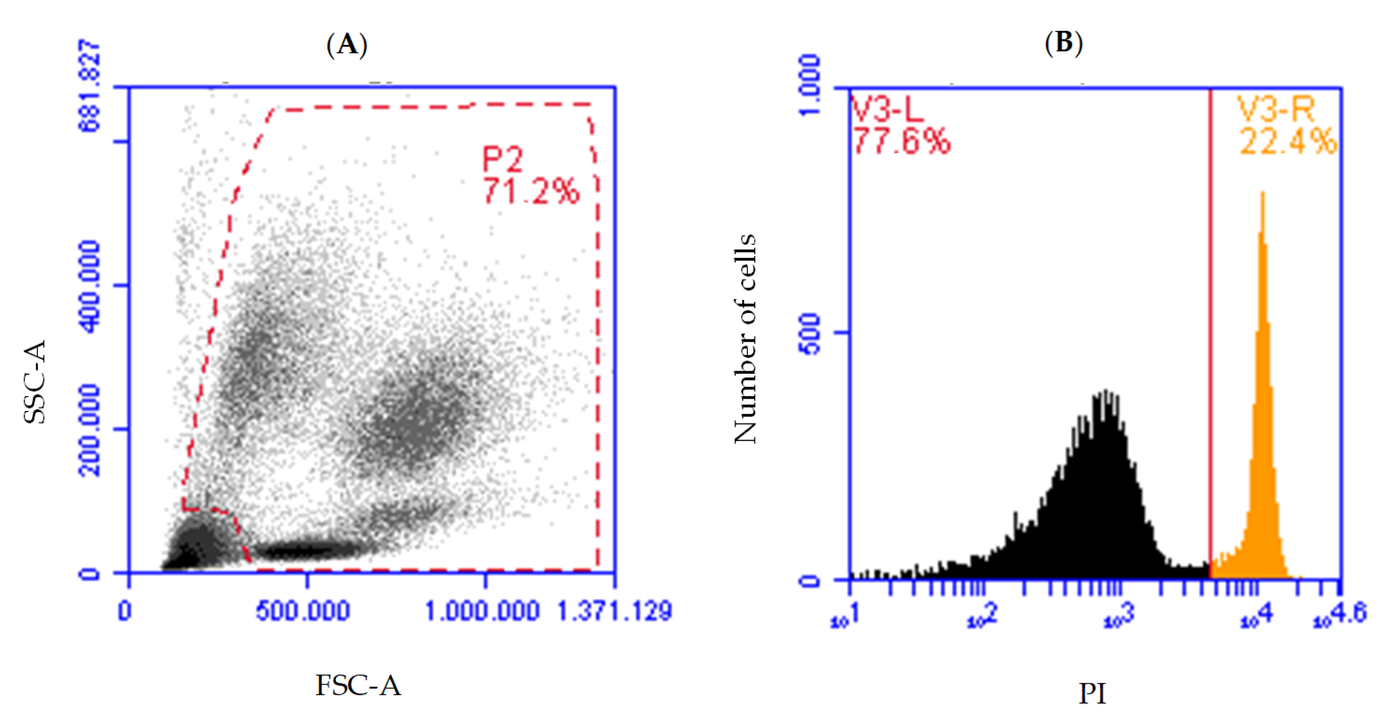
Flow cytometry was used to identify the leukocyte population and its possible variation (in %) caused by the chalcones (Figure 3A) to analyze the fluorescence produced by the intercalation of the PI in the DNA of necrotic leukocytes (Figure 3B).
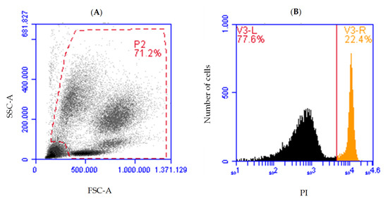
Figure 3.
(A) Dot-plot of forward scatter area (FSC-A)/side scatter area (SSC-A) illustrating the leukocyte population (P2) and (B) histogram illustrating the count of the number of live leukocytes (black) and the necrotic leukocytes, propidium iodide (PI) staining (orange).
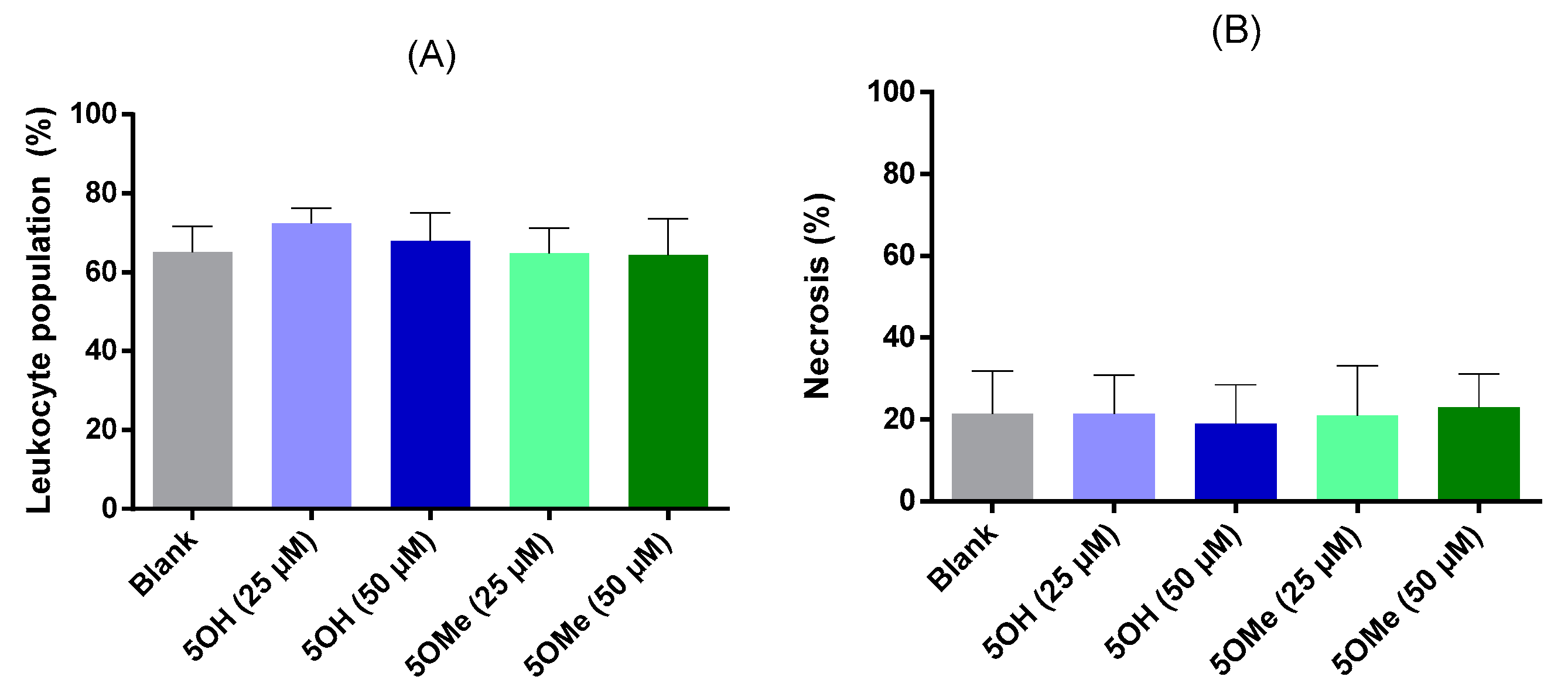
Figure 4A shows the mean values of the defined leukocyte population and Figure 4B shows the percentage of necrosis obtained in this leukocyte population. Figure 4A indicates that the percentage of leukocytes in the blood sample is constant and is always above 60%, which is to be expected, considering the technique used. Figure 4B displays the percentage of necrotic leukocytes in the presence of chalcones under study and in the blank (without chalcones), which is less than 22%. From the statistical analysis of Figure 4B, it is possible to conclude that there is no statistically significant difference between the results obtained for the blank and the chalcones, which shows that, at the concentrations tested, these chalcones are not cytotoxic to leucocytes.
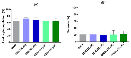
Figure 4.
Effect of chalcones 5OH and 5OMe on (A) leukocyte population and (B) percentage of leukocyte necrosis. Results are expressed as mean ± standard error of the mean (SEM) (n ≥ 3).
The potential anti-inflammatory activity was studied by evaluating the effect of the chalcones under study on the production of PGE2, via COX-2. Figure 5 shows the results obtained for the most active chalcone, butein. Under the present experimental conditions, 5OH and 5OMe were not active. Butein was the only tested chalcone that seems to have concentration-dependent activity and showed a maximum inhibition of 40 ± 8%, at 50 μM. The tested positive control, celecoxib, showed a percentage of inhibition of PGE2 production of 83 ± 7%, at a concentration of 5 μM.
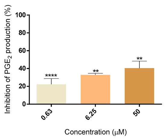
Figure 5.
Percentage of inhibition of PGE2 production of butein (0.63–50 µM). The results are expressed as the mean ± standard error of the mean (SEM) (n ≥ 3). ** p < 0.01, **** p < 0.0001, compared with the blank.
4. Discussion
In this work, the COX-2 inhibitory activity of chalcones 5OH, 5OMe, and butein was evaluated. In the first phase, assays were performed to understand whether the chalcones under study were cytotoxic and which were the maximum innocuous concentrations that could be used in subsequent anti-inflammatory assays. To study the viability of erythrocytes when exposed to the studied chalcones, the percentage of hemolysis was evaluated through the quantification of hemoglobin and methemoglobin. The results showed that none of the chalcones, at any of the concentrations tested, induced hemolysis. In the same way, to study the viability of leucocytes when exposed to the studied chalcones, the percentage of necrosis was evaluated using the PI fluorescent probe. The cytotoxic potential of butein on leukocytes was not assessed, as its innocuousness in cell models has been well-described in the literature, namely for the concentrations currently tested [15,18]. The results showed that none of the chalcones, at any of the concentrations tested, caused necrosis of blood leukocytes. Thus, it was concluded that it was possible to use the highest tested concentration of each compound (50 µM) for the study of PGE2 production, via COX-2, as none of the chalcones under study is cytotoxic. Finally, the evaluation of the potential anti-inflammatory capacity of the chalcones 5OH, 5OMe, and butein was assessed by studying the modulation of PGE2 production, via COX-2, in human whole blood. By analyzing the results, butein is the most active of the evaluated chalcones, since at 50 µM, it presents an inhibition of PGE2 production of 40 ± 8%. As for 5OH and 5OMe, they did not show activity under the present experimental conditions. Butein, identified here as active, still appears to show an activity lower than celecoxib. As far as is known at the time of the completion of this report, there are no studies about the effect of 5OH, 5OMe, and butein in modulating PG production in the blood model and experimental conditions used. However, butein presents a greater relevance in the literature as a compound of interest for the modulation of several diseases related to the inflammatory process, and its anti-inflammatory activity has been described. A study by Lee et al. [15] evaluated the neuroprotective and anti-neuroinflammatory capacity of butein through the evaluation of the production of PGE2 in microglia cells (immune cells of the central nervous system) stimulated with LPS. Additionally, as in the present work, the anti-inflammatory capacity was evaluated by the quantification of PGE2 through an ELISA kit. The obtained results showed that the production of PGE2 decreased, as well as the expression of COX-2, in a concentration-dependent manner. Thus, these results are in line with the anti-inflammatory potential of butein.
5. Conclusions
The high overall consumption of NSAIDs reveals the need to invest in new small molecules with anti-inflammatory capacity as a way of improving efficacy and reducing the adverse effects of the drugs currently available on the market. Chalcones emerge as compounds with high therapeutic potential, being already described as possessing various biological activities, namely anti-inflammatory properties. In this sense, the present study aimed to evaluate, for the first time to the best of our knowledge, the anti-inflammatory potential of three different chalcones, 5OH, 5OMe, and butein, in whole human blood. Butein showed the best inhibition of PGE2 production. In conclusion, this chalcone seems to possess the most promising structural characteristics for the development of new compounds with anti-inflammatory activity, focused on the selectivity towards COX-2.
Author Contributions
Conceptualization, D.R. and E.F.; methodology, A.V. and M.L.; validation, D.R. and E.F.; investigation, A.V. and M.L.; writing—original draft preparation, A.V.; writing—review and editing, A.V., M.L., D.R. and E.F.; supervision, D.R. and E.F.; project administration, E.F. and D.R.; funding acquisition, E.F. and D.R. All authors have read and agreed to the published version of the manuscript.
Funding
This work received financial support from PT national funds (FCT/MCTES, Fundação para a Ciência e Tecnologia and Ministério da Ciência, Tecnologia e Ensino Superior) through the projects UIDB/50006/2020 and UIDP/50006/2020.
Institutional Review Board Statement
The study was conducted following the Declaration of Helsinki and approved by the Ethics Committee of Centro Hospitalar Univesitário de Santo António/Instituto de Ciências Biomédicas Abel Salazar, Oporto, Portugal.
Informed Consent Statement
Informed consent was obtained from all subjects involved in the study.
Data Availability Statement
The data presented in this study are available on request from the corresponding authors.
Acknowledgments
Mariana Lucas thanks FCT/MCTES and ESF (European Social Fund) through NORTE 2020 (Programa Operacional Região Norte) for her PhD grant (ref. 2021.06746.BD).
Conflicts of Interest
The authors declare no conflict of interest.
References
- Zhuang, C.; Zhang, W.; Sheng, C.; Zhang, W.; Xing, C.; Miao, Z. Chalcone: A Privileged Structure in Medicinal Chemistry. Chem. Rev. 2017, 117, 7762–7810. [Google Scholar] [CrossRef] [PubMed]
- Rudrapal, M.; Khan, J.; Bin Dukhyil, A.A.; Alarousy, R.M.I.I.; Attah, E.I.; Sharma, T.; Khairnar, S.J.; Bendale, A.R. Chalcone Scaffolds, Bioprecursors of Flavonoids: Chemistry, Bioactivities, and Pharmacokinetics. Molecules 2021, 26, 7177. [Google Scholar] [CrossRef] [PubMed]
- Wang, T.; Fu, X.; Chen, Q.; Patra, J.K.; Wang, D.; Wang, Z.; Gai, Z. Arachidonic Acid Metabolism and Kidney Inflammation. Int. J. Mol. Sci. 2019, 20, 3683. [Google Scholar] [CrossRef] [PubMed]
- Miller, S.B. Prostaglandins in Health and Disease: An Overview. Semin. Arthritis Rheum. 2006, 36, 37–49. [Google Scholar] [CrossRef] [PubMed]
- Rashid, H.U.; Xu, Y.; Ahmad, N.; Muhammad, Y.; Wang, L. Promising anti-inflammatory effects of chalcones via inhibition of cyclooxygenase, prostaglandin E2, inducible NO synthase and nuclear factor κb activities. Bioorg. Chem. 2019, 87, 335–365. [Google Scholar] [CrossRef] [PubMed]
- Everts, B.; Währborg, P.; Hedner, T. COX-2 specific inhibitors—The emergence of a new class of analgesic and anti-inflammatory drugs. Clin. Rheumatol. 2000, 19, 331–343. [Google Scholar] [CrossRef]
- Borer, J.S.; Simon, L.S. Cardiovascular and gastrointestinal effects of COX-2 inhibitors and NSAIDs: Achieving a balance. Thromb. Haemost. 2005, 7 (Suppl. S4), S14–S22. [Google Scholar] [CrossRef]
- Laufer, S.; Greim, C.; Luik, S.; Ayoub, S.S.; Dehner, F. Human whole blood assay for rapid and routine testing of non-steroidal anti-inflammatory drugs (NSAIDs) on cyclo-oxygenase-2 activity. Inflammopharmacology 2008, 16, 155–161. [Google Scholar] [CrossRef] [PubMed]
- Heireman, L.; Van Geel, P.; Musger, L.; Heylen, E.; Uyttenbroeck, W.; Mahieu, B. Causes, consequences and management of sample hemolysis in the clinical laboratory. Clin. Biochem. 2017, 50, 1317–1322. [Google Scholar] [CrossRef] [PubMed]
- Scholkmann, F.; Restin, T.; Ferrari, M.; Quaresima, V. The Role of Methemoglobin and Carboxyhemoglobin in COVID-19: A Review. J. Clin. Med. 2020, 10, 50. [Google Scholar] [CrossRef] [PubMed]
- Sabino, R.M.; Kauk, K.; Movafaghi, S.; Kota, A.; Popat, K.C. Interaction of blood plasma proteins with superhemophobic titania nanotube surfaces. Nanomed. Nanotechnol. Biol. Med. 2019, 21, 102046. [Google Scholar] [CrossRef] [PubMed]
- Lee, J.; El-Abaddi, N.; Duke, A.; Cerussi, A.E.; Brenner, M.; Tromberg, B.J. Noninvasive in vivo monitoring of methemoglobin formation and reduction with broadband diffuse optical spectroscopy. J. Appl. Physiol. 2006, 100, 615–622. [Google Scholar] [CrossRef] [PubMed]
- Rodi, P.; Gianello, M.B.; Corregido, M.; Gennaro, A. Comparative study of the interaction of CHAPS and Triton X-100 with the erythrocyte membrane. Biochim. Biophys. Acta (BBA)-Biomembr. 2014, 1838, 859–866. [Google Scholar] [CrossRef] [PubMed]
- D’Arcy, M.S. Cell death: A review of the major forms of apoptosis, necrosis and autophagy. Cell Biol. Int. 2019, 43, 582–592. [Google Scholar] [CrossRef] [PubMed]
- Lee, D.S.; Jeong, G.S. Butein provides neuroprotective and anti-neuroinflammatory effects through Nrf2/ARE-dependent haem oxygenase 1 expression by activating the PI3K/Akt pathway. Br. J. Pharmacol. 2016, 173, 2894–2909. [Google Scholar] [CrossRef] [PubMed]
- Kato, K.; Ohkawa, S.; Terao, S.; Terashita, Z.; Nishikawa, K. Thromboxane synthetase inhibitors (TXSI). Design, synthesis, and evaluation of a novel series of ω-pyridylalkenoic acids. J. Med. Chem. 1985, 28, 287–294. [Google Scholar] [CrossRef] [PubMed]
- Patrono, C.; Rocca, B. Aspirin and Other COX-1 Inhibitors. In Handbook of Experimental Pharmacology; Springer: Berlin/Heidelberg, Germany, 2012; pp. 137–164. [Google Scholar] [CrossRef]
- Zheng, W.; Zhang, H.; Jin, Y.; Wang, Q.; Chen, L.; Feng, Z.; Chen, H.; Wu, Y. Butein inhibits IL-1β-induced inflammatory response in human osteoarthritis chondrocytes and slows the progression of osteoarthritis in mice. Int. Immunopharmacol. 2017, 42, 1–10. [Google Scholar] [CrossRef] [PubMed]
Disclaimer/Publisher’s Note: The statements, opinions and data contained in all publications are solely those of the individual author(s) and contributor(s) and not of MDPI and/or the editor(s). MDPI and/or the editor(s) disclaim responsibility for any injury to people or property resulting from any ideas, methods, instructions or products referred to in the content. |
© 2023 by the authors. Licensee MDPI, Basel, Switzerland. This article is an open access article distributed under the terms and conditions of the Creative Commons Attribution (CC BY) license (https://creativecommons.org/licenses/by/4.0/).

