Proceeding Paper

Photodynamic Inactivation of Phage Phi6 as SARS-CoV-2 Model in Wastewater Disinfection: Effectivity and Safety †

by
Maria Bartolomeu
1,
Cátia Vieira
1,
Marta Gomes
1,
Ana T. P. C. Gomes
2,
Maria Amparo F. Faustino
3,*,
Maria Graça P. M. S. Neves
3 and
Adelaide Almeida
1,*
1
Department of Biology and CESAM, University of Aveiro, 3810-193 Aveiro, Portugal
2
Center for Interdisciplinary Investigation (CIIS), Faculty of Dental Medicine, Universidade Católica Portuguesa, 3504-505 Viseu, Portugal
3
Department of Chemistry and LAQV-REQUIMTE, University of Aveiro, 3810-193 Aveiro, Portugal
*
Authors to whom correspondence should be addressed.
Presented at the 2nd International Electronic Conference on Antibiotics—Drugs for Superbugs: Antibiotic Discovery, Modes of Action and Mechanisms of Resistance, 15–30 June 2022; Available online: https://eca2022.sciforum.net/.
Med. Sci. Forum 2022, 12(1), 9; https://doi.org/10.3390/eca2022-12707
Published: 15 June 2022

Abstract

:
The past 2 years have been marked by the severe acute respiratory syndrome coronavirus 2 (SARS-CoV-2) pandemic. This virus is found in the intestinal tract and reaches the wastewater system, and, consequently, the natural receiving water bodies, and inappropriate or/and inefficient WW treatment is a means of contamination. In the present work, we used a SARS-CoV-2 model—the phage Phi6—to evaluate its survival under different environmental conditions (pH, temperature, salinity, solar, and UV-B irradiation). Then, we tested the efficiency of photodynamic inactivation (PDI) as a WW disinfection alternative method, and, additionally, the impact on the cultivable native marine microorganisms of the PDI-treated WW was evaluated.

1. Introduction

Severe acute respiratory syndrome coronavirus 2 (SARS-CoV-2), the causative agent of the COVID-19 pandemic, infects the gastrointestinal tract via the angiotensin-converting enzyme (ACE2) receptor that is expressed by epithelial cells in the gastrointestinal system [1]. Although no cases have yet been described in the literature, the possibility of SARS-CoV-2 transmission via the fecal-oral route is still being discussed, as well as the negative impact that its release via wastewater could have [2,3].
Generally, the wastewater (WW) suffers a secondary stage of treatment in the wastewater treatment plants (WWTPs) where organic matter is subtracted before being released into the environment, though still containing high concentrations of microorganisms, including viruses [4,5]. As SARS-CoV-2 is present in the intestinal tract, the virions are excreted through the feces of infected persons, with the RNA of this virus already being detected in the WW of different countries around the world [6,7,8,9,10]. When applied, the traditional tertiary disinfection treatments can reduce the content of the viruses from WW, but they also bring disadvantages namely being expensive, toxic to aquatic organisms, and inducing genetic damages in microorganisms [2,11]. Due to safety and low-laboratory-cost, bacteriophage Phi6 (or phage Phi6) was used as a SARS-CoV-2 surrogate. The similar structures of both viruses, such as the presence of a lipidic envelope, and their similar behavior under particular environmental conditions, make phage Phi6 a suitable surrogate for SARS-CoV-2 [12,13,14,15]. The persistence of Phi6 over time in different environmental conditions of temperature, pH, salinity, and solar and UV-B irradiation was evaluated to mimic environmental conditions and evaluate its prevalence under different conditions.
This study aims to explore the hypothesis of photodynamic inactivation (PDI) use as an alternative disinfection method to be applied in WW. PDI has already been shown to be an effective approach in the inactivation of both Gram-positive and Gram-negative bacteria, fungi, parasites, and viruses [16,17,18,19]. This approach combines the action of a photosensitizer (PS), a light source with adequate wavelength, and molecular oxygen, generating highly reactive oxygen species (ROS) capable of causing irreversible damage to vital constituents of the microbial entities.
In the present work, PDI was tested using the tetracationic porphyrin 5,10,15,20-tetrakis(1-methylpyridinium-4-yl)porphyrin tetra-iodide (TMPyP) recognized by its efficiency against different types of viruses [18,20]. The viability of the cultivable native marine water microorganisms was evaluated when exposed to the PDI-treated WW, in order to assess possible toxic effects in the microbial community of the natural receiving waters of treated WW.

2. Materials and Methods

Pseudomonas sp. (DSM 21482) was used as the bacterial host of phage Phi6. Bacterial cells glycerol stocks were stored at −80 °C. Before each biological experiment, a frozen stock was aseptically inoculated into 30 mL of tryptic soy broth (TSB) medium and incubated at 25 °C overnight, until the stationary phase of growth was reached. Phage Phi6 stocks were prepared and maintained in SM buffer, with a phage titer of 109 plaque-forming units per mL (PFU/mL).
Wastewater samples were collected from a WWTP located in the littoral center of Portugal after receiving the secondary stage of treatment, on different days. After collection, the samples were processed by filtration to the removal of native microbial entities and kept refrigerated at 4 °C until further use.
The persistence of phage Phi6 was evaluated in WW under different conditions of temperature (17, 25, and 37 °C, in an incubating chamber and protected from light), pH (6.0, 8.0, and 9.0, at the temperature of 17 °C and protected from light; pH desired values were adjusted by addition of acidic/basic solutions of HCl/NaOH), salinity (15 and 34 g/kg, at the temperature of 17 °C and protected from light), and solar and UV-B irradiation (UV-B radiation was given by UV-B lamp, 280–320 nm; solar irradiation ranged between 46.2 and 91.1 mW/cm2 during the experiments). To assess the effects of the selected conditions of temperature, pH, and salinity, aliquots were collected every day during the first week, followed by once-a-week collections until the end of the experiments; when assessing the effects of UV-B and solar irradiation, aliquots were collected after 0, 2, 4, 5, 8, 10, and 12 h, and after 0, 2, 4, and 6 h, respectively. Along all the experiments, appropriate controls were performed in parallel.
PDI experiments were performed, in WW-collected samples, using the tetracationic porphyrin TMPyP at the concentration of 5.0 μM under a LED system adjusted to an irradiance of 50 mW/cm2. Before irradiation, 10 min of dark incubation with stirring was performed to allow the homogeneity of the suspension and the interaction of the PS molecules with the viral particles. During the experiments, aliquots were taken after 0, 5, 10, 15, and 30 min, serially diluted in isotonic phosphate-buffered saline (PBS) solution and drop-plated in Petri dishes previously prepared with tryptic soy agar (TSA) medium and a top layer of TSB 0.6% agar with the phage host Pseudomonas sp. For the phage Phi6 survival monitoring, suitable controls (dark control: phage Phi6 in the presence of the TMPyP, kept in the dark; light control: phage Phi6 irradiated under the same irradiance of the samples) were performed along the PDI samples.
To evaluate the effect of the PDI-treated WW on native marine water microorganisms, samples of coastal marine water were collected in the littoral center of Portugal on the day of the experiments, and the cultivable microorganisms were quantified by platting in plate count agar (PCA) medium. PDI experiments were performed in WW as previously described but without the addition of any biological entity into the samples (TMPyP was used at a concentration of 5.0 μM under light irradiation with an irradiance of 50 mW/cm2 for 30 min). Determined volumes of PDI-treated WW and suitable light and dark controls were added to the marine water samples to achieve the following dilutions 1:2, 1:10, 1:100, and 1:1000. The samples were maintained at 17 °C, and the samples were incubated in the dark or under white irradiation (50 mW/cm2) for 24 h, with aliquots being collected after 0, 6, and 24 h.
For each experiment, at least three independent assays were performed, in duplicate. The results were analyzed, and ANOVA (α 0.05) and Tukey’s multiple comparisons test were applied to assess the significance of the differences among the tested conditions (GraphPad Prism software).

3. Results

The viability of the phage Phi6 showed to be temperature dependent (Figure 1), with the highest decrease being observed during the experiments performed at 37 °C, where the phage viability decreased to the detection limit of the method (2 log PFU/mL) after 24 h (Figure 1a), followed by the experiments where the temperature was maintained at 25 °C (detection limit of the method reached after 35 days) (Figure 1b) and 17 °C (detection limit reached only after 84 days) (Figure 1c). Considering that the persistence of the phage Phi6 was longer at 17 °C, the subsequent experiments were performed at this controlled temperature.
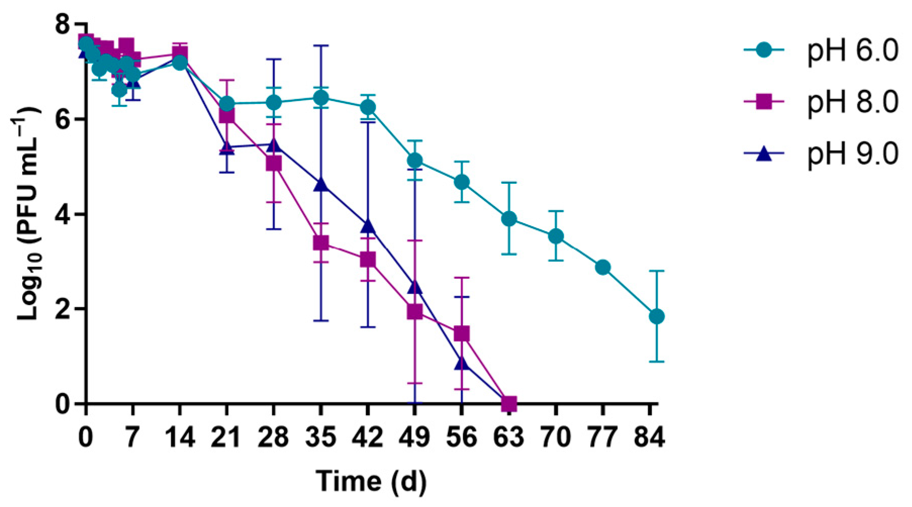
The viability of phage Phi6 at pH values (Figure 2) of 8.0 and 9.0 was similar, with the phage content reaching the detection limit after 63 days. At pH 6.0, the viability of the phage Phi6 was maintained for a longer period, reaching a decrease of 5.7 log PFU/mL after 84 days but not reaching the detection limit of the method.
Under different salinity conditions (Figure 3), a decrease in the phage viability of 7.3 log PFU/mL was reached after 49 days in the conditions of salinity of 34 g/kg, and at the salinity of 15 g/kg a decrease of 5.7 log PFU/mL was reached after 84 days (similar to the control, WW samples without salt addition).
The experiments of phage Phi6 viability under irradiation (Figure 4) showed that when exposed to UV-B irradiation the viability of the phage decreased similarly in WW and PBS, reaching the detection limit after 12 h (Figure 4a). When exposed to solar irradiation, the viability of the phage decreased sooner; after 4 h the detection limit of the method was reached in WW, and a decrease of 2.7 log PFU/mL was reached after 6 h (Figure 4b).
The results obtained in the PDI experiments in PBS and WW with TMPyP at a concentration of 5.0 μM and at an irradiance of 50 mW/cm2 with white light are shown in Figure 5. The PDI assays in PBS reached the detection limit of the method for phage content after 10 min of treatment (>8.0 log PFU/mL) (Figure 5a). When performed in WW, the detection limit was reached after 5 min of treatment (Figure 5b).
When the toxicity effect of PDI-treated WW on the cultivable native marine water microorganisms was tested, the results showed that only when the native microorganisms were exposed to PDI-treated WW at a ratio of 1:2 and subjected to a 50 mW/cm2 white light irradiation during 24 h the survival was affected (Figure 6c). However, at dilution ratios ≥ 1:10, no negative effect was observed. The experiments where the native microorganisms where exposed to PDI-treated WW and kept in the dark for 24 h, no toxic effects were detected (Figure 6d).

4. Discussion and Conclusions

Some studies have already shown that temperature can affect the viability of viruses [21,22]. In this study, three different temperature values (17, 25, and 37 °C) were selected, considering that 17 °C is the closest temperature to the annual average temperature of seawater in central coastal Portugal, where WW is released after treatment on the WWTPs. The obtained results showed that the virus remains viable for a longer period (up to 84 days, 3 months) at lower temperatures (17 °C) (Figure 1).
Also, pH is shown to be an important factor to influence the phage survival [23,24], thus, in this study pH values ranging from 6.0–9.0 (comprehending the pH interval values of 6.5–8.5 for treated effluent to be allowed to be discharged into the environment, and the global average ocean’s surface pH of about 8.1 [25,26]) were used. The results showed that the virus remained active for up to 63 days in WW with pH values of 8.0 and 9.0, and for more than 84 days when the WW pH was adjusted to 6.0 (Figure 2).
Considering that in the region where the WW is released, marine water is near a brackish water lagoon, the environmental conditions of salinity were also mimicked, adjusting the WW salinity to 15 and 34 g/kg. The results showed that, although the higher salinity environment had a higher impact on the phage viability, the virus remained viable for 49 days (Figure 3).
UV radiation (artificial and from solar light) also has a recognized impact on the viruses’ viability and their infectivity capacity, through protein degradation and nucleic acids damage [13,27]. Among the solar radiation, UV-B (280–320 nm) is the most significant in the microorganisms’ inactivation. Hence, we used both solar and UV-B radiation to access the viability of phage particles in both PBS and WW. Under solar radiation, the viability of the phage particles had a decrease of 2.6 log PFU/mL after 6 h in PBS, and a decrease of 7.6 log PFU/mL after 4 h in WW, indicating the presence of certain compounds in the WW samples acting as PS and generating ROS, as dissolved organic matter, antibiotics, nitrates, etc. [28], capable of inactivating the virus particles (Figure 4).
Analyzing the obtained results regarding the phage viability in the different environmental conditions tested, it is reiterated the need for an effective viral inactivation treatment in WW before its release into the environment.
The PDI experiments, with the tetracationic porphyrin TMPyP at the micromolar range (5.0 μM), in PBS and WW demonstrated the effectiveness of the process in the inactivation of the viral particles: a viral load of >8.0 log PFU/mL was photoinactivated after just 10 min in both aqueous matrices (PBS and WW) (Figure 5).
Although some of the traditional disinfection WW methods are also able to achieve complete viral inactivation, some of them like chlorine-based agents lead to the formation of toxic sub-products. UV irradiation disinfection treatment has shown to be less effective than chlorination, and its effectiveness is highly dependent on the external structures of the viral particles. Contrarily to the conventional methods applied in WW disinfection, the PDI approach has shown to be effective in the viral inactivation without producing toxic photoproducts to the marine water native microorganisms under realistic dilution ratios of WW when released into the environment (Figure 6).
In summary, this study allows concluding that the viral particles of the phage Phi6, a suitable SARS-CoV-2 surrogate [13,14,15], were effectively inactivated under PDI treatment, showing the potential value of this method as an innovative WW disinfection approach before the release of the WW into the environment, where the viability experiments showed that the viral particles would remain viable for a considerable amount of time if not inactivated during WW treatment processes. It is important to note that although phage Phi6 is an appropriate surrogate of SARS-CoV-2, further studies are needed with the COVID-19-causing agent. Also, the toxicity experiments showed that under realistic dilution rates, the PDI-treated WW did not affect the viability of the marine water native microorganisms.

Author Contributions

Conceptualization, M.B., A.T.P.C.G., M.A.F.F., M.G.P.M.S.N. and A.A.; methodology, M.B. and M.G.; validation, M.B., M.G., A.T.P.C.G., M.A.F.F., M.G.P.M.S.N. and A.A.; formal analysis, M.B., M.G., A.T.P.C.G., M.A.F.F., M.G.P.M.S.N. and A.A.; investigation, M.B., C.V., M.G., A.T.P.C.G., M.A.F.F., M.G.P.M.S.N., and A.A.; resources, M.A.F.F., M.G.P.M.S.N. and A.A.; data curation, M.B., C.V., M.G., A.T.P.C.G., M.A.F.F., M.G.P.M.S.N. and A.A.; writing—original draft preparation, M.B., M.G., A.T.P.C.G., M.A.F.F., M.G.P.M.S.N. and A.A.; writing—review and editing, M.B., C.V., M.G., A.T.P.C.G., M.A.F.F., M.G.P.M.S.N. and A.A.; supervision, A.T.P.C.G., M.A.F.F., M.G.P.M.S.N. and A.A.; funding acquisition, M.A.F.F., M.G.P.M.S.N. and A.A. All authors have read and agreed to the published version of the manuscript.

Funding

This research was funded by FCT/MCT through financial support for the LAQV-REQUIMTE (UIDB/50006/2020) and CESAM (UID/AMB/50017/2019 and UIDB/50017/2020 + UIDP/50017/2020) research units and for the FCT projects (PREVINE, PTDC/ASP-PES/29576/2017) through national funds (OE), and, where applicable, it was co-financed by the FEDER—Operational Thematic Program for Competitiveness and Internationalization—COMPETE 2020 within the PT2020 Partnership Agreement and the Portuguese NMR Network. The doctoral grants of M.B. (SFRH/BD/121645/2016) and C.V. (SFRH/BD/150358/2019) are funded by FCT.

Institutional Review Board Statement

Not applicable.

Informed Consent Statement

Not applicable.

Acknowledgments

Thanks are due to the University of Aveiro and FCT/MEC for the financial support to LAQV-REQUIMTE and CESAM research units, where the experimental work was carried out, and to the FCT project PREVINE (PTDC/ASP-PES/29576/2017). M.B. and C.V. also thank FCT for the conceded doctoral grants.

Conflicts of Interest

The authors declare no conflict of interest.

References

  1. Xiao, F.; Tang, M.; Zheng, X.; Liu, Y.; Li, X.; Shan, H. Evidence for Gastrointestinal Infection of SARS-CoV-2. Gastroenterology 2020, 158, 1831–1833. [Google Scholar] [CrossRef] [PubMed]
  2. Sobsey, M.D. Absence of virological and epidemiological evidence that SARS-CoV-2 poses COVID-19 risks from environmental fecal waste, wastewater and water exposures. J. Water Health 2022, 20, 126–138. [Google Scholar] [CrossRef]
  3. Westhaus, S.; Weber, F.A.; Schiwy, S.; Linnemann, V.; Brinkmann, M.; Widera, M.; Greve, C.; Janke, A.; Hollert, H.; Wintgens, T.; et al. Detection of SARS-CoV-2 in raw and treated wastewater in Germany—Suitability for COVID-19 surveillance and potential transmission risks. Sci. Total Environ. 2021, 751, 141750. [Google Scholar] [CrossRef] [PubMed]
  4. Bartolomeu, M.; Reis, S.; Fontes, M.; Neves, M.G.P.M.S.; Faustino, M.A.F.; Almeida, A. Photodynamic Action against Wastewater Microorganisms and Chemical Pollutants: An Effective Approach with Low Environmental Impact. Water 2017, 9, 630. [Google Scholar] [CrossRef]
  5. Usman, M.; Farooq, M.; Hanna, K. Existence of SARS-CoV-2 in Wastewater: Implications for Its Environmental Transmission in Developing Communities. Environ. Sci. Technol. 2020, 54, 7758–7759. [Google Scholar] [CrossRef] [PubMed]
  6. Medema, G.; Heijnen, L.; Elsinga, G.; Italiaander, R.; Brouwer, A. Presence of SARS-Coronavirus-2 RNA in Sewage and Correlation with Reported COVID-19 Prevalence in the Early Stage of the Epidemic in The Netherlands. Environ. Sci. Technol. Lett. 2020, 7, 511–516. [Google Scholar] [CrossRef]
  7. Zhang, D.; Ling, H.; Huang, X.; Li, J.; Li, W.; Yi, C.; Zhang, T.; Jiang, Y.; He, Y.; Deng, S.; et al. Potential spreading risks and disinfection challenges of medical wastewater by the presence of Severe Acute Respiratory Syndrome Coronavirus 2 (SARS-CoV-2) viral RNA in septic tanks of Fangcang Hospital. Sci. Total Environ. 2020, 741, 140445. [Google Scholar] [CrossRef] [PubMed]
  8. Lodder, W.; de Roda Husman, A.M. SARS-CoV-2 in wastewater: Potential health risk, but also data source. Lancet Gastroenterol. Hepatol. 2020, 5, 533–534. [Google Scholar] [CrossRef]
  9. Wurtzer, S.; Marechal, V.; Mouchel, J.M.; Maday, Y.; Teyssou, R.; Richard, E.; Almayrac, J.L.; Moulin, L. Evaluation of lockdown impact on SARS-CoV-2 dynamics through viral genome quantification in waste water, Greater Paris, France, 5 March to 23 April 2020. Eurosurveillance 2020, 25, 2000776. [Google Scholar] [CrossRef]
  10. Ahmed, W.; Angel, N.; Edson, J.; Bibby, K.; Bivins, A.; O’Brien, J.W.; Choi, P.M.; Kitajima, M.; Simpson, S.L.; Li, J.; et al. First confirmed detection of SARS-CoV-2 in untreated wastewater in Australia: A proof of concept for the wastewater surveillance of COVID-19 in the community. Sci. Total Environ. 2020, 728, 138764. [Google Scholar] [CrossRef]
  11. Rasero, F.J.R.; Ruano, L.A.M.; Del Real, P.R.; Gómez, L.C.; Lorusso, N. Associations between SARS-CoV-2 RNA concentrations in wastewater and COVID-19 rates in days after sampling in small urban areas of Seville: A time series study. Sci. Total Environ. 2022, 806, 150573. [Google Scholar] [CrossRef]
  12. Fedorenko, A.; Grinberg, M.; Orevi, T.; Kashtan, N. Survival of the enveloped bacteriophage Phi6 (a surrogate for SARS-CoV-2) in evaporated saliva microdroplets deposited on glass surfaces. Sci. Rep. 2020, 10, 1–10. [Google Scholar] [CrossRef] [PubMed]
  13. Aquino de Carvalho, N.; Stachler, E.N.; Cimabue, N.; Bibby, K. Evaluation of Phi6 Persistence and Suitability as an Enveloped Virus Surrogate. Environ. Sci. Technol. 2017, 51, 8692–8700. [Google Scholar] [CrossRef]
  14. Silverman, A.I.; Boehm, A.B. Systematic Review and Meta-Analysis of the Persistence and Disinfection of Human Coronaviruses and Their Viral Surrogates in Water and Wastewater. Environ. Sci. Technol. Lett. 2020, 7, 544–553. [Google Scholar] [CrossRef]
  15. Whitworth, C.; Mu, Y.; Houston, H.; Martinez-Smith, M.; Noble-Wang, J.; Coulliette-Salmond, A.; Rose, L. Persistence of bacteriophage phi 6 on porous and nonporous surfaces and the potential for its use as an ebola virus or coronavirus surrogate. Appl. Environ. Microbiol. 2020, 86, 1–11. [Google Scholar] [CrossRef] [PubMed]
  16. Costa, L.; Tomé, J.P.C.; Neves, M.G.P.M.S.; Tomé, A.C.; Cavaleiro, J.A.S.; Faustino, M.A.F.; Cunha, Â.; Gomes, N.C.; Almeida, A. Evaluation of resistance development and viability recovery by a non-enveloped virus after repeated cycles of aPDT. Antiviral. Res. 2011, 91, 278–282. [Google Scholar] [CrossRef]
  17. Kasimova, K.R.; Sadasivam, M.; Landi, G.; Sarna, T.; Hamblin, M.R. Potentiation of photoinactivation of Gram-positive and Gram-negative bacteria mediated by six phenothiazinium dyes by addition of azide ion. Photochem. Photobiol. Sci. 2014, 13, 1541–1548. [Google Scholar] [CrossRef]
  18. Bartolomeu, M.; Oliveira, C.; Pereira, C.; Neves, M.G.P.M.S.; Faustino, M.A.F.; Almeida, A. Antimicrobial photodynamic approach in the inactivation of viruses in wastewater: Influence of alternative adjuvants. Antibiotics 2021, 10, 767. [Google Scholar] [CrossRef]
  19. Wainwright, M.; Maisch, T.; Nonell, S.; Plaetzer, K.; Almeida, A.; Tegos, G.P.; Hamblin, M.R. Photoantimicrobials—are we afraid of the light? Lancet Infect Dis. 2016, 3099, 1–7. [Google Scholar] [CrossRef]
  20. Costa, L.; Tomé, J.P.C.; Neves, M.G.P.M.S.; Tomé, A.C.; Cavaleiro, J.A.S.; Cunha, Â.; Faustino, M.A.F.; Almeida, A. Susceptibility of non-enveloped DNA- and RNA-type viruses to photodynamic inactivation. Photochem. Photobiol. Sci. 2012, 11, 1520–1523. [Google Scholar] [CrossRef]
  21. de Oliveira, L.C.; Torres-Franco, A.F.; Lopes, B.C.; da Silva Santos, B.S.; Costa, E.A.; Costa, M.S.; Reis, M.T.P.; Melo, M.C.; Polizzi, R.B.; Teixeira, M.M.; et al. Viability of SARS-CoV-2 in river water and wastewater at different temperatures and solids content. Water Res. 2021, 195, 117002. [Google Scholar] [CrossRef] [PubMed]
  22. Pinon, A.; Vialette, M. Survival of viruses in water. Intervirology 2019, 61, 214–222. [Google Scholar] [CrossRef] [PubMed]
  23. Pinheiro, L.A.M.; Pereira, C.; Frazão, C.; Balcão, V.M.; Almeida, A. Efficiency of Phage φ6 for Biocontrol of Pseudomonas syringae pv. syringae: An in Vitro Preliminary Study. Microorganisms 2019, 7, 286. [Google Scholar] [CrossRef]
  24. Jończyk, E.; Kłak, M.; Międzybrodzki, R.; Górski, A. The influence of external factors on bacteriophages-review. Folia Microbiol. (Praha) 2011, 56, 191–200. [Google Scholar] [CrossRef] [PubMed]
  25. Tchobanoglous, G.; Burton, F.L.; Stencel, H.D. Wastewater Engineering: Treatment and Reuse, 4th ed.; Metcalf & Eddy, Inc.: New York, NY, USA, 2003. [Google Scholar]
  26. Jiang, L.Q.; Carter, B.R.; Feely, R.A.; Lauvset, S.K.; Olsen, A. Surface ocean pH and buffer capacity: Past, present and future. Sci. Rep. 2019, 9, 1–11. [Google Scholar] [CrossRef] [PubMed]
  27. Mojica, K.D.A.; Brussaard, C.P.D. Factors affecting virus dynamics and microbial host-virus interactions in marine environments. FEMS Microbiol. Ecol. 2014, 89, 495–515. [Google Scholar] [CrossRef]
  28. Filipe, O.M.S.; Santos, E.B.H.; Otero, M.; Gonçalves, E.A.C.; Neves, M.G.P.M.S. Photodegradation of metoprolol in the presence of aquatic fulvic acids. Kinetic studies, degradation pathways and role of singlet oxygen, OH radicals and fulvic acids triplet states. J. Hazard. Mater. 2020, 385, 121523. [Google Scholar] [CrossRef]
Figure 1. Persistence of phage Phi6 following the exposure to different temperatures, in WW: (a) 37 °C; (b) 25 °C; and (c) 17 °C.
Msf 12 00009 g001
Figure 2. Persistence of phage Phi6 following exposure to different pH values, in WW: pH 6.0, 8.0, and 9.0.
Msf 12 00009 g002
Figure 3. Persistence of phage Phi6 following exposure to different salinity values, in WW: salinities of 15 and 34 g/kg.
Msf 12 00009 g003
Figure 4. Persistence of phage Phi6 following exposure to UV-B (a) and solar irradiation (b), in PBS and WW.
Msf 12 00009 g004
Figure 5. PDI experiments with the tetracationic porphyrin TMPyP at 5.0 μM under white light irradiation at 50 mW/cm2 in PBS (a) and WW (b).
Msf 12 00009 g005
Figure 6. Survival of cultivable native marine water microorganisms after exposure to PDI-treated WW: (a) light controls; (b) dark controls; (c) native microorganisms exposed to PDI-treated WW and irradiated during 24 h; (d) native microorganisms exposed to PDI-treated WW and kept in the dark for 24 h.
Msf 12 00009 g006
Publisher’s Note: MDPI stays neutral with regard to jurisdictional claims in published maps and institutional affiliations.