Proceeding Paper

Photoinactivation of Bacterial and Fungal Planktonic/Biofilm Forms Using the Combination of a Porphyrinic Formulation with Potassium Iodide †

by
Cátia Vieira
1,
Maria Bartolomeu
1,
Adriele R. Santos
1,2,
Mariana Q. Mesquita
3,
Ana T. P. C. Gomes
4,
Maria Graça P. M. S. Neves
3,
Maria Amparo F. Faustino
3,* and
Adelaide Almeida
1,*
1
Department of Biology and CESAM, University of Aveiro, 3810-193 Aveiro, Portugal
2
Department of Clinical Analysis and Biomedicine, State University of Maringá, Maringá-Paraná 87020-900, Brazil
3
Department of Chemistry and LAQV-REQUIMTE, University of Aveiro, 3810-193 Aveiro, Portugal
4
Center for Interdisciplinary Investigation (CIIS), Faculty of Dental Medicine, Universidade Católica Portuguesa, 3504-505 Viseu, Portugal
*
Authors to whom correspondence should be addressed.
Presented at the 2nd International Electronic Conference on Antibiotics—Drugs for Superbugs: Antibiotic Discovery, Modes of Action and Mechanisms of Resistance, 15–30 June 2022; Available online: https://eca2022.sciforum.net/.
Med. Sci. Forum 2022, 12(1), 13; https://doi.org/10.3390/eca2022-12711
Published: 15 June 2022

Abstract

:
Antimicrobial photodynamic therapy (aPDT) is a promising approach against multidrug-resistant microorganisms. In this work, we accessed the photodynamic efficiency of an affordable formulation composed of five cationic porphyrins (FORM) and its combined effect with potassium iodide (KI) on a large spectrum of microorganisms. For this purpose, the aPDT assays were conducted with FORM alone and FORM + KI on planktonic and biofilm forms of Gram(+) (Staphylococcus aureus) and Gram(−) (Escherichia coli) bacteria and of the yeast Candida albicans. The results obtained indicate that FORM, at low concentrations (0.5–5.0 μM), had an efficient photodynamic action on the planktonic forms of E. coli, S. aureus, and C. albicans. Moreover, the combination of FORM with KI improved the photodynamic action of this PS, promoting microbial inactivation with lower PS concentrations and treatment time. The combination of FORM + KI was also extremely efficient in the destruction of bacterial and fungal biofilms. This outstanding effect may be due to the action of longer-lived iodine reactive species produced by the reaction of KI with the ROS generated by FORM during the aPDT treatment.

1. Introduction

Antimicrobial photodynamic therapy (aPDT) is a promising approach against resistant free-living microorganisms and their biofilm counterparts [1,2]. This technique relies on the production of reactive oxygen species (ROS) by a photosensitizer (PS) upon its illumination in the presence of dioxygen, and then their reactions with biological entities causing microbial cell death [3]. Although the efficacy of aPDT against microorganisms is undeniable, there are new improvements required for the application of this technique in the field. In particular, the difficult commercialization of the photosensitizers (PSs) into the market—mainly due to the laborious and expensive processes involved in their synthesis and purification—has been one of the main limitations of this approach [4,5]. Bearing this in mind, the production of affordable PSs through simplified synthetic pathways is therefore indispensable to the success of the aPDT approach. Recently, our research group reported the preparation of a formulation (FORM), based on a non-separated mixture of five cationic meso-tetraarylporphyrins [Mono-Py(+)-Me, Di-Py(+)-Meopp,Di-Py(+)-Meadj, Tri-Py(+)-Me and Tetra-Py(+)-Me] (Figure 1), with low costs and minimized purification steps when compared with other approaches [6]. This FORM exhibited high efficacy against planktonic forms of Gram(–) and Gram(+) bacteria, providing promising indications for its use in the field [6,7,8]. However, its photodynamic efficiency on yeast and microbial biofilms was not studied.
Therefore, this work aimed to evaluate the photodynamic effect of FORM as a cost-effective formulation of porphyrins, as well as in the inactivation of planktonic and biofilm forms of a wide range of microorganisms: a Gram(–) (E. coli), a Gram(+) bacterium (S. aureus), and a fungus (C. albicans). Considering the low susceptibility of biofilms to aPDT in comparison to their planktonic counterpart, requiring higher PS concentrations to be efficiently photoinactivated, we have also evaluated the effect of FORM combined with potassium iodide (KI). This inorganic salt can act as a co-adjuvant, improving the aPDT efficacy of some PSs and allowing the destruction of biofilms with reduced concentrations of PS and time of treatment [8,9,10].

2. Materials and Methods

Stock solutions of FORM and KI were prepared at 500 μM and 1.0 M, in dimethyl sulfoxide (DMSO) and phosphate-buffered saline (PBS), respectively. The preparation and purification of FORM were performed as described in [11].
The photodynamic effect of FORM or FORM + KI was evaluated against a Gram(+) bacterium (Staphylococcus aureus DSM 25693), a Gram(–) bacterium (E. coli Top10), and a yeast (C. albicans ATCC 10231). The bacterial and yeast strains were kept on tryptic soy agar (TSA, Merck) or yeast extract glucose chloramphenicol agar (YGCA, Merck) at 4 °C, respectively. Before each assay, fresh bacterial and yeast cultures were obtained by subculturing in tryptic soy broth (TSB, Merck) or YG (yeast extract (10 g/L) + Glucose (20 g/L)), respectively, followed by overnight incubation at 37 °C until a stationary growth phase was achieved.
For the photodynamic experiments on planktonic cells, microbial cultures at the stationary phase were diluted 10-fold in PBS and incubated with FORM alone or combined with KI at 100 mM. After 15 min of dark incubation under stirring, samples were irradiated with white light radiation (380–700 nm) at an irradiance of 25 W/m2. To evaluate the photodynamic treatments’ effect, aliquots of the samples were collected at predefined times of irradiation, serially diluted in PBS, and pour-plated in duplicate in TSA (E. coli and S. aureus) or YGC agar (C. albicans). Afterwards, the Petri plates were incubated at 37 °C for 24 h (E. coli and S. aureus) or 72 h (C. albicans) and, after this period, the number of colony-forming units per mL (CFU mL−1) was determined. Light and dark controls were also carried out: light controls comprised a bacterial suspension (LC-Bacteria) and a bacterial suspension with KI (LC-KI), both exposed to light; dark control (DC) comprised a bacterial suspension incubated with FORM and KI, protected from light.
To evaluate the effect of FORM and FORM + KI on the bacterial and fungal biofilms, microbial cultures at the stationary phase were diluted 1:100 in fresh TSB or YG medium and aliquots of 200 μL of these suspensions were transferred to polypropylene 96-well microplates and incubated at 37 °C for 24 h. Then, non-adherent cells were removed and replaced with fresh TSB or YG medium and incubated for another 24 h to develop microbial biofilms. Afterwards, the liquid media containing planktonic cells was carefully discharged and the biofilms were gently washed twice with PBS. Solutions of FORM (0.1 to 20 μM) alone or with KI (100 mM) were prepared in PBS. Finally, 200 μL of each solution was transferred to the wells containing biofilms. Light and dark controls were also performed. The 96-well microplate containing the samples was first dark incubated for 30 min and then irradiated at 25 W/m2 for 60 min. At the end of the procedure, the 96-well microplate was sonicated for 10 min (ultrasonic bath, SONOREX SUPER BK 102H, 35 kHz) in order to detach sessile cells, and the suspensions were serially diluted in PBS. Finally, bacterial quantification was performed as previously described. At least three independent biological experiments were performed in duplicate and quintuplicate for each condition in the planktonic and biofilm assays, respectively.

3. Results and Discussion

aPDT is a promising and effective alternative to conventional antimicrobials against a large spectrum of microorganisms [12,13]. However, the translation of aPDT to the field requires the optimization of some key factors of this therapeutic approach. Some important aspects needing improvement are related to the development of synthetic strategies which are able to afford efficient PS at low cost, and of aPDT protocols where the amount of the PS or the treatment time is reduced. Bearing this in mind, in this study we evaluated the efficiency of FORM—a formulation of porphyrins of low-cost production synthesized by our research group [6]—against bacterial and fungal planktonic cells and biofilms. The combined photodynamic effect of FORM with KI was also evaluated, considering this inorganic salt capacity to improve the aPDT efficiency of some PS, allowing the reduction in the PS concentration and irradiation time.

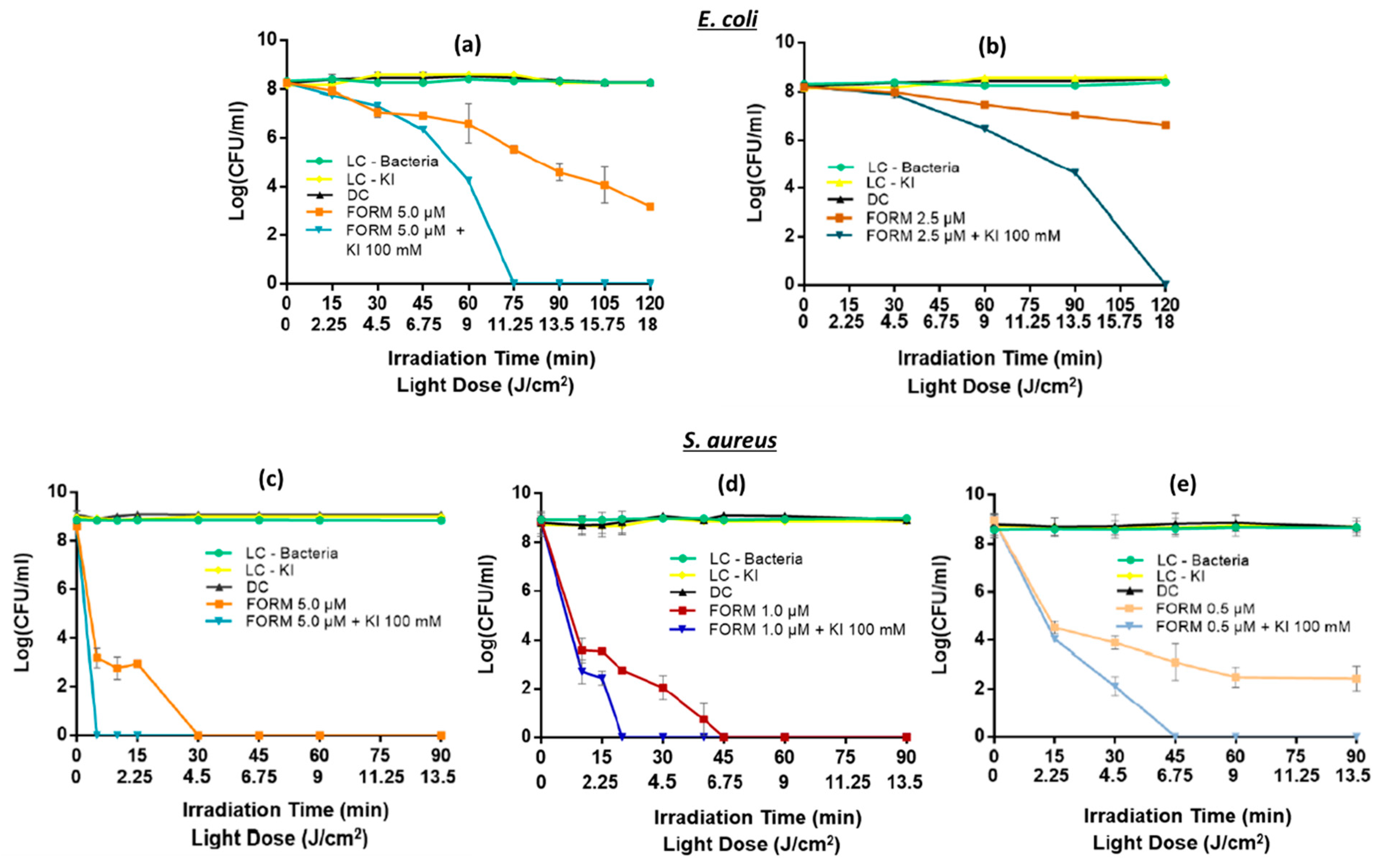
3.1. Photodynamic Inactivation of the Bacterial and Fungal Planktonic Cells

The photodynamic effect of FORM and its combination with KI (100 mM) was first evaluated with regard to planktonic forms of a Gram(–) (E. coli) and a Gram(+) (S. aureus) bacterium, and the results obtained are presented in Figure 2.
The Gram(–)bacterium was the less susceptible microorganism to the aPDT treatments with a 5 log of CFU·mL−1 reduction attained after 120 min of treatment with FORM at 5.0 μM (Figure 2a). On the other hand, this PS was more efficient against S. aureus since a lower PS concentration (1.0 μM) was enough to promote the bacterial reduction until the detection limit of the methodology (~9 log reduction) after 45 min of treatment (Figure 2b). These results are consistent with previous studies, where FORM acts as an efficient PS in the photoinactivation of both Gram(–) and Gram(+) bacteria [6,8].
Furthermore, the application of KI improved the efficiency of photodynamic treatments, allowing a significant reduction in the time required to efficiently photoinactivate E. coli (8.3 log reduction) from 120 min (FORM at 5.0 μM) to 75 min (FORM at 5.0 μM + KI) (Figure 2a,b). This same combination of FORM + KI was highly effective against S. aureus, enabling not only a reduction in the treatment time by at least half (depending on the concentration tested), but also decreasing the PS concentration 10-fold (from 5.0 to 0.5 μM) (Figure 2c,e. In general, the results indicate that—with the combination of FORM with KI— bacterial photoinactivation was obtained using lower concentrations or lower light exposure than those used in non-combined strategies for both Gram(+) and Gram(–) bacteria.
These promising results prompted us to test the effect of FORM and the combined action of FORM + KI in the yeast C. albicans for the first time. The results obtained are summarized in Figure 3 and show that FORM (5.0 and 1.0 μM) was highly efficient in the photoinactivation of C. albicans (reduction of 6.7 logs), reaching the detection limit of the method after 10 and 90 min of irradiation, respectively (Figure 3a,b). The efficacy of FORM against C. albicans is even more noteworthy when the results are compared with those from the literature since high concentrations of PS (>10 μM) or light doses are usually required for the photoinactivation of this microorganism [10,12]. Indeed, when C. albicans was tested with Tetra-Py(+)-Me at 5.0 μM, a porphyrin present in 17% in the FORM, the total photoinactivation of C. albicans was only observed after 270 min of irradiation (total light dose of 64.8 J/cm2) [12]. However, with the FORM alone, the total photoinactivation occurred after 10 min of irradiation (1.5 J/cm2) at the same concentration (5.0 μM). These results highlight FORM as a promising PS to be used against fungal infections.
Similarly to the observed for E. coli and S. aureus, the combination FORM + KI has also reduced the aPDT treatment time (>three-times) and concentration of PS (from 1.0 to 0.5 μM) required to inactivate C. albicans (Figure 3b,c). However, this enhanced effect was only observed with low concentrations of FORM (1.0 and 0.5 μM) (Figure 3b,c). This fact can be justified by the high efficacy of this PS alone at 5.0 μM (Figure 3a), which may have promoted the total inactivation of C. albicans by the ROS species produced by FORM before the formation of iodine-reactive species (formed by the combination of FORM + KI). Overall, these results confirm the enormous potential of FORM for the inactivation of the yeast C. albicans and for the application of KI to enhance the aPDT effect of FORM, enabling a reduction in the treatment time and concentration of PS.

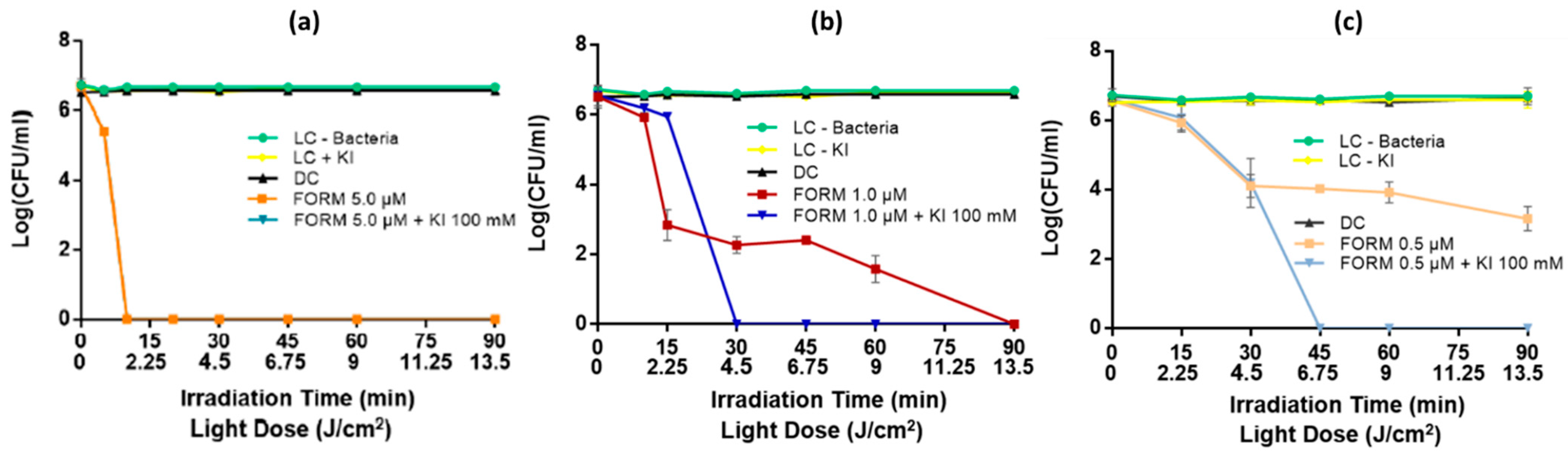
3.2. Photodynamic Inactivation of the Bacterial and Fungal Biofilms

Considering the high incidence of infectious diseases caused by biofilms and the excellent results obtained in the previous assays on planktonic cells, we decided to evaluate the aPDT effect of the combination FORM + KI on biofilms forms of the Gram(–)bacterium E. coli, the Gram(+) bacterium S. aureus, and of the yeast C. albicans. Knowing that biofilms are less susceptible to aPDT than their counterpart planktonic forms, the efficacy of FORM alone was tested at a concentration of 20 μM.
FORM at 20 μM was not able to destroy the biofilm matrix of E. coli, S. aureus, and C. albicans (p > 0.005), proving the difficulty in the photoinactivation of this microbial three-dimensional structure. However, when combinations of FORM with KI were used, a drastic aPDT effect was observed. For E. coli and S. aureus biofilms—when KI was combined with FORM at 20, 10, 5.0, and 1.0 μM—the cells of the biofilms were inactivated approximately in 7.5 log (Figure 4a,b), reaching the detection limit of the method (p < 0.005). These results demonstrate that the combination of a PS + KI allows the significant improvement in the destruction of Gram(+) and Gram(–) bacteria, suggesting its promising application in the controlling clinical infections.
This combined effect of FORM + KI was also observed for the yeast C. albicans (Figure 4c). In fact, C. albicans biofilms were more susceptible to aPDT combination of FORM + KI than bacterial biofilms. In this case, FORM at 0.5 μM + KI was enough to destroy C. albicans biofilms after 60 min of treatment (9.0 J/cm2), while biofilm forms of E. coli and S. aureus required the double concentration of FORM (1.0 μM) to achieve the same effect. These results indicate that under the conditions evaluated (FORM + KI) C. albicans biofilms are easier to destroy than bacterial biofilms. These results are in accordance with the literature since C. albicans biofilms have previously shown higher susceptibility to aPDT treatments than P. aeruginosa and S. aureus biofilms [12].
The enhanced in vitro inactivation of fungal biofilms by the combined application of KI and PS was already reported in the literature. However, the highest biofilm destruction was attained with MB (100 mM) + KI (100 mM)—2.31 log—with a total light dose of 40 J/cm2 [9], corresponding to 267 min under the irradiation conditions used in this study. Under similar conditions, no improvement effect by KI was observed with the analogue new methylene blue (NMB) [9]. Despite the structural differences between these phenothiazinium PSs and FORM, the potential of FORM combined with KI, allowing the destruction of C. albicans biofilms with low light doses (9.0 J/cm2, ≥4 times lower) and lower PS concentrations (200 times lower), is undeniable.

4. Conclusions

In summary, this study demonstrated that FORM is an efficient PS not only in the inactivation of planktonic forms of Gram(+) and Gram(–) bacteria, but also of C. albicans at low concentrations (<5.0 μM). Moreover, the combination of the low-cost FORM with KI was clearly more effective in inactivating all the microorganisms, enabling a reduction in the PS concentration and the treatment time. These results highlight the combination of the affordable and easily synthesizable FORM with KI as a promising approach to destroy, at low concentrations, planktonic and biofilm. These features could be useful to the translation of this aPDT protocol to the field.

Author Contributions

Conceptualization, C.V., A.T.P.C.G., M.A.F.F., M.G.P.M.S.N. and A.A.; methodology, C.V. and A.R.S.; validation, C.V., A.R.S., M.Q.M., A.T.P.C.G., M.A.F.F., M.G.P.M.S.N. and A.A.; formal analysis, C.V., A.R.S., M.Q.M., A.T.P.C.G., M.A.F.F., M.G.P.M.S.N. and A.A.; investigation, C.V., M.B., A.R.S., M.Q.M., A.T.P.C.G., M.A.F.F., M.G.P.M.S.N. and A.A.; resources, M.A.F.F., M.G.P.M.S.N. and A.A.; data curation, C.V., M.B., A.R.S., M.Q.M., A.T.P.C.G., M.A.F.F., M.G.P.M.S.N. and A.A.; writing—original draft preparation, C.V., A.R.S., M.Q.M., A.T.P.C.G., M.A.F.F., M.G.P.M.S.N. and A.A.; writing—review and editing, C.V., M.B., A.R.S., M.Q.M., A.T.P.C.G., M.A.F.F., M.G.P.M.S.N. and A.A.; supervision, A.T.P.C.G., M.A.F.F., M.G.P.M.S.N. and A.A.; project administration, M.A.F.F., M.G.P.M.S.N. and A.A.; funding acquisition, M.A.F.F., M.G.P.M.S.N. and A.A. All authors have read and agreed to the published version of the manuscript.

Funding

This research was funded by FCT/MCT through financial support for the LAQV-REQUIMTE (UIDB/50006/2020) and CESAM (UID/AMB/50017/2019 and UIDB/50017/2020 + UIDP/50017/2020) research units and by the FCT project PREVINE, (PTDC/ASP-PES/29576/2017) through national funds (OE), and, where applicable, it was co-financed by the FEDER—Operational Thematic Program for Competitiveness and Internationalization—COMPETE 2020 within the PT2020 Partnership Agreement and the Portuguese NMR Network.

Institutional Review Board Statement

Not applicable.

Informed Consent Statement

Not applicable.

Data Availability Statement

Not applicable.

Acknowledgments

Thanks are due to the University of Aveiro and FCT/MCT for the financial support to LAQV-REQUIMTE (UIDB/50006/2020) and CESAM (UID/AMB/50017/2019 and UIDB/50017/2020 + UIDP/50017/2020) research units, and to the FCT project PREVINE,(PTDC/ASP-PES/29576/2017) through national funds (OE) and, where applicable, co-financed by the FEDER-Operational Thematic Program for Competitiveness and Internationalization−COMPETE 2020, within the PT2020 Partnership Agreement, and to the Portuguese NMR Network. Thanks are also due to the SANAQUA project MAR02.05.01-FEAMP-0004 and I9+Proalga project MAR2020-16-01-03-FMP-0011. The doctoral grants to C.V., M.B., and M.Q.M. are funded by FCT (SFRH/BD/150358/2019, SFRH/BD/121645/2016, and SFRH/BD/112317/2015, respectively).

Conflicts of Interest

The authors declare no conflict of interest.

References

  1. Alves, E.; Faustino, M.A.F.; Tomé, J.P.C.; Neves, M.G.P.M.S.; Tomé, A.C.; Cavaleiro, J.A.S.; Cunha, A.; Gomes, N.C.M.; Almeida, A. Photodynamic Antimicrobial Chemotherapy in Aquaculture: Photoinactivation Studies of Vibrio fischeri. PLoS ONE 2011, 6, e20970. [Google Scholar] [CrossRef] [PubMed]
  2. Alves, E.; Faustino, M.A.F.; Neves, M.G.P.M.S.; Cunha, A.; Nadais, H.; Almeida, A. Potential Applications of Porphyrins in Photodynamic Inactivation Beyond the Medical Scope. J. Photochem. Photobiol 2015, 22, 34–57. [Google Scholar] [CrossRef]
  3. Hamblin, M.R. Upconversion in Photodynamic therapy: Plumbing the Depths. Dalton Trans 2018, 47, 8571–8580. [Google Scholar] [CrossRef] [PubMed]
  4. Takasaki, A.A.; Aoki, A.; Mizutani, K.; Schwarz, F.; Sculean, A.; Wang, C.-Y.; Koshy, G.; Romanos, G.; Shikawa, I.; Zumi, Y. Application of Antimicrobial Photodynamic Therapy in Periodontal and Peri-implant Diseases. Periodontology 2009, 51, 109–140. [Google Scholar] [CrossRef] [PubMed]
  5. Mesquita, M.Q.; Dias, C.J.; Neves, M.G.P.M.S.; Almeida, A.; Faustino, M.A.F. Revisiting Current Photoactive Materials for Antimicrobial Photodynamic Therapy. Molecules 2018, 23, 2424. [Google Scholar] [CrossRef] [PubMed]
  6. Marciel, L.; Rosalina, F.; Ana, M.; Mariana, M.; Neves, M.G.P.M.S.; Adelaide, A. An Efficient Formulation Based on Cationic Porphyrins to Photoinactivate Staphylococcus aureus and Escherichia coli. Future Med. Chem. 2018, 10, 1821–1833. [Google Scholar] [CrossRef] [PubMed]
  7. Martins, D.; Mesquita, M.Q.; Neves, M.G.P.M.S.; Faustino, M.A.F.; Reis, L.; Figueira, E.; Almeida, A. Photoinactivation of Pseudomonas syringae pv. actinidiae in Kiwifruit Plants by Cationic Porphyrins. Planta 2018, 248, 409–421. [Google Scholar] [CrossRef] [PubMed]
  8. Vieira, C.; Gomes, A.T.P.C.; Mesquita, M.Q.; Moura, N.M.M.; Neves, M.G.P.M.S.; Faustino, M.A.F.; Almeida, A. An Insight Into the Potentiation Effect of Potassium Iodide on aPDT Efficacy. Front. Microbiol. 2018, 9, 2665. [Google Scholar] [CrossRef] [PubMed]
  9. Freire, F.; Ferraresi, C.; Jorge, A.O.C.; Hamblin, M.R. Photodynamic Therapy of Oral Candida Infection in A Mouse Model. Photochem. Photobiol. B 2016, 159, 161–168. [Google Scholar] [CrossRef] [PubMed]
  10. Zhang, Y.; Dai, T.; Wang, M.; Vecchio, D.; Chiang, L.Y.; Hamblin, M.R. Potentiation of Antimicrobial Photodynamic Inactivation Mediated by a Cationic Fullerene by Added Iodide: In vitro and in vivo Studies. Nanomedicine 2015, 10, 603–614. [Google Scholar] [CrossRef]
  11. Simões, C.; Gomes, M.C.; Neves, M.G.P.M.S.; Cunha, A.; Tomé, J.P.C.; Tomé, A.C.; Cavaleiro, J.A.S.; Almeida, A.; Faustino, M.A.F. Photodynamic Inactivation of Escherichia coli with Cationic Meso-tetraarylporphyrins—The Charge Number and Charge Distribution Effects. Catal. Today 2016, 266, 197–204. [Google Scholar] [CrossRef]
  12. Beirão, S.; Fernandes, S.; Coelho, J.; Faustino, M.A.F.; Tomé, J.P.C.; Neves, M.G.P.M.S.; Tomé, A.C.; Almeida, A.; Cunha, A. Photodynamic Inactivation of Bacterial and Yeast Biofilms with A Cationic Porphyrin. Photochem. Photobiol 2014, 90, 1387–1396. [Google Scholar] [CrossRef] [PubMed]
  13. Costa, L.; Faustino, M.A.F.; Neves, M.G.P.M.S.; Cunha, A.; Almeida, A. Photodynamic Inactivation of Mammalian Viruses and Bacteriophages. Viruses 2012, 4, 1034–1074. [Google Scholar] [CrossRef] [PubMed]
Figure 1. Structure of porphyrin derivatives present in FORM.
Msf 12 00013 g001
Figure 2. Photodynamic inactivation of planktonic forms of E. coli (a,b) and S. aureus (ce). The experiments were performed with FORM alone at 5.0 (a,c), 2.5 (b), 1.0 (d), and 0.5 μM (e) and by combination of FORM with KI at 100 mM under irradiation with white light (25 W/m2).
Msf 12 00013 g002
Figure 3. Photodynamic inactivation of planktonic forms of C. albicans by FORM alone at 5.0 μM (a), 1.0 μM (b) or 0.5 μM (c) and by combination FORM with KI at 100 mM under irradiation with white light (25 W/m2).
Msf 12 00013 g003
Figure 4. Photodynamic inactivation of biofilms of E. coli (a), S. aureus (b), or C. albicans (c) by FORM (0.1–20 μM) alone or combined with KI (100 mM). All the assays were performed during 60 min under white light irradiation (25 W/m2, 9.0 J/cm2). Samples that reached the total inactivation are represented by (*).
Msf 12 00013 g004
Publisher’s Note: MDPI stays neutral with regard to jurisdictional claims in published maps and institutional affiliations.

