Proceeding Paper

Exploring Flavonoids and Citric Acid Extraction Yields from Lemon Peels Across Glucanase, Cellulase, and Pectinase Enzymes †

by
Rosa Zapata
,
Lorena Martínez-Zamora
and
Francisco Artés-Hernández
*
Postharvest and Refrigeration Group, Department of Agricultural Engineering and Institute of Plant Biotechnology, Universidad Politécnica de Cartagena, Pº Alfonso XIII, 48, 30203 Cartagena, Murcia, Spain
*
Author to whom correspondence should be addressed.
Presented at the 6th International Electronic Conference on Foods, 28–30 October 2025; Available online: https://sciforum.net/event/foods2025.
Biol. Life Sci. Forum 2026, 56(1), 7; https://doi.org/10.3390/blsf2026056007
Published: 15 January 2026
(This article belongs to the Proceedings of The 6th International Electronic Conference on Foods)

Abstract

Lemon peel, which contains a wide variety of antioxidant compounds and biopolymers, is an interesting byproduct for valorization. In this study, pectin and cellulose were obtained from lemon peel with 36% and 23% extraction yields. Meanwhile, when evaluating the extraction of secondary metabolites using various enzymes, it was found that their concentrations increased the most in the case of pectin methylesterase, by up to 21.7% for eriocitrin, although the solid residue was not affected spectroscopically by the enzymatic activity. In contrast, enzyme mixes with β-glucosidase activity showed no presence of the O-glycosylated flavonoids, suggesting their hydrolysis into the aglycones.

1. Introduction

Lemon fruit (Citrus limon) stands out among other citrus fruits due to its wide range of polar antioxidant compounds. It belongs to the citrus genus, one of the most cultivated worldwide, and together with limes reached a global production of 23.6 Mtons in 2023. Approximately 40% of the total production is destined for the juice industry, where peel, pulp, and seeds, accounting for up to 50% of the whole fruit, are generated as byproducts and pollute the environment. The primary use of lemon peel is as animal feed, although they represent a broad field of research for the development of high-value products containing pectin, as a gelling agent, and antioxidant metabolite compounds [1].
Lemon peels are primarily composed of pectin, cellulose, and hemicellulose, which constitute the cell walls. Pectin is a soluble fiber formed by a chain of α (1→4) d-galacturonic acid monomers, whose carboxyl groups exhibit varying degrees of methyl esterification, while cellulose consists of glucose monomers linked by β (1→4)-glycosidic bonds [2]. Additionally, although in smaller quantities, they contain secondary metabolites, including phenolic compounds, mainly diglycosylated flavonoids, with highly beneficial antioxidant, anti-inflammatory and antimicrobial properties [3]. Since these compounds are commonly bound to the polysaccharides that constitute the cell wall, the application of exogenous enzymes to disrupt these structures is an effective strategy for enhancing extraction yield.
Enzyme-assisted extraction is considered a green technology since it notably improves extraction yield without the need for high temperatures or organic solvents, as has been reported in numerous studies. Li et al. [4] reported that the use of cellulase improved the total phenolic content extracted from lemon peel by up to 40.5%. Wang et al. [5] observed an improvement in total phenolic and flavonoid contents of approximately 2.07- and 2.36-fold compared to conventional solvent extraction using a water-methanol-DMSO mixture (1:4:5, v/v/v), achieving results similar to ultrasound-assisted extraction but requiring a fourfold increase in processing time.
Therefore, the aims of this work were to isolate and characterize pectin and cellulose from lemon peel with high yield via FTIR spectroscopy, comparing them to the freeze-dried peel. The subsequent objective was to assess the activity of various enzymes, specifically, two pectinases, a cellulase and a glucanase, in degrading these structural polysaccharides to extract bioactive secondary metabolites from the peel. This was achieved by analyzing the solid residues via FTIR and quantifying the supernatant concentrations by HPLC-MS.

2. Materials and Methods

2.1. Chemical and Biochemical Reagents

The solvents employed, including 37% hydrochloric acid (HCl), 96% ethanol and LC-MS grade acetonitrile, were supplied by PanReac AppliChem (Castellar del Vallès, Barcelona, Spain), as well as the 98% citric acid reagent. A Millipore Synergy UV ultra-purification system (Bedford, MA, USA) was employed to produce Mili-Q-grade water. The enzymatic preparations Viscozyme®, Pectinex Ultrapulp®, Pectinex Yield-mash® and Celluclast® were provided by Novozymes A/S (Bagsværd, Hovedstaden, Denmark). Among commercial standards, hesperidin and eriocitrin were purchased from CymitQuimica S.L (Sant Martí, Barcelona, Spain), while rutin was supplied by Sigma-Aldrich (Saint Louis, MO, USA).

2.2. Plant Materials

The lemon peels were separated from ‘Fino 95’ organic lemons (Citrus limon), which were grown in open air in southeast mediterranean Spanish lands, handled and provided by Toñifruit S.L (Librilla, Murcia, Spain) at commercial maturity stage, as determined and reported in a previous published work, (4.5 ± 0.7 °Brix, 2.9 ± 0.5 g of citric acid per 100 mL, and 2.8 ± 0.4 of maturity index) [6]. The lemon peels were frozen at −80 °C and freeze-dried using a Telstar LyoBeta freeze-drier (Telstar Technologies S.L.; Terrassa, Barcelona, Spain). They were then ground to obtain ~200 g of homogeneous powdered material using the IKA A 11 basic analytical mill (IKA-Werke GmbH & Co. KG, Staufen, Germany).

2.3. Pectin and Cellulose Extraction

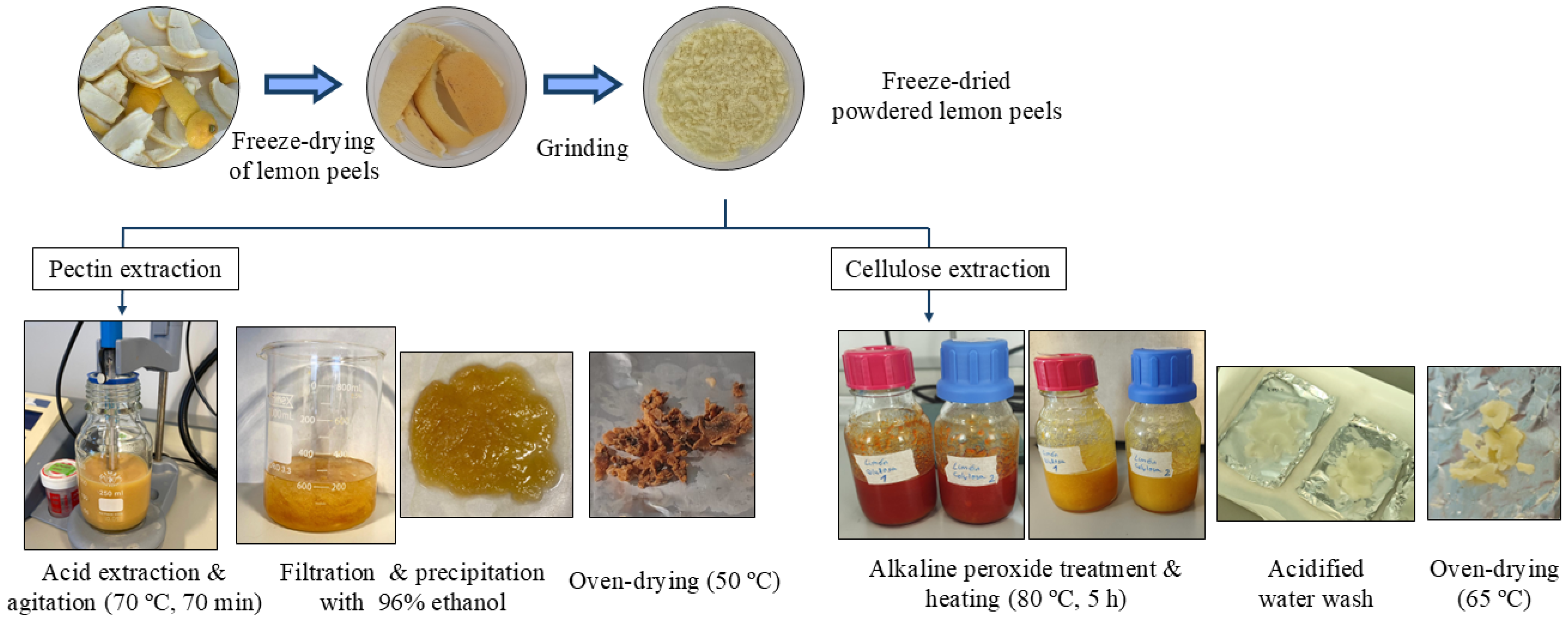
The extraction of pectin was performed following the previously described methods [1,7], with slight modifications (n = 3), as shown in Figure 1. Samples of 5 g of freeze-dried lemon peel powder were treated with 150 mL of miliQ water (1:30 w/v ratio) in glass bottles. The pH was adjusted to 1.3 with 1 M HCl using a digital pH meter (GLP21, Crison Instruments S.A.; Alella, Barcelona, Spain). The bottles were heated in a Unitronic Reciprocating shaking water bath (J.P. Selecta S.A., Abrera, Barcelona, Spain) at 70 °C for 70 min with shaking at 50 oscillations per min. Then, the mixtures were cooled with ice and filtered through three layers of gauze. The pectin was precipitated by adding 96% ethanol at a 1:1 (v/v) ratio to the filtered liquid phase. After 3 h of pectin coagulation at room temperature, the pectin was collected by gravity filtration through filter paper, washed twice with 96% ethanol and dried in a Digitheat-TFT convection oven (J.P. Selecta S.A., Abrera, Barcelona, Spain) at 50 °C for 24 h until reaching constant weight.
The cellulose was extracted following the previously described methods [8,9], with some modifications (n = 3). Samples of 5 g of freeze-dried lemon peel powder were treated with 100 mL of a 4% (w/v) NaOH aqueous solution containing 0.9% (v/v) H2O2 in glass bottles. The mixtures were heated in the same water bath at 80 °C for 5 h. The resulting white residue from each replicate was filtered and then washed sequentially with neutral water and a 10% acetic acid solution until a pH of ~7 was reached. The white solid was dried in the same oven at 65 °C for 48 h until reaching constant weight.

2.4. Enzymatic-Assisted Extraction and HPLC-ESI-QqQ-MS/MS Quantification

Samples of 0.1 g of freeze-dried lemon peel were subjected to enzymatic extraction using 10 mL of 1% aqueous solutions of glucanase, pectin lyase, cellulase, or pectin methylesterase (v/v) (n = 3). Control samples were extracted with 10 mL of ultrapure water. After agitation by vortexing for 3 min, the mixtures were heated at 85 °C for 10 min to stop the enzymatic activity and subsequently filtered through 0.2 µm polyamide (PA) membranes. The liquid filtrates were collected for subsequent HPLC-MS analysis, while the solid residues were oven-dried at 65 °C and analyzed by FTIR spectroscopy.
HPLC-MS analysis was performed as previously described [6], following the same methodology. Briefly, the equipment used was an Agilent 1200 liquid chromatograph (Santa Clara, CA, USA) coupled to a 6420 triple-quadrupole mass spectrometer (QqQ) with an electrospray ionization source (ESI) set in negative ionization mode. The stationary phase was a C18 reversed-phase column, Luna Omega C18 (2.1 × 100 mm; 3 μm), and the mobile phases consisted of a 0.1% formic acid aqueous solution (A) and HPLC-MS grade acetonitrile (B). Quantification of the major analytes, based on previous full-scan studies [6], was carried out using multiple reaction monitoring mode with the following m/z transition: eriocitrin (595→287), hesperidin (609→301), diosmetin-6,8-di-C-glucoside (DG) (623→353), and citric acid (191→111). External calibration was performed in all cases using the corresponding commercial standard, except for DG, which was quantified in rutin equivalents. The values were expressed as g of each analyte per kg of dry weight.

2.5. Attenuated Total Reflection-Fourier Transform Infrared Analysis (ATR-FTIR)

FTIR spectra of the powdered solid samples (n = 3), including the isolated pectin and cellulose fractions, along with the post-enzymatic extraction residue, were recorded using a Thermo Nicolet 5700 FTIR spectrophotometer (Madison, WI, USA). The instrument was equipped with an Attenuated Total Reflectance (ATR) module featuring a diamond crystal and a deuterated triglycine sulfate/KBr detector. Data acquisition and processing were managed by Omnic 9.11.721 software. Each spectrum was acquired in transmittance mode over the range of 4000 to 400 cm−1, with spectral resolution of 4 cm−1 and 32 accumulated scans, as previously described [10]. The spectra were plotted over the 4000–2300 and 1800–400 cm−1 ranges, representing the mean of the three replicates for each sample, using The Unscrambler X 10.4 software (CAMO Software, Oslo, Norway).

2.6. Statistical Analysis

For the individual quantification of secondary metabolites, significant differences among enzyme treatments for each compound were assessed by a one-way ANOVA (p < 0.05), followed by Tukey’s multiple range test for pairwise comparisons. All analyses were performed using Statgraphics Centurion XVI.I version (Statgraphics Technologies, Inc., The Plains, VA, USA).

3. Results and Discussion

3.1. Extraction Yields and FTIR Characterization of Pectin and Cellulose

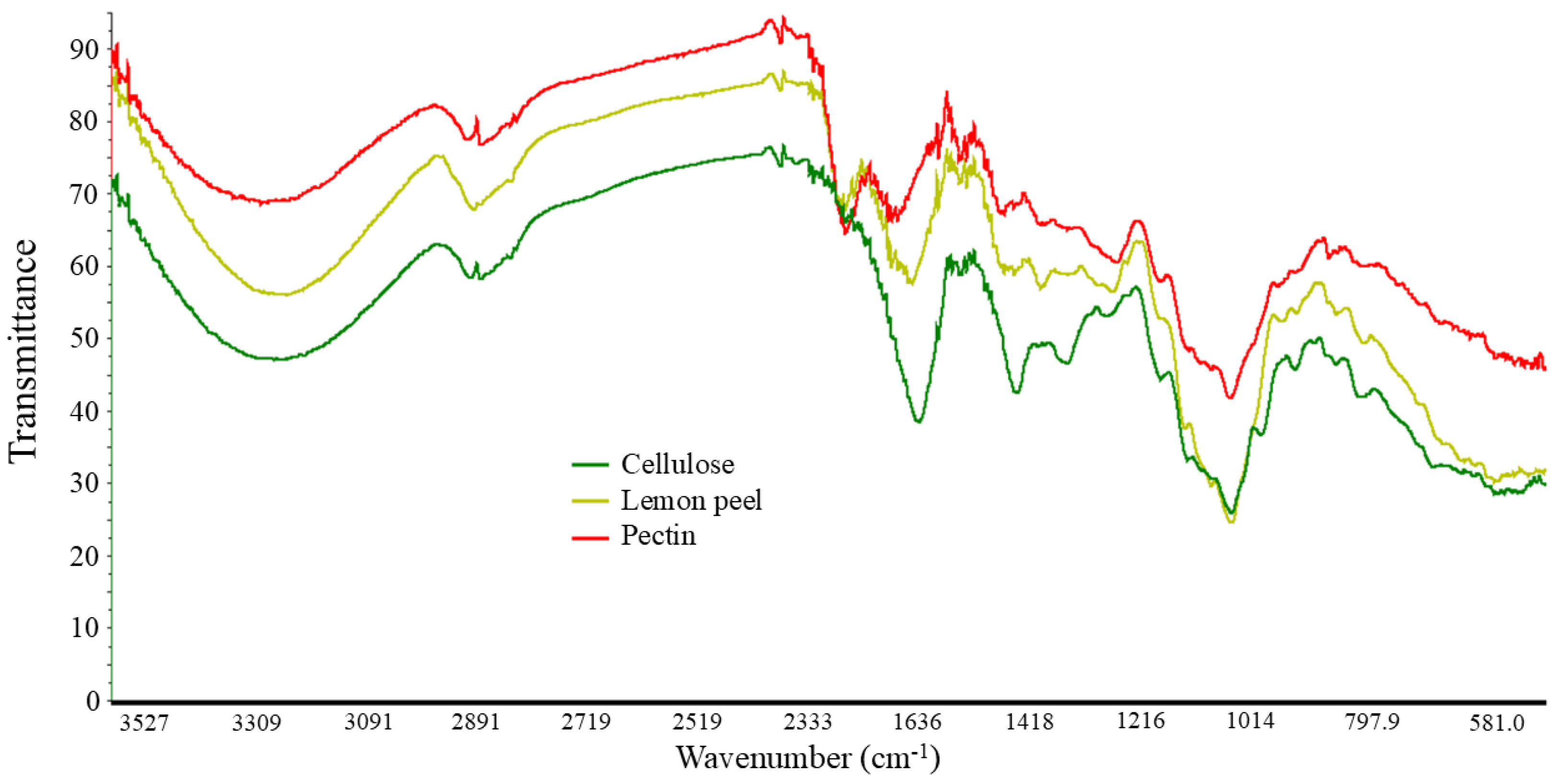
The characterization of the solids obtained by FTIR spectroscopy is characteristic of pectin and cellulose (Figure 2), confirming their extraction [11,12]. The pectin was obtained as an orange solid despite ethanol washing, with a yield of 36 ± 3%, similar to the 30.3 ± 1.9% reported by Chandrasekar et al. [13] for citrus waste. The cellulose was isolated as a white solid with a yield of 23.2 ± 1.4%. In this case, lower than the 57.8 ± 2.4% reported in the literature [13]. In Figure 2, the spectra of cellulose and pectin are also shown superimposed along with that of the freeze-dried lemon peel from which they were extracted. The three spectra exhibited a similar trend, but with some notable differences. In all three, the bands in the 3200–3300 cm−1 spectral range correspond to hydroxyl group stretching, while the bands observed between 2800–3000 cm−1 are related to the ν(C-H) from the methyl and methylene groups, and the pyranose ring.
Moving to lower wavenumbers, the first notable variations are observed in the bands around 1745 and 1650 cm−1, which are produced by ν(C=O) from the ester, the anionic carboxyl and the methyl-esterified carboxyl groups. These bands showed greater similarity between the pectin and lemon peel spectra, whereas the cellulose spectrum exhibited only a band around 1630 cm−1, which could correspond to the H-O-H bending vibration of adsorbed water, as pure cellulose lacks intrinsic carbonyl groups. In the literature, a weaker band near 1410 cm−1 has been reported, corresponding to the stretching of carboxylate groups [11]. However, in our cellulose spectrum, the band appearing around this wavenumber is more intense. This could be possible due to oxidation or residual contamination with other polysaccharides. The most intense band in the three spectra, appearing around 1014 cm−1, is attributed to ν(C-O) and typical of glycosidic bonds, as well as the bands below 930 cm−1, corresponding to the C-O-C bridges. The overlapping signals around 1150 cm−1 likely correspond to δ(C-OH), according to the literature [14].

3.2. Enzymatic Extraction: Solid Residues FTIR and HPLC-MS/MS Quantification

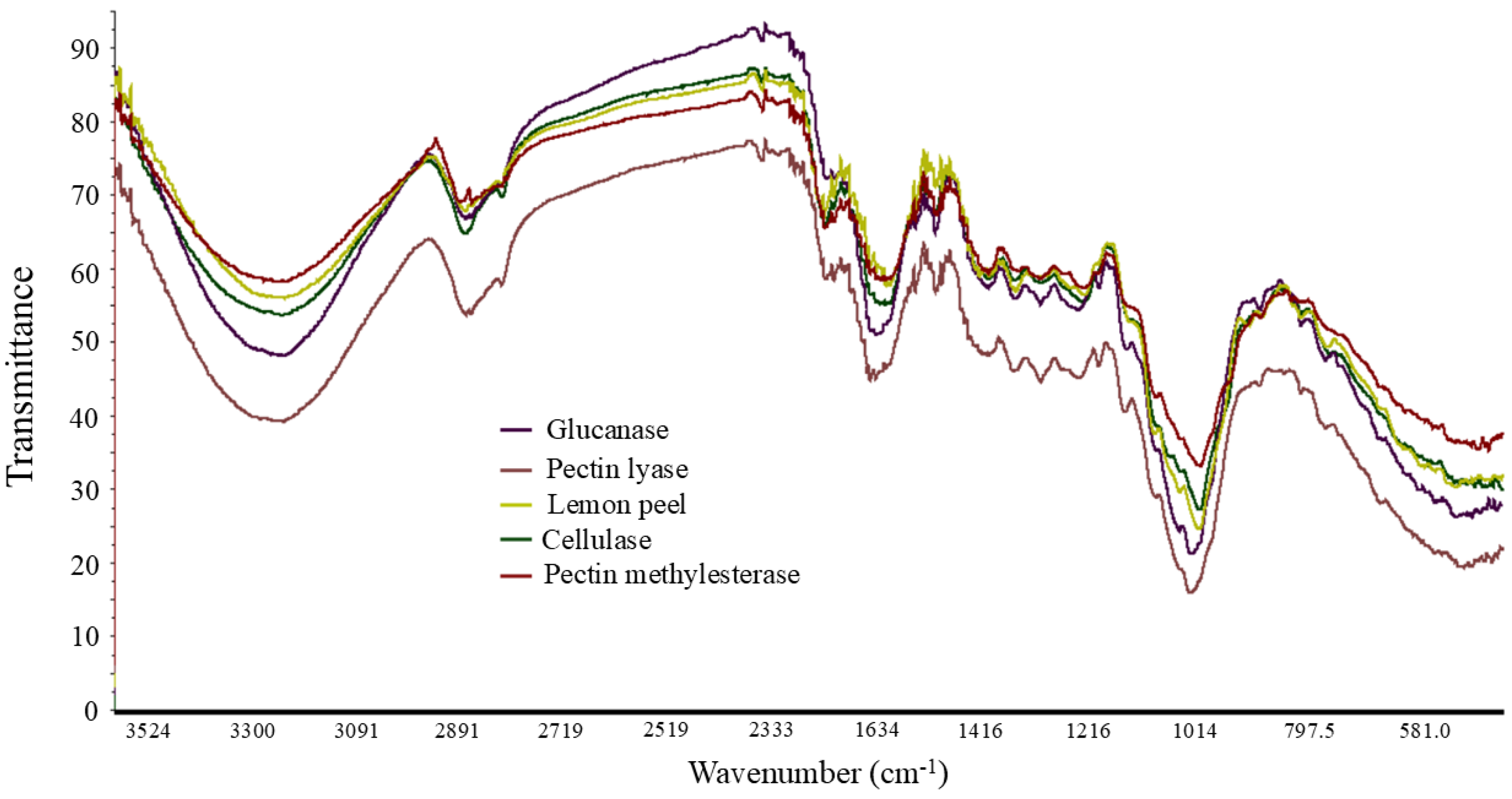
The solid residues recovered after each enzymatic-assisted extraction once oven-dried showed FTIR spectra similar to that of the non-extracted freeze-dried lemon peel, as can be observed in Figure 3. This is possibly indicative that the plant material on which the enzyme acted has passed into the liquid phase in solution, without affecting the residual solid phase. Consequently, the band assignments are consistent with those described in the previous section.
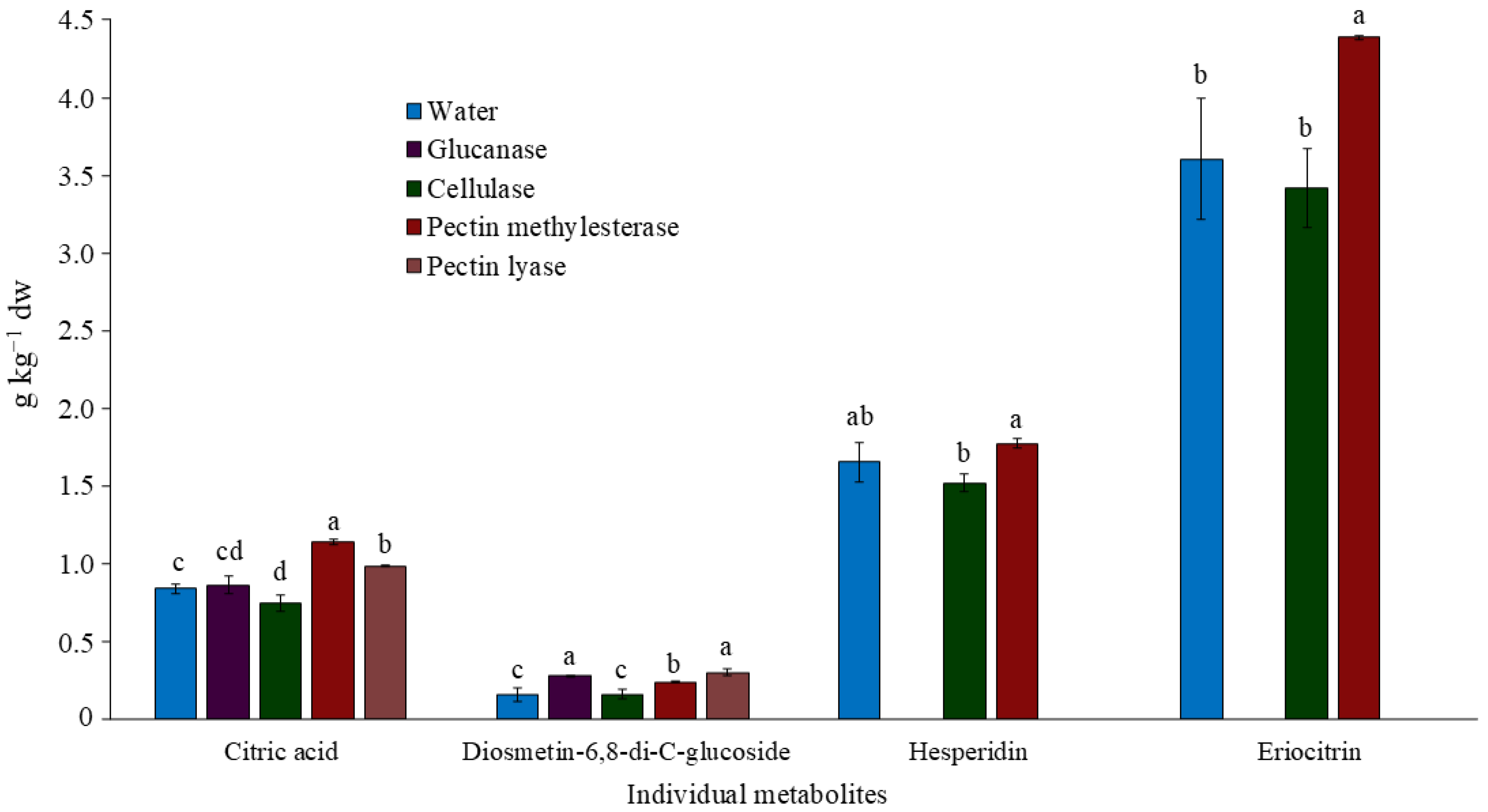
The quantification of the main individual polar compounds extracted from lemon peel and analyzed in the filtrates by HPLC-MS is shown in Figure 4. From previous full-scan analyses of an ethanolic extract, the most intense peaks were identified as citric acid, DG, eriocitrin, and hesperidin, in order of polarity. The latter three are diglycosylated flavonoids. Specifically, DG is a C-glycosyl flavone with linked glucose moieties, whereas eriocitrin and hesperidin are O-glycosyl flavanones. It can be observed that pectin methylesterase is the enzyme that generally most enhanced the extraction of the compounds, assuming an improvement of 35.8%, 51.7%, 7.20%, and 21.7% in the obtained content of citric acid, DG, hesperidin, and eriocitrin, respectively, compared to direct extraction in water. However, the DG concentration resulted in slightly higher levels using glucanase and pectin lyase. The results of the aqueous samples and those with cellulose and pectin methylesterase showed different concentrations in g/kg dw of the analyzed compounds, in descending order: eriocitrin, hesperidin, citric acid, and DG. For citric acid, the main acid in citrus, its notably lower molecular weight compared to flavonoids means its molar concentration is not so different from that of eriocitrin, being 4.36 mmol kg−1 and 6.04 mmol kg−1, respectively in the water-extracted samples. The same trend has been reported in the literature, with higher concentrations of eriocitrin than hesperidin in lemon peel following both aqueous and 80% MeOH extraction [6,15], as well as a lower citric acid content when using 80% MeOH [6].
A notable finding was the absence of hesperidin and eriocitrin in the extracts obtained using glucanase and pectin lyase. Both compounds are O-diglycosides, where the aglycone is conjugated to rutinosyl moiety. The presence of certain amounts of β-glucosidase in the commercial enzyme mixtures Viscozyme® (predominantly β-glucanase) and Pectinex Ultrapulp® (predominantly pectin lyase) may be responsible for cleaving the bonds between both moieties, thereby releasing the corresponding aglycones. This activity has also been reported in the literature [16], where the application of polygalacturonase resulted in an increase in hesperetin and a corresponding decrease in hesperidin, suggesting the hydrolysis of the O-glycosidic bond and the subsequent release of the aglycone. Although DG is also a diglycosylated flavonoid, it is not affected by this enzymatic activity because the glucose units are attached to the flavonoid skeleton via C-glycosidic bonds.

4. Conclusions

Lemon peel is rich in structural cell wall biopolymers, including pectin and cellulose, as confirmed in this study by their successful extraction through acid and alkaline peroxide treatments, with 36 and 23% yields, respectively. This once again confirms their potential valorization as natural sources of pectin, applied in nutrition as a gelling agent with prebiotic and glucose modulation properties, and of cellulose, which is studied in the biopolymer industry. Among the exogenous enzymes tested for the extraction of antioxidants aimed at complete valorization, pectinases showed significantly better results, the methylesterase in the recovery of hesperidin, eriocitrin, and citric acid, and the lyase in the extraction of DG. The apparent cleavage of the O-glycosidic bond in the flavonoids hesperidin and eriocitrin by the enzymatic mixtures of pectin lyase and glucanase, which exhibit β-glucosidase activity, represents an interesting point for future research focused on the analysis of the antioxidant and antimicrobial properties of these aglycones, which are not directly found in the plant tissue otherwise.

Author Contributions

Conceptualization, F.A.-H., L.M.-Z. and R.Z.; methodology, R.Z.; software, R.Z.; validation, F.A.-H., L.M.-Z. and R.Z.; formal analysis, L.M.-Z. and R.Z.; investigation, F.A.-H., L.M.-Z. and R.Z.; resources, F.A.-H.; data curation, R.Z.; writing—original draft preparation, R.Z.; writing—review and editing, F.A.-H., L.M.-Z. and R.Z.; visualization, R.Z.; supervision, F.A.-H.; project administration, F.A.-H.; funding acquisition, F.A.-H. All authors have read and agreed to the published version of the manuscript.

Funding

This research was funded by MICIU/AEI/10.13039/501100011033 and by “ERDF/EU”, grant number PID2021-123857OB-I00. This work is also a result of the AGROALNEXT program and was supported by MICIU with funding from EU NextGeneration (PRTR-C17.I1) and by Seneca Foundation with funding from Autonomous Community of the Region of Murcia (CARM).

Informed Consent Statement

Informed consent was obtained from all subjects involved in the study.

Data Availability Statement

Not applicable to this article.

Acknowledgments

R.Z. thanks the Spanish Ministry of Science, Innovation and Universities for her predoctoral fellowship FPU23/02121. L. M-Z’s contract has been financed by the AGROALNEXT-MRR Project.

Conflicts of Interest

The authors declare no conflicts of interest.

References

  1. Akhter, M.J.; Sarkar, S.; Sharmin, T.; Mondal, S.C. Mondal, Extraction of pectin from powdered citrus peels using various acids: An analysis contrasting orange with lime. Appl. Food Res. 2024, 4, 100614. [Google Scholar] [CrossRef] [Scilit]
  2. Khopade, S.; Gomte, S.S.; Janrao, C.; Bavaskar, A.; Agnihotri, T.G.; Jain, A.; Khatik, R. Peptide and protein delivery through cellulose, hyaluronic acid, and heparin. In Peptide and Protein Drug Delivery Using Polysaccharides; Elsevier: Amsterdam, The Netherlands, 2023; pp. 75–113. [Google Scholar] [CrossRef] [Scilit]
  3. Kumar, V.; Kaur, R.; Aggarwal, P.; Singh, G. Underutilized citrus species: An insight of their nutraceutical potential and importance for the development of functional food. Sci. Hortic. 2022, 296, 110909. [Google Scholar] [CrossRef] [Scilit]
  4. Li, B.; Smith, B.; Hossain, M. Extraction of phenolics from citrus peels: I. Solvent extraction method. Sep. Purif. Technol. 2006, 48, 182–188. [Google Scholar] [CrossRef] [Scilit]
  5. Wang, Z.; Mei, X.; Chen, X.; Rao, S.; Ju, T.; Li, J.; Yang, Z. Extraction and recovery of bioactive soluble phenolic compounds from brocade orange (Citrus sinensis) peels: Effect of different extraction methods thereon. LWT 2023, 173, 114337. [Google Scholar] [CrossRef] [Scilit]
  6. Zapata, R.; Martínez-Zamora, L.; Cano-Lamadrid, M.; Artés-Hernández, F. Wounding Citrus Peel By-Products as Abiotic Stress to Induce the Synthesis of Phenolic Compounds? Horticulturae 2024, 10, 885. [Google Scholar] [CrossRef] [Scilit]
  7. Akachat, B.; Himed, L.; Salah, M.; D’eLia, M.; Zomorroda, S.; Ali, R.; Rastrelli, L.; Barkat, M. Optimization of pectin extraction from lemon peels using hydrochloric acid and response surface methodology. Appl. Food Res. 2025, 5, 100948. [Google Scholar] [CrossRef] [Scilit]
  8. Ciriminna, R.; Petri, G.L.; Angellotti, G.; Fontananova, E.; Luque, R.; Pagliaro, M. Nanocellulose and microcrystalline cellulose from citrus processing waste: A review. Int. J. Biol. Macromol. 2024, 281, 135865. [Google Scholar] [CrossRef] [Scilit] [PubMed]
  9. Naz, S.; Ahmad, N.; Akhtar, J.; Ahmad, N.M.; Ali, A.; Zia, M. Management of citrus waste by switching in the production of nanocellulose. IET Nanobiotechnol. 2016, 10, 395–399. [Google Scholar] [CrossRef] [Scilit] [PubMed]
  10. Zapata, R.; Martínez-Zamora, L.; Artés-Hernández, F. Diversity of Color, Infrared Spectra, and Phenolic Profile Correlation in Citrus Fruit Peels. In Proceedings of the 5th International Electronic Conference on Foods, Basel, Switzerland, 28–30 October 2024; MDPI: Basel, Switzerland, 2025; p. 26. [Google Scholar] [CrossRef] [Scilit]
  11. Kozioł, A.; Środa-Pomianek, K.; Górniak, A.; Wikiera, A.; Cyprych, K.; Malik, M. Structural Determination of Pectins by Spectroscopy Methods. Coatings 2022, 12, 546. [Google Scholar] [CrossRef] [Scilit]
  12. Abderrahim, B.; Abderrahman, E.; Mohamed, A.; Fatima, T.; Abdesselam, T.; Krim, O. Kinetic Thermal Degradation of Cellulose, Polybutylene Succinate and a Green Composite: Comparative Study. World J. Environ. Eng. 2015, 3, 95–110. [Google Scholar]
  13. Chandrasekar, C.M.; Carullo, D.; Saitta, F.; Krishnamachari, H.; Bellesia, T.; Nespoli, L.; Caneva, E.; Baschieri, C.; Signorelli, M.; Barbiroli, A.G.; et al. Valorization of citrus peel industrial wastes for facile extraction of extractives, pectin, and cellulose nanocrystals through ultrasonication: An in-depth investigation. Carbohydr. Polym. 2024, 344, 122539. [Google Scholar] [CrossRef] [Scilit] [PubMed]
  14. Szymanska-Chargot, M.; Zdunek, A. Use of FT-IR Spectra and PCA to the Bulk Characterization of Cell Wall Residues of Fruits and Vegetables Along a Fraction Process. Food Biophys. 2013, 8, 29–42. [Google Scholar] [CrossRef] [Scilit] [PubMed]
  15. Athanasiadis, V.; Chatzimitakos, T.; Mantiniotou, M.; Bozinou, E.; Lalas, S.I. Exploring the Antioxidant Properties of Citrus limon (Lemon) Peel Ultrasound Extract after the Cloud Point Extraction Method. Biomass 2024, 4, 202–216. [Google Scholar] [CrossRef] [Scilit]
  16. Multari, S.; Licciardello, C.; Caruso, M.; Anesi, A.; Martens, S. Flavedo and albedo of five citrus fruits from Southern Italy: Physicochemical characteristics and enzyme-assisted extraction of phenolic compounds. J. Food Meas. Charact. 2021, 15, 1754–1762. [Google Scholar] [CrossRef] [Scilit]
Figure 1. Extraction of pectin and cellulose from lemon peel.
Blsf 56 00007 g001
Figure 2. FTIR spectra of lemon peel freeze-dried powder and its extracted, oven-dried cellulose and pectin in the 3600–2300 and 1800–400 cm−1 regions.
Blsf 56 00007 g002
Figure 3. Comparative FTIR spectra of lemon peel versus the oven-dried solid recovered after enzymatic extractions using glucanase, pectin lyase, cellulase, and pectin methylesterase in the 3600–2300 and 1800–400 cm−1 regions.
Blsf 56 00007 g003
Figure 4. Concentration of the main secondary metabolites from freeze-dried lemon peel obtained by enzymatic extraction using glucanase, pectin lyase, cellulase, and pectin methylesterase at 1% and 0% in water via HPLC-MS/MS (ESI-QqQ). Values are mean ± sd. Different letters indicate significant differences among extraction treatments (p < 0.05).
Blsf 56 00007 g004
Disclaimer/Publisher’s Note: The statements, opinions and data contained in all publications are solely those of the individual author(s) and contributor(s) and not of MDPI and/or the editor(s). MDPI and/or the editor(s) disclaim responsibility for any injury to people or property resulting from any ideas, methods, instructions or products referred to in the content.