Article

In Vitro Effects of Retinoic Acid and Sodium Selenite on Neuroblastoma Cell Line (SH-SY5Y)

by
Milena Mariano Ribeiro
1,2,
Luíza Siqueira Lima
1,2,
Nayara de Souza da Costa
1,2,
Meire Ellen Pereira
1,2,
Aline S. Fonseca
1,2,
Luciane R. Cavalli
1,2,
Quelen I. Garlet
3,
Ana Carolina Irioda
1,2 and
Cláudia S. Oliveira
1,2,*
1
Instituto de Pesquisa Pelé Pequeno Príncipe, Curitiba 80250-060, Brazil
2
Faculdades Pequeno Príncipe, Curitiba 80230-020, Brazil
3
Programa de Pós-Graduação em Farmacologia, Universidade Federal do Paraná, Curitiba 15300-000, Brazil
*
Author to whom correspondence should be addressed.
Future Pharmacol. 2025, 5(4), 65; https://doi.org/10.3390/futurepharmacol5040065
Submission received: 25 September 2025 / Revised: 28 October 2025 / Accepted: 30 October 2025 / Published: 4 November 2025

Abstract

Background/Objectives: Neuroblastoma is a pediatric embryonal tumor of the autonomic nervous system, characterized by high heterogeneity. Recent research has explored the therapeutic potential of retinoic acid and selenium derivatives as antiproliferative agents. This study aims to assess the antiproliferative effects of sodium selenite and retinoic acid, as well as the conventional chemotherapeutic agents, cyclophosphamide and cisplatin, using the SH-SY5Y neuroblastoma cell line. Methods: Cells were treated with the compounds at concentrations ranging from 0 to 1000 µM for 72 h. The following assays were performed: cell viability, clonogenic assay, cell migration, cell cycle analysis, and gene expression (BCL2 and BAX). Data were analyzed using the Kruskal–Wallis test followed by Dunn’s or the Mann–Whitney test (p < 0.05). IC50 values were obtained from dose–response curves. Results: Sodium selenite (100–1000 µM) significantly reduced cell viability by more than 50% (IC50: 166 µM at 72 h). Retinoic acid (300 µM) reduced viability by 65% (IC50: 198 µM at 72 h), and cisplatin (10 µM) reduced viability by 79% (IC50: 3.4 µM at 72 h). All compounds significantly decreased colony formation. Sodium selenite and retinoic acid induced arrest in the G0/G1 phase of the cell cycle. Gene expression analysis revealed downregulation of the BCL2 gene by all compounds and upregulation of BAX only by sodium selenite at IC50 concentration. Conclusions: Sodium selenite and retinoic acid showed antiproliferative effects on neuroblastoma cells, suggesting their potential as adjuvant therapeutic agents. To reach this goal, we suggest further investigation of their mechanisms of action and evaluation of the combined strategies.

Graphical Abstract

1. Introduction

Cancers occurring during childhood and adolescence represent one of the leading causes of mortality among young people worldwide. The most prevalent pediatric cancers include leukemias, central nervous system tumors, lymphomas, and solid tumors (e.g., neuroblastoma and Wilms’ tumor) [1,2]. In high-income countries, more than 80% of childhood cancer cases are curable. However, in low- and middle-income countries, survival drops to approximately 30%, highlighting stark inequities in access to timely diagnosis and effective treatment [2]. Despite substantial advancements in oncology, chemotherapy and radiotherapy remain associated with significant adverse effects and long-term impairments in patients’ quality of life [3]. In this context, the development of more effective and less toxic therapeutic strategies is recognized as a priority, particularly for aggressive pediatric malignancies such as neuroblastoma.
Neuroblastoma is a rare embryonal solid tumor originating from neural crest cells that have undergone incomplete differentiation [4]. This malignancy predominantly affects children under five years of age and, despite its rarity, accounts for a considerable proportion of pediatric cancer cases [5]. The complex biology of neuroblastoma is closely linked to disruptions in cellular migration, adhesion, and differentiation during the development of the peripheral nervous system [6,7].
The SH-SY5Y cell line, widely used as an in vitro model of neuroblastoma, exhibits an N-type phenotype characterized by immature neuronal features derived from the neural crest [8]. In culture, SH-SY5Y cells are small and round, with long, delicate neurofilaments and scant cytoplasm. These characteristics make SH-SY5Y a relevant model for studying neuronal differentiation, neurotoxicity, and signaling pathways involved in neuroblastoma pathophysiology [8,9].
Given the biological complexity of neuroblastoma, treatment strategies vary according to clinical risk classification. In high-risk neuroblastoma, first-line therapy typically involves intensive multimodal regimens that include aggressive chemotherapy with agents such as cisplatin, a platinum-based compound, and cyclophosphamide, a widely used alkylating agent. Both primarily act by inducing DNA damage to inhibit tumor cell proliferation [10]. Although effective in reducing tumor burden, these drugs are associated with significant adverse effects, including nephrotoxicity, myelosuppression, and neurotoxicity [11]. In current clinical practice, retinoids such as retinoic acid are part of the standard maintenance therapy for high-risk neuroblastoma, where they promote tumor cell differentiation and reduce recurrence rates [12]. In contrast, sodium selenite remains an experimental compound with emerging evidence of antiproliferative activity in cancer models [13].
Selenium compounds are widely utilized in cancer research due to their dual redox activity and ability to modulate multiple cellular pathways involved in tumor progression and apoptosis. Both inorganic forms (such as sodium selenite) and organic forms (including selenomethionine, methylseleninic acid, and synthetic organoselenium derivatives) exhibit potential anticancer properties, acting as antioxidants or pro-oxidants depending on their concentration and the cellular context [14]. These selenium compounds can induce apoptosis, inhibit tumor cell proliferation, and enhance the efficacy of conventional chemotherapeutic agents by modulating oxidative stress and interfering with signaling pathways such as NF-κB, MAPK, and PI3K/AKT [15]. Recently, da Costa et al. [16] evaluated both inorganic and organic selenium compounds—selenomethionine, sodium selenate, sodium selenite, ebselen, and diphenyl diselenide—using non-tumorigenic breast cells (MCF-10A) and triple-negative breast cancer cell lines (BT-549 and MDA-MB-231). The results demonstrated that, while selenomethionine and sodium selenate exhibited minimal cytotoxicity, sodium selenite and the synthetic organoselenium compounds ebselen and diphenyl diselenide significantly reduced cell viability and colony formation, inducing apoptotic and necrotic cell death in tumor cells.
Despite extensive research on alternative compounds with lower systemic toxicity, studies directly evaluating the antiproliferative and molecular effects of bioactive agents, such as retinoic acid and selenium, alongside conventional chemotherapeutics under standardized experimental conditions remain limited. This gap hampers the understanding of their potential adjuvant or synergistic roles in neuroblastoma therapy, particularly regarding dose-dependent responses and their effects on key cellular processes such as proliferation, migration, and apoptosis. In this context, the present study aims to evaluate the antiproliferative effects of sodium selenite and retinoic acid on SH-SY5Y cells, contributing to a better understanding of the effect of these compounds on cell viability, cell cycle regulation, proliferation, and gene expression. Ultimately, this study aims to contribute to the development of more effective and less toxic therapeutic strategies for treating pediatric neuroblastoma patients.

2. Materials and Methods

2.1. Neuroblastoma Cell Line

The SH-SY5Y neuroblastoma cell line, classified as stage 4 and non-MYCN-amplified, was derived from the bone marrow of a 4-year-old female patient and exhibits an N-type phenotype [8]. The cell line was obtained from the Rio de Janeiro Cell Bank, Brazil. As this research was conducted exclusively with immortalized human cell lines, ethics committee approval was not required, in accordance with institutional and international guidelines.

2.2. Cell Culture

Cells were cultured in Dulbecco’s Modified Eagle Medium/Nutrient Mixture F-12 (DMEM/F12; Sigma-Aldrich®, St. Louis, MO, USA), supplemented with 10% fetal bovine serum (FBS; Sigma-Aldrich®, St. Louis, MO, USA) and 1% penicillin/streptomycin (Sigma-Aldrich®, St. Louis, MO, USA). Cells were maintained in 75 cm2 culture flasks at 37 °C in a humidified atmosphere containing 5% CO2. The medium was replaced every 2–3 days.

2.3. Compounds

The compounds tested in the present study were retinoic acid (C20H28O2; Sigma-Aldrich®, St. Louis, MO, USA), sodium selenite (Na2SeO3; Sigma-Aldrich®, St. Louis, MO, USA), cyclophosphamide (C7H15Cl2N2O2P—Sigma-Aldrich®, St. Louis, MO, USA), and cisplatin ([Pt(NH3)2Cl2]; Accord Healthcare®, São Paulo, SP, Brazil). Cisplatin and sodium selenite were prepared in phosphate-buffered saline (PBS; Sigma-Aldrich®, St. Louis, MO, USA). Cyclophosphamide and retinoic acid were prepared in dimethyl sulfoxide (DMSO; Êxodo Científica®, Sumaré, SP, Brazil).

2.4. Exposure to the Compounds

SH-SY5Y cells were treated with retinoic acid (concentrations of 0–300 µM), sodium selenite, or cyclophosphamide at concentrations of 0–1000 µM for 24, 48, and 72 h. Since previous studies from our group showed that the half-maximal inhibitory concentration (IC50) of cisplatin was <10 µM (unpublished data), cisplatin was evaluated at concentrations ranging from 0 to 10 µM for 24, 48, and 72 h.

2.5. Cell Viability

SH-SY5Y cells were seeded in 96-well plates at densities of 2.5 × 10⁴ cells/well and exposed to the compounds as described in Section 2.4. Following compound exposure, the treatment medium was replaced with a 1 mg/mL solution of 3-(4,5-dimethylthiazol-2-yl)-2,5-diphenyltetrazolium bromide (MTT; Invitrogen®, São Paulo, SP, Brazil). After 3 h of incubation at 37 °C, the MTT solution was removed, and 100 µL of DMSO was added to each well to dissolve the formazan crystals. Absorbance was measured spectrophotometrically at a wavelength of 595 nm [17]. Results are expressed as a percentage of the control.

2.6. Cell Cycle Analysis

SH-SY5Y cells were seeded in 6-well plates at a density of 5 × 10⁵ cells/well and exposed for 72 h to the IC50 concentration of retinoic acid, sodium selenite, or cisplatin, or to 1000 µM of cyclophosphamide. After the exposure period, adherent cells were washed with PBS and dissociated by trypsinization. The cells were then collected by centrifugation at 300× g for 10 min. The resulting cell pellet was resuspended and washed once with PBS. Subsequently, cells were fixed with 200 µL of cold 70% ethanol (C2H5OH, Êxodo Científica®, Sumaré, SP, Brazil) and incubated for 30 min at 4 °C. Following fixation, 1 mL of PBS containing 2% bovine serum albumin (Sigma-Aldrich®, St. Louis, MO, USA) was added and centrifuged at 300× g for 10 min. The supernatant was discarded, and the pellet was resuspended in 250 µL of a solution containing 100 µg/mL RNase and 0.1% Triton X-100, diluted in PBS. The samples were kept on ice until analysis. Prepared samples were incubated with 5 μL of 7-Amino-Actinomycin D (7-AAD, Invitrogen®, Waltham, MA, USA) for 15 min to process the analysis for cell cycle distribution, using a FACS Canto II flow cytometer (Becton Dickinson, Franklin Lakes, NJ, USA) with FITC and PERCP channels to evaluate the cells. Data analyses were conducted using Infinicyt software, version 6.0. The results obtained were expressed as the percentage of cells in each phase (G0/G1, S, and G2) [18].

2.7. Colony Formation Assay

SH-SY5Y cells were seeded in 96-well plates at a density of 2.5 × 10⁴ cells/well and exposed for 72 h to the IC50 concentrations of retinoic acid, sodium selenite, or cisplatin, or to 1000 µM of cyclophosphamide. After the exposure period, the culture medium was removed, and the cells were washed twice with PBS before being detached using trypsin. Subsequently, 90 cells from each experimental group were seeded into 6-well plates and incubated for 14 days to allow colony formation. After this period, the cells were fixed with cold 70% ethanol for 5 min and stained with 25% crystal violet solution, as described by Sotiropoulou et al. [19]. The results were presented as the number of colonies per well.

2.8. Cell Migration Assay

Cell migration was assessed using the scratch assay [20]. Cells were seeded in 12-well plates at a density of 2 × 10⁵ cells/well. After 24 h, they were pre-treated with mitomycin C (Sigma-Aldrich®, St. Louis, MO, USA) for 2 h to inhibit proliferation. A sterile cell comb was used to create a scratch in the monolayer, after which the medium was removed, cells were washed with PBS, and fresh complete DMEM-F12 was added. Cells were then treated for 24 h with the IC50 (72 h) concentrations of retinoic acid, sodium selenite, and cisplatin, as well as with 1000 µM of cyclophosphamide. Scratch closure was documented at 0 and 24 h post-treatment using an EVOS XL Core inverted microscope (10× magnification). ImageJ Exe® software 1.54r was used to analyze wound closure, expressed as a closed area (m2) of the original scratch area.

2.9. RT-qPCR Analysis

To evaluate the expression of BCL2 (ID#Hs00608023_m1) and BAX (ID#Hs00180269_m1) genes, using the GAPDH (ID#Hs03929097_g1) as a housekeeping gene, SH-SY5Y cells were seeded in 6-well plates at a density of 5 × 10⁵ cells/well and treated for 72 h with the IC50 concentrations of retinoic acid, sodium selenite, or cisplatin, as well as 1000 µM cyclophosphamide. After treatment, adherent cells were washed with PBS and detached by trypsinization. Trypsin activity was neutralized by adding complete culture medium, and cells were collected by centrifugation at 300× g for 5 min. The pellet was resuspended in PBS and washed by repeated centrifugation (4× g) to remove residual medium and trypsin. Total RNA was extracted using TRIzol reagent and quantified with a NanoDrop One spectrophotometer (Thermo Scientific®, Waltham, MA, USA). cDNA was synthesized from 1.0 μg of RNA using the High-Capacity cDNA Reverse Transcription Kit, according to the manufacturer’s instructions (Thermo Scientific®, Waltham, MA, USA). Synthesized cDNA was diluted 1:5 and stored at −80 °C until use. Gene expression was quantified by RT-qPCR using the 2−ΔΔCt method [21].

2.10. Statistical Analyses

For all analyses, at least three independent experiments were performed. Statistical analyses were conducted using GraphPad Prism software, version 6.0. Data distribution was assessed by the Shapiro–Wilk test before selecting the appropriate statistical test. Homoscedasticity was assessed by Levene’s tests. Non-parametric data (cell viability) were analyzed using the Kruskal–Wallis test, with results presented as median ± interquartile range. Other experiments (cell migration, colony formation, RT-qPCR data, and cell cycle) were analyzed using the Mann–Whitney U test, a non-parametric test suitable for comparing two independent groups. As the concentrations were on a logarithmic scale, the IC50 values were calculated using nonlinear regression analysis with the four-parameter logistic (4PL) model (Hill equation). Statistical significance was considered at p < 0.05.

3. Results

3.1. Cell Viability

The cell viability of the neuroblastoma cell line SH-SY5Y, exposed to sodium selenite, retinoic acid, cyclophosphamide, or cisplatin, is shown in Figure 1A–L. The Kruskal–Wallis test revealed a significant effect of sodium selenite at all tested time points: 24 h (H(8) = 26.46; p = 0.0004), 48 h (H(8) = 26.75; p = 0.0004), and 72 h (H(8) = 25.85; p = 0.0005). A statistically significant reduction in cell viability was observed at concentrations ≥ 300 µM after both 24 h (27–58%) and 48 h (70%) of incubation (Figure 1A,B). Moreover, after 72 h, decreased viability (50–70%) was already evident at concentrations ≥ 100 µM (Figure 1C). The Kruskal–Wallis test revealed a significant effect of retinoic acid after 24 h (H(7) = 15.81; p = 0.0148), 48 h (H(7) = 19.44; p = 0.0035), and 72 h (H(7) = 21.37; p = 0.0016) of exposure. Cell viability was reduced at a concentration of 300 µM after 24 h (45%; Figure 1D), 48 h (65%; Figure 1E), and 72 h (65%; Figure 1F)of exposure (Figure 1D,E). The Kruskal–Wallis test indicated significant effects of cyclophosphamide at 24 h (H(8) = 21.28, p = 0.0034; Figure 1G) and 72 h (H(8) = 15.02, p = 0.0357; Figure 1I) after the exposure. At 24 h, reductions in cell viability were observed at 10 µM (27%), 100 µM (25%), and 1000 µM (28%). At 72 h, significant reductions were observed at 300 µM (25%) and 1000 µM (65%). Regarding cisplatin, the Kruskal–Wallis test revealed a statistically significant effect at 24 h (H(6) = 13.50, p = 0.0191; Figure 1J) 48 h (H(6) = 10.70, p = 0.05; Figure 1K), and 72 h (H(6) = 16.44, p = 0.0057; Figure 1L) of exposure. Cell viability was reduced at the highest concentration tested (10 µM) after 24 h (30%), 48 h (30%), and 72 h (65%) of exposure.
Figure S1 in the Supplementary Materials shows the dose–response curves for sodium selenite, retinoic acid, cyclophosphamide, or cisplatin in SH-SY5Y cells after 24, 48, and 72 h of exposure. Data analysis allowed the determination of IC50 values for each time point (except for cyclophosphamide), with the lowest IC50 values observed after 72 h of exposure, except for retinoic acid (Table 1). To standardize the experiments, all assays were performed at the 72 h time point, even though retinoic acid did not present its lowest IC50 value at 72 h but rather at 48 h. Based on these results, a 72 h exposure period was selected for the subsequent experiments. Since the IC50 for cyclophosphamide could not be determined, the maximum concentration tested (1000 µM) was used in further analyses involving this compound.

3.2. Cell Cycle

The cell cycle was analyzed for each compound at its IC50 (72 h) concentration, and the results are presented in Figure 2A–E. The Kruskal–Wallis test revealed significant effects of the compounds at the G0/G1 (H(5) = 9.703, p = 0.0165), S (H(5) = 10.31, p = 0.0093), and G2 (H(5) = 12.90, p = 0.0020) phases. Although not statistically significant, exposure to sodium selenite resulted in an approximately 15% increase in the number of cells in the G0/G1 phase (Figure 2A). Both sodium selenite (Figure 2A) and retinoic acid (Figure 2B) appeared to promote an accumulation of cells in the S phase, although this effect was not statistically significant, suggesting a potential phase-specific disruption in cell cycle progression. No statistically significant difference was observed to cyclophosphamide in any cell cycle phase (Figure 2C). One hundred percent of the cells exposed to cisplatin were in the G0/G1 phase (Figure 2D), suggesting a blockade of cell cycle progression at this checkpoint. In contrast, no cells exposed to cisplatin were detected in the S phase (Figure 2D). Regarding the G2 phase (Figure 2E), no cells exposed to sodium selenite, retinoic acid, or cisplatin were detected in this phase.

3.3. Clonogenic Assay

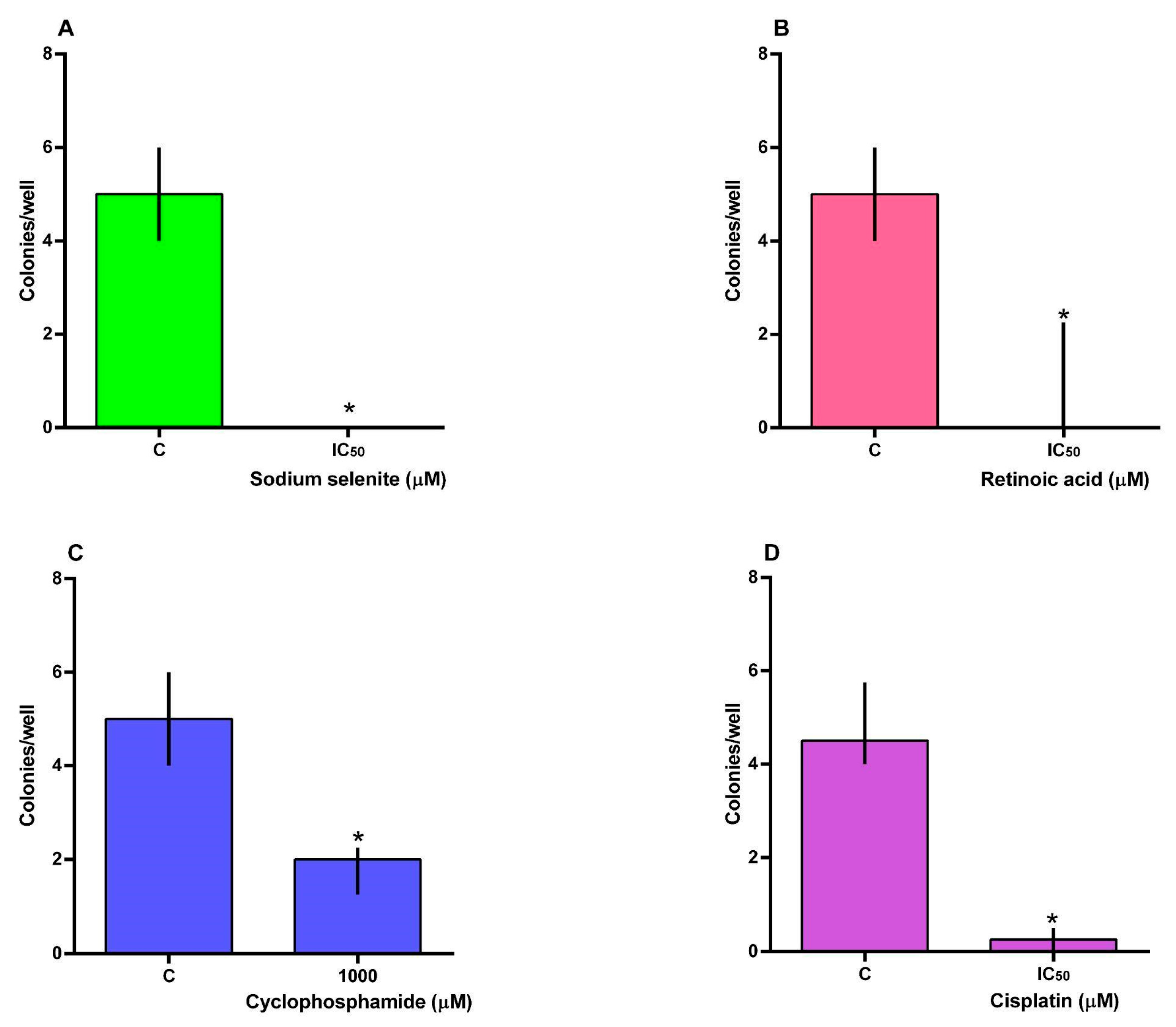
The number of colonies formed after exposure to the compounds is shown in Figure 3A–D. The Mann–Whitney U test revealed significant effects of sodium selenite (U = 0; p = 0.0079; Figure 3A), retinoic acid (U = 0; p = 0.0040; Figure 3B), cyclophosphamide (U = 0; p = 0.0079; Figure 3C), and cisplatin (U = 0; p = 0.0143; Figure 3D). All the compounds significantly decreased the ability of SH-SY5Y cells to form colonies, with a more pronounced effect observed in cells exposed to sodium selenite and retinoic acid.

3.4. Cell Migration

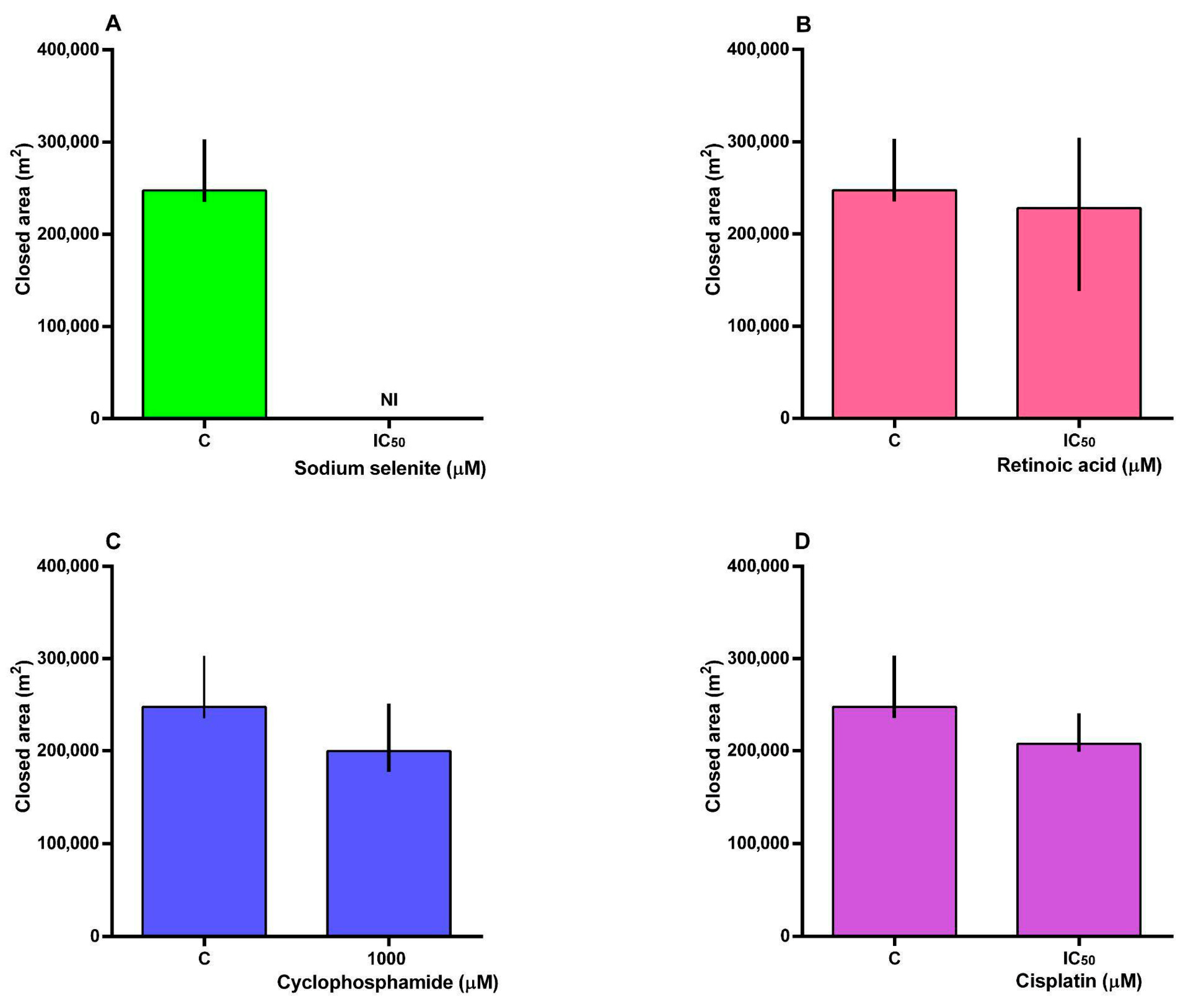
The migration of SH-SY5Y cells exposed to the compounds is shown in Figure 4A–D. The Mann–Whitney U test did not reveal any effect of retinoic acid, cyclophosphamide, or cisplatin exposure on cell migration. However, for sodium selenite at the IC50 concentration (Figure 4A), cell migration could not be assessed, as a large number of cells had detached 24 h after exposure, making it difficult to visualize the scratch area. Representative images of each well at 0 and 24 h after treatments are shown in Figure S2 of the Supplementary Materials.

3.5. BCL2 and BAX Gene Expression

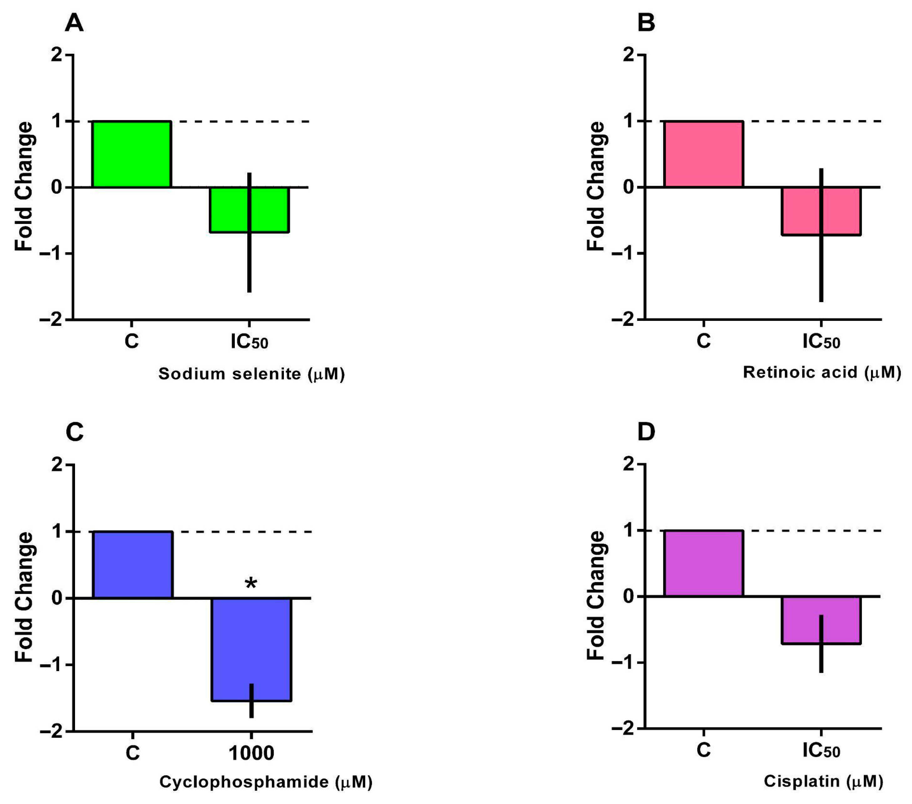
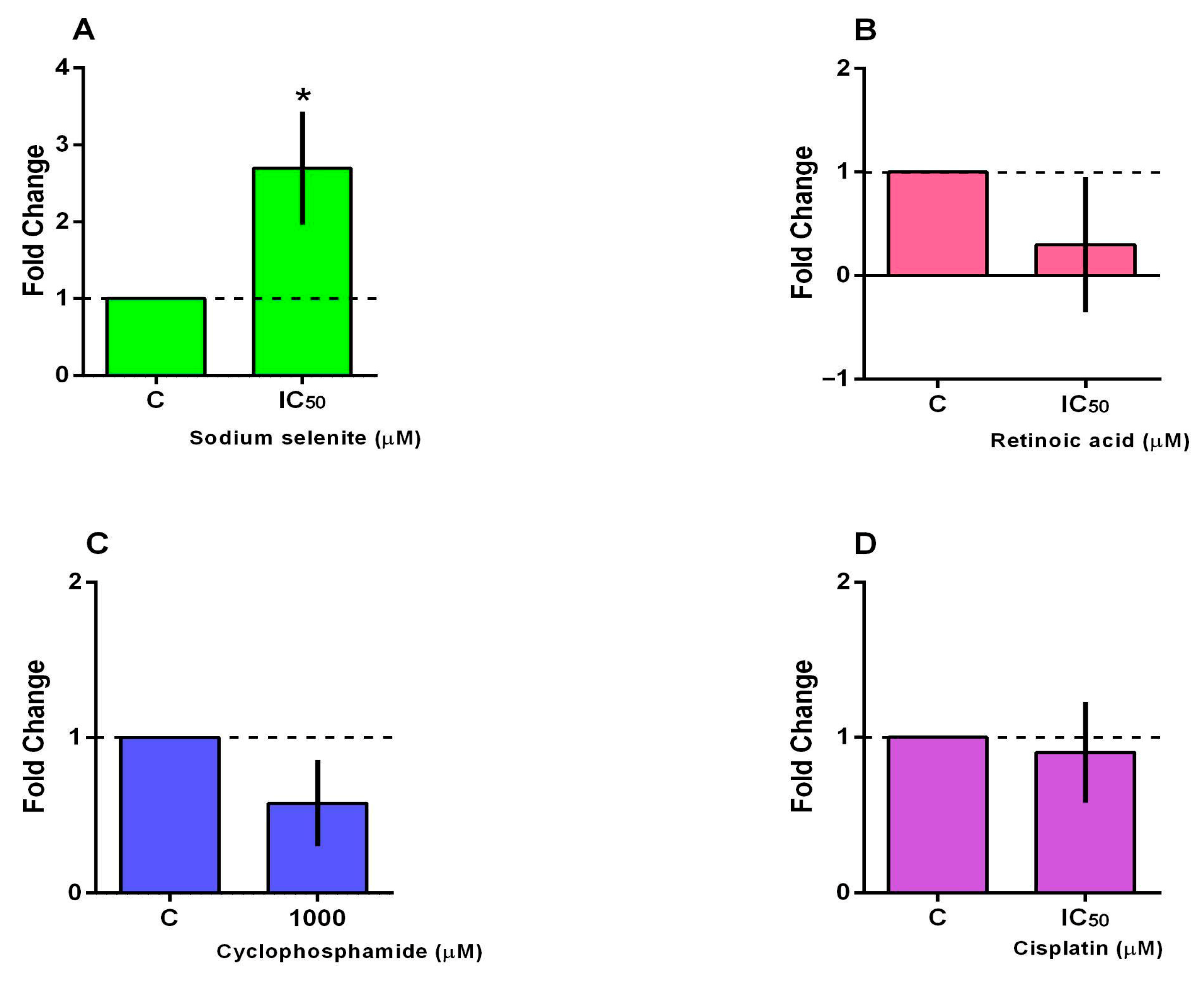
BCL2 expression is shown in Figure 5A–D. The Mann–Whitney U test revealed no significant effect of sodium selenite, retinoic acid, or cisplatin on BCL2 expression. However, the test indicated a significant effect of cyclophosphamide (U = 14; p = 0.0005), which resulted in a marked downregulation of BCL2 expression (Figure 5C). Although not statistically significant, treatments with sodium selenite, retinoic acid, or cisplatin showed a trend toward downregulating BCL2 expression. BAX expression is presented in Figure 6A–D. A Mann–Whitney U test revealed a significant effect of sodium selenite exposure on BAX expression (U = 7.00, p = 0.0025; Figure 6A). In contrast, retinoic acid, cyclophosphamide, or cisplatin had no significant effect on BAX expression, as determined by the same test.

4. Discussion

The search for more effective and less toxic therapies for neuroblastoma remains a major focus in pediatric oncology, especially given the aggressive nature of high-risk cases and the limited success of conventional treatments [22]. In this context, established chemotherapeutic agents remain central to treatment.
In the present study, both sodium selenite and retinoic acid significantly reduced cell viability. These findings suggest enhanced cytotoxic efficacy at this time point, likely due to the compounds’ kinetics of action and the low proliferation rate of the cell line. This discovery is aligned with the results of studies with other human cancer cell lines. Lv et al. [23] reported that sodium selenite significantly inhibited the growth of cervical cancer cells (HeLa and SiHa) in a time- and dose-dependent manner, demonstrating its pro-oxidant and antiproliferative effects. In other cell types, such as synovial sarcoma (SW982), sodium selenite exhibited much lower IC50 values (9.3 µM at 72 h) than those observed in our study, reinforcing its lineage-dependent specificity and suggesting the existence of favorable therapeutic windows [24]. Regarding retinoic acid, Pan et al. [25] demonstrated cytotoxic and differentiation-inducing effects in SH-SY5Y cells following 48 h of exposure.
Retinoic acid has been incorporated into neuroblastoma treatment protocols, particularly as maintenance therapy, to promote tumor cell differentiation and reduce recurrence. Its clinical use is based on its ability to induce the maturation of residual neuroblastoma cells into a less proliferative state [12]. Cyclophosphamide and cisplatin are widely used in neuroblastoma therapy, particularly as part of multimodal protocols for high-risk patients [10]. However, therapeutic response is often limited by intrinsic or acquired resistance, and the underlying mechanisms in neuroblastoma remain incompletely understood [26]. Cyclophosphamide is an alkylating agent that acts as a prodrug, requiring hepatic cytochrome P450-mediated activation to 4-hydroxycyclophosphamide and aldophosphamide [27]. Because most cell culture systems, including SH-SY5Y cells, lack this metabolic capacity, the parent compound remains largely inactive, which explains the absence of significant cytotoxic effects observed in our viability assays [28]. Consequently, the IC50 values obtained in vitro may underestimate the drug’s true cytotoxic potential in vivo, where metabolic activation occurs. Importantly, the clonogenic assay captures reproductive cell death resulting from DNA damage and is more sensitive to delayed cytostatic effects than short-term metabolic viability assays [29]. This difference likely accounts for the significant inhibition of colony formation observed despite the minimal acute cytotoxicity of cyclophosphamide. Furthermore, cyclophosphamide has been shown to modulate the BCL2 family balance, decreasing BCL2 and, in some models, increasing BAX expression—findings consistent with our observation of BCL2 downregulation without a marked reduction in viability [30]. Together, these results suggest that cyclophosphamide can exert sublethal or delayed genotoxic effects in vitro and highlight the importance of testing 4-hydroxycyclophosphamide in future studies to refine potency estimates and clarify underlying mechanisms.
Cell migration is a biological process involving the coordinated movement of cell groups and is a critical event in metastatic progression, during which tumor cells spread to distant organs and establish secondary colonies [31]. The metastatic potential of solid tumor-derived cells is one of the leading causes of cancer-related mortality [32]. In the present study, sodium selenite induced cell detachment, thereby inhibiting the possibility of cell migration. Similarly, da Costa et al. [16] observed that sodium selenite, even at a concentration of 1 µM, caused cell detachment in breast cell lines, preventing the evaluation of cell migration.
During the migration assay, SH-SY5Y cells exposed to sodium selenite for 24 h detached from the plate surface, preventing reliable quantification. This phenomenon does not necessarily indicate cell death, as SH-SY5Y cells can survive in suspension due to their semi-adherent nature [33]. Sodium selenite has been described as a pro-oxidant agent that increases reactive oxygen species (ROS) and disrupts integrin-mediated adhesion and cytoskeletal organization [34]. Such redox-dependent alterations may explain the observed detachment, suggesting that sodium selenite, in addition to inducing cytotoxicity as observed in the present study assays, also affects cell-adhesion dynamics.
BAX and BCL2 genes are well-established molecular markers of apoptosis, with elevated BAX expression indicating a pro-apoptotic state, while increased BCL2 expression reflects enhanced resistance to programmed cell death [35]. While BCL2 functions as an anti-apoptotic protein that stabilizes the mitochondrial membrane and prevents caspase activation, BAX promotes apoptosis by undergoing conformational activation, oligomerization, and membrane insertion, leading to mitochondrial permeabilization and caspase-9 activation [36].
In the present study, exposure to sodium selenite increased the expression of BAX, suggesting a shift toward a pro-apoptotic state and activation of the intrinsic apoptotic pathway. Similar findings have been reported in studies where selenium compounds triggered oxidative stress–mediated apoptosis by modulating the p53–BAXBCL2 axis and disrupting redox homeostasis in cancer cells [15]. These results align with the observed alterations in the cell cycle, where sodium selenite caused arrest and reduction in viable proliferating cells, reinforcing the pro-apoptotic and antiproliferative potential.

5. Conclusions and Limitations

Sodium selenite and retinoic acid have demonstrated significant antiproliferative effects on neuroblastoma cell lines. The results suggest that these compounds may influence cell viability through distinct yet complementary molecular pathways, highlighting their potential as adjuvant therapeutic agents for this pediatric malignancy; however, this possibility was not directly evaluated in the present study, which focused instead on assessing clonogenic potential, migration, cell cycle progression, and the expression of apoptosis-related genes (BCL2 and BAX). We suggest further in-depth studies to clarify their mechanisms of action, determine optimal dosing strategies, and evaluate their efficacy in combination with conventional chemotherapeutic agents. Such investigations could contribute to the development of more effective and less toxic treatment options for neuroblastoma.
Considering the biological complementarity between retinoic acid and selenium, future studies could investigate combined treatments (sodium selenite + retinoic acid) to assess potential synergistic or antagonistic effects in neuroblastoma cells. It would also be valuable to evaluate these compounds in non-tumoral cell lines, allowing the assessment of selectivity and potential cytotoxicity toward normal cells, and expanding future studies to include additional neuroblastoma cell lines with distinct genetic backgrounds and a tumoral cell line of non-neuronal origin to evaluate the reproducibility and specificity of the observed effects. Such investigations could clarify whether the interaction between the mechanisms of each compound enhances therapeutic efficacy while minimizing systemic toxicity. Therefore, exploring their combined action may provide important insights into potential adjuvant strategies for neuroblastoma treatment and improve translational relevance. In addition, considering the potential artificial behavior of immortalized cell lines, the present conclusions are specifically applicable to the SH-SY5Y neuroblastoma model. Further experiments using additional neuroblastoma and non-tumoral cell lines are warranted to confirm and generalize these findings.

Supplementary Materials

The following supporting information can be downloaded at: https://www.mdpi.com/article/10.3390/futurepharmacol5040065/s1. Figure S1: Dose–response curves of the neuroblastoma cell line (SH-SY5Y) exposed to various test compounds at incubation times of 24 (A, D, G, J), 48 (B, E, H, K), and 72 (C, F, I, L) hours. The compounds evaluated were sodium selenite (A–C), retinoic acid (D–F), cyclophosphamide (G–I), and cisplatin (J–L). Results were analyzed and calculated using nonlinear regression analysis with the four-parameter logistic (4PL) model (Hill equation) (n = 3–4). C: control—non-exposed cells; Figure S2: Cell migration. Photographs were taken under an inverted microscope (10x magnification; Evos XL Core) before the addition of the test compounds (time 0) and after exposure to the compounds (time 24 h) of the SH-SY5Y cell line. Control (A). sodium selenite (B), retinoic acid (C), cyclophosphamide (D), and cisplatin (E).

Author Contributions

Conceptualization, M.M.R., L.S.L., Q.I.G., and C.S.O.; investigation, M.M.R., L.S.L., N.d.S.d.C., M.E.P., A.S.F., L.R.C., A.C.I., Q.I.G., and C.S.O.; writing—review and editing, M.M.R., L.S.L., M.E.P., Q.I.G., and C.S.O.; visualization, C.S.O.; supervision, Q.I.G., and C.S.O. All authors have read and agreed to the published version of the manuscript.

Funding

This research received no external funding.

Institutional Review Board Statement

Not applicable.

Informed Consent Statement

Not applicable.

Data Availability Statement

The raw data supporting the conclusions of this article will be made available by the authors on request.

Acknowledgments

During the preparation of this manuscript, the authors used ChatGPT (OpenAI, GPT-5 model) for the purpose of English language editing and text refinement. The authors have carefully reviewed and edited all AI-generated content and take full responsibility for the final version of this publication. This study was financed in part by the Coordenação de Aperfeiçoamento de Pessoal de Nível Superior—Brasil (CAPES)—Finance Code 001 and the Instituto de Pesquisa Pelé. Pequeno Príncipe (IPPPP).

Conflicts of Interest

The authors declare no conflicts of interest.

References

  1. Lam, C.G.; Howard, S.C.; Bouffet, E.; Pritchard-Jones, K. Science and health for all children with cancer. Science 2019, 363, 1182–1186. [Google Scholar] [CrossRef] [PubMed]
  2. World Health Organization (WHO). Childhood Cancer. WHO Website 2025. Available online: https://www.who.int/news-room/fact-sheets/detail/cancer-in-children (accessed on 27 March 2025).
  3. Zafar, A.; Khatoon, S.; Khan, M.J.; Abu, J.; Naeem, A. Advancements and limitations in traditional anti-cancer therapies: A comprehensive review of surgery, chemotherapy, radiation therapy, and hormonal therapy. Discov. Oncol. 2025, 16, 607. [Google Scholar] [CrossRef] [PubMed]
  4. Pudela, C.; Balyasny, S.; Applebaum, M.A. Nervous system: Embryonal tumors: Neuroblastoma. Atlas Genet. Cytogenet. Oncol. Haematol. 2020, 24, 284–290. [Google Scholar] [CrossRef]
  5. Qiu, B.; Matthay, K.K. Advancing therapy for neuroblastoma. Nat. Rev. Clin. Oncol. 2022, 19, 515–533. [Google Scholar] [CrossRef]
  6. Martina-Perez, S.F.; Heirene, L.A.; Kasemeier, J.C.; Kulesa, P.M.; Baker, R.E. Modeling cell differentiation in neuroblastoma: Insights into development, malignancy, and treatment relapse. J. Theor. Biol. 2025, 2502, 20939. [Google Scholar] [CrossRef]
  7. Heinly, B.E.; Grant, C.N. Cell adhesion molecules in neuroblastoma: Complex roles, therapeutic potential. Front. Oncol. 2022, 2, 782186. [Google Scholar] [CrossRef]
  8. Cogo, S.C.; Nascimento, T.G.F.C.; Pinhatti, F.A.B.; Junior, N.F.; Rodrigues, B.S.; Cavalli, L.R.; Elifio-Esposito, S. An overview of neuroblastoma cell lineage phenotypes and in vitro models. Exp. Biol. Med. 2020, 245, 1637–1647. [Google Scholar] [CrossRef]
  9. dos Santos, M.G.; Gomes, J.R.; Costa, M.D.M. Methods used to achieve different levels of neuronal differentiation in SH-SY5Y and Neuro2a cell lines: An integrative review. Cell Biol. Int. 2023, 47, 1883–1894. [Google Scholar] [CrossRef]
  10. Gu, L.; Hickey, R.J.; Malkas, L.H. Therapeutic targeting of DNA replication stress in cancer. Genes 2023, 14, 1346. [Google Scholar] [CrossRef]
  11. Yaster, M.; McNaull, P.P.; Davis, P.J. The opioid epidemic in pediatrics: A 2020 update. Curr. Opin. Anesthesiol. 2020, 33, 327–334. [Google Scholar] [CrossRef] [PubMed]
  12. Makimoto, A.; Fujisaki, H.; Matsumoto, K.; Takahashi, Y.; Cho, Y.; Morikawa, Y.; Yuza, Y.; Tajiri, T.; Iehara, T. Retinoid therapy for neuroblastoma: Historical overview, regulatory challenges, and prospects. Cancers 2024, 16, 544. [Google Scholar] [CrossRef]
  13. Liu, X.; Jiang, M.; Pang, C.; Wang, J.; Hu, L. Sodium selenite inhibits proliferation and metastasis through ROS-mediated NF-κB signaling in renal cell carcinoma. BMC Cancer 2022, 22, 870. [Google Scholar] [CrossRef] [PubMed]
  14. Rocha, J.B.T.; Piccoli, B.C.; Oliveira, C.S. Biological and chemical interest in selenium: A brief historical account. Arkivoc 2017, 2, 457–491. [Google Scholar] [CrossRef]
  15. Rocha, J.B.T.; Oliveira, C.S.; Nogara, P.A. Toxicology and Anticancer Activity of Synthetic Organoselenium Compounds. In Chemistry Beyond Chlorine; Jain, V.K., Priyadarsini, K.I., Eds.; Royal Society of Chemistry: Cambridge, UK, 2017; Chapter 13; pp. 342–376. [Google Scholar]
  16. da Costa, N.S.; Lima, L.S.; Oliveira, F.A.M.; Galiciolli, M.E.A.; Manzano, M.I.; Garlet, Q.I.; Oliveira, C.S. Antiproliferative effect of inorganic and organic selenium compounds in breast cell lines. Biomedicines 2023, 11, 1346. [Google Scholar] [CrossRef]
  17. Mosmann, T. Rapid Colorimetric Assay for Cellular Growth and Survival: Application to Proliferation and Cytotoxicity Assays. J. Immunol. Methods. 1983, 65, 55–63. [Google Scholar] [CrossRef] [PubMed]
  18. Veiga, A.A.; Irioda, A.C.; Mogharbel, B.F.; Bonatto, S.J.R.; Souza, L.M. Quercetin-Rich Extracts from Onions (Allium cepa) Play Potent Cytotoxicity on Adrenocortical Carcinoma Cell Lines, and Quercetin Induces Important Anticancer Properties. Pharmaceuticals 2022, 15, 754. [Google Scholar] [CrossRef]
  19. Sotiropoulou, P.A.; Perez, S.A.; Salagianni, M.; Baxevanis, C.N.; Papamichail, M. Characterization of the optimal culture conditions for clinical scale production of human mesenchymal stem cells. Stem Cells 2006, 24, 462–471. [Google Scholar] [CrossRef]
  20. Almeida, V.M.; Bezerra, M.A., Jr.; Nascimento, J.C.; Amorim, L.M.F. Anticancer drug screening: Standardization of in vitro wound healing assay. J. Bras. Patol. Med. 2019, 55, 606–619. [Google Scholar] [CrossRef]
  21. Livak, K.J.; Schmittgen, T.D. Analysis of relative gene expression data using real-time quantitative PCR and the 2−∆∆CT method. Methods 2001, 25, 402–408. [Google Scholar] [CrossRef]
  22. Wahba, A.; Wolters, R.; Foster, J.H. Neuroblastoma in the era of precision medicine: A clinical review. Cancers 2023, 15, 4722. [Google Scholar] [CrossRef]
  23. Lv, C.; Zeng, Q.; Qi, L.; Wang, Y.; Li, J.; Sun, H.; Du, L.; Hao, S.; Li, G.; Feng, C.; et al. Sodium Selenite Induces Autophagy and Apoptosis in Cervical Cancer Cells via Mitochondrial ROS-Activated AMPK/mTOR/FOXO3a Pathway. Antioxidants 2024, 13, 1004. [Google Scholar] [CrossRef] [PubMed]
  24. Yang, L.; Cai, Y.S.; Xu, K.; Zhu, J.L.; Li, Y.B.; Wu, X.Q.; Sun, J.; Lu, S.M.; Xu, P. Sodium selenite induces apoptosis and inhibits autophagy in human synovial sarcoma cell line SW982 in vitro. Mol. Med. Rep. 2018, 17, 6560–6568. [Google Scholar] [CrossRef] [PubMed]
  25. Pan, M.; Zhang, Y.; Wright, W.C.; Liu, X.; Passaia, B.; Currier, D.; Low, J.; Chapple, R.H.; Steele, J.A.; Connelly, J.P.; et al. Bone morphogenetic protein (BMP) signaling determines neuroblastoma cell fate and sensitivity to retinoic acid. Nat. Commun. 2025, 16, 2036. [Google Scholar] [CrossRef]
  26. Illendula, A.; Fultang, N.; Peethambaran, B. Retinoic acid induces differentiation in neuroblastoma via ROR1 by modulating retinoic acid response elements. Oncol. Rep. 2020, 44, 1013–1024. [Google Scholar] [CrossRef]
  27. Webb, E.R.; Moreno-Vicente, J.; Easton, A.; Lanati, S.; Taylor, M.; James, S.; Williams, E.L.; English, V.; Penfold, C.; Beers, S.A.; et al. Cyclophosphamide depletes tumor infiltrating T regulatory cells and combined with anti-PD-1 therapy improves survival in murine neuroblastoma. iScience. 2022, 25, 104995. [Google Scholar] [CrossRef]
  28. Krüger-Genge, A.; Köhler, S.; Laube, M.; Haileka, V.; Lemm, S.; Majchrzak, K.; Kammerer, S.; Schulz, C.; Storsberg, J.; Pietzsch, J.; et al. Anti-Cancer Prodrug Cyclophosphamide Exerts Thrombogenic Effects on Human Venous Endothelial Cells Independent of CYP450 Activation, Relevance to Thrombosis. Cells 2023, 12, 1965. [Google Scholar] [CrossRef]
  29. Villanova, L.; Careccia, S.; De Maria, R.; Fiori, M.E. Micro-Economics of Apoptosis in Cancer: ncRNAs Modulation of BCL-2 Family Members. Int. J. Mol. Sci. 2018, 19, 958. [Google Scholar] [CrossRef]
  30. Dewidar, S.A.; Hamdy, O.; Soliman, M.M.; El Gayar, A.M.; El-Mesery, M. Enhanced therapeutic efficacy of doxorubicin/cyclophosphamide in combination with pitavastatin or simvastatin against breast cancer cells. Med. Oncol. 2023, 41, 7. [Google Scholar] [CrossRef] [PubMed]
  31. Pally, D.; Naba, A. Extracellular matrix dynamics: A key regulator of cell migration across length-scales and systems. Curr. Opin. Cell Biol. 2024, 86, 102309. [Google Scholar] [CrossRef]
  32. Chastney, M.R.; Kaivola, J.; Leppänen, V.M.; Ivaska, J. The role and regulation of integrins in cell migration and invasion. Nat. Rev. Mol. Cell Biol. 2025, 26, 147–167. [Google Scholar] [CrossRef]
  33. Kovalevich, J.; Langford, D. Considerations for the use of SH-SY5Y neuroblastoma cells in neurobiology. Methods Mol. Biol. 2013, 1078, 9–21. [Google Scholar] [PubMed]
  34. Zhao, S.; Meng, Y.; Cai, W.; Luo, Q.; Gao, H.; Shen, Q.; Shi, D. Docosahexaenoic Acid Coordinating with Sodium Selenite Promotes Paraptosis in Colorectal Cancer Cells by Disrupting the Redox Homeostasis and Activating the MAPK Pathway. Nutrients 2024, 16, 1737. [Google Scholar] [CrossRef] [PubMed]
  35. Youle, R.J.; Strasser, A. The BLC-2 protein family: Opposing activities that mediate cell death. Nat. Rev. Mol. Cell Biol. 2008, 9, 47–59. [Google Scholar] [CrossRef] [PubMed]
  36. Varlamova, E.G.; Baimler, I.V.; Gudkov, S.V.; Turovsky, E.A. Comparative study of the anticancer effects of selenium nanoparticles and nanorods: Regulation os Ca2+ signaling, ER stress, and apoptosis. Appl. Sci. 2023, 13, 10763. [Google Scholar] [CrossRef]
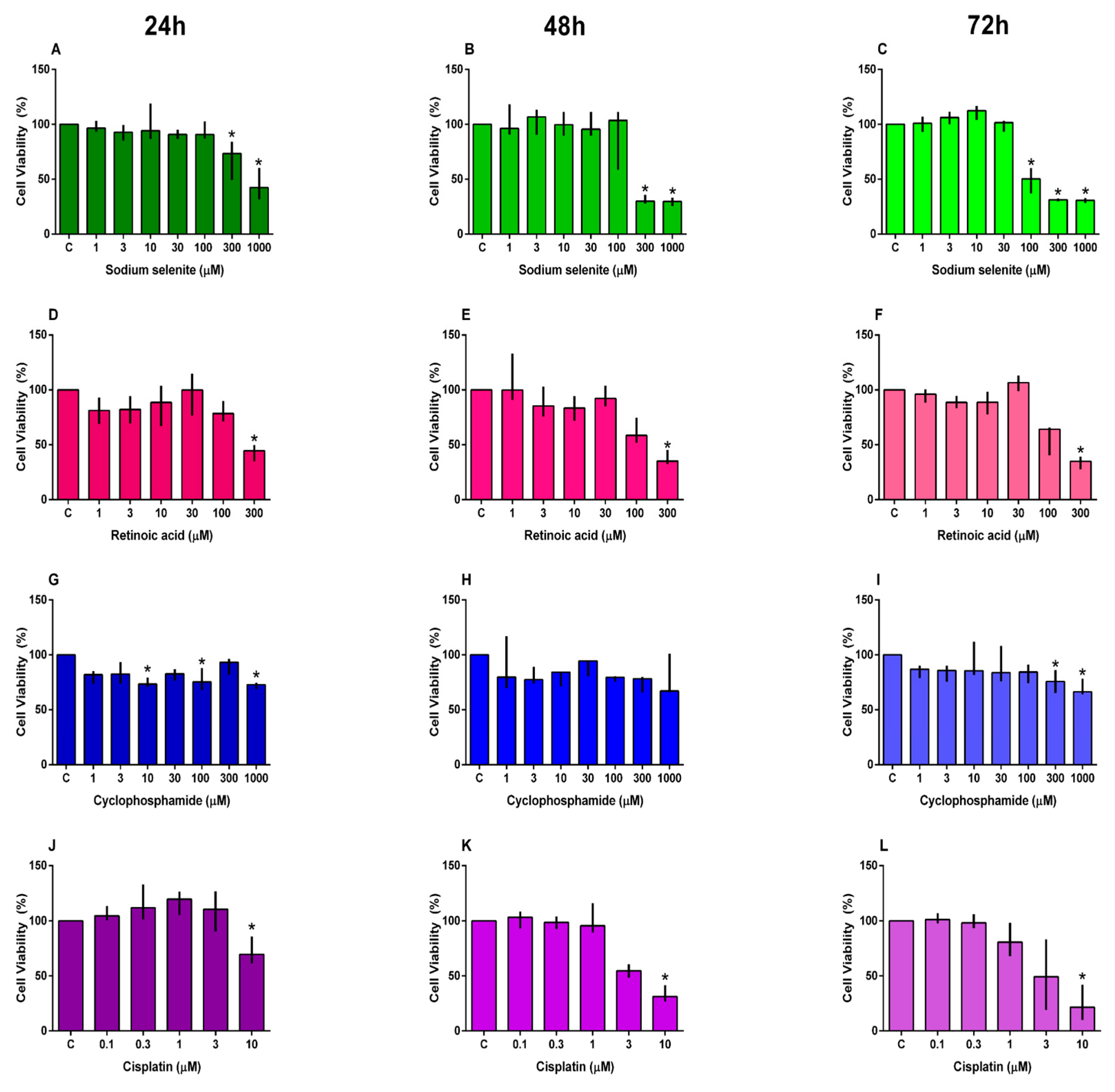
Figure 1. Cell viability of SH-SY5Y cells exposed to sodium selenite (AC), retinoic acid (DF), cyclophosphamide (GI), and cisplatin (JL) was assessed after 24 h (A,D,G,J), 48 h (B,E,H,K), and 72 h (C,F,I,L) of exposure. Results were analyzed using the Kruskal–Wallis test followed by Dunn’s post hoc test, and are presented as median ± interquartile range (n = 3–4). * Statistically different from C (not exposed cells).
Futurepharmacol 05 00065 g001
Figure 2. Cell cycle phases of the SH-SY5Y cell line exposed for 72 h to sodium selenite (A), retinoic acid (B), cyclophosphamide (C), and cisplatin (D). Cell cycle phase distribution (G0/G1, S, G2) after treatment with tested compounds (E). The results were analyzed using the Kruskal–Wallis test followed by Dunn’s post hoc test, and are presented as median ± interquartile range (n = 3–4). * Statistically different from C (not exposed cells).
Futurepharmacol 05 00065 g002aFuturepharmacol 05 00065 g002b
Figure 3. Colonies per well of SH-SY5Y cells exposed for 72 h to sodium selenite (A), retinoic acid (B), cyclophosphamide (C), and cisplatin (D). The results were analyzed using the Mann–Whitney U test, and are presented as median ± interquartile range (n = 4–6). * Statistically different from C (not exposed cells).
Futurepharmacol 05 00065 g003
Figure 4. Cell migration of SH-SY5Y cells exposed for 24 h to sodium selenite (A), retinoic acid (B), cyclophosphamide (C), and cisplatin (D). The results were analyzed using the Mann–Whitney U test, and are presented as median ± interquartile range (n = 3). NI: not identified/detached cells.
Futurepharmacol 05 00065 g004
Figure 5. BCL2 expression in SH-SY5Y cells exposed for 72 h to sodium selenite (A), retinoic acid (B), cyclophosphamide (C), and cisplatin (D). The results were analyzed using the Mann–Whitney U test, and are presented as median ± interquartile range (n = 6). * Statistically different from C (not exposed cells).
Futurepharmacol 05 00065 g005
Figure 6. BAX expression in SH-SY5Y cells exposed for 72 h to sodium selenite (A), retinoic acid (B), cyclophosphamide (C), and cisplatin (D). The results were analyzed using the Mann–Whitney U test, and are presented as median ± interquartile range (n = 6). * Statistically different from C (not exposed cells).
Futurepharmacol 05 00065 g006
Table 1. IC50 values of the tested compounds based on SH-SY5Y cell viability after 24, 48, and 72 h of exposure.
Compound24 h48 h72 h
IC50 (µM)IC50 (µM)IC50 (µM)
Sodium Selenite722.10294.20166.30
Retinoic Acid251.10174.30178.60
Cisplatin-5.133.45
Disclaimer/Publisher’s Note: The statements, opinions and data contained in all publications are solely those of the individual author(s) and contributor(s) and not of MDPI and/or the editor(s). MDPI and/or the editor(s) disclaim responsibility for any injury to people or property resulting from any ideas, methods, instructions or products referred to in the content.