Article

Targeting Oxidative Stress and Inflammation with Vitis vinifera Leaf Extract: A Combined Experimental and Computational Pharmacological Study

1
Department of Pharmacy, Faculty of Medical Sciences, University of Kragujevac, 34000 Kragujevac, Serbia
2
Department of Physiology, Faculty of Medical Sciences, University of Kragujevac, 34000 Kragujevac, Serbia
3
Center of Excellence for Redox Balance Research in Cardiovascular and Metabolic Disorders, University of Kragujevac, 34000 Kragujevac, Serbia
4
Department of Human Pathology, 1st Moscow State Medical, University IM Sechenov, 119991 Moscow, Russia
*
Author to whom correspondence should be addressed.
Future Pharmacol. 2025, 5(3), 52; https://doi.org/10.3390/futurepharmacol5030052
Submission received: 13 July 2025 / Revised: 24 August 2025 / Accepted: 12 September 2025 / Published: 14 September 2025
(This article belongs to the Special Issue Recent Advances in the Discovery of Anti-Inflammatory Compounds)

Abstract

Objectives: Our study aimed to examine the antioxidative and anti-inflammatory potential of the lyophilized aqueous leaf extract of Vitis vinifera. Methods: The antioxidant capacity of the extract was evaluated using the DPPH and FRAP assays. The in vivo phase of the study included 40 male Wistar albino rats. One half of the animals were used to induce the carrageenan model of acute inflammation, while the other half were used for examination of the extract effect on the redox state. Rats from the experimental group drank tap water containing 150 mg/kg Vitis vinifera extract for 14 days, while control animals received saline at the same volume. The molecular docking studies of polyphenols present in the leaf extract were conducted in AutoDock Vina. Results: In vitro assessment of the antioxidative capacity of the applied extract revealed significant free radical scavenging activity (IC50 value 11.63 µg/mL), along with a pronounced ferric reducing ability (0.143 at 700 nm). Moreover, animal treatment with the extract led to significant paw edema inhibition (30.34%, 35.06%, and 41.54% in the second, third, and fourth hours, respectively) and to pro-oxidative marker reduction. Additionally, Vitis vinifera extract significantly increased catalase activity and glutathione levels. The in silico results showed that rutin binds to cyclooxygenase 1 (−8.2 kcal/mol) and 2 (−8.3 kcal/mol), as well as to antioxidant enzymes (catalase: −8.6 kcal/mol, SOD: −7.4 kcal/mol), indicating its key role in mediating the biological activity of the tested extract. Conclusions: This study highlights the significant antioxidant and anti-inflammatory potential of V. vinifera lyophilized aqueous leaf extract from the Serbian market, supported by both in vivo and in silico analyses.

1. Introduction

Free radicals are highly reactive chemical species that possess unpaired electron(s) in the outer orbit, enabling easy participation in chemical chain reactions with biomolecules. According to the reactive nature of the atom, free radicals are classified into three different groups—reactive oxygen species (ROS), reactive nitrogen species (RNS), and reactive chlorine species (RCS)—among which ROS represent the most critical class [1]. ROS are chemically reactive molecules, normally generated in limited quantities within the body during aerobic metabolism, thus regulating processes involved in cell homeostasis [2]. The most important source of intracellular ROS are mitochondria, and the formation of the superoxide anion radical (O2.−) occurs during leakage of electrons from the respiratory chain when an oxygen molecule gains one electron. Moreover, nitric oxide (NO) can also be produced during the respiratory chain reaction, leading to RNS formation [3]. At low levels free radicals help regulate physiological activities; however, under pathophysiological conditions, the redox balance becomes disturbed, leading to oxidative stress. Oxidative stress is defined as a state during which ROS production overwhelms the capacity of an antioxidative defense system, resulting in damage to primary cellular components such as proteins, DNA, and lipids [4]. It has been observed that the redox balance state is disturbed during inflammation, in favor of the release of pro-oxidative molecules, which implies that oxidative stress remains a key component in the inflammation pathogenesis [5]. On the other hand, increased levels of ROS/RNS may act as an activator of the signaling cascade that increases the expression of pro-inflammatory genes [6]. Additionally, there is no doubt that oxidative stress and inflammation are closely related pathophysiological conditions, tightly linked with one another. Besides enzymatic and non-enzymatic antioxidant defense systems, the discovery of natural compounds with both antioxidant and anti-inflammatory properties is still of great importance.
Vitis vinifera L. (V. vinifera; grapevine) is a perennial plant from the Vitaceae family, which is widely used for wine and grape production. Consequently, a huge amount of waste is generated, which has represented a turning point for intensive research regarding the reuse of those by-products [7]. Various studies have demonstrated that grapes are a major source of many bioactive compounds that possess a variety of biological activities [8]. It was shown that V. vinifera plant materials are rich in polyphenols, flavonoids, anthocyanins, vitamins, and stilbenoids, which have made this fruit very popular in cosmetics and supplements. Moreover, this plant is a great source of organic acids, minerals, and other nutrients that are important for human nutrition [9]. Grapevine leaves exhibit several biological properties, including antibacterial, anti-inflammatory, vasorelaxant, etc., activities. In addition, the leaves of V. vinifera are traditionally used as a food for treating diarrhea, hemorrhage, hypertension, and hyperglycemia. They have also been reported to exert hepatoprotective effects against acetaminophen-induced liver damage [10,11,12,13].
Molecular modeling studies nowadays play an important role in the investigation of bioactive molecules derived from natural sources. The application of advanced in silico molecular docking studies provides rapid and reliable binding affinity assessment of various phytochemicals for relevant biological targets. Moreover, in silico results can significantly speed up the identification and development of novel plant bioactive compounds, facilitating their progression through the further stages of drug discovery. Numerous studies have previously investigated the molecular interaction profiles of plant polyphenols to evaluate their potential for activation and inhibition of various molecular targets involved in biological pathways of inflammation, oxidative stress, carcinogenesis, and bacterial and viral infections [14,15,16,17,18].
Given these considerations, the aim of our study was to examine the antioxidative and anti-inflammatory potential of the commercial lyophilized aqueous extract of V. vinifera leaves obtained from the Serbian market. Additionally, molecular docking studies were employed to explore the potential molecular mechanisms underlying the antioxidative and anti-inflammatory activities of polyphenolic compounds present in the plant leaves.

2. Material and Methods

2.1. Ethical Consent

This experimental study was conducted in accordance with the EU Directive for the Protection of the Vertebrate Animals used for Experimental and other Scientific Purposes 86/609/EEC and the principles of Good Laboratory Practice (GLP). Investigation was performed in the Center of Excellence for Redox Balance Research in Cardiovascular and Metabolic Disorders, Kragujevac, Serbia, and was approved by the Ethics Committee for Experimental Animal Well-Being of the Faculty of Medical Sciences of the University of Kragujevac, Serbia (Approval No: 01-6292, date of approval: 31 July 2020).

2.2. Chemicals

The lyophilized aqueous extract of V. vinifera leaves (Floralend®) is an official product of the pharmaceutical company Galenika a.d. (Zemun, Belgrade, Serbia). Reagents and chemicals used for the examination of antioxidative and anti-inflammatory properties were purchased from Sigma Aldrich (St. Louis, MO, USA): sodium tetraborate, sodium chloride, sodium citrate, sulfonylic acid, 5,5`-Dithio-bis-(2-nitrobenzoic acid) (DTNB), ethylenediaminetetraacetic acid disodium salt dehydrate, Glutathione reduced (GSH), gelatin, trichloroacetic acid (TCA), tris(hydroxymethyl)aminomethane, thiobarbituric acid (TBA), L-N-(1-naphthyl)-ethylenediamine-dihydrochloride (NEDA), nitro-blue-tetrazolium (NBT), epinephrine, sodium hydroxide, metaphosphoric acid, phosphoric acid, disodium phosphate, ammonium chloride, sodium carbonate, butylated hydroxytoluene (BHT; IUPAC: 2,6-ditert-butyl-4-methylphenol), 1,1-diphenyl-2-picrylhydrazyl, L-Ascorbic Acid, carrageenan, and indomethacin.

2.3. In Vitro Potential Assessment of V. vinifera Extract

The antioxidant activity of V. vinifera extract was assessed using the DPPH and FRAP assays, as widely accepted in vitro methods. These techniques are known for their strong reproducibility and reliability in evaluating antioxidant potential across a variety of samples.

2.3.1. Assessment of DPPH Radical Scavenging Activity

The DPPH assay was utilized to assess the free radical scavenging potential of V. vinifera extract. Various concentrations of the extract were prepared and in cuvettes mixed with an equal volume of DPPH• radical solution. After the incubation period, the absorption was measured spectrophotometrically at 515 nm. The synthetic antioxidant BHT was used as a positive control to ensure the reliability of the assay. Radical scavenging capacity (RSC) was calculated using the following equation:
RSC % = 100 × (Ac − As)/Ac
Ac = absorbance of the control (which contains all reagents, except the tested extract or compound); As = absorbance of the sample. The extract concentration that results in 50% inhibition of the DPPH radical (IC50) was determined from the RSC concentration curve [19].

2.3.2. Assessment of Ferric Reducing Antioxidant Power (FRAP) Assay

The Ferric Reducing Antioxidant Power (FRAP) assay was employed to evaluate the reducing capacity of V. vinifera extract. A stock solution of the extract was initially prepared and subsequently diluted using phosphate buffer (pH 7.4) to the desired concentrations. The resulting solutions were incubated at room temperature for 10 min, after which their absorbance was recorded at 700 nm. Ascorbic acid, at a concentration of 5 µg/mL, was used as a reference standard to validate the assay [20].

2.4. Study Design

A total of forty healthy male Wistar albino rats, 8 weeks old, average body weight (BW) 200 ± 50 g, were obtained from the Military Medical Academy, Belgrade, Serbia, and involved in this study. After a one-week adaptation period in vivarium within the Centre for Preclinical and Functional Investigations of the Faculty of Medical Sciences, University of Kragujevac, Serbia, the rats were randomly assigned into two groups and housed under controlled environmental conditions (room temperature 22 ± 3 °C, humidity 55 ± 5%, and 12 h/12 h (light/dark) cycle). The animals had free access to water and a standard diet for rats. Out of these forty animals, twenty rats were allocated to the carrageenan-induced acute inflammation model, while the remaining twenty animals were used for the examination of sub-acute treatment with the extract on the rats’ redox state and were further divided into two groups, depending on the applied treatment:
  • CTRL (control; n = 10)—healthy untreated rats;
  • V. vinifera (n = 10) experimental group—rats who drank tap water containing 150 mg/kg V. vinifera water extract for 14 days.
The animals were sacrificed after finishing the experimental period with a single intraperitoneal injection of an anesthetic mixture (ketamine/xylazine—100/10 mg/kg of BW). At the moment of decapitation, blood samples were collected for further biochemical analysis.

2.5. Evaluation of Effect of V. vinifera Extract on Systemic Redox State

The whole blood was centrifuged to isolate plasma and erythrocytes plasma and stored at −20 °C until further analysis. The level of the following pro-oxidative parameters was examined spectrophotometrically in the plasma samples: superoxide anion radical (O2.−), hydrogen peroxide (H2O2), nitrites (NO2), and index of lipid peroxidation measured as thiobarbituric acid (TBA) reactive substances (TBARSs). Erythrocyte lysate samples were used for spectrophotometric determination of the antioxidative defense system parameters: the activity of superoxide dismutase (SOD) and catalase (CAT), and the level of reduced glutathione (GSH).

2.5.1. Determination of Pro-Oxidative Parameters

The assessment of the O2.− level in the plasma samples was based on the nitro blue tetrazolium (NBT) reaction in tris(hydroxymethyl)aminomethane (TRIS) buffer. Fifty microliters of plasma sample were mixed with 950 µL of buffer and measured at a wavelength of λ = 530 nm. The level of H2O2 was examined during phenol red oxidation by H2O2 in a reaction catalyzed by horseradish peroxidase (HRP). Freshly prepared red solution in a volume of 800 µL was added to 200 µL of the sample, and then 10 µL of HRP (1:20) (made ex tempore) was subsequently added. After a 10 min incubation period, measurement was performed at λ = 610 nm. The nitrite level was indirectly measured using the index of NO production. Equal volumes (125 µL) of sample and Griess’s reagent were incubated for 10 min and measured at λ = 550 nm. TBARS level determination was performed using a solution of 100 µL 1% thiobarbituric acid in 0.05 M NaOH incubated with 400 µL of sample at 100 °C for 15 min. Measurement was performed at λ = 530 nm [21].

2.5.2. Determination of Antioxidative Parameters

The SOD activity was determined by adding 100 µL of lysate sample to 1 mL of carbonate buffer. Epinephrine was added into an incubation mixture in the volume equal to sample. Measurement was performed at λ = 470 nm after 10 min of incubation period. Lysate samples diluted with distilled water (1:7 v/v) and a chloroform–ethanol (0.6:1 v/v) mixture were used for CAT activity determination. An amount of 100 μL of this mixture were added to 50 μL of CAT buffer and 1 mL of 10 mM H2O2. Measurement was performed at λ = 230 nm. Determination of the GSH level was based on GSH oxidation with 5,5`-Dithio-bis-(2-nitrobenzoic acid) at λ = 412 nm [22].

2.6. Evaluation of Anti-Inflammatory Potential of V. vinifera Extract

The anti-inflammatory potential of the examined extract was evaluated on the carrageenan-induced paw edema model. Carrageenan (0.5%) was dissolved in 0.9% saline, and 1 mL of the solution was injected into the rat’s left hind paw. One hour before inflammation induction, animals from the experimental group were treated with V. vinifera extract at a volume of 300 μL per os, while animals from the CTRL group received the same volume of saline. Using a digital vernier caliper (Aerospace, Beijing, China), the thickness of the rats’ left paw was measured prior to and after the carrageenan injection, at the 1st, 2nd, 3rd, and 4th hour. According to the following formula, the percentage of paw edema inhibition was calculated [23]:
% Inhibition = 100 × [1 − (Yt/Yc)]
Yt—average increase in paw thickness in the treated group of rats between two measurement moments.
Yc—average increase in paw thickness in the untreated group of rats between two measurement moments.

2.7. In Silico Analysis

A semi-flexible docking protocol utilizing the default scoring function was implemented using AutoDock Vina version 1.1.2 [24]. The polyphenolic compounds were selected for in silico simulations according to previously published HPLC-MS/MS analysis of the grape leaf content [25]. The 3D conformer coordinates of the tested polyphenols were retrieved from the PubChem database (https://pubchem.ncbi.nlm.nih.gov/) accessed on 15 June 2025 in sdf file format and subsequently converted into pdb files using PyMOL version 2.5.5 [26]. The crystallographic structures of the selected biological targets, including human CAT (PDB ID: 1DGF) [27], human SOD (PDB ID: 1PU0) [28], cyclooxygenase-1 (COX-1) (PDB ID: 3N8Z) [29], and cyclooxygenase-2 (COX-2) (PDB ID:1CX2) [30], were obtained from the RCSB Protein Data Bank database (http://www.rcsb.org/, accessed on 15 June 2025). The biological data were further processed using Discovery Studio [31] and AutoDock Tools [32] by removing unnecessary protein chains and co-crystallized ligands, followed by the addition of polar hydrogen atoms and Collman charges. Blind molecular docking studies were conducted on human CAT and SOD using a maximized grid box with dimensions of 126 × 126 × 126 points and a grid spacing of 0.375 Å. Focused molecular docking studies were performed on COX-1 and COX-2, employing the same grid spacing value with grid box dimensions of 38 × 32 × 34 points. The binding site cavities on COX-1 and COX-2 were determined based on the coordinates of co-crystallized ligands flurbiprofen and SC-558. The grid box center coordinates for x, y, and z were defined as 20.945, 60.993, and 58.326 for CAT; 57.948, 43.521, and 16.78 for SOD; −20.975, 50.155, and 10.484 for COX-1; and 23.947, 21.582, and 15.436 for COX-2. The visualization of non-covalent binding interactions of the best-docked binding poses of the investigated polyphenols was carried out using PyMOL. Molecular docking simulations were conducted to assess the category, type, and total number of non-covalent interactions, as well as the docking score, equilibrium binding constant (Kb), and inhibition constant (Ki). The values of equilibrium binding and inhibition constants were calculated from docking score values using the equations ∆G = -RTlnKb and ∆G = RTlnKi, respectively. The temperature was set at 298 K, with R representing the universal gas constant with a value of 1.9872036 × 10⁻³ kcal K−1 mol−1.

2.8. Statistical Analysis

All data were analyzed using GraphPad Prism 8 (Version for Windows, GraphPad Software, La Jolla, CA, USA) and expressed as mean ± standard deviation (SD). The sample size for each experimental group was n = 10. After initial analysis of normality by the Shapiro–Wilk test, samples with a normal distribution were analyzed using the Independent t-test, while the Mann–Whitney U-test was applied to non-normally distributed samples. To estimate the differences in variances between groups, one-way analysis of variance (ANOVA) followed by post hoc comparisons with the Bonferroni correction, as well as the Kruskal–Wallis test, was used. Statistical significance was observed at a value of p < 0.05.

3. Results

3.1. In Vitro Antioxidative Capacity of V. vinifera Extract

The in vitro evaluation of the V. vinifera extract antioxidant capacity demonstrated similar free radical scavenging activity in relation to the BHT used as a standard value, as indicated by its IC50 value in the DPPH assay (Figure 1A).
The antioxidant potential of the V. vinifera extract was further confirmed by the FRAP assay, which clearly demonstrated a strong capacity to reduce ferric (Fe3+) to ferrous ions (Fe2+). This reducing ability reflects the antioxidant efficacy of the phenolic constituents present in the extract, highlighting their potential protective role against oxidative damage in biological systems (Figure 1B).

3.2. Effect of V. vinifera Extract on Pro-Oxidative Markers

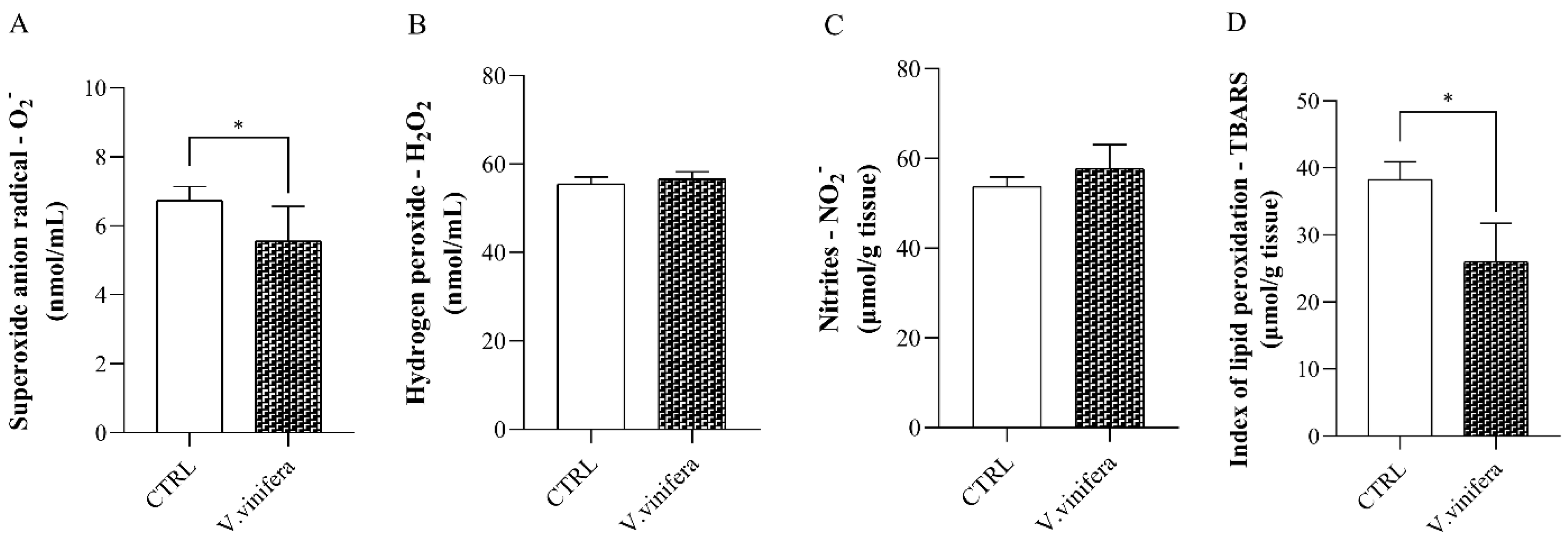
Two-week treatment with lyophilized aqueous extract of V. vinifera resulted in a significantly lower release of O2.− compared to the untreated group of animals (Figure 2A). On the other hand, there were no statistical differences in H2O2 and NO2 plasma concentrations between the CTRL and V. vinifera groups (Figure 2B,C). However, the TBARS level was significantly reduced in the experimental group compared to the CTRL group (Figure 2D).

3.3. Effect of V. vinifera Extract on Antioxidative Markers

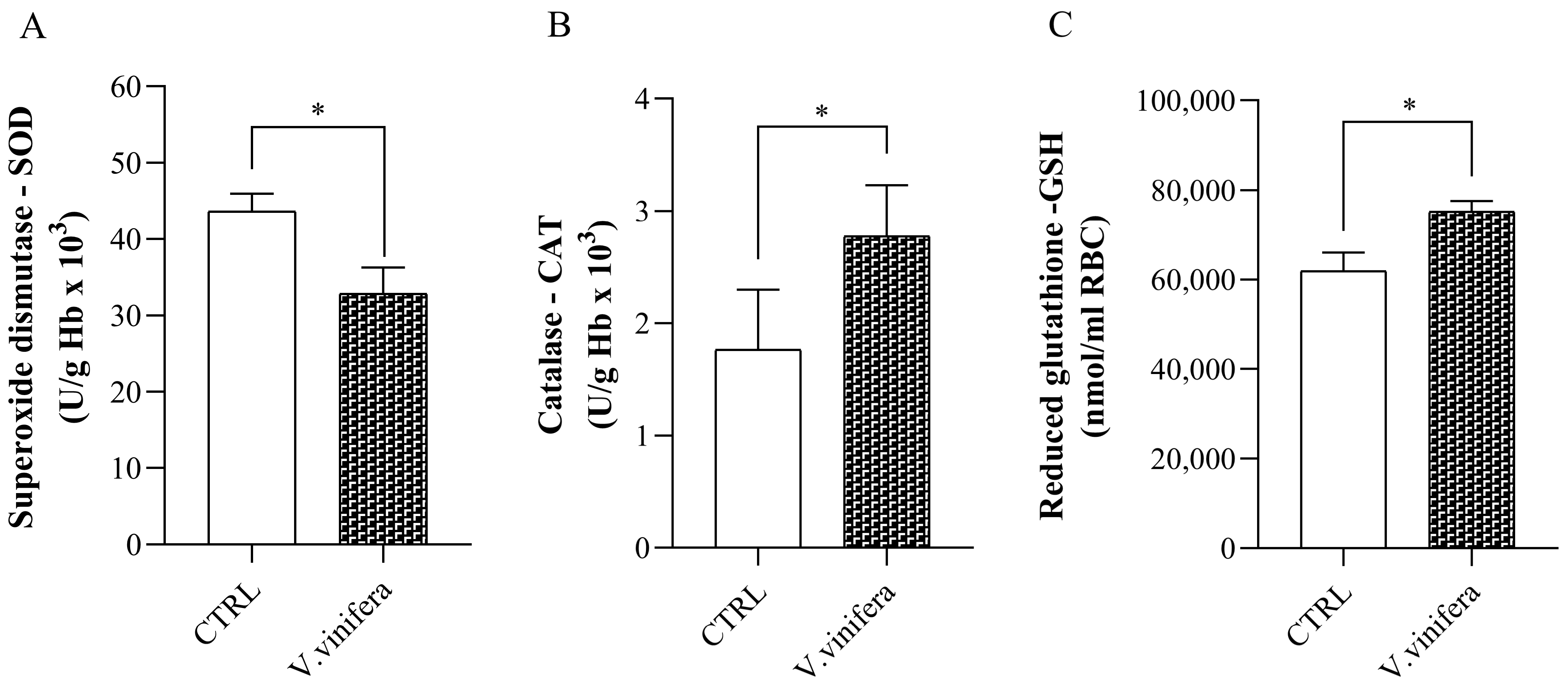
Sub-acute application of V. vinifera lyophilized aqueous extract led to significantly lower SOD activity in comparison to healthy control rats (Figure 3A). However, the antioxidative potential of V. vinifera was proved due to significantly higher CAT activity (Figure 3B) and GSH levels (Figure 3C) in rats from the experimental group compared to the CTRL animals.

3.4. Anti-Inflammatory Activity of V. vinifera Extract

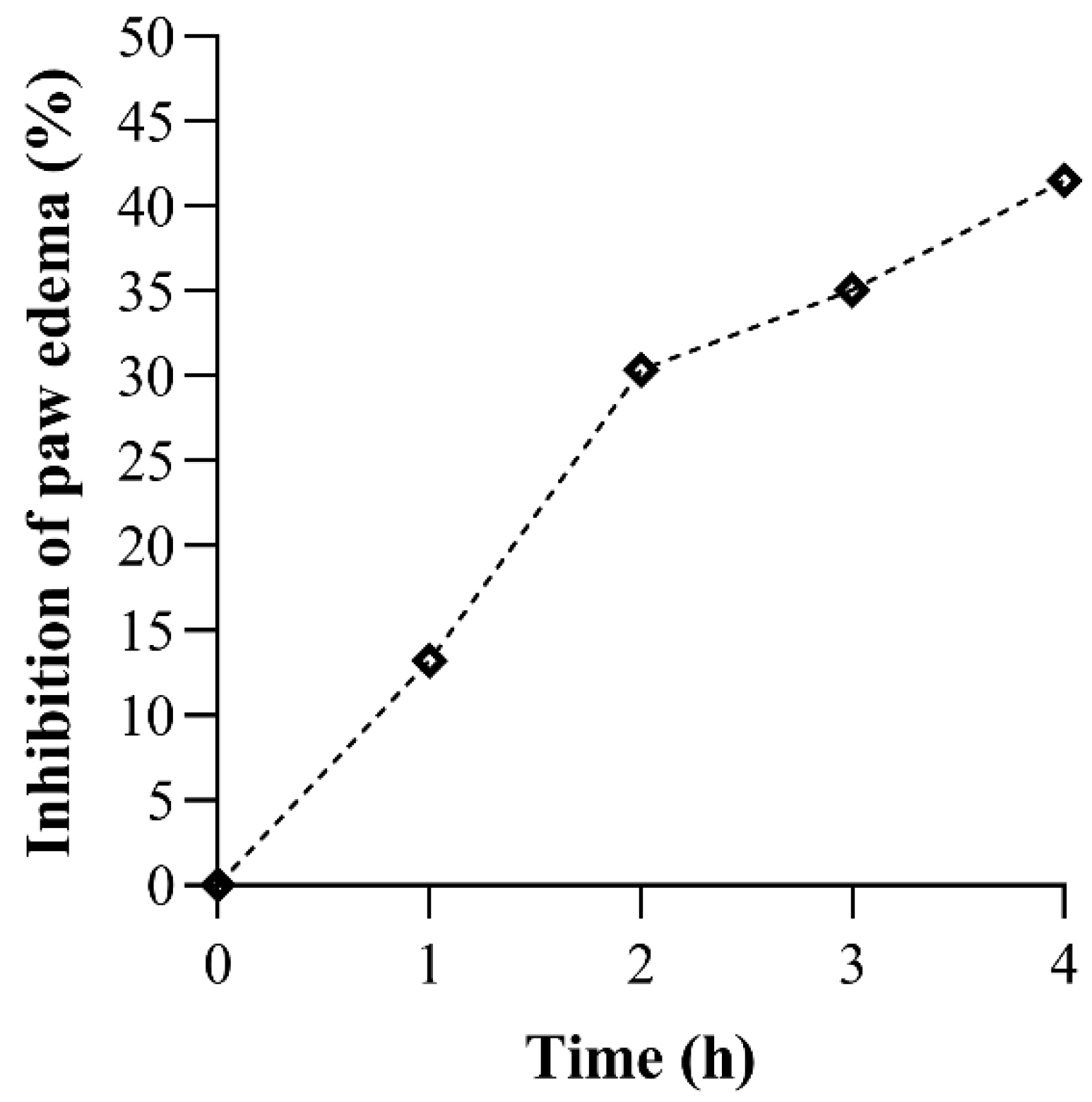
The results of the anti-inflammatory activity of V. vinifera extract are shown in Table 1 and Figure 4. There were no statistical differences between the control and experimental groups before and one hour after carrageenan injection. However, administration of the extract led to significant inhibition of paw edema after the second, third, and fourth hour of inflammation induction compared to the CTRL group.

3.5. In Silico Simulations

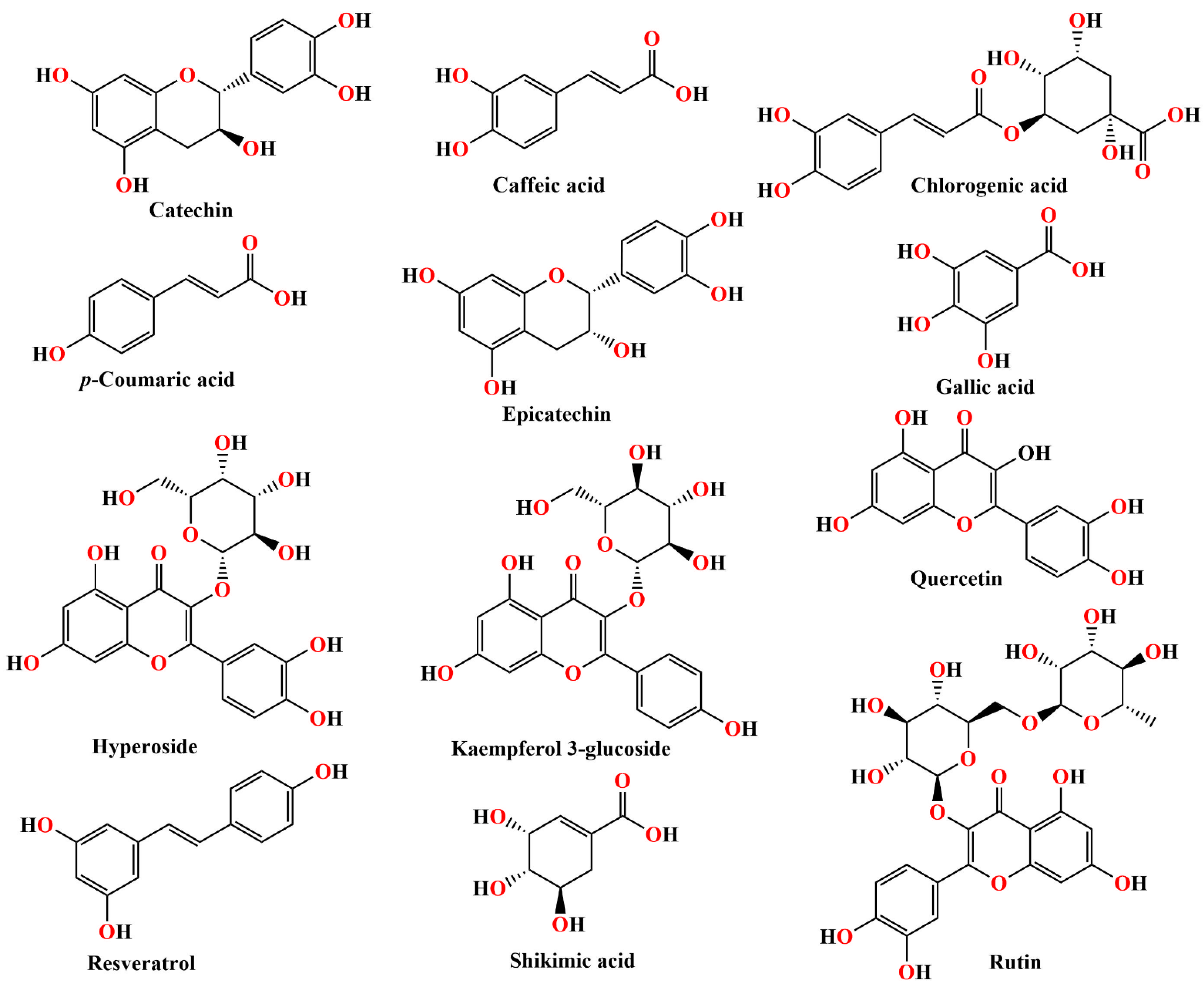
Molecular docking simulations were conducted to examine the interaction profiles of polyphenolic compounds present in V. vinifera leaves (Figure 5) with biological targets involved in molecular mechanisms of their antioxidative and anti-inflammatory activity. Consequently, focused and blind molecular docking studies were performed to assess the binding potential of the investigated polyphenolic compounds for CAT, SOD, COX-1, and COX-2. The binding affinity was assessed based on the following variables: docking score (∆G), equilibrium binding constant (Kb), inhibition constant (Ki), and established non-covalent binding interactions. A lower value of docking score, a higher value of the equilibrium binding constant, and a lower value of the inhibition constant indicate a stronger molecular interaction of the tested compounds with the selected biological targets.

3.5.1. Blind Molecular Docking Studies

Molecular docking data for the best-docked conformations of the investigated polyphenolic compounds within the CAT are outlined in Table 2.
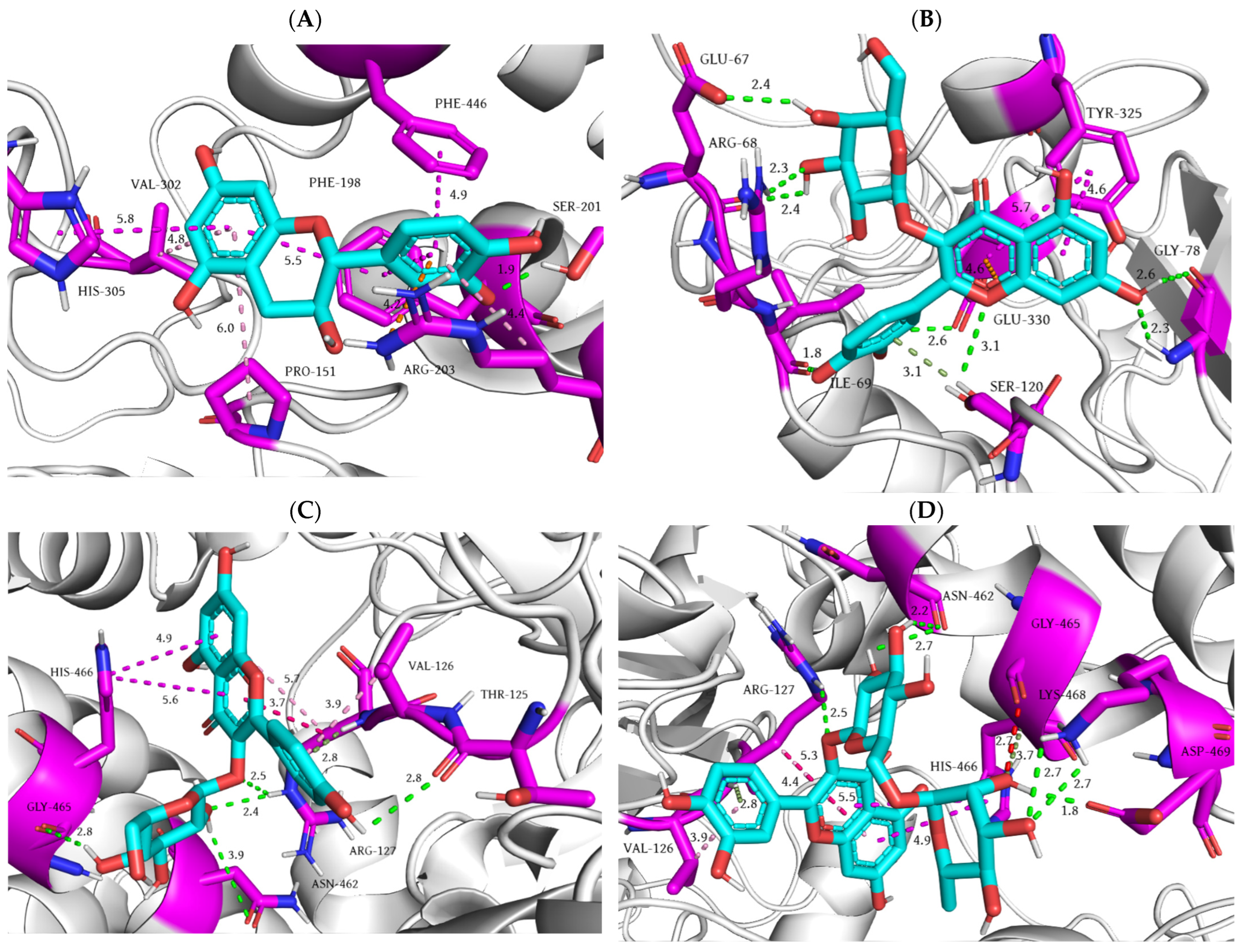
According to the obtained molecular docking data, the most stable ligand–protein complex, as determined by docking score and equilibrium binding constant, was the rutin–CAT complex, whereas the highest docking score was observed during molecular docking of p-coumaric acid into the CAT. In terms of non-covalent contacts, rutin, hyperoside, and quercetin exhibit the highest number of binding interactions. Regarding conventional hydrogen bonds, the highest number is established by hyperoside and shikimic acid with eight and nine hydrogen bonds, respectively. Interestingly, shikimic acid interacts with CAT predominantly through hydrogen bonds, mostly involving its carboxyl group. Additionally, amino acid Arg127 is the most frequent residue involved in the formation of non-conventional π-donor hydrogen bonds established by kaempferol 3-glucoside, quercetin, and rutin. Residues Lys77, His372, and His466 of CAT serve as carbon–hydrogen bond donors in the molecular interactions with resveratrol, epicatechin, and rutin, respectively, while Gln387 and His421 act as carbon–hydrogen bond acceptors in interaction with chlorogenic acid and epicatechin. Histidine residues at positions 372 and 466 are the most common residues engaged in electrostatic interactions established between epicatechin, gallic acid, quercetin, and CAT, while Glu330 predominantly participates in π-anion electrostatic interactions. The most prevalent type of hydrophobic interaction is π-alkyl contact, which is established by all polyphenolic compounds, except for shikimic acid. Furthermore, steric bumps are observed during the molecular docking of shikimic acid and rutin involving Glu330 and Gly465, respectively. Based on the displayed molecular docking results, catechin, hyperoside, kaempferol 3-glucoside, and rutin exhibited the highest binding affinity for CAT. The molecular docking of these compounds will be presented in detail (Figure 6).
During the molecular docking of catechin into the CAT, a single conventional hydrogen bond is formed between the hydroxymethyl group of Ser201 and the hydroxyl group of 3,4-dihydroxyphenyl moiety. The remaining seven non-covalent interactions belong to hydrophobic interactions, along with a single π-cation electrostatic interaction formed between the guanidine moiety of Arg203 and the 3,4-dihydroxyphenyl moiety of catechin. The π-alkyl interactions involve residues Pro151, Arg203, and Val302, while π-π interactions are observed of residues Phe198, His305, and Phe446 with the dihydropyran core of catechin (Figure 6A).
In the molecular interaction of hyperoside and CAT, a total of twelve non-covalent interactions are formed, including eight hydrogen bonds. Specifically, five conventional hydrogen bonds are formed involving 4H-chromen-4-one core, whereas the hydroxyl groups of galactose moiety additionally participate in the formation of four hydrogen bonds. Furthermore, the 4H-chromen-4-one ring of hyperoside is involved in the formation of two π-π stacked interactions with residue Tyr325 and a single π-anion interaction with residue Glu330 (Figure 6B).
Kaempferol 3-glucoside establishes a total of five conventional hydrogen bonds, whereby hydroxyl groups of glucose moiety are engaged in the formation of four interactions with residues Arg127, Asn462, and Gly465. The 4-hydroxyphenyl moiety participates as a hydrogen bond donor in a conventional hydrogen bond with the main chain of Thr125, serves as an acceptor in a π-donor hydrogen bond with Arg127, and forms a single π-alkyl interaction with a side chain of Val126. Furthermore, the 4H-1-benzopyran-4-one ring contributes to four hydrophobic interactions involving residues Val126, Arg127, and His466 (Figure 6C).
The rutin–CAT complex is stabilized by a total of thirteen non-covalent binding interactions, including six conventional and two non-conventional hydrogen bonds, as well as five hydrophobic interactions. Specifically, the hydroxyl groups of disaccharide rutinose components participate in six hydrogen bonds, while the dihydroxyphenyl ring and glucose moiety establish π-donor and carbon–hydrogen bonds, respectively. Moreover, the 4H-chromen-4-one core and dihydroxyphenyl moiety form five hydrophobic interactions, including one π-σ, two π-π, and two π–alkyl interactions, further stabilizing the rutin–CAT complex. The hydroxyl group of the rhamnose moiety establishes an unfavorable steric bump with residue Gly465 (Figure 6D).
The molecular docking results for the most favorable conformations of the investigated polyphenolic compounds within the SOD are summarized in Table 3.
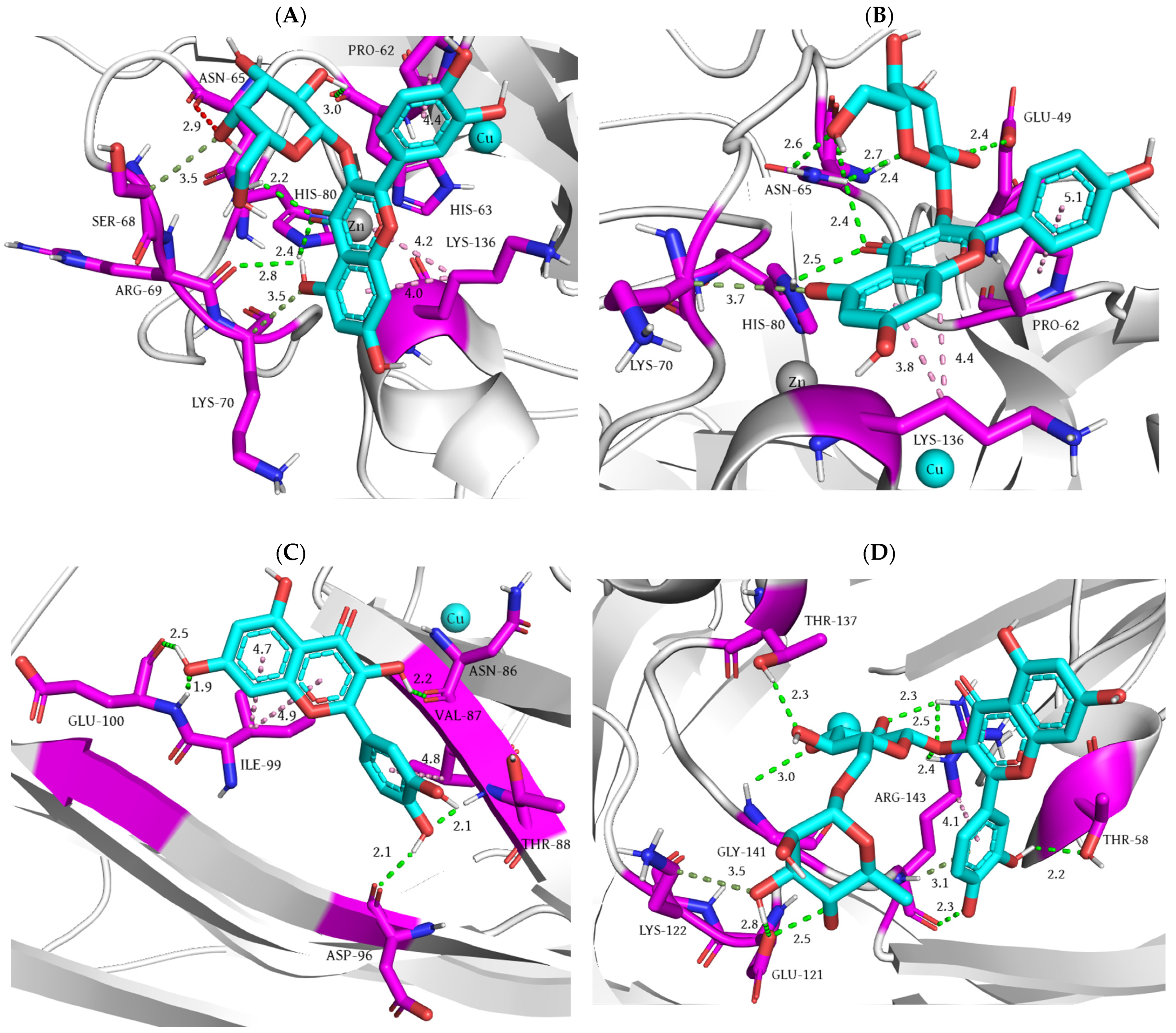
The lowest value of docking score and the highest value of equilibrium binding constant obtained for rutin suggest the formation of the most stable rutin–SOD complex, compared to other polyphenolic compounds. On the other hand, shikimic acid exhibits the highest docking score and interacts with SOD exclusively through hydrogen bond formation, similar to its molecular interactions with CAT. The highest number of favorable binding interactions is observed for rutin, including the highest number of conventional hydrogen bonds—nine. Among the non-conventional hydrogen bonds, carbon–hydrogen bonds are predominant, involving residues His43, Ser68, Lys70, Val103, Lys122, and Gly141, and polyphenols catechin, gallic acid, catechin, kaempferol, shikimic acid, and chlorogenic acid. Moreover, only rutin establishes a single π-donor hydrogen bond with residue Arg143. Considering electrostatic non-covalent interactions, only one π–anion interaction is formed between epicatechin and residue Glu100. The most frequent type of hydrophobic interaction is π–alkyl contact, which is present during molecular docking of all tested polyphenols except catechin and shikimic acid. Established π-σ interactions are detected only for catechin, p-coumaric acid, and gallic acid, while π-π stacking interactions are observed for caffeic acid and epicatechin. With respect to unfavorable binding contacts, a steric bump is observed during molecular interaction between hyperoside and residue Asn65. Based on the molecular docking results, hyperoside, kaempferol 3-glucoside, quercetin, and rutin demonstrated the highest binding affinity for SOD. A detailed analysis of the molecular docking interactions of these compounds is presented in Figure 7.
During the molecular fitting of hyperoside into the SOD, Asn65, Arg69, and His80 are hydrogen-bonded by the oxychromen-4-one core, wherein the hydroxyl group of the mentioned ring acts as a hydrogen bond donor, while the adjacent carbonyl group serves as a hydrogen bond acceptor. The hydroxyl group of the galactose moiety is a hydrogen bond donor in its interaction with the main chain of His63. However, another hydroxyl group of the sugar moiety forms an unfavorable steric bump with Asn65. Residues Ser68 and Lys70 interact with the hydroxyl groups of the dihydroxyphenyl ring and galactose moiety, forming two non-conventional hydrogen bonds. The remaining three interactions belong to π–alkyl contacts, which engage oxychromen-4-one and dihydroxyphenyl rings (Figure 7A).
The molecular interaction of kaempferol 3–glucoside and SOD revealed a total of six conventional hydrogen bonds, including four interactions established by the glucose moiety, wherein hydroxyl groups act as hydrogen bond acceptors, except for one hydroxyl group that interacts with residue Glu49 as a hydrogen bond donor. The remaining two conventional hydrogen bonds are formed by the carbonyl group of 4H-1-benzopyran-4-one core, while the adjacent hydroxyl group of the same ring establishes an additional carbon–hydrogen bond with the carbon atom of the Lys70 side chain. The complex kaempferol 3–glucoside–SOD is further stabilized through the formation of three π–alkyl interactions, involving aromatic rings of the 4H-chromen-4-one and 4-hydroxyphenyl moieties (Figure 7B).
During the molecular docking of quercetin into the SOD, the trihydroxychromen-4-one ring forms three hydrogen bonds with residues Asn86 and Glu100, whereas a hydroxyl group of the 3,4-dihydroxyphenyl moiety participates in two hydrogen bonds, acting as a hydrogen bond acceptor and donor in molecular interaction with Thr88 and Asp96, respectively. Similarly to kaempferol 3–glucoside, the aromatic rings of the 3,4-dihydroxyphenyl and 4H-1-benzopyran-4-one moieties form three π–alkyl interactions with Val87 and Ile99, respectively (Figure 7C).
Within the SOD, rutin establishes a grid of even nine conventional and two non-conventional hydrogen bonds, involving the multiple functional moieties of rutin. Specifically, the glucose moiety participates in five hydrogen bonds, while the rhamnose and 3,4-dihydroxyphenyl moieties each establish three hydrogen bonds. In addition to hydrogen bonding, the aromatic ring of the 3,4-dihydroxyphenyl moiety interacts with the carbon atoms of the Arg143 side chain, forming an additional π–alkyl hydrophobic contact. In contrast to the molecular interaction of rutin and CAT, the oxychromen-4-one core does not interact with any residues within the SOD (Figure 7D).

3.5.2. Focused Molecular Docking Studies

To investigate the potential mechanisms underlying the anti-inflammatory activity of V. vinifera, a focused molecular docking study was conducted to assess the binding affinity of key compounds from the extract for COX-1 and COX-2. The results for the best-docked conformations of the examined polyphenolic compounds within the active site of COX-1 are summarized in Table 4.
The presence of phenolic groups in the structures of the tested compounds facilitates the formation of at least one conventional hydrogen bond, a key interaction contributing significantly to the stability of the ligand–target protein complex. This is particularly characteristic for compounds such as shikimic acid, which exclusively forms conventional hydrogen bonds, as well as caffeic acid, chlorogenic acid, epicatechin, hyperoside, and rutin, which establish only one non-conventional hydrogen interaction. As shown in Table 4, residue Gln192 most frequently participates in the formation of conventional hydrogen bonds, either as a donor or an acceptor. Other hydrogen-bonded residues include Arg97, Asn122, and Ala199, with Asn122 consistently establishing multiple conventional hydrogen bonds as a hydrogen bond donor. On the other hand, the co-crystallized ligand flurbiprofen forms conventional hydrogen bonds with residues Arg120 and Ser530. It can be assumed that the inability of the tested compounds to establish these specific interactions contributes to their lower binding affinity and higher docking score values compared to the co-crystallized ligand. Among other types of hydrogen interactions, carbon–hydrogen bonds particularly contribute to the stabilization of catechin, gallic acid, hyperoside, and kaempferol-3–glucoside complexes with COX-1. Regarding hydrophobic interactions, the primary role is played by the π-electrons of the investigated compounds’ nonpolar regions, enabling the formation of predominantly amide–π, π–cation, π-π, and π–alkyl interactions. Such interactions are particularly important for the binding of catechin, p-coumaric acid, quercetin, and resveratrol, which are unable to form a significant number of hydrogen bonds, so hydrophobic interactions predominantly contribute to their overall binding affinity. Based on two primary criteria (number of binding interactions and docking score values), the results indicate that hyperoside, resveratrol, and rutin exhibit the highest binding potential toward COX-1. Resveratrol and rutin simultaneously established the highest number of interactions and the lowest free binding energy values. Hyperoside, despite forming only four interactions, which is half the number observed for catechin, achieved significantly lower docking score values, presumably due to a higher number of conventional hydrogen bond interactions. The binding modes of co-crystallized ligand and the best-docked conformations of these three compounds are illustrated in Figure 8.
Upon the binding of flurbiprofen into the active site of COX-1, two conventional hydrogen bonds are established. The Arg120 residue, through its guanidine group as a hydrogen bond donor, forms a conventional hydrogen interaction with the carbonyl oxygen of the flurbiprofen’s carboxyl group, while the hydroxyl group of serine at position 530 establishes hydrogen contact with the fluorine atom of the co-crystallized ligand. Additionally, the guanidinium ion of Arg120 can form a salt bridge with the carboxylate group of flurbiprofen. The π-electrons of the flurbiprofen’s benzene ring bind the fluorine atom to form three hydrophobic contacts, including π-σ interactions with Val349 and Ala527, as well as a single π–alkyl interaction with Leu531. The remaining ligand’s benzene ring establishes a π–alkyl contact with the isobutyl moiety of Leu352 (Figure 8A).
The molecular docking of hyperoside into the COX-1 active site is characterized by the formation of three conventional hydrogen bond interactions. The Gln192 residue simultaneously forms two conventional hydrogen bonds. Specifically, the glutamine’s nitrogen atom of the amide group acts as a proton donor and the carbonyl oxygen atom of the peptide bond acts as a proton acceptor, establishing these contacts with two hydroxyl groups of the hyperoside glycosidic part. The remaining conventional hydrogen bond is formed between the carbonyl oxygen atom of the carboxyl group within the Glu347 side chain and the phenolic group of the quercetin aglycone. A carbon–hydrogen bond is established between the alpha carbon atom of Phe356 (carbon–hydrogen bond donor) and the oxygen of benzopyran-4-one phenolic group (Figure 8B).
When fitting into the catalytic site of COX-1, resveratrol establishes a single conventional hydrogen interaction via its para-phenolic hydroxyl group as a proton donor with the carbonyl oxygen atom of the Trp387 residue. The His386 residue forms an electrostatic π–cation interaction through the protonated nitrogen atom of the imidazole ring with the π-electrons of the resveratrol benzene ring, while the π-electron cloud of imidazole establishes a π-π contact with the same part of the resveratrol molecule. The phenylalanine at position 210 forms a π-π contact with the benzene ring of the ligand through its π-electron cloud, as well as a π-donor hydrogen bond with the meta-phenolic hydroxyl group of the same part of resveratrol. The remaining benzene ring of resveratrol establishes amide–π contacts with Ala202 and Gln203 residues, as well as a single π–alkyl interaction with Ala202. A potential factor limiting resveratrol’s binding affinity to the target enzyme is the formation of steric bump with the Asn382 residue (Figure 8C).
Upon binding to COX-1, rutin exclusively forms conventional hydrogen bond interactions, except for a single π–alkyl interaction (His581). This interaction profile may explain its lower docking score and inhibition constant in comparison to other polyphenolic compounds. The only two conventional hydrogen bonds, in which the rutin molecule acts as a hydrogen bond acceptor, include the interaction of the benzopyran-4-one carbonyl oxygen atom with the amide group of Gln351 and the interaction of rutinoside hydroxyl group with the 1H nitrogen of the His581 imidazole ring. All remaining conventional hydrogen bond interactions are established between hydroxyl groups of the rutin molecule as proton donors and Gln192, Gln351, Pro514, and Asn515 residues (Figure 8D).
The molecular fitting results for the best-docked conformations of SC-558 and V. vinifera polyphenols into the catalytic site of COX-2 are listed in Table 5.
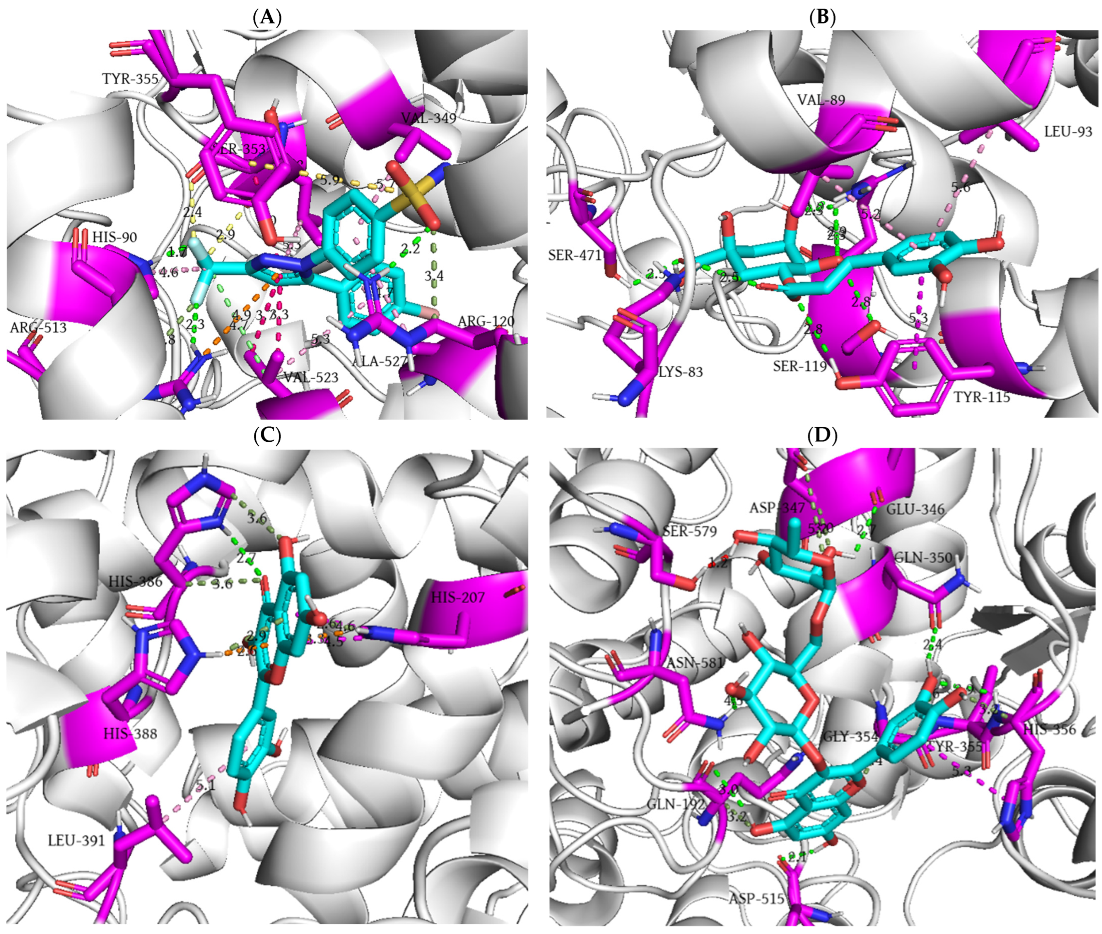
The results of investigated compounds’ molecular fitting into the active site of COX-2 indicate a similar binding model of the active components of V. vinifera as observed for the COX-1 isoform. Based on docking score values, inhibition constants, and the number of interactions, it can be concluded that chlorogenic acid, quercetin, and rutin exhibit the highest affinity for COX-2. Among them, quercetin achieved the lowest docking score value (−8.6 kcal/mol), which is lower than that of the co-crystallized ligand, while rutin formed the highest number of interactions (13). The substantial number of polar hydroxyl groups in the tested molecules enables a high prevalence of conventional hydrogen bonding interactions. All tested compounds, except resveratrol, form at least one conventional hydrogen bond, with rutin standing out by establishing a total of seven such interactions. In addition to conventional hydrogen bonds, a considerable presence of carbon–hydrogen interactions contributes to the enhanced binding of caffeic acid, epicatechin, hyperoside, kaempferol-3-glucoside, quercetin, and rutin. This type of interaction is characteristic of amino acid residues Gln192, Gln203, His207, Lys342, Asp347, Gly354, His356, and His386. Other types of hydrogen bonding interactions include π-donor hydrogen bonds, where Arg120 interacts with resveratrol, and histidine residues at positions 207 and 388 engage with quercetin. Regarding hydrophobic interactions, π-π stacking contacts are the most prevalent, occurring in all tested compounds except hyperoside, kaempferol-3-glucoside, resveratrol, and shikimic acid. Additional hydrophobic contacts include π–alkyl interactions (chlorogenic acid, p-coumaric acid, epicatechin, quercetin, resveratrol) and amide–π interactions (catechin, gallic acid, and resveratrol). Unfavorable binding contacts were also observed, with steric bumps detected between catechin, caffeic acid, and rutin with Thr206, Tyr385, and Ser579, respectively. A graphical representation of the molecular docking of the co-crystallized ligand and the three compounds with the highest COX-2 affinity (chlorogenic acid, quercetin, and rutin) is shown in Figure 9.
The selective COX-2 inhibitor (SC-558) forms a total of 19 interactions within the enzyme’s active site, including three key conventional hydrogen bonds. His90 and Arg513 act as proton donors, interacting via their imidazole and guanidine moieties with fluorine atoms from the trifluoromethyl group of the inhibitor. Arg120 establishes another conventional hydrogen bond by acting as a proton donor in interaction with the oxygen atom of the sulfonamide group in SC-558. Additionally, Arg120 and Arg513 establish carbon–hydrogen interactions as hydrogen donors, while Arg513 further stabilizes the complex through a π–cation interaction mediated by an argininium ion in its side chain. Leu352 and Ser353 via carbonyl oxygen atoms form halogen-type interactions with fluorine atoms from the trifluoromethyl group. Additional interactions contributing to the complex’s stability include π-σ, π–alkyl, π–sulfur, and alkyl hydrophobic contacts (Figure 9A).
The docking of chlorogenic acid into the COX-2 binding site is characterized by the formation of eight conventional hydrogen bonds. Arginine at position 120, through its guanidine group as a proton donor, establishes four conventional hydrogen bonds with oxygen atoms of hydroxyl groups within the quinic acid part of the molecule. This molecular fragment of chlorogenic acid acts as a proton acceptor and forms a conventional hydrogen bond with the phenol group of Tyr115 through its ester oxygen. Two remaining hydrogen bonds involve interactions with the hydroxyl groups of Ser119 (H-bond acceptor) and Ser471 (H-bond donor). Additionally, the carbonyl oxygen atom in the caffeic acid moiety of chlorogenic acid, as a proton acceptor, forms a conventional hydrogen bond with the amino group of Lys83. The chlorogenic acid–COX-2 complex is further stabilized by hydrophobic contacts between the benzene ring of ligand and Val89 (π–alkyl), Leu93 (π–alkyl), and Tyr115 (π-π) residues (Figure 9B).
The molecular fitting of quercetin indicate that its unconjugated benzene ring forms a single π–alkyl hydrophobic contact with the isobutyl group of Leu391. The sole conventional hydrogen bond is formed by the 1H nitrogen of the imidazole ring in His386 as a proton donor and interacts with the carbonyl oxygen atom of the benzopyran-4-one moiety of quercetin. The same amino acid acts as a C–hydrogen bond donor via carbon atoms interacting with the carbonyl and hydroxyl oxygen atoms of the benzopyran-4-one moiety. Additionally, His207 establishes four interactions: two π-π interactions, one π–cation interaction, and one π–donor interaction with the same moiety of quercetin molecule. His388 further stabilizes the complex through π–donor and π–cation interactions via its 1H nitrogen of the imidazole ring (Figure 9C).
The primary feature of rutin binding to COX-2 is the dominant formation of hydrogen bond interactions, with seven conventional hydrogen bonds. Specifically, Tyr355, His356, and Asn581, as proton donors, interact with the aglycone part of the ligand. The remaining conventional hydrogen bonds involve contacts with Gln192, Glu346, Gln350, and Asp515 residues as proton acceptors. Among these, only Glu346 interacts with the rutinose part of the molecule. The rutin–COX-2 complex is further stabilized by carbon–hydrogen bonds with Gln192, Asp347, and Gly354. Additionally, imidazole π-electrons of His356 forms a π-π contact with the non-conjugated benzene ring of rutin. However, the presence of a steric bump with Ser579 slightly disrupts the stability of the complex (Figure 9D).

4. Discussion

The interrelation between oxidative stress and inflammation has been confirmed in vast numbers of studies [33,34]. Due to increased ROS production, different transcription factors trigger production of a wide range of cells, including inflammatory cytokines, soluble mediators, chemokines, monocytes, adhesion molecules, and some enzymes. Consequently, mediators secreted from inflammatory cells in turn increased ROS production, thus forming a vicious feed-forward cycle [35]. In that way, this pathological condition that closely links oxidative stress and inflammation is increasingly recognized as the basis of numerous disease pathogenesis, including diabetes, obesity, metabolic syndrome, cardiovascular, neurodegenerative diseases, and others [36,37]. Additionally, understanding the molecular pathway that lies in the relationship between inflammation and oxidative stress may result in finding novel strategies for the prevention or treatment of different diseases. Although cells are supplied with enzymatic as well as nonenzymatic scavengers, frequently the capacity of these molecules is not able to diminish excessive ROS production [38]. Therefore, agents that enhance antioxidant defense systems and decrease excessive ROS production may serve as an effective treatment option for diseases associated with oxidative stress and inflammation.
Plants such as grapevine, known for its abundance of bioactive compounds, are commonly used in different products development [39]. However, the winemaking industry involves the production of large amounts of waste, thus having a significant environmental impact. The reuse of agro-industrial biomasses during wine production contributes not only to waste reduction and consequent socio-economic impact, but also to the production of grape-based products due to their rich composition [40,41]. Therefore, grapevine leaves serve as a great raw material because of the health-promoting properties of compounds found in this part of the plant [42].
In this study, we investigated the potential of commercially obtained lyophilized aqueous extract of V. vinifera to attenuate acute inflammatory response, expose antioxidant potential, and improve redox balance in rats. We used the extract that is available in the Serbian market, containing at least 30% polyphenols, 0.3% anthocyanins, and 0.005% trans-resveratrol. The results of our research showed that oral administration of V. vinifera extract exerted a significant time-dependent anti-inflammatory effect, which was manifested due to different percentages of inhibition during four hours of the follow-up period. Namely, the reduction in rat paw edema started from the second hour after the carrageenan administration and continued to increase until the fourth hour. The highest inhibition of paw edema (41.538%) was observed in the fourth hour following carrageenan injection. Based on the literature data, subplantar carrageenan injection is a well-known model of acute inflammation that induces biphasic edema. The early phase (one hour after carrageenan injection) is associated with the production of histamine, serotonin, and bradykinin as COX products. On the other hand, a delayed phase occurs up to two hours following carrageenan injection, and it is related to neutrophil infiltration and the ongoing prostaglandin production. Also, the release of pro-inflammatory cytokines, neutrophil-derived free radicals, and NO follow delayed phase of carrageenan-induced acute inflammation [43]. Therefore, the administration of agents that target COX enzymes and pathways responsible for free radicals’ generation, could offer better control over inflammatory conditions. Due to the most prominent reduction in paw edema observed in the fourth hour, the anti-inflammatory effect of the applied V. vinifera extract may be attributed to its potential to reduce COX enzyme activity. Findings obtained from this study are consistent with previously published research conducted on healthy mice, which found the most potent anti-inflammatory effect of grapevine leaf extract in the last hour of the experimental period [44]. In addition, there are vast quantities of evidence indicating the potential of grape-derived products to suppress inflammatory markers due to diminished COX-2 and iNOS protein expression, which are closely related to inflammatory processes [39,45]. Moreover, polyphenols as well as flavonoids such as quercetin, luteolin, caffeic acid, found in V. vinifera extract, have been identified as potent COX inhibitors [46,47]. These reports, along with our results, indicate a possible connection between the anti-inflammatory potential of V. vinifera leaf extract and its rich content of polyphenols.
Previously published studies have demonstrated that polyphenolic compounds present in plant extracts garner increasing attention as potential therapeutic agents for the prevention and treatment of inflammation-associated human diseases [48]. In this context, an in silico study was conducted to evaluate the binding potential of polyphenolic compounds from V. vinifera extract to COX enzymes, which play a crucial role in inflammatory processes. The molecular docking analysis revealed that among the tested compounds, rutin exhibited the highest binding affinity for both COX isoforms. This suggests that rutin may be the primary contributor to the anti-inflammatory properties of V. vinifera extract. Using AutoDock Vina version 1.1.2, Merecz-Sadowska et al. performed molecular docking studies on the active components of L. sibiricus extract, which shares a similar polyphenolic composition with V. vinifera. Their findings demonstrated that rutin exhibited the highest binding affinity toward COX-2, with a docking score of −13.83 kcal/mol [49]. In addition to rutin, quercetin also showed a notable binding potential. In particular, quercetin was the only compound with a lower docking score compared to the co-crystallized ligand when docked into the active site of COX-2. Previous studies have identified quercetin as the major bioactive component responsible for the anti-inflammatory activity of V. vinifera and C. olitorius extracts [50]. Specifically, a study assessing the affinity of quercetin and its derivatives for both COX isoforms demonstrated that quercetin exhibited a higher binding affinity for COX-2, with a docking score of −9.6 kcal/mol that is lower but comparable to the value obtained in our study (−8.6 kcal/mol) [51]. These computational findings were further corroborated by in vitro assays, which confirmed that rutin and quercetin exhibit high and relatively selective affinity toward COX-2. This binding preference is likely responsible for the significant anti-inflammatory activity associated with the application of V. vinifera extract.
To provide information regarding the antioxidant potential of V. vinifera leaf extract, we performed in vitro complementary assays followed by in vivo experimental study. Recognized as a reliable method for evaluating antioxidant potential, the DPPH assay provides valuable information regarding the total antioxidant capacities of plant extracts [19]. In this study, V. vinifera leaf extract demonstrated significant antioxidant activity, confirmed by an IC50 value of 11.63 µg/mL. Since phenolic compounds are known to contribute substantially to antioxidant potential, a close correlation was observed between the phenolic content in the V. vinifera extract and its ability to neutralize various free radicals. This finding was further supported by the FRAP assay, resulting in 0.1430 at 700 nm for V. vinifera extract, which evidently indicates the extract`s potential to reduce ferric (Fe3+) to ferrous ions (Fe2+). The observed reducing capacity not only highlights the antioxidant properties of the phenolic compounds present, but also explains the notable bioactivity exhibited in both in vitro assays. To further evaluate the antioxidant potential of the extract, we extended our study to an in vivo model by analyzing redox homeostasis in the blood samples of rats treated for two weeks with the V. vinifera lyophilized aqueous extract. Our results have shown a significant drop in the O2.− and TBARS levels in rats from the experimental group, indicating that pretreatment with V. vinifera leaf extract was able to prevent fluctuations in the level of the pro-oxidative markers and thus inhibit oxidative stress. Additionally, the antioxidant potential of V. vinifera extract was also demonstrated due to markedly improved CAT activity along with increased GSH level. The powerful antioxidant potential of grape leaf extract has been considered in vitro and is attributed to its high content of polyphenols and flavonoids that suppress free radical generation. There is evidence that the antioxidants found in vine leaves are more potent than vitamin C and E due to strong activity of these compounds in terms of inhibiting linoleic acid peroxidation [8]. Quercetin, as one compound of bioactive vine extract, acts as a scavenger of some radicals, such as superoxide, hydroxyl, and lipid peroxide radicals, due to its ability to increase both enzymatic and non-enzymatic antioxidants. It was demonstrated that supplementation with quercetin may protect liver damage caused by an imbalance in redox homeostasis. Also, this flavonoid normalizes NO levels, along with improved glutathione as well as glutathione peroxidase levels [52]. Quercetin glycoside, rutin, has demonstrated significant antioxidant potential in vitro, exhibiting notable activation of CAT and other antioxidant enzymes [53]. Molecular docking analysis provides strong support for in vivo assay findings on CAT activity, revealing that catechin, hyperoside, kaempferol 3-glucoside, and rutin exhibit the strongest molecular interaction with the enzyme compared to other investigated polyphenolic compounds. Specifically, the lowest calculated docking score, the highest equilibrium binding constant, and the highest number of non-covalent binding interactions strongly support the formation of the most stable rutin-CAT complex. In a similar in silico study conducted in AutoDock [54], Iqbal and colleagues reported that rutin exhibited a comparable docking score (−8.9 kcal/mol) in its interaction with CAT, as obtained in present study (−8.6 kcal/mol), but with significantly lower number of established non-covalent interactions—nine versus thirteen in ongoing analysis. While both studies identified the same double π-π stacking interaction with His466, and π–alkyl contacts with Val126 and Arg127, the present molecular docking analysis revealed a significantly larger number of conventional hydrogen bonds, non-conventional hydrogen bonds, and hydrophobic interactions during molecular docking of rutin into the CAT.
Besides polyphenols, V. vinifera leaves are a great source of organic acids, including malic, oxalic, and tartaric acids. These acids work as direct antioxidants by preventing lipid oxidation, but also can fulfill the antioxidative defense system, acting as synergists, thus improving antioxidants` capacity to control free radicals [55]. Although the antioxidative potential of grapevine was described in previous studies, most of them examined the effects of bioactive molecules from grape seed extract [56,57,58], while only a few investigated extracts obtained from V. vinifera leaves [59,60]. Due to the high content of a variety phytoconstituents that are found in grapevine leaves and exert antioxidant potential, V. vinifera extracts have been employed for treatment of different diseases. Acting as both anti-inflammatory and antioxidant agent, vine leaf extract managed to improve keratinocyte function and reduce the typical psoriatic lesion markers [60]. Besides skin diseases, oxidative stress is an important factor involved in many diseases, including cardiovascular, as one of the most common worldwide. Complex pathological mechanisms underline the occurrence of oxidative stress in cardiovascular diseases. However, the role of SOD, as an enzymatic constituent of an antioxidant defense system, has been described previously in cardiovascular diseases. It has been shown that inhibition of SOD expression leads to mitochondrial oxidative stress and consequent hypertrophy of cardiomyocytes [61]. Although it has been shown that grape seed extract, which contains a high concentration of proanthocyanidins, may be protective against cardiovascular diseases by diminishing elevated oxidative stress [62]. On the contrary, our research demonstrated no significant changes in SOD activity in rats treated with V. vinifera leaf extract for two weeks. An earlier study supported the findings obtained from our research regarding the potential of V. vinifera extract to decrease pro-oxidative markers, as well as that grape product consumption does not affect SOD activity [63]. Probably, the most important antioxidant enzyme, SOD, neutralizes O2.− and transforms this very harmful radical into one of a lower toxicity, such as H2O2 [64]. Therefore, the findings obtained from our study, showing no significant changes in SOD activity along with markedly lower O2.− level following a 2-week treatment with V. vinifera extract, can be explained by the fact that the entire amount of produced SOD was utilized in the O2.− dismutation reaction, which is reflected in the significantly lower values of this ROS. The extensive utilization of SOD may be attributed to the strong molecular interaction between bioactive polyphenolic compounds in V. vinifera extract and the enzyme’s binding domain. Namely, the obtained molecular docking data demonstrated that hyperoside, kaempferol 3–glucoside, quercetin, and rutin possess the highest antioxidative potential, regarding SOD activity. Analogous to its molecular interaction with CAT, rutin exhibited the most favorable binding profile with SOD in comparison to other tested polyphenols, as evidenced by the lowest docking score, the highest value of equilibrium binding constant and the highest number of non-covalent binding interactions. Zhao and co-workers investigated the molecular interaction between rutin and SOD in AutoDock [65]. In this study, the lowest value of docking score for rutin was −4.77 kcal/mol, which significantly differs from the docking score obtained in the present study (−7.4 kcal/mol). Furthermore, the rutin–SOD complex was stabilized by the formation of fourteen non-covalent interactions, including six hydrogen bonds and eight hydrophobic interactions, which contrasts with the binding pattern observed for rutin in the present study. While the binding profiles are somewhat comparable in terms of the total number of interactions, the present analysis revealed that rutin interacted with SOD through twelve binding interactions, including nine conventional hydrogen bonds, two non-conventional hydrogen bonds, and only a single hydrophobic interaction. Considering its strong molecular interactions with both antioxidative enzymes, CAT, and SOD, rutin emerges as a polyphenolic compound with significant antioxidative potential, which may further explain the observed antioxidant activity of V. vinifera leaf extract. However, this study has some limitations, including the absence of dose–response testing, which could provide more detailed information on the most effective dose of V. vinifera lyophilized aqueous leaf extract for its antioxidant and anti-inflammatory effects. Additionally, this study examined the antioxidant potential of V. vinifera extract after sub-acute treatment. Therefore, investigating its long-term effects could be of interest and represents another limitation of the present study, which could be addressed in future research.

5. Conclusions

In summary, our study indicates the strong potential of V. vinifera lyophilized aqueous leaf extract, found in the Serbian market, to improve redox homeostasis due to significantly decreased pro-oxidative markers and increased catalase activity, as well as glutathione level in healthy rats. Moreover, the efficacy of the applied extract in reducing inflammation in rats opens the door for its application as a potential anti-inflammatory product in the treatment of various inflammatory-mediated diseases. The obtained in silico results are in good agreement with the in vivo results, supporting the antioxidative and anti-inflammatory potential of the tested V. vinifera extract. Namely, molecular docking analysis provides valuable insights into the potential molecular mechanism underlying the therapeutic effects of bioactive polyphenolic compounds present in V. vinifera extract for the treatment of inflammation and oxidative stress-related diseases. A comprehensive binding analysis revealed that rutin exhibits moderately binding affinity for COX enzymes, along with strong molecular interaction with catalase and superoxide dismutase, suggesting that this molecule is most likely responsible for the biological activity of V. vinifera extract. Despite these promising preliminary results, further research is required to deeply explore the proposed molecular mechanism that lies in the basis of the beneficial biological effects of V. vinifera polyphenols on redox balance recovery and inflammatory suppression.

Author Contributions

Conceptualization, I.S. and V.J.; data curation, I.S.; investigation, M.N. (Marina Nikolic), N.N., M.K., J.B., and M.N. (Milos Nikolic); methodology, S.D., M.N. (Marina Nikolic), J.B., M.A., and M.N. (Milos Nikolic); supervision, V.J.; writing—original draft, S.D., M.N. (Marina Nikolic), N.N., M.K., M.A., and M.N. (Milos Nikolic); writing—review and editing, M.N. (Marina Nikolic) and M.N. (Milos Nikolic). All authors have read and agreed to the published version of the manuscript.

Funding

The research was supported by the Ministry of Science, Technological Development and Innovation of the Republic of Serbia through a Grant Agreement with the University of Kragujevac, Faculty of Medical Sciences, No. 451-03-137/2025-03/200111.

Data Availability Statement

The data that support the findings of this study are available from the corresponding author upon reasonable request.

Acknowledgments

The authors would like to express gratitude to the Faculty of Medical Sciences, University of Kragujevac, but also to the laboratory technician Andrijana Vicentijevic for her help in conducting the experimental part of this study. This study was supported by the Faculty of Medical Sciences, University of Kragujevac.

Conflicts of Interest

All authors declare that they have no conflicts of interest.

References

  1. Aranda-Rivera, A.K.; Cruz-Gregorio, A.; Arancibia-Hernández, Y.L.; Hernández-Cruz, E.Y.; Pedraza-Chaverri, J. RONS and Oxidative Stress: An Overview of Basic Concepts. Oxygen 2022, 2, 437–478. [Google Scholar] [CrossRef]
  2. Demirci-Çekiç, S.; Özkan, G.; Avan, A.N.; Uzunboy, S.; Çapanoğlu, E.; Apak, R. Biomarkers of Oxidative Stress and Antioxidant Defense. J. Pharm. Biomed. Anal. 2022, 209, 114477. [Google Scholar] [CrossRef] [PubMed]
  3. Sies, H.; Jones, D.P. Reactive oxygen species (ROS) as pleiotropic physiological signalling agents. Nat. Rev. Mol. Cell Biol. 2020, 21, 363–383. [Google Scholar] [CrossRef]
  4. Silvestrini, A.; Meucci, E.; Ricerca, B.M.; Mancini, A. Total Antioxidant Capacity: Biochemical Aspects and Clinical Significance. Int. J. Mol. Sci. 2023, 24, 10978. [Google Scholar] [CrossRef]
  5. Zhang, H.; Dhalla, N.S. The Role of Pro-Inflammatory Cytokines in the Pathogenesis of Cardiovascular Disease. Int. J. Mol. Sci. 2024, 25, 1082. [Google Scholar] [CrossRef]
  6. Biswas, S.K. Does the Interdependence between Oxidative Stress and Inflammation Explain the Antioxidant Paradox? Oxidative Med. Cell. Longev. 2016, 2016, 5698931. [Google Scholar] [CrossRef]
  7. Nassiri-Asl, M.; Hosseinzadeh, H. Review of the Pharmacological Effects of Vitis vinifera (Grape) and its Bioactive Constituents: An Update. Phytother. Res. 2016, 30, 1392–1403. [Google Scholar] [CrossRef]
  8. Singh, J.; Rasane, P.; Kaur, R.; Kaur, H.; Garg, R.; Kaur, S.; Ercisli, S.; Choudhary, R.; Skrovankova, S.; Mlcek, J. Valorization of grape (Vitis vinifera) leaves for bioactive compounds: Novel green extraction technologies and food-pharma applications. Front. Chem. 2023, 11, 1290619. [Google Scholar] [CrossRef]
  9. Pantelić, M.M.; Zagorac, D.Č.; Ćirić, I.Ž.; Pergal, M.V.; Relić, D.J.; Todić, S.R.; Natić, M.M. Phenolic profiles, antioxidant activity and minerals in leaves of different grapevine varieties grown in Serbia. J. Food Compos. Anal. 2017, 62, 76–83. [Google Scholar] [CrossRef]
  10. Kosar, M.; Küpeli, E.; Malyer, H.; Uylaser, V.; Türkben, C.; Baser, K.H.C. Effect of brining on biological activity of leaves of Vitis vinifera L. (cv. Sultani Cekirdeksiz) from Turkey. J. Agric. Food. Chem. 2007, 55, 4596–4603. [Google Scholar] [CrossRef]
  11. Dani, C.; Oliboni, L.S.; Agostini, F.; Funchal, C.; Serafini, L.; Henriques, J.A.; Salvador, M. Phenolic content of grapevine leaves (Vitis labrusca var. Bordo) and its neuroprotective effect against peroxide damage. Toxicol. Vitr. 2010, 24, 148–153. [Google Scholar] [CrossRef]
  12. Monagas, M.; Núñez, V.; Bartolomé, B.; Gómezcordoves, C. Anthocyanin derived pigments in Graciano, Tempramillo, and Carbernet Sauvignon wines 446 produced in Spain. Am. J. Enol. Vitic. 2003, 54, 163–169. [Google Scholar] [CrossRef]
  13. Orhan, D.D.; Orhan, N.; Ergun, F. Hepatoprotective effect of Vitis vinifera L. leaves on carbon tetrachloride-induced acute liver damage in rats. J. Ethnopharmacol. 2007, 112, 145–151. [Google Scholar] [CrossRef]
  14. Gheraibia, S.; Belattar, N.; Hassan, M.E.; El-Nekeety, A.A.; El-Sawy, E.R.; Abdel-Wahhab, M.A. Molecular docking of polyphenol compounds and exploring the anticoagulant activity of Costus speciosus extracts in vitro and in vivo. Toxicol. Rep. 2025, 14, 101961. [Google Scholar] [CrossRef]
  15. Ma, Y.; Tao, Y.; Qu, H.; Wang, C.; Yan, F.; Gao, X.; Zhang, M. Exploration of plant-derived natural polyphenols toward COVID-19 main protease inhibitors: DFT, molecular docking approach, and molecular dynamics simulations. RSC Adv. 2022, 12, 5357–5368. [Google Scholar] [CrossRef]
  16. Kiani, H.S.; Ahmad, W.; Nawaz, S.; Farah, M.A.; Ali, A. Optimized Extraction of Polyphenols from Unconventional Edible Plants: LC-MS/MS Profiling of Polyphenols, Biological Functions, Molecular Docking, and Pharmacokinetics Study. Molecules 2023, 28, 6703. [Google Scholar] [CrossRef] [PubMed]
  17. Dilkalal, A.; Annapurna, A.S.; Umesh, T.G. In vitro antioxidant, anticancer and in silico studies of polyphenol enriched leaf extract of Asystasia gangetica. Sci. Rep. 2024, 14, 28374. [Google Scholar] [CrossRef] [PubMed]
  18. Aljarba, N.H.; Hasnain, M.S.; Bin-Meferij, M.M.; Alkahtani, S. An in-silico investigation of potential natural polyphenols for the targeting of COVID main protease inhibitor. J. King Saud Univ. Sci. 2022, 34, 102214. [Google Scholar] [CrossRef]
  19. Bradic, J.; Petrovic, A.; Kocovic, A.; Mitrovic, S.; Jakovljevic, V.; Lazarevic, N.; Bolevich, S.; Simanic, I. Hypotensive and Cardioprotective Potential of Yellow Bedstraw Extract-Based Oral Liquid in Spontaneously Hypertensive Rats. Int. J. Mol. Sci. 2024, 25, 8346. [Google Scholar] [CrossRef] [PubMed]
  20. Bradic, J.; Petrovic, A.; Kocovic, A.; Ugrinovic, V.; Popovic, S.; Ciric, A.; Markovic, Z.; Avdovic, E. Development and Optimization of Grape Skin Extract-Loaded Gelatin-Alginate Hydrogels: Assessment of Antioxidant and Antimicrobial Properties. Pharmaceutics 2025, 17, 790. [Google Scholar] [CrossRef]
  21. Rankovic, M.; Krivokapic, M.; Bradic, J.; Petkovic, A.; Zivkovic, V.; Sretenovic, J.; Jeremic, N.; Bolevich, S.; Kartashova, M.; Jeremic, J.; et al. New Insight Into the Cardioprotective Effects of Allium ursinum L. Extract Against Myocardial Ischemia-Reperfusion Injury. Front. Physiol. 2021, 12, 690696. [Google Scholar] [CrossRef]
  22. Radonjic, T.; Rankovic, M.; Ravic, M.; Zivkovic, V.; Srejovic, I.; Jeremic, J.; Jeremic, N.; Sretenovic, J.; Matic, S.; Jakovljevic, V.; et al. The Effects of Thiamine Hydrochloride on Cardiac Function, Redox Status and Morphometric Alterations in Doxorubicin-Treated Rats. Cardiovasc. Toxicol. 2020, 20, 111–120. [Google Scholar] [CrossRef] [PubMed]
  23. Nedeljković, N.; Dobričić, V.; Bošković, J.; Vesović, M.; Bradić, J.; Anđić, M.; Kočović, A.; Jeremić, N.; Novaković, J.; Jakovljević, V.; et al. Synthesis and Investigation of Anti-Inflammatory Activity of New Thiourea Derivatives of Naproxen. Pharmaceuticals 2023, 16, 666. [Google Scholar] [CrossRef] [PubMed]
  24. Trott, O.; Olson, A.J. AutoDockVina: Improving the speed and accuracy of docking with a new scoring function, efficient optimization, and multithreading. J. Comput. Chem. 2010, 31, 455–461. [Google Scholar] [CrossRef]
  25. Nzekoue, F.K.; Nguefang, M.L.K.; Alessandroni, L.; Mustafa, A.M.; Vittori, S.; Caprioli, G. Grapevine leaves (Vitis vinifera): Chemical characterization of bioactive compounds and antioxidant activity during leave development. Food Biosci. 2022, 50, 102120. [Google Scholar] [CrossRef]
  26. Schrödinger, L.; DeLano, W. PyMOL. Available online: http://www.pymol.org/pymol (accessed on 1 July 2025).
  27. Putnam, C.D.; Arvai, A.S.; Bourne, Y.; Tainer, J.A. Active and inhibited human catalase structures: Ligand and NADPH binding and catalytic mechanism. J. Mol. Biol. 2000, 296, 295–309. [Google Scholar] [CrossRef]
  28. DiDonato, M.; Craig, L.; Huff, M.E.; Thayer, M.M.; Cardoso, R.M.; Kassmann, C.J.; Lo, T.P.; Bruns, C.K.; Powers, E.T.; Kelly, J.W.; et al. ALS mutants of human superoxide dismutase form fibrous aggregates via framework destabilization. J. Mol. Biol. 2003, 332, 601–615. [Google Scholar] [CrossRef] [PubMed]
  29. Sidhu, R.S.; Lee, J.Y.; Yuan, C.; Smith, W.L. Comparison of cyclooxygenase-1 crystal structures: Cross-talk between monomers comprising cyclooxygenase-1 homodimers. Biochemistry 2010, 49, 7069–7079. [Google Scholar] [CrossRef]
  30. Kurumbail, R.G.; Stevens, A.M.; Gierse, J.K.; McDonald, J.J.; Stegeman, R.A.; Pak, J.Y.; Gildehaus, D.; Miyashiro, J.M.; Penning, T.D.; Seibert, K.; et al. Structural basis for selective inhibition of cyclooxygenase-2 by anti-inflammatory agents. Nature 1996, 384, 644–648. [Google Scholar] [CrossRef]
  31. Biovia, D.S.; Berman, H.M.; Westbrook, J.; Feng, Z.; Gilliland, G.; Bhat, T.N.; Richmond, T.J. Dassault Systèmes BIOVIA, Discovery Studio Visualizer, V. 17.2, San Diego: Dassault Systèmes. J. Chem. Phys. 2000, 10, 21–9991. [Google Scholar]
  32. Morris, G.M.; Huey, R.; Lindstrom, W.; Sanner, M.F.; Belew, R.K.; Goodsell, D.S.; Olson, A.J. AutoDock4 and AutoDockTools4: Automated docking with selective receptor flexibility. J. Comput. Chem. 2009, 30, 2785–2791. [Google Scholar] [CrossRef]
  33. Mahmoud, A.M.; Wilkinson, F.L.; Sandhu, M.A.; Lightfoot, A.P. The interplay of oxidative stress and inflammation: Mechanistic insights and therapeutic potential of antioxidants. Oxidative Med. Cell. Longev. 2021, 2021, 9851914. [Google Scholar] [CrossRef]
  34. Lugrin, J.; Rosenblatt-Velin, N.; Parapanov, R.; Liaudet, L. The role of oxidative stress during inflammatory processes. Biol. Chem. 2014, 395, 203–230. [Google Scholar] [CrossRef]
  35. Hulsmans, M.; Holvoet, P. The vicious circle between oxidative stress and inflammation in atherosclerosis. J. Cell. Mol. Med. 2010, 14, 70–78. [Google Scholar] [CrossRef]
  36. Scarian, E.; Viola, C.; Dragoni, F.; Di Gerlando, R.; Rizzo, B.; Diamanti, L.; Gagliardi, S.; Bordoni, M.; Pansarasa, O. New Insights into Oxidative Stress and Inflammatory Response in Neurodegenerative Diseases. Int. J. Mol. Sci. 2024, 25, 2698. [Google Scholar] [CrossRef]
  37. Yang, X.; Li, Y.; Li, Y.; Ren, X.; Zhang, X.; Hu, D.; Gao, Y.; Xing, Y.; Shang, H. Oxidative Stress-Mediated Atherosclerosis: Mechanisms and Therapies. Front. Physiol. 2017, 8, 600. [Google Scholar] [CrossRef]
  38. Wang, Y.; Ji, D.; Chen, T.; Li, B.; Zhang, Z.; Qin, G.; Tian, S. Production, Signaling, and Scavenging Mechanisms of Reactive Oxygen Species in Fruit-Pathogen Interactions. Int. J. Mol. Sci. 2019, 20, 2994. [Google Scholar] [CrossRef]
  39. de Almeida Sousa Cruz, M.A.; de Barros Elias, M.; Calina, D.; Sharifi-Rad, J.; Junger Teodoro, A. Insights into grape-derived health benefits: A comprehensive overview. Food Prod. Process. Nutr. 2024, 6, 91. [Google Scholar] [CrossRef]
  40. Vinci, G.; Prencipe, S.A.; Abbafati, A.; Filippi, M. Environmental Impact Assessment of an Organic Wine Production in Central Italy: Case Study from Lazio. Sustainability 2022, 14, 15483. [Google Scholar] [CrossRef]
  41. Armari, M.; Zavattaro, E.; Trejo, C.F.; Galeazzi, A.; Grossetti, A.; Veronese, F.; Savoia, P.; Azzimonti, B. Vitis vinifera L. Leaf Extract, a Microbiota Green Ally against Infectious and Inflammatory Skin and Scalp Diseases: An In-Depth Update. Antibiotics 2024, 13, 697. [Google Scholar] [CrossRef] [PubMed]
  42. Martin, M.E.; Grao-Cruces, E.; Millan-Linares, M.C.; Montserrat-de la Paz, S. Grape (Vitis vinifera L.) Seed Oil: A Functional Food from the Winemaking Industry. Foods 2020, 9, 1360. [Google Scholar] [CrossRef]
  43. Mansouri, M.T.; Hemmati, A.A.; Naghizadeh, B.; Mard, S.A.; Rezaie, A.; Ghorbanzadeh, B. A study of the mechanisms underlying the anti-inflammatory effect of ellagic acid in carrageenan-induced paw edema in rats. Indian J. Pharmacol. 2015, 47, 292–298. [Google Scholar]
  44. Aouey, B.; Samet, A.M.; Fetoui, H.; Simmonds, M.S.J.; Bouaziz, M. Anti-oxidant, anti-inflammatory, analgesic and antipyretic activities of grapevine leaf extract (Vitis vinifera) in mice and identification of its active constituents by LC-MS/MS analyses. Biomed. Pharmacother. 2016, 84, 1088–1098. [Google Scholar] [CrossRef]
  45. Bocsan, I.C.; Măgureanu, D.C.; Pop, R.M.; Levai, A.M.; Macovei, Ș.O.; Pătrașca, I.M.; Chedea, V.S.; Buzoianu, A.D. Antioxidant and Anti-Inflammatory Actions of Polyphenols from Red and White Grape Pomace in Ischemic Heart Diseases. Biomedicines 2022, 10, 2337. [Google Scholar] [CrossRef]
  46. Liu, W.; Cui, X.; Zhong, Y.; Ma, R.; Liu, B.; Xia, Y. Phenolic metabolites as therapeutic in inflammation and neoplasms: Molecular pathways explaining their efficacy. Pharmacol. Res. 2023, 193, 106812. [Google Scholar] [CrossRef]
  47. Serreli, G.; Deiana, M. Role of Dietary Polyphenols in the Activity and Expression of Nitric Oxide Synthases: A Review. Antioxidants 2023, 12, 147. [Google Scholar] [CrossRef] [PubMed]
  48. Katalinic, V.; Mozina, S.; Generalic, I.; Skroza, D.; Ljubenkov, I.; Klancnik, A. Phenolic Profile, Antioxidant Capacity, and Antimicrobial Activity of Leaf Extracts from Six Vitis vinifera L. Varieties. Int. J. Food Prop. 2013, 16, 45–60. [Google Scholar] [CrossRef]
  49. Merecz-Sadowska, A.; Sitarek, P.; Kowalczyk, T.; Palusiak, M.; Hoelm, M.; Zajdel, K.; Zajdel, R. In vitro evaluation and In Silico calculations of the antioxidant and anti-inflammatory properties of secondary metabolites from Leonurus sibiricus L. root extracts. Molecules 2023, 28, 6550. [Google Scholar] [CrossRef] [PubMed]
  50. Handoussa, H.; Hanafi, R.; Eddiasty, I.; El-Gendy, M.; El Khatib, A.; Linscheid, M. Anti-Inflammatory and Cytotoxic Activities of Dietary Phenolics Isolated from Corchorus olitorius and Vitis vinifera. J. Funct. Foods 2013, 5, 1204–1216. [Google Scholar] [CrossRef]
  51. Mandour, Y.; Handoussa, H.; Swilam, N.; Hanafi, R.; Mahran, L. Structural docking studies of COX-II inhibitory activity for metabolites derived from Corchorus olitorius and Vitis vinifera. Int. J. Food Prop. 2016, 19, 2377–2384. [Google Scholar] [CrossRef]
  52. Amen, Y.; Sherif, A.E.; Shawky, N.M.; Abdelrahman, R.S.; Wink, M.; Sobeh, M. Grape-leaf extract attenuates alcohol-induced liver injury via interference with NF-κBsignaling pathway. Biomolecules 2020, 10, 558. [Google Scholar] [CrossRef]
  53. Magalingam, K.B.; Radhakrishnan, A.; Haleagrahara, N. Rutin, a bioflavonoid antioxidant protects rat pheochromocytoma (PC-12) cells against 6-hydroxydopamine (6-OHDA)-induced neurotoxicity. Int. J. Mol. Med. 2013, 32, 235–240. [Google Scholar] [CrossRef]
  54. Iqbal, A.; Hafeez Kamran, S.; Siddique, F.; Ishtiaq, S.; Hameed, M.; Manzoor, M. Modulatory effects of rutin and vitamin A on hyperglycemia induced glycation, oxidative stress and inflammation in high-fat-fructose diet animal model. PLoS ONE 2024, 19, e0303060. [Google Scholar] [CrossRef] [PubMed]
  55. Robles, A.; Fabjanowicz, M.; Chmiel, T.; Płotka-Wasylka, J. Determination and identification of organic acids in wine samples. Problems and challenges. TrAC Trends Anal. Chem. 2019, 120, 115630. [Google Scholar] [CrossRef]
  56. Al-Warhi, T.; Zahran, E.M.; Selim, S.; Al-Sanea, M.M.; Ghoneim, M.M.; Maher, S.A.; Mostafa, Y.A.; Alsenani, F.; Elrehany, M.A.; Almuhayawi, M.S.; et al. Antioxidant and Wound Healing Potential of Vitis vinifera Seeds Supported by Phytochemical Characterization and Docking Studies. Antioxidants 2022, 11, 881. [Google Scholar] [CrossRef]
  57. Pozzo, L.; Grande, T.; Raffaelli, A.; Longo, V.; Weidner, S.; Amarowicz, R.; Karamać, M. Characterization of Antioxidant and Antimicrobial Activity and Phenolic Compound Profile of Extracts from Seeds of Different Vitis Species. Molecules 2023, 28, 4924. [Google Scholar] [CrossRef]
  58. Shaban, N.Z.; El-Faham, A.A.; Abu-Serie, M.M.; Habashy, N.H. Targeting apoptosis in MCF-7 and Ehrlich ascites carcinoma cells by saponifiable fractions from green and black Vitis vinifera seed oil. Biomed. Pharmacother. 2023, 157, 114017. [Google Scholar] [CrossRef]
  59. Fernandes, F.; Ramalhosa, E.; Pires, P.; Verdial, J.; Valentão, P.; Andrade, P.; Bento, A.; Pereira, J.A. Vitis vinifera leaves towards bioactivity. Ind. Crops Prod. 2013, 43, 434–440. [Google Scholar] [CrossRef]
  60. Sangiovanni, E.; Di Lorenzo, C.; Piazza, S.; Manzoni, Y.; Brunelli, C.; Fumagalli, M.; Magnavacca, A.; Martinelli, G.; Colombo, F.; Casiraghi, A.; et al. Vitis vinifera L. Leaf Extract Inhibits In Vitro Mediators of Inflammation and Oxidative Stress Involved in Inflammatory-Based Skin Diseases. Antioxidants 2019, 8, 134. [Google Scholar] [CrossRef]
  61. Dubois-Deruy, E.; Peugnet, V.; Turkieh, A.; Pinet, F. Oxidative Stress in Cardiovascular Diseases. Antioxidants 2020, 9, 864. [Google Scholar] [CrossRef]
  62. Nunes, M.A.; Pimentel, F.; Costa, A.S.G.; Alves, R.C.; Oliveira, M.B.P.P. Cardioprotective properties of grape seed proanthocyanidins: An update. Trends Food Sci. Technol. 2016, 57, 31–39. [Google Scholar] [CrossRef]
  63. Di Renzo, L.; Carraro, A.; Valente, R.; Iacopino, L.; Colica, C.; De Lorenzo, A. Intake of red wine in different meals modulates oxidized LDL level, oxidative and inflammatory gene expression in healthy people: A randomized crossover trial. Oxidative Med. Cell. Longev. 2014, 2014, 681318. [Google Scholar] [CrossRef] [PubMed]
  64. Andrés, C.M.C.; Pérez de la Lastra, J.M.; Andrés Juan, C.; Plou, F.J.; Pérez-Lebeña, E. Superoxide Anion Chemistry-Its Role at the Core of the Innate Immunity. Int. J. Mol. Sci. 2023, 24, 1841. [Google Scholar] [CrossRef]
  65. Zhao, Y.; Chen, K.; Rao, L.; Hu, Y.; Yang, H.; Wang, Y.; Zhao, L.; Liao, X. Investigating the non-covalent interactions between rutin and superoxide dismutase: Focus on thermal stability, structure, and in vitro digestion. LWT 2024, 212, 116977. [Google Scholar] [CrossRef]
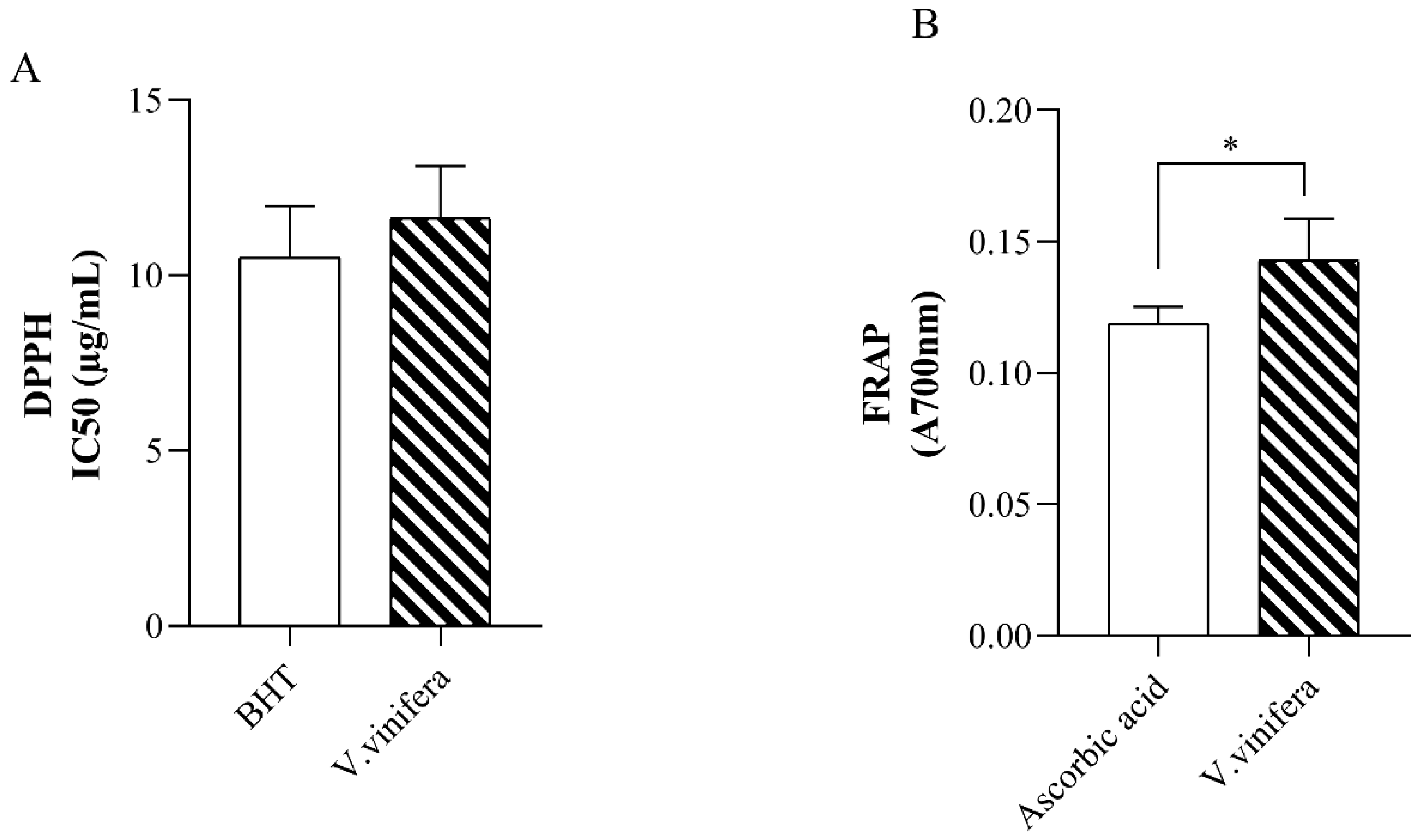
Figure 1. (A) DPPH radical scavenging activity (IC50 (µg/mL). (B) Ferric Reducing Antioxidant Power (FRAP) Assay. BHT—butylated hydroxytoluene; V. viniferaV. vinifera extract. Data is presented as means ± standard deviation (SD). * Statistically significant difference at levels of p < 0.05 in relation to the control group.
Futurepharmacol 05 00052 g001
Figure 2. Effect of V. vinifera extract on the pro-oxidative markers. (A) Superoxide anion radical—O2.−; (B) hydrogen peroxide—H2O2; (C) nitrites—NO2; and (D) index of lipid peroxidation—TBARS. CTRL—healthy untreated rats; V. vinifera—rats who drank tap water containing 150 mg/kg V. vinifera water extract for 14 days. Data is presented as means ± standard deviation (SD). * Statistically significant difference at levels of p < 0.05 in relation to the control group.
Futurepharmacol 05 00052 g002
Figure 3. Effect of V. vinifera extract on the antioxidant markers. (A) Superoxide dismutase—SOD; (B) catalase—CAT; and (C) reduced glutathione—GSH. CTRL—healthy untreated rats; V. vinifera—rats who drank tap water containing 150 mg/kg V. vinifera water extract for 14 days. Data is presented as means ± standard deviation (SD). * Statistically significant difference at levels of p < 0.05 in relation to the control group.
Futurepharmacol 05 00052 g003
Figure 4. Percentage of paw edema inhibition in rats treated with V. vinifera extract.
Futurepharmacol 05 00052 g004
Figure 5. Chemical structures of V. Vinifera polyphenols.
Futurepharmacol 05 00052 g005
Figure 6. Three-dimensional representation of catechin (A), hyperoside (B), kaempferol 3-glucoside (C), and rutin (D) binding interactions within the binding pocket of catalase. Illustrated interactions along with their corresponding bond lengths (Å) include conventional hydrogen bonds (green dashed lines), C-H and π-donor hydrogen bonds (smudged dashed lines), π-σ interactions (hot pink dashed lines), π-π interactions (magenta dashed lines), π-alkyl interactions (pink dashed lines), π-cation and π-anion interactions (orange dashed lines), and unfavorable steric bumps (red dashed lines).
Futurepharmacol 05 00052 g006
Figure 7. Three-dimensional representation of hyperoside (A), kaempferol 3-glucoside (B), quercetin (C), and rutin (D) binding interactions within the binding pocket of SOD. Illustrated interactions along with their corresponding bond lengths (Å) include conventional hydrogen bonds (green dashed lines), C-H and π-donor hydrogen bonds (smudge dashed lines), π-σ interactions (hot pink dashed lines), π–alkyl interactions (pink dashed lines), and unfavorable steric bumps (red dashed lines).
Futurepharmacol 05 00052 g007
Figure 8. Three-dimensional representation of flurbiprofen (A), hyperoside (B), resveratrol (C), and rutin (D) binding interactions within the binding pocket of COX-1. Illustrated interactions along with their corresponding bond lengths (Å) include conventional hydrogen bonds (green dashed lines), C-H and π-donor hydrogen bonds (smudged dashed lines), π–cation interactions (orange dashed lines), π-π interactions (magenta dashed lines), π-σ interactions (hot pink dashed lines), π–alkyl interactions (pink dashed lines), amide–π interactions (blue dashed lines), salt bridges (gray dashed lines), and unfavorable steric bumps (red dashed lines).
Futurepharmacol 05 00052 g008
Figure 9. Three-dimensional representation of SC-558 (A), chlorogenic acid (B), quercetin (C), and rutin (D) binding interactions within the binding pocket of COX-2. Illustrated interactions along with their corresponding bond lengths (Å) include conventional hydrogen bonds (green dashed lines), C-H and π-donor hydrogen bonds (smudged dashed lines), π-σ interactions (hot pink dashed lines), π-π interactions (magenta dashed lines), π–cation interactions (orange dashed lines), π–alkyl interactions (pink dashed lines), halogen interactions (pale-yellow dashed lines), π–sulfur interactions (orange-yellow dashed lines), alkyl interactions (green lime dashed lines), and unfavorable steric bumps (red dashed lines).
Futurepharmacol 05 00052 g009
Table 1. Anti-inflammatory activity of V. vinifera lyophilized aqueous extract in the carrageenan-induced rat paw edema model.
Rat Paw Thickness (mm) (% of Inhibition)
Groups0 h1 h2 h3 h4 h
CTRL4.30 ± 0.406.00 ± 0.306.50 ± 0.306.20 ± 0.405.90 ± 0.40
V. vinifera4.37 ± 0.156.57 ± 1.16 (13.235%)6.07 ± 0.72 (30.337%) *5.77 ± 0.42 (35.065%) *5.30 ± 0.26 (41.538%) *
The results are presented as mean value ± standard deviation (SD). * Statistically significant difference at levels of p < 0.05 in relation to the control group.
Table 2. Molecular docking results of Vitis vinifera polyphenols in interaction with human catalase.
LigandDocking Score (kcal/mol)Kb
(M−1)
Number of Favorable Binding InteractionInteracting Residue *
Catechin−8.31.22 × 1069Pro151 (π-alkyl), Phe198 (π-π x 2), Ser201 (HBD), Arg203 (π-alkyl), Arg203 (π-cation), Val302 (π-alkyl), His305 (π-π), Phe446 (π-π)
Caffeic acid−6.12.98 × 1044Asn369 (HBD), Ile373 (π-alkyl), Met392 (π-alkyl), Met394 (π-alkyl)
Chlorogenic acid−6.44.94 × 10411Lys221 (HBD x 2), Ser337 (HBA x 2), Met339 (HBA), Met339 (π-σ), Ala345 (π-alkyl), His421 (HBA x 2), His421 (CHBA), Tyr425 (HBA)
p-coumaric acid−5.15.5 × 1036Pro7 (HBA), His14 (HBD), Phe266 (π-π), Ile269 (HBA), Ala270 (π-alkyl), Asn321 (HBD)
Epicatechin −6.91.15 × 1059Arg363 (HBD), Pro368 (HBA), Pro368 (π-alkyl), His372 (CHBD), His372 (π-cation x 2), His372 ((π-π), Gln387 (HBA), Gln387 (CHBA)
Gallic acid−5.81.79 × 1048Asn369 (HBA), His372 (π-cation x 2), His372 (π-π) Asn385 (HBD), Asp389 (HBA x 2), Gln398 (HBD),
Hyperoside−7.77.37 × 10512Glu67 (HBA x 2), Arg68 (HBD), Ile69 (HBA), Gly78 (HBD), Gly78 (HBA), Ser120 (HBD), Ser120 (π-donor HBD), Tyr325 (π-π x 2), Glu330 (HBA), Glu330
(π-anion)
Kaempferol 3-glucoside−7.85.25 × 10511Thr125 (HBA), Val126 (π-alkyl), Arg127 (HBD x 2), Arg127 (π-donor HBD), Arg127 (π-alkyl), Arg127 (π-σ), Asn462 (HBA), Gly465 (HBA), His466 (π-π x 2)
Quercetin−7.42.67 × 10512Arg127 (HBD), Arg127 (π-donor HBD),
Arg127 (π-cation), Arg127 (π-alkyl x 3), Gln168 (HBD), Val247 (π-alkyl), Asn462 (HBA), His466 (HBD), His466 (π-cation), His466 (π-π)
Resveratrol−6.44.94 × 1046Glu67 (HBA), Arg68 (π-alkyl), Ile69 (HBA), Pro70 (HBA), Lys77 (CHBD), Glu330 (π-anion)
Rutin−8.62.03 × 10613Val126 (π-alkyl), Arg127 (HBD), Arg127 (π-donor HBD), Arg127 (π-σ), Arg127 (π-alkyl),
Asn462 (HBA x 2), His466 (CHBD), Gly465
(steric bump), His466 (π-π x 2), Lys468 (HBD x 2), Asp469 (HBA)
Shikimic acid−5.26.51 × 1037Ala76 (HBA), Gly78 (HBD), Ser120 (HBA x 3), Gly121 (HBA), Tyr325 (HBD),
Glu330 (steric bump)
* Residues engaged in conventional hydrogen bonding are denoted in bold. HBD—hydrogen bond donor, HBA—hydrogen bond acceptor, π-donor HBD—π-donor hydrogen bond donor, CHBD—carbon–hydrogen bond donor, CHBA—carbon–hydrogen bond acceptor, and steric bump—unfavorable binding interaction.
Table 3. Molecular docking results of V. vinifera polyphenols in interaction with human SOD.
LigandDocking Score (kcal/mol)Kb
(M−1)
Number of Favorable Binding InteractionInteracting Residue *
Catechin−5.92.12 × 1049Arg69 (HBD), Ser102 (HBA), Ser107 (HBA), His110 (HBD x 2), His110 (π-σ), Val103 (CHBA), Val103
(π-σ x 2)
Caffeic acid−4.52.00 × 1036Thr39 (HBD), His43 (CHBD), His43 (π-π), Glu121 (HBA), Lys122 (π-alkyl), Ala123 (π-alkyl)
Chlorogenic acid−5.71.51 × 1047Glu133 (HBA x 2), Ala140 (HBD), Gly141 (HBD), Gly141 (CHBA), Arg143 (HBA), Arg143 (π-alkyl)
p-coumaric acid−4.62.36 × 1037Asp11 (HBD), Gly12 (HBD), Pro13 (π-alkyl), Val14
(π-alkyl), Gly37 (HBA), Leu144 (HBA), Leu144 (π-σ)
Epicatechin −6.02.51 × 1047Lys30 (π-alkyl x 2), Trp32 (π-π x 2), Ser98 (HBD), Glu100 (HBD), Glu100 (π-anion)
Gallic acid−4.72.80 × 1036Ala140 (HBD), Gly141 (HBD), Glu133 (HBA), Thr137 (π-σ), Arg143 (HBD x 2)
Hyperoside−6.12.98 × 1049Asn65 (steric bump), Pro62 (π-alkyl), His63 (HBA), Asn65 (HBD), Ser68 (CHBD), Arg69 (HBA), Lys70 (CHBD), His80 (HBD), Lys136 (π-alkyl x 2)
Kaempferol 3-glucoside−6.23.52 × 10410Glu49 (HBA), Pro62 (π-alkyl), Asn65 (HBD x 4), Lys70 (CHBD), His80 (HBD), Lys136 (π-alkyl x 2)
Quercetin−6.34.17 × 1048Asn86 (HBA), Val87 (π-alkyl), Thr88 (HBD), Asp96 (HBA), Ile99 (π-alkyl x 2), Glu100 (HBD), Glu100 (HBA)
Resveratrol−5.51.08 × 1047Val7 (π-alkyl), Lys9 (π-alkyl x 2), Asp11 (HBA), Cys146 (π-S x 2), Val148 (HBD)
Rutin−7.42.67 × 10512Thr58 (HBA), Glu121 (HBA x 2), Lys122 (CHBD), Thr137 (HBD), Gly141 (HBD), Arg143 (HBD x 3), Arg143 (π-donor HBD), Arg143 (HBA), Arg143 (π-alkyl)
Shikimic acid−4.21.20 × 1034His80 (HBD), Glu132 (HBA x 2), Thr135 (HBA)
* Residues engaged in conventional hydrogen bonding are denoted in bold. HBD—hydrogen bond donor, HBA—hydrogen bond acceptor, π-donor HBD—π-donor hydrogen bond donor, CHBD—carbon–hydrogen bond donor, CHBA—carbon–hydrogen bond acceptor, steric bump—unfavorable binding interaction.
Table 4. Molecular docking results of flurbiprofen and V. vinifera polyphenols in interaction with COX-1.
LigandDocking Score (kcal/mol)Ki (M)Number of Favorable Binding InteractionInteracting Residue *
Flurbiprofen−9.31.50 × 10−77Arg120 (HBD), Arg120 (salt bridge), Val349
(π-σ), Leu 352 (π-alkyl), Ala527 (π-σ), Ser530 (HBD), Leu531 (π-alkyl)
Catechin−7.72.24 × 10−68Ala199 (HBA), Ala202 (amide-π), Gln203
(amide-π), Thr206 (HBD), Tyr385 (CHBA), His386 (π-cation), His386 (π-π), Met391 (π-S)
Caffeic acid−5.59.20 × 10−55Asn122 (HBA x 2), Pro125 (π-alkyl), Thr129 (steric bump), Tyr130 (HBD), Arg469 (HBD)
Chlorogenic acid−7.53.14 × 10−65Thr94 (π-σ), Arg97 (HBD), Gln192 (HBD), Gln351 (HBA), Tyr355 (HBA)
p-coumaric acid−6.51.70 × 10−53Ala202 (π-alkyl), Trp387 (HBA), Trp387
(π-donor HBD)
Epicatechin −7.62.65 × 10−66Asn122 (HBA x 3), Pro125 (π-alkyl), Arg469 (HBD x 2)
Gallic acid−6.13.34 × 10−55Ala199 (HBA), Ala202 (π-alkyl), Gln203 (CHBD), Thr206 (HBD), His207 (HBD)
Hyperoside−8.11.14 × 10−64Gln192 (HBD), Gln192 (HBA), Glu347 (HBA), Phe356 (CHBD)
Kaempferol 3-glucoside−7.53.14 × 10−66Arg97 (HBD), Gln192 (HBD), Gln350 (CHBA), Gly354 (CHBD), Tyr355 (HBA), Asn515 (HBA)
Quercetin−7.43.71 × 10−66Asn122 (HBA x 2), Pro125 (π-alkyl), Pro125
(π-σ), Ser126 (HBD), Ser126 (π-donor HBD), Gln372 (steric bump)
Resveratrol−7.91.60 × 10−68Ala202 (amide-π), Ala202 (π-alkyl), Gln203 (amide-π), Phe210 (π-π), Phe210 (π-donor HBA), Asn382 (steric bump), His386 (π-cation), His386 (π-π), Trp387 (HBA)
Rutin−8.29.61 × 10−79Gln192 (HBA), Gln351 (HBD), Gln351 (HBA), Pro514 (HBA x 2), Asn515 (HBA x 2), His581 (HBD), His581 (π-alkyl)
Shikimic acid−5.85.54 × 10−55Ala199 (HBA), His207 (HBD), Tyr385 (HBA), Trp387 (HBD), His388 (HBD)
* Residues engaged in conventional hydrogen bonding are denoted in bold. HBD—hydrogen bond donor, HBA—hydrogen bond acceptor, π-donor HBD—π-donor hydrogen bond donor, CHBD—carbon–hydrogen bond donor, CHBA—carbon–hydrogen bond acceptor, steric bump—unfavorable binding interaction.
Table 5. Molecular docking results of SC-558 and V. vinifera polyphenols in interaction with COX-2.
LigandDocking Score (kcal/mol)Ki (M)Number of Favorable Binding InteractionInteracting Residue *
SC-558−8.55.79 × 10−719His90 (HBD), His90 (π-alkyl),
Arg120 (HBD), Arg120 (CHBD), Val349 (π-alkyl), Leu352 (halogen interaction), Leu352 (π-alkyl x 2), Ser353 (halogen interaction), Ser353 (π-σ), Tyr355
(π-S), Arg513 (HBD), Arg513 (CHBD), Arg513
(π-cation), Val523 (π-σ x 2), Val523 (alkyl), Val523
(π-alkyl), Ala527 (π-alkyl)
Catechin−8.29.61 × 10−77Ala202 (amide-π), Gln203 (amide-π), Thr206
(steric bump), His207 (π-cation), His207 (π-π), Asn382 (HBD), His386 (π-cation), His388 (HBD)
Caffeic acid−7.07.30 × 10−63Ala202 (HBA), His207 (CHBD), Tyr385 (steric bump), His388 (π-π)
Chlorogenic acid−7.81.89 × 10−611Lys83 (HBD), Val89 (π-alkyl), Leu93 (π-alkyl), Tyr115 (HBD), Tyr115 (π-π), Ser119 (HBA), Arg120 (HBD x 4), Ser471 (HBD)
p-coumaric acid−7.16.16 × 10−68Ile124 (π-alkyl), Asp125 (HBD), Thr149 (HBA), Asn375 (HBD), Ala378 (π-alkyl), Phe381 (π-π), Phe529 (HBA), Phe529 (π-π)
Epicatechin −7.72.24 × 10−68Gln203 (CHBD), Thr206 (HBD), His207 (π-cation), His207 (π-π), Asn382 (HBA), His386 (π-cation), His388 (HBD), Leu390 (π-alkyl)
Gallic acid−6.51.70 × 10−55Ala202 (amide-π), Gln203 (amide-π), Thr206 (HBD), Tyr385 (HBA), His388 (π-π)
Hyperoside−7.72.24 × 10−65Lys342 (CHBD), Asp347 (π-anion), Thr578 (HBA x 2), Phe580 (HBD)
Kaempferol 3-glucoside−7.43.71 × 10−67Asp347 (HBA), Gln350 (HBA), Gly354 (CHBD), His356 (HBD), Asp515 (HBA), Asn581 (HBD x 2)
Quercetin−8.64.89 × 10−710His207 (π-cation), His207 (π-donor HBD), His207
(π-π x 2), His386 (HBD), His386 (CHBD x 2), His388 (π-donor HBD), His388 (π-cation), Leu391 (π-alkyl)
Resveratrol−7.62.65 × 10−69Arg120 (π-donor HBD), Val349 (π-alkyl), Leu352
(π-alkyl), Leu359 (π-alkyl), Gly526 (amide-π), Ala527 (amide-π), Ala527 (π-alkyl x 2), Leu531 (π-alkyl)
Rutin−8.38.12 × 10−713Gln192 (HBA), Gln192 (CHBD), Glu346 (HBA), Asp347 (CHBA), Asp347 (CHBD), Gln350 (HBA), Gly354 (CHBD), Tyr355 (HBD), His356 (HBD), His356 (CHBD), His356 (π-π), Asp515 (HBA), Asn581(HBD), Ser579 (steric bump)
Shikimic acid−5.76.56 × 10−54Ala199 (HBA), Gln203 (HBA), Thr206 (HBD), Tyr385 (HBA)
* Residues engaged in conventional hydrogen bonding are denoted in bold. HBD—hydrogen bond donor, HBA—hydrogen bond acceptor, π-donor HBD—π-donor hydrogen bond donor, CHBD—carbon–hydrogen bond donor, CHBA—carbon–hydrogen bond acceptor, steric bump—unfavorable binding interaction.
Disclaimer/Publisher’s Note: The statements, opinions and data contained in all publications are solely those of the individual author(s) and contributor(s) and not of MDPI and/or the editor(s). MDPI and/or the editor(s) disclaim responsibility for any injury to people or property resulting from any ideas, methods, instructions or products referred to in the content.