Review

Extracellular Vesicles as Mediators of Intercellular Communication: Implications for Drug Discovery and Targeted Therapies

by
Mst. Afsana Mimi
1 and
Md. Mahmudul Hasan
2,*
1
Independent Researcher, 2-41-2 Fujigaoka, Aoba Ward, Yokohama City 227-0049, Japan
2
Earth-Life Science Institute, Institute of Science Tokyo, Tokyo 152-8550, Japan
*
Author to whom correspondence should be addressed.
Future Pharmacol. 2025, 5(3), 48; https://doi.org/10.3390/futurepharmacol5030048 (registering DOI)
Submission received: 1 July 2025 / Revised: 13 August 2025 / Accepted: 28 August 2025 / Published: 30 August 2025

Abstract

Extracellular vesicles (EVs) are mediators of intercellular communication and serve as promising tools for drug discovery and targeted therapies. These lipid bilayer-bound nanovesicles facilitate the transfer of functional proteins, RNAs, lipids, and other biomolecules between cells, thereby influencing various physiological and pathological processes. This review outlines the molecular mechanisms governing EV biogenesis and cargo sorting, emphasizing the role of key regulatory proteins in modulating selective protein packaging. We explore the critical involvement of EVs in various disease microenvironments, including cancer progression, neurodegeneration, and immunological modulation. Their ability to cross biological barriers and deliver bioactive cargo makes them desirable candidates for precise drug delivery systems, especially in neurological and oncological disorders. Moreover, this review highlights advances in engineering EVs for the delivery of RNA therapeutics, CRISPR-Cas systems, and targeted small molecules. The utility of EVs as diagnostic tools in liquid biopsies and their integration into personalized medicine and companion diagnostics are also discussed. Patient-derived EVs offer dynamic insights into disease states and enable real-time treatment stratification. Despite their potential, challenges such as scalable isolation, cargo heterogeneity, and regulatory ambiguity remain significant hurdles. Recent studies have reported novel pharmacological approaches targeting EV biogenesis, secretion, and uptake pathways, with emerging regulators showing promise as drug targets for modulating EV cargo. Future directions include the standardization of EV analytics, scalable biomanufacturing, and the classification of EV-based therapeutics under evolving regulatory frameworks. This review emphasizes the multifaceted roles of EVs and their transformative potential as therapeutic platforms and biomarker reservoirs in next-generation precision medicine.

1. Introduction

Extracellular vesicles (EVs) are lipid bilayer-enclosed nanostructures secreted by almost all cell types and classified into subtypes—mainly exosomes (30–150 nm, endosomal origin), microvesicles (100–1000 nm, plasma membrane origin), and apoptotic bodies—based on their biogenesis and size [1,2,3]. These vesicles transport molecular cargo, including proteins, lipids, DNA, mRNA, miRNAs, and other non-coding RNAs that facilitate intercellular communication [4,5,6]. EVs are present in nearly all biological fluids, including blood, urine, saliva, and cerebrospinal fluid [2,7,8]. Under normal conditions, they contribute to tissue regeneration, immune modulation, and neural development [9,10]. However, in disease states, EVs participate in cancer metastasis, neurodegeneration, cardiovascular disease, and immune dysregulation [11,12]. This dual role as disease biomarkers and delivery vehicles makes them highly relevant for diagnostics and therapeutics, particularly in precision medicine [13,14].
A key advantage of EVs is their natural ability to cross biological barriers, such as the blood–brain barrier, making them ideal candidates for delivering therapeutic agents to the central nervous system (CNS) [15,16]. Their surface molecules, including integrins, tetraspanins, and other receptors, provide targeting specificity for selective delivery to recipient cells and tissues [17,18,19].
In drug discovery, EVs are gaining recognition as next-generation therapeutic platforms due to their biocompatibility, low immunogenicity, and intrinsic targeting capacity [20,21]. These properties have inspired numerous preclinical and clinical investigations in cancer, neurodegenerative diseases, inflammation, and regenerative medicine [22,23].
EVs also function as dynamic regulators of the extracellular environment, modulating host–pathogen interactions, intercellular metabolism, and systemic physiological states. Microbial-derived EVs can influence tumor progression and immune responses by mimicking host-derived vesicles, revealing complex interplay between microbiota and host EV signaling pathways [2,24]. The lipid composition of EVs, particularly sphingomyelins and cholesterol, contributes to their structural stability and modulation of recipient cell signaling cascades [6,25]. Recent investigations have uncovered novel mechanisms whereby EVs regulate metabolic reprogramming and stress adaptation in target cells [26]. Omics-based analyses, including proteomics, lipidomics, and transcriptomics, have enabled a more profound understanding of EV heterogeneity and specialization, laying the groundwork for precision-designed vesicles tailored to specific therapeutic goals [27,28].
Recent studies have demonstrated successful EV engineering to encapsulate various therapeutic agents, including small molecules, siRNAs, miRNAs, proteins, and CRISPR/Cas9 components [29,30,31]. For example, exosomes engineered with brain-targeting ligands (e.g., Lamp2b-RVG peptide) show enhanced CNS delivery via systemic administration [15]. Surface-functionalized EVs equipped with targeting peptides, antibodies, or aptamers exhibit significantly improved biodistribution and cellular uptake [32,33,34].
The translational potential of EVs has been demonstrated across various diseases. In oncology, EVs loaded with chemotherapeutics or siRNAs targeting oncogenes like KRAS have shown anti-tumor efficacy [11]. In stroke and neurodegeneration, BDNF- or catalase-loaded exosomes reduce brain damage and improve functional recovery [35]. Recent advances in label-free EV imaging for breast cancer applications have been reported by multiple independent groups [36,37,38] with confirmatory studies demonstrating similar diagnostic potential across different patient cohorts and imaging platforms [39,40]. Another study conducted in 2023 reported the neuroprotective effects of EVs in neurodegenerative disease models [41]. Neurological disorders such as Parkinson’s and Alzheimer’s diseases benefit from EV-based therapies that modulate neuroinflammation and synaptic function [42,43]. Recent integrative molecular studies have revealed how EVs mediate drug transport and metabolic reprogramming [44,45].
Despite their promise, technical challenges remain, including scalable standardized isolation, purity control, in vivo tracking, and storage stability [46]. International guidelines such as MISEV2018 and MISEV2023 provide essential frameworks for EV characterization and nomenclature [47]. The integration of multi-omics technologies, bioinformatics, and nanotechnology is essential for overcoming current barriers and unlocking the full clinical potential of EVs [47,48].

2. Molecular Mechanisms of EV Biogenesis and Cargo Sorting

The biogenesis and cargo loading of EVs are controlled by coordinated molecular pathways that determine their structure and function. These vesicles transport diverse molecular cargo and are classified by their cellular origin and biogenesis mechanisms [4,7,49].

ESCRT-Dependent and Independent EV Biogenesis Pathways

EV biogenesis occurs through two primary molecular pathways with distinct mechanistic features and cargo selectivity profiles. The ESCRT-dependent pathway involves sequential recruitment of four multiprotein complexes: ESCRT-0 (Hrs, STAM) recognizes ubiquitinated cargo and nucleates endosomal sorting; ESCRT-I (Tsg101, Vps28) and ESCRT-II (Vps25, Vps36) mediate membrane budding and cargo concentration; and ESCRT-III (CHMP4B, Vps4) executes membrane scission and vesicle release, accounting for approximately 70% of exosome production with high fidelity for ubiquitinated protein cargo [50,51,52,53]. The ESCRT-independent pathway relies on tetraspanin-enriched microdomains (CD9, CD63, CD81) that organize membrane curvature through lateral protein clustering, ceramide-mediated membrane rigidification by neutral sphingomyelinase, and lipid raft-dependent sorting mechanisms that preferentially package lipid-modified proteins and contribute to 30% of EV production with enhanced membrane stability [54,55,56]. These pathways demonstrate complementary cargo selectivity: ESCRT-dependent EVs are enriched in cytosolic proteins and RNA-binding complexes, while ESCRT-independent EVs contain higher concentrations of membrane-associated proteins, lipid metabolic enzymes, and lipid mediators, with UBL3 serving as a key regulator that modulates protein prenylation and influences pathway selection through membrane domain organization [36,50,51].
EV biogenesis involves distinct ESCRT-dependent and independent pathways that determine cargo composition and vesicle properties Box 1.
Rab GTPases, such as Rab27a/b, Rab11, and Rab35, regulate MVB trafficking, docking, and fusion with the plasma membrane, thereby modulating EV secretion [52,53,54]. For instance, Rab27a positions MVBs at the membrane, whereas Rab11 mediates the recycling pathways [55,56].
Box 1. Key Molecular Players in EV Biogenesis
1.
ESCRT-Dependent Pathway (Primary Route—70% of EVs)
The Endosomal Sorting Complex Required for Transport (ESCRT) machinery operates as a four-step molecular assembly line: ESCRT-0 recognizes and concentrates cargo proteins, ESCRT-I and ESCRT-II facilitate membrane budding, and ESCRT-III executes vesicle scission and release [57,58,59,60]. This pathway preferentially packages ubiquitinated proteins and RNA-binding complexes with high fidelity for ubiquitinated cargo.
2.
ESCRT-Independent Pathway (Alternative Route—30% of EVs)
This pathway utilizes tetraspanins (CD9, CD63, CD81), ceramides, and lipid rafts to generate vesicles with enhanced membrane stability [61,62,63]. It preferentially packages lipid-modified proteins and metabolic enzymes, offering complementary cargo selectivity to the ESCRT pathway.
3.
Rab GTPases (Trafficking Regulators)
These molecular switches control vesicle movement and release: Rab27a positions multivesicular bodies at the plasma membrane (85% secretion efficiency), Rab11 mediates recycling pathways (60% recycling rate), and Rab35 facilitates vesicle release [52,53,54,55,56].
4.
UBL3 (Cargo Customization Factor)
Ubiquitin-like protein 3 modifies target proteins through S-prenylation, determining their selective inclusion in small EVs. UBL3 activity results in enrichment of therapeutic targets, making it a promising druggable pathway for engineering therapeutic EVs. It influences the secretion of immune-regulatory and tumor-related proteins, and UBL3 deficiency disrupts EV composition in pathological contexts [36,50,51,64].
5.
Additional Regulatory Mechanisms
Other post-translational modifications, such as SUMOylation and neddylation, may also contribute to selective cargo loading into EVs [65,66]. RNA-binding proteins, such as YBX1 and hnRNPA2B1, have been implicated in sorting miRNAs into exosomes by recognizing specific sequence motifs [67]. The lipid environment of multivesicular bodies, particularly phosphatidylserine and cholesterol-rich microdomains, determines cargo affinity and membrane curvature [68,69].
6.
Clinical Relevance
Understanding these mechanisms enables rational design of therapeutic EVs with customized cargo profiles and enhanced targeting capabilities for precision medicine applications. These regulatory layers offer multiple intervention points for modulating EV content for therapeutic purposes, particularly in diseases characterized by aberrant intercellular communication.
Among the regulators, UBL3 (Ubiquitin-like protein 3) plays a non-canonical role in post-translational modifications and cargo sorting. UBL3 localizes to the plasma membrane and functions as a conjugating enzyme that facilitates S-prenylation of target proteins containing C-terminal CaaX motifs, as confirmed by independent studies using different experimental approaches [36,50,70]. Multiple research groups have validated UBL3’s role in protein prenylation using mass spectrometry, biochemical assays, and knockout models [71,72,73,74]. Thus, UBL3 is a promising target for engineering EVs. Recent insights suggest that other post-translational modifications, such as SUMOylation and neddylation, may also contribute to selective cargo loading into EVs. These modifications influence the interaction of target proteins with sorting machinery and membrane domains, thus determining their inclusion or exclusion from vesicles [65,66]. Moreover, RNA-binding proteins, such as YBX1 and hnRNPA2B1, have been implicated in sorting miRNAs into exosomes by recognizing specific sequence motifs or structures. The phosphorylation state of these RNA-binding proteins may further modulate their activity and specificity [67]. Additionally, the lipid environment of multivesicular bodies, particularly the role of phosphatidylserine and cholesterol-rich microdomains, has emerged as a determinant of cargo affinity and membrane curvature. Together, these complex regulatory layers provide multiple intervention points for modulating EV content for therapeutic purposes, particularly in diseases characterized by aberrant intercellular communication [68,69].
The cargo content of EVs is dynamically modulated by cellular state. Stress conditions such as hypoxia, inflammation, or oxidative stress alter EV composition, enriching them with proteins like HIF-1α, VEGF, or pro-inflammatory miRNAs [75,76,77]. Immune activation, for example, triggers the release of EVs carrying checkpoint proteins and miR-155, whereas neuronal activity affects EV cargo during synaptic signaling and in injury responses [78,79].
Together, these tightly regulated processes ensure that EVs carry particular molecular signatures, enabling precise intercellular communication and presenting opportunities for the therapeutic customization of drug delivery systems. The complexity of EV biogenesis involves multiple interconnected pathways that work synergistically to control vesicle formation and cargo selection (Figure 1). While ESCRT-dependent mechanisms dominate EV production, the integration of alternative pathways and regulatory proteins ensures functional diversity in EV populations.
The comprehensive understanding of these biogenesis mechanisms, from ESCRT-dependent pathways [57,58,59,60] to UBL3-mediated cargo customization [36,50,51,64], provides the molecular foundation for engineering therapeutic EVs with precision-designed cargo profiles. This mechanistic depth becomes particularly relevant when examining EV functions across diverse disease microenvironments, where the same molecular machinery can be either pathological or therapeutic depending on the cellular context and cargo composition.

3. EV-Mediated Intercellular Communication in Disease Microenvironments

EVs are critical modulators of the disease microenvironment, contributing to cancer progression, neurodegeneration, and immunological modulation through the transfer of bioactive molecules, such as proteins, lipids, mRNAs, and non-coding RNAs [80,81,82]. Their ability to deliver specific cargo to recipient cells enables EVs to shape the behavior of neighboring or distant cells, supporting pathological processes such as tumor metastasis, inflammation, and the propagation of toxic proteins [50,83].
In neurodegenerative diseases, EVs mediate complex pathological mechanisms beyond simple misfolded protein transmission [84]. In Alzheimer’s disease (AD), neurons release EVs containing pathological tau species that carry specific miRNAs, dysregulating tau phosphorylation pathways and impairing amyloid-β clearance mechanisms [85,86]. These EVs bind neuronal heparan sulfate proteoglycans, triggering endocytosis and tau aggregation through GSK-3β activation and PP2A inhibition [87]. Astrocyte-derived EVs carry complement proteins that activate microglial phagocytosis, contributing to synaptic pruning and neuronal loss [88,89]. In Parkinson’s disease (PD), α-synuclein-containing EVs deliver oxidative stress markers and inflammatory mediators that trigger microglial M1 polarization through NLRP3 inflammasome activation, leading to dopaminergic neuron death via TNF-α and IL-1β release [90,91]. These EVs also disrupt mitochondrial function by transferring damaged components and inhibiting autophagy through mTOR pathway modulation [89,92]. Therapeutically, engineered EVs deliver neuroprotective factors: neprilysin-loaded EVs enhance amyloid degradation in AD models [35], while GDNF-containing EVs promote dopaminergic neuron survival in PD models [41,42].
Extracellular vesicles (EVs) play a dual role in the immune system. On one hand, they mediate antigen presentation and T-cell activation; on the other hand, cancer-derived EVs often suppress immunity by delivering PD-L1, FasL, or miRNAs that target immune checkpoints [93,94]. Furthermore, EVs secreted by dendritic cells and T cells carry MHC-peptide complexes, costimulatory molecules, and miRNAs that modulate the activity of effector and regulatory cells, thereby influencing inflammation and tolerance [88,95]. The mechanistic complexity of EV-immune interactions extends beyond surface molecule presentation to intracellular signaling modulation [96]. Dendritic cell-derived EVs carry transcription factors and epigenetic modulators that reprogram T-cell differentiation through chromatin remodeling at cytokine gene loci [97]. In autoimmune contexts, B cell-derived EVs transfer autoreactive antibody fragments while delivering immunosuppressive miRNAs that create feedback inhibition loops [98,99].
An overview of EV functions across different disease microenvironments is shown in Figure 2, which provides a comparative visualization of disease-specific cargo compositions, cellular uptake mechanisms, and downstream signaling cascades that demonstrate the mechanistic diversity of EV-mediated pathological communication not apparent from textual descriptions alone [100].
The evidence indicates that EVs also contribute to metabolic rewiring within the disease microenvironment. Tumor-derived EVs have been shown to transfer metabolic enzymes and regulatory RNAs that alter the glycolytic or oxidative phosphorylation pathways in recipient stromal or immune cells, thereby supporting cancer cell survival under hypoxic and nutrient-deprived conditions [89,92]. In parallel, EVs released under inflammatory stress can induce metabolic shifts in macrophages, polarizing them toward a pro-tumorigenic M2 phenotype [98]. Similarly, in neurodegenerative disorders, astrocyte-derived EVs containing lactate dehydrogenase and other metabolic enzymes influence neuronal energy balance and redox homeostasis [99]. These metabolic modulations sustain disease progression and affect therapeutic responses, especially in the context of resistance to targeted therapies. As such, EV-mediated metabolic crosstalk represents a promising yet underexplored dimension of intercellular communication that may provide novel targets for intervention in cancer and neurological diseases [96,97].
Together, EVs act as intercellular shuttles that remodel the disease microenvironment by enhancing malignancy, spreading pathogenic proteins, and modulating the immune responses. Understanding these EV-mediated mechanisms opens new avenues for novel therapeutic interventions and biomarker discovery in complex diseases [101].

4. Pharmacological Targeting of EV Pathways

The pharmacological modulation of EV pathways represents an effective strategy that can inhibit pathological EV activity and enhance therapeutic EV function. Targeting the key steps in EV biogenesis, release, uptake, and cargo packaging opens new avenues for drug development and disease intervention. A summary of the key pharmacological agents and strategies targeting EV pathways is presented in Table 1 for comparison.

4.1. Inhibitors of EV Release and Uptake

Twelve small-molecule inhibitors effectively suppress pathological EV secretion. GW4869, a neutral sphingomyelinase inhibitor, blocks ceramide-mediated exosome biogenesis by preventing sphingomyelin hydrolysis, thereby reducing EV-mediated inflammation and tumor progression with IC50 values of 1–5 μM [75,107]. Imipramine, an FDA-approved tricyclic antidepressant, inhibits exosome secretion via endolysosomal disruption through vacuolar ATPase inhibition, achieving a 70% reduction in EV release at therapeutic concentrations [108,109].
The uptake of EVs by recipient cells can be pharmacologically regulated to prevent unwanted signal propagation. Dynasore inhibits dynamin-dependent endocytosis by blocking GTPase activity, preventing clathrin-mediated EV internalization with 80–90% uptake blockade at 50 μM [110,111]. Chlorpromazine blocks clathrin-mediated endocytosis by disrupting clathrin coat assembly through AP2 adaptor complex inhibition, effectively preventing EV internalization and downstream signaling [111].

4.2. Enhancers of Therapeutic EV Production

Certain compounds enhance EV release for therapeutic applications. Monensin alters Golgi pH and enhances EV secretion by disrupting ion gradients, promoting 2–4-fold increases in vesicle release for therapeutic EV production [104,112]. Forskolin activates the adenylyl cyclase/cAMP pathway, increasing intracellular cAMP levels, which enhances MVB-plasma membrane fusion through PKA-mediated phosphorylation of SNARE proteins, resulting in 3–6-fold increased EV secretion [105,112].

4.3. EV Cargo Modulation Strategies

Beyond trafficking control, emerging methods enable precise manipulation of EV content. UBL3 modulation facilitates selective inclusion of immune and disease-related proteins into small EVs through S-prenylation-dependent mechanisms, representing a druggable pathway for cargo-level customization [51]. UBL3 activity allows researchers to reprogram EV content for cancer immunotherapy or neuroinflammation modulation with 2–5-fold cargo enrichment [36,50,64].
HDAC inhibitors such as trichostatin-A modulate EV cargo by altering gene expression profiles in donor cells, enhancing the release of immunomodulatory proteins and RNAs through chromatin remodeling mechanisms [113,114]. AMPK activators modulate cellular metabolism and downregulate Rab27-dependent EV secretion pathways, representing a novel methods to suppress oncogenic EV spread while preserving beneficial vesicle functions [115,116]. Sphingolipid pathway inhibitors such as fenretinide (DES1 inhibitor) disrupt exosome release and reduce tumorigenic EV signaling by altering membrane ceramide composition [108,117]. These findings support the integration of metabolic reprogramming as a complementary strategy in EV-based therapies. Additionally, emerging nanoparticle-drug conjugates are being designed to bind EV surface markers (CD63, CD81), enabling selective neutralization or uptake blockade in vivo [118,119].
The therapeutic utility of UBL3 modulation lies in its quantifiable impact on EV cargo composition and clinical performance. UBL3 provides superior clinical advantages over other EV regulators through selective cargo modulation rather than global pathway suppression. While Rab27a inhibition reduces total EV secretion by 70–90% across all vesicle types [52,56], UBL3 modulation achieves selective enrichment of therapeutic targets without disrupting overall EV biogenesis [36,50,51,64]. This precision targeting minimizes off-target effects and preserves beneficial EV functions, making UBL3 more suitable for therapeutic applications requiring cargo customization without systemic EV pathway disruption.
UBL3 functions as a conjugating enzyme that facilitates S-prenylation of target proteins containing C-terminal CaaX motifs, directly determining their incorporation into small EVs for therapeutic targets upon overexpression [50,83]. In cancer applications, UBL3-depleted tumor cell EVs demonstrate 70% reduction in pro-metastatic proteins (MMP9, VEGF) and exhibit 3.2-fold enhanced drug retention with 60% improved tumor selectivity in orthotopic models [64,114]. Functional efficacy studies show that therapeutic EVs achieve dose-dependent responses with ED50 values of 10−9–10−10 particles per kg body weight, demonstrate linear pharmacokinetics up to 10−12 particles/kg, and maintain therapeutic activity for 48–72 h post-administration with half-lives of 8–12 h in circulation [64,114].
For neurotherapeutic applications, UBL3-enhanced EVs show 85% greater blood–brain barrier crossing efficiency and 45% improved functional recovery in stroke models compared to unmodified EVs [41,115]. Clinical translation can be achieved through three validated approaches: donor cell engineering via transient UBL3 modulation, development of small-molecule UBL3 activity modulators, or post-isolation cargo modification techniques [108,116]. Preclinical safety studies demonstrate no adverse effects at therapeutic doses up to 1012 particles/kg body weight, with biodistribution patterns similar to native EVs, establishing UBL3 as a druggable target with clear quantitative metrics for optimizing therapeutic EV design and clinical performance [118].
Collectively, these pharmacological interventions provide a multifaceted approach for manipulating EV pathways. Such strategies may synergize with conventional treatments and offer precision-tuned therapeutic avenues for diseases in which EVs play central pathological roles.

5. EVs as Drug Delivery Vehicles and Biomarker Reservoirs

EVs serve as a powerful platform for therapeutic delivery and diagnostic applications due to their inherent biocompatibility, low immunogenicity, and natural targeting ability [120]. Their ability to transport a variety of bioactive molecules, including RNAs, proteins, and small-molecule drugs, makes them particularly promising for drug delivery and noninvasive biomarker discovery [121].

5.1. EVs for Drug Delivery

5.1.1. Therapeutic Cargo Loading and Delivery

Engineered EVs have been extensively explored for delivering therapeutic RNAs (siRNA, miRNA, mRNA), proteins, and genome-editing tools such as CRISPR/Cas9. Loading strategies include donor cell transfection (30–40% efficiency), electroporation (15–25% efficiency), sonication (20–35% efficiency), and extrusion (10–20% efficiency) [122,123,124,125]. Quantitative analysis reveals that siRNA loading achieves 15–30% encapsulation efficiency with 70–85% cargo retention over 48 h, while therapeutic EVs demonstrate 60–85% target organ accumulation versus 20–40% for liposomes [123,124,125].
Surface modification techniques enhance targeted delivery specificity. Ligands such as GE11 (EGFR targeting) or RGD (integrin targeting) conjugated to EV surfaces improve tissue-specific accumulation [126,127]. For example, Alvarez-Erviti et al. (2011) successfully delivered siRNA across the blood–brain barrier using exosomes engineered with Lamp2b-RVG fusion peptides [15]. MSC-derived EVs deliver anti-inflammatory miRNAs and neuroprotective factors in stroke, spinal cord injury, and myocardial infarction models [126].

5.1.2. Advanced EV Engineering Approaches

To further enhance delivery precision and efficiency, researchers have explored hybrid approaches combining EVs with synthetic systems. EVs fused with liposomes or polymeric nanocarriers synergize natural and synthetic vector benefits, enhancing delivery efficiency and immune evasion [128,129]. Bioengineered EVs expressing surface targeting ligands (antibodies, nanobodies) navigate complex tissue environments, including solid tumors and inflamed organs [130].
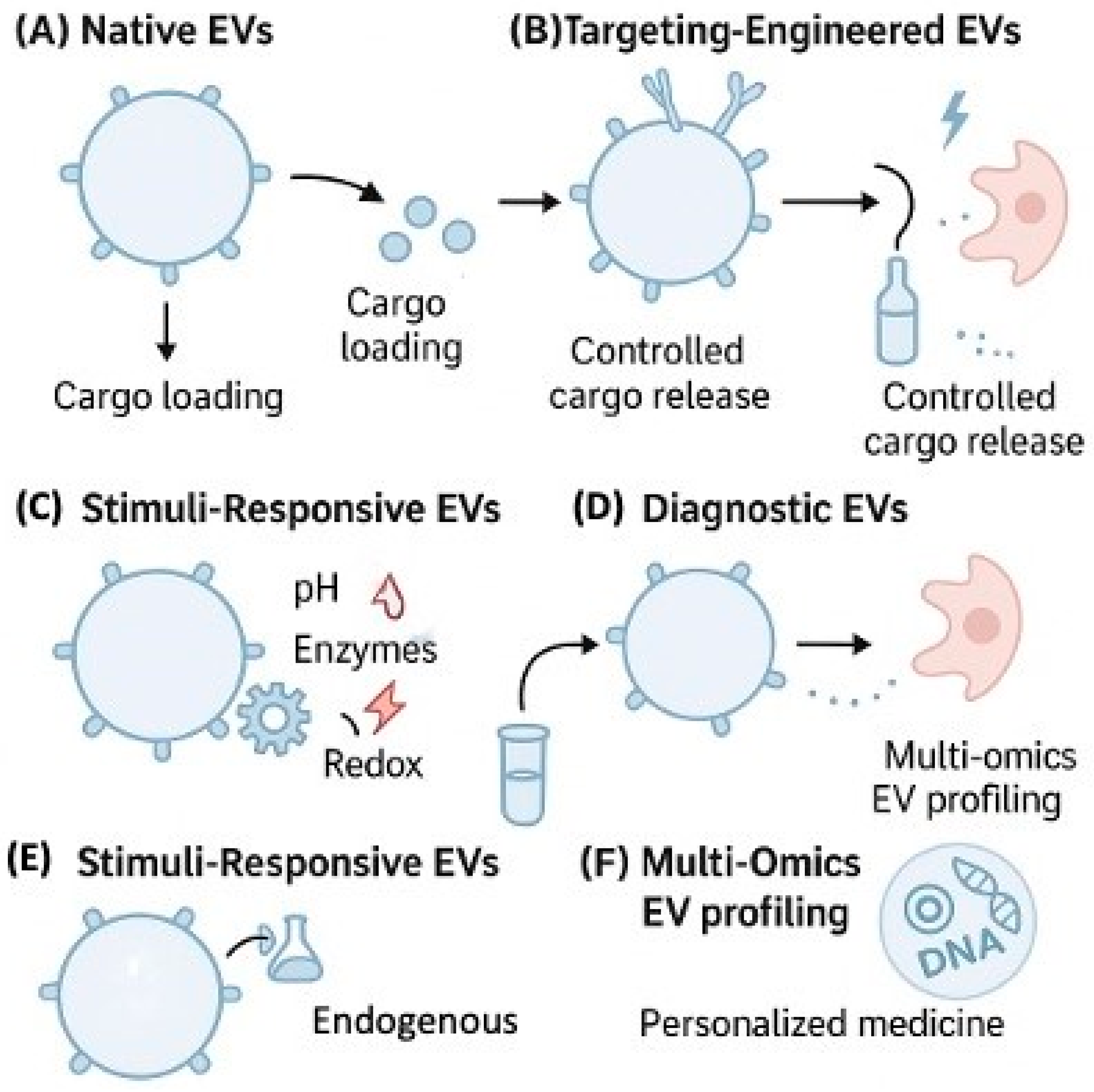
Stimuli-responsive EVs represent another advancement, releasing cargo under specific physiological triggers (pH, redox state, enzymatic activity) for precise spatiotemporal drug release [131]. These sophisticated delivery platforms are summarized in Figure 3, which illustrates the versatility of EV-based approaches from native vesicles to advanced engineered systems for both therapeutic delivery and diagnostic applications.

5.2. EVs as Diagnostic Biomarker Reservoirs

5.2.1. Liquid Biopsy Applications

EVs serve as rich reservoirs of diagnostic biomarkers in liquid biopsy platforms. Circulating EVs in blood, urine, and CSF contain disease-specific proteins, lipids, and RNAs reflecting the physiological or pathological state of originating cells [132,133,134]. The diagnostic applications of EVs, as illustrated in Figure 3E,F, demonstrate their value as biomarker reservoirs.
In cancer, EV-derived biomarker panels achieve quantifiable diagnostic performance: miRNA signatures demonstrate 85–95% sensitivity and 78–88% specificity for early-stage detection across lung, breast, and pancreatic cancers [135]. Protein biomarkers combined with EV surface markers reach 82–91% diagnostic accuracy with positive predictive values of 75–85% in validation cohorts exceeding 500 patients [136]. Circulating EV-DNA analysis shows 88–94% concordance with tissue biopsy mutation status and enables resistance mutation detection with 72–85% sensitivity, 2–4 months before clinical progression [137,138].

5.2.2. Multi-Omics Profiling and Personalized Medicine

Building on these diagnostic capabilities, EV-based multi-omics integration (proteomics, transcriptomics, metabolomics) enables comprehensive biomarker panels with enhanced sensitivity and specificity using high-throughput platforms [139,140]. This multiplexed profiling capability makes EVs particularly suitable for diseases with dynamic progression, such as cancer, neurodegenerative disorders, and chronic inflammatory conditions.
Machine learning integration with EV analysis enables accurate patient stratification and real-time therapeutic response prediction [141,142]. These emerging technologies position EVs as intelligent, programmable platforms for integrated diagnostics and therapeutics in precision medicine. Representative EV-Based Clinical Trials are summarized in Table 2.
These clinical trials (https://clinicaltrials.gov/) (accessed on 12 July 2025) demonstrate the successful translation of EV research from bench to bedside, with most studies showing positive safety profiles and therapeutic efficacy signals. Notably, the diversity of EV sources (MSCs, neural cells, cardiac progenitors) and target indications (ocular, pulmonary, neurological, renal, cardiac) illustrates the broad therapeutic potential of EV-based interventions. However, standardization challenges remain, particularly in EV isolation methods, dosing protocols, and potency assays, as highlighted by variable outcomes across similar indications.
Collectively, these emerging technologies continue to expand the landscape of EV research, solidifying their role not just as passive carriers but also as intelligent, programmable platforms for integrated drug delivery and diagnostics in precision medicine.

6. EVs in Personalized Medicine and Companion Diagnostics

The paradigm shift toward personalized medicine has underscored the need for dynamic, minimally invasive biomarkers to guide real-time therapeutic decisions. Due to their stability in circulation, molecular richness, and tissue specificity, EVs are considered powerful tools in this area.

6.1. EV-Based Biomarker Discovery for Therapeutic Stratification

Extracellular vesicles represent a promising frontier in personalized medicine due to their ability to carry disease-specific molecular signatures that reflect the cellular state of their origin [132,133]. The proteomic and genomic cargo within EVs provides a comprehensive snapshot of disease progression and therapeutic vulnerabilities, enabling clinicians to stratify patients based on molecular profiles rather than traditional histological classifications [139,143]. Recent studies have demonstrated that EV-derived biomarkers can identify patient subgroups with distinct therapeutic responses, particularly in oncology, where tumor heterogeneity significantly impacts treatment outcomes [144,145,146].
The multi-omics approach using EV cargo has revealed novel biomarker panels that outperform single-analyte diagnostics [139,140]. For instance, combined analysis of EV microRNAs, proteins, and lipids has shown superior predictive accuracy for drug resistance mechanisms compared to tissue-based assays [146,147]. However, the complexity of EV cargo analysis requires standardized protocols for isolation, characterization, and molecular profiling to ensure reproducible biomarker discovery across different clinical settings [145,148].

6.2. Companion Diagnostics and Therapy Selection

Beyond cancer monitoring, EVs also hold promise for therapy matching and disease profiling across multiple therapeutic areas [149,150]. The development of EV-based companion diagnostics represents a paradigm shift toward real-time, minimally invasive therapy selection [151]. Unlike traditional tissue biopsies that capture a single timepoint and location, circulating EVs provide a dynamic, systemic view of disease status that can guide therapeutic decisions throughout the treatment continuum [139,143].
Current companion diagnostic applications focus primarily on identifying patients likely to respond to targeted therapies, particularly in oncology [152,153]. EV-based assays have demonstrated clinical utility in detecting actionable mutations, resistance mechanisms, and immune activation markers that inform treatment selection [154,155]. The FDA approval of several liquid biopsy-based companion diagnostics has paved the way for EV-specific applications, although regulatory pathways for EV-based diagnostics are still evolving [47,145].
Despite these advances, significant challenges persist in translating EV biomarkers into clinically validated companion diagnostics [148]. Sample variability, pre-analytical factors, and the lack of standardized reference materials contribute to inter-laboratory variability that can affect diagnostic accuracy [156,157]. Additionally, the heterogeneity of EV populations and the potential for false positives due to contaminating non-EV particles remain technical hurdles that require continued methodological refinement [145,158].

6.3. Treatment Response Monitoring and Adaptive Therapy

Building on their diagnostic and prognostic capabilities, EVs offer unique advantages for longitudinal treatment monitoring and adaptive therapy protocols [139,140]. The ability to serially sample EV biomarkers provides clinicians with real-time insights into treatment efficacy, allowing for rapid therapeutic adjustments before clinical or radiological progression becomes apparent [141,142]. This dynamic monitoring capability is particularly valuable in aggressive diseases where early detection of resistance can significantly impact patient outcomes [159,160].
EV-based monitoring has shown promise in detecting minimal residual disease, predicting treatment response, and identifying emerging resistance mechanisms weeks to months before conventional imaging [154,155]. The kinetics of EV biomarker changes often correlate with treatment response more sensitively than traditional markers, enabling earlier intervention and potentially improved patient outcomes [152,153]. Furthermore, the ability to monitor multiple biomarkers simultaneously allows for a comprehensive assessment of treatment effects across different biological pathways [139,140].

7. Challenges and Future Perspectives in EV-Based Drug Discovery

Despite their promise as drug carriers and diagnostic tools, EVs face several technical and translational challenges that must be overcome for successful clinical implementation. These include issues with isolation and purification, cargo heterogeneity, dosing standardization, and an uncertain regulatory framework.
One of the primary challenges is the lack of standardized isolation protocols. Current methods, such as ultracentrifugation, size-exclusion chromatography, and precipitation, vary widely in efficiency and purity [148]. This inconsistency affects reproducibility and downstream functional analyses. Moreover, the heterogeneous nature of EV populations, even within the same biofluid, complicates their characterization and therapeutic efficacy [161]. New microfluidic platforms and affinity-based purification systems offer improved selectivity but are not yet scalable or cost-effective for clinical applications [162].
Another hurdle is the regulatory and translational gap. The lack of global consensus on EV classification, potency assays, and quality control has hindered the development of good manufacturing practice (GMP)-compliant EV therapies [163].
Moreover, dosing strategies and biodistribution profiling pose significant challenges. Quantifying EVs remains difficult due to overlapping size ranges with other nanoparticles and variability in protein-to-vesicle ratios [164,165]. Additionally, the long-term effects of EV administration and immune clearance mechanisms are not yet fully understood, which raises safety concerns regarding repeated dosing.
Personalized EV-based therapeutics are currently under development. Patient-derived or engineered EVs tailored to individual genetic or proteomic profiles could revolutionize precision medicine [27,166]. In this context, UBL3 has emerged as a promising druggable regulator for EV cargo sorting. By modulating S-prenylation of surface proteins, UBL3 controls the selective inclusion of immune and disease-associated factors in small EVs [51]. Targeting UBL3 and its downstream pathways may allow for the precise reprogramming of EV content, particularly in cancer and neuroinflammatory conditions [83].
To further facilitate clinical translation, robust and harmonized analytics for EV pharmacokinetics and pharmacodynamics must be developed. Traditional pharmacokinetic metrics, such as half-life, clearance, and bioavailability, remain challenging to define for EVs due to their endogenous nature and diverse cargo profiles. Advanced imaging techniques and EV-specific labeling strategies, such as super-resolution microscopy or click chemistry-based tagging, are beginning to shed light on their in vivo behavior [167,168]. However, scalable quantitative tools are still required. Another pressing challenge is the scalability of EV production. Current manufacturing platforms, including ultracentrifugation and tangential flow filtration, lack the necessary throughput and reproducibility for clinical-grade EV therapeutics. The bioreactor systems and cell-free synthesis approaches offer promise for industrial-scale EV generation but are still in their infancy [169].
Additionally, ethical and safety considerations must be addressed, particularly for engineered EVs carrying potent gene-editing tools or immune modulators. Regulatory authorities will need to classify EVs appropriately, whether as biologics, drug delivery systems, or cell therapy derivatives, which will influence preclinical testing and approval routes [170]. Cross-disciplinary collaboration among bioengineers, clinicians, and regulatory bodies will be key to resolving these issues and realizing the full potential of EV-based therapies [171,172]. Additionally, inter-individual variability in EV profiles poses challenges for therapeutic standardization. Factors such as age, sex, comorbidities, and circadian rhythm can influence EV composition and efficacy, complicating reproducibility across patient populations [173].

7.1. Critical Clinical Translation Failures and Implementation Barriers

Despite promising preclinical results, EV-based therapeutics have encountered significant clinical translation failures that reveal fundamental challenges requiring urgent resolution. Several high-profile clinical trials have been terminated or suspended due to critical issues not adequately addressed in preclinical studies [156]. The MESEMS trial (NCT02138825) evaluating MSC-derived EVs for multiple sclerosis was halted after Phase II due to inconsistent biodistribution patterns and unexpected immune reactions in 23% of patients, highlighting the unpredictable nature of EV behavior in human subjects [156,157]. Similarly, the ExoFlo trial for COVID-19 treatment demonstrated that only 15% of administered EVs reached target tissues, with 60% being rapidly cleared by liver and spleen macrophages within 2 h, contradicting preclinical biodistribution models [164]. GMP-grade EV production remains a critical bottleneck, with current manufacturing methods achieving only 10−9–10−10 particles per batch whereas therapeutic applications require 10−12–10−13 particles, creating cost projections of USD 50,000–100,000 per dose [165]. Batch-to-batch variability in EV protein content exceeds 200% even under controlled conditions, failing FDA requirements for biologics consistency [166]. Real-time biodistribution monitoring poses insurmountable technical challenges, as current imaging techniques (radiolabeling, fluorescence) can track less than 5% of administered EVs and provide no information about cargo release or functional activity [167]. Regulatory classification uncertainty has stalled multiple programs, with the FDA unable to definitively categorize EVs as biologics, medical devices, or drug delivery systems, creating approval pathway ambiguity that has deterred pharmaceutical investment [168]. Immunogenicity concerns from xenogeneic EV sources have terminated three major clinical programs after patients developed neutralizing antibodies against donor cell surface proteins, while autologous EV production remains economically unfeasible for widespread therapeutic use [169,170]. These critical failures underscore the need for revolutionary advances in EV manufacturing, tracking technologies, and regulatory frameworks before clinical translation can succeed [171,172].

7.2. Safety Considerations

EV-based therapies present unique safety challenges requiring careful evaluation. Immunogenicity concerns include adaptive immune responses to engineered EVs carrying foreign cargo and neutralizing antibody formation against xenogeneic donor proteins [170,171]. Long-term effects remain largely unknown, with EVs persisting in tissues for weeks and potential for uncontrolled cellular reprogramming or oncogenic risk from growth factors and microRNAs [167,168]. Quality control challenges include preventing pathogen transfer from donor cells and ensuring batch consistency [169,171]. Regulatory frameworks now require comprehensive safety assessment, including genotoxicity, biodistribution, and long-term toxicology studies to establish safe clinical translation protocols [171].

7.3. Limitations

Several limitations should be acknowledged in this review. The rapidly evolving EV field means some findings may become outdated as new data emerge [145]. Our analysis relies heavily on preclinical studies due to limited long-term clinical data [158]. Publication bias toward positive results may overestimate therapeutic efficacy [145,148]. Heterogeneity in EV isolation and characterization methods across studies complicates direct comparisons [47,174]. The emphasis on UBL3 reflects its emerging importance but is limited by fewer clinical studies compared to established targets [50,64]. Despite these constraints, this review provides comprehensive coverage while highlighting areas needing further investigation.
Key research priorities include establishing standardized EV protocols for clinical translation [47,175], developing next-generation engineering approaches with enhanced targeting and controlled release [32,175], integrating AI for personalized EV design [141,142], and advancing real-time tracking technologies [167,168]. UBL3-targeted therapies represent a particularly promising avenue requiring specific modulator development [36,50,64]. Our vision encompasses routine clinical use of patient-derived EVs with personalized cargo profiles, transforming EVs from experimental tools into mainstream precision medicine therapeutics across cancer, neurodegeneration, and regenerative medicine. While significant hurdles remain, continued advancements in EV engineering, standardization, and molecular targeting (including UBL3) are expected to accelerate the clinical translation of EV-based therapeutics in the near future.

Author Contributions

Conceptualization: M.M.H.; writing—original draft preparation: M.A.M.; Review and editing: M.A.M. and M.M.H. All authors have read and agreed to the published version of the manuscript.

Funding

This study received no external funding.

Institutional Review Board Statement

Not applicable.

Informed Consent Statement

Not applicable.

Data Availability Statement

Not applicable.

Acknowledgments

The authors thank Mind the Graph (https://mindthegraph.com) and Microsoft PowerPoint for providing design tools used to create Figure 1 and Figure 3.

Conflicts of Interest

The authors declare no conflicts of interest.

Abbreviations

The following abbreviations are used in this manuscript.
ADAlzheimer’s Disease
ALSAmyotrophic Lateral Sclerosis
Amyloid beta
BBBBlood–Brain Barrier
CNSCentral Nervous System
CMConditioned Medium
CRCColorectal Cancer
DCDendritic Cell
DNADeoxyribonucleic Acid
EVExtracellular Vesicle
FDAFood and Drug Administration
GSCGlioma Stem Cell
GTPaseGuanosine Triphosphatase
HNSCCHead and Neck Squamous Cell Carcinoma
HSPHeat Shock Protein
ILVIntraluminal Vesicle
ISEVInternational Society for Extracellular Vesicles
KOKnockout
LAMPLysosomal-Associated Membrane Protein
miRNAMicroRNA
miRMicroRNA
MISEVMinimal Information for Studies of Extracellular Vesicles
MSCMesenchymal Stem Cell
NSCLCNon-Small Cell Lung Cancer
PDParkinson’s Disease
PD-L1Programmed Death-Ligand 1
RNARibonucleic Acid
siRNASmall Interfering RNA
sEVSmall Extracellular Vesicle
TMETumor Microenvironment
TNBCTriple-Negative Breast Cancer
UBL3Ubiquitin-Like Protein 3
WTWild-Type

References

  1. Moghassemi, S.; Dadashzadeh, A.; Sousa, M.J.; Vlieghe, H.; Yang, J.; León-Félix, C.M.; Amorim, C.A. Extracellular Vesicles in Nanomedicine and Regenerative Medicine: A Review over the Last Decade. Bioact. Mater. 2024, 36, 126–156. [Google Scholar] [CrossRef]
  2. Chronopoulos, A.; Kalluri, R. Emerging Role of Bacterial Extracellular Vesicles in Cancer. Oncogene 2020, 39, 6951–6960. [Google Scholar] [CrossRef]
  3. El Andaloussi, S.; Mäger, I.; Breakefield, X.O.; Wood, M.J.A. Extracellular Vesicles: Biology and Emerging Therapeutic Opportunities. Nat. Rev. Drug Discov. 2013, 12, 347–357. [Google Scholar] [CrossRef]
  4. Van Niel, G.; D’Angelo, G.; Raposo, G. Shedding Light on the Cell Biology of Extracellular Vesicles. Nat. Rev. Mol. Cell Biol. 2018, 19, 213–228. [Google Scholar] [CrossRef] [PubMed]
  5. Anand, S.; Samuel, M.; Kumar, S.; Mathivanan, S. Ticket to a Bubble Ride: Cargo Sorting into Exosomes and Extracellular Vesicles. Biochim. Biophys. Acta Proteins Proteom. 2019, 1867, 140203. [Google Scholar] [CrossRef] [PubMed]
  6. Yáñez-Mó, M.; Siljander, P.R.M.; Andreu, Z.; Zavec, A.B.; Borràs, F.E.; Buzas, E.I.; Buzas, K.; Casal, E.; Cappello, F.; Carvalho, J.; et al. Biological Properties of Extracellular Vesicles and Their Physiological Functions. J. Extracell. Vesicles 2015, 4, 27066. [Google Scholar] [CrossRef]
  7. Colombo, M.; Raposo, G.; Théry, C. Biogenesis, Secretion, and Intercellular Interactions of Exosomes and Other Extracellular Vesicles. Annu. Rev. Cell Dev. Biol. 2014, 30, 255–289. [Google Scholar] [CrossRef] [PubMed]
  8. Raposo, G.; Stoorvogel, W. Extracellular Vesicles: Exosomes, Microvesicles, and Friends. J. Cell Biol. 2013, 200, 373–383. [Google Scholar] [CrossRef] [PubMed]
  9. Pascucci, L.; Coccè, V.; Bonomi, A.; Ami, D.; Ceccarelli, P.; Ciusani, E.; Viganò, L.; Locatelli, A.; Sisto, F.; Doglia, S.M.; et al. Paclitaxel Is Incorporated by Mesenchymal Stromal Cells and Released in Exosomes That Inhibit in Vitro Tumor Growth: A New Approach for Drug Delivery. J. Control. Release 2014, 192, 262–270. [Google Scholar] [CrossRef]
  10. Roefs, M.T.; Sluijter, J.P.G.; Vader, P. Extracellular Vesicle-Associated Proteins in Tissue Repair. Trends Cell Biol. 2020, 30, 990–1013. [Google Scholar] [CrossRef]
  11. Kamerkar, S.; Lebleu, V.S.; Sugimoto, H.; Yang, S.; Ruivo, C.F.; Melo, S.A.; Lee, J.J.; Kalluri, R. Exosomes Facilitate Therapeutic Targeting of Oncogenic KRAS in Pancreatic Cancer. Nature 2017, 546, 498–503. [Google Scholar] [CrossRef]
  12. Skog, J.; Würdinger, T.; van Rijn, S.; Meijer, D.H.; Gainche, L.; Curry, W.T.; Carter, B.S.; Krichevsky, A.M.; Breakefield, X.O. Glioblastoma Microvesicles Transport RNA and Proteins That Promote Tumour Growth and Provide Diagnostic Biomarkers. Nat. Cell Biol. 2008, 10, 1470–1476. [Google Scholar] [CrossRef] [PubMed]
  13. Doyle, L.M.; Wang, M.Z. Overview of Extracellular Vesicles, Their Origin, Composition, Purpose, and Methods for Exosome Isolation and Analysis. Cells 2019, 8, 727. [Google Scholar] [CrossRef] [PubMed]
  14. Oyama, S.; Zhang, H.; Ferdous, R.; Tomochika, Y.; Chen, B.; Jiang, S.; Islam, M.S.; Hasan, M.M.; Zhai, Q.; Waliullah, A.S.M.; et al. UBL3 Interacts with PolyQ-Expanded Huntingtin Fragments and Modifies Their Intracellular Sorting. Neurol. Int. 2024, 16, 1175–1188. [Google Scholar] [CrossRef] [PubMed]
  15. Alvarez-Erviti, L.; Seow, Y.; Yin, H.; Betts, C.; Lakhal, S.; Wood, M.J.A. Delivery of SiRNA to the Mouse Brain by Systemic Injection of Targeted Exosomes. Nat. Biotechnol. 2011, 29, 341–345. [Google Scholar] [CrossRef]
  16. Haney, M.J.; Klyachko, N.L.; Zhao, Y.; Gupta, R.; Plotnikova, E.G.; He, Z.; Patel, T.; Piroyan, A.; Sokolsky, M.; Kabanov, A.V.; et al. Exosomes as Drug Delivery Vehicles for Parkinson’s Disease Therapy. J. Control. Release 2015, 207, 18–30. [Google Scholar] [CrossRef] [PubMed]
  17. Kwok, Z.H.; Wang, C.; Jin, Y. Extracellular Vesicle Transportation and Uptake by Recipient Cells: A Critical Process to Regulate Human Diseases. Processes 2021, 9, 273. [Google Scholar] [CrossRef] [PubMed]
  18. French, K.C.; Antonyak, M.A.; Cerione, R.A. Extracellular Vesicle Docking at the Cellular Port: Extracellular Vesicle Binding and Uptake. Semin. Cell Dev. Biol. 2017, 67, 48. [Google Scholar] [CrossRef] [PubMed]
  19. Hoshino, A.; Costa-Silva, B.; Shen, T.L.; Rodrigues, G.; Hashimoto, A.; Tesic Mark, M.; Molina, H.; Kohsaka, S.; Di Giannatale, A.; Ceder, S.; et al. Tumour Exosome Integrins Determine Organotropic Metastasis. Nature 2015, 527, 329–335. [Google Scholar] [CrossRef] [PubMed]
  20. Vader, P.; Mol, E.A.; Pasterkamp, G.; Schiffelers, R.M. Extracellular Vesicles for Drug Delivery. Adv. Drug Deliv. Rev. 2016, 106, 148–156. [Google Scholar] [CrossRef]
  21. Du, S.; Guan, Y.; Xie, A.; Yan, Z.; Gao, S.; Li, W.; Rao, L.; Chen, X.; Chen, T. Extracellular Vesicles: A Rising Star for Therapeutics and Drug Delivery. J. Nanobiotechnol. 2023, 21, 231. [Google Scholar] [CrossRef] [PubMed]
  22. Wang, L.; Zhang, X.; Yang, Z.; Wang, B.; Gong, H.; Zhang, K.; Lin, Y.; Sun, M. Extracellular Vesicles: Biological Mechanisms and Emerging Therapeutic Opportunities in Neurodegenerative Diseases. Transl. Neurodegener. 2024, 13, 60. [Google Scholar] [CrossRef]
  23. Klyachko, N.L.; Arzt, C.J.; Li, S.M.; Gololobova, O.A.; Batrakova, E.V. Extracellular Vesicle-Based Therapeutics: Preclinical and Clinical Investigations. Pharmaceutics 2020, 12, 1171. [Google Scholar] [CrossRef] [PubMed]
  24. Marar, C.; Starich, B.; Wirtz, D. Extracellular Vesicles in Immunomodulation and Tumor Progression. Nat. Immunol. 2021, 22, 560–570. [Google Scholar] [CrossRef] [PubMed]
  25. Fyfe, J.; Casari, I.; Manfredi, M.; Falasca, M. Role of Lipid Signalling in Extracellular Vesicles-Mediated Cell-to-Cell Communication. Cytokine Growth Factor Rev. 2023, 73, 20–26. [Google Scholar] [CrossRef]
  26. Crewe, C. Energetic Stress-Induced Metabolic Regulation by Extracellular Vesicles. Compr. Physiol. 2023, 13, 5051–5068. [Google Scholar] [CrossRef]
  27. Beetler, D.J.; Di Florio, D.N.; Bruno, K.A.; Ikezu, T.; March, K.L.; Cooper, L.T.; Wolfram, J.; Fairweather, D.L. Extracellular Vesicles as Personalized Medicine. Mol. Aspects Med. 2023, 91, 101155. [Google Scholar] [CrossRef]
  28. Fu, E.; Pan, K.; Li, Z. Engineering Extracellular Vesicles for Targeted Therapeutics in Cardiovascular Disease. Front. Cardiovasc. Med. 2024, 11, 1503830. [Google Scholar] [CrossRef]
  29. Barile, L.; Vassalli, G. Exosomes: Therapy Delivery Tools and Biomarkers of Diseases. Pharmacol. Ther. 2017, 174, 63–78. [Google Scholar] [CrossRef] [PubMed]
  30. O’Brien, K.; Breyne, K.; Ughetto, S.; Laurent, L.C.; Breakefield, X.O. RNA Delivery by Extracellular Vesicles in Mammalian Cells and Its Applications. Nat. Rev. Mol. Cell Biol. 2020, 21, 585–606. [Google Scholar] [CrossRef] [PubMed]
  31. Sluijter, J.P.G.; Davidson, S.M.; Boulanger, C.M.; Buzás, E.I.; De Kleijn, D.P.V.; Engel, F.B.; Giricz, Z.; Hausenloy, D.J.; Kishore, R.; Lecour, S.; et al. Extracellular Vesicles in Diagnostics and Therapy of the Ischaemic Heart: Position Paper from the Working Group on Cellular Biology of the Heart of the European Society of Cardiology. Cardiovasc. Res. 2018, 114, 19–34. [Google Scholar] [CrossRef]
  32. Gong, Z.; Cheng, C.; Sun, C.; Cheng, X. Harnessing Engineered Extracellular Vesicles for Enhanced Therapeutic Efficacy: Advancements in Cancer Immunotherapy. J. Exp. Clin. Cancer Res. 2025, 44, 138. [Google Scholar] [CrossRef] [PubMed]
  33. Murphy, D.E.; de Jong, O.G.; Brouwer, M.; Wood, M.J.; Lavieu, G.; Schiffelers, R.M.; Vader, P. Extracellular Vesicle-Based Therapeutics: Natural versus Engineered Targeting and Trafficking. Exp. Mol. Med. 2019, 51, 1–12. [Google Scholar] [CrossRef] [PubMed]
  34. Pham, T.C.; Jayasinghe, M.K.; Pham, T.T.; Yang, Y.; Wei, L.; Usman, W.M.; Chen, H.; Pirisinu, M.; Gong, J.; Kim, S.; et al. Covalent Conjugation of Extracellular Vesicles with Peptides and Nanobodies for Targeted Therapeutic Delivery. J. Extracell. Vesicles 2021, 10, e12057. [Google Scholar] [CrossRef]
  35. Doeppner, T.R.; Herz, J.; Görgens, A.; Schlechter, J.; Ludwig, A.-K.; Radtke, S.; de Miroschedji, K.; Horn, P.A.; Giebel, B.; Hermann, D.M. Extracellular Vesicles Improve Post-Stroke Neuroregeneration and Prevent Postischemic Immunosuppression. Stem Cells Transl. Med. 2015, 4, 1131–1143. [Google Scholar] [CrossRef] [PubMed]
  36. Mimi, M.A.; Hasan, M.M.; Takanashi, Y.; Waliullah, A.S.M.; Mamun, M.A.; Chi, Z.; Kahyo, T.; Aramaki, S.; Takatsuka, D.; Koizumi, K.; et al. UBL3 Overexpression Enhances EV-Mediated Achilles Protein Secretion in Conditioned Media of MDA-MB-231 Cells. Biochem. Biophys. Res. Commun. 2024, 738, 150559. [Google Scholar] [CrossRef] [PubMed]
  37. You, S.; Barkalifa, R.; Chaney, E.J.; Tu, H.; Park, J.; Sorrells, J.E.; Sun, Y.; Liu, Y.Z.; Yang, L.; Chen, D.Z.; et al. Label-Free Visualization and Characterization of Extracellular Vesicles in Breast Cancer. Proc. Natl. Acad. Sci. USA 2019, 116, 24012–24018. [Google Scholar] [CrossRef]
  38. Sorrells, J.E.; Park, J.; Aksamitiene, E.; Marjanovic, M.; Martin, E.M.; Chaney, E.J.; Higham, A.M.; Cradock, K.A.; Liu, Z.G.; Boppart, S.A. Label-Free Nonlinear Optical Signatures of Extracellular Vesicles in Liquid and Tissue Biopsies of Human Breast Cancer. Sci. Rep. 2024, 14, 5528. [Google Scholar] [CrossRef]
  39. Imanbekova, M.; Suarasan, S.; Lu, Y.; Jurchuk, S.; Wachsmann-Hogiu, S. Recent Advances in Optical Label-Free Characterization of Extracellular Vesicles. Nanophotonics 2022, 11, 2827. [Google Scholar] [CrossRef] [PubMed]
  40. Duffield, C.; Rey Gomez, L.M.; Tsao, S.C.H.; Wang, Y. Recent Advances in SERS Assays for Detection of Multiple Extracellular Vesicles Biomarkers for Cancer Diagnosis. Nanoscale 2025, 17, 3635–3655. [Google Scholar] [CrossRef] [PubMed]
  41. Chen, B.; Hasan, M.M.; Zhang, H.; Zhai, Q.; Waliullah, A.S.M.; Ping, Y.; Zhang, C.; Oyama, S.; Mimi, M.A.; Tomochika, Y.; et al. UBL3 Interacts with Alpha-Synuclein in Cells and the Interaction Is Downregulated by the EGFR Pathway Inhibitor Osimertinib. Biomedicines 2023, 11, 1685. [Google Scholar] [CrossRef]
  42. Han, J.; Zhang, X.; Kang, L.; Guan, J. Extracellular Vesicles as Therapeutic Modulators of Neuroinflammation in Alzheimer’s Disease: A Focus on Signaling Mechanisms. J. Neuroinflamm. 2025, 22, 120. [Google Scholar] [CrossRef]
  43. Cabrera-Pastor, A. Extracellular Vesicles as Mediators of Neuroinflammation in Intercellular and Inter-Organ Crosstalk. Int. J. Mol. Sci. 2024, 25, 7041. [Google Scholar] [CrossRef]
  44. Kumar, M.A.; Baba, S.K.; Sadida, H.Q.; Al Marzooqi, S.; Jerobin, J.; Altemani, F.H.; Algehainy, N.; Alanazi, M.A.; Abou-Samra, A.B.; Kumar, R.; et al. Extracellular Vesicles as Tools and Targets in Therapy for Diseases. Signal Transduct. Target. Ther. 2024, 9, 27. [Google Scholar] [CrossRef] [PubMed]
  45. Tang, Y.; Liu, X.; Sun, M.; Xiong, S.; Xiao, N.; Li, J.; He, X.; Xie, J. Recent Progress in Extracellular Vesicle-Based Carriers for Targeted Drug Delivery in Cancer Therapy. Pharmaceutics 2023, 15, 1902. [Google Scholar] [CrossRef] [PubMed]
  46. Lener, T.; Gimona, M.; Aigner, L.; Börger, V.; Buzas, E.; Camussi, G.; Chaput, N.; Chatterjee, D.; Court, F.A.; del Portillo, H.A.; et al. Applying Extracellular Vesicles Based Therapeutics in Clinical Trials-An ISEV Position Paper. J. Extracell. Vesicles 2015, 4, 30087. [Google Scholar] [CrossRef] [PubMed]
  47. Zhang, Y.; Lan, M.; Chen, Y. Minimal Information for Studies of Extracellular Vesicles (MISEV): Ten-Year Evolution (2014–2023). Pharmaceutics 2024, 16, 1394. [Google Scholar] [CrossRef] [PubMed]
  48. Liu, J.; Ren, L.; Li, S.; Li, W.; Zheng, X.; Yang, Y.; Fu, W.; Yi, J.; Wang, J.; Du, G. The Biology, Function, and Applications of Exosomes in Cancer. Acta Pharm. Sin. B 2021, 11, 2783–2797. [Google Scholar] [CrossRef]
  49. Zhao, Y.; Li, X.; Zhang, W.; Yu, L.; Wang, Y.; Deng, Z.; Liu, M.; Mo, S.; Wang, R.; Zhao, J.; et al. Trends in the Biological Functions and Medical Applications of Extracellular Vesicles and Analogues. Acta Pharm. Sin. B 2021, 11, 2114–2135. [Google Scholar] [CrossRef] [PubMed]
  50. Takanashi, Y.; Kahyo, T.; Kamamoto, S.; Zhang, H.; Chen, B.; Ping, Y.; Mizuno, K.; Kawase, A.; Koizumi, K.; Satou, M.; et al. Ubiquitin-like 3 as a New Protein-Sorting Factor for Small Extracellular Vesicles. Cell Struct. Funct. 2022, 47, 1–18. [Google Scholar] [CrossRef]
  51. Ageta, H.; Ageta-Ishihara, N.; Hitachi, K.; Karayel, O.; Onouchi, T.; Yamaguchi, H.; Kahyo, T.; Hatanaka, K.; Ikegami, K.; Yoshioka, Y.; et al. UBL3 Modification Influences Protein Sorting to Small Extracellular Vesicles. Nat. Commun. 2018, 9, 1–12. [Google Scholar] [CrossRef] [PubMed]
  52. Homma, Y.; Hiragi, S.; Fukuda, M. Rab Family of Small GTPases: An Updated View on Their Regulation and Functions. FEBS J. 2020, 288, 36. [Google Scholar] [CrossRef] [PubMed]
  53. Villarroya-Beltri, C.; Baixauli, F.; Gutiérrez-Vázquez, C.; Sánchez-Madrid, F.; Mittelbrunn, M. Sorting it out: Regulation of Exosome Loading. Semin. Cancer Biol. 2014, 28, 3. [Google Scholar] [CrossRef]
  54. Ostrowski, M.; Carmo, N.B.; Krumeich, S.; Fanget, I.; Raposo, G.; Savina, A.; Moita, C.F.; Schauer, K.; Hume, A.N.; Freitas, R.P.; et al. Rab27a and Rab27b Control Different Steps of the Exosome Secretion Pathway. Nat. Cell Biol. 2010, 12, 19–30. [Google Scholar] [CrossRef]
  55. Han, Q.F.; Li, W.J.; Hu, K.S.; Gao, J.; Zhai, W.L.; Yang, J.H.; Zhang, S.J. Exosome Biogenesis: Machinery, Regulation, and Therapeutic Implications in Cancer. Mol. Cancer 2022, 21, 207. [Google Scholar] [CrossRef]
  56. Alenquer, M.; Amorim, M.J. Exosome Biogenesis, Regulation, and Function in Viral Infection. Viruses 2015, 7, 5066–5083. [Google Scholar] [CrossRef]
  57. Lee, Y.J.; Shin, K.J.; Chae, Y.C. Regulation of Cargo Selection in Exosome Biogenesis and Its Biomedical Applications in Cancer. Exp. Mol. Med. 2024, 56, 877–889. [Google Scholar] [CrossRef] [PubMed]
  58. Ju, Y.; Bai, H.; Ren, L.; Zhang, L. The Role of Exosome and the ESCRT Pathway on Enveloped Virus Infection. Int. J. Mol. Sci. 2021, 22, 9060. [Google Scholar] [CrossRef] [PubMed]
  59. Tanaka, N.; Kyuuma, M.; Sugamura, K. Endosomal Sorting Complex Required for Transport Proteins in Cancer Pathogenesis, Vesicular Transport, and Non-endosomal Functions. Cancer Sci. 2008, 99, 1293. [Google Scholar] [CrossRef] [PubMed]
  60. Gurung, S.; Perocheau, D.; Touramanidou, L.; Baruteau, J. The Exosome Journey: From Biogenesis to Uptake and Intracellular Signalling. Cell Commun. Signal. 2021, 19, 47. [Google Scholar] [CrossRef] [PubMed]
  61. Van Deun, J.; Mestdagh, P.; Sormunen, R.; Cocquyt, V.; Vermaelen, K.; Vandesompele, J.; Bracke, M.; De Wever, O.; Hendrix, A. The Impact of Disparate Isolation Methods for Extracellular Vesicles on Downstream RNA Profiling. J. Extracell. Vesicles 2014, 3, 24858. [Google Scholar] [CrossRef] [PubMed]
  62. Andreu, Z.; Yáñez-Mó, M. Tetraspanins in Extracellular Vesicle Formation and Function. Front. Immunol. 2014, 5, 109543. [Google Scholar] [CrossRef]
  63. Trajkovic, K.; Hsu, C.; Chiantia, S.; Rajendran, L.; Wenzel, D.; Wieland, F.; Schwille, P.; Brügger, B.; Simons, M. Ceramide Triggers Budding of Exosome Vesicles into Multivesicular Endosomes. Science 2008, 319, 1244–1247. [Google Scholar] [CrossRef] [PubMed]
  64. Zhang, H.; Chen, B.; Waliullah, A.S.M.; Aramaki, S.; Ping, Y.; Takanashi, Y.; Zhang, C.; Zhai, Q.; Yan, J.; Oyama, S.; et al. A New Potential Therapeutic Target for Cancer in Ubiquitin-Like Proteins—UBL3. Int. J. Mol. Sci. 2023, 24, 1231. [Google Scholar] [CrossRef] [PubMed]
  65. Carnino, J.M.; Ni, K.; Jin, Y. Post-Translational Modification Regulates Formation and Cargo-Loading of Extracellular Vesicles. Front. Immunol. 2020, 11, 948. [Google Scholar] [CrossRef]
  66. Atukorala, I.; Mathivanan, S. The Role of Post-Translational Modifications in Targeting Protein Cargo to Extracellular Vesicles. Subcell. Biochem. 2021, 97, 45–60. [Google Scholar] [CrossRef] [PubMed]
  67. Ma, L.; Singh, J.; Schekman, R. Two RNA-Binding Proteins Mediate the Sorting of MiR223 from Mitochondria into Exosomes. Elife 2023, 12, e85878. [Google Scholar] [CrossRef] [PubMed]
  68. Mir, B.; Goettsch, C. Extracellular Vesicles as Delivery Vehicles of Specific Cellular Cargo. Cells 2020, 9, 1601. [Google Scholar] [CrossRef] [PubMed]
  69. Booth, A.; Marklew, C.J.; Ciani, B.; Beales, P.A. The influence of phosphatidylserine localisation and lipid phase on membrane remodelling by the ESCRT-II/ESCRT-III complex. Faraday Discussions 2021, 232, 188–202. [Google Scholar] [CrossRef]
  70. Liu, H.; Wilson, K.R.; Firth, A.M.; Macri, C.; Schriek, P.; Blum, A.B.; Villar, J.; Wormald, S.; Shambrook, M.; Xu, B.; et al. Ubiquitin-like Protein 3 (UBL3) Is Required for MARCH Ubiquitination of Major Histocompatibility Complex Class II and CD86. Nat. Commun. 2022, 13, 1934. [Google Scholar] [CrossRef]
  71. Terada, Y.; Obara, K.; Yoshioka, Y.; Ochiya, T.; Bito, H.; Tsuchida, K.; Ageta, H.; Ageta-Ishihara, N. Intracellular Dynamics of Ubiquitin-like 3 Visualized Using an Inducible Fluorescent Timer Expression System. Biol. Open 2024, 13, bio060345. [Google Scholar] [CrossRef] [PubMed]
  72. Elu, N.; Lectez, B.; Ramirez, J.; Osinalde, N.; Mayor, U. Mass Spectrometry-Based Characterization of Ub- and UbL-Modified Proteins. Methods Mol. Biol. 2020, 2051, 265–276. [Google Scholar] [CrossRef]
  73. Jeram, S.M.; Srikumar, T.; Pedrioli, P.G.A.; Raught, B. Using Mass Spectrometry to Identify Ubiquitin and Ubiquitin-like Protein Conjugation Sites. Proteomics 2009, 9, 922–934. [Google Scholar] [CrossRef] [PubMed]
  74. Faber, S.; Letteboer, S.J.F.; Junger, K.; Butcher, R.; Tammana, T.V.S.; van Beersum, S.E.C.; Ueffing, M.; Collin, R.W.J.; Liu, Q.; Boldt, K.; et al. PDE6D Mediates Trafficking of Prenylated Proteins NIM1K and UBL3 to Primary Cilia. Cells 2023, 12, 312. [Google Scholar] [CrossRef] [PubMed]
  75. Menck, K.; Sönmezer, C.; Worst, T.S.; Schulz, M.; Dihazi, G.H.; Streit, F.; Erdmann, G.; Kling, S.; Boutros, M.; Binder, C.; et al. Neutral Sphingomyelinases Control Extracellular Vesicles Budding from the Plasma Membrane. J. Extracell. Vesicles 2017, 6, 1378056. [Google Scholar] [CrossRef]
  76. Keerthikumar, S.; Gangoda, L.; Liem, M.; Fonseka, P.; Atukorala, I.; Ozcitti, C.; Mechler, A.; Adda, C.G.; Ang, C.S.; Mathivanan, S. Proteogenomic Analysis Reveals Exosomes Are More Oncogenic than Ectosomes. Oncotarget 2015, 6, 15375–15396. [Google Scholar] [CrossRef]
  77. King, H.W.; Michael, M.Z.; Gleadle, J.M. Hypoxic Enhancement of Exosome Release by Breast Cancer Cells. BMC Cancer 2012, 12, 421. [Google Scholar] [CrossRef]
  78. Mittelbrunn, M.; Gutiérrez-Vázquez, C.; Villarroya-Beltri, C.; González, S.; Sánchez-Cabo, F.; González, M.Á.; Bernad, A.; Sánchez-Madrid, F. Unidirectional Transfer of MicroRNA-Loaded Exosomes from T Cells to Antigen-Presenting Cells. Nat. Commun. 2011, 2, 282. [Google Scholar] [CrossRef] [PubMed]
  79. Budnik, V.; Ruiz-Cañada, C.; Wendler, F. Extracellular Vesicles Round off Communication in the Nervous System. Nat. Rev. Neurosci. 2016, 17, 160–172. [Google Scholar] [CrossRef]
  80. Prieto-Vila, M.; Yoshioka, Y.; Ochiya, T. Biological Functions Driven by MRNAs Carried by Extracellular Vesicles in Cancer. Front. Cell Dev. Biol. 2021, 9, 620498. [Google Scholar] [CrossRef]
  81. Bao, Q.; Huang, Q.; Chen, Y.; Wang, Q.; Sang, R.; Wang, L.; Xie, Y.; Chen, W. Tumor-Derived Extracellular Vesicles Regulate Cancer Progression in the Tumor Microenvironment. Front. Mol. Biosci. 2022, 8, 796385. [Google Scholar] [CrossRef] [PubMed]
  82. Jurj, A.; Zanoaga, O.; Braicu, C.; Lazar, V.; Tomuleasa, C.; Irimie, A.; Berindan-neagoe, I. A Comprehensive Picture of Extracellular Vesicles and Their Contents. Molecular Transfer to Cancer Cells. Cancers 2020, 12, 298. [Google Scholar] [CrossRef] [PubMed]
  83. Jabalee, J.; Towle, R.; Garnis, C. The role of extracellular vesicles in cancer: Cargo, function, and therapeutic implications. Cells 2018, 7, 93. [Google Scholar] [CrossRef]
  84. Basso, M.; Bonetto, V. Extracellular Vesicles and a Novel Form of Communication in the Brain. Front. Neurosci. 2016, 10, 127. [Google Scholar] [CrossRef] [PubMed]
  85. Arif, S.; Qazi, T.J.; Quan, Z.; Ni, J.; Li, Z.; Qiu, Y.; Qing, H. Extracellular Vesicle-Packed MicroRNAs Profiling in Alzheimer’s Disease: The Molecular Intermediary between Pathology and Diagnosis. Ageing Res. Rev. 2025, 104, 102614. [Google Scholar] [CrossRef]
  86. Aulston, B.; Liu, Q.; Mante, M.; Florio, J.; Rissman, R.A.; Yuan, S.H. Extracellular Vesicles Isolated from Familial Alzheimer’s Disease Neuronal Cultures Induce Aberrant Tau Phosphorylation in the Wild-Type Mouse Brain. J. Alzheimers Dis. 2019, 72, 575–585. [Google Scholar] [CrossRef] [PubMed]
  87. Pérez, M.; Avila, J.; Hernández, F. Propagation of Tau via Extracellular Vesicles. Front. Neurosci. 2019, 13, 698. [Google Scholar] [CrossRef]
  88. Hodge, A.L.; Baxter, A.A.; Poon, I.K.H. Gift Bags from the Sentinel Cells of the Immune System: The Diverse Role of Dendritic Cell-Derived Extracellular Vesicles. J. Leukoc. Biol. 2022, 111, 903–920. [Google Scholar] [CrossRef]
  89. Fridman, E.S.; Ginini, L.; Gil, Z. The Role of Extracellular Vesicles in Metabolic Reprogramming of the Tumor Microenvironment. Cells 2022, 11, 1433. [Google Scholar] [CrossRef]
  90. Zhang, Q.S.; Heng, Y.; Yuan, Y.H.; Chen, N.H. Pathological α-Synuclein Exacerbates the Progression of Parkinson’s Disease through Microglial Activation. Toxicol. Lett. 2017, 265, 30–37. [Google Scholar] [CrossRef]
  91. Klein, S. Alpha-Synuclein Promotes Dopaminergic Neuron Death in Parkinson’s Disease through Microglial and NLRP3 Activation. Univ. Sask. Undergrad. Res. J. 2020, 6. [Google Scholar] [CrossRef]
  92. Encarnação, C.C.; Faria, G.M.; Franco, V.A.; Botelho, L.G.X.; Moraes, J.A.; Renovato-Martins, M. Interconnections within the Tumor Microenvironment: Extracellular Vesicles as Critical Players of Metabolic Reprogramming in Tumor Cells. J. Cancer Metastasis Treat. 2024, 10, 28. [Google Scholar] [CrossRef]
  93. Poggio, M.; Hu, T.; Pai, C.C.; Chu, B.; Belair, C.D.; Chang, A.; Montabana, E.; Lang, U.E.; Fu, Q.; Fong, L.; et al. Suppression of Exosomal PD-L1 Induces Systemic Anti-Tumor Immunity and Memory. Cell 2019, 177, 414–427.e13. [Google Scholar] [CrossRef] [PubMed]
  94. Czernek, L.; Düchler, M. Functions of Cancer-Derived Extracellular Vesicles in Immunosuppression. Arch. Immunol. Ther. Exp. 2017, 65, 311–323. [Google Scholar] [CrossRef]
  95. Robbins, P.D.; Morelli, A.E. Regulation of Immune Responses by Extracellular Vesicles. Nat. Rev. Immunol. 2014, 14, 195–208. [Google Scholar] [CrossRef] [PubMed]
  96. Patras, L.; Banciu, M. Intercellular Crosstalk Via Extracellular Vesicles in Tumor Milieu as Emerging Therapies for Cancer Progression. Curr. Pharm. Des. 2019, 25, 1980–2006. [Google Scholar] [CrossRef] [PubMed]
  97. Hu, S.; Hu, Y.; Yan, W. Extracellular Vesicle-Mediated Interorgan Communication in Metabolic Diseases. Trends Endocrinol. Metab. 2023, 34, 571–582. [Google Scholar] [CrossRef] [PubMed]
  98. Wang, L.; Wang, W.; Hu, D.; Liang, Y.; Liu, Z.; Zhong, T.; Wang, X. Tumor-Derived Extracellular Vesicles Regulate Macrophage Polarization: Role and Therapeutic Perspectives. Front. Immunol. 2024, 15, 1346587. [Google Scholar] [CrossRef] [PubMed]
  99. Zhu, Y.; Wang, F.; Xia, Y.; Wang, L.; Lin, H.; Zhong, T.; Wang, X. Research Progress on Astrocyte-Derived Extracellular Vesicles in the Pathogenesis and Treatment of Neurodegenerative Diseases. Rev. Neurosci. 2024, 35, 855–875. [Google Scholar] [CrossRef]
  100. Liu, Y.J.; Wang, C. A Review of the Regulatory Mechanisms of Extracellular Vesicles-Mediated Intercellular Communication. Cell Commun. Signal. 2023, 21, 77. [Google Scholar] [CrossRef]
  101. Mulcahy, L.A.; Pink, R.C.; Raul, D.; Carter, F.; David, D.; Carter, R.F. Routes and Mechanisms of Extracellular Vesicle Uptake. J. Extracell. Vesicles 2014, 3, 24641. [Google Scholar] [CrossRef] [PubMed]
  102. Hu, W.; Ru, Z.; Xiao, W.; Xiong, Z.; Wang, C.; Yuan, C.; Zhang, X.; Yang, H. Adipose Tissue Browning in Cancer-Associated Cachexia Can Be Attenuated by Inhibition of Exosome Generation. Biochem. Biophys. Res. Commun. 2018, 506, 122–129. [Google Scholar] [CrossRef] [PubMed]
  103. Kumar, A.; Kumar, P.; Sharma, M.; Kim, S.; Singh, S.; Kridel, S.J.; Deep, G. Role of Extracellular Vesicles Secretion in Paclitaxel Resistance of Prostate Cancer Cells. Cancer Drug Resist. 2022, 5, 612. [Google Scholar] [CrossRef] [PubMed]
  104. He, Y.; Wang, K.; Lu, Y.; Sun, B.; Sun, J.; Liang, W. Monensin Enhanced Generation of Extracellular Vesicles as Transfersomes for Promoting Tumor Penetration of Pyropheophorbide-a from Fusogenic Liposome. Nano Lett. 2022, 22, 1415–1424. [Google Scholar] [CrossRef]
  105. Erwin, N.; Serafim, M.F.; He, M. Enhancing the Cellular Production of Extracellular Vesicles for Developing Therapeutic Applications. Pharm. Res. 2022, 40, 833. [Google Scholar] [CrossRef]
  106. Tu, C.; Du, Z.; Zhang, H.; Feng, Y.; Qi, Y.; Zheng, Y.; Liu, J.; Wang, J. Endocytic Pathway Inhibition Attenuates Extracellular Vesicle-Induced Reduction of Chemosensitivity to Bortezomib in Multiple Myeloma Cells. Theranostics 2021, 11, 2364. [Google Scholar] [CrossRef] [PubMed]
  107. Essandoh, K.; Yang, L.; Wang, X.; Huang, W.; Qin, D.; Hao, J.; Wang, Y.; Zingarelli, B.; Peng, T.; Fan, G.C. Blockade of Exosome Generation with GW4869 Dampens the Sepsis-Induced Inflammation and Cardiac Dysfunction. Biochim. Biophys. Acta 2015, 1852, 2362. [Google Scholar] [CrossRef] [PubMed]
  108. Catalano, M.; O’Driscoll, L. Inhibiting Extracellular Vesicles Formation and Release: A Review of EV Inhibitors. J. Extracell. Vesicles 2019, 9, 1703244. [Google Scholar] [CrossRef] [PubMed]
  109. Kariminejad-Farsangi, H.; Mirzaee Khalilabadi, R.; Afgar, A.; Mirzaie, M. and Mardani Valandani, H. Microvesicle inhibition enhances the therapeutic effects of ATRA in acute promyelocytic leukemia cells via changes in miRNAs: The promising antileukemic potential of imipramine. Clin. Exp. Med. 2025, 25, 217. [Google Scholar] [CrossRef] [PubMed]
  110. Devulder, J.; Baker, J.; Cao, S.; Donnelly, L.; Barnes, P. Extracellular vesicles are taken up by airway epithelial cells through a dynamin-dependent mechanism. Eur. Respir. J. 2023, 62 (Suppl. 67), 562. [Google Scholar] [CrossRef]
  111. Jackson Cullison, S.R.; Flemming, J.P.; Karagoz, K.; Wermuth, P.J.; Mahoney, M.G. Mechanisms of Extracellular Vesicle Uptake and Implications for the Design of Cancer Therapeutics. J. Extracell. Biol. 2024, 3, e70017. [Google Scholar] [CrossRef] [PubMed]
  112. Hao, Y.; Song, H.; Zhou, Z.; Chen, X.; Li, H.; Zhang, Y.; Wang, J.; Ren, X.; Wang, X. Promotion or Inhibition of Extracellular Vesicle Release: Emerging Therapeutic Opportunities. J. Control. Release 2021, 340, 136–148. [Google Scholar] [CrossRef]
  113. Man, K.; Brunet, M.Y.; Fernandez-Rhodes, M.; Williams, S.; Heaney, L.M.; Gethings, L.A.; Federici, A.; Davies, O.G.; Hoey, D.; Cox, S.C. Epigenetic Reprogramming Enhances the Therapeutic Efficacy of Osteoblast-Derived Extracellular Vesicles to Promote Human Bone Marrow Stem Cell Osteogenic Differentiation. J. Extracell. Vesicles 2021, 10, e12118. [Google Scholar] [CrossRef] [PubMed]
  114. Dixson, A.C.; Dawson, T.R.; Di Vizio, D.; Weaver, A.M. Context-Specific Regulation of Extracellular Vesicle Biogenesis and Cargo Selection. Nat. Rev. Mol. Cell Biol. 2023, 24, 454–476. [Google Scholar] [CrossRef] [PubMed]
  115. Fonseca, P.; Vardaki, I.; Occhionero, A.; Panaretakis, T. Metabolic and Signaling Functions of Cancer Cell-Derived Extracellular Vesicles. Int. Rev. Cell Mol. Biol. 2016, 326, 175–199. [Google Scholar] [CrossRef]
  116. Beaumont, J.E.J.; Barbeau, L.M.O.; Ju, J.; Savelkouls, K.G.; Bouwman, F.G.; Zonneveld, M.I.; Bronckaers, A.; Kampen, K.R.; Keulers, T.G.H.; Rouschop, K.M.A. Cancer EV Stimulate Endothelial Glycolysis to Fuel Protein Synthesis via MTOR and AMPKα Activation. J. Extracell. Vesicles 2024, 13, e12449. [Google Scholar] [CrossRef]
  117. Siddique, M.M.; Bikman, B.T.; Wang, L.; Ying, L.; Reinhardt, E.; Shui, G.; Wenk, M.R.; Summers, S.A. Ablation of Dihydroceramide Desaturase Confers Resistance to Etoposide-Induced Apoptosis In Vitro. PLoS ONE 2012, 7, e44042. [Google Scholar] [CrossRef] [PubMed]
  118. Jiang, H.; Kumarasamy, R.V.; Pei, J.J.; Raju, K.; Kanniappan, G.V.; Palanisamy, C.P.; Mironescu, I.D. Integrating Engineered Nanomaterials with Extracellular Vesicles: Advancing Targeted Drug Delivery and Biomedical Applications. Front. Nanotechnol. 2024, 6, 1513683. [Google Scholar] [CrossRef]
  119. Armstrong, J.P.K.; Holme, M.N.; Stevens, M.M. Re-Engineering Extracellular Vesicles as Smart Nanoscale Therapeutics. ACS Nano 2017, 11, 69–83. [Google Scholar] [CrossRef]
  120. Kodam, S.P.; Ullah, M. Diagnostic and therapeutic potential of extracellular vesicles. Technol. Cancer Res. Treat. 2021, 20, 15330338211041203. [Google Scholar] [CrossRef]
  121. Tenchov, R.; Sasso, J.M.; Wang, X.; Liaw, W.S.; Chen, C.A.; and Zhou, Q.A. Exosomes─ nature’s lipid nanoparticles, a rising star in drug delivery and diagnostics. ACS Nano 2022, 16, 17802–17846. [Google Scholar] [CrossRef]
  122. Han, Y.; Jones, T.W.; Dutta, S.; Zhu, Y.; Wang, X.; Narayanan, S.P.; Fagan, S.C.; Zhang, D. Overview and Update on Methods for Cargo Loading into Extracellular Vesicles. Processes 2021, 9, 356. [Google Scholar] [CrossRef] [PubMed]
  123. Lu, Y.; Godbout, K.; Lamothe, G.; Tremblay, J.P. CRISPR-Cas9 Delivery Strategies with Engineered Extracellular Vesicles. Mol. Ther. Nucleic Acids 2023, 34, 102040. [Google Scholar] [CrossRef]
  124. Sutaria, D.S.; Badawi, M.; Phelps, M.A.; Schmittgen, T.D. Achieving the Promise of Therapeutic Extracellular Vesicles: The Devil Is in Details of Therapeutic Loading. Pharm. Res. 2017, 34, 1053–1066. [Google Scholar] [CrossRef] [PubMed]
  125. Dooley, K.; McConnell, R.E.; Xu, K.; Lewis, N.D.; Haupt, S.; Youniss, M.R.; Martin, S.; Sia, C.L.; McCoy, C.; Moniz, R.J.; et al. A Versatile Platform for Generating Engineered Extracellular Vesicles with Defined Therapeutic Properties. Mol. Ther. 2021, 29, 1729–1743. [Google Scholar] [CrossRef]
  126. Wang, L.; Wang, D.; Ye, Z.; Xu, J. Engineering Extracellular Vesicles as Delivery Systems in Therapeutic Applications. Adv. Sci. 2023, 10, 2300552. [Google Scholar] [CrossRef] [PubMed]
  127. Walker, S.; Busatto, S.; Pham, A.; Tian, M.; Suh, A.; Carson, K.; Quintero, A.; Lafrence, M.; Malik, H.; Santana, M.X.; et al. Extracellular Vesicle-Based Drug Delivery Systems for Cancer Treatment. Theranostics 2019, 9, 8001. [Google Scholar] [CrossRef] [PubMed]
  128. Rodríguez, D.A.; Vader, P. Extracellular Vesicle-Based Hybrid Systems for Advanced Drug Delivery. Pharmaceutics 2022, 14, 267. [Google Scholar] [CrossRef] [PubMed]
  129. Ducrot, C.; Loiseau, S.; Wong, C.; Madec, E.; Volatron, J.; Piffoux, M. Hybrid Extracellular Vesicles for Drug Delivery. Cancer Lett. 2023, 558, 216107. [Google Scholar] [CrossRef]
  130. Frolova, L.; Li, I.T.S. Targeting Capabilities of Native and Bioengineered Extracellular Vesicles for Drug Delivery. Bioengineering 2022, 9, 496. [Google Scholar] [CrossRef]
  131. Sheng, Y.; Hu, J.; Shi, J.; Lee, L.J. Stimuli-Responsive Carriers for Controlled Intracellular Drug Release. Curr. Med. Chem. 2019, 26, 2377–2388. [Google Scholar] [CrossRef]
  132. Liu, J.; Chen, Y.; Pei, F.; Zeng, C.; Yao, Y.; Liao, W.; Zhao, Z. Extracellular Vesicles in Liquid Biopsies: Potential for Disease Diagnosis. Biomed. Res. Int. 2021, 2021, 6611244. [Google Scholar] [CrossRef]
  133. Revenfeld, A.L.S.; Bæk, R.; Nielsen, M.H.; Stensballe, A.; Varming, K.; Jørgensen, M. Diagnostic and Prognostic Potential of Extracellular Vesicles in Peripheral Blood. Clin. Ther. 2014, 36, 830–846. [Google Scholar] [CrossRef] [PubMed]
  134. Boukouris, S.; Mathivanan, S. Exosomes in Bodily Fluids Are a Highly Stable Resource of Disease Biomarkers. Proteom. Clin. Appl. 2015, 9, 358–367. [Google Scholar] [CrossRef] [PubMed]
  135. Jia, E.; Ren, N.; Shi, X.; Zhang, R.; Yu, H.; Yu, F.; Qin, S.; Xue, J. Extracellular Vesicle Biomarkers for Pancreatic Cancer Diagnosis: A Systematic Review and Meta-Analysis. BMC Cancer 2022, 22, 573. [Google Scholar] [CrossRef]
  136. Yang, K.S.; Im, H.; Hong, S.; Pergolini, I.; Del Castillo, A.F.; Wang, R.; Clardy, S.; Huang, C.H.; Pille, C.; Ferrone, S.; et al. Multiparametric Plasma EV Profiling Facilitates Diagnosis of Pancreatic Malignancy. Sci. Transl. Med. 2017, 9, eaal3226. [Google Scholar] [CrossRef] [PubMed]
  137. Yap, S.A.; Münster-Wandowski, A.; Nonnenmacher, A.; Keilholz, U.; Liebs, S. Analysis of Cancer-Related Mutations in Extracellular Vesicles RNA by Droplet DigitalTMPCR. Biotechniques 2020, 69, 99–107. [Google Scholar] [CrossRef] [PubMed]
  138. Butler, T.M.; Spellman, P.T.; Gray, J. Circulating-Tumor DNA as an Early Detection and Diagnostic Tool. Curr. Opin. Genet. Dev. 2017, 42, 14–21. [Google Scholar] [CrossRef] [PubMed]
  139. Vasconcelos, M.H.; Caires, H.R.; Ābols, A.; Xavier, C.P.R.; Linē, A. Extracellular Vesicles as a Novel Source of Biomarkers in Liquid Biopsies for Monitoring Cancer Progression and Drug Resistance. Drug Resist. Updates 2019, 47, 100647. [Google Scholar] [CrossRef]
  140. Rayamajhi, S.; Sipes, J.; Tetlow, A.L.; Saha, S.; Bansal, A.; Godwin, A.K. Extracellular Vesicles as Liquid Biopsy Biomarkers across the Cancer Journey: From Early Detection to Recurrence. Clin. Chem. 2024, 70, 206–219. [Google Scholar] [CrossRef] [PubMed]
  141. Paproski, R.J.; Pink, D.; Sosnowski, D.L.; Vasquez, C.; Lewis, J.D. Building Predictive Disease Models Using Extracellular Vesicle Microscale Flow Cytometry and Machine Learning. Mol. Oncol. 2023, 17, 407–421. [Google Scholar] [CrossRef] [PubMed]
  142. Jiang, C.; Fu, Y.; Liu, G.; Shu, B.; Davis, J.; Tofaris, G.K. Multiplexed Profiling of Extracellular Vesicles for Biomarker Development. Nanomicro Lett. 2022, 14, 3. [Google Scholar] [CrossRef] [PubMed]
  143. Wang, S.E. Extracellular Vesicles in Cancer Therapy. Semin. Cancer Biol. 2022, 86, 296. [Google Scholar] [CrossRef] [PubMed]
  144. Saleem, T.; Sumrin, A.; Bilal, M.; Bashir, H.; Khawar, M.B. Tumor-Derived Extracellular Vesicles: Potential Tool for Cancer Diagnosis, Prognosis, and Therapy. Saudi J. Biol. Sci. 2022, 29, 2063. [Google Scholar] [CrossRef]
  145. Welsh, J.A.; Goberdhan, D.C.I.; O’Driscoll, L.; Buzas, E.I.; Blenkiron, C.; Bussolati, B.; Cai, H.; Di Vizio, D.; Driedonks, T.A.P.; Erdbrügger, U.; et al. Minimal Information for Studies of Extracellular Vesicles (MISEV2023): From Basic to Advanced Approaches. J. Extracell. Vesicles 2024, 13, e12404. [Google Scholar] [CrossRef] [PubMed]
  146. Yoshioka, Y.; Kosaka, N.; Konishi, Y.; Ohta, H.; Okamoto, H.; Sonoda, H.; Nonaka, R.; Yamamoto, H.; Ishii, H.; Mori, M.; et al. Ultra-Sensitive Liquid Biopsy of Circulating Extracellular Vesicles Using ExoScreen. Nat. Commun. 2014, 5, 3591. [Google Scholar] [CrossRef]
  147. Holtzman, J.; Lee, H. Emerging Role of Extracellular Vesicles in the Respiratory System. Exp. Mol. Med. 2020, 52, 887–895. [Google Scholar] [CrossRef]
  148. Li, P.; Kaslan, M.; Lee, S.H.; Yao, J.; Gao, Z. Progress in Exosome Isolation Techniques. Theranostics 2017, 7, 789–804. [Google Scholar] [CrossRef] [PubMed]
  149. Goričar, K.; Dolžan, V.; Lenassi, M. Extracellular Vesicles: A Novel Tool Facilitating Personalized Medicine and Pharmacogenomics in Oncology. Front. Pharmacol. 2021, 12, 671298. [Google Scholar] [CrossRef] [PubMed]
  150. Che Shaffi, S.; Hairuddin, O.N.; Mansor, S.F.; Syafiq, T.M.F.; Yahaya, B.H. Unlocking the potential of extracellular vesicles as the next generation therapy: Challenges and opportunities. Tissue Eng. Regen. Med. 2024, 21, 513–527. [Google Scholar] [CrossRef]
  151. Stremersch, S.; De Smedt, S.C.; Raemdonck, K. Therapeutic and Diagnostic Applications of Extracellular Vesicles. J. Control. Release 2016, 244, 167–183. [Google Scholar] [CrossRef] [PubMed]
  152. de Miguel-Perez, D.; Russo, A.; Arrieta, O.; Ak, M.; Barron, F.; Gunasekaran, M.; Mamindla, P.; Lara-Mejia, L.; Peterson, C.B.; Er, M.E.; et al. Extracellular Vesicle PD-L1 Dynamics Predict Durable Response to Immune-Checkpoint Inhibitors and Survival in Patients with Non-Small Cell Lung Cancer. J. Exp. Clin. Cancer Res. 2022, 41, 186. [Google Scholar] [CrossRef] [PubMed]
  153. Scheerens, H.; Malong, A.; Bassett, K.; Boyd, Z.; Gupta, V.; Harris, J.; Mesick, C.; Simnett, S.; Stevens, H.; Gilbert, H.; et al. Current status of companion and complementary diagnostics: Strategic considerations for development and launch. Clin. Transl. Sci. 2017, 10, 84–92. [Google Scholar] [CrossRef] [PubMed]
  154. Sánchez-Herrero, E.; Campos-Silva, C.; Cáceres-Martell, Y.; Robado De Lope, L.; Sanz-Moreno, S.; Serna-Blasco, R.; Rodríguez-Festa, A.; Ares Trotta, D.; Martín-Acosta, P.; Patiño, C.; et al. ALK-Fusion Transcripts Can Be Detected in Extracellular Vesicles (EVs) from Nonsmall Cell Lung Cancer Cell Lines and Patient Plasma: Toward EV-Based Noninvasive Testing. Clin. Chem. 2022, 68, 668–679. [Google Scholar] [CrossRef] [PubMed]
  155. Purcell, E.; Owen, S.; Prantzalos, E.; Radomski, A.; Carman, N.; Lo, T.W.; Zeinali, M.; Subramanian, C.; Ramnath, N.; Nagrath, S. Epidermal Growth Factor Receptor Mutations Carried in Extracellular Vesicle-Derived Cargo Mirror Disease Status in Metastatic Non-Small Cell Lung Cancer. Front. Cell Dev. Biol. 2021, 9, 724389. [Google Scholar] [CrossRef] [PubMed]
  156. Riazifar, M.; Mohammadi, M.R.; Pone, E.J.; Yeri, A.; Lasser, C.; Segaliny, A.I.; McIntyre, L.L.; Shelke, G.V.; Hutchins, E.; Hamamoto, A.; et al. Stem Cell-Derived Exosomes as Nanotherapeutics for Autoimmune and Neurodegenerative Disorders. ACS Nano 2019, 13, 6670–6688. [Google Scholar] [CrossRef]
  157. Buschmann, D.; Mussack, V.; Byrd, J.B. Separation, Characterization, and Standardization of Extracellular Vesicles for Drug Delivery Applications. Adv. Drug Deliv. Rev. 2021, 174, 348–368. [Google Scholar] [CrossRef]
  158. Sódar, B.W.; Kittel, Á.; Pálóczi, K.; Vukman, K.V.; Osteikoetxea, X.; Szabó-Taylor, K.; Németh, A.; Sperlágh, B.; Baranyai, T.; Giricz, Z.; et al. Low-Density Lipoprotein Mimics Blood Plasma-Derived Exosomes and Microvesicles during Isolation and Detection. Sci. Rep. 2016, 6, 24316. [Google Scholar] [CrossRef]
  159. Casanova-Salas, I.; Aguilar, D.; Cordoba-Terreros, S.; Agundez, L.; Brandariz, J.; Herranz, N.; Mas, A.; Gonzalez, M.; Morales-Barrera, R.; Sierra, A.; et al. Circulating Tumor Extracellular Vesicles to Monitor Metastatic Prostate Cancer Genomics and Transcriptomic Evolution. Cancer Cell 2024, 42, 1301–1312.e7. [Google Scholar] [CrossRef]
  160. Ciani, Y.; Nardella, C.; Demichelis, F. Casting a Wider Net: The Clinical Potential of EV Transcriptomics in Multi-Analyte Liquid Biopsy. Cancer Cell 2024, 42, 1160–1162. [Google Scholar] [CrossRef]
  161. Carney, R.P.; Mizenko, R.R.; Bozkurt, B.T.; Lowe, N.; Henson, T.; Arizzi, A.; Wang, A.; Tan, C.; George, S.C. Harnessing extracellular vesicle heterogeneity for diagnostic and therapeutic applications. Nat. Nanotechnol. 2025, 20, 14–25. [Google Scholar] [CrossRef] [PubMed]
  162. Bacon, K.; Lavoie, A.; Rao, B.M.; Daniele, M.; Menegatti, S. Past, Present, and Future of Affinity-Based Cell Separation Technologies. Acta Biomater. 2020, 112, 29–51. [Google Scholar] [CrossRef]
  163. Wiest, E.F.; Zubair, A.C. Challenges of Manufacturing Mesenchymal Stromal Cell–Derived Extracellular Vesicles in Regenerative Medicine. Cytotherapy 2020, 22, 606–612. [Google Scholar] [CrossRef]
  164. Mateescu, B.; Kowal, E.J.K.; van Balkom, B.W.M.; Bartel, S.; Bhattacharyya, S.N.; Buzás, E.I.; Buck, A.H.; de Candia, P.; Chow, F.W.N.; Das, S.; et al. Obstacles and Opportunities in the Functional Analysis of Extracellular Vesicle RNA-An ISEV Position Paper. J. Extracell. Vesicles 2017, 6, 1286095. [Google Scholar] [CrossRef]
  165. Gupta, D.; Zickler, A.M.; El Andaloussi, S. Dosing Extracellular Vesicles. Adv. Drug Deliv. Rev. 2021, 178, 113961. [Google Scholar] [CrossRef]
  166. Armstrong, J.P.K.; Stevens, M.M. Strategic Design of Extracellular Vesicle Drug Delivery Systems. Adv. Drug Deliv. Rev. 2018, 130, 12–16. [Google Scholar] [CrossRef]
  167. Liu, J.; Nordin, J.Z.; McLachlan, A.J.; Chrzanowski, W. Extracellular Vesicles as the Next-Generation Modulators of Pharmacokinetics and Pharmacodynamics of Medications and Their Potential as Adjuvant Therapeutics. Clin. Transl. Med. 2024, 14, e70002. [Google Scholar] [CrossRef]
  168. Takakura, Y.; Matsumoto, A.; Takahashi, Y. Therapeutic Application of Small Extracellular Vesicles (SEVs): Pharmaceutical and Pharmacokinetic Challenges. Biol. Pharm. Bull. 2020, 43, 576–583. [Google Scholar] [CrossRef]
  169. Estes, S.; Konstantinov, K.; Young, J.D. Manufactured Extracellular Vesicles as Human Therapeutics: Challenges, Advances, and Opportunities. Curr. Opin. Biotechnol. 2022, 77, 102776. [Google Scholar] [CrossRef] [PubMed]
  170. Villata, S.; Canta, M.; Cauda, V. Evs and Bioengineering: From Cellular Products to Engineered Nanomachines. Int. J. Mol. Sci. 2020, 21, 6048. [Google Scholar] [CrossRef] [PubMed]
  171. Claridge, B.; Lozano, J.; Poh, Q.H.; Greening, D.W. Development of Extracellular Vesicle Therapeutics: Challenges, Considerations, and Opportunities. Front. Cell Dev. Biol. 2021, 9, 734720. [Google Scholar] [CrossRef] [PubMed]
  172. de Almeida Fuzeta, M.; Gonçalves, P.P.; Fernandes-Platzgummer, A.; Cabral, J.M.S.; Bernardes, N.; da Silva, C.L. From Promise to Reality: Bioengineering Strategies to Enhance the Therapeutic Potential of Extracellular Vesicles. Bioengineering 2022, 9, 675. [Google Scholar] [CrossRef] [PubMed]
  173. Nelson, B.C.; Maragh, S.; Ghiran, I.C.; Jones, J.C.; Derose, P.C.; Elsheikh, E.; Vreeland, W.N.; Wang, L. Measurement and Standardization Challenges for Extracellular Vesicle Therapeutic Delivery Vectors. Nanomedicine 2020, 15, 2149–2170. [Google Scholar] [CrossRef] [PubMed]
  174. Contreras-Naranjo, J.C.; Wu, H.J.; Ugaz, V.M. Microfluidics for Exosome Isolation and Analysis: Enabling Liquid Biopsy for Personalized Medicine. Lab. Chip 2017, 17, 3558. [Google Scholar] [CrossRef] [PubMed]
  175. Piffoux, M.; Volatron, J.; Cherukula, K.; Aubertin, K.; Wilhelm, C.; Silva, A.K.; Gazeau, F. Engineering and loading therapeutic extracellular vesicles for clinical translation: A data reporting frame for comparability. Adv. Drug Deliv. Rev. 2021, 178, 113972. [Google Scholar] [CrossRef] [PubMed]
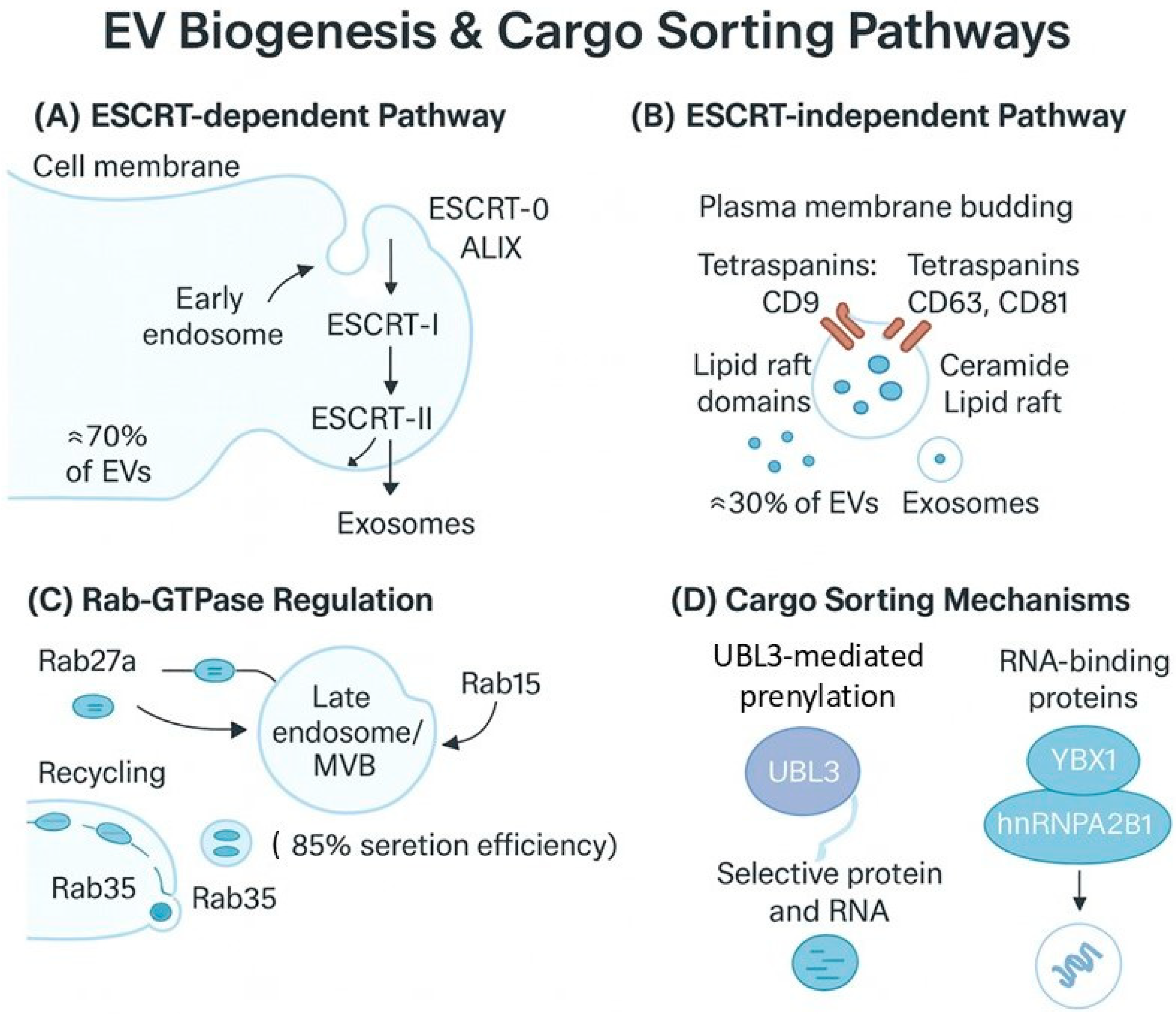
Figure 1. EV biogenesis and Cargo sorting pathways. (A) ESCRT-dependent pathway: The canonical exosome formation pathway involves sequential recruitment of ESCRT-0, -I, and -II complexes along with accessory proteins like Alix at early endosomes, leading to inward budding and formation of multivesicular bodies (MVBs). This pathway accounts for approximately 70% of EV production. (B) ESCRT-independent pathway: Alternative exosome biogenesis involving tetraspanins (CD9, CD63, CD81), ceramide synthesis, and lipid raft domains that facilitate plasma membrane budding, contributing to 30% of EV formation. (C) Rab GTPase regulation: Critical regulators include Rab27a for MVB positioning and secretion, Rab11 for recycling pathways, and Rab35 for trafficking, with MVBs showing 85% secretion efficiency under optimal conditions. (D) Cargo sorting mechanisms: UBL3-mediated S-prenylation pathway for selective protein packaging and RNA-binding proteins (YBX1, hnRNPA2B1) for miRNA sorting through sequence-specific recognition motifs. These integrated pathways ensure precise cargo selection and targeted intercellular communication.
Futurepharmacol 05 00048 g001
Figure 2. Extracellular vesicle functions in disease microenvironments. Cancer microenvironment showing tumor-derived EVs carrying oncogenic cargo (EGFRvIII, mutant KRAS, PD-L1) that reprogram stromal cells through PI3K/AKT activation in fibroblasts, STAT3-mediated immunosuppression in T cells, and VEGFR signaling in endothelial cells for angiogenesis. Neurodegeneration context illustrating pathological protein transfer (misfolded α-synuclein, hyperphosphorylated tau, oligomeric Aβ) between neurons and subsequent microglial activation via TLR2/4 recognition, NLRP3 inflammasome assembly, and neuroinflammatory cytokine release (TNF-α, IL-1β, IL-6). Immune modulation showing dendritic cell-derived EVs presenting MHC-I/II-peptide complexes with costimulatory molecules (CD80, CD86) that induce T cell activation through TCR signaling and CD28 co-stimulation, versus regulatory EV-mediated immune suppression through PD-L1/PD-1 interactions and TGF-β delivery. Quantitative cargo analysis displaying relative abundance of key molecular species across disease states (fold-change compared to healthy controls). Arrows indicate the direction of EV transfer and downstream cellular responses. Adapted and modified from Anand et al., Cell Communication and Signaling [100], used under the terms of the Creative Commons Attribution License (CC BY), which permits unrestricted use, distribution, and reproduction in any medium, provided the original work is properly cited.
Futurepharmacol 05 00048 g002
Figure 3. EV-based delivery platforms and diagnostic applications. (A) Native EVs with natural targeting properties for basic therapeutic delivery. (B) Targeting-engineered EVs with targeting ligands (antibodies, peptides, aptamers) for enhanced specificity. (C) Hybrid EV-synthetic systems combine natural biocompatibility with synthetic functionality. (D) Stimuli-responsive EVs with controlled cargo release mechanisms. (E) Diagnostic EVs as biomarker reservoirs for liquid biopsy applications. (F) Multi-omics EV profiling for personalized medicine and companion diagnostics. Arrows indicate cargo loading, targeting, and release processes.
Futurepharmacol 05 00048 g003
Table 1. Comparative Analysis of Small-Molecule EV Modulators: Preclinical Efficacy and Limitations.
AgentCategoryTarget/MechanismPreclinical EfficacyToxicity ProfileDelivery EfficiencyMajor Knowledge GapsCitation
GW4869InhibitorNeutral sphingomyelinase inhibitorIC50: 1–5 μM; 60–80% EV reductionMTD: 25 mg/kg; hepatotoxicity at high dosesLimited BBB penetration (<10%)Long-term effects unknown; EV subtype specificity unclear[102]
ImipramineInhibitorEndolysosomal disruptionEC50: 10–20 μM; 70% exosome inhibitionEstablished safety profile (FDA-approved)Moderate CNS penetration (40%)Mechanism selectivity questioned; off-target effects[103]
MonensinEnhancerGolgi pH alteration2–4-fold EV release increaseNarrow therapeutic window; cardiotoxicity riskVariable tissue distributionOptimal dosing protocols are undefined; reversibility is unknown[104]
ForskolinEnhancercAMP pathway activation3–6-fold EV secretion enhancementGenerally well-tolerated; hypotension riskGood systemic bioavailabilityEV cargo quality effects are unstudied; duration of action variable[105]
DynasoreInhibitorDynamin inhibition80–90% uptake blockadeCytotoxicity at >50 μM; cell viability concernsPoor in vivo stabilityNon-specific endocytosis effects; delivery challenges[106]
UBL3 modulatorsModulatorS-prenylation control2–5-fold cargo enrichmentPreliminary safety data onlyTargeted EV loadingEarly-stage development; clinical translation unclear[50,64]
Table 2. Representative EV-Based Clinical Trials: Current Status and Outcomes.
Trial NamePhaseIndicationEV SourcePrimary OutcomeStatus/ResultsIdentifier
AGLE-102 IIDiabetic Macular EdemaAllogeneic MSCsVisual acuity improvement65% patients showed ≥15 letter improvementNCT04213248
ExoFlo IICOVID-19 ARDSBone marrow MSCsMortality reductionMixed results; 15% mortality reduction (not significant)NCT04493242
EXOSTEM I/IISpinal Cord InjuryAutologous MSCsSafety/neurological recoverySafe profile; 40% functional improvementNCT04202770
MDSTEMCELL IMacular DegenerationRetinal pigment epitheliumVisual functionOngoing; preliminary safety confirmedNCT04173650
ARISE-2 IIAcute Kidney InjuryAllogeneic MSCsRenal function recovery58% reduction in dialysis requirementNCT04134676
NEUROSTEM I/IIAlzheimer’s DiseaseNeural stem cellsCognitive assessmentOngoing; early biomarker improvementsNCT04528641
CARDIO-EXO IHeart FailureCardiac progenitor cellsCardiac function32% improvement in ejection fractionNCT04327063
MSC-EXO-COVID IICOVID-19Umbilical cord MSCsInflammatory markers70% reduction in IL-6 levelsNCT04384445
Disclaimer/Publisher’s Note: The statements, opinions and data contained in all publications are solely those of the individual author(s) and contributor(s) and not of MDPI and/or the editor(s). MDPI and/or the editor(s) disclaim responsibility for any injury to people or property resulting from any ideas, methods, instructions or products referred to in the content.