Review

Thallium Toxicity: Mechanisms of Action, Available Therapies, and Experimental Models

by
Karla Alejandra Avendaño-Briseño
1,2,
Jorge Escutia-Martínez
1,2,
José Pedraza-Chaverri
1,* and
Estefani Yaquelin Hernández-Cruz
1,*
1
Laboratory F-315, Department of Biology, Faculty of Chemistry, National Autonomous University of Mexico, Mexico City 04510, Mexico
2
Graduate Program in Biological Sciences, National Autonomous University of Mexico (UNAM), University City, Mexico City 04510, Mexico
*
Authors to whom correspondence should be addressed.
Future Pharmacol. 2025, 5(3), 49; https://doi.org/10.3390/futurepharmacol5030049 (registering DOI)
Submission received: 12 July 2025 / Revised: 18 August 2025 / Accepted: 27 August 2025 / Published: 30 August 2025
(This article belongs to the Special Issue Feature Papers in Future Pharmacology 2025)

Abstract

Thallium (Tl) is a non-essential and highly toxic heavy metal capable of replacing potassium (K+) in biological systems, leading to mitochondrial dysfunction, oxidative stress, and inhibition of protein synthesis. In humans, the estimated oral lethal dose ranges from 10 to 15 mg/kg, with acute mortality rates of 6–15% and chronic neurological sequelae in up to 55% of survivors. Environmental releases of thallium of up to 5000 metric tons annually from industrial and mining activities, combined with its high oral bioavailability and nonspecific multisystemic symptoms, underscore the urgent need for more effective therapeutic strategies. This review summarizes current evidence on Tl toxicity, including its mechanisms of action, clinical manifestations, and available treatments. It emphasizes the strategic selection of biological models: simple organisms such as Caenorhabditis elegans and Drosophila melanogaster enable high-throughput screening and early biomarker detection; zebrafish (Danio rerio) provide vertebrate-level evaluation of multi-organ effects; and rodent models offer systemic toxicokinetic and therapeutic validation. Human-derived organoids and induced pluripotent stem cell (iPSC) systems recreate tissue-specific microenvironments, allowing translational assessment of mitochondrial, neuronal, and cardiac toxicity. Integrating these models within a tiered and complementary framework, alongside environmental and clinical surveillance, can accelerate the development of targeted treatments and strengthen public health responses to Tl exposure.

Graphical Abstract

1. Introduction

Thallium (Tl) is a heavy metal that is non-essential for living organisms and characterized by its high toxicity, even at low concentrations [1]. In recent decades, its presence as a trace contaminant has gained increasing relevance in both environmental and health contexts, driven by its expanding industrial use and the recognition of its adverse effects on human health and ecosystems. Although Tl can be released through natural processes, the main sources today are anthropogenic, including mining, non-ferrous metal smelting, coal combustion, and cement production [2,3].
A particularly concerning feature of many Tl compounds is their solubility in water and the absence of odor, taste, or color, which has facilitated both accidental and intentional poisonings [4]. In mammals, its toxicity exceeds that of other heavy metals, such as lead and mercury, and it manifests through nonspecific symptoms that complicate early clinical diagnosis. In humans, exposure can result in abdominal pain, alopecia, peripheral neuropathies, psychiatric disorders, cardiac abnormalities, and, in severe cases, death [2,5].
Because Tl can simultaneously affect multiple organs and systems, its study requires a multidisciplinary approach encompassing environmental toxicology as well as cellular and molecular biology. Although some therapeutic strategies have been approved, such as the use of Prussian blue, there are still no effective treatments to reverse mitochondrial and neurological damage [4,6]. In this scenario, biological models have become essential tools for elucidating the mechanisms of Tl toxicity and evaluating potential therapeutic interventions.
Traditional animal models, such as rats and mice, have been instrumental in describing tissue distribution, systemic effects, and toxicokinetics. However, the incorporation of alternative models, including simple organisms such as Caenorhabditis elegans (C. elegans), Drosophila melanogaster (D. melanogaster), and Danio rerio (D. rerio), has expanded research opportunities by enabling high-throughput assays and the identification of conserved molecular pathways [7,8,9,10]. More recently, human organoids and induced pluripotent stem cell (iPSC)-derived systems provide a translational bridge to human physiology, recreating relevant tissue microenvironments to directly assess mitochondrial, neuronal, and cardiac toxicity [11].
In this context, the present review synthesizes current knowledge on Tl toxicity, with an emphasis on the molecular mechanisms involved, clinical manifestations, and available therapeutic strategies. In particular, it highlights the biological models used to study Tl, analyzing their advantages, limitations, and complementary applications in toxicological research. By integrating mechanistic findings with experimental approaches, this work aims to provide a broader and more critical perspective on Tl as both an environmental and clinical risk, while guiding future research and therapeutic strategies.

2. Literature Search Strategy

This article is presented as a narrative review. The literature was retrieved mainly from the PubMed, Scopus, and Web of Science databases, and complemented by reports from regulatory agencies [The United States Environmental Protection Agency (EPA), World Health Organization (WHO), Secretaría de Medio Ambiente y Recursos Naturales (SEMARNAT)]. Articles were selected according to their relevance to Tl toxicity, mechanisms of action, clinical manifestations, therapeutic approaches, and biological models. Priority was given to studies published in the last 15 years, although seminal works and key historical reports were also included when relevant.

3. Characteristics of Tl

Tl is a soft, malleable heavy metal with a bluish-white color. Its atomic number is 81, and it has an atomic mass of 204.38. In nature, Tl exists in two oxidation states: I (Tl+) and III (Tl3+). Although the monovalent state is more stable than the trivalent form (Tl3+), both are highly toxic. The element’s name derives from the Greek word thallos, meaning “green shoot”, in reference to the bright green spectral line observed by the English chemist William Crookes during his discovery of Tl in selenium samples [12].
Despite being present in very low concentrations in the environment, Tl is classified as a non-essential and highly toxic element. The EPA has listed it as a priority pollutant [13].
The most common commercial forms of Tl are thallium acetate (TlCH3COO) and thallium sulfate (Tl2SO4), in which Tl is present in the +1 oxidation state. These compounds have industrial applications with significant toxicological implications for human health. C2H6O2Tl was historically used in pharmaceutical products for the treatment of fungal infections of the scalp. Tl2SO4, on the other hand, was used as a pesticide, particularly in syrups and gels for ant control [14,15].

4. Environmental Contamination by Tl

Tl represents a significant environmental concern due to its release during industrial activities such as mining, metal smelting, cement production, and coal combustion [2]. Although Tl occurs naturally, its abundance is generally low. Globally, the Earth’s crust contains approximately 0.7 mg of Tl per kilogram (0.7 ppm), primarily associated with potassium (K+)-rich minerals such as clays, granitic soils, and certain feldspars [3,16]. In uncontaminated freshwater systems, Tl concentrations generally remain below 1 µg/L, while in open-ocean waters, they rarely exceed 20 ng/L [17].
However, human activities have significantly increased Tl concentrations in air, soil, and water, with global emissions reaching up to 5000 tons per year. This poses a substantial risk of bioaccumulation in food chains [12,18]. For instance, in certain regions of China, soil concentrations of Tl have reached up to 124 mg/kg, and crops such as lettuce, carrots, and chili peppers have shown levels as high as 500 mg/kg (dry weight), far exceeding international safety limits and posing a threat to food security [19]. In the Silesia–Cracow region (Poland), surface soils have exhibited Tl concentrations of up to 150 mg/kg, with values ranging between 4.4 and 49.8 mg/kg in historically mined areas, averaging 20.8 mg/kg [20]. In old mining waste deposits near Olkusz, average levels reach 43 mg/kg, with localized peaks of 78 mg/kg [21]. Beyond these areas, the Allchar site (Republic of North Macedonia) is notable for its extreme concentrations, ranging from 0.11 to 20,000 mg/kg, with an average of 660 mg/kg in surface soils [20].
Moreover, documented incidents between 2010 and 2018 in major rivers highlight the ongoing contamination linked to mining and metallurgical activities, despite the fact that the use of Tl-containing minerals in industry dates back to the 1960s, when specific regulations were still lacking [22]. A notable case is Lake Sanliqi in Daye, which has experienced chronic contamination by Tl and other metals for more than 60 years due to copper smelting waste, affecting even nearby agricultural areas [23,24].
At the regulatory level, there are significant differences between countries and regions regarding Tl regulation, which has been focused mainly on drinking water. In the United States, the EPA has set a maximum contaminant level (MCL) of 2 µg/L for Tl in drinking water, with a non-enforceable health goal (MCLG) of 0.5 µg/L. California has proposed a more stringent limit of 0.1 µg/L [25]. In Canada, the Canadian Council of Ministers of the Environment (CCME) recommends a guideline value of 0.8 µg/L [26]. In Europe, Directive 98/83/EC requires member states to establish limits for chemical contaminants in drinking water; for example, Italy has adopted a maximum limit of 2 µg/L in groundwater [27]. China enforces one of the strictest limits globally, at 0.1 µg/L for human consumption, reflecting a response to severe contamination events [28] In Mexico, although Tl and its salts have been banned as rodenticides for decades, there is no specific Official Mexican Standard (NOM) for Tl in drinking water. The only applicable standard is NOM-147-SEMARNAT/SSA1-2004 [29], which establishes a limit of 5.2 mg/kg in soils for agricultural, residential, or commercial use. However, increasing industrial activity, illegal mining, and inadequate wastewater disposal in remote areas may contribute to a rise in Tl contamination in the country. In contrast, the WHO has not established a specific guideline value for Tl in drinking water.
In terms of future trends, although emissions from traditional industries may decline with the shift to clean energy and stricter environmental controls, the rapid expansion of lithium and battery production poses a new challenge [30]. Minerals used in these industries often contain Tl, potentially increasing contamination risks if proper regulations are not enforced [30]. Furthermore, the persistence of Tl in marine sediments and agricultural soils represents a long-term hazard, especially under changing redox conditions that may remobilize the metal into water systems, enhancing its bioavailability and bioaccumulation [31]. Given these concerns, effective Tl management requires harmonized regulations, continuous monitoring, and the implementation of efficient remediation technologies to minimize both environmental and public health impacts.

5. Statistics and Reported Cases of Tl Poisoning in Humans

Unlike other heavy metals, there are currently no consolidated global statistics on Tl poisoning, likely due to its low incidence. However, its effects can be severe: the acute mortality rate is estimated to be between 6% and 15%, with a lethal dose of approximately 10–15 mg/kg in humans, although deaths at lower doses have been documented. Additionally, between 33% and 55% of survivors develop chronic neurological or visual sequelae [32,33].
One factor that complicates surveillance is the lack of adequate toxicological monitoring systems in many countries, which contributes to the underreporting of cases, delayed diagnoses, or confusion with other syndromes of neurological or hepatic origin. In the United States, the National Poison Data System reported 60 unique cases of Tl exposure in 2023, mostly in adults over the age of 20. No deaths were reported, and only one case presented severe complications [34]. This figure represents an increase compared to 2019, when 49 exposures were recorded, also without fatalities [35].
Various clinical studies have documented the manifestations and progression of Tl poisoning. A case reported in 2023 by the Journal of Yeungnam Medical Science described a 51-year-old Argentine woman who developed myalgia, vertigo, jaundice, and alopecia. Urinary Tl levels of 540 µg/g creatinine were detected (normal range: 0.4–10 µg/g). She was successfully treated with D-penicillamine, achieving full clinical recovery after one year [36].
In China, a retrospective study (2000–2010) analyzed 13 patients initially diagnosed with other pathologies, such as Guillain–Barré syndrome. Most exposures were accidental or due to deliberate poisoning. In some cases, the source was contaminated water or food, including edible mushrooms harvested from Tl-contaminated soils. The average hospital stay was 44 days. One person died, and among the survivors, long-term sequelae were documented: peripheral neuropathy, cognitive impairment (33%), anxiety (50%), and depression (42%) [37].
That same year, also in China, the poisoning of a family of 14 people was reported after they consumed Tl-contaminated bread. Although initial symptoms were mild to moderate (vomiting, painful neuropathy, alopecia, fatigue, among others), one patient with pulmonary fibrosis died due to respiratory failure. During follow-up, some survivors developed complications such as venous thrombosis and residual paresthesia, although most recovered without permanent sequelae [38].
One of the most notorious cases of deliberate Tl exposure is that of Zhu Ling, a student at Tsinghua University (Beijing), who was chronically poisoned between 1994 and 1995. She presented with abdominal pain, alopecia, coma, and severe neurological damage. Although she initially survived, she was left with permanent disabilities and died in 2013. Her case was pioneering in the use of telemedicine for diagnosis, as it was internationally discussed in online medical forums. She received treatment with plasmapheresis, hemodialysis, and Prussian blue, although she developed severe complications during hospitalization [39].
In Mexico, the case of a 19-year-old pregnant woman who ingested Tl-containing rat poison with suicidal intent was reported. Within a week, she developed characteristic symptoms such as alopecia, Mees’ lines, and palmar erythema. Her newborn presented with stupor, distal cyanosis, and alopecia. Although the child showed favorable progression, psychomotor delays were detected at three years of age [40].
Taken together, clinical cases reflect variability in routes of exposure and clinical manifestations, but also highlight frequent signs, such as abdominal pain, vomiting, tachycardia, fatigue, alopecia, and neurological disturbances. Mees’ lines on the nails and generalized alopecia are distinctive findings, although they appear late (2 to 3 weeks after exposure) [36,37,38,39,41]. Neurological complications include peripheral neuropathies, ataxia, motor dysfunction, and psychiatric disorders such as anxiety, depression, or hallucinations [42,43].
Table 1 summarizes the most representative clinical cases described in the recent literature.

6. Toxicokinetics of Tl

Tl enters the body through various routes, including the skin, inhalation, and primarily the gastrointestinal tract, where its absorption is particularly efficient, reaching an oral bioavailability close to 100% [49,50]. Water-soluble Tl+ salts can enter the body using the same cellular transport mechanisms as K+, due to their ionic similarity [50]. It is important to note that both Tl+ and Tl3+ are present under physiological conditions. The estimated oral lethal dose of Tl for humans ranges from 10 to 15 mg/kg [32].
The stability of Tl+ in aqueous solutions is notably influenced by pH and redox potential (Eh). Tl+ is highly soluble and mobile across a wide pH range (approximately 3 to 12), whereas Tl3+ rapidly reduces to Tl+, and its solubility is limited by hydrolysis reactions [51]. Monovalent Tl+ is the predominant and thermodynamically most stable form in aquatic systems, whereas Tl3+, although it can exist under specific conditions, is typically unstable [3].
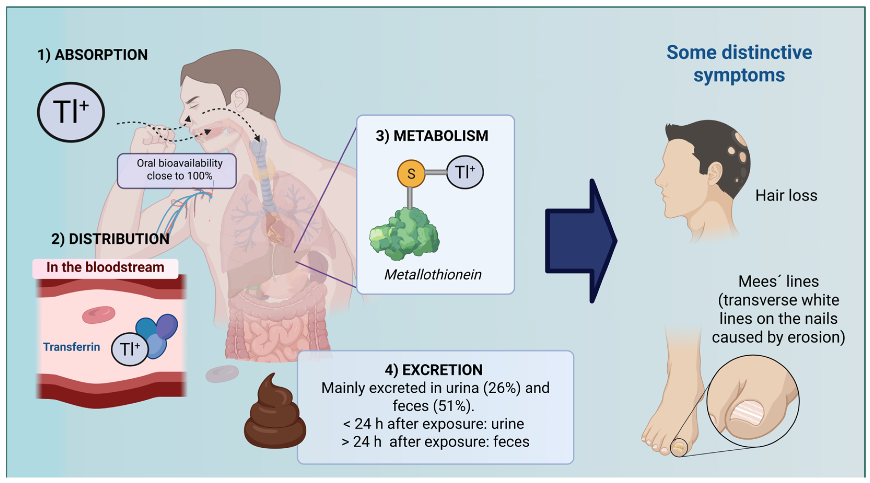
In the gastrointestinal tract, pH varies significantly: it is acidic in the stomach (pH 1.5–3.5), increases in the duodenum to approximately pH 5–6, and reaches pH 7–8 in the distal small intestine. Although specific studies on gastric pH’s influence on Tl+ absorption are limited, the high solubility of Tl+ ions in water suggests that they may remain in solution under acidic conditions, facilitating absorption in the stomach [52]. Once absorbed, Tl is widely distributed throughout the body, with the highest concentrations found in the kidneys, although it also accumulates in the liver, heart, and brain [5]. Its transport is facilitated by binding to proteins such as transferrin, and it exhibits a high volume of distribution (3–10 L/kg) [53]. Experimental studies have shown heterogeneous distribution within the brain, with greater accumulation in the hypothalamus [54]. Furthermore, Tl has been shown to cross both the blood–brain barrier and the placental barrier, increasing its toxicological risk, particularly during pregnancy [55,56]. Regarding metabolism, Tl exposure induces the expression of metallothionein (MT), a cysteine-rich protein that plays a protective role by neutralizing the metal and scavenging free radicals (FRs) [57,58,59]. It is noteworthy that Tl does not undergo enzymatic or chemical transformations (as many xenobiotics do).
Tl elimination primarily occurs through urine (26%) within the first 24 h; however, over time, feces become the predominant excretion route (51%) [5]. Under normal conditions, urinary Tl concentrations in humans do not exceed 1 μg/g of creatinine [12]. Additionally, Tl can be excreted via saliva, tears, sweat, and breast milk. Due to its strong affinity for cysteine residues in keratin, Tl is deposited in tissues such as hair and nails, leading to very slow elimination and enabling its detection even months after exposure [49,60]. This slow excretion results in a risk of accumulation even at low levels, and its biological half-life can extend up to 30 days, potentially causing long-lasting or irreversible neurological consequences [5,60] (Figure 1).

7. Mechanisms of Toxicity

Exposure to Tl poses a significant risk to human health due to its ability to induce systemic and cellular alterations in a progressive and silent manner. Although its initial clinical manifestations may be nonspecific, the toxicity of this heavy metal extends to various organs and tissues, affecting key physiological functions and compromising cellular integrity. Its chemical similarity to K+, particularly the comparable ionic radius and monovalent charge of Tl, allows it to enter cells through K+ transport systems. Once inside, Tl+ can partially substitute for K+ in metabolic processes but fails to sustain its essential biochemical functions. This misreplacement disrupts critical processes such as energy production, protein synthesis, and redox balance, ultimately leading to cellular dysfunction and toxicity. These properties make it a highly harmful agent with both clinical and molecular implications [61,62].
Below, the main effects of Tl in the body are described in detail at the organ level, along with metabolic alterations that characterize its toxic mechanism.

7.1. Cellular Effects

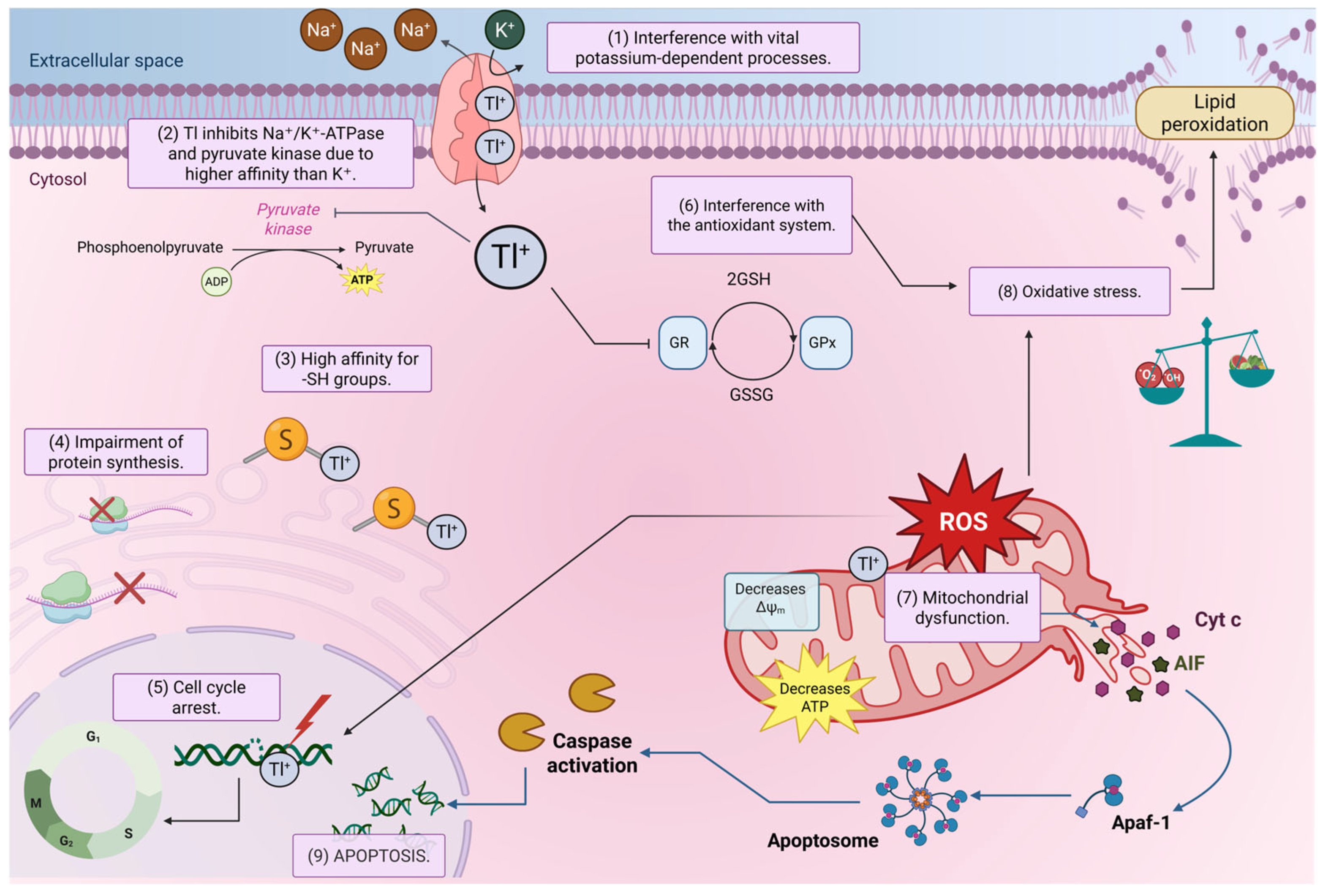
Tl enters cells through the same channels and transporters as K+, disrupting ionic gradients and altering membrane potential, processes essential for the excitability of neurons and muscles. Once inside the cell, Tl promotes the production of reactive oxygen species (ROS); however, the biochemical mechanisms are not yet fully understood. Some studies suggest that Tl interferes with the cellular antioxidant system [63,64]. For example, Tl(OH)3 has been reported to inhibit the activity of key enzymes such as glutathione peroxidase (GPx) and glutathione reductase (GR) in rat brain tissue [63,64]. The increased generation of ROS leads to the oxidation of fatty acids in the membrane, which in turn enhances membrane fluidity [65]. In fact, the interaction of Tl with membrane phospholipids, especially those found in brain tissue, such as phosphatidylcholine and phosphatidylserine, alters the physical properties of the bilayer. This is important because these changes may affect various membrane-dependent processes, such as the activity of associated enzymes and intracellular transport, which could contribute to the neurotoxicity associated with Tl poisoning [66].
Additionally, the increase in ROS production due to Tl exposure appears to be a key factor in disrupting the cell cycle. It has been shown that incubation with C2H6O2Tl halts cell cycle progression at the G2/M phase, which is associated with decreased expression of cyclin-dependent kinase 2 (CDK2) and upregulation of p21 [67]. More recently, it was reported that Tl, even at concentrations that do not affect cell viability, disrupts the cell cycle in response to DNA base oxidation mediated by oxidative stress [68].

7.2. Effects on Organelles

Mitochondria are among the primary subcellular targets of Tl toxicity, largely be-cause these organelles rely on ATP-sensitive K+ channels, which can also permit Tl entry [69]. Once inside the mitochondria, Tl has been shown to induce loss of mitochondrial membrane potential [70,71]. This is relevant because it prevents the maintenance of the electrochemical gradient required for oxidative phosphorylation.
It has also been demonstrated that in certain cells, Tl reduces the activity of mitochondrial complexes in the electron transport chain (ETC) [72], which likely explains, at least in part, the significant increase in hydrogen peroxide and other ROS reported [73].
Furthermore, Tl has been documented to promote the opening of the mitochondrial permeability transition pore (mPTP) [74]. This pore is known to form under conditions of extreme mitochondrial stress, allowing the free entry of water and small molecules and causing changes in mitochondrial morphology, such as organelle swelling. Another consequence of mPTP opening is the release of cytochrome c and other pro-apoptotic proteins into the cytosol, leading to activation of the intrinsic apoptotic pathway and subsequent cell death [73].
Taken together, these alterations converge in the disruption of adenosine triphosphate (ATP) synthesis. Given the high energy demand of nervous tissue, Tl-induced mitochondrial dysfunction has deleterious effects on neuronal function.
Moreover, Tl negatively affects protein synthesis in animals by interfering with ribosome stability. For example, administration of thallium nitrate (TlNO3) to mice results in reduced incorporation of amino acids into proteins in vivo, as well as disaggregation of hepatic polyribosomes [75]. It was reported that Tl significantly reduced levels of the 60S ribosomal subunit. This reduction was found not to result from increased degradation, but rather from a blockade in subunit biogenesis. The 60S subunit plays a crucial role in peptide elongation and overall protein synthesis, and its disruption can severely compromise cellular function. Consequently, ribosome assembly is impaired, leading to a global halt in protein synthesis and underscoring the importance of the 60S subunit in maintaining cellular homeostasis and viability [74].
Additionally, Tl also affects the endoplasmic reticulum (ER). Following metal exposure, structural changes, such as organelle dilation, increased membrane abundance, and even fragmentation, have been observed. Recently, it was found that Tl may induce ER stress, as significant increases in two key ER stress response proteins were detected 24 h after exposure in Madin–Darby canine kidney (MDCK) cells [76,77,78].

7.3. Effects on Metabolic Pathways

Due to Tl’s interference with metabolic processes that depend on K+, this metal can alter the enzymatic activation of not only Na+/K+ ATPase but also other enzymes that require the presence of K+ ions to function properly. Among these is pyruvate kinase [79], a key enzyme for cellular energy production and essential for the proper maintenance and metabolism of red blood cells.
Another major mechanism of Tl toxicity is related to its high affinity for the sulfhydryl groups present in many proteins. The binding of Tl to these sulfhydryl groups interferes with the active sites of several enzymes critical to cellular metabolism, including hydrolases, transferases, and oxidoreductases [80]. In particular, inhibition of pyruvate dehydrogenase (PDH) and succinate dehydrogenase (SDH) has been reported [81]. Both enzymes are located in mitochondria and are essential for energy metabolism. PDH inhibition limits the conversion of pyruvate into acetyl coenzyme A (CoA), thereby reducing the oxidative metabolism of glucose. SDH inhibition, since this enzyme is central to both the Krebs cycle and the electron transport chain, leads to decreased ATP production.
Tl also interferes with sulfhydryl groups in cysteine residues, preventing the formation of disulfide bonds necessary for keratin synthesis. This disruption compromises the structural integrity of the protein and clinically manifests as abnormalities in hair (alopecia) and nails (the appearance of Mees’ lines) [82].
Figure 2 summarizes the main toxic mechanisms exerted by Tl at the cellular level.

7.4. Other Mechanisms of Toxic Action

7.4.1. Effect of Tl on Calcium Homeostasis

Calcium is an essential regulator of cellular function, and its imbalance can lead to severe alterations. It has been reported that Tl significantly interferes with this homeostasis, causing mitochondrial dysfunction, cytoplasmic calcium overload, and alterations in the expression of key regulatory genes. The following studies illustrate these effects.
Exposure to low concentrations of Tl (1 µg/L) for 48 h has been shown to induce a significant increase in cytoplasmic calcium levels in the murine hippocampal neuronal cell line HN9.10e. These results were accompanied by early mitochondrial dysfunction, evidenced by alterations in both mitochondrial ROS production and mitochondrial membrane potential. Based on these findings, the authors emphasize the central role of mitochondria in calcium storage and release, warning that prolonged mitochondrial dysfunction may lead to cytosolic calcium overload, thereby contributing to severe cellular dysfunction [33]. Furthermore, it has been reported that mitochondrial calcium overload, together with the presence of Tl, may promote the mPTP. In particular, in the case of Tl, this opening has been reported to depend on a prior calcium load within the mitochondria [83]. The above highlights the close relationship between the metal’s toxicity and calcium imbalance, both in the cytosol and in the mitochondrial matrix.
Furthermore, transcriptomic analyses of zebrafish embryos exposed to 100 µM Tl revealed significant alterations in calcium signaling pathways. In total, 46 differentially expressed genes (DEGs) were identified, among which slc8a3, slc8a4a, and atp2b3b stand out as key regulators of calcium homeostasis. The authors discuss that the downregulation of these genes compromises calcium efflux, promoting intracellular calcium overload. To further investigate, the researchers employed the Tg(elavl3:GCamp6f) transgenic zebrafish line, which expresses GCamp6f, a fluorescent calcium indicator protein in neuronal cells under the control of the elavl3 gene. Fluorescence imaging revealed notable changes in calcium levels in the brain and spinal cord of embryos exposed to Tl. These results were corroborated by quantification using Fluo-4 AM, a fluorescent dye used to measure intracellular calcium, which showed a 1.3-fold increase in Tl-treated embryos (100 μg/L) compared to the control group. Additionally, the researchers noted that increased intracellular calcium could disrupt both mitochondrial and endoplasmic reticulum function, triggering oxidative stress and subsequently apoptosis. To confirm this, acridine orange staining was used, revealing a higher proportion of apoptotic cells in the head region of Tl-exposed embryos. Finally, ultrastructural analysis by transmission electron microscopy confirmed typical apoptotic features, such as nuclear condensation and morphological alterations in mitochondria, observed in the Tl-treated group [84].
At the systemic level, Tl toxicity affects key organs involved in calcium handling, such as the kidney. Intraperitoneal administration of Tl (30 mg/kg) in rats has been shown to induce severely compromised renal function. Low-vacuum scanning electron microscopy revealed calcium deposits in the renal medulla, specifically in the thick ascending limb of the loop of Henle. Furthermore, a reduction in the expression of essential transporters for calcium reabsorption, such as the Na+–K+–2Cl cotransporter 2 (NKCC2), was observed, affecting overall body ion regulation and contributing to acute kidney injury. In addition, ROS generation and bioenergetic dysfunction further exacerbate calcium dysregulation, intensifying renal damage [85]. Based on the above, it appears that exposure to Tl triggers a disruption of calcium homeostasis, which, as discussed, manifests at multiple levels, from mitochondrial to cellular and systemic. Although further studies are still needed, this calcium imbalance may emerge as a key mechanism underlying metal-induced toxicity.

7.4.2. Effects of Tl on Myelin

Tl can cause severe neurological damage, particularly in the peripheral nervous system. Some authors have sought to characterize the metal’s ability to induce structural alterations in nerve fibers, with particular attention paid to its impact on myelin, given that it is essential for the efficient conduction of nerve impulses. The following summarizes the available evidence from the literature regarding these effects.
A case was reported of a man who, after ingesting 5 to 10 g of thallium nitrate, died nine days later. On the seventh day, a sural nerve biopsy was performed, revealing that the large myelinated fibers were the most affected. Many axons appeared swollen, were devoid of organelles, and contained large vacuoles; however, in most cases, the surrounding myelin sheaths remained intact or exhibited only mild alterations [86]. In another report, a 40-year-old man was admitted to the neurology unit with symptoms of neuropathy after attending a party and noticing a peculiar taste in a candy he was offered. On day 18 of the illness, blood Tl levels were confirmed at 40,980 µg/mL. A sural nerve biopsy performed on day 40 revealed axonal loss and active axonal degeneration, and Kulchitsky Pal staining showed associated demyelination [87].
Experimental studies available in the literature addressing the effects of Tl on myelin are scarce and mostly date from the 1970s to 1990s. For example, in 1973, it was demonstrated that in functionally coupled organotypic cultures of dorsal root ganglia, peripheral spinal nerves, and muscles, exposure to Tl salts (C2H6O2Tl and Tl2SO4, 10–20 µM) in a liquid medium caused significant alterations. After two hours of exposure, enlarged mitochondria were observed in the axons of peripheral nerve fibers, which progressively swelled. This led to the transformation of the mitochondria into axonal vacuoles. The resulting swelling of the fibers caused retraction of myelin from the nodes of Ranvier, but not degeneration. Impulses were still able to propagate along the nerve fibers during the experiment [88].
In a second study, myelinated dorsal root ganglia were used, and it was observed that treatment with Tl (around 100 µM) inhibited neurite outgrowth; however, its ability to block primary myelination was very low compared to that observed for other heavy metals, such as lead [89]. In animal models, a single intraperitoneal administration of TlCH3COO (16 mg/kg) to newborn Wistar rats caused, by day 8, a moderate reduction in large and medium sural nerve fibers, with early signs of myelin degeneration along the axons. By day 50, the myelinated fibers exhibited tortuous courses and fragmentation. Additionally, axonal and myelin alterations were observed along the entire length of the axon, suggesting a progressive distal axonopathy [90].
Taken together, these findings suggest that Tl (at least in the early stages) causes axonal damage with secondary myelin loss, without primarily blocking the myelination process.

7.4.3. Mutagenic and Genotoxic Effects of Tl

The genotoxic and mutagenic effects of Tl have not been conclusively characterized. Researchers have evaluated the metal’s capacity to induce DNA damage and have sought markers of genetic instability following its administration. A summary of the available evidence from the literature is presented below.
Results from bacterial studies are contradictory. In Rec assays with Bacillus subtilis, Tl (1 mM) showed a moderate effect, indicating that the metal may cause DNA damage in recombination-deficient bacteria. However, in reversion (mutagenicity) tests with Escherichia coli and Salmonella, Tl did not produce positive results [91].
In eukaryotic cell models, the findings are more consistent, although not entirely uniform. In rodent embryonic fibroblasts, Tl induced single-strand DNA breaks and reduced cell survival in a dose-dependent manner after 24 h of exposure [92]. In human cell hemocultures, Tl (0.5–100 µg/mL) was reported to reduce the mitotic and replicative indices in a concentration-dependent manner. Furthermore, a significant increase in chromosomal aberrations was observed, and its clastogenic effect and induction of oxidative DNA damage were confirmed using the single-cell gel electrophoresis assay [93].
In peripheral lymphocytes from 13 patients poisoned by Tl, chromosomal alterations were observed, mainly chromatid aberrations. However, in the subgroup of eight individuals evaluated with the micronucleus assay, only one showed a markedly increased frequency of micronuclei. No correlation was found between the frequency of chromosomal aberrations and the results of the micronucleus test [94]. On the other hand, in this same model, genotoxicity was evaluated for six metal salts using the micronucleus assay and fluorescence in situ hybridization (FISH) analysis. It was found that all compounds, except Tl, produced a significant increase in micronuclei at least at one dose. FISH analysis, in turn, revealed that only antimony (Sb) and mercury (Hg) were highly genotoxic [95]. In conclusion, Tl exhibits genotoxic properties that some authors have considered weak and inconclusive. These properties have been more extensively studied in eukaryotic cells, yet they lack consistency across studies. This complicates the classification of the metal as a mutagen and underscores the need for standardized and comparative investigations.

8. Treatment of Tl Poisoning in Humans

Various therapeutic regimens have been used to treat Tl poisoning; however, there is no consensus among physicians regarding the effectiveness of different strategies, and no single treatment has been proven effective in severe cases of toxicity. As a result, combined therapies have been employed, including gastric lavage, potassium ferric hexacyanoferrate (commonly known as Prussian blue), activated charcoal, and extracorporeal drug removal methods such as hemodialysis and forced diuresis [12]. In some cases, D-penicillamine has also been used, although its efficacy is limited and it is not recommended as a first-line treatment; however, it may be considered an alternative when Prussian blue or other more effective options are unavailable [44,48]. On the other hand, exogenous administration of MT has been shown to be effective in reducing Tl-induced liver damage in animal models, acting as a metal-chelating and antioxidant agent [59].
Prussian blue is the antidote of choice and is approved by the U.S. Food and Drug Administration (FDA) for the treatment of Tl poisoning. Prussian blue is a crystalline network with high affinity for Tl and cesium. The drug is available in both soluble (potassium ferric hexacyanoferrate) and insoluble (ferric hexacyanoferrate) forms. Both forms have been used therapeutically, although the literature highlights the greater efficacy of the soluble form in Tl poisoning [96,97].
The mechanism of action of Prussian blue involves binding to Tl as it passes through the gastrointestinal tract, exchanging it for K+ on the surface of the crystal in the intestinal lumen. This process reduces the gastrointestinal reabsorption of the metal and facilitates the excretion of the insoluble complex. The K+ that is exchanged originates from within the drug’s own crystalline lattice [98]. In Mexico, it has been reported that administration of 3 g/day of Prussian blue accelerates Tl elimination from the body, resulting in a 70% reduction in plasma concentrations in approximately four days [40]. Although Prussian blue has few adverse effects, one of its limitations is that it can only bind Tl present in the gastrointestinal tract, and its attributable effect remains unclear since it is typically administered in combination with other therapies [98].
On the other hand, activated charcoal is a processed form of common charcoal that results in a porous network capable of trapping other chemicals and preventing their absorption by the body, which is why it has been used as a decontaminant. Activated charcoal binds to certain toxic substances, such as Tl, and retains them within the gastrointestinal tract, thereby reducing systemic absorption and promoting toxin elimination from the body [99]. The administration of multiple doses (two or more sequential doses) of activated charcoal, in combination with Prussian blue, has been shown to be effective in treating Tl poisoning [99].
Among the experimental strategies recommended for managing acute Tl poisoning are forced diuresis and hemodialysis [100]. Although this therapy has been one of the most commonly used in acute poisoning scenarios, complications such as fluid overload, pulmonary edema, and acid–base disturbances have led clinicians to reduce its use in recent years [101]. However, there are reports of successful recovery in patients treated with a combination of Prussian blue and forced diuresis [96,102].
By contrast, hemodialysis has proven to be more effective than forced diuresis in treating Tl poisoning [103]. On average, a standard session lasts about four hours. However, long-term hemodialysis (200 h over 10 days) in combination with forced diuresis and oral Prussian blue administration has resulted in the complete recovery of a patient who ingested Tl2SO4. Moreover, hemodialysis has been reported to be effective even in the third week after Tl exposure [87].
In addition to Prussian blue, several antioxidant compounds have been investigated for their potential to reverse Tl-induced oxidative stress. These compounds offer promising advantages, such as free radical scavenging, preservation of glutathione (GSH) levels, and prevention of lipid peroxidation in tissues, thus complementing the chelating action of Prussian blue. Among the antioxidants evaluated are N-acetylcysteine (NAC), α-tocopherol, ascorbic acid, and S-allyl-L-cysteine, all of which have demonstrated improvements in cell viability, reductions in oxidative stress markers such as malondialdehyde (MDA), and the preservation of antioxidant enzyme activities such as superoxide dismutase (SOD) in cell and animal models [53,104,105].
However, these compounds have limitations, including their inability to significantly reduce the total body burden of Tl, lack of standardized dosing, and absence of robust clinical evidence in humans. In a comparative study in mice, Meegs et al. [105] evaluated NAC (200 mg/kg i.p.) versus Prussian blue (50 mg/kg oral) after a lethal dose of Tl. Although NAC improved survival (35% vs. 10%), it did not match the efficacy of Prussian blue and did not provide additional benefit when combined with it.
Table 2 lists the antioxidants evaluated against Tl, and Table 3 compares their effects with the standard treatment using Prussian blue.

9. Biological Models for the Study of Tl Poisoning

Understanding the mechanisms underlying Tl toxicity and the development of effective therapies largely depends on the biological models employed. The use of animal models has been invaluable in characterizing Tl distribution within the body, with rodents being the most used. Table 4 summarizes various studies conducted in these models, encompassing different Tl compounds, doses, and exposure times.
While rodents have formed the cornerstone of research, the integration of simpler models, organoids, and systems derived from iPSCs offers new opportunities for mechanistic analysis and preclinical evaluation. This section outlines the advantages, limitations, and potential applications of both traditional and emerging models, emphasizing their complementary role in the study of toxicity and the development of therapeutic strategies.

9.1. Traditional Models: Rodents

Both rats and mice remain the standard for studying tissue distribution, toxicokinetic, and the systemic effects of Tl. The advantages of these models include their physiological similarity to humans, the availability of surgical techniques, and established genetic models [74,76]. However, they also present several limitations, such as high costs, ethical considerations, and the extended duration required for experimentation.

9.1.1. Rats

The rat has been the preferred biological model due to its larger size compared to the mouse, which facilitates handling and allows for a wide range of surgical procedures [72,114]. This model has been pivotal in characterizing Tl accumulation, particularly in the kidneys, the main organ of deposition, and in elucidating its mechanisms of toxicity at the cellular and subcellular levels.
In hepatocytes and isolated mitochondria from rats, Tl3+ has been shown to be more toxic than Tl+, increasing ROS production, lipid peroxidation, and membrane potential loss, effects that can be mitigated by lipid antioxidants such as α-tocopherol and hydroxyl radical scavengers such as mannitol [71,115]. Oxidative damage has also been observed in isolated brain mitochondrial fractions, along with marked inhibition of Na+/K+-ATPase, underscoring the metal’s impact on energy metabolism [116].
Studies in rats have further confirmed that Tl can substitute for K+ in the activation of key enzymes such as Na+/K+-ATPase and pyruvate kinase, displaying an affinity even greater than that of K, which explains its strong capacity to disrupt essential biochemical processes [79]. The activation or inhibition of these enzymes is concentration-dependent; at higher Tl concentrations (>10 mM), enzymatic inhibition has been observed [56]. Additionally, Tl has been shown to inhibit sulfhydryl-dependent enzymes, such as porphobilinogen synthase (PBGS), through direct oxidation of cysteine residues. However, this effect can be significantly attenuated by the addition of glutathione and DL-dithiothreitol, both reducing agents that protect sulfhydryl groups [80].
Taken together, the evidence from rat models has been fundamental in understanding Tl toxicity, from mitochondrial dysfunction and oxidative stress to enzymatic inhibition, cementing this model as a cornerstone in toxicological research and the evaluation of potential therapies.

9.1.2. Mice

The high genomic similarity between mice and humans, the availability of a wide range of genetic and molecular tools, as well as their small size and ease of handling, have established the mouse as a highly valuable biological model for studying the toxic effects of Tl [72].
At the hepatic level, administration of thallium nitrate at lethal doses (100 and 150 mg/kg body weight) in mice caused polyribosome disaggregation and marked inhibition of protein synthesis [75]. Similarly, exposure to different cationic forms of Tl (10 ppm) for two weeks led to its accumulation in the liver and severe histological damage [105].
The utility of the mouse model has also extended to the study of Tl reproductive toxicity. Oral exposure reduced sperm viability and increased mortality, in addition to inducing DNA damage in spermatozoa [117]. Finally, in the immune system, Tl was observed to inhibit the expression of key genes involved in B cell development in the bone marrow [118].
These findings confirm that the mouse is a versatile and sensitive model for elucidating the toxic mechanisms of Tl in critical organs and functions.

9.2. Simple Biological Models

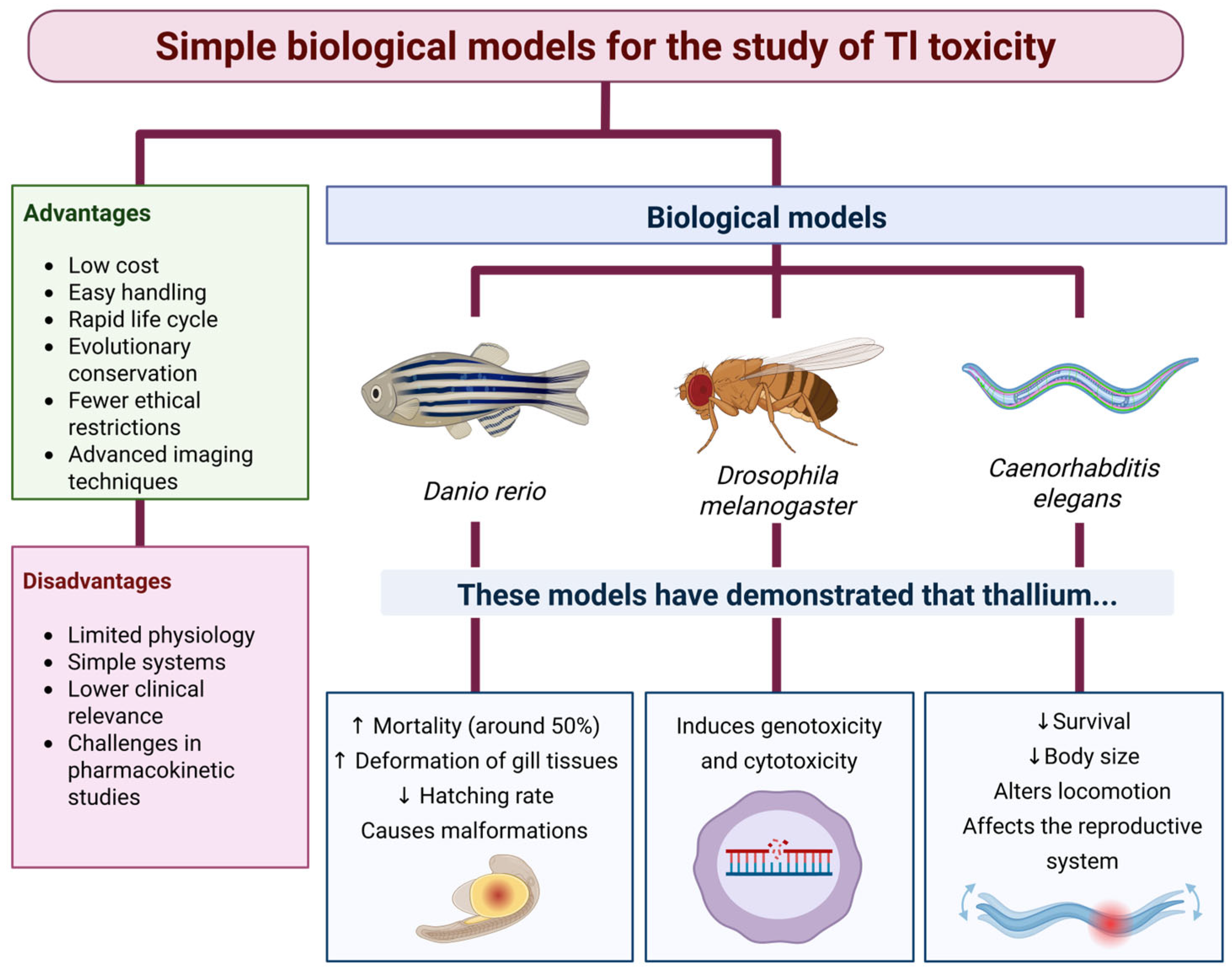
The use of simple biological models (lower animals) offers advantages over mammalian models for toxicological research. Lower animals have short generation times, are cost-effective to maintain, and pose fewer ethical constraints, allowing for multiple concentration–response tests for a given substance [119]. Additionally, the main signaling pathways in these organisms are highly conserved, and their genetic homology with humans exceeds 60%. Among these model organisms are the zebrafish (D. rerio), the fruit fly (D. melanogaster), and the soil nematode (C. elegans) (Figure 3).

9.2.1. Zebrafish (D. rerio)

The zebrafish (D. rerio) has established itself as a valuable biological model for assessing toxicity during embryonic development and detecting early cardiovascular or neurological effects. Its ability to absorb compounds directly from water enables high-throughput toxicity assays, which, combined with its transparent embryos developed ex vivo, facilitates controlled substance exposure and direct observation of morphological and functional abnormalities [120,121]. Considering that Tl can enter aquatic ecosystems through erosion and mining leachates, atmospheric deposition, or industrial wastewater discharges [122], this model provides a realistic approximation of environmental exposure scenarios.
Various studies have shown that exposure of embryos and larvae to Tl can lead to reduced hatching rates and malformations, as well as cardiac alterations, neuronal damage, and oxidative stress [7,8]. In juveniles and adults, Tl increased mortality and caused reproductive dysfunction, gill and liver lesions, and changes in the intestinal microbiota [123].
However, the use of zebrafish presents important limitations: its physiology differs in several aspects from humans, it lacks certain organs present in mammals (e.g., lungs), and its xenobiotic metabolism is not always comparable to that of mammals, which can complicate the direct extrapolation of doses and effects [121,124]. Nevertheless, the combination of its ecological relevance and experimental versatility positions zebrafish as a highly sensitive model for investigating toxicity in aquatic systems.

9.2.2. Fruit Fly (D. melanogaster)

The fruit fly (D. melanogaster) stands out as an ideal biological model for toxicological studies due to its extremely short life cycle, approximately 60 to 80 days, allowing the assessment of Tl effects across multiple generations with high efficiency [125]. Moreover, these organisms share metal tolerance mechanisms similar to those in humans, including homologous genes involved in metal transport, storage, and excretion [126].
Despite the limited research on Tl toxicity in this model, its potential for detecting mutagenic effects has been demonstrated using the somatic mutation and recombination test (SMART), in which 600 µM of thallium sulfate induced genotoxicity with 87.6% somatic recombination and cytotoxicity by interfering with cell division in the wing imaginal discs [127].
These findings highlight a significant gap in the study of Tl toxicity in this model, representing an opportunity to further investigate its mechanisms of action.

9.2.3. Nematode (C. elegans)

The nematode (C. elegans) has established itself as a powerful biological model for studying environmental toxicology due to its fully characterized genome, high genetic homology with mammals, and sensitivity to contaminants [128]. Its small size, short life cycle, and body transparency enable high-throughput assays and direct observations of cellular and molecular processes, ranging from morphological changes to the activation of fluorescent reporter genes [9,129,130]. These characteristics make it a highly valuable tool for elucidating the toxic mechanisms of Tl, a highly toxic metal that has been little studied in this organism.
Available studies indicate that Tl critically affects the survival, development, and reproduction of C. elegans. The main mechanisms observed include ROS generation and the activation of highly conserved antioxidant pathways such as SKN-1/Nrf2 and DAF-16/FoxO [131,132]. This molecular parallelism with humans gives the nematode remarkable predictive potential for understanding toxic mechanisms and exploring therapeutic interventions. For instance, exposure to S-allylcysteine (SAC) was found to attenuate Tl-induced effects in C. elegans [10].
Despite its advantages, C. elegans has certain limitations that must be considered when interpreting results. As an invertebrate, it lacks organs and systems found in mammals, such as a circulatory system, lungs, and an adaptive immune system, and exhibits differences in xenobiotic metabolism, which limits the direct extrapolation of doses and effects [10,131]. Additionally, exposure to toxicants typically occurs in aqueous media, which does not always replicate the routes of entry and distribution in higher organisms [133,134]. Nevertheless, its unique combination of experimental simplicity and molecular relevance positions it as a strategic model for investigating Tl toxicity in a mechanistic context with translational applications.

9.3. Emerging Models: Organoids and iPSC-Derived Systems

Three-dimensional organoids (liver, heart, and brain) and human iPSC-derived systems provide tissue microenvironments with architecture and functions closely resembling those of humans, thereby enhancing translational relevance and enabling direct assessment of mitochondrial and neuronal toxicity, oxidative stress, inflammation, and cell death [135]. In screening platforms, these models facilitate the identification of early biomarkers through metabolomics/proteomics and the evaluation of combination therapies (e.g., chelators with antioxidants) under controlled conditions.
The utility of these models has already been demonstrated in the study of metals: in human liver and cardiac organoids, Tl was identified as the most toxic compound among several environmental toxins, with a mean inhibitory concentration (IC50) of 13.5 µM in liver and 1.35 µM in heart [136]. This study validates the discriminatory power of these biological models for risk prioritization and mechanistic target identification. Recent reviews on organoid use also highlight the finding of Tl as the most toxic compound within tested panels in these models [137]. Furthermore, brain organoids have been systematically employed to model neurotoxicity induced by arsenic and lead, including proteomic alterations [138].
In the case of iPSCs, brain microphysiologies derived from them have been developed to study diseases and toxicity, reinforcing their utility for the exploration of mechanisms and biomarkers with potential for clinical translation [11].

10. Strategic Selection of Biological Models for Tl Toxicity: A Tiered and Complementary Approach

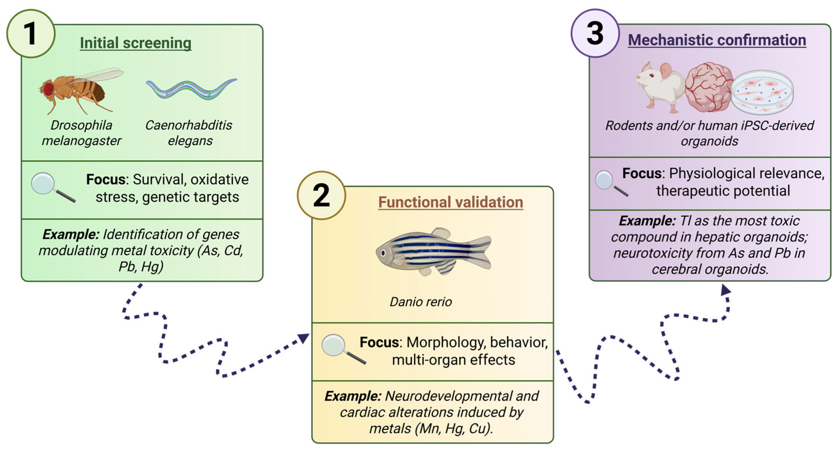
The appropriate selection of biological models should align with the specific objectives of the research; however, a tiered and complementary approach is recommended to maximize experimental outcomes and optimize resource use when studying Tl or other metal toxicities. This integrated framework combines models of increasing complexity, resulting in an effective combination of speed, functional validation, and translational relevance.
First, for initial screening, the use of simple models such as C. elegans and D. melanogaster is advised. Both organisms are characterized by short life cycles, high reproductive rates, and the conservation of key cellular pathways involved in oxidative stress responses [125]. These features make them ideal tools for high-throughput screening and early detection of molecular biomarkers.
The second stage corresponds to functional validation using zebrafish (D. rerio), a vertebrate animal that allows the assessment of multi-organ effects and behavioral alterations within a single organism, thanks to its transparency, rapid development, and high genetic homology with humans [120,121]. This platform has been widely used to study environmental toxicity and to develop phenotypic and genetic screens [139].
Finally, for mechanistic confirmation, the use of rodents and/or human iPSC-derived organoids is recommended. Rodents enable systemic, pharmacokinetic, and therapeutic efficacy studies, while organoids and iPSCs provide a human tissue context with relevant architecture and functionality [135]. Additionally, organoids represent sensitive platforms for testing toxicity through viability assays and functional changes (such as beating activity in cardiac organoids), and allow the exploration of strategies with a high degree of translatability [136] (Figure 4).

11. Conclusions and Future Perspectives

Tl toxicity remains a persistent challenge in clinical and environmental toxicology due to its unique ability to substitute for K+ and disrupt essential cellular processes. This property profoundly alters mitochondrial function, generating oxidative stress and causing loss of mitochondrial membrane potential, which severely compromises cell viability in high-energy-demand tissues, such as the nervous system and the heart. Moreover, the lack of specific symptoms and the limited availability of effective treatments hinder timely diagnosis and clinical management, underscoring the need for innovative therapeutic strategies.
Recent advances demonstrate that the understanding and management of Tl toxicity largely depends on the proper selection and combination of biological models. Traditional models remain essential for studying toxicokinetics and therapeutic efficacy; however, the incorporation of simple models, such as C. elegans, D. melanogaster, and D. rerio, offers a cost-effective and agile platform for compound screening, biomarker identification, and the analysis of conserved molecular pathways.
In parallel, human organoids and iPSC-derived systems represent a key bridge toward translational relevance by recreating human tissue microenvironments and enabling the direct evaluation of mitochondrial, neuronal, and cardiac toxicity. These emerging models not only facilitate the validation of results obtained in animal models but could also enable the application of omics tools to uncover metabolic and proteomic alterations prior to the onset of clinical symptoms.
Advancing toward more effective treatments requires a tiered and integrated approach that uses simple models for initial screening, vertebrate models for functional validation, and organoids/iPSC-derived systems for mechanistic confirmation and the development of combination therapies. Furthermore, the use of these models should be accompanied by environmental and clinical surveillance strategies, with standardized protocols for Tl detection and regulatory limits based on the most recent scientific evidence.
Overall, the strategic integration of multiple biological models—from nematodes to human organoids—will not only accelerate the discovery of novel therapeutic interventions but also strengthen the prevention of and response to Tl exposure, closing the gap between mechanistic knowledge and its clinical and environmental application.

Author Contributions

Conceptualization, K.A.A.-B.; formal analysis, K.A.A.-B.; investigation, K.A.A.-B. and E.Y.H.-C.; resources, J.P.-C.; writing—original draft preparation, K.A.A.-B.; writing—review and editing, E.Y.H.-C., J.E.-M. and J.P.-C.; visualization, K.A.A.-B., J.E.-M. and E.Y.H.-C.; supervision, E.Y.H.-C. and J.P.-C.; funding acquisition, J.P.-C. All authors have read and agreed to the published version of the manuscript.

Funding

The funding for this article was supported by Consejo Nacional de Humanidades, Ciencia y Tecnología (CONAHCyT; CBF2023-2024-190), Programa de Apoyo a Proyectos de Investigación e Innovación Tecnológica (PAPIIT IN202725) and Programa de Apoyo a la Investigación y al Posgrado (PAIP, 5000-9105).

Institutional Review Board Statement

Not applicable.

Informed Consent Statement

Not applicable.

Data Availability Statement

Not applicable.

Acknowledgments

All the authors are thankful to their respective affiliated institutions for providing research facilities to carry out this work. Karla Alejandra Avendaño Briseño and Jorge Escutia- Martínez are doctoral students from Programa de Doctorado en Ciencias Biológicas from Universidad Nacional Autónoma de México (UNAM). They received a fellowship (No. CVU: 1035130 and 1035117) from Secretaría de Ciencia, Humanidades, Tecnología e Innovación (SECIHTI). During the preparation of this manuscript, the authors used ChatGPT (GPT-5 mini) to improve the writing, review and refine the English, search for references, and assist in integrating and organizing ideas. The authors have reviewed and edited the output and take full responsibility for the content of this publication.

Conflicts of Interest

The authors declare that they have no known competing financial interests or personal relationships that could have appeared to influence the work reported in this paper.

Abbreviations

The following abbreviations are used in this manuscript:
ADMEAbsorption, Distribution, Metabolism, and Excretion
AIFApoptosis-Inducing Factor
Apaf-1Apoptotic Protease Activating Factor 1
ARDSAcute Respiratory Distress Syndrome
ATPAdenosine Triphosphate
CCMECanadian Council of Ministers of the Environment
CDK2Cyclin-Dependent Kinase 2
C. elegansCaenorhabditis elegans
CoACoenzyme A
CVVHContinuous Veno Venous Hemofiltration
Cyt cCytochrome c
ΔψmMitochondrial Membrane Potential
DEGsDifferentially Expressed Genes
DMSDimercaptosuccinic Acid
EEGElectroencephalogram
EhRedox Potential
EMGElectromyography
ETCElectron Transport Chain
EPAEnvironmental Protection Agency (USA)
EREndoplasmic Reticulum
FDAFood and Drug Administration (USA)
FISHFluorescence In Situ Hybridization
FRsFree Radicals
GPxGlutathione Peroxidase
GRGlutathione Reductase
GSHGlutathione
GSSGOxidized Glutathione (Glutathione Disulfide)
GT1-7Immortalized Neuronal Cell Line from Mouse Hypothalamus
i.p.Intraperitoneal (route of administration)
iPSCInduced Pluripotent Stem Cell
K+Potassium Ion
LD90–LD100Lethal Dose for 90–100% of Animals
MDAMalondialdehyde
MDCKsMadin–Darby Canine Kidney Cells
MK-801NMDA Receptor Antagonist (Dizocilpine)
mPTPMitochondrial Permeability Transition Pore
MTMetallothionein
NACN-acetylcysteine
Na+Sodium Ion
NPDSsNational Poison Data System (USA)
NOMNorma Oficial Mexicana (Mexican Official Standard)
PBGSPorphobilinogen Synthase
PDHPyruvate Dehydrogenase
ROSReactive Oxygen Species
SODSuperoxide Dismutase
SDHSuccinate Dehydrogenase
SMARTSomatic Mutation and Recombination Test
TlThallium
Tl+Thallium Ion in +1 Oxidation State
Tl3+Thallium Ion in +3 Oxidation State
TlCH3COOThallium Acetate
TIDTer In Die.
TlNO3Thallium Nitrate
Tl2SO4Thallium Sulfate
USGSUnited States Geological Survey
WHOWorld Health Organization

References

  1. Duan, W.; Wang, Y.; Li, Z.; Fu, G.; Mao, L.; Song, Y.; Qu, Y.; Ye, L.; Zhou, Q.; Yang, F.; et al. Thallium Exposure at Low Concentration Leads to Early Damage on Multiple Organs in Children: A Case Study Followed-up for Four Years. Environ. Pollut. 2020, 258, 113319. [Google Scholar] [CrossRef]
  2. Peter, A.L.J.; Viraraghavan, T. Thallium: A Review of Public Health and Environmental Concerns. Environ. Int. 2005, 31, 493–501. [Google Scholar] [CrossRef]
  3. Karbowska, B. Presence of Thallium in the Environment: Sources of Contaminations, Distribution and Monitoring Methods. Environ. Monit. Assess. 2016, 188, 640. [Google Scholar] [CrossRef]
  4. Soave, P.M.; Chirico, F.; Pallocchi, M.; Magnavita, N. Thallium Poisoning: Case Report and Scoping Review on Diagnostic Delay and Therapeutic Outcome. Appl. Sci. 2025, 15, 1732. [Google Scholar] [CrossRef]
  5. Fujihara, J.; Nishimoto, N. Thallium—Poisoner’s Poison: An Overview and Review of Current Knowledge on the Toxicological Effects and Mechanisms. Curr. Res. Toxicol. 2024, 6, 100157. [Google Scholar] [CrossRef] [PubMed]
  6. Hoffman, R.S. Thallium Toxicity and the Role of Prussian Blue in Therapy. Toxicol. Rev. 2003, 22, 29–40. [Google Scholar] [CrossRef] [PubMed]
  7. Chang, Y.; Tsai, J.-F.; Chen, P.-J.; Huang, Y.-T.; Liu, B.-H. Thallium Exposure Interfered with Heart Development in Embryonic Zebrafish (Danio Rerio): From Phenotype to Genotype. Sci. Total Environ. 2023, 878, 162901. [Google Scholar] [CrossRef]
  8. Wei, X.; Li, X.; Liu, P.; Li, L.; Chen, H.; Li, D.; Liu, J.; Xie, L. Integrated Physiological, Biochemical, and Transcriptomic Analysis of Thallium Toxicity in Zebrafish (Danio Rerio) Larvae. Sci. Total Environ. 2023, 859, 160265. [Google Scholar] [CrossRef]
  9. Pitchakarn, P.; Inthachat, W.; Karinchai, J.; Temviriyanukul, P. Human Hazard Assessment Using Drosophila Wing Spot Test as an Alternative In Vivo Model for Genotoxicity Testing—A Review. Int. J. Mol. Sci. 2021, 22, 9932. [Google Scholar] [CrossRef]
  10. Hurtado-Díaz, M.E.; Estrada-Valencia, R.; Rangel-López, E.; Maya-López, M.; Colonnello, A.; Galván-Arzate, S.; Verstraeten, S.V.; Karasu, C.; Túnez, I.; Aschner, M.; et al. Thallium Toxicity in Caenorhabditis Elegans: Involvement of the SKN-1 Pathway and Protection by S-Allylcysteine. Neurotox. Res. 2020, 38, 287–298. [Google Scholar] [CrossRef]
  11. Wu, X.; Chen, Y.; Kreutz, A.; Silver, B.; Tokar, E.J. Pluripotent Stem Cells for Target Organ Developmental Toxicity Testing. Toxicol. Sci. 2024, 199, 163–171. [Google Scholar] [CrossRef] [PubMed]
  12. Genchi, G.; Carocci, A.; Lauria, G.; Sinicropi, M.S.; Catalano, A. Thallium Use, Toxicity, and Detoxification Therapy: An Overview. Appl. Sci. 2021, 11, 8322. [Google Scholar] [CrossRef]
  13. Unites States Environmental Protection Agency (US EPA). Toxic and Priority Pollutants under the Clean Water Act. Available online: https://www.epa.gov/eg/toxic-and-priority-pollutants-under-clean-water-act (accessed on 16 January 2024).
  14. Clarkson, T.W. Inorganic and Organometal Pesticides. In Handbook of Pesticide Toxicology; Elsevier: Amsterdam, The Netherlands, 2001; pp. 1357–1428. [Google Scholar]
  15. Ingram, J.T. Thallium Acetate In The Treatment Of Ringworm Of The Scalp. BMJ 1932, 1, 8–26. [Google Scholar] [CrossRef]
  16. United States Geological Survey Thallium Statistics and Information. Available online: https://www.usgs.gov/centers/national-minerals-information-center/thallium-statistics-and-information (accessed on 10 August 2025).
  17. Turetta, C.; Barbante, C.; Capodaglio, G.; Gambaro, A.; Cescon, P. The Distribution of Dissolved Thallium in the Different Water Masses of the Western Sector of the Ross Sea (Antarctica) during the Austral Summer. Microchem. J. 2010, 96, 194–202. [Google Scholar] [CrossRef]
  18. Li, S.; Xue, W.; Gao, N.; Niu, X.; Wang, T.; Zhu, B.; Zhang, X.; Wang, Z. A Multi-Biomarker Approach to Study the Toxic Effects of Thallium on Earthworms (Eisenia Fetida) Fed with Different Food Sources. Appl. Soil. Ecol. 2025, 213, 106246. [Google Scholar] [CrossRef]
  19. Xiao, T.; Guha, J.; Boyle, D.; Liu, C.-Q.; Chen, J. Environmental Concerns Related to High Thallium Levels in Soils and Thallium Uptake by Plants in Southwest Guizhou, China. Sci. Total Environ. 2004, 318, 223–244. [Google Scholar] [CrossRef]
  20. Migaszewski, Z.M.; Gałuszka, A. Abundance and Fate of Thallium and Its Stable Isotopes in the Environment. Rev. Environ. Sci. Biotechnol. 2021, 20, 5–30. [Google Scholar] [CrossRef]
  21. Wierzbicka, M.; Szarek-Łukaszewska, G.; Grodzińska, K. Highly Toxic Thallium in Plants from the Vicinity of Olkusz (Poland). Ecotoxicol. Environ. Saf. 2004, 59, 84–88. [Google Scholar] [CrossRef]
  22. Liu, J.; Wang, J.; Tsang, D.C.W.; Xiao, T.; Chen, Y.; Hou, L. Emerging Thallium Pollution in China and Source Tracing by Thallium Isotopes. Environ. Sci. Technol. 2018, 52, 11977–11979. [Google Scholar] [CrossRef]
  23. Daye Municipal People’s Government. Statistical Bulletin on the Economic and Social Development of Agriculture and Rural Areas in Daye in 2024. Available online: https://hbdaye.gov.cn/zfxxgk/dfbmptlj/szbm/dysnyncjjgsz/fdzdgknr_16507/tjxx_20101/202505/t20250506_1219064.html (accessed on 17 June 2025).
  24. Wang, J.; Zhou, Y.; Dong, X.; Yin, M.; Tsang, D.C.W.; Sun, J.; Liu, J.; Song, G.; Liu, Y. Temporal Sedimentary Record of Thallium Pollution in an Urban Lake: An Emerging Thallium Pollution Source from Copper Metallurgy. Chemosphere 2020, 242, 125172. [Google Scholar] [CrossRef] [PubMed]
  25. EWG’s Tap Water Database. Thallium. Available online: https://www.ewg.org/tapwater/contaminant.php?contamcode=1085&utm_source=chatgpt.com (accessed on 2 July 2025).
  26. Canadian Council of Ministers of the Environment. Thallium. Available online: https://ccme.ca/en/chemical/211 (accessed on 2 July 2025).
  27. Azara, A.; Castiglia, P.; Piana, A.; Masia, M.D.; Palmieri, A.; Arru, B.; Maida, G.; Dettori, M. Derogation from Drinking Water Quality Standards in Italy According to the European Directive 98/83/EC and the Legislative Decree 31/2001—a Look at the Recent Past. Ann. Ig. 2018, 30, 517–526. [Google Scholar] [CrossRef]
  28. Xia, Y.; Liao, G.; Wang, Z.; Wei, Y.; Liu, H.; Tang, Y.; Tang, H.; Liu, X.; Shi, J.; Liu, C. Efficient and Deep Adsorption of Thallium(I) from Complex Water Based on Hard-Soft Acid-Base Theory. Sep. Purif. Technol. 2025, 360, 131221. [Google Scholar] [CrossRef]
  29. SEMARNAT NORMA Oficial Mexicana NOM-147-SEMARNAT/SSA1-2004. Available online: https://www.dof.gob.mx/nota_detalle.php?codigo=4964569&fecha=02/03/2007#gsc.tab=0 (accessed on 26 August 2025).
  30. Liu, J.; Yuan, W.; Lin, K.; Wang, J.; Sonne, C.; Rinklebe, J. Thallium Pollution from the Lithium Industry Calls for Urgent International Action on Regulations. Environ. Sci. Technol. 2023, 57, 19099–19101. [Google Scholar] [CrossRef] [PubMed]
  31. Antić-Mladenović, S.; Frohne, T.; Kresović, M.; Stärk, H.-J.; Savić, D.; Ličina, V.; Rinklebe, J. Redox-Controlled Release Dynamics of Thallium in Periodically Flooded Arable Soil. Chemosphere 2017, 178, 268–276. [Google Scholar] [CrossRef]
  32. Moore, D.; House, I.; Dixon, A. Thallium Poisoning. Diagnosis May Be Elusive but Alopecia Is the Clue. BMJ 1993, 306, 1527–1529. [Google Scholar] [CrossRef]
  33. Kemnic, T.R.; Coleman, M. Thallium Toxicity. Available online: https://www.ncbi.nlm.nih.gov/books/NBK513240/ (accessed on 6 August 2025).
  34. Gummin, D.D.; Mowry, J.B.; Beuhler, M.C.; Spyker, D.A.; Rivers, L.J.; Feldman, R.; Brown, K.; Pham, N.P.T.; Bronstein, A.C.; DesLauriers, C. 2023 Annual Report of the National Poison Data System® (NPDS) from America’s Poison Centers®: 41st Annual Report. Clin. Toxicol. 2024, 62, 793–1027. [Google Scholar] [CrossRef]
  35. Gummin, D.D.; Mowry, J.B.; Beuhler, M.C.; Spyker, D.A.; Brooks, D.E.; Dibert, K.W.; Rivers, L.J.; Pham, N.P.T.; Ryan, M.L. 2019 Annual Report of the American Association of Poison Control Centers’ National Poison Data System (NPDS): 37th Annual Report. Clin. Toxicol. 2020, 58, 1360–1541. [Google Scholar] [CrossRef]
  36. Jimenez, O.; Cáceres, H.; Gimenez, L.; Soto, L.; Montenegro, M.; Rueda, J.A.A. Thallium Poisoning: A Case Report. J. Yeungnam Med. Sci. 2022, 40, 311–314. [Google Scholar] [CrossRef] [PubMed]
  37. Li, J.M.; Wang, W.; Lei, S.; Zhao, L.L.; Zhou, D.; Xiong, H. Misdiagnosis and Long-Term Outcome of 13 Patients with Acute Thallium Poisoning in China. Clin. Toxicol. 2014, 52, 181–186. [Google Scholar] [CrossRef]
  38. Sun, T.-W.; Xu, Q.-Y.; Zhang, X.-J.; Wu, Q.; Liu, Z.-S.; Kan, Q.-C.; Sun, C.-Y.; Wang, L. Management of Thallium Poisoning in Patients with Delayed Hospital Admission. Clin. Toxicol. 2012, 50, 65–69. [Google Scholar] [CrossRef]
  39. Ash, R.D.; He, M. Details of a Thallium Poisoning Case Revealed by Single Hair Analysis Using Laser Ablation Inductively Coupled Plasma Mass Spectrometry. Forensic Sci. Int. 2018, 292, 224–231. [Google Scholar] [CrossRef]
  40. Rangel- Guerra, R.; Martínez, H.R.; Villarreal, H.J.; Montoya Cabrera, M.A. Intoxicación Por Talio. Experiencia Con 50 Pacientes. Gac. Med. Mex. 1990, 126, 487–495. [Google Scholar]
  41. Yang, G.; Li, C.; Long, Y.; Sheng, L. Hair Loss: Evidence to Thallium Poisoning. Case Rep. Emerg. Med. 2018, 2018, 1–3. [Google Scholar] [CrossRef]
  42. Tsai, Y.-T.; Huang, C.-C.; Kuo, H.-C.; Wang, H.-M.; Shen, W.-S.; Shih, T.-S.; Chu, N.-S. Central Nervous System Effects in Acute Thallium Poisoning. Neurotoxicology 2006, 27, 291–295. [Google Scholar] [CrossRef] [PubMed]
  43. Munch, J.C.; Olden, G.; Ginsburg, H.M.; Nixon, C. The 1932 Thallotoxicosis Outbreak in California. JAMA J. Am. Med. Assoc. 1933, 100, 1315–1319. [Google Scholar] [CrossRef]
  44. Huang, C.; Zhang, X.; Li, G.; Jiang, Y.; Wang, Q.; Tian, R. A Case of Severe Thallium Poisoning Successfully Treated with Hemoperfusion and Continuous Veno-Venous Hemofiltration. Hum. Exp. Toxicol. 2014, 33, 554–558. [Google Scholar] [CrossRef] [PubMed]
  45. Spadaro, A.; Lee, A.S.Y.; Pineda, H.; Ruck, B.; Calello, D.P.; Greller, H.A.; Nelson, L.S.; Parris, M.A. Attempted Self-Harm with Elemental Thallium Purchased Online: Case Report with Analytical Confirmation. J. Med. Toxicol. 2024, 20, 416–421. [Google Scholar] [CrossRef]
  46. Hirata, M.; Taoda, A.; Ono-Ogasawara, M.; Takaya, M.; Hisanaga, N. A Probable Case of Chronic Occupational Thallium Poisoning in a Glass Factory. Ind. Health 1998, 36, 300–303. [Google Scholar] [CrossRef]
  47. Villanueva, E.; Hernandez-Cueto, C.; Lachica, E.; Rodrigo, M.D.; Ramos, V. Poisoning by Thallium A Study of Five Cases. Drug Saf. 1990, 5, 384–389. [Google Scholar] [CrossRef]
  48. Salas Pérez, G. Intoxicación Por Talio: Reporte de 5 Casos. Trabajo de Grado de Especialización; Universidad Nacional Autónoma de México: Mexico, Mexico, 1993. [Google Scholar]
  49. Rodríguez-Mercado, J.J.; Altamirano-Lozano, M.A. Genetic Toxicology of Thallium: A Review. Drug Chem. Toxicol. 2013, 36, 369–383. [Google Scholar] [CrossRef]
  50. Mulkey, J.P.; Oehme, F.W. A Review of Thallium Toxicity. Vet. Hum. Toxicol. 1993, 35, 445–453. [Google Scholar]
  51. Ren, X.; Feng, H.; Zhao, M.; Zhou, X.; Zhu, X.; Ouyang, X.; Tang, J.; Li, C.; Wang, J.; Tang, W.; et al. Recent Advances in Thallium Removal from Water Environment by Metal Oxide Material. Int. J. Environ. Res. Public. Health 2023, 20, 3829. [Google Scholar] [CrossRef]
  52. Asadoulahi, T.; Dadfarnia, S.; Shabani, A.M.H. Determination of Thallium Traces by ETAAS after On-Line Matrix Separation and Preconcentration in a Flow Injection System. J. Braz. Chem. Soc. 2007, 18, 1353–1359. [Google Scholar] [CrossRef]
  53. De Groot, G.; Van Heijst, A.N.P. Toxicokinetic Aspects of Thallium Poisoning. Methods of Treatment by Toxin Elimination. Sci. Total Environ. 1988, 71, 411–418. [Google Scholar] [CrossRef]
  54. Ríos, C.; Galván-Arzate, S.; Tapia, R. Brain Regional Thallium Distribution in Rats Acutely Intoxicated with Tl2SO4. Arch. Toxicol. 1989, 63, 34–37. [Google Scholar] [CrossRef]
  55. Galván-Arzate, S.; Martínez, A.; Medina, E.; Santamaría, A.; Ríos, C. Subchronic Administration of Sublethal Doses of Thallium to Rats: Effects on Distribution and Lipid Peroxidation in Brain Regions. Toxicol. Lett. 2000, 116, 37–43. [Google Scholar] [CrossRef] [PubMed]
  56. Hoffman, R.S.; Hoffman, R. Thallium Poisoning During Pregnancy: A Case Report and Comprehensive Literature Review. J. Toxicol. Clin. Toxicol. 2000, 38, 767–775. [Google Scholar] [CrossRef]
  57. Nordberg, M.; Nordberg, G.F. Toxicological Aspects of Metallothionein. Cell Mol. Biol. (Noisy-Le-Grand) 2000, 46, 451–463. [Google Scholar] [PubMed]
  58. Sugiura, T.; Yamashita, U. B Cell Stimulating Activity of Metallothionein in Vitro. Int. J. Immunopharmacol. 2000, 22, 113–122. [Google Scholar] [CrossRef] [PubMed]
  59. Kılıç, G.A.; Kutlu, M. Effects of Exogenous Metallothionein against Thallium-Induced Oxidative Stress in Rat Liver. Food Chem. Toxicol. 2010, 48, 980–987. [Google Scholar] [CrossRef]
  60. Liu, Y.; Wei, L.; Luo, D.; Xiao, T.; Lekhov, A.; Xie, X.; Huang, X.; Su, X. Geochemical Distribution and Speciation of Thallium in Groundwater Impacted by Acid Mine Drainage (Southern China). Chemosphere 2021, 280, 130743. [Google Scholar] [CrossRef] [PubMed]
  61. Favari, L.; Mourelle, M. Thallium Replaces Potassium in Activation of the (Na+, K+)-atpase of Rat Liver Plasma Membranes. J. Appl. Toxicol. 1985, 5, 32–34. [Google Scholar] [CrossRef] [PubMed]
  62. Britten, J.S.; Blank, M. Thallium Activation of the (Na+-K+)-Activated ATPase of Rabbit Kidney. Biochim. Biophys. Acta (BBA) Enzymol. 1968, 159, 160–166. [Google Scholar] [CrossRef]
  63. Schmidt, D. A Case of Localized Retinal Damage in Thallium Poisoning. Int. Ophthalmol. 1997, 21, 143–147. [Google Scholar] [CrossRef]
  64. Hamilton, E.I.; Sabbioni, E.; Van der Venne, M.T. Element Reference Values in Tissues from Inhabitants of the European Community. VI. Review of Elements in Blood, Plasma and Urine and a Critical Evaluation of Reference Values for the United Kingdom Population. Sci. Total Environ. 1994, 158, 165–190. [Google Scholar] [CrossRef]
  65. Hanzel, C.E.; Villaverde, M.S.; Verstraeten, S.V. Glutathione Metabolism Is Impaired in Vitro by Thallium(III) Hydroxide. Toxicology 2005, 207, 501–510. [Google Scholar] [CrossRef] [PubMed]
  66. Villaverde, M.S.; Hanzel, C.E.; Verstraeten, S.V. In Vitro Interactions of Thallium with Components of the Glutathione-Dependent Antioxidant Defence System. Free Radic. Res. 2004, 38, 977–984. [Google Scholar] [CrossRef]
  67. Verstraeten, S.V. Relationship between Thallium(I)-Mediated Plasma Membrane Fluidification and Cell Oxidants Production in Jurkat T Cells. Toxicology 2006, 222, 95–102. [Google Scholar] [CrossRef]
  68. Villaverde, M.S.; Verstraeten, S. V Effects of Thallium(I) and Thallium(III) on Liposome Membrane Physical Properties. Arch. Biochem. Biophys. 2003, 417, 235–243. [Google Scholar] [CrossRef]
  69. Chia, C.; Chen, S.; Chen, C.; Shih, C.; Lee, H.; Wu, C. Thallium Acetate Induces C6 Glioma Cell Apoptosis. Ann. N. Y. Acad. Sci. 2005, 1042, 523–530. [Google Scholar] [CrossRef]
  70. López-Lanuza, A.; Álvarez-Barrera, L.; López-Muñoz, H.; Mateos-Nava, R.A.; Rodríguez-Mercado, J.J. Thallium (III) Disrupts the Cell Cycle and Induces Oxidative DNA Damage in Human Lymphocytes in Vitro. Curr. Res. Toxicol. 2025, 8, 100240. [Google Scholar] [CrossRef]
  71. Nikitina, E.R.; Glazunov, V.V. Involvement of K+–ATP-Dependent Channel in Transport of Monovalent Thallium (Tl+) across the Inner Membrane of Rat Liver Mitochondria. Dokl. Biochem. Biophys. 2003, 392, 244–246. [Google Scholar] [CrossRef]
  72. Pourahmad, J.; Eskandari, M.R.; Daraei, B. A Comparison of Hepatocyte Cytotoxic Mechanisms for Thallium (I) and Thallium (III). Environ. Toxicol. 2010, 25, 456–467. [Google Scholar] [CrossRef]
  73. Hanzel, C.; Verstraeten, S. Thallium Induces Hydrogen Peroxide Generation by Impairing Mitochondrial Function. Toxicol. Appl. Pharmacol. 2006, 216, 485–492. [Google Scholar] [CrossRef]
  74. Eskandari, M.R.; Mashayekhi, V.; Aslani, M.; Hosseini, M. Toxicity of Thallium on Isolated Rat Liver Mitochondria: The Role of Oxidative Stress and MPT Pore Opening. Environ. Toxicol. 2015, 30, 232–241. [Google Scholar] [CrossRef]
  75. Korotkov, S.M.; Nesterov, V.P.; Sobol, K.V. The Effects of Thallium on the Spontaneous Contraction of the Heart Muscle and the Energetic Processes in Cardiomyocyte Mitochondria. Biophysics 2019, 64, 777–785. [Google Scholar] [CrossRef]
  76. Hultin, T.; Näslund, P.H. Effects of Thallium (I) on the Structure and Functions of Mammalian Ribosomes. Chem. Biol. Interact. 1974, 8, 315–328. [Google Scholar] [CrossRef] [PubMed]
  77. Chou, Y.-T.; Lo, K.-Y. Thallium(I) Treatment Induces Nucleolar Stress to Stop Protein Synthesis and Cell Growth. Sci. Rep. 2019, 9, 6905. [Google Scholar] [CrossRef]
  78. Morel Gómez, E.; Casali, C.I.; Fernández, M.d.C.; Verstraeten, S.V. Tl(I) and Tl(III) Induce Reticulum Stress in MDCK Cells. Environ. Toxicol. Pharmacol. 2023, 101, 104192. [Google Scholar] [CrossRef] [PubMed]
  79. Woods, J.S.; Fowler, B.A. Alteration of Hepatocellular Structure and Function by Thallium Chloride: Ultrastructural, Morphometric, and Biochemical Studies. Toxicol. Appl. Pharmacol. 1986, 83, 218–229. [Google Scholar] [CrossRef]
  80. Ozgul Ozalp, F.; Kutlu, M.; Iscan, A. The Effects of Thallium Acetate on Hepatopancreatic Cells of Gammarus Pulex (Crustacea: Amphipoda). Ekoloji 2011, 20, 15–20. [Google Scholar] [CrossRef]
  81. Kayne, F.J. Thallium (I) Activation of Pyruvate Kinase. Arch. Biochem. Biophys. 1971, 143, 232–239. [Google Scholar] [CrossRef] [PubMed]
  82. Rocha, J. Effect of Group 13 Metals on Porphobilinogen Synthase in Vitro. Toxicol. Appl. Pharmacol. 2004, 200, 169–176. [Google Scholar] [CrossRef] [PubMed]
  83. Nava- Ruiz, C.; Méndez- Armenta, M. Efectos Neurotóxicos de Metales Pesados (Cadmio, Plomo, Arsénico y Talio). Arch. Neurocienc. 2011, 16, 140–147. [Google Scholar]
  84. Bramanti, E.; Onor, M.; Colombaioni, L. Neurotoxicity Induced by Low Thallium Doses in Living Hippocampal Neurons: Evidence of Early Onset Mitochondrial Dysfunction and Correlation with Ethanol Production. ACS Chem. Neurosci. 2019, 10, 451–459. [Google Scholar] [CrossRef]
  85. Korotkov, S.M. Mitochondrial Oxidative Stress Is the General Reason for Apoptosis Induced by Different-Valence Heavy Metals in Cells and Mitochondria. Int. J. Mol. Sci. 2023, 24, 14459. [Google Scholar] [CrossRef]
  86. Liu, J.; Guan, A.; Huo, Z.; Li, X.; Zhu, Y.; Liang, H.; Liu, W.; Zhou, H.; Lin, Z.; Yan, B. Distinct Neurotoxic Mechanisms of Thallium and Lead: Calcium-Mediated Apoptosis and Iron-Induced Ferroptosis in Zebrafish at Environmental Concentrations. J. Hazard. Mater. 2025, 492, 138288. [Google Scholar] [CrossRef]
  87. Unuma, K.; Wen, S.; Sugahara, S.; Nagano, S.; Aki, T.; Ogawa, T.; Takeda-Homma, S.; Oikawa, M.; Tojo, A. Thallium Reabsorption via NKCC2 Causes Severe Acute Kidney Injury with Outer Medulla-Specific Calcium Crystal Casts in Rats. Arch. Toxicol. 2024, 98, 3973–3986. [Google Scholar] [CrossRef]
  88. Davis, L.E.; Standefer, J.C.; Kornfeld, M.; Abercrombie, D.M.; Butler, C. Acute Thallium Poisoning: Toxicological and Morphological Studies of the Nervous System. Ann. Neurol. 1981, 10, 38–44. [Google Scholar] [CrossRef]
  89. Misra, U.K.; Kalita, J.; Yadav, R.K.; Ranjan, P. Thallium Poisoning: Emphasis on Early Diagnosis and Response to Haemodialysis. Postgrad. Med. J. 2003, 79, 103–105. [Google Scholar] [CrossRef] [PubMed]
  90. Spencer, P.S.; Peterson, E.R.; Madrid, A.R.; Raine, C.S. Effects of Thallium Salts on Neuronal Mitochondria in Organotypic Cord-Ganglia-Muscle Combination Cultures. J. Cell Biol. 1973, 58, 79–95. [Google Scholar] [CrossRef]
  91. Windebank, A.J. Specific Inhibition of Myelination by Lead in Vitro; Comparison with Arsenic, Thallium, and Mercury. Exp. Neurol. 1986, 94, 203–212. [Google Scholar] [CrossRef]
  92. Barroso-Moguel, R.; Mendez-Armenta, M.; Villeda-Hernandez, J.; Rios, C.; Galvan-Arzate, S. Experimental Neuromyopathy Induced by Thallium in Rats. J. Appl. Toxicol. 1996, 16, 385–389. [Google Scholar] [CrossRef]
  93. Kanematsu, N.; Hara, M.; Kada, T. Rec Assay and Mutagenicity Studies on Metal Compounds. Mutat. Res. /Genet. Toxicol. 1980, 77, 109–116. [Google Scholar] [CrossRef]
  94. Zasukhina, G.D.; Vasilyeva, I.M.; Sdirkova, N.I.; Krasovsky, G.N.; Vasyukovich, L.Y.; Kenesariev, U.I.; Butenko, P.G. Mutagenic Effect of Thallium and Mercury Salts on Rodent Cells with Different Repair Activities. Mutat. Res. /Genet. Toxicol. 1983, 124, 163–173. [Google Scholar] [CrossRef]
  95. Rodríguez-Mercado, J.J.; Hernández-de la Cruz, H.; Felipe-Reyes, M.; Jaramillo-Cruz, E.; Altamirano-Lozano, M.A. Evaluation of Cytogenetic and DNA Damage Caused by Thallium(I) Acetate in Human Blood Cells. Environ. Toxicol. 2015, 30, 572–580. [Google Scholar] [CrossRef] [PubMed]
  96. Nikiforov Alexey, S.N. Cytogenetic Investigation of Thallium-Poisoned People: Pilot Study. J. Toxicol. Environ. Health A 1999, 58, 465–468. [Google Scholar] [CrossRef] [PubMed]
  97. Migliore, L.; Cocchi, L.; Nesti, C.; Sabbioni, E. Micronuclei Assay and FISH Analysis in Human Lymphocytes Treated with Six Metal Salts. Environ. Mol. Mutagen. 1999, 34, 279–284. [Google Scholar] [CrossRef]
  98. Vrij, A. Successful Recovery of a Patient with Thallium Poisoning. Neth. J. Med. 1995, 47, 121–126. [Google Scholar] [CrossRef]
  99. Thompson, D.F.; Callen, E.D. Soluble or Insoluble Prussian Blue for Radiocesium and Thallium Poisoning? Ann. Pharmacother. 2004, 38, 1509–1514. [Google Scholar] [CrossRef]
  100. Silberman, J.; Galuska, M.A.; Taylor, A. Activated Charcoal. Available online: https://www.ncbi.nlm.nih.gov/books/NBK482294/ (accessed on 27 August 2025).
  101. Wallbridge, T.; James, S.; Lee, R.; Khan, A.; Bradberry, S.; Elamin, M.E.M.O. Successful Treatment of Potentially Lethal Dose Thallium Sulfate Poisoning with Sequential Use of Prussian Blue and Multiple-Dose Activated Charcoal. Clin. Toxicol. 2023, 61, 200–201. [Google Scholar] [CrossRef] [PubMed]
  102. Zimmerman, J.L.; Rudis, M. Poisonings. In Critical Care Medicine; Elsevier: Amsterdam, The Netherlands, 2008; pp. 1453–1474. [Google Scholar]
  103. Ghannoum, M.; Gosselin, S. Enhanced Poison Elimination in Critical Care. Adv. Chronic Kidney Dis. 2013, 20, 94–101. [Google Scholar] [CrossRef]
  104. Nogué, S.; Mas, A.; Parés, A.; Nadal, P.; Bertrán, A.; Millá, J.; Carrera, M.; To, J.; Pazos, M.R.; Corbella, J. Acute Thallium Poisoning: An Evaluation of Different Forms of Treatment. J. Toxicol. Clin. Toxicol. 1982, 19, 1015–1021. [Google Scholar] [CrossRef]
  105. Maya-López, M.; Mireles-García, M.V.; Ramírez-Toledo, M.; Colín-González, A.L.; Galván-Arzate, S.; Túnez, I.; Santamaría, A. Thallium-Induced Toxicity in Rat Brain Crude Synaptosomal/Mitochondrial Fractions Is Sensitive to Anti-Excitatory and Antioxidant Agents. Neurotox. Res. 2018, 33, 634–640. [Google Scholar] [CrossRef] [PubMed]
  106. Mizuno, D.; Kawahara, M.; Konoha-Mizuno, K.; Ogawara, T.; Hama, R.; Yamazaki, K. Toxic Effects of Two Redox States of Thallium on Immortalised Hypothalamic GT1-7 Neuronal Cells. Int. J. Mol. Sci. 2023, 24, 11583. [Google Scholar] [CrossRef]
  107. Meggs, W.J.; Cahill-Morasco, R.; Shih, R.D.; Goldfrank, L.R.; Hoffman, R.S. Effects of Prussian Blue and N-Acetylcysteine on Thallium Toxicity in Mice. J. Toxicol. Clin. Toxicol. 1997, 35, 163–166. [Google Scholar] [CrossRef] [PubMed]
  108. Osorio-Rico, L.; Villeda-Hernández, J.; Santamaría, A.; Königsberg, M.; Galván-Arzate, S. The N-Methyl-d-Aspartate Receptor Antagonist MK-801 Prevents Thallium-Induced Behavioral and Biochemical Alterations in the Rat Brain. Int. J. Toxicol. 2015, 34, 505–513. [Google Scholar] [CrossRef]
  109. Alonzo, R.R.; Núñez Tapia, F.A.; Rodríguez Mercado, J.J.; González Calixto, C.; Ortuño Pineda, C.; Urióstegui-Acosta, M. Genotoxic and Cytotoxic Effect of Exposure to Thallium on Mature Spermatozoa of Mice. Cienc. Lat. Rev. Científica Multidiscip. 2022, 6, 5538–5555. [Google Scholar] [CrossRef]
  110. Li, D.; Li, L.; Yao, H.; Su, Q.; Ye, J. Thallium Exposure Induces Changes in B and T Cell Generation in Mice. Toxicology 2023, 492, 153532. [Google Scholar] [CrossRef]
  111. Lie, R.; Thomas, R.G.; Scott, J.K. The Distribution and Excretion of Thallium-204 in the Rat, With Suggested MPC’s and a Bio-Assay Procedure. Health Phys. 1960, 2, 334–340. [Google Scholar] [CrossRef]
  112. Aoyama, H. Distribution and Excretion of Thallium after Oral and Intraperitoneal Administration of Thallous Malonate and Thallous Sulfate in Hamsters. Bull. Environ. Contam. Toxicol. 1989, 42, 456–463. [Google Scholar] [CrossRef]
  113. Galvanarzate, S.; Rios, C. Thallium Distribution in Organs and Brain Regions of Developing Rats. Toxicology 1994, 90, 63–69. [Google Scholar] [CrossRef]
  114. Abdel-Daim, M.M.; Abdou, R.H. Protective Effects of Diallyl Sulfide and Curcumin Separately against Thal-lium-Induced Toxicity in Rats. Cell J. 2015, 17, 379–388. [Google Scholar] [CrossRef]
  115. Szpirer, C. Rat Models of Human Diseases and Related Phenotypes: A Systematic Inventory of the Causative Genes. J. Biomed. Sci. 2020, 27, 84. [Google Scholar] [CrossRef] [PubMed]
  116. Modlinska, K.; Pisula, W. The Norway Rat, from an Obnoxious Pest to a Laboratory Pet. Elife 2020, 9. [Google Scholar] [CrossRef]
  117. Vandamme, T. Use of Rodents as Models of Human Diseases. J. Pharm. Bioallied. Sci. 2014, 6, 2. [Google Scholar] [CrossRef] [PubMed]
  118. Li, D.; Yao, H.; Du, L.; Zeng, X.; Xiao, Q. Thallium (I and III) Exposure Leads to Liver Damage and Disorders of Fatty Acid Metabolism in Mice. Chemosphere 2022, 307, 135618. [Google Scholar] [CrossRef] [PubMed]
  119. Leung, K.M.; Ooi, V.E.C. Studies on Thallium Toxicity, Its Tissue Distribution and Histopathological Effects in Rats. Chemosphere 2000, 41, 155–159. [Google Scholar] [CrossRef]
  120. Salehi, S.; Saljooghi, A.S.; Badiee, S.; Moqadam, M.M. Chelation of Thallium (III) in Rats Using Combined Deferasirox and Deferiprone Therapy. Toxicol. Res. 2017, 33, 299–304. [Google Scholar] [CrossRef]
  121. Sandner, G.; König, A.; Wallner, M.; Weghuber, J. Alternative Model Organisms for Toxicological Fingerprinting of Relevant Parameters in Food and Nutrition. Crit. Rev. Food Sci. Nutr. 2022, 62, 5965–5982. [Google Scholar] [CrossRef]
  122. Alper, S.R.; Dorsky, R.I. Unique Advantages of Zebrafish Larvae as a Model for Spinal Cord Regeneration. Front. Mol. Neurosci. 2022, 15, 983336. [Google Scholar] [CrossRef] [PubMed]
  123. Spitsbergen, J.M.; Kent, M.L. The State of the Art of the Zebrafish Model for Toxicology and Toxicologic Pathology Research—Advantages and Current Limitations. Toxicol. Pathol. 2003, 31, 62–87. [Google Scholar] [CrossRef]
  124. Zhao, R.; Hu, Y.; Li, B.; Chen, M.; Ren, Z. Potential Effects of Internal Physio-Ecological Changes on the Online Biomonitoring of Water Quality: The Behavior Responses with Circadian Rhythms of Zebrafish (Danio Rerio) to Different Chemicals. Chemosphere 2020, 239, 124752. [Google Scholar] [CrossRef] [PubMed]
  125. Wang, Y.; Zhou, Y.; Wei, X.; Chen, Y.; Beiyuan, J.; She, J.; Wang, L.; Liu, J.; Liu, Y.; Wang, J.; et al. Effects of Thallium Exposure on Intestinal Microbial Community and Organ Functions in Zebrafish (Danio Rerio). Elementa 2021, 9, 92. [Google Scholar] [CrossRef]
  126. Adhish, M.; Manjubala, I. Effectiveness of Zebrafish Models in Understanding Human Diseases—A Review of Models. Heliyon 2023, 9, e14557. [Google Scholar] [CrossRef]
  127. Fernández-Moreno, M.A.; Farr, C.L.; Kaguni, L.S.; Garesse, R. Drosophila Melanogaster as a Model System to Study Mitochondrial Biology. Methods Mol Biol. 2007, 372, 33–49. [Google Scholar]
  128. Calap-Quintana, P.; González-Fernández, J.; Sebastiá-Ortega, N.; Llorens, J.; Moltó, M. Drosophila Melanogaster Models of Metal-Related Human Diseases and Metal Toxicity. Int. J. Mol. Sci. 2017, 18, 1456. [Google Scholar] [CrossRef]
  129. Wittkowski, P.; Marx-Stoelting, P.; Violet, N.; Fetz, V.; Schwarz, F.; Oelgeschläger, M.; Schönfelder, G.; Vogl, S. Caenorhabditis Elegans As a Promising Alternative Model for Environmental Chemical Mixture Effect Assessment—A Comparative Study. Environ. Sci. Technol. 2019, 53, 12725–12733. [Google Scholar] [CrossRef]
  130. Leung, C.K.; Wang, Y.; Malany, S.; Deonarine, A.; Nguyen, K.; Vasile, S.; Choe, K.P. An Ultra High-Throughput, Whole-Animal Screen for Small Molecule Modulators of a Specific Genetic Pathway in Caenorhabditis Elegans. PLoS ONE 2013, 8, e62166. [Google Scholar] [CrossRef] [PubMed]
  131. Varão, A.M.; Silva, J.D.S.; Amaral, L.O.; Aleixo, L.L.P.; Onduras, A.; Santos, C.S.; Silva, L.P.D.; Ribeiro, D.E.; Filho, J.L.L.; Bornhorst, J.; et al. Toxic Effects of Thallium Acetate by Acute Exposure to the Nematode C. Elegans. J. Trace Elem. Med. Biol. 2021, 68, 126848. [Google Scholar] [CrossRef]
  132. Hernández-Cruz, E.Y.; Aparicio-Trejo, O.E.; Eugenio-Pérez, D.; Juárez-Peredo, E.; Zurita-León, M.; Valdés, V.J.; Pedraza-Chaverri, J. Sulforaphane Exposure Prevents Cadmium-Induced Toxicity and Mitochondrial Dysfunction in the Nematode Caenorhabditis Elegans by Regulating the Insulin/Insulin-like Growth Factor Signaling (IIS) Pathway. Antioxidants 2024, 13, 584. [Google Scholar] [CrossRef]
  133. Hernández-Cruz, E.Y.; Eugenio-Pérez, D.; Ramírez-Magaña, K.J.; Pedraza-Chaverri, J. Effects of Vegetal Extracts and Metabolites against Oxidative Stress and Associated Diseases: Studies in Caenorhabditis Elegans. ACS Omega 2023, 8, 8936–8959. [Google Scholar] [CrossRef] [PubMed]
  134. Gershon, H.; Gershon, D. Caenorhabditis Elegans—A Paradigm for Aging Research: Advantages and Limitations. Mech. Ageing Dev. 2002, 123, 261–274. [Google Scholar] [CrossRef] [PubMed]
  135. Queirós, L.; Pereira, J.L.; Gonçalves, F.J.M.; Pacheco, M.; Aschner, M.; Pereira, P. Caenorhabditis Elegans as a Tool for Environmental Risk Assessment: Emerging and Promising Applications for a “Nobelized Worm. ” Crit. Rev. Toxicol. 2019, 49, 411–429. [Google Scholar] [CrossRef]
  136. Hartman, J.H.; Widmayer, S.J.; Bergemann, C.M.; King, D.E.; Morton, K.S.; Romersi, R.F.; Jameson, L.E.; Leung, M.C.K.; Andersen, E.C.; Taubert, S.; et al. Xenobiotic Metabolism and Transport in Caenorhabditis Elegans. J. Toxicol. Environ. Health Part. B 2021, 24, 51–94. [Google Scholar] [CrossRef]
  137. Alnasser, S.M. From Gut to Liver: Organoids as Platforms for next-Generation Toxicology Assessment Vehicles for Xenobiotics. Stem Cell Res. Ther. 2025, 16, 150. [Google Scholar] [CrossRef]
  138. Forsythe, S.D.; Devarasetty, M.; Shupe, T.; Bishop, C.; Atala, A.; Soker, S.; Skardal, A. Environmental Toxin Screening Using Human-Derived 3D Bioengineered Liver and Cardiac Organoids. Front. Public Health 2018, 6, 103. [Google Scholar] [CrossRef]
  139. Caipa Garcia, A.L.; Arlt, V.M.; Phillips, D.H. Organoids for Toxicology and Genetic Toxicology: Applications with Drugs and Prospects for Environmental Carcinogenesis. Mutagenesis 2022, 37, 143–154. [Google Scholar] [CrossRef] [PubMed]
Figure 1. Absorption, distribution, metabolism, and excretion (ADME) of thallium (Tl+), along with its characteristic symptoms. (1) Absorption: Tl+ has an oral bioavailability close to 100%, making ingestion highly efficient. (2) Distribution: Once absorbed, Tl+ is transported in the bloodstream bound to transferrin. (3) Metabolism: Within cells, Tl+ binds to sulfhydryl (–SH) groups in proteins such as metallothioneins. (4) Excretion: Tl+ is mainly eliminated via feces (51%) and urine (26%). In the first 24 h post-exposure, excretion occurs primarily through urine, while after 24 h, fecal excretion predominates. Characteristic symptoms of Tl poisoning include hair loss and Mees’ lines (transverse white lines on the nails caused by matrix damage and keratin disruption). Image created with BioRender.com.
Futurepharmacol 05 00049 g001
Figure 2. Mechanisms of thallium (Tl) toxicity. Tl interferes with multiple essential cellular processes due to its chemical similarity to potassium (K+), allowing it to substitute for K+ in transporters and enzymes. (1) Tl disrupts vital K+-dependent processes. (2) It inhibits Na+/K+-ATPase and pyruvate kinase by exhibiting greater binding affinity than K+. (3) It has a high affinity for sulfhydryl groups (–SH), compromising the function of proteins rich in these groups. (4) It interferes with protein synthesis by affecting ribosomal stability. (5) Its accumulation in cells causes cell cycle arrest. (6) By altering the glutathione-based antioxidant system (GSH/GSSG), it promotes redox imbalance. (7) This leads to mitochondrial dysfunction, with loss of mitochondrial membrane potential (Δψm), decreased ATP levels, and release of pro-apoptotic factors such as cytochrome c (Cyt c) and apoptosis-inducing factor (AIF). (8) The resulting oxidative stress includes reactive oxygen species (ROS) such as superoxide radical (O2) and hydroxyl radical (·OH), promoting lipid peroxidation. (9) These events culminate in caspase activation, apoptosome (Apaf-1) formation, and cell death by apoptosis. GSSG: glutathione disulfide, Na+: sodium. Image created with BioRender.com.
Futurepharmacol 05 00049 g002
Figure 3. Studies on thallium (Tl) toxicity in simple biological models. This figure illustrates the advantages and disadvantages of using simple biological models and summarizes the main toxic effects of Tl in organisms such as the zebrafish (Danio rerio), the fruit fly (Drosophila melanogaster), and the soil nematode (Caenorhabditis elegans). ↑ indicates an increase, and ↓ indicates a decrease Created with BioRender.com.
Futurepharmacol 05 00049 g003
Figure 4. Tiered and complementary approach to biological models for Tl toxicity. From simple organisms (Caenorhabditis elegans, Drosophila melanogaster) to Danio rerio and advanced systems (rodents, iPSC-derived organoids), models of increasing complexity provide rapid screening, functional validation, and mechanistic confirmation with translational relevance. Image created with BioRender.com.
Futurepharmacol 05 00049 g004
Table 1. Representative cases of thallium (Tl) poisoning in humans.
Case (Location/Year)Source of ExposureDetected DosesDiagnosis (Methodology)Main SymptomsTreatmentOutcomeRef.
Zhu Ling (Beijing, China/1994–1995)Unknown (criminal suspicion)Urine: 275 µg/L; blood: 31 µg/L; hair: 531 µg/kg; nails: 22,824 µg/kgICP-MS in blood, urine, hair, nails, and CSF; lumbar puncture; epidemiological tracing via internet; time reconstruction in hair using laser ablation ICP-MSAbdominal pain, alopecia, intense leg pain, paresthesia, paralysis, comaPlasmapheresis (seven sessions, ≈10,000 mL total), ventilatory support, mechanical ventilation, Prussian blue, chelators, and metabolic supportChronic lower limb paralysis, optic atrophy, generalized brain atrophy, permanent intellectual and physical impairment[39]
14 family members (mean age 36) with late hospitalization (China/2008–2018)Ingestion of contaminated food (accidental/criminal)At 2 weeks: Blood: 219–1414 µg/L (median 535);
urine: 956–11,285 µg/L (median 7460)
Atomic absorption or ICP in blood and urine; EMG, brain imaging/EEG in neurological casesVomiting, pain, painful neuropathy, alopecia, fatigue, hyperpigmentation, mild hepatitis, confusion or comaFirst: chelator sodium dimercaptosulfonate (5 mg/kg IM 1–4x/day); second: oral Prussian blue (250 mg/kg/day in 50 mL 20% mannitol, divided in four doses); daily hemodialysis (4–6 h/day)One died due to previous pulmonary fibrosis/ARDS, one developed deep vein thrombosis in left leg, and one had residual paresthesia and generalized anxiety. Most recovered without sequelae[38]
40-year-old male (China, 2014)Acute unknown ingestion (possible criminal exposure)Blood: “Supralethal” levels > estimated lethal range of 10–15 mg/kg in humansTl monitoring in blood and urine; neurological and metabolic clinical assessmentSevere abdominal pain, intense vomiting, ascending dystrophic neuropathy, confusionHemoperfusion (three rounds; reduced Tl in blood by 20–35%), followed by continuous CVVH (5 rounds; 40–64% reduction each round), chelation with 2,3-DMS (250 mg/day IM), nutritional and metabolic support (including Prussian blue 250 mg/kg/day orally)Fully recovered without significant neurological sequelae[44]
51-year-old female (Buenos Aires, 2018)Unidentified accidental exposureUrine: 540 µg/g creatinine (healthy range: 0.410 µg/g)Normal heavy-metal screening (Pb, As, Hg, Cu); thallium measured in urineMyalgia, vertigo, asthenia, abdominal pain, jaundice, confusion, asterixis, thrombocytopenia, alopecia (eyebrows, scalp, armpits), plantar ulcersOral D-penicillamine 1000 mg/day (preferred over Prussian blue due to contraindication); supportive care and annual follow-upFull resolution after one year, normalized urinary excretion, no apparent sequelae[36]
18-year-old male (USA, 2024)Ingested elemental thallium bar (~90% pure–100 g) purchased online, suicide attemptPeak serum: 423.5 µg/L (normal <5.1); urine: 1850.5 µg/g creatinine (normal <0.4); metal fragment ~100 gSerial ICP-MS in serum and urine; abdominal X-ray; colonoscopy for fragment extractionNo classical symptoms (no vomiting, diarrhea, alopecia, neuropathic pain, limb weakness, or systemic signs)Activated charcoal, oral Prussian blue (3 g TID), endoscopic fragment removal, intestinal lavage with polyethylene glycolCompletely asymptomatic; negative serum and urine at 44 days; no alopecia or neurological deficits[45]
Glass factory worker (Japan, 1998)Daily exposure to raw materials containing Tl at glass plant (chronic occupational exposure, ~4 years)Hair: ~20 ng/g (above healthy range ~5–10 ng/g)ICP-MS in hair sample; neurological exam; nerve conduction velocity; work historyMild alopecia, chronic diarrhea, colicky abdominal pain, paresthesia in hands/feet, glove-and-stocking neuropathyImmediate removal from exposure and symptomatic treatment; no chelators reportedPersistent mild neuropathy; no progression or severe sequelae[46]
Adulterated herbal infusion, family (Granada, Spain/1985–1987)Infusion consumed by all; adulterated with thallium thiosulfate (presumed criminal act)Not quantified (clinical information based on symptoms and family evolution)Clinical exam + family history; diagnosis confirmed by thallium lab analysis (unspecified method)Vomiting, abdominal pain, GI bleeding, severe headache, paresthesia, sensory–motor polyneuritisSymptomatic treatment (decontamination, fluids, neuromuscular support); Prussian blue use not always documentedGradual recovery in 3–9 weeks without lasting sequelae in most survivors[47]
Five patients (Mexico, 1989)Three suicide attempts; two unknown accidental ingestionsDetectable Tl in blood and urine (not publicly quantified)Blood and urine analysis; scalp skin biopsy (telogen phase, pigment in keratin, hypotrophic follicles)Diffuse alopecia (all cases); GI and neurological symptoms in accidental cases. Radiopacities in tibial metaphyses in childrenOral D-penicillamine (dosage not specified); dermatological follow-upPartial restoration of neuropathy in some cases[48]
13 cases (Sichuan, China/2000–2010)Oral ingestion of thallium (criminal or accidental, including contaminated water, food, and mushrooms from Tl-rich soil)Elevated Tl levels in urine and blood (specific values not reported)Clinical–toxicological diagnosis; majority initially misdiagnosed (e.g., Guillain–Barré syndrome); later confirmed via toxicological analysisPeripheral neuropathy, cognitive impairment, alopecia, anxiety, depression, digestive symptomsSupportive care, Prussian blue, hemodialysis in some casesOne death during hospitalization; follow-up up to 12 years: persistent neurological sequelae in several patients (33% cognitive decline, 50% anxiety, 42% depression)[37]
ARDS: Acute Respiratory Distress Syndrome, CVVH: Continuous Veno Venous Hemofiltration, DMS: Dimercaptosuccinic Acid, EEG: Electroencephalogram, EMG: Electromyography, ICP MS: Inductively Coupled Plasma Mass Spectrometry, TID: ter in die.
Table 2. Antioxidant compounds vs. thallium (Tl) toxicity.
ModelTl CompoundAntioxidant (Dose)Key FindingsRef.
Female Swiss albino miceSubcutaneous Tl acetate 70–85 mg/kg (LD90–LD100)Prussian blue (50 mg/kg oral); NAC (200 mg/kg i.p.)Prussian blue increased survival from 10% to 50% (p = 0.014); NAC to 35% (p = 0.13); NAC + Prussian blue showed no additional benefit over Prussian blue alone[105]
GT1-7 neuronal cellsTl(I), Tl(III)Mannitol, ascorbic acid, α-tocopherol (dose not specified)Protection: ↑ viability, ↓ apoptosis, ↓ MDA, SOD stabilization[104]
Adult rats (i.p.)Tl+ acetate 32 mg/kgMK-801 (1 mg/kg)Reduced lipid peroxidation and GSH depletion in brain[106]
Rat synaptosomes/mitochondria (ex vivo)Tl+ 5–250 µMS-allyl-L-cysteine (100 µM)Mitochondrial and lipid peroxidation protection; less effective than neuroexcitatory antagonists[53]
RatsTl (dose not specified)Diallyl sulfide, curcuminHepatoprotective effect: ↓ inflammatory cytokines and liver enzymes[107]
Isolated hepatocytes/mitochondriaTl(I)α-tocopherol, deferoxamine, carnitine, L-glutamine, fructose, xylitolInhibition of caspase-3 and apoptosis[108]
µM: micromolar, GSH: glutathione, GT1-7: immortalized neuronal cell line derived from the mouse hypothalamus, i.p.: intraperitoneal administration, LD90–LD100: lethal dose for 90–100% of the animals, MDA: malondialdehyde, MK-801: NMDA receptor antagonist (dizocilpine), NAC: N-acetylcysteine, SOD: superoxide dismutase, Tl(I), Tl(III): oxidation states of thallium, +1 and +3, respectively; ↑ indicates an increase, and ↓ indicates a decrease.
Table 3. Comparison between Prussian blue and antioxidant compounds studied against thallium (Tl) toxicity.
AspectPrussian BlueAntioxidants
Main mechanismReduces Tl absorption in the intestine by increasing excretionCellular protection by reducing oxidative stress
Preclinical evidenceStrong: animal models and human case reportsLimited: in vitro and cell-based studies only
Clinical evidenceYes: supported by multiple clinical applicationsNo clinical evidence in humans to date
Potential benefitRemoves Tl from the bodyMay reduce residual Tl-induced organ damage
Substitute or complement?Clinically validated substituteComplementary therapy
Table 4. Thallium (Tl) accumulation in the organism according to the route of administration and dose.
Study ModelsTl CompoundRoute of AdministrationAdministered DoseTimeTl Accumulation in the OrganismRef.
RatsTl 204Oral0.76 mg/kgApproximately 7 daysHighest amount in the kidneys, followed by the salivary glands, testes, muscles, bones, lymph nodes, gastrointestinal tract, spleen, and liver.[109]
Rats (Wistar)Tl2SO4Intraperitoneal16 mg/kg24 h afterGreater accumulation of Tl in the kidneys, followed by the testes, spleen, lungs, heart, and liver.[110]
HamsterC3H2O4Tl2 and
Tl2SO4
Oral (single dose)C3H2O4Tl2:
12.5 mg/kg
Tl2SO4:
12.35 mg
12, 24, and 72 h afterThe Tl content was higher in the kidneys regardless of the post-treatment time, followed by the testes (starting at 24 h post-treatment), the heart, and the liver.[111]
Rats (Wistar)
(newborn)
C2H6O2TlIntraperitoneal16 mg/kg24 h afterHighest content in the testes, heart, and kidneys.[54]
Albino ratsTl2SO4Intraperitoneal30 mg/kg4 daysThe Tl content was highest in the kidney, followed by the ileum, stomach, and liver.[112]
Rats (Wistar)Tl 3+Oral20 mg/kg/día60 daysGreater amount in the kidney, followed by the liver, spleen, intestines, and heart.[113]
TINO3: thallium nitrate. Tl2SO4, thallium sulfate. C3H2O4Tl2, thallium malonate. C2H6O2Tl, thallium acetate.
Disclaimer/Publisher’s Note: The statements, opinions and data contained in all publications are solely those of the individual author(s) and contributor(s) and not of MDPI and/or the editor(s). MDPI and/or the editor(s) disclaim responsibility for any injury to people or property resulting from any ideas, methods, instructions or products referred to in the content.